Embed Size (px)
Citation preview
ARTICLE
Editing an α-globin enhancer in primary humanhematopoietic stem cells as a treatment forβ-thalassemiaSachith Mettananda 1,2, Chris A. Fisher1, Deborah Hay1, Mohsin Badat1, Lynn Quek1, Kevin Clark3,
Philip Hublitz3, Damien Downes1, Jon Kerry1, Matthew Gosden1, Jelena Telenius1, Jackie A. Sloane-Stanley1,
Paula Faustino4,5, Andreia Coelho4, Jessica Doondeea1, Batchimeg Usukhbayar1, Paul Sopp3,
Jacqueline A. Sharpe1, Jim R. Hughes 1, Paresh Vyas 1,6, Richard J. Gibbons1 & Douglas R. Higgs 1,6
β-Thalassemia is one of the most common inherited anemias, with no effective cure for most
patients. The pathophysiology reflects an imbalance between α- and β-globin chains with an
excess of free α-globin chains causing ineffective erythropoiesis and hemolysis. When
α-thalassemia is co-inherited with β-thalassemia, excess free α-globin chains are reduced
significantly ameliorating the clinical severity. Here we demonstrate the use of CRISPR/Cas9
genome editing of primary human hematopoietic stem/progenitor (CD34+) cells to emulate
a natural mutation, which deletes the MCS-R2 α-globin enhancer and causes α-thalassemia.
When edited CD34+ cells are differentiated into erythroid cells, we observe the expected
reduction in α-globin expression and a correction of the pathologic globin chain imbalance in
cells from patients with β-thalassemia. Xenograft assays show that a proportion of the edited
CD34+ cells are long-term repopulating hematopoietic stem cells, demonstrating the
potential of this approach for translation into a therapy for β-thalassemia.
DOI: 10.1038/s41467-017-00479-7 OPEN
1Medical Research Council (MRC) Molecular Hematology Unit, MRCWeatherall Institute of Molecular Medicine, University of Oxford, Oxford OX3 9DS, UK.2 Department of Paediatrics, Faculty of Medicine, University of Kelaniya, Ragama 11010, Sri Lanka. 3MRC Weatherall Institute of Molecular Medicine,University of Oxford, Oxford OX3 9DS, UK. 4Human Genetics Department, National Institute of Health Dr. Ricardo Jorge, Av. Padre Cruz, Lisbon 1649-016,Portugal. 5 Institute of Environmental Health, Faculty of Medicine, University of Lisbon, Av. Prof. Egas Moniz, Lisbon 1649-028, Portugal. 6 Oxford NationalInstitute for Health Research Biomedical Research Centre, Blood Theme, Oxford University Hospital, Oxford OX3 9DU, UK. Richard J. Gibbons and Douglas R.Higgs contributed equally to this work. Correspondence and requests for materials should be addressed to D.R.H. (email: [email protected])
NATURE COMMUNICATIONS |8: 424 |DOI: 10.1038/s41467-017-00479-7 |www.nature.com/naturecommunications 1
Thalassemia is a disorder of hemoglobin synthesischaracterized by severe anemia, which requires intensivesupportive treatment from early childhood1. The most
common and severe form of this disease (β-thalassemia) resultsfrom an absent or reduced production of normal β-globin chains2.Most aspects of the pathophysiology of β-thalassemia can beexplained by the presence of excess α-globin chains, which can nolonger pair with the reduced numbers of β-globin chains, found inpatients with β-thalassemia, to produce normal hemoglobin tet-ramers (α2β2). Excess α-globin chains precipitate both in red cellprecursors (causing ineffective erythropoiesis) and mature redcells (causing hemolysis). Clinical and genetic data have clearlyshown that when α-thalassemia is co-inherited with β-thalassemia,there is reduced expression of the α-globin genes, less globin chainimbalance, and reduced numbers of free α-globin chains, sig-nificantly ameliorating the clinical severity of β-thalassemia3. Bycontrast, inheritance of a higher than normal number of α-globingenes (5 or 6 rather than 4) substantially worsens the diseasephenotype, emphasizing that excess α-globin chains are the majordeterminant of the clinical severity of β-thalassemia4, 5.
The human α-globin gene locus is situated in the short arm ofchromosome 16 with two copies of the α-globin gene oneach chromosome (αα/αα). Expression of the α-globin genes iscontrolled by four enhancers (MCS-R1 to R4) located 10–50 kbupstream of the genes6, 7. Previous studies, including transgenicexperiments combined with observations of naturally occurringmutations, have shown that a multi-species conserved sequence,which lies 40 kb upstream of the α-globin locus (MCS-R2, alsoknown as HS-40), is the most powerful enhancer of α-globin geneexpression8–10. We have previously characterized MCS-R2 indetail and shown that its activity is contained within a ~260 bpcore fragment, including several well-conserved erythroidtranscription factor binding sites11. A 1.1 kb deletion removingMCS-R2 in a humanized mouse model has been shown to resultin a significant reduction of human α-globin expression9. Morerecently, Coelho et al.12 reported a patient homozygous for a rare3.3 kb deletion, which uniquely removes MCS-R2 and results in asignificant downregulation of α-globin gene expression. Thus wehypothesized that if a targeted mutation of MCS-R2 was to becreated it should result in a reduction of α-globin expression tolevels beneficial to patients with β-thalassemia.
Genome editing using the CRISPR/Cas9 (clustered, regularlyinterspaced, short palindromic repeat/CRISPR-associated protein9) system provides a realistic approach to the treatment of humangenetic diseases including hemoglobinopathies13. These nucleasescreate double-strand breaks at specific, chosen locations in thegenome and, when repaired, create mutations at the targetedsites14. Although CRISPR/Cas9 can be used to promote eitherhomology-directed recombination (HDR) or non-homologousend joining, hematopoietic stem cells (HSC) are currently largelylimited to the latter form of editing since, despite some recentdevelopments15, HDR remains an inefficient process in HSCs16.This means that deleting and inactivating an enhancer iscurrently a much more tractable approach to amelioratingβ-thalassemia than directly repairing the β-globin gene orremoving the α-globin genes by HDR.
Here we show the use of CRISPR/Cas9 genome editing tech-nology to create a targeted mutation of the MCS-R2 core elementin human HSCs to mimic the effects of natural mutations,which knockdown α-globin expression. We demonstratesuccessful knockdown of α-globin expression in vitro in erythroidcells generated by genome-edited HSCs to levels beneficial inβ-thalassemia without perturbing erythroid differentiation orhaving detectable off-target events. Finally, we show thatthis form of engineering occurs in long-term repopulating HSC(LT-HSC), the cell population currently used in clinical practice
for HSC therapy in blood diseases, demonstrating the potential ofthis approach for translation into a therapy for β-thalassemia.
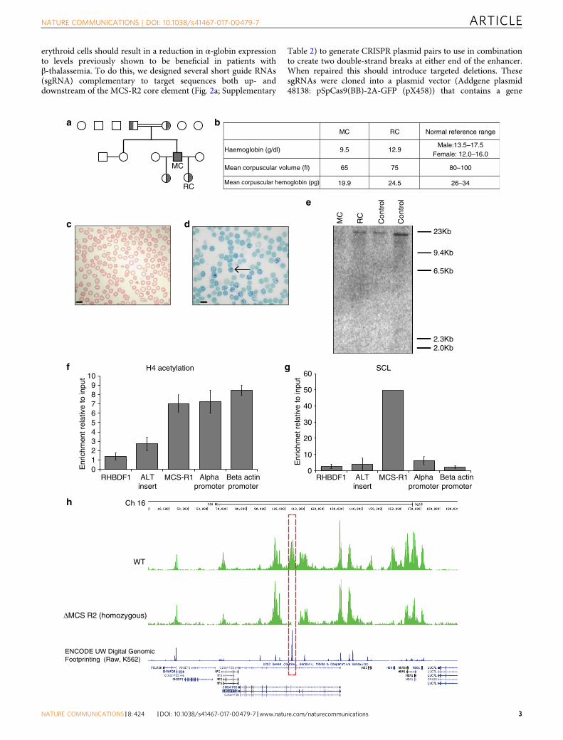
ResultsA natural deletion that uniquely removes MCS-R2 enhancer.A previously reported patient (MC) is homozygous for a veryrare 3.3 kb deletion (called (αα)ALT), which uniquely removesMCS-R2 but leaves the α-globin genes and all other enhancerelements intact12. MC is a member of a pedigree (Fig. 1a) thatoriginates from Portugal and was evaluated in greater detail(Supplementary Table 1). Hematological analysis confirmedthat MC has hypochromic microcytic anemia (Figs. 1b, c); 51% ofhis peripheral blood red blood cells (RBC) were positive forhemoglobin H (HbH) inclusion bodies confirming a diagnosisof α-thalassemia leading to HbH disease (Fig. 1d). α-Globinmessenger RNA (mRNA) levels of MC’s peripheral blood arereduced and multiplex ligation-dependent probe amplificationanalysis12 confirmed the absence of MCS-R2 in its natural geno-mic location, but did not exclude the possibility of its presence inan ectopic locus. To examine this we performed Southern blotanalysis using a probe specific to MCS-R2, which confirmed theabsence of the MCS-R2 region anywhere in the genome (Fig. 1e).
Interestingly, in the (αα)ALT mutation, the 3.3 kb deletion isassociated with the insertion of a 39 nucleotide orphan segment,whose sequence was not found anywhere in the human genome.Our search for canonical binding motifs for both generaltranscription factors, and erythroid-specific factors within thisorphan sequence did not reveal motifs with any known regulatorypotential. Next, we examined the presence of histone 4 acetylation(H4Ac) or binding of the stem cell leukemia (SCL) complex,which are signatures of active chromatin within this orphan insertsequence. Chromatin immunoprecipitation (ChIP-qPCR) acrossthis sequence confirmed the absence of enrichment for theseactivating chromatin marks (Figs. 1f, g).
To assess whether the function of MCS-R2 was assumed by anyother region within or beyond the α-globin cluster, we undertookChIP-Seq (using an anti-pan-H4 acetylation antibody) to give thegreatest likelihood of detecting any newly activated chromatin(Fig. 1h). No new putative regulatory elements were detected,with the only additional finding of note being the loss of a singleH4Ac peak at the HBM promoter. This corresponds to a shortdeletion in the region of the HBM promoter, confirmed bySouthern blot. This region does not contribute to normal α-globintranscription17 and its loss is not associated with any expectedreduction in α-globin levels.
These data support experimental studies in the mouse18, whichdefine MCS-R2 as the major (but not the only) regulatory elementcontrolling expression of α-globin RNA. Its loss from the naturalchromosomal environment results in a significant reduction ofα-globin transcription in cis. The clinical phenotypes ofthalassemia trait in the heterozygous patient RC and of HbHdisease in patient MC, who carries this deletion in homozygosity,demonstrate that deletion of MCS-R2 causes a significant(>50%), but not total reduction in α-globin expression in bothhomozygous and heterozygous states. However, of importance forthis study, the absence of MCS-R2 in the homozygous state didnot result in any other phenotypic abnormalities in MC,demonstrating that its unique functional significance is confinedto expression of the α-globin locus.
Targeted in vitro deletion of MCS-R2 to knockdown α-globin.Next, we hypothesized that creating a targeted deletion ofMCS-R2 would phenocopy the effects seen in MC and RC inhomozygous and heterozygous states, respectively. Thus theremoval of the ~260 bp core element of MCS-R2 in human
ARTICLE NATURE COMMUNICATIONS | DOI: 10.1038/s41467-017-00479-7
2 NATURE COMMUNICATIONS |8: 424 |DOI: 10.1038/s41467-017-00479-7 |www.nature.com/naturecommunications
erythroid cells should result in a reduction in α-globin expressionto levels previously shown to be beneficial in patients withβ-thalassemia. To do this, we designed several short guide RNAs(sgRNA) complementary to target sequences both up- anddownstream of the MCS-R2 core element (Fig. 2a; Supplementary
Table 2) to generate CRISPR plasmid pairs to use in combinationto create two double-strand breaks at either end of the enhancer.When repaired this should introduce targeted deletions. ThesesgRNAs were cloned into a plasmid vector (Addgene plasmid48138: pSpCas9(BB)-2A-GFP (pX458)) that contains a gene
0
10
20
30
40
50
60
RHBDF1 ALTinsert
MCS-R1 Alphapromoter
Beta actinpromoter
Enr
ichm
et r
elat
ive
to in
put
SCL
a b
c Con
trol
Con
trol
RC
MC
e
MC RC Normal reference range
Haemoglobin (g/dl) 9.5 12.9Male:13.5–17.5
Female: 12.0–16.0
Mean corpuscular volume (fl) 65 75 80–100
Mean corpuscular hemoglobin (pg) 19.9 24.5 26–34
d
f g
MC
RC
Ch 16
ΔMCS R2 (homozygous)
WT
ENCODE UW Digital GenomicFootprinting (Raw, K562)
0123456789
10
RHBDF1 ALTinsert
MCS-R1 Alphapromoter
Beta actinpromoter
Enr
ichm
ent r
elat
ive
to in
put
H4 acetylation
23Kb
9.4Kb
6.5Kb
2.3Kb2.0Kb
h
NATURE COMMUNICATIONS | DOI: 10.1038/s41467-017-00479-7 ARTICLE
NATURE COMMUNICATIONS |8: 424 |DOI: 10.1038/s41467-017-00479-7 |www.nature.com/naturecommunications 3
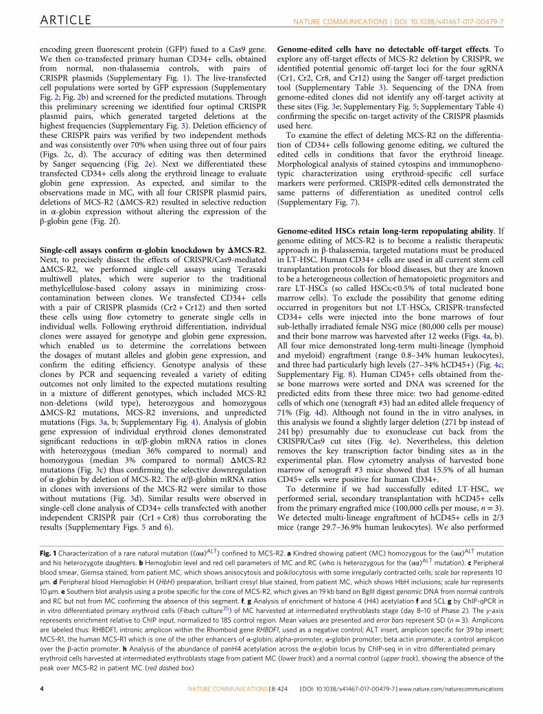
encoding green fluorescent protein (GFP) fused to a Cas9 gene.We then co-transfected primary human CD34+ cells, obtainedfrom normal, non-thalassemia controls, with pairs ofCRISPR plasmids (Supplementary Fig. 1). The live-transfectedcell populations were sorted by GFP expression (SupplementaryFig. 2; Fig. 2b) and screened for the predicted mutations. Throughthis preliminary screening we identified four optimal CRISPRplasmid pairs, which generated targeted deletions at thehighest frequencies (Supplementary Fig. 3). Deletion efficiency ofthese CRISPR pairs was verified by two independent methodsand was consistently over 70% when using three out of four pairs(Figs. 2c, d). The accuracy of editing was then determinedby Sanger sequencing (Fig. 2e). Next we differentiated thesetransfected CD34+ cells along the erythroid lineage to evaluateglobin gene expression. As expected, and similar to theobservations made in MC, with all four CRISPR plasmid pairs,deletions of MCS-R2 (ΔMCS-R2) resulted in selective reductionin α-globin expression without altering the expression of theβ-globin gene (Fig. 2f).
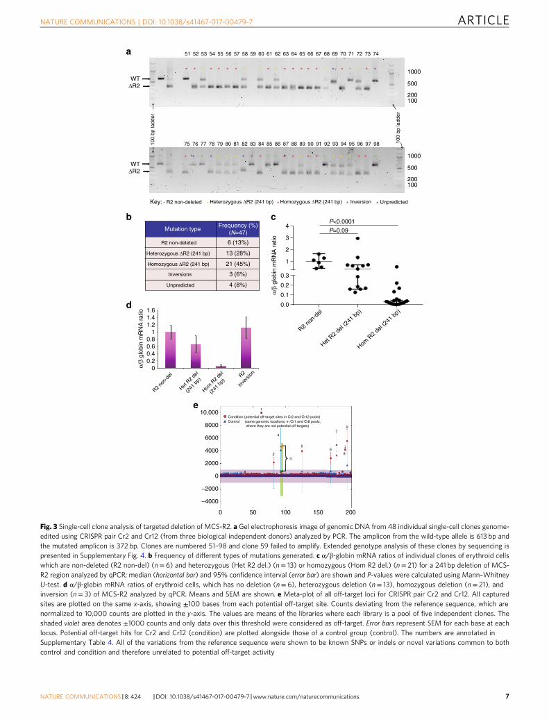
Single-cell assays confirm α-globin knockdown by ΔMCS-R2.Next, to precisely dissect the effects of CRISPR/Cas9-mediatedΔMCS-R2, we performed single-cell assays using Terasakimultiwell plates, which were superior to the traditionalmethylcellulose-based colony assays in minimizing cross-contamination between clones. We transfected CD34+ cellswith a pair of CRISPR plasmids (Cr2 + Cr12) and then sortedthese cells using flow cytometry to generate single cells inindividual wells. Following erythroid differentiation, individualclones were assayed for genotype and globin gene expression,which enabled us to determine the correlations betweenthe dosages of mutant alleles and globin gene expression, andconfirm the editing efficiency. Genotype analysis of theseclones by PCR and sequencing revealed a variety of editingoutcomes not only limited to the expected mutations resultingin a mixture of different genotypes, which included MCS-R2non-deletions (wild type), heterozygous and homozygousΔMCS-R2 mutations, MCS-R2 inversions, and unpredictedmutations (Figs. 3a, b; Supplementary Fig. 4). Analysis of globingene expression of individual erythroid clones demonstratedsignificant reductions in α/β-globin mRNA ratios in cloneswith heterozygous (median 36% compared to normal) andhomozygous (median 3% compared to normal) ΔMCS-R2mutations (Fig. 3c) thus confirming the selective downregulationof α-globin by deletion of MCS-R2. The α/β-globin mRNA ratiosin clones with inversions of the MCS-R2 were similar to thosewithout mutations (Fig. 3d). Similar results were observed insingle-cell clone analysis of CD34+ cells transfected with anotherindependent CRISPR pair (Cr1 + Cr8) thus corroborating theresults (Supplementary Figs. 5 and 6).
Genome-edited cells have no detectable off-target effects. Toexplore any off-target effects of MCS-R2 deletion by CRISPR, weidentified potential genomic off-target loci for the four sgRNA(Cr1, Cr2, Cr8, and Cr12) using the Sanger off-target predictiontool (Supplementary Table 3). Sequencing of the DNA fromgenome-edited clones did not identify any off-target activity atthese sites (Fig. 3e; Supplementary Fig. 5; Supplementary Table 4)confirming the specific on-target activity of the CRISPR plasmidsused here.
To examine the effect of deleting MCS-R2 on the differentia-tion of CD34+ cells following genome editing, we cultured theedited cells in conditions that favor the erythroid lineage.Morphological analysis of stained cytospins and immunopheno-typic characterization using erythroid-specific cell surfacemarkers were performed. CRISPR-edited cells demonstrated thesame patterns of differentiation as unedited control cells(Supplementary Fig. 7).
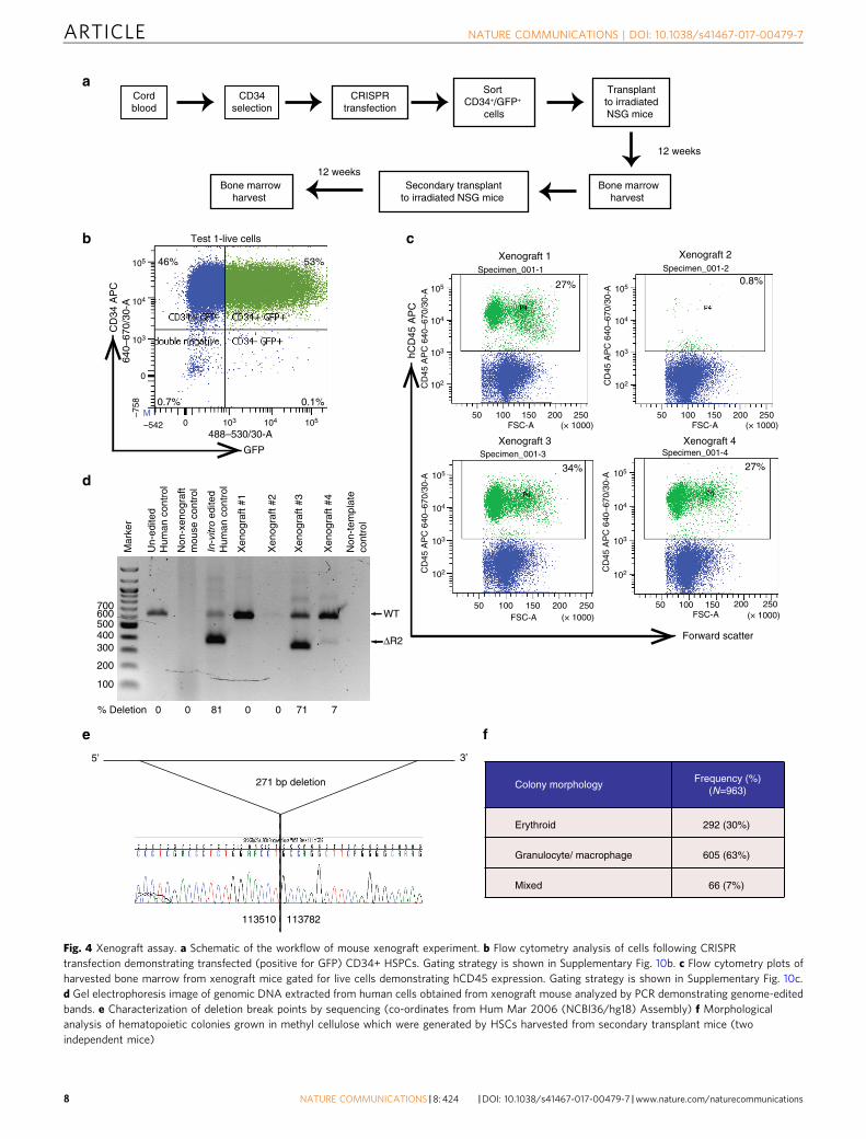
Genome-edited HSCs retain long-term repopulating ability. Ifgenome editing of MCS-R2 is to become a realistic therapeuticapproach in β-thalassemia, targeted mutations must be producedin LT-HSC. Human CD34+ cells are used in all current stem celltransplantation protocols for blood diseases, but they are knownto be a heterogeneous collection of hematopoietic progenitors andrare LT-HSCs (so called HSCs;<0.5% of total nucleated bonemarrow cells). To exclude the possibility that genome editingoccurred in progenitors but not LT-HSCs, CRISPR-transfectedCD34+ cells were injected into the bone marrows of foursub-lethally irradiated female NSG mice (80,000 cells per mouse)and their bone marrow was harvested after 12 weeks (Figs. 4a, b).All four mice demonstrated long-term multi-lineage (lymphoidand myeloid) engraftment (range 0.8–34% human leukocytes),and three had particularly high levels (27–34% hCD45+) (Fig. 4c;Supplementary Fig. 8). Human CD45+ cells obtained from the-se bone marrows were sorted and DNA was screened for thepredicted edits from these three mice: two had genome-editedcells of which one (xenograft #3) had an edited allele frequency of71% (Fig. 4d). Although not found in the in vitro analyses, inthis analysis we found a slightly larger deletion (271 bp instead of241 bp) presumably due to exonuclease cut back from theCRISPR/Cas9 cut sites (Fig. 4e). Nevertheless, this deletionremoves the key transcription factor binding sites as in theexperimental plan. Flow cytometry analysis of harvested bonemarrow of xenograft #3 mice showed that 15.5% of all humanCD45+ cells were positive for human CD34+.
To determine if we had successfully edited LT-HSC, weperformed serial, secondary transplantation with hCD45+ cellsfrom the primary engrafted mice (100,000 cells per mouse, n= 3).We detected multi-lineage engraftment of hCD45+ cells in 2/3mice (range 29.7–36.9% human leukocytes). We also performed
Fig. 1 Characterization of a rare natural mutation ((αα)ALT) confined to MCS-R2. a Kindred showing patient (MC) homozygous for the (αα)ALT mutationand his heterozygote daughters. b Hemoglobin level and red cell parameters of MC and RC (who is heterozygous for the (αα)ALT mutation). c Peripheralblood smear, Giemsa stained, from patient MC, which shows anisocytosis and poikilocytosis with some irregularly contracted cells; scale bar represents 10μm. d Peripheral blood Hemoglobin H (HbH) preparation, brilliant cresyl blue stained, from patient MC, which shows HbH inclusions; scale bar represents10 μm. e Southern blot analysis using a probe specific for the core of MCS-R2, which gives an 19 kb band on BglII digest genomic DNA from normal controlsand RC but not from MC confirming the absence of this segment. f, g Analysis of enrichment of histone 4 (H4) acetylation f and SCL g by ChIP-qPCR inin vitro differentiated primary erythroid cells (Fibach culture35) of MC harvested at intermediated erythroblasts stage (day 8–10 of Phase 2). The y-axisrepresents enrichment relative to ChIP input, normalized to 18S control region. Mean values are presented and error bars represent SD (n= 3). Ampliconsare labeled thus: RHBDF1, intronic amplicon within the Rhomboid gene RHBDF1, used as a negative control; ALT insert, amplicon specific for 39 bp insert;MCS-R1, the human MCS-R1 which is one of the other enhancers of α-globin; alpha-promoter, α-globin promoter; beta actin promoter, a control ampliconover the β-actin promoter. h Analysis of the abundance of panH4 acetylation across the α-globin locus by ChIP-seq in in vitro differentiated primaryerythroid cells harvested at intermediated erythroblasts stage from patient MC (lower track) and a normal control (upper track), showing the absence of thepeak over MCS-R2 in patient MC (red dashed box)
ARTICLE NATURE COMMUNICATIONS | DOI: 10.1038/s41467-017-00479-7
4 NATURE COMMUNICATIONS |8: 424 |DOI: 10.1038/s41467-017-00479-7 |www.nature.com/naturecommunications
methylcellulose colony assays with CD34+ cells harvestedfrom the secondary xenograft, which again demonstrated thepresence of granulocyte/macrophage and erythroid progenitoractivity (Fig. 4f). Sanger sequencing of 40 colonies (20 erythroid
and 20 myeloid) confirmed deletion of MCS-R2. Thesedata confirm that we have successfully edited LT-HSC, whichare able to reconstitute myeloid, lymphoid, and erythroidhematopoiesis.
0%
20%
40%
60%
80%
100%
Cr1+8 Cr2+12 Cr9+12 Cr10+12
PCR band intensity Droplet digital PCR
0
0.2
0.4
0.6
0.8
1
1.2
1.4
NTC C9 Cr1+8 Cr2+12 Cr9+12 Cr10+12
α-globin β-globin α/β-globin ratio
a
Humanchromosome 16
10 Kb
α1α2ζM
CS
-R1
MC
S-R
4
MC
S-R
3M
CS
-R2
50 bp
258 bp MCS-R2 core element
sgRNA target sites
cr9cr12
cr7cr2cr8cr10
cr1
GATA1 binding motif
NF-E2 binding motif
Globin genes
cis-acting regulatory elements
b
100
500400300
200
600
100b
pla
dder
C9
Cr2
+12
Cr1
0+12
Cr9
+12
Cr1
+8
WT
100
bpla
dder
cC
RIS
PR
effi
cien
cy
f
* *
**
*
**
**Rel
ativ
e ex
pres
sion
d
FS
NTC
105
104
103
102
105
104
103
102
105
104
103
102
105
104
103
102
105
104
103
102
105
104
103
102
50 100 150 200 250 50 100 150 200 250 50 100 150 200 250
50 100 150 200 250 50 100 150 200 250 50 100 150 200 250
C9 Cr1+8
Cr2+12 Cr10+12Cr9+12
0±0%
49±6%54±10%51±4%
56±10%60±9%
e
27±5%25±4%29±3%
22±8%24±3%100±0%
cr1
cr8
5’
cr2
cr10
cr9cr12
113496
113497 / 113808
113808
cr1 cr8
310+311 bp deletion
113511 113753
cr2 cr12
241 bp deletion
113444 113752
cr12
cr12
cr9
cr10
307 bp deletion
113499 113752 252 bp deletion
GF
P
NATURE COMMUNICATIONS | DOI: 10.1038/s41467-017-00479-7 ARTICLE
NATURE COMMUNICATIONS |8: 424 |DOI: 10.1038/s41467-017-00479-7 |www.nature.com/naturecommunications 5
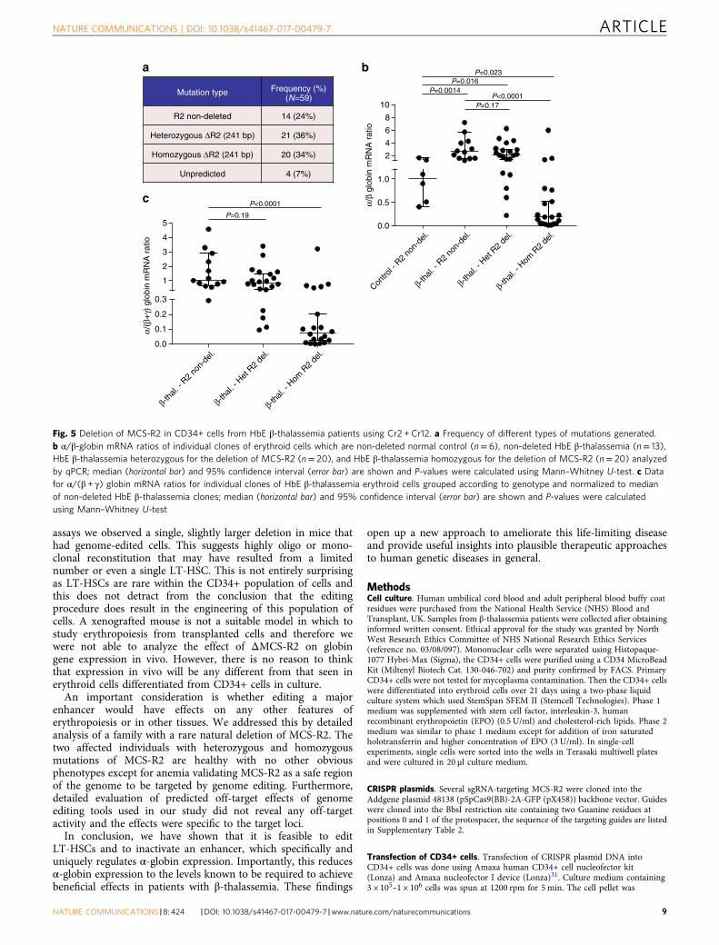
ΔMCS-R2 restores globin balance in β-thalassemia cells.Finally, to examine the ability of the edited deletion of MCS-R2 torectify globin chain imbalance in the erythroid cells of patientswith β-thalassemia, we performed single colony analysison CRISPR-edited CD34+ cells obtained from patients withHbE β-thalassemia (Supplementary Table 5). Genome editing ofCD34+ cells obtained from patients with HbE β-thalassemiaproduced a mixture of different genotypes similar to thoseobserved in normal controls with comparable mutationfrequencies (Supplementary Fig. 9; Fig. 5a). As expected, the α/βglobin mRNA ratios in unmodified erythroid cells from patientswith HbE β-thalassemia were higher than α/β globin mRNAratios in unmodified, normal erythroid cells. However whena heterozygous ΔMCS-R2 mutation was edited into the HbEβ-thalassemia cells, the α/β globin mRNA ratio decreased andmany individual clones had α/β globin mRNA ratios similar tonormal control clones (Fig. 5b). When both alleles at MCS-R2were edited in cells derived from patients with HbE β-thalasse-mia, the α/β globin ratios were rebalanced and many individualclones had α/β globin mRNA ratios similar to those foundin normal control cells (Fig. 5b). Analysis of α/all β-like globins(α/(α + γ)) ratios demonstrated similar trends (Fig. 5c). The-se observations confirm that editing of MCS-R2 can efficientlyrestore globin chain balance in erythroid cells in patients withβ-thalassemia and we predict that this would result in phenotypicimprovements and improved survival of genome-edited cellswith reduced levels of erythroid cell destruction and anemiain vivo.
DiscussionThe clinical management of β-thalassemia still largely depends onsupportive treatment with RBC transfusions and iron chelation inthe majority of patients1. Allogenic bone marrow transplantationremains the only curative treatment; however its usefulness islimited to a minority of patients who have HLA-matched siblingdonors19. Several new therapies for β-thalassemia are currentlybeing investigated20. Except for a few studies21 all of these newand experimental therapies aim to resolve globin chain imbalanceby increasing the production of γ-globin and fetal hemoglobin bygenome editing of transcription factors22, 23 (eg: BCL11A24 andLRF/ZBTB7A25) or by pharmacological methods (eg: histonedeacetylase inhibitors26 and lysine-specific demethylase 127).Alternatively, here we present a novel approach by directlyreducing expression of α-globin. Considering the central role ofexcess α-globin chains in the pathophysiology of β-thalassemia,and the extensive clinical evidence showing how co-inheritanceof α-thalassemia ameliorates β-thalassemia, this presents anextremely promising approach to curing this disease untilefficient gene correction of the defective β-globin by HDR in stemcells becomes routinely possible. This approach can be used on itsown or to complement other on-going efforts of increasing theproduction of fetal hemoglobin.
We have previously summarized the findings fromseveral clinical studies showing that a reduction of α-globinexpression to 75–25% of normal is an effective, safe, and tolerablelevel to provide sustainable beneficial effects in patients withβ-thalassemia3. In genome-edited normal control cells manyof the clones with heterozygous or homozygous deletions ofMCS-R2 demonstrated reduction of the α/β ratio to this desirablerange. In cells with HbE β-thalassemia mutations, targeteddeletion of MCS-R2 in both heterozygous and homozygous statesresulted in amelioration of the disturbed globin ratios.
In this paper we have demonstrated the use of CRISPR/Cas9 system for genome editing of MCS-R2, the major α-globinenhancer, in human primary LT-HSCs. CD34+ cells, includingLT-HSCs, are readily obtainable at a clinical scale and arecurrently used in all stem cell transplantation protocols for blooddiseases. Thus the hematopoietic system offers the ideal model fortranslation of genome editing for clinical benefit, and successfulgenome editing of lymphoid cells using a zinc-finger nuclease-based technique has already progressed to clinical trials28.Translation of our strategy into clinical application would requirea harvest of CD34+ cells from patients with β-thalassemia, ex vivogenome editing, and an autograft. This would therefore avoid thepotentially life-threatening complications of graft rejection andgraft versus host disease associated with allografting for patientswith hemoglobinopathies.
Using our protocol, we currently achieve transfection efficiencyas high as 75% and in transfected cells the editing efficiencywas 70–80%, providing an overall editing rate of 50–60% oflive CD34+ cells. Previously, it has been shown that chimerismlevels of only 10–20% of normal HSCs are sufficient to resultin nearly complete hematologic and pathologic correction ofβ-thalassemia29, 30. Therefore, using our strategy, we should be ableto provide sufficient numbers of genome-edited HSCs to produce aclinically significant beneficial effect in patients with β-thalassemia.
One potential limitation of this strategy is that deletion ofMCS-R2 could lead to profound reduction in α-globin expressionthus causing a critical decrease in total globin synthesis in someedited cells. However, it is likely that erythroid cells with morebalanced globin chain synthesis generated from genome-editedHSCs will have less ineffective erythropoiesis and hemolysis andremain in the circulation for the full life-span thus demonstratingselective advantage for survival in vivo. Furthermore, genomeengineering of the MCS-R2 enhancer could be used to fine-tunethe level of α-globin expression by limiting the edit to alreadyidentified transcription factor binding sites within the enhancer.Therefore, the issue of excessive downregulation of α-globin neednot be a significant barrier during future clinical use.
Xenograft assays performed here show that CRISPR/Cas9genome editing is not limited to progenitor cells but also occursin LT-HSCs. These are the cells, which must be edited if a sus-tainable improvement is to be achieved in patients withβ-thalassemia. We have shown that edited HSCs give rise tomultiple lineages of hematopoietic cells in vivo. In the xenograft
Fig. 2 Deletion of MCS-R2 using CRISPR/Cas9 genome editing in human CD34+ cells. a Schematic of sgRNA target sites. Four sgRNAs (Cr1, Cr2, Cr9, andCr10) were designed to target the 5′ end of the MCS-R2 core element, whereas three sgRNAs (Cr7, Cr8, and Cr12) target the 3′ end. b Representative flowcytometry plots showing GFP expression and forward scatter (FS) after gating for live cells in non-transfection control (NTC), Cas9 control (C9), andCRISPR/Cas9 plasmid pair-transfected cells. Orange: GFP negative, blue: GFP positive (low), and green: GFP positive (high). Mean and SD of the percentageof cells within each region is indicated (n= 3). Gating strategy is shown in Supplementary Fig. 10a. c Representative gel electrophoresis image of genomicDNA extracted from cells targeted by four CRISPR/Cas9 plasmid pairs analyzed by PCR. d Gene editing deletion induction efficiency as measuredindependently by percentages of mutated alleles determined by band size in end-point PCR and subsequent Sanger sequence analysis and by determininginverse of proportions of amplicons inside: outside deletion (amplicons of same length) by multiplexed droplet digital PCR; mean values are presented anderror bars represent SD (n= 3). e Characterization of deletion break points by sequencing (co-ordinates from Hum Mar 2006 (NCBI36/hg18) assembly).f α-and β-globin gene expression normalized to the expression of RPL13A and α/β-globin mRNA ratios relative to Cas9 control (C9) analyzed by qPCR; errorbars represent SD (n= 3); *P< 0.05 and **P< 0.01 relative to C9 (Student’s t-test). C9 Cas9-only control
ARTICLE NATURE COMMUNICATIONS | DOI: 10.1038/s41467-017-00479-7
6 NATURE COMMUNICATIONS |8: 424 |DOI: 10.1038/s41467-017-00479-7 |www.nature.com/naturecommunications
R2 non
-del
Het R2 d
el (2
41 bp
)
Hom R
2 del
(241
bp)
0.0
0.1
0.2
0.3
1
2
3
4
a
100
500
200
1000WT
ΔR2
** *
*
* * **Key: R2 non-deleted Heterozygous ΔR2 (241 bp) Homozygous ΔR2 (241 bp) Unpredicted
WTΔR2
100
500
200
1000
51 605958575655545352 61 706968676665646362 71 747372
100
bp la
dder
100
bp la
dder
75 848382818079787776 85 949392919089888786 95 989796
bMutation type
R2 non-deleted 6 (13%)
Heterozygous ΔR2 (241 bp) 13 (28%)
Homozygous ΔR2 (241 bp) 21 (45%)
Unpredicted
Frequency (%)(N=47)
4 (8%)
Inversions 3 (6%)α/
β gl
obin
mR
NA
rat
io
c
* Inversion
**** *****
*****
***
*********
****
***
*****
**
*
*
*
P=0.09
P<0.0001
00.20.40.60.8
11.21.41.6
R2 no
n-de
l
Het R
2 de
l
(241
bp)
Hom R
2 de
l
(241
bp)
R2
inver
sion
d
α/β
glob
in m
RN
A r
atio
1
2
4
3
56
79
8
Condition (potential off-target sites in Cr2 and Cr12 pools)Control (same genomic locations, in Cr1 and Cr8 pools, where they are not potential off-targets)
e10,000
8000
6000
4000
2000
0
–2000
–4000
0 50 100 150 200
Fig. 3 Single-cell clone analysis of targeted deletion of MCS-R2. a Gel electrophoresis image of genomic DNA from 48 individual single-cell clones genome-edited using CRISPR pair Cr2 and Cr12 (from three biological independent donors) analyzed by PCR. The amplicon from the wild-type allele is 613 bp andthe mutated amplicon is 372 bp. Clones are numbered 51–98 and clone 59 failed to amplify. Extended genotype analysis of these clones by sequencing ispresented in Supplementary Fig. 4. b Frequency of different types of mutations generated. c α/β-globin mRNA ratios of individual clones of erythroid cellswhich are non-deleted (R2 non-del) (n= 6) and heterozygous (Het R2 del.) (n= 13) or homozygous (Hom R2 del.) (n= 21) for a 241 bp deletion of MCS-R2 region analyzed by qPCR; median (horizontal bar) and 95% confidence interval (error bar) are shown and P-values were calculated using Mann–WhitneyU-test. d α/β-globin mRNA ratios of erythroid cells, which has no deletion (n= 6), heterozygous deletion (n= 13), homozygous deletion (n= 21), andinversion (n= 3) of MCS-R2 analyzed by qPCR. Means and SEM are shown. e Meta-plot of all off-target loci for CRISPR pair Cr2 and Cr12. All capturedsites are plotted on the same x-axis, showing ±100 bases from each potential off-target site. Counts deviating from the reference sequence, which arenormalized to 10,000 counts are plotted in the y-axis. The values are means of the libraries where each library is a pool of five independent clones. Theshaded violet area denotes ±1000 counts and only data over this threshold were considered as off-target. Error bars represent SEM for each base at eachlocus. Potential off-target hits for Cr2 and Cr12 (condition) are plotted alongside those of a control group (control). The numbers are annotated inSupplementary Table 4. All of the variations from the reference sequence were shown to be known SNPs or indels or novel variations common to bothcontrol and condition and therefore unrelated to potential off-target activity
NATURE COMMUNICATIONS | DOI: 10.1038/s41467-017-00479-7 ARTICLE
NATURE COMMUNICATIONS |8: 424 |DOI: 10.1038/s41467-017-00479-7 |www.nature.com/naturecommunications 7
GFP
a
b
53%
0.1%0.7%
46%
Test 1-live cells c
Forward scatter
hCD
45 A
PC
27%
27%34%
0.8%
d
Mar
ker
Un-
edite
dH
uman
con
trol
Non
-xen
ogra
ftm
ouse
con
trol
100
500400300
200
600700
WT
% Deletion 0 7810
In-vitro
edite
dH
uman
con
trol
Xen
ogra
ft #1
Xen
ogra
ft #4
Xen
ogra
ft #3
Xen
ogra
ft #2
Non
-tem
plat
eco
ntro
l
ΔR2
7100
Cordblood
CD34selection
CRISPRtransfection
Sort CD34+/GFP+
cells
Transplantto irradiatedNSG mice
Bone marrow harvest
12 weeks
Xenograft 3 Xenograft 4
Xenograft 1 Xenograft 2
e f
Colony morphology
Erythroid 292 (30%)
Granulocyte/ macrophage 605 (63%)
Mixed 66 (7%)
Frequency (%)(N=963)
5’ 3’
271 bp deletion
113510 113782
Secondary transplantto irradiated NSG mice
Bone marrow harvest
CD
34 A
PC
105
104
103
102
50 100 150FSC-A (× 1000)
200 250
50 100 150FSC-A (× 1000)
200 250 50 100 150FSC-A (× 1000)
200 250
50 100 150FSC-A (× 1000)
200 250
103
104
105
102
103
104
105
Specimen_001-1
Specimen_001-3 Specimen_001-4
Specimen_001-2
103 104 105
0
–758
–542 0M
640–
670/
30-A
CD
45 A
PC
640
–670
/30-
A
102
103
104
105
102
103
104
105
CD
45 A
PC
640
–670
/30-
A
CD
45 A
PC
640
–670
/30-
AC
D45
AP
C 6
40–6
70/3
0-A
488–530/30-A
12 weeks
Fig. 4 Xenograft assay. a Schematic of the workflow of mouse xenograft experiment. b Flow cytometry analysis of cells following CRISPRtransfection demonstrating transfected (positive for GFP) CD34+ HSPCs. Gating strategy is shown in Supplementary Fig. 10b. c Flow cytometry plots ofharvested bone marrow from xenograft mice gated for live cells demonstrating hCD45 expression. Gating strategy is shown in Supplementary Fig. 10c.d Gel electrophoresis image of genomic DNA extracted from human cells obtained from xenograft mouse analyzed by PCR demonstrating genome-editedbands. e Characterization of deletion break points by sequencing (co-ordinates from Hum Mar 2006 (NCBI36/hg18) Assembly) f Morphologicalanalysis of hematopoietic colonies grown in methyl cellulose which were generated by HSCs harvested from secondary transplant mice (twoindependent mice)
ARTICLE NATURE COMMUNICATIONS | DOI: 10.1038/s41467-017-00479-7
8 NATURE COMMUNICATIONS |8: 424 |DOI: 10.1038/s41467-017-00479-7 |www.nature.com/naturecommunications
assays we observed a single, slightly larger deletion in mice thathad genome-edited cells. This suggests highly oligo or mono-clonal reconstitution that may have resulted from a limitednumber or even a single LT-HSC. This is not entirely surprisingas LT-HSCs are rare within the CD34+ population of cells andthis does not detract from the conclusion that the editingprocedure does result in the engineering of this population ofcells. A xenografted mouse is not a suitable model in which tostudy erythropoiesis from transplanted cells and therefore wewere not able to analyze the effect of ΔMCS-R2 on globingene expression in vivo. However, there is no reason to thinkthat expression in vivo will be any different from that seen inerythroid cells differentiated from CD34+ cells in culture.
An important consideration is whether editing a majorenhancer would have effects on any other features oferythropoiesis or in other tissues. We addressed this by detailedanalysis of a family with a rare natural deletion of MCS-R2. Thetwo affected individuals with heterozygous and homozygousmutations of MCS-R2 are healthy with no other obviousphenotypes except for anemia validating MCS-R2 as a safe regionof the genome to be targeted by genome editing. Furthermore,detailed evaluation of predicted off-target effects of genomeediting tools used in our study did not reveal any off-targetactivity and the effects were specific to the target loci.
In conclusion, we have shown that it is feasible to editLT-HSCs and to inactivate an enhancer, which specifically anduniquely regulates α-globin expression. Importantly, this reducesα-globin expression to the levels known to be required to achievebeneficial effects in patients with β-thalassemia. These findings
open up a new approach to ameliorate this life-limiting diseaseand provide useful insights into plausible therapeutic approachesto human genetic diseases in general.
MethodsCell culture. Human umbilical cord blood and adult peripheral blood buffy coatresidues were purchased from the National Health Service (NHS) Blood andTransplant, UK. Samples from β-thalassemia patients were collected after obtaininginformed written consent. Ethical approval for the study was granted by NorthWest Research Ethics Committee of NHS National Research Ethics Services(reference no. 03/08/097). Mononuclear cells were separated using Histopaque-1077 Hybri-Max (Sigma), the CD34+ cells were purified using a CD34 MicroBeadKit (Miltenyl Biotech Cat. 130-046-702) and purity confirmed by FACS. PrimaryCD34+ cells were not tested for mycoplasma contamination. Then the CD34+ cellswere differentiated into erythroid cells over 21 days using a two-phase liquidculture system which used StemSpan SFEM II (Stemcell Technologies). Phase 1medium was supplemented with stem cell factor, interleukin-3, humanrecombinant erythropoietin (EPO) (0.5 U/ml) and cholesterol-rich lipids. Phase 2medium was similar to phase 1 medium except for addition of iron saturatedholotransferrin and higher concentration of EPO (3 U/ml). In single-cellexperiments, single cells were sorted into the wells in Terasaki multiwell platesand were cultured in 20 μl culture medium.
CRISPR plasmids. Several sgRNA-targeting MCS-R2 were cloned into theAddgene plasmid 48138 (pSpCas9(BB)-2A-GFP (pX458)) backbone vector. Guideswere cloned into the BbsI restriction site containing two Guanine residues atpositions 0 and 1 of the protospacer, the sequence of the targeting guides are listedin Supplementary Table 2.
Transfection of CD34+ cells. Transfection of CRISPR plasmid DNA intoCD34+ cells was done using Amaxa human CD34+ cell nucleofector kit(Lonza) and Amaxa nucleofector I device (Lonza)31. Culture medium containing3 × 105–1 × 10⁶ cells was spun at 1200 rpm for 5 min. The cell pellet was
Contro
l - R
2 no
n-de
l.
β-tha
l. - R
2 no
n-de
l.
β-tha
l. - H
et R
2 de
l.
β-tha
l. - H
om R
2 de
l.
β-tha
l. - R
2 no
n-de
l.
β-tha
l. - H
et R
2 de
l.
β-tha
l. - H
om R
2 de
l.
0.0
0.5
1.0
2
4
6
8
10
a
Mutation type(N=59)
R2 non-deleted 14 (24%)
Heterozygous ΔR2 (241 bp) 21 (36%)
Homozygous ΔR2 (241 bp) 20 (34%)
Unpredicted 4 (7%)
Frequency (%)
b
α/β
glob
in m
RN
A r
atio
P=0.0014
P=0.023P=0.016
0.0
0.1
0.2
0.3
1
2
3
4
5
c
α/(β
+γ)
glo
bin
mR
NA
rat
io
P<0.0001
P=0.19
P<0.0001P=0.17
Fig. 5 Deletion of MCS-R2 in CD34+ cells from HbE β-thalassemia patients using Cr2 + Cr12. a Frequency of different types of mutations generated.b α/β-globin mRNA ratios of individual clones of erythroid cells which are non-deleted normal control (n= 6), non-deleted HbE β-thalassemia (n= 13),HbE β-thalassemia heterozygous for the deletion of MCS-R2 (n= 20), and HbE β-thalassemia homozygous for the deletion of MCS-R2 (n= 20) analyzedby qPCR; median (horizontal bar) and 95% confidence interval (error bar) are shown and P-values were calculated using Mann–Whitney U-test. c Datafor α/(β + γ) globin mRNA ratios for individual clones of HbE β-thalassemia erythroid cells grouped according to genotype and normalized to medianof non-deleted HbE β-thalassemia clones; median (horizontal bar) and 95% confidence interval (error bar) are shown and P-values were calculatedusing Mann–Whitney U-test
NATURE COMMUNICATIONS | DOI: 10.1038/s41467-017-00479-7 ARTICLE
NATURE COMMUNICATIONS |8: 424 |DOI: 10.1038/s41467-017-00479-7 |www.nature.com/naturecommunications 9
resuspended in 100 µl nucleofection solution and was nucleofected with 10 μgCRISPR plasmid DNA in the cuvette provided using the U-08 program. Cellsuspension was incubated at room temperature for 10 min in nucleofectionsolution before transferring it to antibiotic-free medium.
Flow cytometry. Antibody labeling of washed cells was done using the followinganti-human antibodies; allophycocyanin (APC)-conjugated anti-CD34,fluorescein isothiocyanate (FITC)-conjugated anti-CD71, phycoerythrin (PE)conjugated anti-CD235a, APC-conjugated anti-CD45, FITC-conjugated anti-CD19, and PE-conjugated anti-CD33. Dead cells were identified by Hoechst 33258pentahydrate nucleic acid stain (Invitrogen) and were excluded. Analysis wasperformed on Cyan ADP (Beckman Coulter) analyzer using Summit v4.3 andFlowJo V10 softwares and cell sorting was performed in a BD FACSAria Fusion cellsorter (BD Biosciences). Details of flow cytometry gating strategy are given inSupplementary Fig. 10.
Antibodies used for flow cytometry. Antibodies to the following proteins wereused: APC-conjugated anti-CD34 (Miltenyl Biotec Cat. 130-090-654; dilution1:100), FITC-conjugated anti-CD71 (BD Biosciences Cat. 555536; dilution 1:100),PE-conjugated anti-CD235a (BD Biosciences Cat. 340947; dilution 1:500),APC-conjugated anti-CD45 (BioLegend Cat. 304012 Clone HI30; dilution 1:100),FITC-conjugated anti-CD19 (BioLegend Cat. 302206 Clone HIB19; dilution 1:100),and PE-conjugated anti-CD33 (BioLegend Cat. 303404 Clone WM53; dilution1:100). Dead cells were identified by Hoechst 33258 pentahydrate nucleic acid stain(Invitrogen; dilution 1:10,000) and were excluded.
DNA extraction and PCR. Genomic DNA from cell pellets was extracted usingDNeasy blood and tissue kit (Qiagen) and genomic DNA from small cell numberswas amplified directly from cell lysate using Illustra single-cell GenomiPhi DNAamplification kit (GE Healthcare). PCR reactions across MCS-R2 region wereperformed with AmpliTaq Gold 360 PCR master mix (Invitrogen) using a customdesigned primer pair (forward 5′-TGGTCCTGAAGGATGAGAAG-3′; reverse5′-AGCAACAGTCCTTTCTCTGG-3′). Uncropped scans of all PCR gels areshown in the Supplementary Fig. 11.
Droplet digital PCR. About 20 ng genomic DNA was amplified with a primerpair, one set with a primer within the deletion (5′-GGCCCAGTTATCTGCTCCCTCAAGT-3′) and a primer upstream (5′-AGGCCCATATCTCTGCCCAA-GAGC-3′) and the other set both 3.9 kb downstream (forward 5′-GGGGACTTTTGCCATGCCTGAAGTAGA-3′; reverse 5′-GGCCCCACTCCCTGATCT-TAACCATTT-3′), using the QX200 Droplet Digital PCR System. Around 20,000droplets were evaluated and the gene editing efficiency of the different CRISPRsgRNA pairs was estimated by comparing the two primer sets against a negativecontrol (vector only).
Identification of potential off-target loci for CRISPR plasmids. Potentialgenic off-target loci were identified using the Sanger off-target prediction tool(http://www.sanger.ac.uk/htgt/wge/find_off_targets_by_seq_allowing_up_to_four_mismatches) (Supplementary Table 3). These loci were captured byhybridization to complementary 50 bp biotinylated oligos prior to sequencing;these oligonucleotides (IDT) were designed to bind within 50 bp of either side ofthe CRISPR/Cas9 target and off-target sites using an in-house PERL script (Off-TargetProbes.pl). For each site, one oligo from each adjacent window was selected;selected oligos had the lowest BLAT density score and were only used if the scorewas below 35 and no simple repeats were present32. Chromosome capture oligoswere successfully designed for 53/57 potential off-target sites: 22 potential off-targetsites for Cr1, 6 potential off-target sites for Cr8, 8 potential off-target sites for Cr2,and 17 potential off-target sites for Cr12. (details in the Supplementary Table 3).Most sites had two capture oligos, but for some it was possible to design only one.
Analysis of off-target loci. Whole-genome amplified DNA was prepared (Illustrasingle-cell GenomiPhi DNA amplification kit, GE Healthcare) from 30 clones andcombined in pools of five clones, each contributing equal amounts of DNA, andsonicated to 200 bp (Covaris S220 sonicator). Sonicated DNA was purified withAgencourt AMPure XP beads (Beckman Coulter) and indexed using NebNextUltra II (New England Biolabs). Fragment size was confirmed before and afterindexing using the D1000 Tapestation (Agilent).
The capture was performed using four pools of five clones for Cr2 and Cr12,and for two pools of five clones for Cr1 and Cr8. Up to 2 µg of each library poolwas combined for multiplexed capture with capture-based enrichment of targetand off-target regions. This was carried out in two successive rounds ofhybridization, biotin pull down, and amplification with SeqCap EZ (Nimblegen)and M-280 Streptavadin Dynabeads (ThermoFisher) following the NG Capture-Cmethod33. Each of these pools was sequenced as a separate library with thecaptured fragments sequenced in a 150 bp paired end (300 cycles mid output) runon the MiSeq Platform (Illumina).
The reads were mapped to the hg38 genome build, using bowtie2,(default parameters, —maxins 700), allowing only paired concordant mappingto be reported. The un-mapping read pairs were trimmed with trim_galore
(default parameters), and re-mapped in bowtie2. This resulted in 91–93% mapping.STAR mapping was used to map the remaining reads (first end-to-end mapping,then setting insertion elongation penalty to zero, and finally also allowing singleend mapping). Each of these rounds mapped ~90% of the previously unmappedreads, resulting in overall mapping of 99.9%. No new sequencing artifacts wereintroduced by this process. The mapped data were compared to the referencesequence, by using samtools mpileup, and further excluding all bases in readswith lower quality score than 30, by using varscan. This resulted in read depth foreach base of 40,000–200,000 (except sample 4 for Cr2 and 12, which had 13,000).The counts were normalized to 10,000 counts.
RNA extraction and qRT-PCR. Total RNA was purified using RNeasy mini kit(Qiagen) and cDNA from small number of cells was directly prepared from cell lysateusing TaqMan gene expression cells-to-CT kit (Life Technologies). All qRT-PCRreactions were performed using inventoried TaqMan assays (Applied Biosystems;TaqMan IDs: HBA2/HBA1-Hs00361191_g1, HBB-Hs00747223_g1, HBG-Hs00361131_g1 and RPL13A-Hs03043885_g1) in technical triplicate in 7500 fast real-time PCR system (Applied Biosystems) according to the manufacturer’s protocol.Data were analyzed by 7500 software v2.0.6 using the delta delta CT method.
ChIP and ChIP-seq. Chromatin immunopreciptation was performed as previouslydescribed34. Briefly, 1 × 10⁷ cells from primary erythroid cultures were fixed with1% formaldehyde, before neutralization, and lysis in SDS ChIP lysis buffer(Millipore ChIP kit, 17-295). Pre-clearing was performed with protein agarose Abeads before overnight incubation with antibody, and subsequent agarose beadprecipitation. Washes were performed according to the manufacturer’s protocol.Antibodies used were anti-pan-H4 acetylation and anti-SCL. ChIP-seq librarieswere prepared and sequenced using the standard Illumina paired-end protocol.
Antibodies used for ChIP. Antibodies to the following proteins were used:anti-pan-H4 acetylation (Upstate 17-630; dilution 1:200) and anti-SCL (kindlysupplied by Professor Catherine Porcher, MRC Molecular Haematology Unit,Weatherall Institute of Molecular Medicine, Oxford; dilution 1:200).
Southern blotting. Southern blotting was performed using standard methods. A 538bp probe binding the region over MCS-R2 (chr16: 163380—163907 on build hg19)was generated by PCR, using primers FWD 5′-AACAAGAAAACCAGCAGGCTCC-3′ and REV 5′-TGTAAGTCCATCCAGGTGTGAGTTC-3′. This wascloned into the vector pGEM-T-easy (Promega) for confirmation by sequencing priorto excision and purification. About 50 ng of probe was end-labeled with α-P32 usingthe megaprime kit (Amersham, RPN1604) and hybridized to blotted genomic DNAfrom patient MC, RC, and a normal control, digested using BamH1. A labeledfragment size of 19 kb is expected when hybridized with the MCS-R2 probe.
Sequence analysis. PCR sequencing was performed on PCR products purified withQiaquick Gel Extraction Kit (Manufacturer Qiagen Cat No 28704) or Qiaquick PCRPurification Kit (Manufacturer Qiagen Cat No 28104) and then sequenced withthe amplification primers using BigDye Terminator v3.1 Cycle Sequencing Kit(Manufacturer ThermoFisher Scientific Cat No 4337455) as per the manufacturer’sinstructions. The products were then cleaned with a standard ethanol precipitationand run on an AB3730 DNA Analyzer (ThermoFisher Scientific) and analyzedusing DNA analysis software Sequencer 5.0.1 (Gene Codes Corporation).
Colony forming unit assay. Single-cell suspensions were mixed into MethocultOptimum (Manufacturer Stemcell Technologies Cat No H4034) to allowprogenitor cells to expand and differentiate into myeloid or erythroid coloniesas per the manufacturer’s instructions. Colonies were inspected and pickedmanually into PBS using a light inverted microscope, EVOS XL Imaging System(Manufacturer ThermoFisher Scientific Cat No AME3300).
Animal models and xenotransplantation assay. Female NSG (NOD.Cg-Prkdcscid
Il2rgtm1Wjl/SzJ, Jackson Laboratory, USA) mice (species-Mus musculus) were usedfor xenotransplantation assays. Ten- to 14-week-old NSG mice were irradiated twicewith 100 cGy, 4 h apart. Cells were administered via intra-tibial injection within 24 hof the second irradiation. Four mice were injected during the first transplantationexperiment and three mice received injections during the secondary transplantationexperiment. Human CD45(hCD45)+33 + 19− (myeloid) or hCD45+33-19+(B-lymphoid) engraftment was analyzed by FACS and defined as ≥0.1% of livemononuclear cell gate. Mice were killed for cell harvesting from lower limb, pelvic,and vertebral bones at 12 weeks post injection. Permission was granted to performanimal experiments by the UK Government Home Office through the ProjectLicense 30/2465
Statistical analysis. Exact sample sizes and representations of error bars areindicated in each figure. All experiments were performed in triplicate unlessspecified otherwise. A two-tailed unpaired Student’s t-test was used in statisticalanalysis between groups for normally distributed data and Mann–Whitney U-testwas used for data, which are not normally distributed; comparison groups are
ARTICLE NATURE COMMUNICATIONS | DOI: 10.1038/s41467-017-00479-7
10 NATURE COMMUNICATIONS |8: 424 |DOI: 10.1038/s41467-017-00479-7 |www.nature.com/naturecommunications
mentioned in each figure. Differences corresponding to P< 0.05 were consideredstatistically significant. No statistical method was used to predetermine sample sizeand data analysis was not blinded. No samples, mice, or data points were excludedfrom the reported analysis. No randomization method was used in experiments.
Data availability. All sequencing data sets are available at the National Center forBiotechnology Information Gene Expression Omnibus (NCBI GEO) with accessionnumbers GSE95370, GSM2508191, GSM2508192, GSM2508193, GSM2508194,GSM2508195, GSM2508196.
Received: 5 April 2017 Accepted: 30 June 2017
References1. Higgs, D. R., Engel, J. D. & Stamatoyannopoulos, G. Thalassaemia. Lancet 379,
373–383 (2012).2. Weatherall D. J. & Clegg J. B. in The Thalassaemia Syndromes, 4 edn
(Blackwell Science, 2001).3. Mettananda, S., Gibbons, R. J. & Higgs, D. R. Alpha-Globin as a molecular
target in the treatment of beta-thalassemia. Blood 125, 3694–3701 (2015).4. Sollaino, M. C. et al. Association of alpha globin gene quadruplication and
heterozygous beta thalassemia in patients with thalassemia intermedia.Haematologica 94, 1445–1448 (2009).
5. Premawardhena, A. et al. A novel molecular basis for beta thalassemiaintermedia poses new questions about its pathophysiology. Blood 106,3251–3255 (2005).
6. Mettananda, S., Gibbons, R. J. & Higgs, D. R. Understanding alpha-globin generegulation and implications for the treatment of beta-thalassemia. Ann. N YAcad. Sci. 1368, 16–24 (2016).
7. Higgs, D. R. & Wood, W. G. Long-range regulation of alpha globin geneexpression during erythropoiesis. Curr. Opin. Hematol. 15, 176–183 (2008).
8. Sharpe, J. A. et al. Analysis of the human alpha globin upstream regulatoryelement (HS-40) in transgenic mice. EMBO J. 11, 4565–4572 (1992).
9. Wallace, H. A. C. et al. Manipulating the mouse genome to engineer precisefunctional syntenic replacements with human sequence. Cell 128, 197–209 (2007).
10. Harteveld, C. L. & Higgs, D. R. Alpha-thalassaemia. Orphanet J. Rare Dis. 5, 13(2010).
11. Hughes, J. R. et al. Annotation of cis-regulatory elements by identification,subclassification, and functional assessment of multispecies conservedsequences. Proc. Natl Acad. Sci. USA 102, 9830–9835 (2005).
12. Coelho, A., Picanco, I., Seuanes, F., Seixas, M. T. & Faustino, P. Novel largedeletions in the human alpha-globin gene cluster: clarifying the HS-40 long-range regulatory role in the native chromosome environment. Blood Cells Mol.Dis. 45, 147–153 (2010).
13. Jinek, M. et al. A programmable dual-RNA-guided DNA endonuclease inadaptive bacterial immunity. Science 337, 816–821 (2012).
14. Ran, F. A. et al. Genome engineering using the CRISPR-Cas9 system.Nat. Protoc. 8, 2281–2308 (2013).
15. DeWitt, M. A. et al. Selection-free genome editing of the sickle mutation in humanadult hematopoietic stem/progenitor cells. Sci. Transl. Med. 8, 360ra134 (2016).
16. Genovese, P. et al. Targeted genome editing in human repopulatinghaematopoietic stem cells. Nature 510, 235–240 (2014).
17. Rugless, M. J. et al. A large deletion in the human alpha-globin cluster causedby a replication error is associated with an unexpectedly mild phenotype.Hum. Mol. Genet. 17, 3084–3093 (2008).
18. Anguita, E. et al. Deletion of the mouse alpha-globin regulatory element(HS -26) has an unexpectedly mild phenotype. Blood 100, 3450–3456 (2002).
19. Angelucci, E. et al. Hematopoietic stem cell transplantation in thalassemiamajor and sickle cell disease: indications and management recommendationsfrom an international expert panel. Haematologica 99, 811–820 (2014).
20. Sankaran, V. G. & Weiss, M. J. Anemia: progress in molecular mechanisms andtherapies. Nat. Med. 21, 221–230 (2015).
21. Mettananda, S. et al. Selective silencing of alpha-globin by the histonedemethylase inhibitor IOX1: a potentially new pathway for treatment of beta-thalassemia. Haematologica 102, e80–e84 (2017).
22. Canver, M. C. & Orkin, S. H. Customizing the genome as therapy for the beta-hemoglobinopathies. Blood 127, 2536–2545 (2016).
23. Ludwig, L. S., Khajuria, R. K. & Sankaran, V. G. Emerging cellular and genetherapies for congenital anemias. Am. J. Med. Genet. C Semin. Med. Genet. 172,332–348 (2016).
24. Canver, M. C. et al. BCL11A enhancer dissection by Cas9-mediated in situsaturating mutagenesis. Nature 527, 192–197 (2015).
25. Masuda, T. et al. Transcription factors LRF and BCL11A independently repressexpression of fetal hemoglobin. Science 351, 285–289 (2016).
26. Bradner, J. E. et al. Chemical genetic strategy identifies histone deacetylase 1(HDAC1) and HDAC2 as therapeutic targets in sickle cell disease. Proc. NatlAcad. Sci. USA 107, 12617–12622 (2010).
27. Shi, L., Cui, S., Engel, J. D. & Tanabe, O. Lysine-specific demethylase 1 is atherapeutic target for fetal hemoglobin induction. Nat. Med. 19, 291–294(2013).
28. Tebas, P. et al. Gene editing of CCR5 in autologous CD4 T cells of personsinfected with HIV. N. Engl. J. Med. 370, 901–910 (2014).
29. Persons, D. A. et al. Functional requirements for phenotypic correction ofmurine beta-thalassemia: implications for human gene therapy. Blood 97,3275–3282 (2001).
30. Arumugam, P. & Malik, P. Genetic therapy for beta-thalassemia: from thebench to the bedside. Hematology Am. Soc. Hematol. Educ. Program 2010,445–450 (2010).
31. Meissner, T. B., Mandal, P. K., Ferreira, L. M., Rossi, D. J. & Cowan, C. A.Genome editing for human gene therapy. Methods Enzymol. 546, 273–295(2014).
32. Hughes, J. R. et al. Analysis of hundreds of cis-regulatory landscapes at highresolution in a single, high-throughput experiment. Nat. Genet. 46, 205–212(2014).
33. Davies, J. O. et al. Multiplexed analysis of chromosome conformation at vastlyimproved sensitivity. Nat. Methods 13, 74–80 (2016).
34. De Gobbi, M. et al. Tissue-specific histone modification and transcription factorbinding in alpha globin gene expression. Blood 110, 4503–4510 (2007).
35. Fibach, E., Manor, D., Oppenheim, A. & Rachmilewitz, E. A. Proliferation andmaturation of human erythroid progenitors in liquid culture. Blood 73,100–103 (1989).
AcknowledgementsThis work was supported by grants to D.R.H. by the UK Medical Research Council(grant number MC_UU_12025/ unit programme MC_UU_12009/4) and the NIHROxford Biomedical Research Center. S.M. is a Commonwealth Scholar, funded by theUK government. We also acknowledge funding from Helmut Horten Foundation. Wethank Anuja Premawardana of University of Kelaniya, Sri Lanka for providingsamples from β-thalassemia patients and Gathsaurie Neelika Malavige of University ofSri Jayawardenapura, Sri Lanka for providing facilities for some of the tissue culturework. We thank Yale Michaels for advice using Droplet Digital PCR. Addgene plasmid48138 (pSpCas9(BB)-2A-GFP (PX458)) was a gift from Feng Zhang. We thank family(C) for their kind collaboration. We also thank Dr João Lavinha for critically reading thepaper and Dr Tudor Fulga for helpful discussion.
Author contributionsS.M., C.A.F., D.H., L.Q., P.V., R.J.G., and D.R.H. designed the research; P.F. and A.C.provided patient data and samples; S.M., C.A.F., D.H., M.B., L.Q., K.C., P.H., D.D., J.K.,M.G., J.A.S-S., J.D., B.U., P.S., J.A.S. performed the experiments; S.M., C.A.F., D.H., M.B.,L.Q., J.T., J.R.H., R.J.G. and D.R.H. analyzed and interpreted the data; and S.M., C.A.F.,D.H., L.Q., R.J.G., and D.R.H. wrote the manuscript.
Additional informationSupplementary Information accompanies this paper at doi:10.1038/s41467-017-00479-7.
Competing interests: The authors declare no competing financial interests.
Reprints and permission information is available online at http://npg.nature.com/reprintsandpermissions/
Publisher's note: Springer Nature remains neutral with regard to jurisdictional claims inpublished maps and institutional affiliations.
Open Access This article is licensed under a Creative CommonsAttribution 4.0 International License, which permits use, sharing,
adaptation, distribution and reproduction in any medium or format, as long as you giveappropriate credit to the original author(s) and the source, provide a link to the CreativeCommons license, and indicate if changes were made. The images or other third partymaterial in this article are included in the article’s Creative Commons license, unlessindicated otherwise in a credit line to the material. If material is not included in thearticle’s Creative Commons license and your intended use is not permitted by statutoryregulation or exceeds the permitted use, you will need to obtain permission directly fromthe copyright holder. To view a copy of this license, visit http://creativecommons.org/licenses/by/4.0/.
© The Author(s) 2017
NATURE COMMUNICATIONS | DOI: 10.1038/s41467-017-00479-7 ARTICLE
NATURE COMMUNICATIONS |8: 424 |DOI: 10.1038/s41467-017-00479-7 |www.nature.com/naturecommunications 11

![Limited Number of Globin Genes in HumanDNA10-7, or 0.198 ngof globin DNA.FromEq. [1] wecan calculate the %hybridization, P, expected for anynumberof globin gene copiespresent.Forexample,inExp.1,](https://img.pdfslide.net/doc/110x75/60e570f3b76c9678502ef0c0/limited-number-of-globin-genes-in-humandna-10-7-or-0198-ngof-globin-dnafromeq.jpg)