Embed Size (px)
Citation preview
EFFECTS OF PHYSICAL THERAPY FOR PATIENTS WITH CERVICAL RADICULOPATHY
A literature review
NICHOLAS KING
Akademin för hälsa, vård och välfärd Fysioterapi Avancerad 15 hp Examensarbete i sjukgymnastik VSG010
Handledare: Thomas Overmeer Examinator: Maria Sandborgh Datum: 12 juni 2015
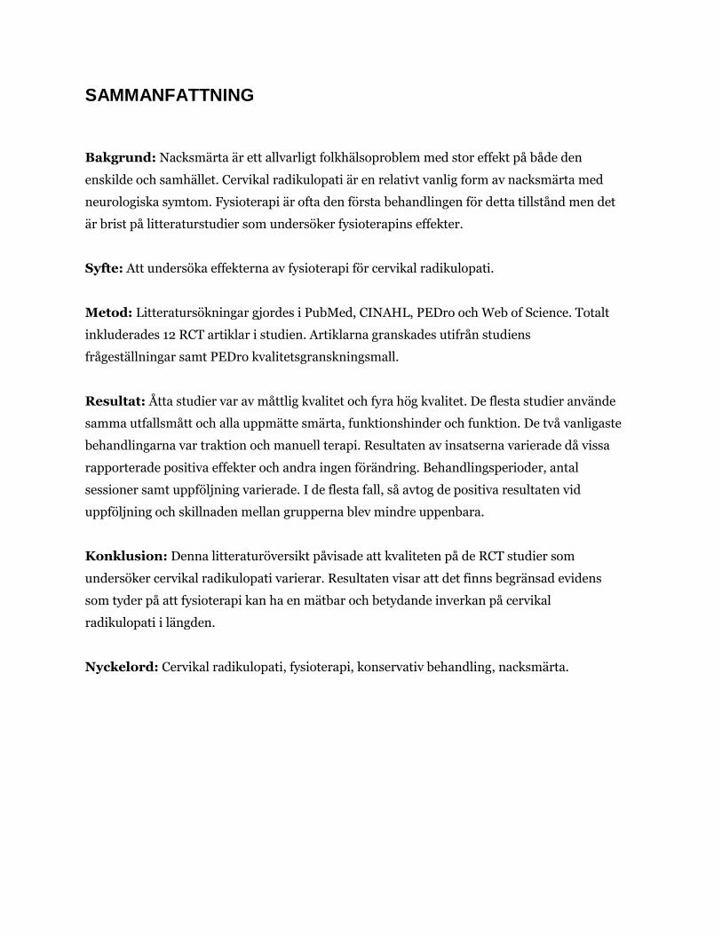
SAMMANFATTNING
Bakgrund: Nacksmärta är ett allvarligt folkhälsoproblem med stor effekt på både den
enskilde och samhället. Cervikal radikulopati är en relativt vanlig form av nacksmärta med
neurologiska symtom. Fysioterapi är ofta den första behandlingen för detta tillstånd men det
är brist på litteraturstudier som undersöker fysioterapins effekter.
Syfte: Att undersöka effekterna av fysioterapi för cervikal radikulopati.
Metod: Litteratursökningar gjordes i PubMed, CINAHL, PEDro och Web of Science. Totalt
inkluderades 12 RCT artiklar i studien. Artiklarna granskades utifrån studiens
frågeställningar samt PEDro kvalitetsgranskningsmall.
Resultat: Åtta studier var av måttlig kvalitet och fyra hög kvalitet. De flesta studier använde
samma utfallsmått och alla uppmätte smärta, funktionshinder och funktion. De två vanligaste
behandlingarna var traktion och manuell terapi. Resultaten av insatserna varierade då vissa
rapporterade positiva effekter och andra ingen förändring. Behandlingsperioder, antal
sessioner samt uppföljning varierade. I de flesta fall, så avtog de positiva resultaten vid
uppföljning och skillnaden mellan grupperna blev mindre uppenbara.
Konklusion: Denna litteraturöversikt påvisade att kvaliteten på de RCT studier som
undersöker cervikal radikulopati varierar. Resultaten visar att det finns begränsad evidens
som tyder på att fysioterapi kan ha en mätbar och betydande inverkan på cervikal
radikulopati i längden.
Nyckelord: Cervikal radikulopati, fysioterapi, konservativ behandling, nacksmärta.
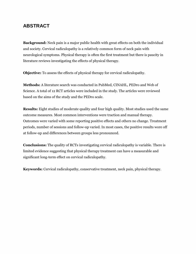
ABSTRACT
Background: Neck pain is a major public health with great effects on both the individual
and society. Cervical radiculopathy is a relatively common form of neck pain with
neurological symptoms. Physical therapy is often the first treatment but there is paucity in
literature reviews investigating the effects of physical therapy.
Objective: To assess the effects of physical therapy for cervical radiculopathy.
Methods: A literature search was conducted in PubMed, CINAHL, PEDro and Web of
Science. A total of 12 RCT articles were included in the study. The articles were reviewed
based on the aims of the study and the PEDro scale.
Results: Eight studies of moderate quality and four high quality. Most studies used the same
outcome measures. Most common interventions were traction and manual therapy.
Outcomes were varied with some reporting positive effects and others no change. Treatment
periods, number of sessions and follow-up varied. In most cases, the positive results wore off
at follow-up and differences between groups less pronounced.
Conclusions: The quality of RCTs investigating cervical radiculopathy is variable. There is
limited evidence suggesting that physical therapy treatment can have a measurable and
significant long-term effect on cervical radiculopathy.
Keywords: Cervical radiculopathy, conservative treatment, neck pain, physical therapy.
1 BACKGROUND......................................................................... 1
1.1 Introduction ....................................................................................... 1
1.2 Cervical radiculopathy ..................................................................... 2
1.3 Epidemiology and prevalence ......................................................... 2
1.4 Pathophysiology ............................................................................... 3
1.5 Treatment for patients with cervical radiculopathy ....................... 3
1.6 Definition of problem ........................................................................ 4
2 PURPOSE ................................................................................. 4
2.1 Research questions .......................................................................... 4
3 METHODS ................................................................................. 5
3.1 Study design ...................................................................................... 5
3.2 Data collection ................................................................................... 5
3.3 Study selection ....................................................................................... 6
3.4 Inclusion- and exclusion criteria ..................................................... 8
3.4 Data analysis ..................................................................................... 9
4.4.1 Quality assessment .............................................................................. 9
4 RESULTS ................................................................................ 10
4.2 Description of studies .................................................................... 10
4.2.1 Quality of studies ................................................................................ 10
4.2.2 Participants ........................................................................................ 11
4.2.3 Measurement interval, retention rate and control group ..................... 12
4.3 Outcome measures ......................................................................... 13
4.3.1 Primary outcome measures ............................................................... 13
4.3.2 Secondary outcome measures ........................................................... 15
4.4 Interventions ............................................................................. 16
4.4.1 Traction ......................................................................................... 16
4.4.2 Manual therapy ............................................................................. 19
4.4.3 Electrotherapy, exercise and acupuncture .................................... 20
4.5 Treatment period ............................................................................. 21
4.6 Outcome of intervention .......................................................... 22
4.6.1 Outcome at the end of treatment ....................................................... 22
4.6.2 Outcome at follow-up ........................................................................ 25
5 DISCUSSION .......................................................................... 27
5.1 Summary of results......................................................................... 27
5.2 Discussion of results ...................................................................... 28
5.2.1 Quality of studies ................................................................................ 28
5.2.2 Outcome measures ............................................................................ 28
5.2.3 Interventions and outcomes ............................................................... 29
5.3 Discussion of method ..................................................................... 31
5.4 Ethical discussion ........................................................................... 33
6 CONCLUSION ......................................................................... 33
REFERENCES ............................................................................... 34

APPENDIX A: CHARACTERISTICS OF STUDIES INCLUDED IN THE REVIEW
APPENDIX B: SUMMARY OF STUDIES INCLUDED IN THE REVIEW
APPENDIX C: CRITICAL APPRAISAL OF INCLUDED STUDIES USING THE PEDro SCALE
1
1 BACKGROUND
1.1 Introduction
Neck pain is a major public health problem which has a great effect on both the individual
and society in terms of pain and suffering, lost work days and health care costs (Fejer, Kyvik,
& Hartvigsen, 2006). Within the general and workforce population the annual prevalence is
30-50 percent (Haldeman, Carroll, & Cassidy, 2010) while the lifetime prevalence of neck
pain is about 70 percent (Fejer et al., 2006). In a relatively recent report on the global burden
of disease, where 291 conditions were studied, neck pain was ranked 21 st in terms of overall
burden and fourth when measured by years lived with disability (Hoy et al., 2014). Back and
neck pain can have an impact on not just function but also give rise to anxiety and depression
and affects the individual, their families, communities, health-care systems and businesses
(Manchikanti et al., 2009; Statens Beredning för Medicinsk Utvärdering, 2000). Neck pain
can be very disabling and the individual may have difficulty with a wide range of activities
such as driving, turning the head and working at a desk (Manchikanti et al., 2009). Ability to
participate in social activities and sports attendance can be affected which might further
increase the burden associated with neck pain (Manchikanti et al., 2009). Recent figures of
the cost from spinal pain to Swedish society could not be found. However, in the United
States the cost has been to be nine percent of the total healthcare expenditure (Martin et al.,
2008). The natural history of the condition appears to be favorable but rates of recurrence
and chronicity appear high (Childs et al., 2008; Luime, Koes, Miedem, Verhaar, & Burdorf,
2005). Neck pain is often characterized by exacerbations, and more than one third of patients
will develop chronic symptoms lasting more than six months (Cote, Cassidy, Carroll, &
Kristman, 2004). Integration of evidence-based practice with clinical expertise and patients’
preferences, that aim to reduce pain and improve function, is of paramount importance for
increasing the quality of life and maintain the work capacity of individuals with neck pain
(Sackett, Rosenberg, Gray, Haynes, & Richardson, 1996). In terms of physical therapy
interventions there is evidence suggesting that physical exercise and manual therapy is
effective in increasing function and decreasing pain in the acute and chronic patients (Gross
et al., 2007; Statens Beredning för Medicinsk Utvärdering, 2000).
2
1.2 Cervical radiculopathy
Cervical radiculopathy is a term used to describe neck pain that radiates with radicular
distribution into the upper extremities and is often accompanied by paresthesia, weakness or
reflex changes (Corey & Comeau, 2014). Pain tends to be unilateral and may be described as
sharp, achy or burning and located in the neck, shoulder arm or chest (Abbed & Coumans,
2007; Corey & Comeau, 2014). The condition can be divided into acute, sub acute and chronic
stages where the first stage usually occurs in the younger patient with a disc tear, resulting in
prolapse of the nucleus pulposus (Abbed & Coumans, 2007). The sub acute phase occurs in
patients with pre-existing cervical spondylosis that is insidious in nature, except for
intermittent neck pain (Abbed & Coumans, 2007). The final stage develops from acute or sub
acute cervical radiculopathies that have not responded to treatment (Abbed & Coumans,
2007).
1.3 Epidemiology and prevalence
Epidemiology is the study of how often diseases occur in different groups of people and why
(British Medical Journal, n.d.). Epidemiological data on cervical radiculopathy is sparse
(Thoomes, Scholten-Peeters, Koes, Falla, & Verhagen, 2013). In an epidemiological review of
a prospectively collected military database in the United States, it was found that the
incidence was 1.79 per 1000 person-years (Schoenfeld, George, Bader, & Caram, 2012).
Person years describes the accumulated amount of time that all the people in the study were
being followed up (NHS, n.d.). Prevalence is the proportion of a population who have (or
had) a specific illness, condition or risk factor in a given time period (National Institutes of
Mental Health, n.d.). The mean world-wide one-year prevalence of neck pain was found to be
37.2% in 17-70 year olds and in a recent study from China it was reported that the prevalence
of cervical radiculopathy was 11 per cent. (Fejer et al., 2006; Zhu, Wei, & Wang, 2015) In
three separate studies the peak incidence has been found to be in the fourth and fifth decade
of life (Dubuisson, Lenelle, & Stevenaert, 1993; Jensen, Tuchsen, & Orhede, 1996;
Radhakrishnan, Litchy, O'Fallon, & Kurland, 1994). In the older population
spondyloarthrosis is normally the cause of cervical radiculopathy whereas disc herniation is
more common in the younger population (Dubuisson et al., 1993; Radhakrishnan et al., 1994;
Salemi et al., 1996). The findings from Schoenfeld et al. (2012) indicate that age is most likely
the greatest risk factor for developing cervical radiculopathy.
3
1.4 Pathophysiology
Before the age of 20 few morphological changes occur in the cervical spine but beginning in
the third decade the water content of the disc begins to decrease from about 90 percent to
less than 70 percent in the eighth decade (Abbed & Coumans, 2007). The loss of water causes
the disc to become more compressible and less elastic (Blumenkrantz, Sylvest, & Asboe-
Hansen, 1977). As a consequence the disc loses its height and bulges dorsally into the spinal
canal (Abbed & Coumans, 2007). As the discs become thinner the vertebral bodies start to
approximate, causing the ligaments and joint capsules to fold leading to further decrease of
the space in the intervertebral foramina and the spinal canal (Abbed & Coumans, 2007).
When the vertebral bodies meet it leads to a reactive process and ultimately results in
osteophyte formation (Goel, 2013). The bulging discs and osteophytes are the primary cause
of the nerve root compression that results in radiculopathy (Goel, 2013). The mechanisms
behind the pain in not known however, as direct and steady pressure do not give rise to pain
(Statens Beredning för Medicinsk Utvärdering, 2000). One theory is that chemical irritation
and/or edema of the nerve causes increased pain sensibility (Statens Beredning för Medicinsk
Utvärdering, 2000). Treatments that are primarily aimed at reducing the inflammation and
relieving the pressure of the nerve roots are thought to be the most effective in the treatment
of cervical radiculopathy (Hurwitz et al., 2008).
1.5 Treatment for patients with cervical radiculopathy
The natural course of cervical radiculopathy is generally self-limiting (Decker, 2011). It has
been demonstrated that 45 per cent of patients with this diagnosis had only a single episode
of pain without recurrence and 30 per cent had only mild symptoms (Lees & Turner, 1963).
However, the same authors also found that 25 per cent of patients had persistent or
worsening symptoms (Lees & Turner, 1963). In the treatment of cervical radiculopathy, pain
relief, improvement of neurologic function, and prevention of recurrence are the objectives
(Corey & Comeau, 2014). Treatment options for this condition can be divided into
pharmacological, conservative, or surgical (Corey & Comeau, 2014). There is however no
reliably effective treatment option (Cohen et al., 2014). Pharmacotherapy such as
nonsteroidal anti-inflammatory drugs (NSAIDs), muscle relaxants and steroids may be
helpful in the management and relief of symptoms (Corey & Comeau, 2014). However, few
well made studies have been published to address their use in cervical radiculopathy (Onks &
4
Billy, 2013). Typically, patients are first managed with conservative therapy including
exercise, manual therapy and electrotherapy (Cohen et al., 2014). When patients do not
respond to conservative treatment after three months of persistent pain, they are often
referred for further evaluation, including a surgical consultation (Engquist et al., 2013).
1.6 Definition of problem
Cervical radiculopathy is a relatively common form of neck pain. Physical therapists mostly
treat this patient group as out-patients as first line of treatment. As clinicians we are to offer
evidence based treatments yet in the case of patients with cervical radiculopathy they are
often excluded from clinical trials (Dedering, Halvorsen, Cleland, Svensson, & Peolsson,
2014). There is therefore a need for an overview of the current literature to aid the clinician in
their choice of treatment for this patient group.
2 PURPOSE
The aim of this literature review is to describe physical therapy interventions and assess the
effects of these, for patients with cervical radiculopathy.
2.1 Research questions
1. What was the quality of the studies measured with the PEDro scale?
2. What were the baseline characteristics and the recruitment procedure of the
participants?
3. What were the study characteristics in terms of treatment periods, measurement
intervals, retention rate and use of control groups?
4. What primary and secondary outcomes were used to demonstrate the effects of
the intervention?
5. What interventions were used in the treatment of the cervical radiculopathy?
6. What was the outcome of the intervention with regards to the primary outcomes
at the end of treatment and at follow-up?
5
3 METHODS
3.1 Study design
The design of the current study is a literature review. The design was chosen to give an
overview of previous research about physical therapy treatment for cervical radiculopathy.
3.2 Data collection
Prior to carrying out the structured literature search several preliminary searches were made
to find out what MeSH terms and keywords to use in the various databases. This initial search
was done with the help of a librarian at the Karolinska University Hospital. The database
searches for the studies were performed using PICO (University of Oxford, n.d.). As an
essential first step in Evidence Based Medicine, PICO is used to formulate answerable
questions by dissect and restructuring questions into its component parts (Nasjonalt
kunnskapssenter for helsetjenesten, 2014). PICO is an acronym for Population; Intervention;
Comparison; Outcome. Many clinical questions can be broken down to these four parts. A
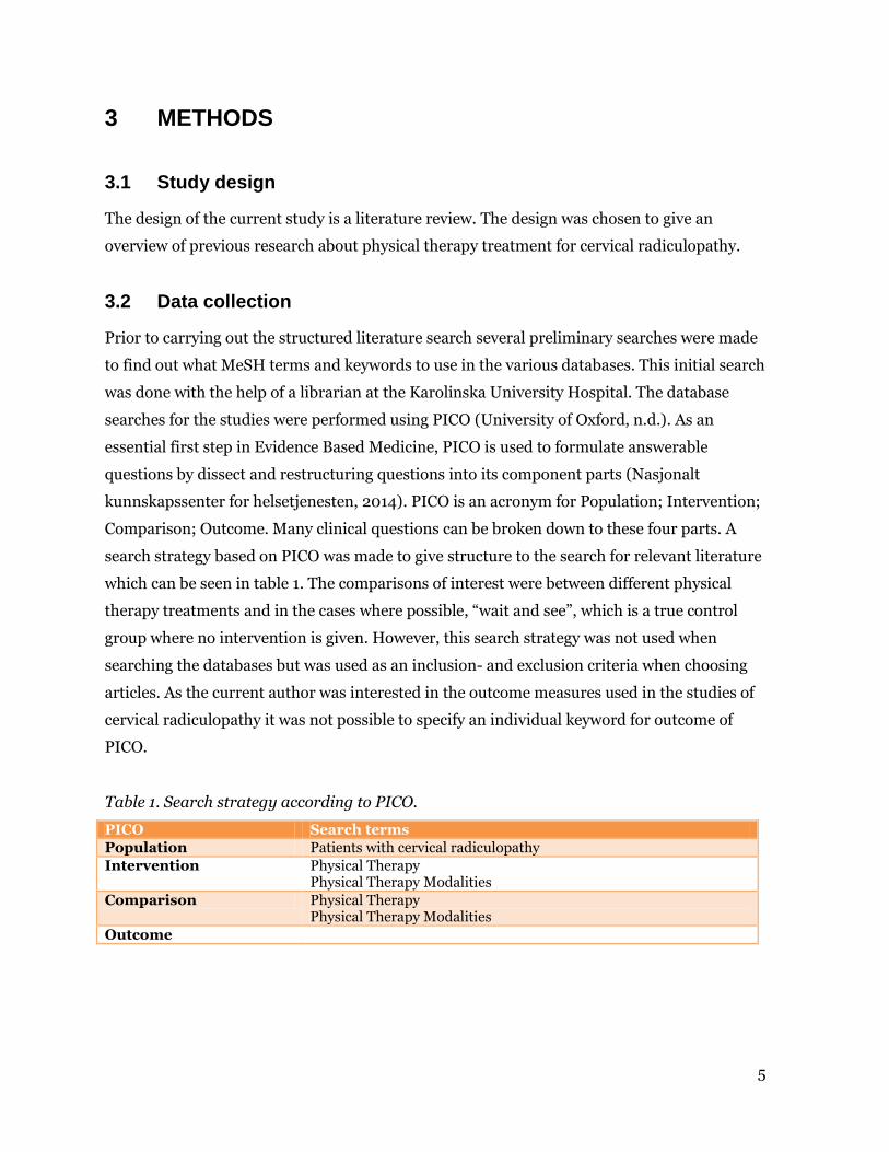
search strategy based on PICO was made to give structure to the search for relevant literature
which can be seen in table 1. The comparisons of interest were between different physical
therapy treatments and in the cases where possible, “wait and see”, which is a true control
group where no intervention is given. However, this search strategy was not used when
searching the databases but was used as an inclusion- and exclusion criteria when choosing
articles. As the current author was interested in the outcome measures used in the studies of
cervical radiculopathy it was not possible to specify an individual keyword for outcome of
PICO.
Table 1. Search strategy according to PICO.
PICO Search terms Population Patients with cervical radiculopathy Intervention Physical Therapy
Physical Therapy Modalities Comparison Physical Therapy
Physical Therapy Modalities Outcome
6
In April 2015, a search of four electronic databases: PubMed, CINAHL, PEDro and Web of
Science was carried out using detailed search terms in order to identify potential articles for
inclusion in this review. The review author used MeSH (PubMed), Thesaurus (CINAHL) and
free text words. Combinations were made based on (1) location (cervical); (2) disorder
(cervical radiculopathy); (3) intervention (physical therapy, physiotherapy, conservative
treatment); and (4) design; randomized controlled trial. Manual searches of reference list of
included articles was undertaken to search for possible studies not captured by the electronic
searches. The review author performed the electronic search together with a librarian at
Karolinska University Hospital. First, the title and abstract were screened for eligibility.
Second, the full text papers were assessed to verify whether the study met the inclusion
criteria regarding design, participants and intervention.
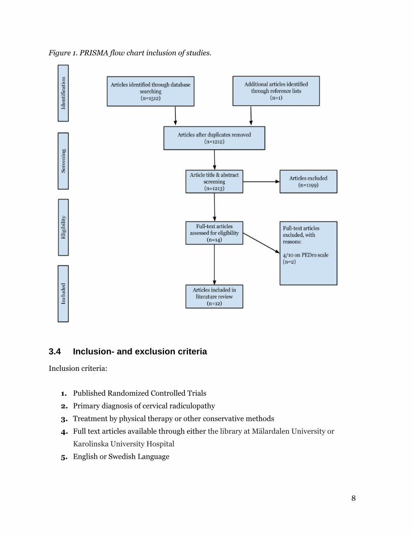
3.3 Study selection
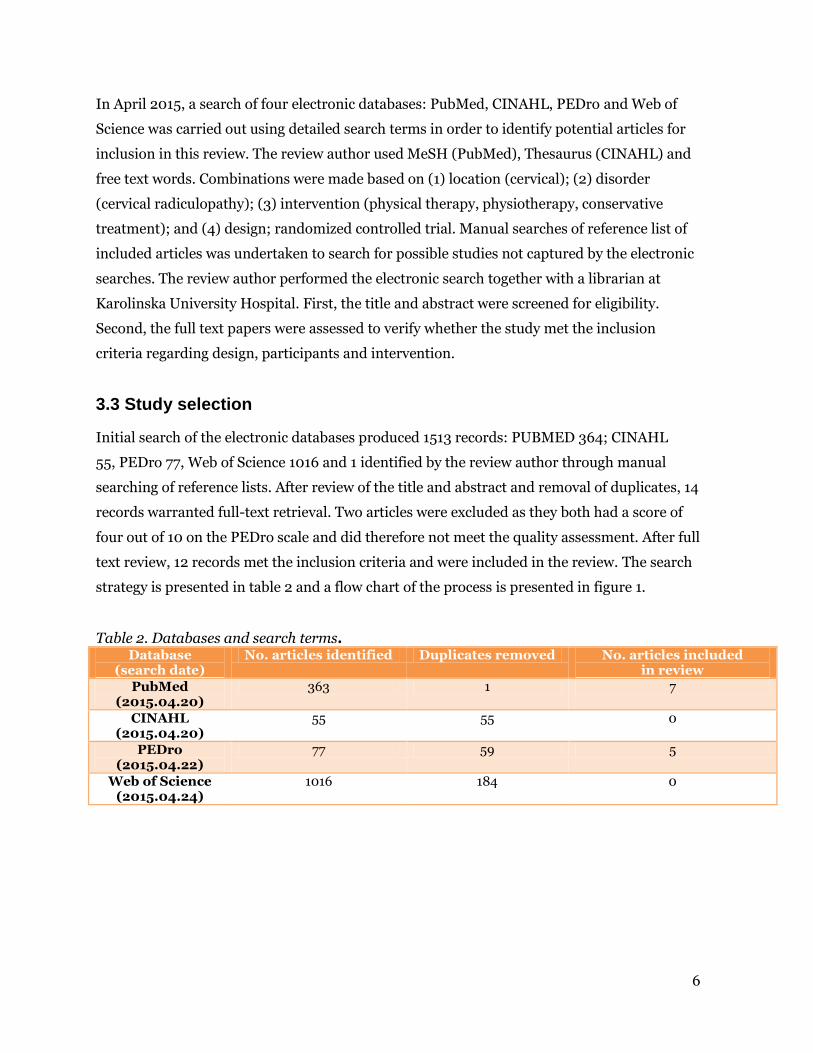
Initial search of the electronic databases produced 1513 records: PUBMED 364; CINAHL
55, PEDro 77, Web of Science 1016 and 1 identified by the review author through manual
searching of reference lists. After review of the title and abstract and removal of duplicates, 14
records warranted full-text retrieval. Two articles were excluded as they both had a score of
four out of 10 on the PEDro scale and did therefore not meet the quality assessment. After full
text review, 12 records met the inclusion criteria and were included in the review. The search
strategy is presented in table 2 and a flow chart of the process is presented in figure 1.
Table 2. Databases and search terms. Database
(search date) No. articles identified Duplicates removed No. articles included
in review PubMed
(2015.04.20)
363 1 7
CINAHL
(2015.04.20)
55 55 0
PEDro
(2015.04.22)
77 59 5
Web of Science
(2015.04.24)
1016 184 0
7
Search terms PubMed
(Physical Therapy Modalities) AND (((("prolapsed intervertebral disc") OR ((((((("neck disability") OR
"referred pain") OR "cervical spine injuries") OR "neck pain") OR "nerve root compression") OR
Radiculopathy) OR Intervertebral Disc Displacement)) OR "displaced intervertebral disc") OR "slipped
disc") Filters: Randomized Controlled Trial; English
Search terms CINAHL
(MH "Physical Therapy") AND ((MH "Radiculopathy") OR (MH "Neck Pain") OR "nerve root
compression" OR (MH "Referred Pain") OR "cervical spine injuries" OR "neck disability" OR (MH
"Intervertebral Disk Displacement") OR "prolapsed intervertebral disc" OR "displaced intervertebral
disc" OR "slipped disc") Filters: Randomized Controlled Trial; English
Search terms PEDro
"Radiculopathy"
Search terms Web of Science
("Physical Therapy" AND "Radiculopathy" OR "Neck Pain" OR "nerve root compression" OR "Referred
Pain" OR "cervical spine injuries" OR "neck disability" OR "Intervertebral Disk Displacement" OR
"prolapsed intervertebral disc" OR "displaced intervertebral disc" OR "slipped disc") Filters: English;
Clinical trial
8
Figure 1. PRISMA flow chart inclusion of studies.
3.4 Inclusion- and exclusion criteria
Inclusion criteria:
1. Published Randomized Controlled Trials
2. Primary diagnosis of cervical radiculopathy
3. Treatment by physical therapy or other conservative methods
4. Full text articles available through either the library at Mälardalen University or
Karolinska University Hospital
5. English or Swedish Language
9
Exclusion criteria:
1. Study designs focusing on surgical intervention
2. Study design comparing physical therapy with surgery or other non-conservative
treatment forms
3. Articles with a PEDro score of 4/10 or less.
3.4 Data analysis
4.4.1 Quality assessment
Selected research articles were first evaluated for quality using the PEDro scale which is an
11-item scale developed to rate methodological quality of randomized clinical trials (PEDro,
n.d.). The scale searches for literal evidence in the articles for true randomization,
concealment of randomization, blinded subjects, blinded therapists, blinded assessors,
appropriate statistical analysis, measures of variability and intention-to-treat analyses and
the number of “dropouts” (Harvey, Herbert, & Crosbie, 2002). It is important to note that
external validity, or generalizability, is not rated and neither is the size of the treatment effect
(PEDro, n.d.). One point is awarded for each item that meets the criteria, giving a total of ten
points. The first item, which is related to external validity, is not included in the final score. It
is important to note that trials are rated on basis of what they report. If no evidence is found
that a criterion is met then no score will be given for that particular criterion (PEDro, n.d.).
The scale has been shown to be both a reliable and valid measure (de Morton, 2009; Maher,
Sherrington, Herbert, Moseley, & Elkins, 2003). In the current study, articles with a PEDro
score of 7/10 or more are considered “high quality”, those with a score of 5/10 or 6/10 are
considered “moderate quality” and a score of 4 or less are considered “poor quality” in terms
of study methods and susceptibility to bias (Harvey et al., 2002). Articles that score 4/10 or
less will be excluded from this review. Once the current author has graded an article the score
will be compared with that given by PEDro. If there are discrepancies the score will be
amended to match the PEDro score.
10
4 RESULTS
4.2 Description of studies
Tables A and B in the appendix presents the characteristics and summary of the studies
included in the review. There were in total 12 studies included in this review. The studies took
place in eight different countries including Turkey, Egypt, The United States of America,
Tunisia, Iran, The Netherlands, Canada and China. Nine of the studies were carried out by
physical therapists with another two being conducted by medical doctors (Kuijper et al.,
2009; Zhang, Lin, & Yuan, 2011) and one by acupuncturists (Zhou et al., 2014).
4.2.1 Quality of studies
Table 5, found in appendix C, details the critical appraisal of the included studies. There were
no discrepancies between the scores given by the current author and those given by PEDro.
All of the studies were of moderate or high quality. No study scored the full 10 points but
Langevin et al. (2015) received nine points followed by Fritz et al. (2014) and Young et al.
(2009) that both received eight points. These latter two studies lost points because they failed
to blind the subjects and therapists. Langevin et al. (2015) was the only study that had a
double blind design but the study also lost one point as the therapists were not blinded. The
two lowest scores of five out of 10 were achieved by Albayrak Aydin et al. (2012) and Zhou et
al. (2014). These two studies lost points because of lack of blinding; no concealment of group
allocation and no “intention to treat” analysis were done. Diab et al. (2012) also lacked
blinding but received seven points and were considered high quality because the internal
validity and statistical reporting was otherwise sound. The remaining six studies that all
received six points tended to lack blinding, “intention to treat” analysis, group allocation
concealment, measures of key outcomes and heterogenic groups at baseline (Jellad et al.,
2009; Joghataei, Arab, & Khaksar, 2004; Kuijper et al., 2009; Moustafa & Diab, 2014;
Ragonese, 2009; Zhang et al., 2011).
11
4.2.2 Participants
The baseline characteristics were similar in all studies with three exceptions (Fritz,
Thackeray, Brennan, & Childs, 2014; Jellad et al., 2009; Kuijper et al., 2009). The most
notable of these exceptions was found in the study be Fritz et al. (2014) where the groups
were different in terms of symptom duration, gender, marital- and education status. The
groups were however similar regarding the most important prognostic factors such as age
and outcome measure scores. In the study by Jellad et al. (2009) one of the treatment groups
had higher scores in self-perceived disability whereas one of the groups in Kuijper et al.
(2009) study a higher number of participants reported right arm pain. Ragonese et al. (2009)
did not present any data on baseline characteristics making an evaluation of any potential
differences impossible.
There were marginally more women (55.3%) than men in the studies. The age of the patients
were for the most part reported as a mean number for each patient group which had a span
between 23.3 years (Zhang et al., 2011) to 46.9 in Fritz et al. (2014) study. The number of
patients in each study varied widely from 27 (Albayrak Aydin & Yazicioglu, 2012) to 216
(Moustafa & Diab, 2014) with a median number of patients of 70.5 and a total of 1026.
With regards to symptom duration there were four studies with acute patients (symptoms of
three months or less) (Fritz et al., 2014; Jellad et al., 2009; Kuijper et al., 2009; Langevin,
Desmeules, Lamothe, Robitaille, & Roy, 2015) and another four with chronic patients with a
symptom duration of more than three months (Albayrak Aydin & Yazicioglu, 2012; Diab &
Moustafa, 2012; Joghataei et al., 2004; Moustafa & Diab, 2014). In three of the studies the
symptom duration was not specified (Ragonese, 2009; Young, Michener, Cleland, Aguilera, &
Snyder, 2009; Zhang et al., 2011) and in the study by Zhou et al. (2014) there were both acute
and chronic patients.
Most of the participants were recruited directly from physical therapy or physician outpatient
clinics but there were exceptions. In the study by Jellad et al. (2009) the patients were
referred from rheumatologists, orthopedic surgeons and neurologists from a university
hospital or medical practitioners. Diab et al. (2011) recruited patients from the physical
therapy clinic at the academic institution where the researchers were based. Joghataei et al.
12
(2004) and Kuijper et al. (2009) received their patients via GP and physician referral. Two of
the studies recruited their patients from hospitals (Zhang et al., 2011; Zhou et al., 2014).
4.2.3 Measurement interval, retention rate and control group
All included studies measured the patients at baseline but the follow-up periods varied
widely. Two of the studies had one year follow-ups (Fritz et al., 2014; Moustafa & Diab, 2014)
while five studies, including Fritz et al. (2014) had their final follow-up at six months (Diab &
Moustafa, 2012; Jellad et al., 2009; Kuijper et al., 2009; Zhang et al., 2011). In the other
extreme there was the study by Zhou et al. (2014) where there was only a follow-up on day 10,
at the last treatment session. Albayrak Aydin et al. (2012) also had one follow-up which was
on the third and final week of treatment. Ragonese et al. (2009) had weekly follow-ups
ending on the third week upon completion of the study. In nearly all studies the follow-up
interval was between two to three weeks.
The retention rate was generally very high with eight out of twelve studies having no drop-
outs (Albayrak Aydin & Yazicioglu, 2012; Diab & Moustafa, 2012; Jellad et al., 2009;
Joghataei et al., 2004; Langevin et al., 2015; Ragonese, 2009; Zhang et al., 2011; Zhou et al.,
2014). Fritz et al. (2014) had the lowest retention rate at 62.8 percent, followed by Moustafa
et al. (2014) at 87.5 percent and Young et al. (2009) and Kuijper et al. (2009) that had 93 and
93.7 percent retention rate respectively. The reason for the dropouts was not given in any of
the studies. Langevin et al. (2015) and Kuijper et al. (2009) reported that there were no
differences in baseline characteristics between those lost to follow-up and those who
completed follow-up. In the study by Young et al. (2009) it was reported that there was an
equal amount of dropouts in both groups (n=6).
There was a control group in all but one (Ragonese, 2009) of the included studies. Kuijper et
al. (2009) was the only study to include a “Wait-and-see” group. In this latter study there was
also a group that wore a semi-hard collar without receiving any other treatment. The control
group in the study by Young et al. (2009) received manual therapy and exercise in addition to
sham traction. Four of the studies, including Kuijper et al. (2009), had two treatment groups
and one control while Ragonese et al. (2009) had three treatment groups comparing manual
physical therapy with therapeutic exercise and a combination of these two approaches. In the
13
remaining studies the control groups received standard treatment which consisted of
electrotherapy, exercise, manual therapy, and traction. In the study by Zhou et al. (2014) the
control group received acupuncture, which was considered standard treatment to the
researchers.
4.3 Outcome measures
An outcome measure, also known as the end point, is an outcome variable of interest in a
clinical trial (Consolidated Standards of Reporting Trials, n.d.). A randomized controlled trial
often has both primary and secondary outcomes. The primary outcome measure represents
the one which has the greatest therapeutic benefit while secondary outcomes measures are
used to evaluate additional effects of secondary importance (British Medical Journal, 2010;
Consolidated Standards of Reporting Trials, n.d.). In the present review information on
which were the primary and secondary outcomes was based on the information extracted
from the included articles.
4.3.1 Primary outcome measures
All 12 studies had primary outcome measures that measured pain, disability or function. The
most common measure of pain was the Visual Analogue Scale (VAS), which was used to
assess neck, arm and radicular pain (Albayrak Aydin & Yazicioglu, 2012; Jellad et al., 2009;
Kuijper et al., 2009; Zhou et al., 2014). The VAS has been found to have moderately to good
reliability in patients with chronic musculoskeletal pain (Boonstra, Schiphorst Preuper,
Reneman, Posthumus, & Stewart, 2008). The minimum clinically important difference
(MCID) for the VAS has been found to be 12 mm in a study conducted on adult patients in an
urban emergency department (Kelly, 2001). MCID is the minimal change in the score of an
instrument used to measure a symptom (Cook, 2008). Another clinimetric concept, which
will not be used in this review, is the minimal detectable change (MDC). The MDC is the
minimal change of an outcome measure that falls out of the measurement of error (Kovacs et
al., 2008). One of the functions of these concepts is to interpret the clinical relevance of
results in studies on the effectiveness of treatments (Kovacs et al., 2008). It is important to
note that a statistically significant change does not have to be clinically important. In the four
studies where VAS was used the reliability, validity and sensitivity to change in patients with
cervical radiculopathy has not been discussed. The other outcome measure for pain was the
14
Numeric Pain-Rating Scale (NPRS) which was used in three studies (Ragonese, 2009; Young
et al., 2009; Zhang et al., 2011). Disability was measured with the Neck Disability Index
(NDI) in six of the studies (Fritz et al., 2014; Kuijper et al., 2009; Langevin et al., 2015;
Moustafa & Diab, 2014; Ragonese, 2009; Young et al., 2009). The NPRS and the NDI have
been found to exhibit fair to moderate test-retest reliability in patients with mechanical neck
pain (Cleland, Childs, & Whitman, 2008). In two separate studies it was found that that the
MCID for the NDI was 14/50 points and one point for the NPRS (Cleland, Fritz, Whitman, &
Palmer, 2006; MacDermid et al., 2009). The MDC for the NDI has been found to be 10/50
points (MacDermid et al., 2009). Apart from the NDI, self perceived disability was measured
with VAS and the Patient Specific Functional Scale (PSFS) (Jellad et al., 2009; Young et al.,
2009). Jellad et al. 2009 do not discuss using VAS as a measure of self-perceived disability
and the current author has not found any studies where this outcome measure has been
validated for that purpose. It has been found that the PSFS is reliable, valid, and responsive
to change in patients with cervical radiculopathy (Horn et al., 2012). The MCID of the PSFS is
according to another study between 2.0 and 3.0 points (Cleland et al., 2006). Grip strength
was used as a primary outcome measure in two studies (Albayrak Aydin & Yazicioglu, 2012;
Joghataei et al., 2004). Albayrak Aydin et al. (2012) used a Jamar dynamometer while
Joghataei et al. (2004) used a Martin vigorimeter. The validity of the Jamar dynanometer has
not, to the knowledge of the current author, been established for patients with cervical
radiculopathy. There are however studies that demonstrate that the Jamar dynamometer is
both reliable and valid in healthy adults and in stroke patients (Boissy, Bourbonnais, Carlotti,
Gravel, & Arsenault, 1999; Mathiowetz, Weber, Volland, & Kashman, 1984). MCID values for
stroke patients has been found to be 5.0 and 6.2 kg for the affected dominant and
nondominant sides, respectively (Lang, Edwards, Birkenmeier, & Dromerick, 2008). In a
study comparing the Jamar dynamometer and the Martin vigorimeter the results indicated a
very high correlation between the two measures (Desrosiers, Hebert, Bravo, & Dutil, 1995).
Diab et al. (2011) assessed nerve root function of C6 and C7 using an EMG device by
measuring peak-to-peak amplitude of dermatomal somatosensory evoked potentials. Jellad
et al. (2009) measured the average number of analgesics that the patients took in one week.
Zhang et al (2011) and Zhou et al. (2014) used scales that measured cervical function in the
former study and in the latter; a scale measuring symptoms of spondylotic radiculopathy was
used. The assessment scale used by Zhang et al. (2011) consists of three major items: Clinical
symptoms, Clinical examination and Daily activities. Each item is further subdivided: Clinical
15
symptoms includes referred to pain and/ or numbness of neck, shoulder, back or upper
limbs, headache or dizziness; Clinical examination: tenderness, Spurlings test, Brachial
plexus stretch test, Neck hyperextension test, sensation disorders, muscle strength and test
for Hoffmann syndrome. The last item, Daily activities involve neck movement, bed mobility
and upper extremity loading. The scales for symptoms of cervical spondylotic radiculopathy
are a 20-score scale that assesses neurological function, movement, strength, pain and
discomfort of the neck and upper limbs (Zhou et al., 2014). The current author was unable to
find any information on the validity and reliability of the outcome measures used in the the
two studies from China.
4.3.2 Secondary outcome measures
Most of the studies had secondary outcomes to measure the effects of their treatments. In
contrast with the primary outcome measures there was a high heterogeneity amongst the
tests for secondary outcomes. Two of the outcomes assessed satisfaction with one focusing on
the patient (Young et al., 2009) and the other one on the treatment (Kuijper et al., 2009).
Four studies assessed pain as a secondary outcome and did so with either the Visual Analogue
Scale or the Numeric Pain-Rating Scale (Diab & Moustafa, 2012; Fritz et al., 2014; Langevin
et al., 2015; Moustafa & Diab, 2014). Kuijper et al. (2009) measured use of non-steroidal
anti-inflammatory drugs and opiates and working status by recording how many patients
were on partial or complete sick leave. Langevin et al. (2015) used the QuickDASH to
measure disability. The QuickDASH is a region specific questionnaire that evaluates physical
disability and symptoms of the arm, shoulder and hand in individuals with upper extremity
disorders and is valid in the measurement in patients with neck pain (Langevin et al., 2015).
Langevin et al. (2015) also measured cervicothoracic range of movement using a CROM
device. Young et al. used a whole battery of secondary outcome measures including the Fear
Avoidance Beliefs Questionnaire. The test is used to predict individuals that have high pain
avoidance behavior and may therefore be in more need of supervision than those who
confront their pain (Young et al., 2009). In the aforementioned study other secondary
outcome measures include body diagram of symptom distribution, the Global Rating of
Change Scale to quantify the patient’s improvement and grip strength using a dynamometer.
Moustafa et al. (2014) used an electromyograph to measure latency and peak-to-peak
amplitude of the flexor Carpi radialis H-reflex, which can be used in the diagnosis of patients
with suspected C6/7 nerve root lesions.
16
4.4 Interventions
The interventions in the studies can roughly be divided into two main categories: traction,
and manual therapy. In addition, electrotherapy, acupuncture and exercise were used in three
separate studies. Many of the studies have treatments that span over two or more categories
and these will be presented in the category that the current author considers the most
appropriate.
4.4.1 Traction
Traction was used in six of the twelve studies and was therefore the most frequent
intervention used.
Fritz et al. (2014) compared two cervical traction methods with a control group receiving neck
strengthening exercises. In the study 86 patients were randomized to one of three groups and
all three groups received an active exercise program to be performed daily in between therapy
sessions. In addition, the two treatment groups received 10 sessions over four weeks of either
mechanical or over-door traction. In the mechanical traction group the subjects were lying
supine on a bench with the neck in 15◦ flexion. A traction device was used to give intermittent
60 second tractions with 20 seconds of relaxation for a total of 15 minutes. An initial force of
5.4kg was used which was increased based on patient tolerance and symptom response. The
goal was to maximize symptom reduction and centralize symptoms. The over-door traction
group was given a traction device to be used daily at home. The device was set up, by the
patients, with an over-door bracket-and-pulley assembly on the top-edge of a door, with a
straight-back chair directly beneath the assembly. Traction was applied with the patient
seated facing the door, with the feet flat on the floor. The initial traction force was 3.6 to 5.4
kg with the goal of maximizing symptom reduction and centralization. Each session lasted 15
minutes.
Albayrak Aydin et al. (2012) randomized 27 patients into either a manual traction group or a
“standard” treatment group. The standard treatment involved: ultrasound (1 watt/cm2 for 10
min.), hot pack for 20 minutes, transcutaneous electrical nerve stimulation once a day for 20
minutes (60 Hz and impulse duration of 100 μsec) and isometric cervical muscle
strengthening, followed by stretching exercises for the spinal muscles. In the traction group,
17
intermittent tractions (7 s. traction and 5 s. rest, for 20 minutes) were performed by a
physical therapist. The patient was lying supine with the head in the most pain free position
and the weight gradually increased from five to twelve kilograms. Both groups received five
sessions a week to a total of 15.
Thirty-nine patients were randomized into three groups in the study by Jellad et al. (2009)
where a comparison was made between mechanical traction, manual traction and “standard”
physical therapy. In this study “standard” physical therapy consisted of 12 sessions (three per
week) of ultrasound, infrared radiation therapy, massage, cervical spine mobilization and
isometric strengthening of flexor and extensor muscles, followed by stretching of the spinal
muscles. No information on treatment variables were given in the article. Both traction
groups also received the standard treatment in addition to the traction. In the manual
traction group, intermittent cervical tractions (20 20-second tractions, a 10-second inter-
traction rest period) were performed by a physical therapist with a force of 6 kg. In the
mechanical traction group the patient was lying in supine with a weight bearing pulley
system. Each session comprised two 25 minute tractions, with a 10-minute rest interval. The
weight was gradually increased from five to 12 kg. During the traction, the neck was
maintained in the most pain-free position.
Joghataei et al. (2004) recruited 30 patients to participate in their four week study comparing
electrotherapy and exercise with mechanical traction. Patients in both groups received three
weekly sessions for a total of 10 treatments. The treatment and control group received
ultrasound (no parameters given) and isometric neck strengthening exercises (25 x 20
seconds, twice daily). The treatment group also received mechanical intermittent traction.
The subjects were in supine lying with a special pad under the head. A 14 kg traction force at
an angle of 24◦ was used for a period of 7 seconds, followed by a 5 second rest for a total of 20
minutes.
Young et al. (2009) compared mechanical traction with sham traction in a month long study
with 81 subjects. Both groups received an average of seven sessions with the same
standardized treatment protocol. All the sessions were carried out sequentially to include
postural education, manual therapy, and exercise. The difference between the two groups was
that one received mechanical intermittent traction, whilst the other group received sham
traction at the end of each treatment. Patients were informed about the importance of correct
18
posture and spinal alignment at the initial session and then only when deemed necessary at
subsequent sessions. Manual therapy was defined as high-velocity, low-amplitude thrust
manipulation or mobilizations. The therapist was required to perform at least one technique
each targeting the upper- and mid thoracic spine during each visit. Following treatment
directed at the thoracic spine, at least one set (30 seconds or 15–20 repetitions) of a
mobilization was directed at each desired level of the cervical spine. The cervical spine
techniques could include retractions, rotations, lateral glides in the ULTT1 position, and P-A
glides. The exercises included cervical retraction- and extension, deep cervical flexor- and
scapular strengthening of which at least one was used during each visit. At the end of the
session the subjects received either traction or sham traction for 15 minutes. The patient was
positioned in supine, with the cervical spine placed at an angle of approximately 15◦ of
flexion. The traction force was started at 9.1 kg or 10% of the patient’s body weight and
increased every visit depending on centralization or reduction of symptoms to a maximum of
15.9 kg. The on/off cycle was set at 50/10. The sham traction protocol included the identical
set-up; however, only 2.27 kg or less of force was applied. All other traction parameters were
the same as for the group that received intermittent cervical traction.
With 216 subjects the study by Moustafa et al. (2014) was the most numerous. The subjects
were randomly assigned to three groups where they all received “standard” physical therapy.
In addition, two groups received intermittent cervical mechanical traction for 20 minutes,
three times per week for four weeks. The “standard” treatment consisted of: infrared
radiation to the neck (15 minutes with lamp at 50-75cm distance); interferential therapy (100
Hz constant frequency and 125 μs pulse duration for 20 minutes); soft tissue mobilization
(deep, stroking massage to neck and shoulder muscles for pain relief and increased soft tissue
mobility); isometric muscle strengthening of deep cervical flexors, shoulder retractors and
serratus anterior; thrust manipulations to the thoracic spine. This “standard” treatment was
received three times per week for four weeks. In addition the subjects were asked to perform
the strengthening exercises twice daily at home. The first traction group received
ventroflexion traction with the patient in supine with the cervical spine at a 24◦ angle. The
same traction protocol used by Young et al. (2009), which is described above, was used in the
first traction group. With the second traction group the researchers used the flexor Carpi
radialis H-reflex to determine the optimal head posture in which to perform the traction. The
H-reflex amplitude was recorded using an electromyogram device and the optimal posture
19
was achieved when there was decompression of the nerve root. The traction protocol as in the
first group was used with the exception that the head posture in the second group was
variable at either 24◦ of flexion, neutral or 5◦ of neck extension, depending on the H-reflex
findings.
4.4.2 Manual therapy
Manual therapy was used in three of the included studies.
The first one of these studies was carried out by Langevin et al. (2015). In the study the
researchers randomized 36 subjects into two groups that attended eight physical therapy
sessions for four weeks. In the control group the physical therapist could choose four manual
therapy techniques, at any spinal level, from the following: rotations, lateral glides in neutral,
posteroanterior glides, posteroinferior medial glides, or anterosuperior anterior glides. Each
technique was performed for 10 repetitions of 30 seconds. After the manual therapy, a 5-
minute nonspecific manual traction was applied. A standardized home exercise program
aimed at strength and mobility of the neck and shoulders was given to the participants at the
end of the session. Any techniques or exercises aimed at increasing the size of the
intervertebral foramen were not allowed. The treatment group received the same
rehabilitation program as the control group, except that two of the four mobilizations had to
be techniques thought to increase the size of the intervertebral foramen and that the third
exercise of the home program was a repeated movement known to increase the size of the
intervertebral foramen (10 repetitions, 10 times per day).
In the second manual therapy study, 30 patients were randomized into three groups where
one received manual therapy, the second group therapeutic exercise and the third group
received a combination of the two treatment approaches (Ragonese, 2009). All groups made
three visits per week for three weeks to their designated physical therapist. The manual
therapy group received: cervical lateral glides (30-45 seconds at each cervical level); thoracic
mobilizations (30-45 seconds at each selected thoracic levels) and neural dynamic
mobilizations for the median nerve. The exercise group performed all exercises with the
physical therapist at their clinic and no home exercise program was given. This group
performed deep neck flexor strengthening exercises (10 repetitions held for 10 seconds);
lower- and middle trapezius strengthening exercises (two sets of 15 repetitions) and serratus
20
anterior exercises doing “push-up plus” against the wall. The third group did a combination
of the two aforementioned treatment protocols.
In the third study, by Zhang et al. (2011), one hundred and twenty subjects between the ages
of 18-35 were randomized into two groups comparing Tuina therapy with manual traction.
Tuina therapy is a form of manual therapy used in traditional Chinese medicine. The subjects
in the former group received pressing, kneading, plucking manipulation, shoulder grasping
and neck stretching. The kneading was done to certain acupuncture points (GB 20, GV 16 and
Ashi). The manual intermittent traction, which was received by the control group, was carried
out using an occipital-jaw belt with the neck in 15° of flexion; a force of eight to ten kg; for a
traction period of 60 seconds, followed by a 10 second rest for a total of 15 minutes. Both
groups received treatment every other day for a total of 10 sessions.
4.4.3 Electrotherapy, exercise and acupuncture
The three remaining studies included various treatment methods with a mixture of
ultrasound, exercise, acupuncture, cervical collar and a “Wait-and-see”-group.
The first of these three was the study by Diab et al. (2011) with 96 subjects randomized into
two groups where both received infrared radiation and ultrasound. In addition, the
experimental group received posture corrective exercise. Infrared radiation was given for 10
minutes to the neck followed by continuous ultrasound application on upper trapezius (10
minutes; 1.5 w/cm² intensity). This conventional treatment was given three times per week
for 10 weeks. The experiment group also received two strengthening exercises of the deep
cervical flexors and shoulder retractors (three sets of 12 repetitions) and two stretching
exercises of the cervical extensors and pectoral muscles (held for 30 seconds each). This
program was carried out four times per week for 10 weeks. Both groups were asked to avoid
any other exercise programs.
Kuijper et al. (2009) was the only study out of all twelve included in this review that included
a true control group. Two hundred and five subjects were randomized into three groups with
the first group receiving a semi-hard cervical collar that patients were advised to wear during
the day for three weeks and then gradually decrease the use of the collar over the following
three weeks. After week six the patients were advised to stop wearing the collar completely.
21
During the first three weeks the patients were asked to rest as much as possible. The second
group received twice weekly physical therapy sessions for six weeks consisting of “hands-off”
graded activity exercises to strengthen the superficial and deep neck muscles. This group was
also given a home exercise program to be performed daily. The control group was advised to
continue with their daily activities and was not given any form of treatment.
The last of the three studies compared two types of acupuncture (Zhou, 2014). Sixty patients
were randomly divided into either a group receiving conventional acupuncture or
acupuncture plus warming-needle moxibustion. The latter treatment involves burning herb
cone attached to the acupuncture needle which produces heat. Needles were applied at
acupuncture points: GV 14, BL 11, SI 11, GB 21 and LI4. In the conventional acupuncture
group, needles were inserted into the following points: TE 14, LI 15, LI 11, TE 10, TE 4 and TE
5. Treatment was given for both groups every day for a total of 10 days.
4.5 Treatment period
In six of the studies the treatment period lasted for four weeks with the majority having three
sessions per week (Fritz et al., 2014; Jellad et al., 2009; Joghataei et al., 2004; Moustafa &
Diab, 2014). In the studies by Young et al. (2009) and Langevin et al. (2015) the patients had
two sessions per week. Three of the studies lasted for three weeks with the ones by Ragonese
(2009) and Zhang et al. (2011) having three sessions per week while the patients in Albayrak
Aydin et al. (2012) study had five sessions per week. Diab et al. (2012) study was the longest
and lasted for 10 weeks while the one by Zhou et al. (2014) was the shortest with 10 days. In
the latter study the patients came for treatment every day while the control group in Diab et
al. (2012) study had three sessions per week and the treatment group had four. In the study
by Kuijper et al. (2009) the physical therapy group had two sessions per week for a total of six
weeks while the other two groups did not receive any sessions. Each session lasted between
10-20 minutes three times a week to 30 minutes per day for 10 days. The average session
lasted 25-30 minutes.
22
4.6 Outcome of intervention
In the twelve studies covered in this review nine different primary outcome measures were
used to assess the effect of the interventions. Treatment times varied between 10 days to six
weeks with follow-ups from 10 days to 12 months.
4.6.1 Outcome at the end of treatment
The study by Albayrak Aydin et al. (2012) lasted for three weeks and at the end of treatment
there had been significant improvements in both groups on the primary outcome measures
neck pain and max grip strength (p=0.001) but the treatment group had a significantly
greater improvement in grip strength than the control group (p=0.037 and p=0.042,
respectively). The MCID for neck pain measured with VAS was achieved in both groups.
MCID for grip strength was not achieved by either group. Both groups had received 15
sessions of “standard” physical therapy. The treatment group also received traction but the
frequency and length of this additional treatment is not reported.
Diab et al. (2011) reported that at the end of the 10 week treatment the experimental group
had improved significantly more than the control group on the primary outcome measure,
which was nerve root function of C6-7 in (p=0.001). The posture corrective exercises that
were carried out by the experimental group were done at home. It is reported that the
subjects in this group were contacted on a weekly basis but the compliance rate of the
exercise program is not reported. Both groups, that also received ultrasound and infrared
radiation, were instructed not to engage in any exercise program that could interfere with the
results of the study.
In the study by Fritz et al. (2014) there was, as mentioned above, a high dropout rate at just
over 37 per cent but the baseline characteristics of those lost to follow-up was no different to
those who stayed for follow-up. The study, which lasted four weeks, showed that the exercise,
mechanical traction and over-door traction groups all had made improvements at the end of
treatment. However, no significant differences between the groups were found at the end of
treatment when the results were analyzed with an “intention-to-treat” method. The p-values
for the group comparisons were all greater than 0.05: exercise versus mechanical traction
(p=0.36); exercise versus over-door traction (p=0.48) and mechanical traction versus over-
23
door traction (p=0.84). The primary outcome was disability measured with the NDI and
MCID was achieved in all three groups.
The mechanical- and manual traction groups in the study by Jellad et al. (2009) improved
their neck- and radicular pain and self perceived disability at the end of the four week
treatment (p<0.05). The “standard” physical therapy group also made significant
improvements in self perceived disability but not in neck- and radicular pain (p>0.05). The
analgesic consumption fell significantly in all three groups at the end of treatment but no
between group differences was seen.
In the month long study by Joghataei et al. (2004) both the observation and treatment
groups received electrotherapy and neck exercise with the treatment group receiving
additional mechanical traction. Grip strength was measured with a Vigorimeter in week two
and at the end of treatment another two weeks later. At the end of treatment significant gains
had been made in both the traction group and the control group but there was no statistical
difference between the groups. There was, however a difference between the groups at two
weeks but the control group made up for the difference in the last half of the study period.
At the end of the six week treatment in the study by Kuijper et al. (2009) it could be seen that
the cervical collar group and physical therapy group had made the greatest improvements in
pain and neck disability, but the “wait-and-see” group had also made significant
improvements. The collar group fared slightly better in the primary outcomes neck-and arm
pain and disability than the therapy groups at the 6 week follow-up. MCID was achieved in
both treatment groups in all three primary outcomes. The control group achieved MCID in
arm pain measured with VAS at the 6 week follow-up.
The study by Langevin et al. (2015) lasted four weeks and at the end of the treatment it was
found that the two groups that had both received manual therapy had made statistically
significant improvement in the Neck Disability Index scores with a p-value of less than 0.05.
However, no significant between group difference could be observed (p=0.44). The MCID of
disability, measured with the NDI, was achieved in both groups.
24
Moustafa et al. (2014) found that the H-reflex based traction group scored better on the Neck
Disability index than the “standard” physical therapy group and “standard” traction group at
the end of the four week study period (p<0.005). No significant difference was seen between
the other traction group and the “standard” treatment group. The MCID was achieved in the
H-reflex based traction when measured with the NDI. It should be noted that there was no
blinding of subjects, assessors or therapists in this study.
In the study by Ragonese et al. (2009) there were three groups where one received exercise, a
second one received manual therapy and a third group a combination of the two. It was found
that the all three groups made significant improvements in both pain (p<0.01) and function
measured with the Neck Disability Index (p<0.05). The greatest gains were made by the
group that combined manual therapy and exercise (p<0.05). The MCID of disability
measured with the NDI was achieved by the treatment groups at the end of treatment. Pain
measured with VAS was achieved in all three groups at the end of treatment. All three groups
received the same number of sessions but the length of the treatments was not given.
Young et al. (2009) treated 81 patients for four weeks with manual therapy, exercise and
either traction or placebo traction. At the end of treatment both groups had made significant
gains in pain, disability and function but no significant between group difference was found
when measured with the NPRS, NDI and PSFS (p>0.24). When looking at the MCID
disability measured with the NDI was not achieved by either group. Pain and function did
show MCID when measured with the NPRS and the PSFS. At the end of treatment the
subjects were not asked if they could identify which group they were assigned to. The
effectiveness of the subject blinding is therefore not known.
The subjects in the study by Zhang et al. (2011) were the youngest out of the included studies
with an average age of 23. The 120 patients were divided into a Tuina therapy group and a
traction group for a total of 10 sessions given every other day. Both groups made
improvements in pain and cervical function at the end of treatment (p<0.05 for both
measures), with the Tuina therapy group making significantly greater gains. The MCID for
pain measured with NPRS was achieved in both groups.
25
In the study by Zhou et al. (2014) it was found that the acupuncture group with moxibustion
had made greater improvements in the Scales for symptom of cervical spondylotic
radiculopathy and pain than the group that received “regular” acupuncture (p<0.05). The
MCID for pain measured with VAS was achieved in both groups.
4.6.2 Outcome at follow-up
Seven out of the 12 studies included in this review had follow-up assessments. Fritz et al.
(2014) and Jellad et al. (2009) had follow-ups at six- and 12 months and three- and six
months respectively. The other studies had follow-ups at eight weeks, six months or one year.
In the study by Diab et al. (2011) the difference between groups was maintained at the six
month follow-up with a probability value of less than 0.001.
Fritz et al. (2014) had two follow-ups at six months and one year after completion of the
study. At the end of treatment no difference was seen between groups but at the follow-ups
the mechanical traction group demonstrated better results than the exercise group at both
six- and twelve months and at six months when compared to the over-door traction group.
No difference was seen between the over-door traction group and the exercise group at either
of the follow-ups.
In the study by Jellad et al. (2009) there were two follow-up assessments held. Neck pain,
which had improved in both the traction groups at the end of treatment, did not further
improve at the subsequent follow-ups. However, when compared to baseline, the two groups
had reduced neck pain at the six month follow-up. The mechanical traction group made
continuous improvements in radicular pain until six months post treatment while the manual
traction group showed improvements up until the three month follow-up and then made no
further improvement. The improvements these groups had made when compared to baseline
was still significant at the six month follow-up. The self-perceived disability continued to
improve in the traction groups until the six month follow-up. Compared to baseline
assessment the improvement was significant at the six month follow-up. The “standard”
treatment group had made no significant improvements in neck- or radicular pain or self-
perceived disability at the follow-ups when compared to baseline. The analgesic consumption
26
that had decreased for all three groups at the end of treatment did not change any further at
the follow-ups but was still significantly lower at the final follow-up.
When Kuijper et al. (2009) had a follow-up six months after the end of treatment it was
found that the subjects in all groups had limited or no neck- and arm pain with VAS scores
for neck pain of no greater than two out of 10 points. The Neck Disability Index had
decreased slightly in all three groups at the six month follow-up but no between group
differences could be seen.
Langevin et al. (2015) had a follow-up at four weeks after the end of treatment. No difference
was seen between groups at the end of treatment and neither at the follow-up. There was
however continued improvements in the Neck Disability Scores for both groups at the follow-
up (p<0.05).
At the one year follow-up it was only the H-reflex based traction group, in the study by
Moustafa et al (2014) that had maintained the improvements that were seen at the end of
treatment.
Zang et al. (2011) had a follow-up at six months post treatment however no figures are
presented in the paper for the primary outcomes. Instead the long term effect of the
treatment was measured with the rate of relapse. No further explanation was given by the
authors about this measure but it was reported that relapse rate for the observation group
was zero and 16 percent for the control group.
27
5 DISCUSSION
The aim of this review is to assess the effects of physical therapy for patients with cervical
radiculopathy. The following questions were being asked: (1) What is the quality of the
studies measured with the PEDro scale? (2) What primary and secondary outcomes were
used to demonstrate the effects of the intervention? (3) What interventions were used in
the treatment of the cervical radiculopathy? (4) What was the outcome of the
intervention with regards to the primary outcomes at the end of treatment and at follow-
up?
5.1 Summary of results
Out of the 12 studies that were included in this review, eight were of moderate quality and
four of high quality when measured with the PEDro scale. The lowest score, of five points,
was given to two studies while the highest score of nine points was awarded to one study. The
average score was 6.5 which indicate that overall the methodological quality of the included
studies was moderate.
Most of the included studies shared the same outcome measures, but there were some
notable exceptions. All studies did however have outcome measures that measured pain,
disability and function. The most common outcome measure for pain was the Visual
Analogue Scale followed by the Numeric Pain-Rating Scale. Disability was in most cases
measured with the Neck Disability Index but also with the Visual Analogue Scale and the
Patient Specific Functional Scale. Grip strength, nerve root function and use of analgesics
were also measured. In two studies, which were both conducted in China, outcome measures
that measured cervical function and symptoms of radiculopathy were used. These particular
outcome measures were not used in any of the other studies. Five of the studies did not have
secondary outcome measures and the number of outcome measures varied from just one to
eight.
The two most common interventions in the included were traction and manual therapy, with
the former being by far the most common. In several of the studies manual traction was
compared with mechanical traction but traction was also compared with “standard”
treatment for cervical radiculopathy. Other interventions included electrotherapy,
28
acupuncture, collar and exercise. One study included a true control in the form of a “Wait-
and-see” group and another performed a placebo treatment.
The outcomes of the interventions were varied with some reporting positive effects and
others no change of the symptoms. The treatment periods, number of sessions and use of
follow-up varied across the studies. In most cases, the positive results that were seen at the
end of treatment wore off at follow-up and the differences between groups became less
pronounced.
5.2 Discussion of results
5.2.1 Quality of studies
Due to the inclusion criteria that stated that only studies that had received a score of five or
higher on PEDro scale were to be included, the quality of the studies were moderate to high.
That is not to say that there were no methodological flaws in several of the studies. The lack of
blinding was an issue in all studies. Only Langevin et al. (2015) had a design where both the
subjects and the assessors were blinded. In most cases in physical therapy research it can be
difficult if not impossible to blind the treating therapist. However, efforts should be made in
future research to blind the subjects and the assessors. With lack of blinding there is an
obvious risk of bias and placebo effects. Seven of the 12 studies did not include an “intention-
to-treat” analysis which is done to avoid the effects of subjects’ crossing over to different
groups or those that drop out. This could lead to misleading results of the studies and thus
making it difficult to draw any conclusions from the findings.
5.2.2 Outcome measures
The large variability in the outcome measures makes comparisons between studies difficult
which is something that was also noted in an earlier systematic review by Thoomes et al.
(2013). The authors of that study found that only four out of 11 studies measured pain with
either VAS or NPRS (Thoomes et al., 2013). In contrast, in the current review all but one of
the 12 included studies measured pain with VAS or NPRS as either a primary or secondary
outcome measure. However, only five of these described the actual tests and how they were
administered (Albayrak Aydin & Yazicioglu, 2012; Diab & Moustafa, 2012; Fritz et al., 2014;
29
Langevin et al., 2015; Moustafa & Diab, 2014; Zhou et al., 2014). Although the VAS and NPRS
are standardized tests there are variations of these, which was demonstrated in the study by
Albayrak Aydin et al. (2012). In that study it was described that 0 (mm) on the VAS scale
represented “the least pain imaginable” whereas in the other studies 0 (mm) represented “no
pain”. Such subtle differences may influence how the subjects rate their pain and how the
results are interpreted. It can also be discussed if treatment effects are best measured with
pain scales. Perhaps disability is a more appropriate measure for assessing treatment effects.
An indication of the importance of this outcome may be that it was used as a primary
assessment tool in nine of the studies. Seven of these measured disability with the patient-
completed, condition-specific functional status questionnaire NDI while Jellad et al. (2009)
used VAS and Zhang et al. (2011) used a scale called the Assessment scales for cervical
spondylosis. None of the studies reported on the test-retest reliability of any of the outcomes
and only three studies provided data on the responsiveness of VAS, NPRS and NDI in
patients with neck- and arm pain patients (Albayrak Aydin & Yazicioglu, 2012; Langevin et
al., 2015; Young et al., 2009). This lack of data makes evaluation of the effects of the
interventions difficult.
5.2.3 Interventions and outcomes
The most frequently used intervention was traction and in five of the studies it was treatment
given to the observation group (Albayrak Aydin & Yazicioglu, 2012; Fritz et al., 2014; Jellad et
al., 2009; Moustafa & Diab, 2014; Ragonese, 2009; Young et al., 2009). In the study by Zhou
et al. (2014) traction was used in the control group. Previous reviews have found that traction
was no more effective than placebo traction (Thoomes et al., 2013). This evidence was partly
based on the study by Young et al. (2009), which is included in the current review, and
another randomized controlled trial from 1990 (Thoomes et al., 2013). The subjects in the
latter trial were mostly diagnosed with cervical spondylosis. The results are therefore not
necessarily transferable to other patient groups such as those with cervical radiculopathy. The
study by Young et al. (2009) was methodologically sound and was considered high quality
according to the PEDro scale. However, the diagnostic tool used was not validated so the
diagnostic accuracy was not guaranteed. Secondly the effectiveness of the blinding was not
known. It is also important to note that both the traction group and the placebo group
achieved scores that exceeded the MCID on both the NPRS and the Patient Functional Scale
(Young et al., 2009). In one study on MCID of the NDI and NPRS with nonspecific neck
30
patients it was found that a score of 10.5 points for the NDI and 4.3 points for the NPRS was
required to establish a treatment effect (Pool, Ostelo, Hoving, Bouter, & de Vet, 2007).
Another study conducted on patients with nonspecific neck pain found that 1.5 points or
more was needed for a minimal detectable change on the NPRS (Kovacs et al., 2008). Fritz et
al. (2014) found that adding mechanical traction to exercise resulted in lower scores on the
NDI at the six- and 12 month follow-ups. The researcher did not however provide data on the
minimal clinically important change and the high loss to follow-up might have biased the
results. Albayrak Aydin et al. (2012) found that traction and “regular” physical therapy
reduced arm and neck pain on VAS more than “regular” physical therapy alone. Both groups
did however exceed the minimal clinically important change (9 mm) with -45±16 and -34±12
respectively. This study did receive one of the lowest score and lacked blinding, concealed
allocation and “intention to treat” analysis which affects the methodological quality. Zhang et
al. (2011) concluded that manual therapy (Tuina therapy) was more effective than manual
traction when measuring pain with the NPRS. Again, no minimal clinically important change
was given by the authors. A difficulty in the comparison of the traction methods is the variety
in treatment protocols and methods used. In the latter study an occipital-jaw belt was used;
Young et al. (2009) used a mechanical device for traction and over-door traction while Jellad
et al. (2009) used hands-on manual traction and weights and a pulley system; Moustafa et al.
(2014), like Young et al. (2009) used a mechanical device. The manual therapy treatment,
which was used in four studies, was also, like the traction, heterogeneous (Langevin et al.,
2015; Ragonese, 2009; Young et al., 2009; Zhang et al., 2011). Zhang et al. (2012) treated the
patients with a form of Chinese manual therapy called Tuina. No further explanation about
the therapy other than what actual techniques were used. For the uninitiated it is difficult to
get an idea of what the therapy entails, making comparisons difficult. Langevin et al. (2015)
was the study that received the highest PEDro score out of all the included studies. The high
methodological quality was clearly noticeable when studying the paper. The subjects in
Langevin et al. (2015) study received manual therapy based on Maitland’s methods. One
group was given additional techniques thought to increase the size of the intervertebral
foramen. No difference was found between the groups but both groups made significant
improvements when measured with the NDI. However, as previously discussed there is an
element of spontaneous resolution of symptoms in CR and with the absence of a “no
treatment group” the possibility spontaneous recovery cannot be excluded. Ragonese et al.
(2009) found that a combination of manual therapy techniques including neural dynamic
31
techniques for the median and exercise was superior to manual therapy or exercise alone.
However, there was data on the patient characteristics, there was lack of blinding and no
“intention to treat” analysis was made. Finally, the study by Zhou et al. (2014) compared two
types of acupuncture in 30 patients divided into two groups. In common with Zhang et al.
(2011) it was difficult to follow the treatment protocol and understand the treatment
rationale. There were explanations given about the combustion acupuncture technique but
without knowledge in Traditional Chinese medicine it is difficult to grasp the concepts. Also,
in common with the study by Zhang et al. (2011), some of the outcome measures used are not
used in Western Evidence Based Medicine and no information is given about the validity,
reliability and responsiveness of the outcome measures. This makes interpretation of the
results difficult, if not impossible.
5.3 Discussion of method
The design was chosen to give an overview of the available research on physical therapy
treatment methods for cervical radiculopathy.
The choice of databases was determined in conjunction with a research librarian at
Karolinska University Hospital. As the focus of this review was physical therapy and
conservative treatment then PubMed was chosen as the main database to find relevant
literature. Most of the articles included in this review were found through PubMed. CINAHL,
which is a database with a focus on mainly nursing and allied health, was also used in this
review. All of the hits made through CINAHL had already been identified through PubMed
which suggests that the search through the latter database had been thorough. The PEDro
database proved to be a good source of literature for the purpose of this review. The database
contains randomized trials, systematic reviews and guidelines related to physical therapy.
Five of the 12 articles included in this review were found through PEDro and none of these
were duplicates. Web of Science was the last database to be searched. Over a thousand hits
were made through this database and even after duplicates had been removed there were
nearly nine hundred articles left. All of these articles were excluded after title and abstract
were read as they did not fit the inclusion criteria of this review. Only one article was found
through manual search of reference lists of included articles. It is unlikely that more relevant
articles would have been located if more databases had been searched as the most
comprehensive databases for physical therapy researched were used.
32
When searching PubMed and CINAHL, controlled vocabulary (MeSH) was used to facilitate
the searches and make the results more relevant. In PEDro and Web of Science there is no
controlled vocabulary function so free text searches were made in these databases. In PEDro
only the word “radiculopathy” was used to search for articles and this gave 77 hits. A
combination of words and terms such as those used in Web of Science could have been done
in order to increase the number of hits. However, this method would have resulted in a very
small number of hits. There was also no guarantee that more relevant articles would have
been found. Free text search was also done in both PubMed and CINAHL. With the inclusion
of freer search words and MeSH terms it is possible that more articles would have been
located. However, after having located 12 articles together with time constraint it was decided
that no further search would be carried out. The searches were limited to randomized
controlled trials and articles written in the English language. By excluding all research other
than randomized controlled trials it is possible that it led to publication bias. It may also be
that valuable insights and information could have been found within the excluded research.
However, as a means of minimizing risk of bias and to increase generalizability and
transferability only randomized controlled trials were included in the current review.
Furthermore, the language restriction may also have been associated with bias.
Several large randomized controlled trials comparing physical therapy with surgery have
been carried out. If these would have been included then a greater number of articles could
have been included. However, the focus and interest of this review was to compare the effects
of physical therapy and conservative treatment as this is the area in which the current author
operates. Also, a limited amount of literature reviews with this focus have been carried out.
The quality control of the studies was done using the PEDro scale and a cut-off point of five
was chosen. The reasons for the five point limit have been discussed above. If studies of “low
quality” would have been included this could have potentially affected the validity and
reliability of the current review. Using the PEDro scale can be difficult without qualified
guidance and the potential for awarding the studies the wrong score is always present. In the
current review the scores awarded by the current author was compared with those available
on the PEDro website. This ensured that the scoring had been done correctly. The quality
control could have been done with other instruments such as the one by SBU. However, the
33
current author was already familiar with the PEDro scale and the scale has been found to be
both valid and reliable, so it was decided that that instrument was to be used.
There was a large variability in the patient population, symptom duration, number of
treatment sessions, length of the treatment periods and outcome measures used. This made
comparisons between studies difficult.
One major limitation of the current study is that the current author did all the work alone.
This meant that the article screening, selection and assessment were done by one person.
This may lead to articles being missed, erroneously excluded or included and wrongly
assessed, and could be a source of bias.
5.4 Ethical discussion
This review did only include studies that had received ethical approval or in the cases where
this was not reported, the subjects had given their written consent to partake in the study.
6 CONCLUSION
This review found that the quality of the randomized controlled trials investigating cervical
radiculopathy is variable. Most of the studies reported effects of the treatment but in nearly
all cases both the control group and the treatment group had made improvements. This was
also the case in the only study to include a non-treatment group. There were a number of
outcome measures used in the included studies but the primary outcomes were in all but one
study measuring pain with VAS or NPRS or pain related disability with NDI. Traction was the
treatment investigated in the majority of studies followed by manual therapy, electrotherapy,
exercise and acupuncture. The outcomes of the interventions show that there is limited
evidence to suggest that physical therapy treatment, as carried out in the included studies,
can have a measurable and significant long-term effect on cervical radiculopathy when
compared to control groups. However there is a need for more randomized controlled trials
with blinded subjects and assessors in addition to no-treatment groups.
34
REFERENCES
Abbed, K. M., & Coumans, J. V. (2007). Cervical radiculopathy: pathophysiology, presentation, and clinical evaluation. Neurosurgery, 60(1 Supp1 1), S28-34.
Albayrak Aydin, N., & Yazicioglu, K. (2012). Cervical intermittent traction: does it really work in cervical radiculopathy due to herniated disc? Turkiye Fiziksel Tip ve Rehabilitasyon Dergisi [Turkish Journal of Physical Medicine and Rehabilitation], 58(4), 277-282.
Blumenkrantz, N., Sylvest, J., & Asboe-Hansen, G. (1977). Local low-collagen content may allow herniation of intervertebral disc: biochemical studies. Biochem Med, 18(3), 283-290.
Boissy, P., Bourbonnais, D., Carlotti, M. M., Gravel, D., & Arsenault, B. A. (1999). Maximal grip force in chronic stroke subjects and its relationship to global upper extremity function. Clin Rehabil, 13(4), 354-362.
Boonstra, A. M., Schiphorst Preuper, H. R., Reneman, M. F., Posthumus, J. B., & Stewart, R. E. (2008). Reliability and validity of the visual analogue scale for disability in patients with chronic musculoskeletal pain. Int J Rehabil Res, 31(2), 165-169.
British Medical Journal. (2010). Primary and secondary outcome measures. Retrieved 06 June, 2015, from http://www.bmj.com/content/340/bmj.c1938
British Medical Journal. (n.d.). What is epidemiology. Retrieved 16 June, 2015, from http://www.bmj.com/about-bmj/resources-readers/publications/epidemiology-uninitiated/1-what-epidemiology
Childs, J. D., Cleland, J. A., Elliott, J. M., Teyhen, D. S., Wainner, R. S., Whitman, J. M., et al. (2008). Neck pain: Clinical practice guidelines linked to the International Classification of Functioning, Disability, and Health from the Orthopedic Section of the American Physical Therapy Association. J Orthop Sports Phys Ther, 38(9), A1-A34.
Cleland, J. A., Childs, J. D., & Whitman, J. M. (2008). Psychometric properties of the Neck Disability Index and Numeric Pain Rating Scale in patients with mechanical neck pain. Arch Phys Med Rehabil, 89(1), 69-74.
Cleland, J. A., Fritz, J. M., Whitman, J. M., & Palmer, J. A. (2006). The reliability and construct validity of the Neck Disability Index and patient specific functional scale in patients with cervical radiculopathy. Spine (Phila Pa 1976), 31(5), 598-602.
Cohen, S. P., Hayek, S., Semenov, Y., Pasquina, P. F., White, R. L., Veizi, E., et al. (2014). Epidural steroid injections, conservative treatment, or combination treatment for cervical radicular pain: a multicenter, randomized, comparative-effectiveness study. Anesthesiology, 121(5), 1045-1055.
35
Consolidated Standards of Reporting Trials. (n.d.). Glossary. CONSORT Glossary Retrieved 06 June, 2015, from http://www.consort-statement.org/resources/glossary#O
Corey, D. L., & Comeau, D. (2014). Cervical radiculopathy. Med Clin North Am, 98(4), 791-799, xii.
Cote, P., Cassidy, J. D., Carroll, L. J., & Kristman, V. (2004). The annual incidence and course of neck pain in the general population: a population-based cohort study. Pain, 112(3), 267-273.
de Morton, N. A. (2009). The PEDro scale is a valid measure of the methodological quality of clinical trials: a demographic study. Aust J Physiother, 55(2), 129-133.
Decker, R. C. (2011). Surgical treatment and outcomes of cervical radiculopathy. Phys Med Rehabil Clin N Am, 22(1), 179-191.
Dedering, A., Halvorsen, M., Cleland, J., Svensson, M., & Peolsson, A. (2014). Neck-specific training with a cognitive behavioural approach compared with prescribed physical activity in patients with cervical radiculopathy: a protocol of a prospective randomised clinical trial. BMC Musculoskelet Disord, 15, 274.
Desrosiers, J., Hebert, R., Bravo, G., & Dutil, E. (1995). Comparison of the Jamar dynamometer and the Martin vigorimeter for grip strength measurements in a healthy elderly population. Scand J Rehabil Med, 27(3), 137-143.
Diab, A. A., & Moustafa, I. M. (2012). The efficacy of forward head correction on nerve root function and pain in cervical spondylotic radiculopathy: a randomized trial. Clin Rehabil, 26(4), 351-361.
Dubuisson, A., Lenelle, J., & Stevenaert, A. (1993). Soft cervical disc herniation: a retrospective study of 100 cases. Acta Neurochir (Wien), 125(1-4), 115-119.
Engquist, M., Lofgren, H., Oberg, B., Holtz, A., Peolsson, A., Soderlund, A., et al. (2013). Surgery versus nonsurgical treatment of cervical radiculopathy: a prospective, randomized study comparing surgery plus physiotherapy with physiotherapy alone with a 2-year follow-up. Spine (Phila Pa 1976), 38(20), 1715-1722.
Fejer, R., Kyvik, K. O., & Hartvigsen, J. (2006). The prevalence of neck pain in the world population: a systematic critical review of the literature. Eur Spine J, 15(6), 834-848.
Fritz, J. M., Thackeray, A., Brennan, G. P., & Childs, J. D. (2014). Exercise only, exercise with mechanical traction, or exercise with over-door traction for patients with cervical radiculopathy, with or without consideration of status on a previously described subgrouping rule: a randomized clinical trial. J Orthop Sports Phys Ther, 44(2), 45-57.
Goel, A. (2013). Is it necessary to resect osteophytes in degenerative spondylotic myelopathy? Journal of Craniovertebral Junction and Spine, 4(1), 1-2.
36
Gross, A. R., Goldsmith, C., Hoving, J. L., Haines, T., Peloso, P., Aker, P., et al. (2007). Conservative management of mechanical neck disorders: a systematic review. J Rheumatol, 34(5), 1083-1102.
Haldeman, S., Carroll, L., & Cassidy, J. D. (2010). Findings from the bone and joint decade 2000 to 2010 task force on neck pain and its associated disorders. J Occup Environ Med, 52(4), 424-427.
Harvey, L., Herbert, R., & Crosbie, J. (2002). Does stretching induce lasting increases in joint ROM? A systematic review. Physiother Res Int, 7(1), 1-13.
Horn, K. K., Jennings, S., Richardson, G., Vliet, D. V., Hefford, C., & Abbott, J. H. (2012). The patient-specific functional scale: psychometrics, clinimetrics, and application as a clinical outcome measure. J Orthop Sports Phys Ther, 42(1), 30-42.
Hoy, D., March, L., Brooks, P., Blyth, F., Woolf, A., Bain, C., et al. (2014). The global burden of low back pain: estimates from the Global Burden of Disease 2010 study. Ann Rheum Dis, 73(6), 968-974.
Hurwitz, E. L., Carragee, E. J., van der Velde, G., Carroll, L. J., Nordin, M., Guzman, J., et al. (2008). Treatment of neck pain: noninvasive interventions: results of the Bone and Joint Decade 2000-2010 Task Force on Neck Pain and Its Associated Disorders. Spine (Phila Pa 1976), 33(4 Suppl), S123-152.
Jellad, A., Ben Salah, Z., Boudokhane, S., Migaou, H., Bahri, I., & Rejeb, N. (2009). The value of intermittent cervical traction in recent cervical radiculopathy. Ann Phys Rehabil Med, 52(9), 638-652.
Jensen, M. V., Tuchsen, F., & Orhede, E. (1996). Prolapsed cervical intervertebral disc in male professional drivers in Denmark, 1981-1990. A longitudinal study of hospitalizations. Spine (Phila Pa 1976), 21(20), 2352-2355.
Joghataei, M. T., Arab, A. M., & Khaksar, H. (2004). The effect of cervical traction combined with conventional therapy on grip strength on patients with cervical radiculopathy. Clin Rehabil, 18(8), 879-887.
Kelly, A. M. (2001). The minimum clinically significant difference in visual analogue scale pain score does not differ with severity of pain. Emerg Med J, 18(3), 205-207.
Kovacs, F. M., Abraira, V., Royuela, A., Corcoll, J., Alegre, L., Tomas, M., et al. (2008). Minimum detectable and minimal clinically important changes for pain in patients with nonspecific neck pain. BMC Musculoskelet Disord, 9, 43.
Kuijper, B., Tans, J. T., Schimsheimer, R. J., van der Kallen, B. F., Beelen, A., Nollet, F., et al. (2009). Degenerative cervical radiculopathy: diagnosis and conservative treatment. A review. Eur J Neurol, 16(1), 15-20.
Lang, C. E., Edwards, D. F., Birkenmeier, R. L., & Dromerick, A. W. (2008). Estimating minimal clinically important differences of upper-extremity measures early after stroke. Arch Phys Med Rehabil, 89(9), 1693-1700.
37
Langevin, P., Desmeules, F., Lamothe, M., Robitaille, S., & Roy, J. S. (2015). Comparison of 2 manual therapy and exercise protocols for cervical radiculopathy: a randomized clinical trial evaluating short-term effects. J Orthop Sports Phys Ther, 45(1), 4-17.
Lees, F., & Turner, J. W. (1963). Natural History and Prognosis of Cervical Spondylosis. Br Med J, 2(5373), 1607-1610.
Luime, J. J., Koes, B. W., Miedem, H. S., Verhaar, J. A., & Burdorf, A. (2005). High incidence and recurrence of shoulder and neck pain in nursing home employees was demonstrated during a 2-year follow-up. J Clin Epidemiol, 58(4), 407-413.
MacDermid, J. C., Walton, D. M., Avery, S., Blanchard, A., Etruw, E., McAlpine, C., et al. (2009). Measurement properties of the neck disability index: a systematic review. J Orthop Sports Phys Ther, 39(5), 400-417.
Maher, C. G., Sherrington, C., Herbert, R. D., Moseley, A. M., & Elkins, M. (2003). Reliability of the PEDro scale for rating quality of randomized controlled trials. Phys Ther, 83(8), 713-721.
Manchikanti, L., Singh, V., Datta, S., Cohen, S. P., Hirsch, J. A., & American Society of Interventional Pain, P. (2009). Comprehensive review of epidemiology, scope, and impact of spinal pain. Pain Physician, 12(4), E35-70.
Martin, B. I., Deyo, R. A., Mirza, S. K., Turner, J. A., Comstock, B. A., Hollingworth, W., et al. (2008). Expenditures and health status among adults with back and neck problems. Jama, 299(6), 656-664.
Mathiowetz, V., Weber, K., Volland, G., & Kashman, N. (1984). Reliability and validity of grip and pinch strength evaluations. J Hand Surg Am, 9(2), 222-226.
Moustafa, I. M., & Diab, A. A. (2014). Multimodal treatment program comparing 2 different traction approaches for patients with discogenic cervical radiculopathy: a randomized controlled trial. J Chiropr Med, 13(3), 157-167.
Nasjonalt kunnskapssenter for helsetjenesten. (2014). Slik oppsummerer vi forskning. 3rd. Retrieved 26 May, 2015, from http://www.kunnskapssenteret.no/verktoy/slik-oppsummerer-vi-forskning
National Institutes of Mental Health. (n.d.). What is prevalence? Retrieved 16 June, 2015, from http://www.nimh.nih.gov/health/statistics/prevalence/index.shtml
NHS. (n.d.). Health news glossary. Retrieved 16 June, 2015, from http://www.nhs.uk/news/Pages/Newsglossary.aspx
Onks, C. A., & Billy, G. (2013). Evaluation and treatment of cervical radiculopathy. Prim Care, 40(4), 837-848, vii-viii.
PEDro. (n.d.). Frequently asked questions. Retrieved 26 May 2015, from http://www.pedro.org.au/english/faq/
38
Pool, J. J., Ostelo, R. W., Hoving, J. L., Bouter, L. M., & de Vet, H. C. (2007). Minimal clinically important change of the Neck Disability Index and the Numerical Rating Scale for patients with neck pain. Spine (Phila Pa 1976), 32(26), 3047-3051.
Radhakrishnan, K., Litchy, W. J., O'Fallon, W. M., & Kurland, L. T. (1994). Epidemiology of cervical radiculopathy. A population-based study from Rochester, Minnesota, 1976 through 1990. Brain, 117 ( Pt 2), 325-335.
Ragonese, J. (2009). A randomized trial comparing manual physical therapy to therapeutic exercises, to a combination of therapies, for the treatment of cervical radiculopathy. Orthopaedic Physical Therapy Practice, 21(3), 71-76.
Sackett, D. L., Rosenberg, W. M., Gray, J. A., Haynes, R. B., & Richardson, W. S. (1996). Evidence based medicine: what it is and what it isn't. BMJ, 312(7023), 71-72.
Salemi, G., Savettieri, G., Meneghini, F., Di Benedetto, M. E., Ragonese, P., Morgante, L., et al. (1996). Prevalence of cervical spondylotic radiculopathy: a door-to-door survey in a Sicilian municipality. Acta Neurol Scand, 93(2-3), 184-188.
Schoenfeld, A. J., George, A. A., Bader, J. O., & Caram, P. M., Jr. (2012). Incidence and epidemiology of cervical radiculopathy in the United States military: 2000 to 2009. J Spinal Disord Tech, 25(1), 17-22.
Statens Beredning för Medicinsk Utvärdering. (2000). SBU:s sammanfattning och slutsatser. Rapportnr: 145/1, 145/2. Retrieved 26 May, 2015, from http://www.sbu.se/sv/Publicerat/Gul/Ont-i-ryggen-ont-i-nacken/
Thoomes, E. J., Scholten-Peeters, W., Koes, B., Falla, D., & Verhagen, A. P. (2013). The effectiveness of conservative treatment for patients with cervical radiculopathy: a systematic review. Clin J Pain, 29(12), 1073-1086.
University of Oxford. (n.d.). P I C O: Formulate an Answerable Question. Retrieved 28 May, 2015, from http://learntech.physiol.ox.ac.uk/cochrane_tutorial/cochlibd0e84.php
Young, I. A., Michener, L. A., Cleland, J. A., Aguilera, A. J., & Snyder, A. R. (2009). Manual therapy, exercise, and traction for patients with cervical radiculopathy: a randomized clinical trial. Phys Ther, 89(7), 632-642.
Zhang, J., Lin, Q., & Yuan, J. (2011). Therapeutic efficacy observation on tuina therapy for cervical spondylotic radiculopathy in adolescence: A randomized controlled trial. Journal of Acupuncture and Tuina Science, 9(4), 249-252.
Zhou, J.-f., Li, X.-w., Zhao, J.-c., Wang, L.-s., Wang, L., & Yang, Y. (2014). Treatment of 30 patients with cervical spondylotic radiculopathy by acupuncture plus warming-needle moxibustion: a randomized controlled trial. World Journal of Acupuncture - Moxibustion, 24(4), 24-28.
Zhu, L., Wei, X., & Wang, S. (2015). Does cervical spine manipulation reduce pain in people with degenerative cervical radiculopathy? A systematic review of the evidence, and a meta-analysis. Clin Rehabil.
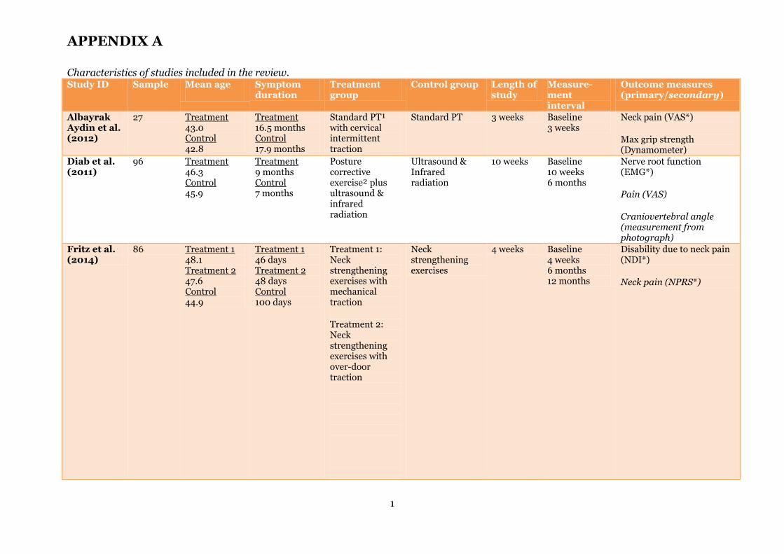
APPENDIX A
1
Characteristics of studies included in the review. Study ID Sample Mean age
Symptom duration
Treatment group
Control group Length of study
Measure- ment interval
Outcome measures (primary/secondary)
Albayrak Aydin et al. (2012)
27 Treatment 43.0 Control 42.8
Treatment 16.5 months Control 17.9 months
Standard PT¹ with cervical intermittent traction
Standard PT 3 weeks Baseline 3 weeks
Neck pain (VAS*) Max grip strength (Dynamometer)
Diab et al. (2011)
96 Treatment 46.3 Control 45.9
Treatment 9 months Control 7 months
Posture corrective exercise² plus ultrasound & infrared radiation
Ultrasound & Infrared radiation
10 weeks Baseline 10 weeks 6 months
Nerve root function (EMG*) Pain (VAS) Craniovertebral angle (measurement from photograph)
Fritz et al. (2014)
86 Treatment 1 48.1 Treatment 2 47.6 Control 44.9
Treatment 1 46 days Treatment 2 48 days Control 100 days
Treatment 1: Neck strengthening exercises with mechanical traction Treatment 2: Neck strengthening exercises with over-door traction
Neck strengthening exercises
4 weeks Baseline 4 weeks 6 months 12 months
Disability due to neck pain (NDI*) Neck pain (NPRS*)
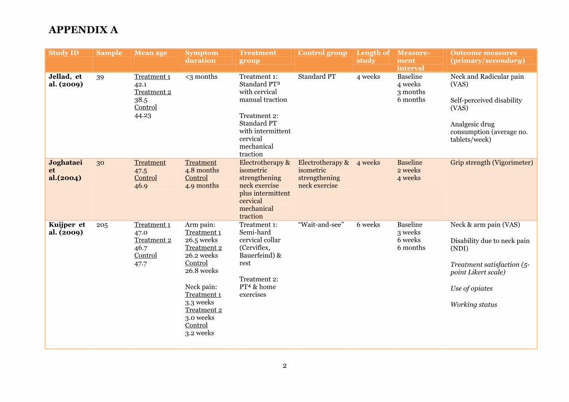
APPENDIX A
2
Study ID Sample Mean age
Symptom duration
Treatment group
Control group Length of study
Measure- ment interval
Outcome measures (primary/secondary)
Jellad, et al. (2009)
39 Treatment 1 42.1 Treatment 2 38.5 Control 44.23
<3 months Treatment 1: Standard PT³ with cervical manual traction Treatment 2: Standard PT with intermittent cervical mechanical traction
Standard PT 4 weeks Baseline 4 weeks 3 months 6 months
Neck and Radicular pain (VAS) Self-perceived disability (VAS) Analgesic drug consumption (average no. tablets/week)
Joghataei et al.(2004)
30 Treatment 47.5 Control 46.9
Treatment 4.8 months Control 4.9 months
Electrotherapy & isometric strengthening neck exercise plus intermittent cervical mechanical traction
Electrotherapy & isometric strengthening neck exercise
4 weeks Baseline 2 weeks 4 weeks
Grip strength (Vigorimeter)
Kuijper et al. (2009)
205 Treatment 1 47.0 Treatment 2 46.7 Control 47.7
Arm pain: Treatment 1 26.5 weeks Treatment 2 26.2 weeks Control 26.8 weeks Neck pain: Treatment 1 3.3 weeks Treatment 2 3.0 weeks Control 3.2 weeks
Treatment 1: Semi-hard cervical collar (Cerviflex, Bauerfeind) & rest Treatment 2: PT⁴ & home exercises
“Wait-and-see” 6 weeks Baseline 3 weeks 6 weeks 6 months
Neck & arm pain (VAS) Disability due to neck pain (NDI) Treatment satisfaction (5-point Likert scale) Use of opiates Working status
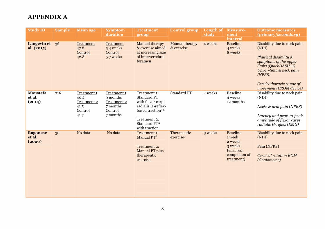
APPENDIX A
3
Study ID Sample Mean age
Symptom duration
Treatment group
Control group Length of study
Measure- ment interval
Outcome measures (primary/secondary)
Langevin et al. (2015)
36 Treatment 47.8 Control 42.8
Treatment 5.4 weeks Control 5.7 weeks
Manual therapy & exercise aimed at increasing size of intervertebral foramen
Manual therapy & exercise
4 weeks Baseline 4 weeks 8 weeks
Disability due to neck pain (NDI) Physical disability & symptoms of the upper limbs (QuickDASH¹²) Upper-limb & neck pain (NPRS) Cervicothoracic range of movement (CROM device)
Moustafa et al. (2014)
216 Treatment 1 40.2 Treatment 2 41.5 Control 41.7
Treatment 1 9 months Treatment 2 7 months Control 7 months
Treatment 1: Standard PT with flexor carpi radialis H-reflex-based traction¹⁵ Treatment 2: Standard PT⁵ with traction
Standard PT 4 weeks Baseline 4 weeks 12 months
Disability due to neck pain (NDI) Neck- & arm pain (NPRS) Latency and peak-to-peak amplitude of flexor carpi radialis H-reflex (EMG)
Ragonese et al. (2009)
30 No data No data Treatment 1: Manual PT⁶ Treatment 2: Manual PT plus therapeutic exercise
Therapeutic exercise⁷
3 weeks Baseline 1 week 2 weeks 3 weeks Final (on completion of treatment)
Disability due to neck pain (NDI) Pain (NPRS) Cervical rotation ROM (Goniometer)
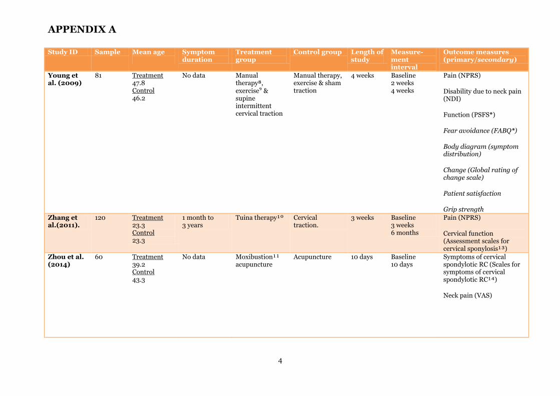
APPENDIX A
4
Study ID Sample Mean age
Symptom duration
Treatment group
Control group Length of study
Measure- ment interval
Outcome measures (primary/secondary)
Young et al. (2009)
81 Treatment 47.8 Control 46.2
No data Manual therapy⁸, exercise⁹ & supine intermittent cervical traction
Manual therapy, exercise & sham traction
4 weeks Baseline 2 weeks 4 weeks
Pain (NPRS) Disability due to neck pain (NDI) Function (PSFS*) Fear avoidance (FABQ*) Body diagram (symptom distribution) Change (Global rating of change scale) Patient satisfaction Grip strength
Zhang et al.(2011).
120 Treatment 23.3 Control 23.3
1 month to 3 years
Tuina therapy¹º Cervical traction.
3 weeks Baseline 3 weeks 6 months
Pain (NPRS) Cervical function (Assessment scales for cervical sponylosis¹³)
Zhou et al. (2014)
60 Treatment 39.2 Control 43.3
No data Moxibustion¹¹ acupuncture
Acupuncture 10 days Baseline 10 days
Symptoms of cervical spondylotic RC (Scales for symptoms of cervical spondylotic RC¹⁴) Neck pain (VAS)
APPENDIX A
5
*Abbreviations: Cervical radiculopathy (CR); Electromyogram (EMG); Fear-Avoidance Beliefs Questionnaire (FABQ); Interferential therapy (IFT); Neck Disability Index (NDI); Numeric Pain-Rating scale (NPRS); Patient specific functional scale (PSFS); Physical therapy (PT); Radiculopathy (RC); Randomized controlled trial (RCT); Range of movement (ROM) Transcutaneous nerve stimulation (TENS); Visual analogue scale (VAS) ¹US, hotpack, TENS, cervical muscle isometric strengthening, spinal muscle stretching. ²Strengthening- & stretching exercises of neck- & shoulder muscles. ³US, IR, massage, cervical spine mobilizations, cervical neck strengthening- & stretching exercises. ⁴“Hands off” graded activity exercises to strengthen neck muscles. ⁵IR, IFT, massage, isometric muscle strengthening & thoracic spine manipulation. ⁶Cervical lateral glides, thoracic mobilization, neural dynamic techniques for median nerve. ⁷Strengthening of deep neck flexors, trapezius & serratus anterior. ⁸Thrust manipulation or non thrust manipulation of cervical & thoracic spine. ⁹Cervical retraction & extension, cervical flexor & scapular muscle strengthening. ¹ºManual therapy used in Chinese medicine. ¹¹Chinese acupuncture technique where a type of herb is burnt on the needle. ¹²Shortened version of the Disabilities of the Arm, Shoulder and Hand Questionnaire. ¹³Clinical symptoms, clinical examination & daily activities. For more info on the test please see: Wang XH, He CQ, Ding MF, Zhang LM, Han M, Zhang YM & Lan Q. (2005). Assessment scales for cervical spondylosis. Hua Xi Yi Xue. 20 (2); 232-233. ¹⁴Evaluation of neurological function including shoulder pain & discomfort, pain & numbness of fingers, neck movement, local muscle strength & tendon reflexes. For more info on the test please see: Jiang H & Shi Q (1998). The introduction to a therapeutic effect evaluation method of cervical spondylotic radiculopathy. Chinese Journal of Orthopedics. 18 (6); 381. ¹⁵The H-reflex is a monosynaptic segmental reflex used to assess the neuropathologic changes in the nerve root and spinal excitability at lumbosacral and cervical level.
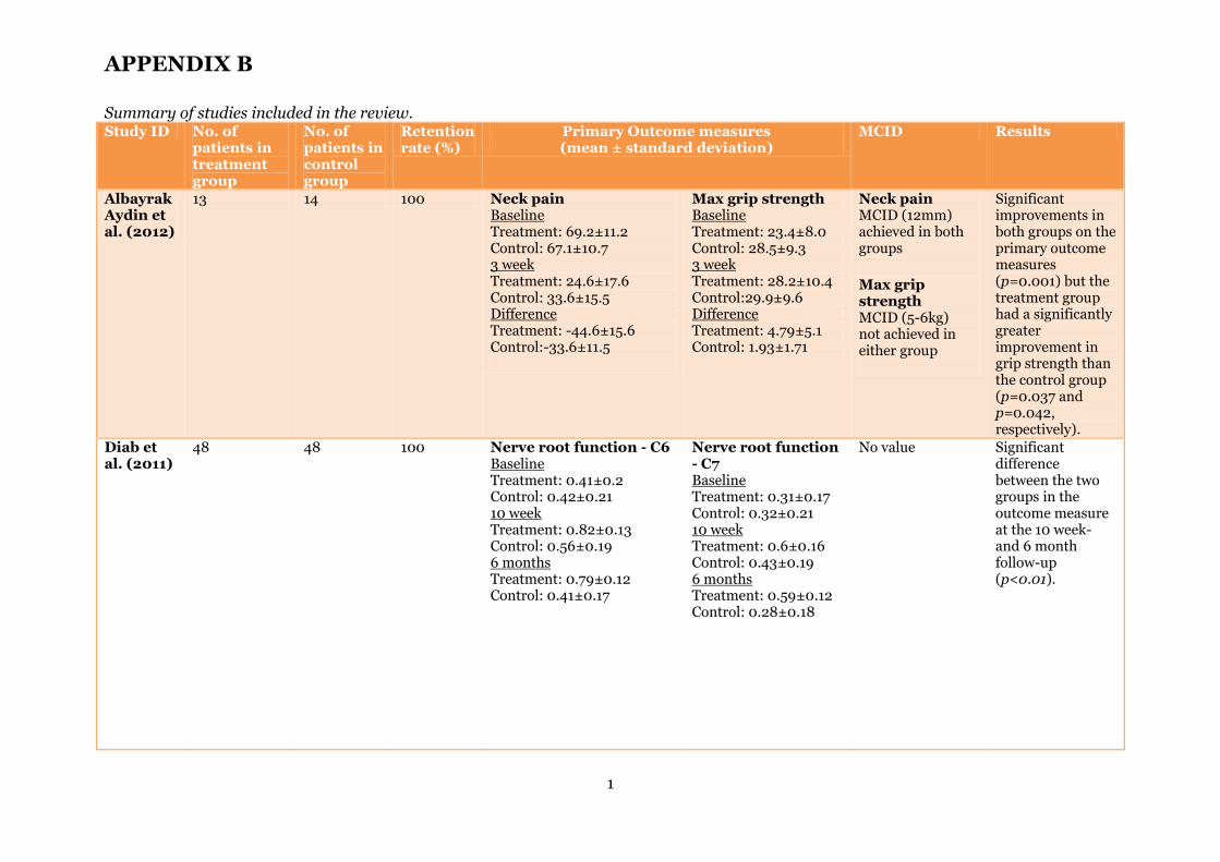
APPENDIX B
1
Summary of studies included in the review. Study ID No. of
patients in treatment group
No. of patients in control group
Retention rate (%)
Primary Outcome measures (mean ± standard deviation)
MCID Results
Albayrak Aydin et al. (2012)
13 14 100 Neck pain Baseline Treatment: 69.2±11.2 Control: 67.1±10.7 3 week Treatment: 24.6±17.6 Control: 33.6±15.5 Difference Treatment: -44.6±15.6 Control:-33.6±11.5
Max grip strength Baseline Treatment: 23.4±8.0 Control: 28.5±9.3 3 week Treatment: 28.2±10.4 Control:29.9±9.6 Difference Treatment: 4.79±5.1 Control: 1.93±1.71
Neck pain MCID (12mm) achieved in both groups Max grip strength MCID (5-6kg) not achieved in either group
Significant improvements in both groups on the primary outcome measures (p=0.001) but the treatment group had a significantly greater improvement in grip strength than the control group (p=0.037 and p=0.042, respectively).
Diab et al. (2011)
48 48 100 Nerve root function - C6 Baseline Treatment: 0.41±0.2 Control: 0.42±0.21 10 week Treatment: 0.82±0.13 Control: 0.56±0.19 6 months Treatment: 0.79±0.12 Control: 0.41±0.17
Nerve root function - C7 Baseline Treatment: 0.31±0.17 Control: 0.32±0.21 10 week Treatment: 0.6±0.16 Control: 0.43±0.19 6 months Treatment: 0.59±0.12 Control: 0.28±0.18
No value Significant difference between the two groups in the outcome measure at the 10 week- and 6 month follow-up (p<0.01).
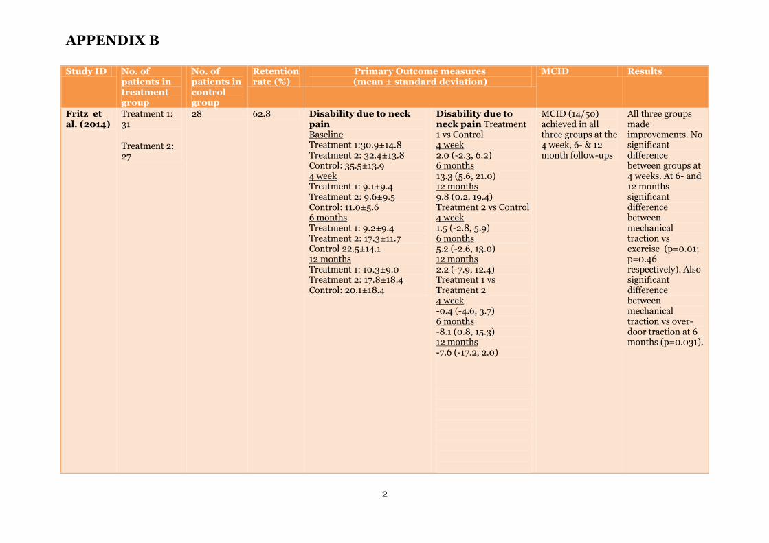
APPENDIX B
2
Study ID No. of patients in treatment group
No. of patients in control group
Retention rate (%)
Primary Outcome measures (mean ± standard deviation)
MCID Results
Fritz et al. (2014)
Treatment 1: 31 Treatment 2: 27
28 62.8 Disability due to neck pain Baseline Treatment 1:30.9±14.8 Treatment 2: 32.4±13.8 Control: 35.5±13.9 4 week Treatment 1: 9.1±9.4 Treatment 2: 9.6±9.5 Control: 11.0±5.6 6 months Treatment 1: 9.2±9.4 Treatment 2: 17.3±11.7 Control 22.5±14.1 12 months Treatment 1: 10.3±9.0 Treatment 2: 17.8±18.4 Control: 20.1±18.4
Disability due to neck pain Treatment 1 vs Control 4 week 2.0 (-2.3, 6.2) 6 months 13.3 (5.6, 21.0) 12 months 9.8 (0.2, 19.4) Treatment 2 vs Control 4 week 1.5 (-2.8, 5.9) 6 months 5.2 (-2.6, 13.0) 12 months 2.2 (-7.9, 12.4) Treatment 1 vs Treatment 2 4 week -0.4 (-4.6, 3.7) 6 months -8.1 (0.8, 15.3) 12 months -7.6 (-17.2, 2.0)
MCID (14/50) achieved in all three groups at the 4 week, 6- & 12 month follow-ups
All three groups made improvements. No significant difference between groups at 4 weeks. At 6- and 12 months significant difference between mechanical traction vs exercise (p=0.01; p=0.46 respectively). Also significant difference between mechanical traction vs over-door traction at 6 months (p=0.031).
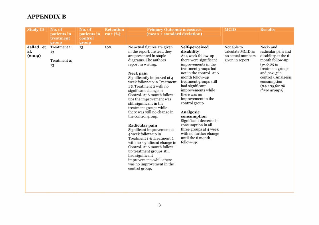
APPENDIX B
3
Study ID No. of patients in treatment group
No. of patients in control group
Retention rate (%)
Primary Outcome measures (mean ± standard deviation)
MCID Results
Jellad, et al. (2009)
Treatment 1: 13 Treatment 2: 13
13 100 No actual figures are given in the report. Instead they are presented in staple diagrams. The authors report in writing: Neck pain Significantly improved at 4 week follow-up in Treatment 1 & Treatment 2 with no significant change in Control. At 6 month follow-ups the improvement was still significant in the treatment groups while there was still no change in the control group. Radicular pain Significant improvement at 4 week follow-up in Treatment 1 & Treatment 2 with no significant change in Control. At 6 month follow-up treatment groups still had significant improvements while there was no improvement in the control group.
Self-perceived disability At 4 week follow-up there were significant improvements in the treatment groups but not in the control. At 6 month follow-up treatment groups still had significant improvements while there was no improvement in the control group. Analgesic consumption Significant decrease in consumption in all three groups at 4 week with no further change until the 6 month follow-up.
Not able to calculate MCID as no actual numbers given in report
Neck- and radicular pain and disability at the 6 month follow-up: (p<0.05 in treatment groups and p>0.5 in control). Analgesic consumption (p<0.05 for all three groups).
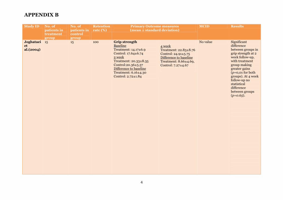
APPENDIX B
4
Study ID No. of patients in treatment group
No. of patients in control group
Retention rate (%)
Primary Outcome measures (mean ± standard deviation)
MCID Results
Joghataei et al.(2004)
15 15 100 Grip strength Baseline Treatment: 14.17±6.9 Control: 17.64±6.74 2 week Treatment: 20.33±8.35 Control:20.36±5.57 Difference to baseline Treatment: 6.16±4.30 Control: 2.72±1.84
4 week Treatment: 22.83±8.76 Control: 24.91±5.75 Difference to baseline Treatment: 8.66±4.69, Control: 7.27±4.67
No value Significant difference between groups in grip strength at 2 week follow-up, with treatment group making greater gains (p=0,01 for both groups). At 4 week follow-up no statistical difference between groups (p=0.65).
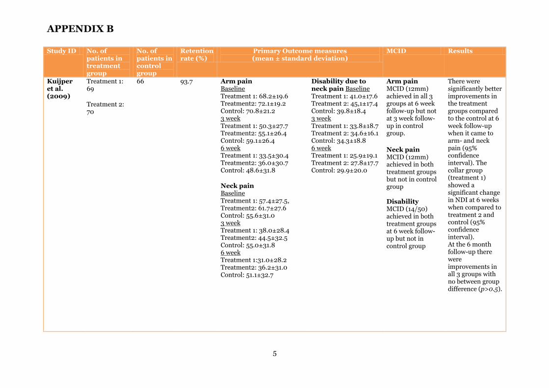
APPENDIX B
5
Study ID No. of patients in treatment group
No. of patients in control group
Retention rate (%)
Primary Outcome measures (mean ± standard deviation)
MCID Results
Kuijper et al. (2009)
Treatment 1: 69 Treatment 2: 70
66 93.7 Arm pain Baseline Treatment 1: 68.2±19.6 Treatment2: 72.1±19.2 Control: 70.8±21.2 3 week Treatment 1: 50.3±27.7 Treatment2: 55.1±26.4 Control: 59.1±26.4 6 week Treatment 1: 33.5±30.4 Treatment2: 36.0±30.7 Control: 48.6±31.8 Neck pain Baseline Treatment 1: 57.4±27.5, Treatment2: 61.7±27.6 Control: 55.6±31.0 3 week Treatment 1: 38.0±28.4 Treatment2: 44.5±32.5 Control: 55.0±31.8 6 week Treatment 1:31.0±28.2 Treatment2: 36.2±31.0 Control: 51.1±32.7
Disability due to neck pain Baseline Treatment 1: 41.0±17.6 Treatment 2: 45,1±17.4 Control: 39.8±18.4 3 week Treatment 1: 33.8±18.7 Treatment 2: 34.6±16.1 Control: 34.3±18.8 6 week Treatment 1: 25.9±19.1 Treatment 2: 27.8±17.7 Control: 29.9±20.0
Arm pain MCID (12mm) achieved in all 3 groups at 6 week follow-up but not at 3 week follow-up in control group. Neck pain MCID (12mm) achieved in both treatment groups but not in control group Disability MCID (14/50) achieved in both treatment groups at 6 week follow-up but not in control group
There were significantly better improvements in the treatment groups compared to the control at 6 week follow-up when it came to arm- and neck pain (95% confidence interval). The collar group (treatment 1) showed a significant change in NDI at 6 weeks when compared to treatment 2 and control (95% confidence interval). At the 6 month follow-up there were improvements in all 3 groups with no between group difference (p>0.5).
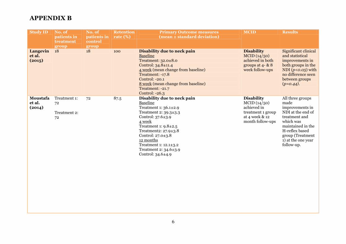
APPENDIX B
6
Study ID No. of patients in treatment group
No. of patients in control group
Retention rate (%)
Primary Outcome measures (mean ± standard deviation)
MCID Results
Langevin et al. (2015)
18 18 100 Disability due to neck pain Baseline Treatment: 32.0±8.0 Control: 34.8±11.4 4 week (mean change from baseline) Treatment: -17.8 Control: -20.1 8 week (mean change from baseline) Treatment: -21.7 Control: -26.3
Disability MCID (14/50) achieved in both groups at 4- & 8 week follow-ups
Significant clinical and statistical improvements in both groups in the NDI (p<0.05) with no difference seen between groups (p=0.44).
Moustafa et al. (2014)
Treatment 1: 72 Treatment 2: 72
72 87.5 Disability due to neck pain Baseline Treatment 1: 36.1±2.9 Treatment 2: 39.3±3.3 Control: 37.6±3.9 4 week Treatment 1: 9.8±2.5 Treatment2: 27.9±3.8 Control: 27.0±3.8 12 months Treatment 1: 12.1±3.2 Treatment 2: 34.6±3.9 Control: 34.6±4.9
Disability MCID (14/50) achieved in treatment 1 group at 4 week & 12 month follow-ups
All three groups made improvements in NDI at the end of treatment and which was maintained in the H-reflex based group (Treatment 1) at the one year follow-up.
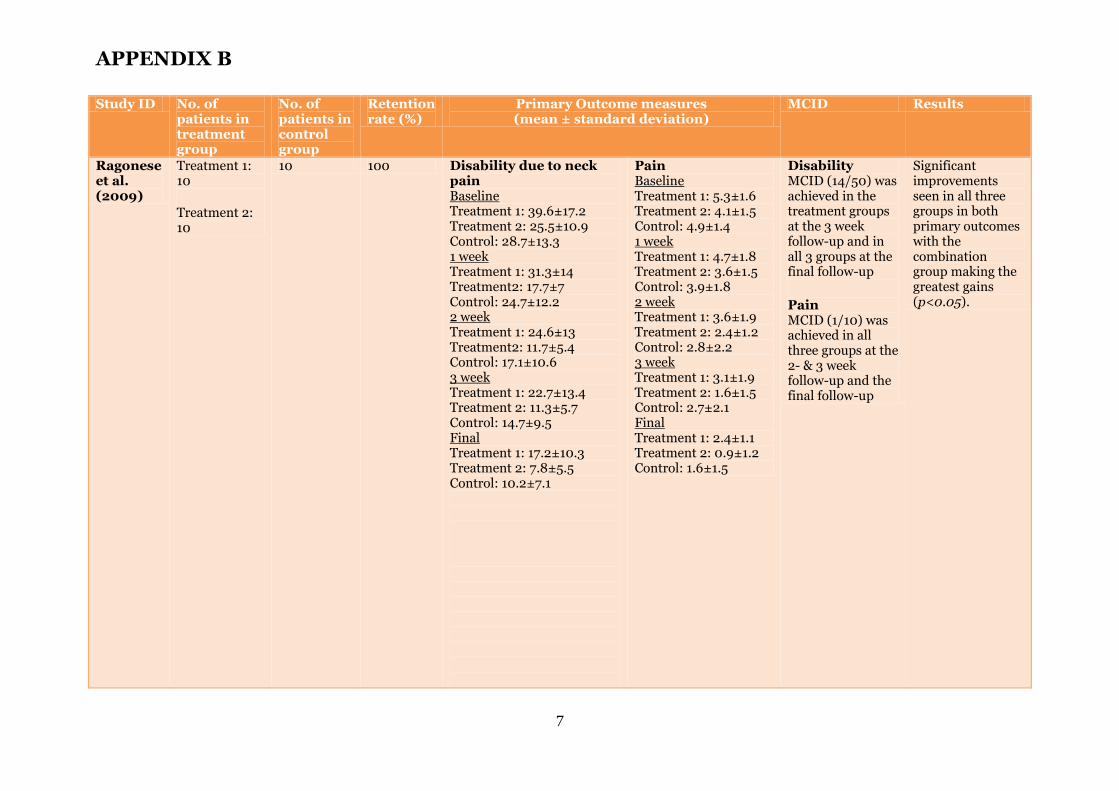
APPENDIX B
7
Study ID No. of patients in treatment group
No. of patients in control group
Retention rate (%)
Primary Outcome measures (mean ± standard deviation)
MCID Results
Ragonese et al. (2009)
Treatment 1: 10 Treatment 2: 10
10 100 Disability due to neck pain Baseline Treatment 1: 39.6±17.2 Treatment 2: 25.5±10.9 Control: 28.7±13.3 1 week Treatment 1: 31.3±14 Treatment2: 17.7±7 Control: 24.7±12.2 2 week Treatment 1: 24.6±13 Treatment2: 11.7±5.4 Control: 17.1±10.6 3 week Treatment 1: 22.7±13.4 Treatment 2: 11.3±5.7 Control: 14.7±9.5 Final Treatment 1: 17.2±10.3 Treatment 2: 7.8±5.5 Control: 10.2±7.1
Pain Baseline Treatment 1: 5.3±1.6 Treatment 2: 4.1±1.5 Control: 4.9±1.4 1 week Treatment 1: 4.7±1.8 Treatment 2: 3.6±1.5 Control: 3.9±1.8 2 week Treatment 1: 3.6±1.9 Treatment 2: 2.4±1.2 Control: 2.8±2.2 3 week Treatment 1: 3.1±1.9 Treatment 2: 1.6±1.5 Control: 2.7±2.1 Final Treatment 1: 2.4±1.1 Treatment 2: 0.9±1.2 Control: 1.6±1.5
Disability MCID (14/50) was achieved in the treatment groups at the 3 week follow-up and in all 3 groups at the final follow-up Pain MCID (1/10) was achieved in all three groups at the 2- & 3 week follow-up and the final follow-up
Significant improvements seen in all three groups in both primary outcomes with the combination group making the greatest gains (p<0.05).
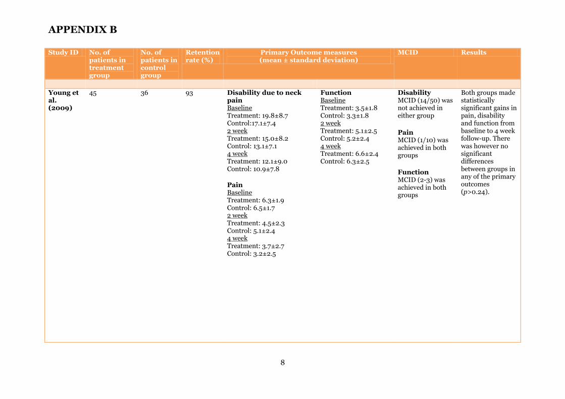
APPENDIX B
8
Study ID No. of patients in treatment group
No. of patients in control group
Retention rate (%)
Primary Outcome measures (mean ± standard deviation)
MCID Results
Young et al. (2009)
45 36 93 Disability due to neck pain Baseline Treatment: 19.8±8.7 Control:17.1±7.4 2 week Treatment: 15.0±8.2 Control: 13.1±7.1 4 week Treatment: 12.1±9.0 Control: 10.9±7.8 Pain Baseline Treatment: 6.3±1.9 Control: 6.5±1.7 2 week Treatment: 4.5±2.3 Control: 5.1±2.4 4 week Treatment: 3.7±2.7 Control: 3.2±2.5
Function Baseline Treatment: 3.5±1.8 Control: 3.3±1.8 2 week Treatment: 5.1±2.5 Control: 5.2±2.4 4 week Treatment: 6.6±2.4 Control: 6.3±2.5
Disability MCID (14/50) was not achieved in either group Pain MCID (1/10) was achieved in both groups Function MCID (2-3) was achieved in both groups
Both groups made statistically significant gains in pain, disability and function from baseline to 4 week follow-up. There was however no significant differences between groups in any of the primary outcomes (p>0.24).
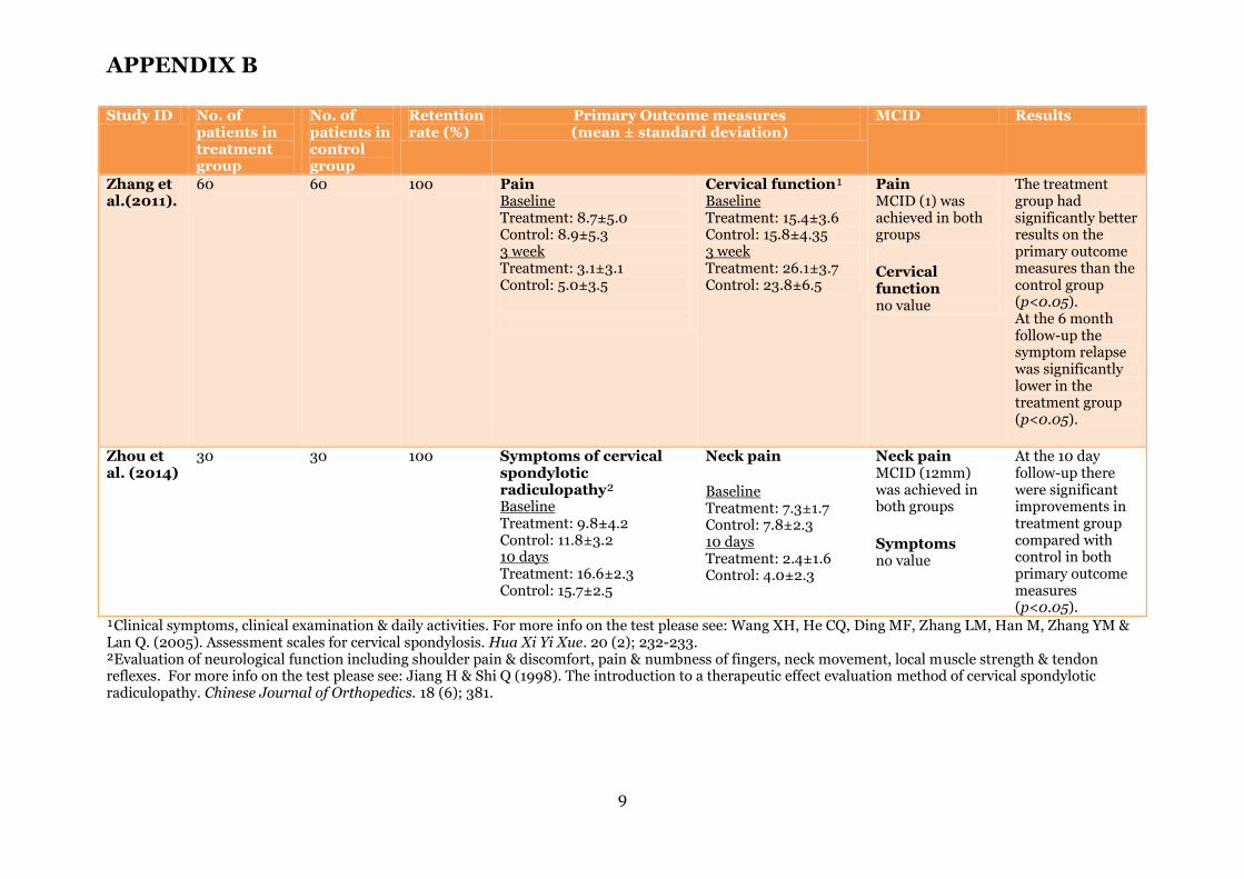
APPENDIX B
9
Study ID No. of patients in treatment group
No. of patients in control group
Retention rate (%)
Primary Outcome measures (mean ± standard deviation)
MCID Results
Zhang et al.(2011).
60 60 100 Pain Baseline Treatment: 8.7±5.0 Control: 8.9±5.3 3 week Treatment: 3.1±3.1 Control: 5.0±3.5
Cervical function¹ Baseline Treatment: 15.4±3.6 Control: 15.8±4.35 3 week Treatment: 26.1±3.7 Control: 23.8±6.5
Pain MCID (1) was achieved in both groups Cervical function no value
The treatment group had significantly better results on the primary outcome measures than the control group (p<0.05). At the 6 month follow-up the symptom relapse was significantly lower in the treatment group (p<0.05).
Zhou et al. (2014)
30 30 100 Symptoms of cervical spondylotic radiculopathy² Baseline Treatment: 9.8±4.2 Control: 11.8±3.2 10 days Treatment: 16.6±2.3 Control: 15.7±2.5
Neck pain Baseline Treatment: 7.3±1.7 Control: 7.8±2.3 10 days Treatment: 2.4±1.6 Control: 4.0±2.3
Neck pain MCID (12mm) was achieved in both groups Symptoms no value
At the 10 day follow-up there were significant improvements in treatment group compared with control in both primary outcome measures (p<0.05).
¹Clinical symptoms, clinical examination & daily activities. For more info on the test please see: Wang XH, He CQ, Ding MF, Zhang LM, Han M, Zhang YM & Lan Q. (2005). Assessment scales for cervical spondylosis. Hua Xi Yi Xue. 20 (2); 232-233. ²Evaluation of neurological function including shoulder pain & discomfort, pain & numbness of fingers, neck movement, local muscle strength & tendon reflexes. For more info on the test please see: Jiang H & Shi Q (1998). The introduction to a therapeutic effect evaluation method of cervical spondylotic radiculopathy. Chinese Journal of Orthopedics. 18 (6); 381.
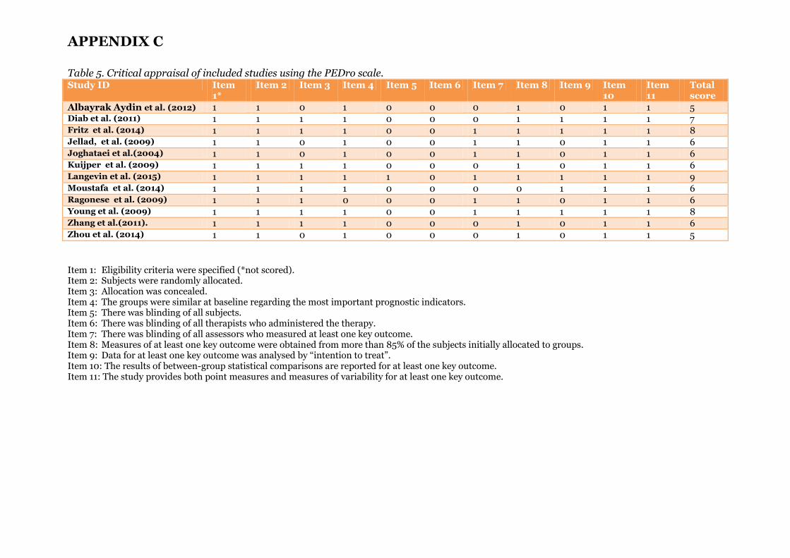
APPENDIX C
Table 5. Critical appraisal of included studies using the PEDro scale. Study ID Item
1* Item 2 Item 3 Item 4 Item 5 Item 6 Item 7 Item 8 Item 9 Item
10 Item 11
Total score
Albayrak Aydin et al. (2012) 1 1 0 1 0 0 0 1 0 1 1 5 Diab et al. (2011) 1 1 1 1 0 0 0 1 1 1 1 7 Fritz et al. (2014) 1 1 1 1 0 0 1 1 1 1 1 8 Jellad, et al. (2009) 1 1 0 1 0 0 1 1 0 1 1 6 Joghataei et al.(2004) 1 1 0 1 0 0 1 1 0 1 1 6 Kuijper et al. (2009) 1 1 1 1 0 0 0 1 0 1 1 6 Langevin et al. (2015) 1 1 1 1 1 0 1 1 1 1 1 9 Moustafa et al. (2014) 1 1 1 1 0 0 0 0 1 1 1 6 Ragonese et al. (2009) 1 1 1 0 0 0 1 1 0 1 1 6 Young et al. (2009) 1 1 1 1 0 0 1 1 1 1 1 8 Zhang et al.(2011). 1 1 1 1 0 0 0 1 0 1 1 6 Zhou et al. (2014) 1 1 0 1 0 0 0 1 0 1 1 5
Item 1: Eligibility criteria were specified (*not scored). Item 2: Subjects were randomly allocated. Item 3: Allocation was concealed. Item 4: The groups were similar at baseline regarding the most important prognostic indicators. Item 5: There was blinding of all subjects. Item 6: There was blinding of all therapists who administered the therapy. Item 7: There was blinding of all assessors who measured at least one key outcome. Item 8: Measures of at least one key outcome were obtained from more than 85% of the subjects initially allocated to groups. Item 9: Data for at least one key outcome was analysed by “intention to treat”. Item 10: The results of between-group statistical comparisons are reported for at least one key outcome. Item 11: The study provides both point measures and measures of variability for at least one key outcome.