Embed Size (px)
Citation preview
Effects of Upper Extremity Mobilization Techniques on Fine-Motor Performance in Children with Neuromotor Disorders
Presented by W. Michael Magrun, MS, OTR/L
Learning Outcomes:
1. List the postural factors involved in fine-motor performance. 2. Describe the developmental characteristics of normal development and atypical development related to hand function. 3. Describe the proximal and distal mobilization techniques and how the contribute to fine-motor function.
Introduction
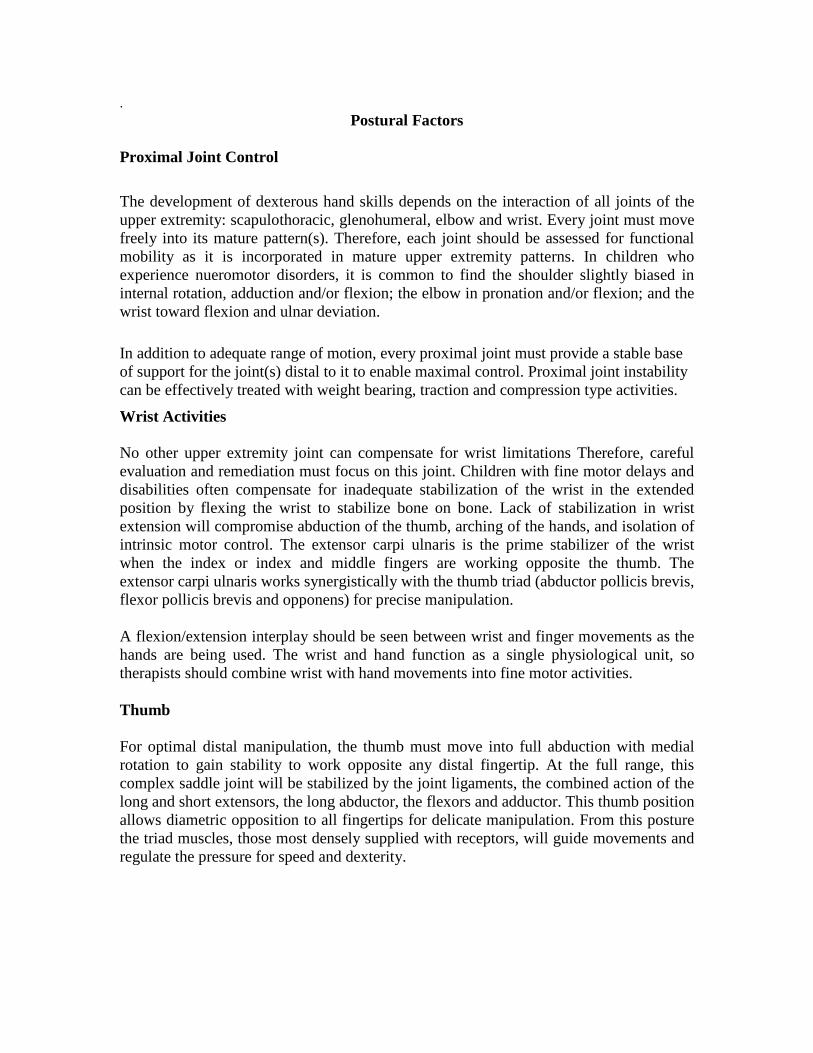
Three children with neuromotor disorders were evaluated pre and post mobilization techniques for a period of 6 days. The purpose was to determine the relationship between soft tissue and joint restrictions and fine motor performance. Children with neuromotor disorders develop patterns of movement that are characterized by limitations in joint range and muscle function. These patterns are often described as abnormal or atypical patterns of movement, characterized by flexion and pronation of the wrist, pronation of the forearm with elbow flexion, and protraction of the shoulder with scapular abduction and general limitation of shoulder mobility. When there is a restriction of joint mobility and dynamic musculoskeletal function, there are corresponding changes in tissue structure and function. Fascia restrictions interfere with effective elongation and folding of tissue. They adapt to places in the body where movement is inefficient. Tissue contracts and binds down in areas of postural stress, which can profoundly affect posture and movement. These characteristics inhibit the child with neuromotor disorders to develop more dynamic function, particularly in the upper extremities and specifically hand function. Hand function that is efficient requires a complex interaction of postural factors.
. Postural Factors
Proximal Joint Control
The development of dexterous hand skills depends on the interaction of all joints of the upper extremity: scapulothoracic, glenohumeral, elbow and wrist. Every joint must move freely into its mature pattern(s). Therefore, each joint should be assessed for functional mobility as it is incorporated in mature upper extremity patterns. In children who experience nueromotor disorders, it is common to find the shoulder slightly biased in internal rotation, adduction and/or flexion; the elbow in pronation and/or flexion; and the wrist toward flexion and ulnar deviation. In addition to adequate range of motion, every proximal joint must provide a stable base of support for the joint(s) distal to it to enable maximal control. Proximal joint instability can be effectively treated with weight bearing, traction and compression type activities.
Wrist Activities No other upper extremity joint can compensate for wrist limitations Therefore, careful evaluation and remediation must focus on this joint. Children with fine motor delays and disabilities often compensate for inadequate stabilization of the wrist in the extended position by flexing the wrist to stabilize bone on bone. Lack of stabilization in wrist extension will compromise abduction of the thumb, arching of the hands, and isolation of intrinsic motor control. The extensor carpi ulnaris is the prime stabilizer of the wrist when the index or index and middle fingers are working opposite the thumb. The extensor carpi ulnaris works synergistically with the thumb triad (abductor pollicis brevis, flexor pollicis brevis and opponens) for precise manipulation. A flexion/extension interplay should be seen between wrist and finger movements as the hands are being used. The wrist and hand function as a single physiological unit, so therapists should combine wrist with hand movements into fine motor activities. Thumb
For optimal distal manipulation, the thumb must move into full abduction with medial rotation to gain stability to work opposite any distal fingertip. At the full range, this complex saddle joint will be stabilized by the joint ligaments, the combined action of the long and short extensors, the long abductor, the flexors and adductor. This thumb position allows diametric opposition to all fingertips for delicate manipulation. From this posture the triad muscles, those most densely supplied with receptors, will guide movements and regulate the pressure for speed and dexterity.
Distal Finger Skills
Wrist stabilization combined with fine manipulative activities should develop the essential hand components for all high level hand skills. The essential components include:
• develop and stabilize the arches of the hand. • develop the two divisions of precision handling - precision translation and
precision rotation. • motorically separate the two sides of the hand. • open and stabilize the thumb-index web space.
Developmental Aspects of Hand Function
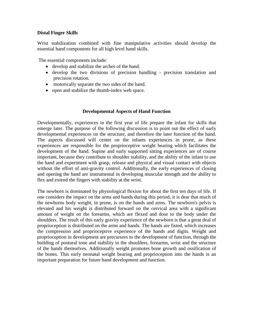
Developmentally, experiences in the first year of life prepare the infant for skills that emerge later. The purpose of the following discussion is to point out the effect of early developmental experiences on the structure, and therefore the later function of the hand. The aspects discussed will center on the infants experiences in prone, as these experiences are responsible for the proprioceptive weight bearing which facilitates the development of the hand. Supine and early supported sitting experiences are of course important, because they contribute to shoulder stability, and the ability of the infant to use the hand and experiment with grasp, release and physical and visual contact with objects without the effort of anti-gravity control. Additionally, the early experiences of closing and opening the hand are instrumental in developing muscular strength and the ability to flex and extend the fingers with stability at the wrist. The newborn is dominated by physiological flexion for about the first ten days of life. If one considers the impact on the arms and hands during this period, it is dear that much of the newborns body weight, in prone, is on the hands and arms. The newborn's pelvis is elevated and his weight is distributed forward on the cervical area with a significant amount of weight on the forearms, which are flexed and dose to the body under the shoulders. The result of this early gravity experience of the newborn is that a great deal of proprioception is distributed on the arms and hands. The hands are fisted, which increases the compression and proprioceptive experience of the hands and digits. Weight and proprioception in development are precursors to the development of function, through the building of postural tone and stability in the shoulders, forearms, wrist and the structure of the hands themselves. Additionally weight promotes bone growth and ossification of the bones. This early neonatal weight bearing and proprioception into the hands is an important preparation for future hand development and function.
Newbor n in Pr one
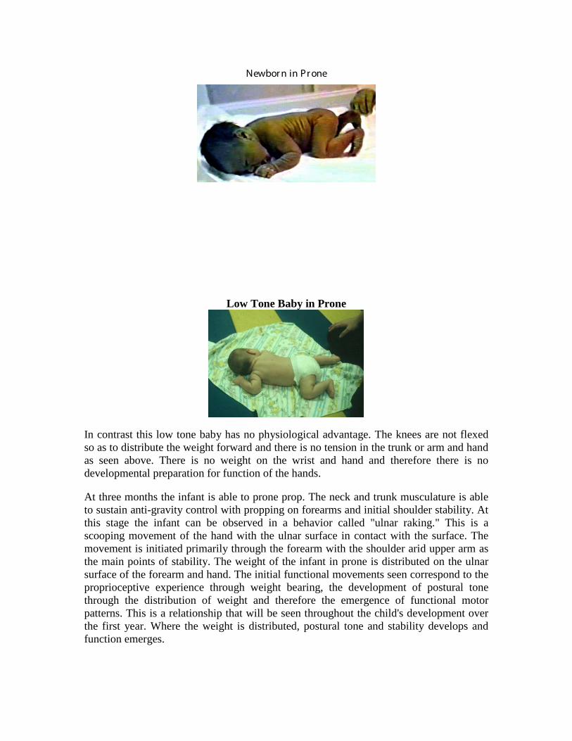
Low Tone Baby in Prone
In contrast this low tone baby has no physiological advantage. The knees are not flexed so as to distribute the weight forward and there is no tension in the trunk or arm and hand as seen above. There is no weight on the wrist and hand and therefore there is no developmental preparation for function of the hands. At three months the infant is able to prone prop. The neck and trunk musculature is able to sustain anti-gravity control with propping on forearms and initial shoulder stability. At this stage the infant can be observed in a behavior called "ulnar raking." This is a scooping movement of the hand with the ulnar surface in contact with the surface. The movement is initiated primarily through the forearm with the shoulder arid upper arm as the main points of stability. The weight of the infant in prone is distributed on the ulnar surface of the forearm and hand. The initial functional movements seen correspond to the proprioceptive experience through weight bearing, the development of postural tone through the distribution of weight and therefore the emergence of functional motor patterns. This is a relationship that will be seen throughout the child's development over the first year. Where the weight is distributed, postural tone and stability develops and function emerges.
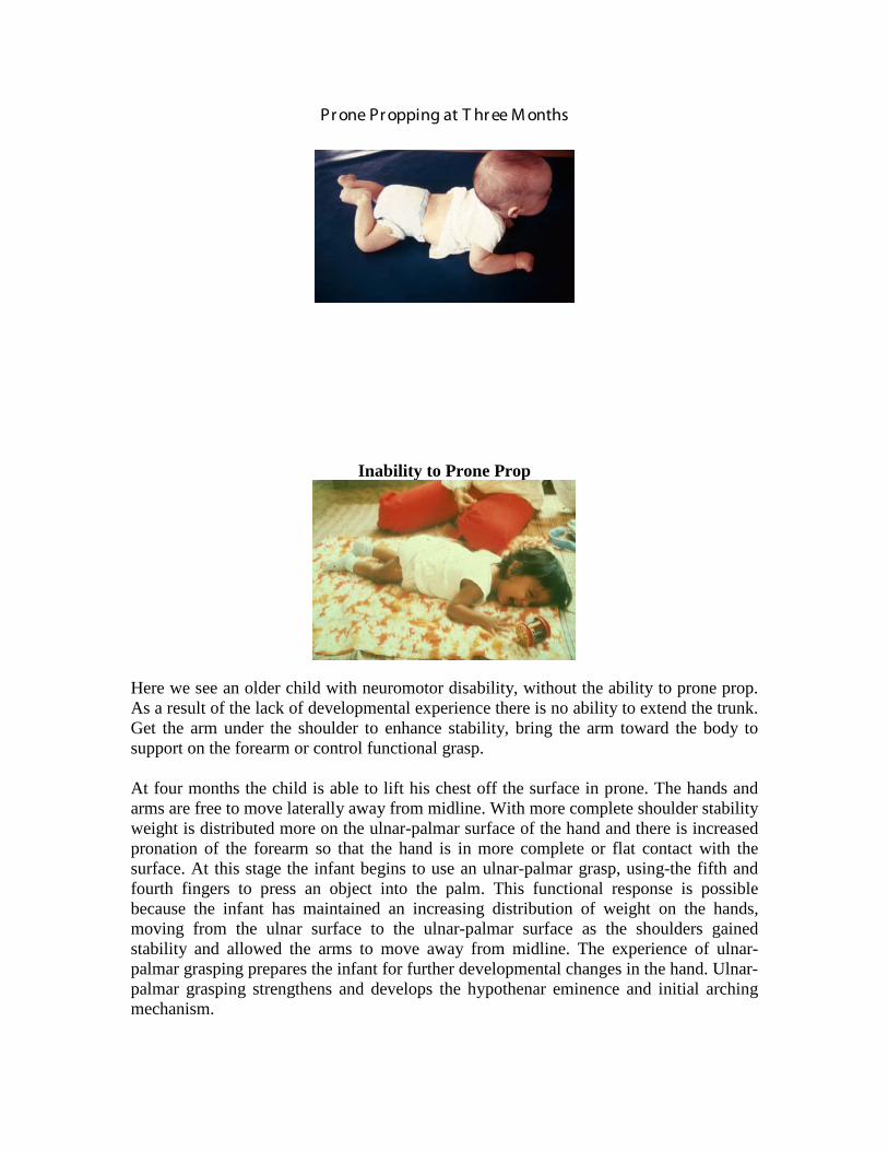
Pr one Pr opping at T hr ee M onths
Inability to Prone Prop
Here we see an older child with neuromotor disability, without the ability to prone prop. As a result of the lack of developmental experience there is no ability to extend the trunk. Get the arm under the shoulder to enhance stability, bring the arm toward the body to support on the forearm or control functional grasp. At four months the child is able to lift his chest off the surface in prone. The hands and arms are free to move laterally away from midline. With more complete shoulder stability weight is distributed more on the ulnar-palmar surface of the hand and there is increased pronation of the forearm so that the hand is in more complete or flat contact with the surface. At this stage the infant begins to use an ulnar-palmar grasp, using-the fifth and fourth fingers to press an object into the palm. This functional response is possible because the infant has maintained an increasing distribution of weight on the hands, moving from the ulnar surface to the ulnar-palmar surface as the shoulders gained stability and allowed the arms to move away from midline. The experience of ulnar-palmar grasping prepares the infant for further developmental changes in the hand. Ulnar-palmar grasping strengthens and develops the hypothenar eminence and initial arching mechanism.
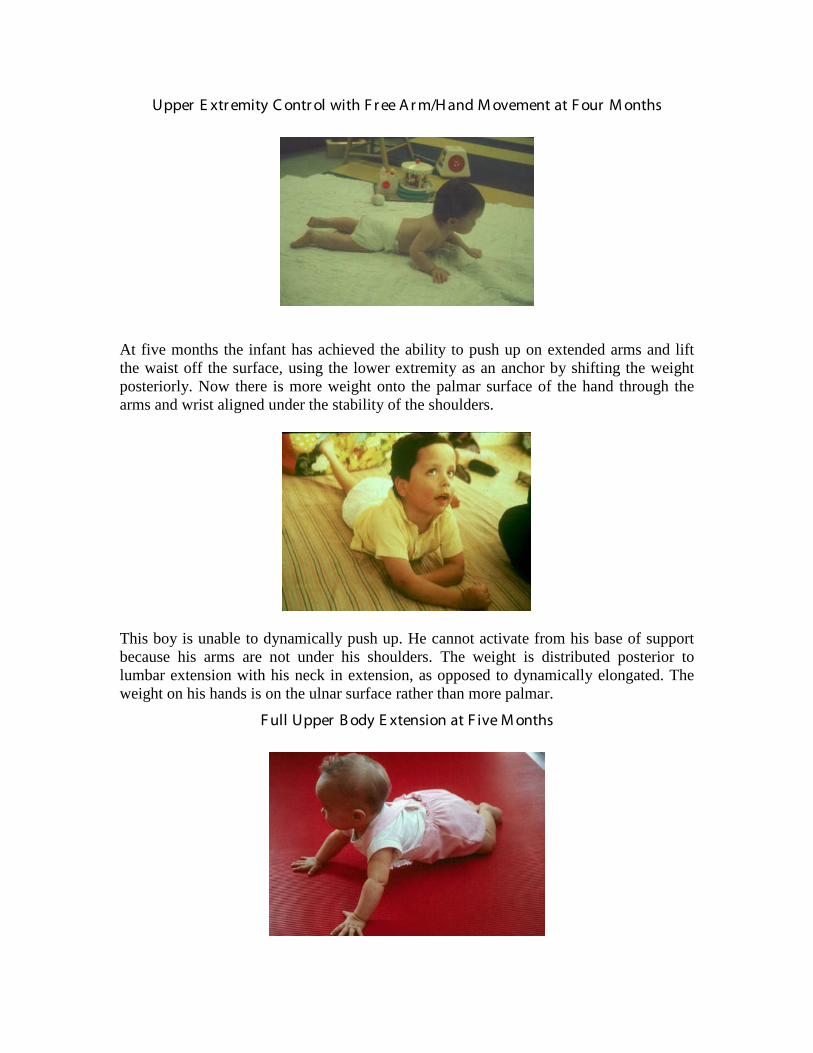
Upper E xtr emity C ontr ol with F r ee A r m/H and M ovement at F our M onths
At five months the infant has achieved the ability to push up on extended arms and lift the waist off the surface, using the lower extremity as an anchor by shifting the weight posteriorly. Now there is more weight onto the palmar surface of the hand through the arms and wrist aligned under the stability of the shoulders.
This boy is unable to dynamically push up. He cannot activate from his base of support because his arms are not under his shoulders. The weight is distributed posterior to lumbar extension with his neck in extension, as opposed to dynamically elongated. The weight on his hands is on the ulnar surface rather than more palmar.
F ull Upper B ody E xtension at F ive M onths
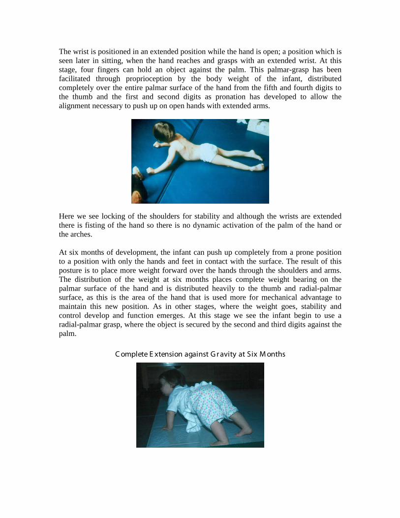
The wrist is positioned in an extended position while the hand is open; a position which is seen later in sitting, when the hand reaches and grasps with an extended wrist. At this stage, four fingers can hold an object against the palm. This palmar-grasp has been facilitated through proprioception by the body weight of the infant, distributed completely over the entire palmar surface of the hand from the fifth and fourth digits to the thumb and the first and second digits as pronation has developed to allow the alignment necessary to push up on open hands with extended arms.
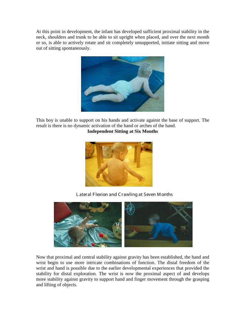
Here we see locking of the shoulders for stability and although the wrists are extended there is fisting of the hand so there is no dynamic activation of the palm of the hand or the arches. At six months of development, the infant can push up completely from a prone position to a position with only the hands and feet in contact with the surface. The result of this posture is to place more weight forward over the hands through the shoulders and arms. The distribution of the weight at six months places complete weight bearing on the palmar surface of the hand and is distributed heavily to the thumb and radial-palmar surface, as this is the area of the hand that is used more for mechanical advantage to maintain this new position. As in other stages, where the weight goes, stability and control develop and function emerges. At this stage we see the infant begin to use a radial-palmar grasp, where the object is secured by the second and third digits against the palm.
C omplete E xtension against G r avity at Six M onths
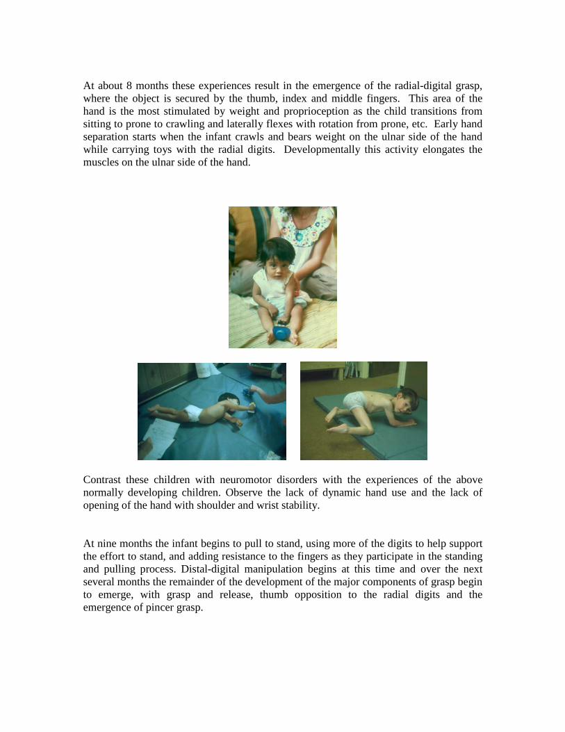
At this point in development, the infant has developed sufficient proximal stability in the neck, shoulders and trunk to be able to sit upright when placed, and over the next month or so, is able to actively rotate and sit completely unsupported, initiate sitting and move out of sitting spontaneously.
This boy is unable to support on his hands and activate against the base of support. The result is there is no dynamic activation of the hand or arches of the hand.
Independent Sitting at Six Months
L ater al F lexion and C r awling at Seven M onths
Now that proximal and central stability against gravity has been established, the hand and wrist begin to use more intricate combinations of function. The distal freedom of the wrist and hand is possible due to the earlier developmental experiences that provided the stability for distal exploration. The wrist is now the proximal aspect of and develops more stability against gravity to support hand and finger movement through the grasping and lifting of objects.
At about 8 months these experiences result in the emergence of the radial-digital grasp, where the object is secured by the thumb, index and middle fingers. This area of the hand is the most stimulated by weight and proprioception as the child transitions from sitting to prone to crawling and laterally flexes with rotation from prone, etc. Early hand separation starts when the infant crawls and bears weight on the ulnar side of the hand while carrying toys with the radial digits. Developmentally this activity elongates the muscles on the ulnar side of the hand.
Contrast these children with neuromotor disorders with the experiences of the above normally developing children. Observe the lack of dynamic hand use and the lack of opening of the hand with shoulder and wrist stability. At nine months the infant begins to pull to stand, using more of the digits to help support the effort to stand, and adding resistance to the fingers as they participate in the standing and pulling process. Distal-digital manipulation begins at this time and over the next several months the remainder of the development of the major components of grasp begin to emerge, with grasp and release, thumb opposition to the radial digits and the emergence of pincer grasp.
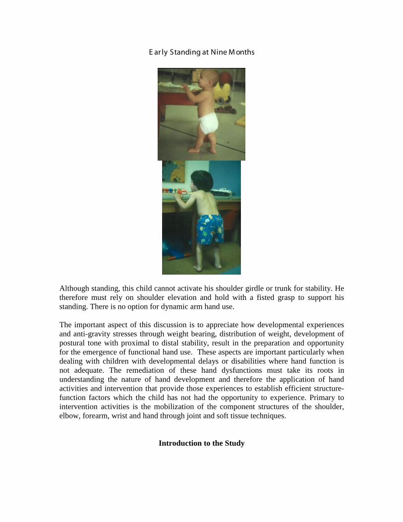
E ar ly Standing at Nine M onths
Although standing, this child cannot activate his shoulder girdle or trunk for stability. He therefore must rely on shoulder elevation and hold with a fisted grasp to support his standing. There is no option for dynamic arm hand use. The important aspect of this discussion is to appreciate how developmental experiences and anti-gravity stresses through weight bearing, distribution of weight, development of postural tone with proximal to distal stability, result in the preparation and opportunity for the emergence of functional hand use. These aspects are important particularly when dealing with children with developmental delays or disabilities where hand function is not adequate. The remediation of these hand dysfunctions must take its roots in understanding the nature of hand development and therefore the application of hand activities and intervention that provide those experiences to establish efficient structure-function factors which the child has not had the opportunity to experience. Primary to intervention activities is the mobilization of the component structures of the shoulder, elbow, forearm, wrist and hand through joint and soft tissue techniques.
Introduction to the Study
Three ten year old children with different types of cerebral palsy were evaluated with selected fine-motor activities from the Brigance Inventory of Early Development. Activities were evaluated before and directly after upper extremity mobilization techniques, daily, over a period of six days. Results indicated a positive learning trend over the treatment period in pre-treatment activities and a greater improvement in performance in post-treatment activities.
Definition of Mobilization Used
Mobilization is a term which is defined in standard dictionaries as; to make mobile or capable of movement. Therapeutically, it is generally accepted as a term which implies techniques capable of improving the movement or mobility of a joint and its surrounding tissue, such that, greater degrees of freedom for motoric expression can be achieved with corresponding release of soft tissue and fascia involved in joint mobility. For the purpose of this study, a combination of joint and soft tissue release techniques was used to reduce tightness and increase the range of joint movement. Techniques included deep pressure, compression and joint mobility techniques to proximal areas of the shoulder girdle, elbow wrist, and hand.
Experimental Design
Three children with cerebral palsy were studied, based on their performance on selected fine-motor tasks before and after mobilization techniques, over a period of two weeks, which included six treatment days; 3 days each week. Each child was presented with four fine-motor tasks, three requiring pencil performance and one requiring scissor performance. The tasks were presented each day prior to mobilization and again directly after mobilization, on each of the six treatment days. Mobilization techniques were applied to the dominant limb of the shoulder girdle, elbow wrist, and hand for approximately ten minutes each day. Each subject served as his own control for purposes of analyzing data. Data was analyzed individually for each task..
Subjects Each child was ten years old at the time of the study and was mainstreamed in a public school setting. Subject A was diagnosed with athetoid cerebral palsy with fluctuating muscle tone and proximal tightness and was independent in ambulation with tenuous balance. Subject B was diagnosed with spastic diplegia, with primary involvement in the lower extremity, evident spasticity in the upper extremity displayed as trunk flexion, protraction of the shoulders, mild elbow contractures, wrist flexion but good isolated control of hands and fingers. Subject B was ambulatory with extreme difficulty for short distances and primarily used a wheel chair for daily mobility.
Subject C was diagnosed with spastic quadrapegia, with significant involvement in both upper and lower extremities. Subject C was non-ambulatory, used a wheelchair throughout the day and was not independent in transfers.
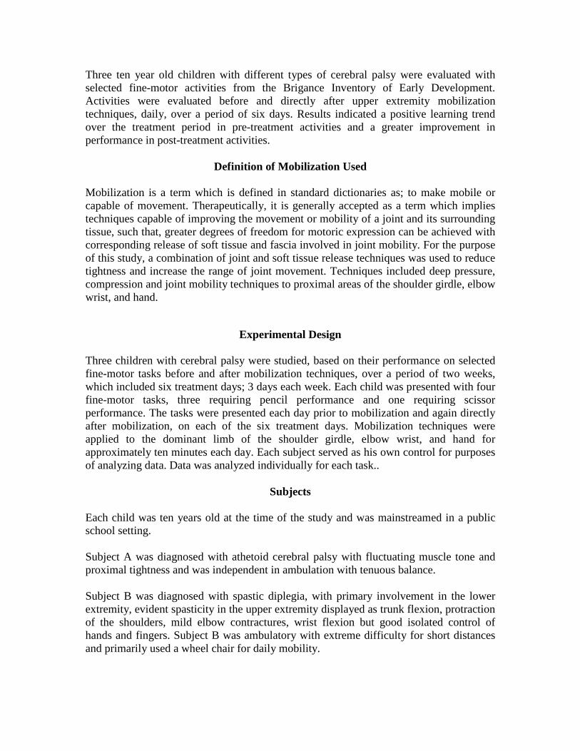
Fine-Motor Tasks Four fine-motor performance tasks were used from worksheets included in the Brigance Inventory of Early Development. Task 1 was a horizontal line drawing between two dots four inches apart.(See Fig. 1) This task was evaluated on two measures; speed in seconds to connect the lines and accuracy within 1/8" of parallel between the drawn line as measured by a transparent grid.
Fig. 1 Task 1: Horizontal line drawing between two dots
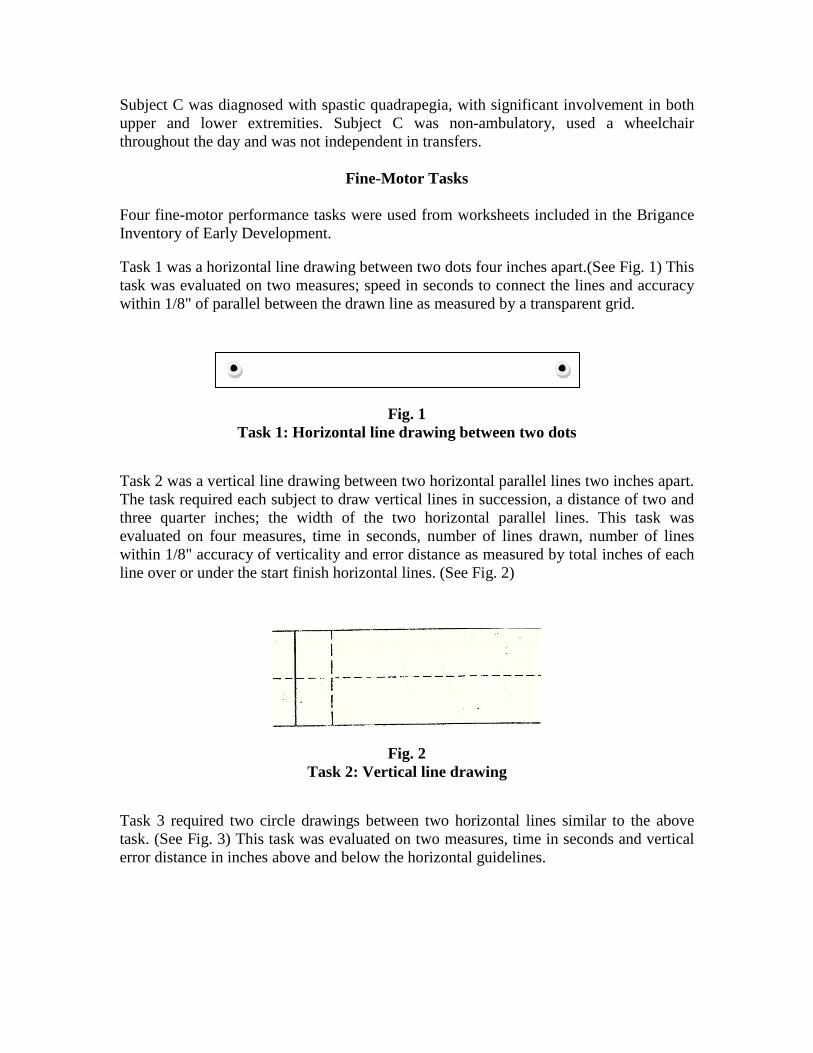
Task 2 was a vertical line drawing between two horizontal parallel lines two inches apart. The task required each subject to draw vertical lines in succession, a distance of two and three quarter inches; the width of the two horizontal parallel lines. This task was evaluated on four measures, time in seconds, number of lines drawn, number of lines within 1/8" accuracy of verticality and error distance as measured by total inches of each line over or under the start finish horizontal lines. (See Fig. 2)
Fig. 2 Task 2: Vertical line drawing
Task 3 required two circle drawings between two horizontal lines similar to the above task. (See Fig. 3) This task was evaluated on two measures, time in seconds and vertical error distance in inches above and below the horizontal guidelines.
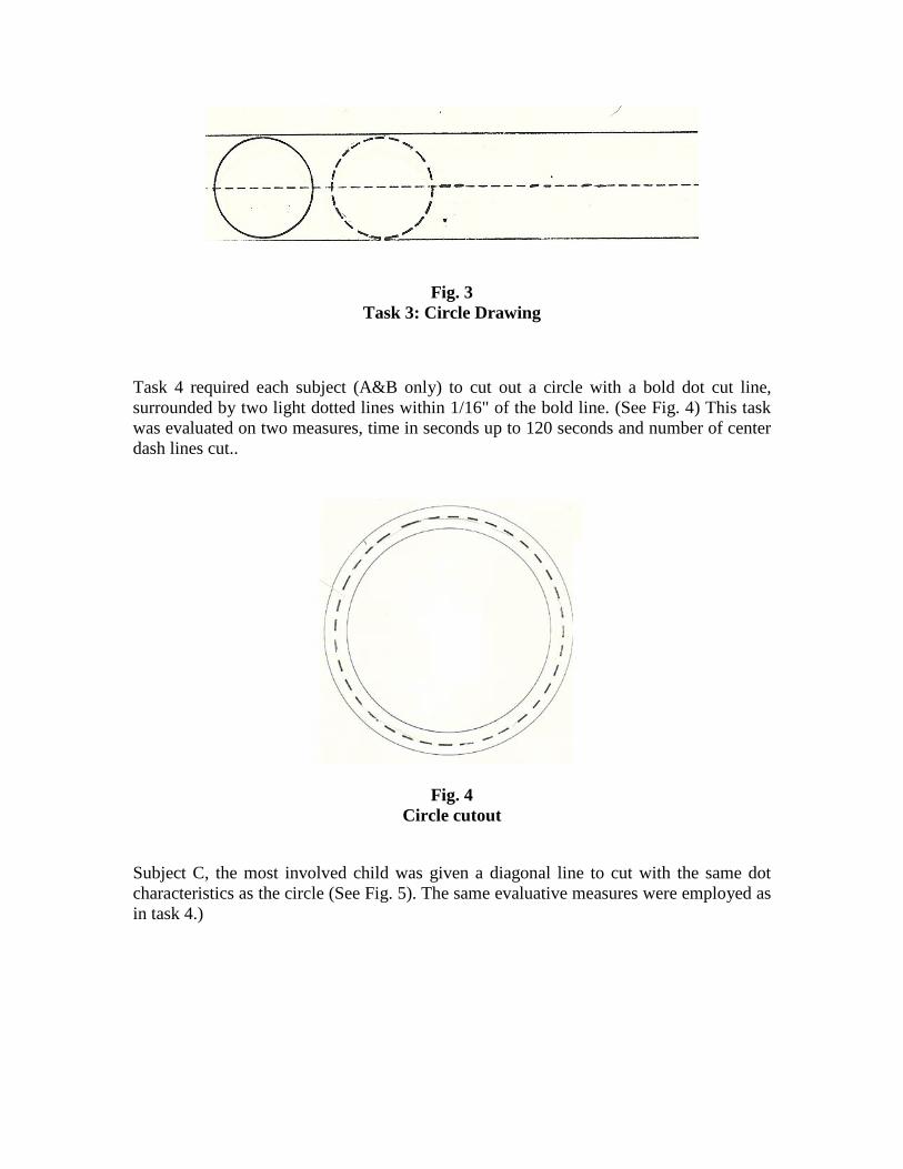
Fig. 3 Task 3: Circle Drawing
Task 4 required each subject (A&B only) to cut out a circle with a bold dot cut line, surrounded by two light dotted lines within 1/16" of the bold line. (See Fig. 4) This task was evaluated on two measures, time in seconds up to 120 seconds and number of center dash lines cut..
Fig. 4 Circle cutout
Subject C, the most involved child was given a diagonal line to cut with the same dot characteristics as the circle (See Fig. 5). The same evaluative measures were employed as in task 4.)
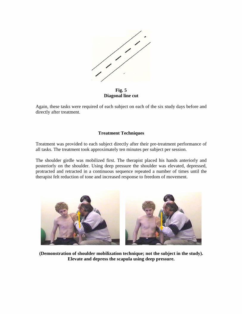
Fig. 5 Diagonal line cut
Again, these tasks were required of each subject on each of the six study days before and directly after treatment.
Treatment Techniques Treatment was provided to each subject directly after their pre-treatment performance of all tasks. The treatment took approximately ten minutes per subject per session. The shoulder girdle was mobilized first. The therapist placed his hands anteriorly and posteriorly on the shoulder. Using deep pressure the shoulder was elevated, depressed, protracted and retracted in a continuous sequence repeated a number of times until the therapist felt reduction of tone and increased response to freedom of movement.
(Demonstration of shoulder mobilization technique; not the subject in the study). Elevate and depress the scapula using deep pressure.
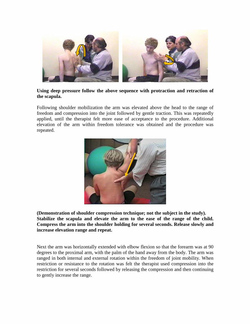
Using deep pressure follow the above sequence with protraction and retraction of the scapula. Following shoulder mobilization the arm was elevated above the head to the range of freedom and compression into the joint followed by gentle traction. This was repeatedly applied, until the therapist felt more ease of acceptance to the procedure. Additional elevation of the arm within freedom tolerance was obtained and the procedure was repeated.
(Demonstration of shoulder compression technique; not the subject in the study). Stabilize the scapula and elevate the arm to the ease of the range of the child. Compress the arm into the shoulder holding for several seconds. Release slowly and increase elevation range and repeat. Next the arm was horizontally extended with elbow flexion so that the forearm was at 90 degrees to the proximal arm, with the palm of the hand away from the body. The arm was ranged in both internal and external rotation within the freedom of joint mobility. When restriction or resistance to the rotation was felt the therapist used compression into the restriction for several seconds followed by releasing the compression and then continuing to gently increase the range.
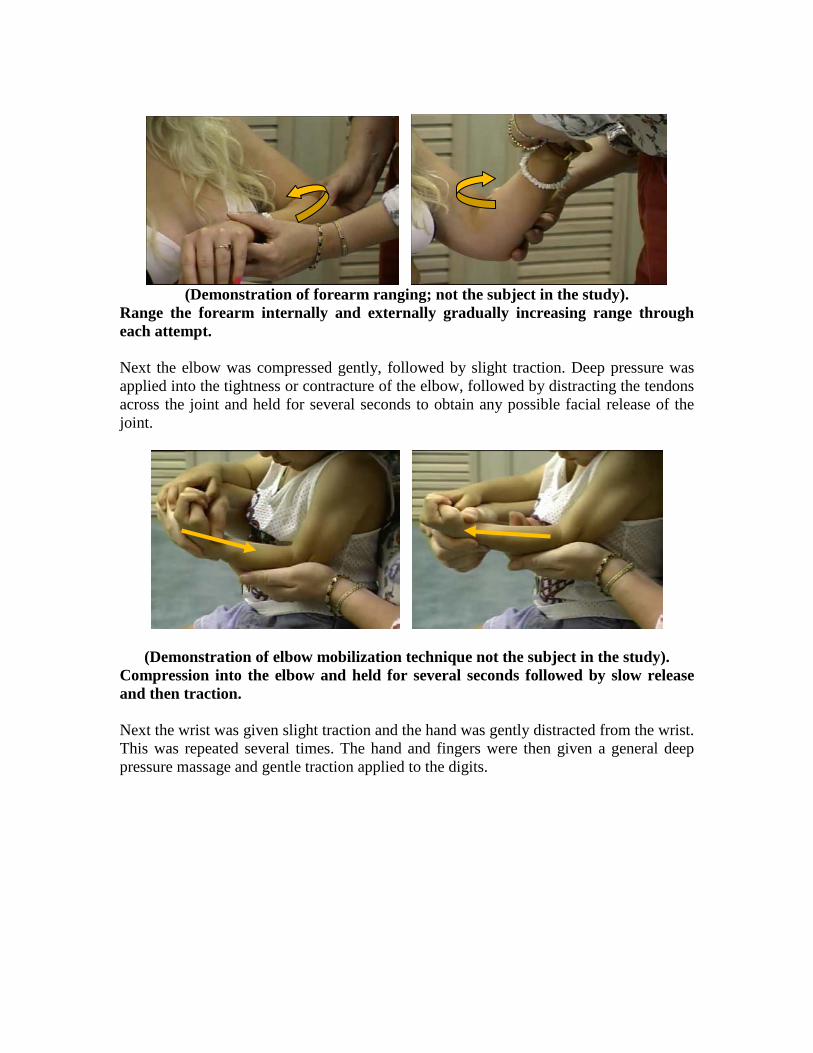
(Demonstration of forearm ranging; not the subject in the study).
Range the forearm internally and externally gradually increasing range through each attempt.
Next the elbow was compressed gently, followed by slight traction. Deep pressure was applied into the tightness or contracture of the elbow, followed by distracting the tendons across the joint and held for several seconds to obtain any possible facial release of the joint.
(Demonstration of elbow mobilization technique not the subject in the study). Compression into the elbow and held for several seconds followed by slow release and then traction. Next the wrist was given slight traction and the hand was gently distracted from the wrist. This was repeated several times. The hand and fingers were then given a general deep pressure massage and gentle traction applied to the digits.
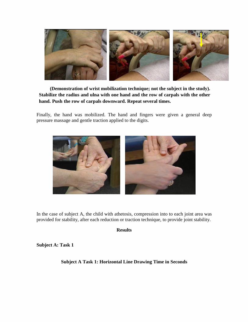
(Demonstration of wrist mobilization technique; not the subject in the study). Stabilize the radius and ulna with one hand and the row of carpals with the other hand. Push the row of carpals downward. Repeat several times.
Finally, the hand was mobilized. The hand and fingers were given a general deep pressure massage and gentle traction applied to the digits.
In the case of subject A, the child with athetosis, compression into to each joint area was provided for stability, after each reduction or traction technique, to provide joint stability.
Results Subject A: Task 1
Subject A Task 1: Horizontal Line Drawing Time in Seconds
0
5
10
15
20
25
Day 1 Day 2 Day 3 Day 4 Day 5 Day 6
Pre-TX
Post-TX
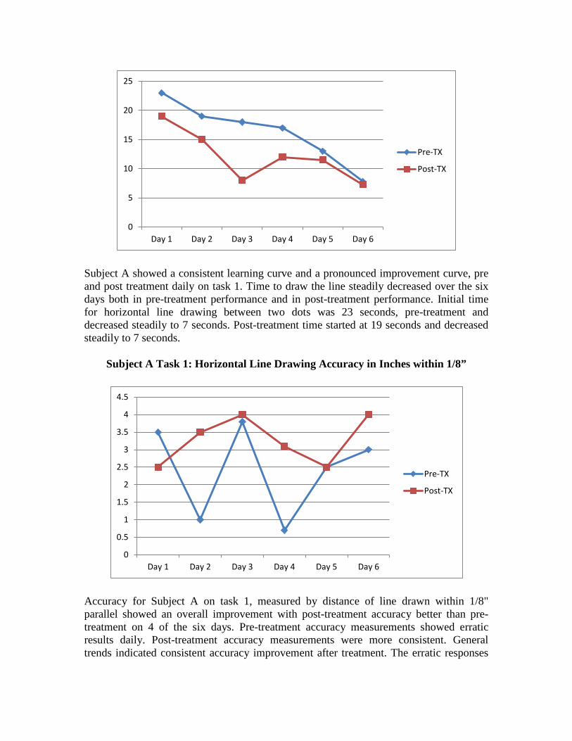
Subject A showed a consistent learning curve and a pronounced improvement curve, pre and post treatment daily on task 1. Time to draw the line steadily decreased over the six days both in pre-treatment performance and in post-treatment performance. Initial time for horizontal line drawing between two dots was 23 seconds, pre-treatment and decreased steadily to 7 seconds. Post-treatment time started at 19 seconds and decreased steadily to 7 seconds.
Subject A Task 1: Horizontal Line Drawing Accuracy in Inches within 1/8”
0
0.5
1
1.5
2
2.5
3
3.5
4
4.5
Day 1 Day 2 Day 3 Day 4 Day 5 Day 6
Pre-TX
Post-TX
Accuracy for Subject A on task 1, measured by distance of line drawn within 1/8" parallel showed an overall improvement with post-treatment accuracy better than pre-treatment on 4 of the six days. Pre-treatment accuracy measurements showed erratic results daily. Post-treatment accuracy measurements were more consistent. General trends indicated consistent accuracy improvement after treatment. The erratic responses
pre-treatment most likely relate to the subject’s fluctuating tone. After treatment the fluctuation in tone was less evident.
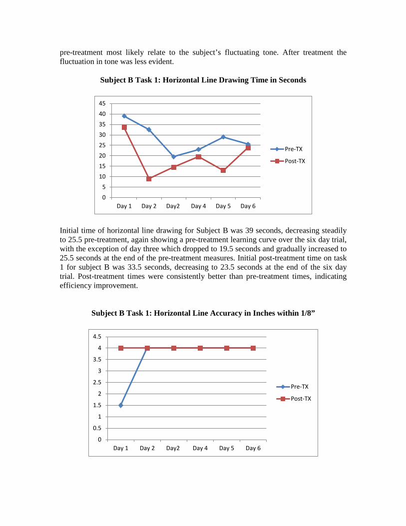
Subject B Task 1: Horizontal Line Drawing Time in Seconds
0
5
10
15
20
25
30
35
40
45
Day 1 Day 2 Day2 Day 4 Day 5 Day 6
Pre-TX
Post-TX
Initial time of horizontal line drawing for Subject B was 39 seconds, decreasing steadily to 25.5 pre-treatment, again showing a pre-treatment learning curve over the six day trial, with the exception of day three which dropped to 19.5 seconds and gradually increased to 25.5 seconds at the end of the pre-treatment measures. Initial post-treatment time on task 1 for subject B was 33.5 seconds, decreasing to 23.5 seconds at the end of the six day trial. Post-treatment times were consistently better than pre-treatment times, indicating efficiency improvement.
Subject B Task 1: Horizontal Line Accuracy in Inches within 1/8”
0
0.5
1
1.5
2
2.5
3
3.5
4
4.5
Day 1 Day 2 Day2 Day 4 Day 5 Day 6
Pre-TX
Post-TX
Accuracy measurements for task 1 for subject B indicated consistent accuracy for each pre-treatment to post-treatment time with the exception of the first pre-treatment measure.
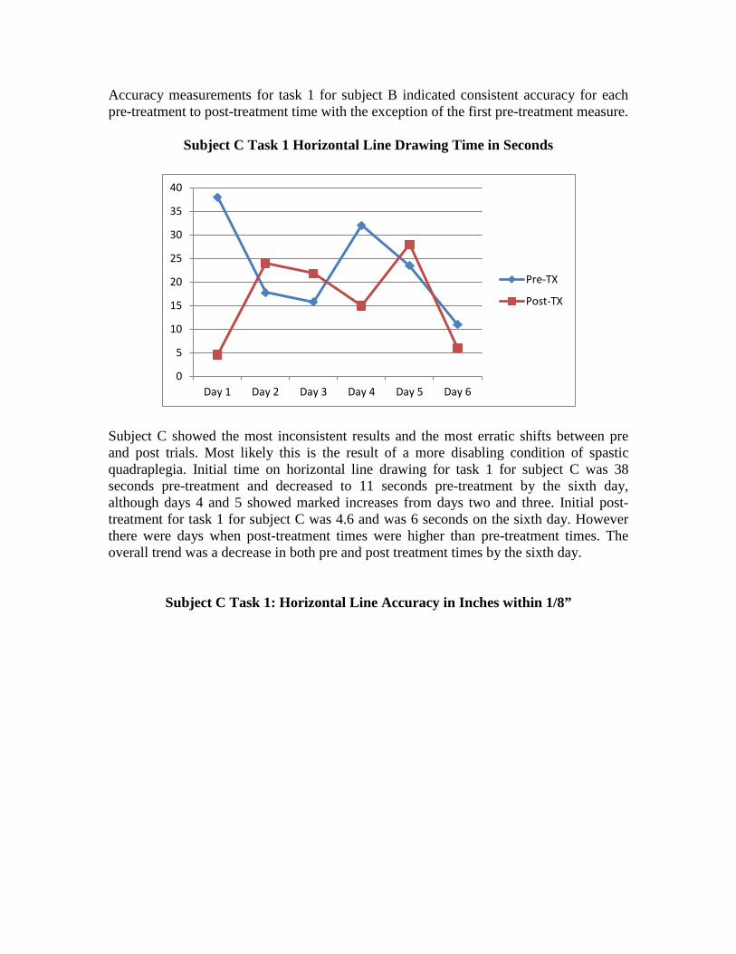
Subject C Task 1 Horizontal Line Drawing Time in Seconds
0
5
10
15
20
25
30
35
40
Day 1 Day 2 Day 3 Day 4 Day 5 Day 6
Pre-TX
Post-TX
Subject C showed the most inconsistent results and the most erratic shifts between pre and post trials. Most likely this is the result of a more disabling condition of spastic quadraplegia. Initial time on horizontal line drawing for task 1 for subject C was 38 seconds pre-treatment and decreased to 11 seconds pre-treatment by the sixth day, although days 4 and 5 showed marked increases from days two and three. Initial post-treatment for task 1 for subject C was 4.6 and was 6 seconds on the sixth day. However there were days when post-treatment times were higher than pre-treatment times. The overall trend was a decrease in both pre and post treatment times by the sixth day.
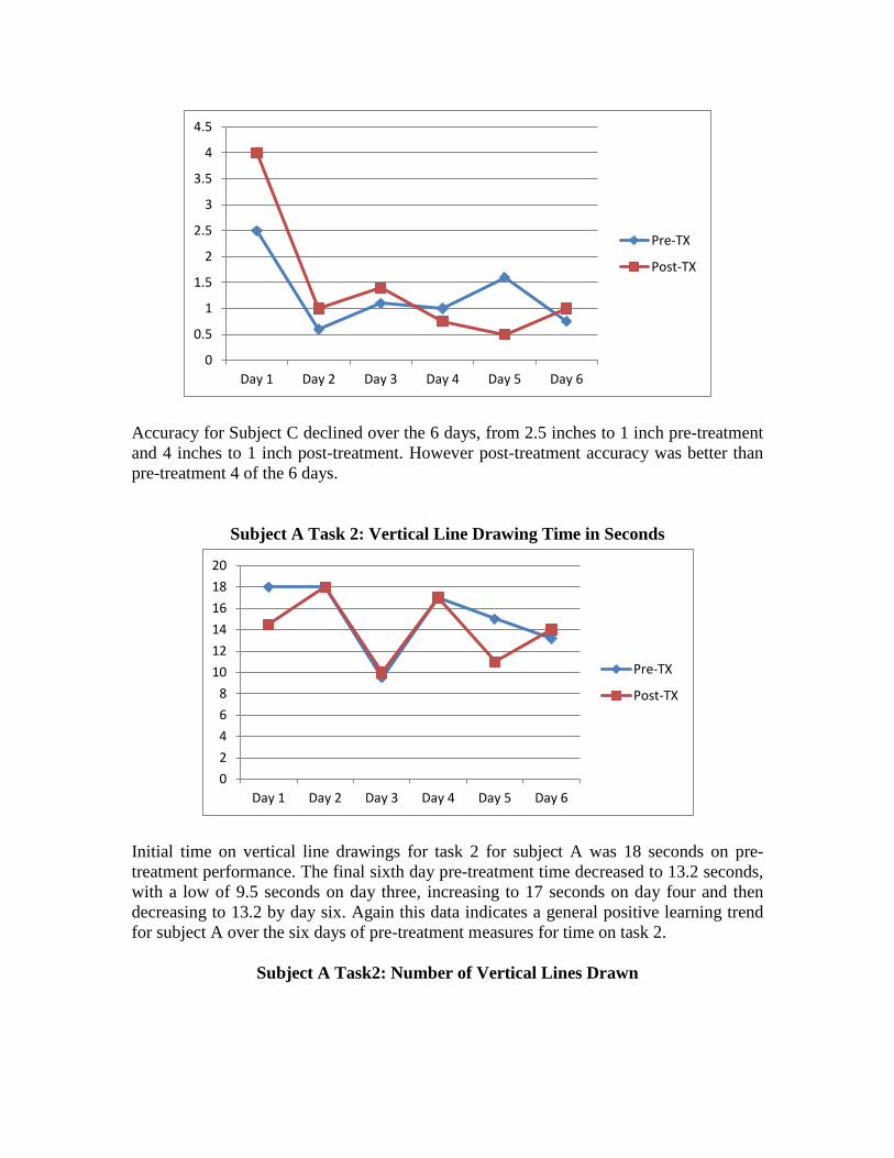
Subject C Task 1: Horizontal Line Accuracy in Inches within 1/8”
0
0.5
1
1.5
2
2.5
3
3.5
4
4.5
Day 1 Day 2 Day 3 Day 4 Day 5 Day 6
Pre-TX
Post-TX
Accuracy for Subject C declined over the 6 days, from 2.5 inches to 1 inch pre-treatment and 4 inches to 1 inch post-treatment. However post-treatment accuracy was better than pre-treatment 4 of the 6 days.
Subject A Task 2: Vertical Line Drawing Time in Seconds
0
2
4
6
8
10
12
14
16
18
20
Day 1 Day 2 Day 3 Day 4 Day 5 Day 6
Pre-TX
Post-TX
Initial time on vertical line drawings for task 2 for subject A was 18 seconds on pre-treatment performance. The final sixth day pre-treatment time decreased to 13.2 seconds, with a low of 9.5 seconds on day three, increasing to 17 seconds on day four and then decreasing to 13.2 by day six. Again this data indicates a general positive learning trend for subject A over the six days of pre-treatment measures for time on task 2.
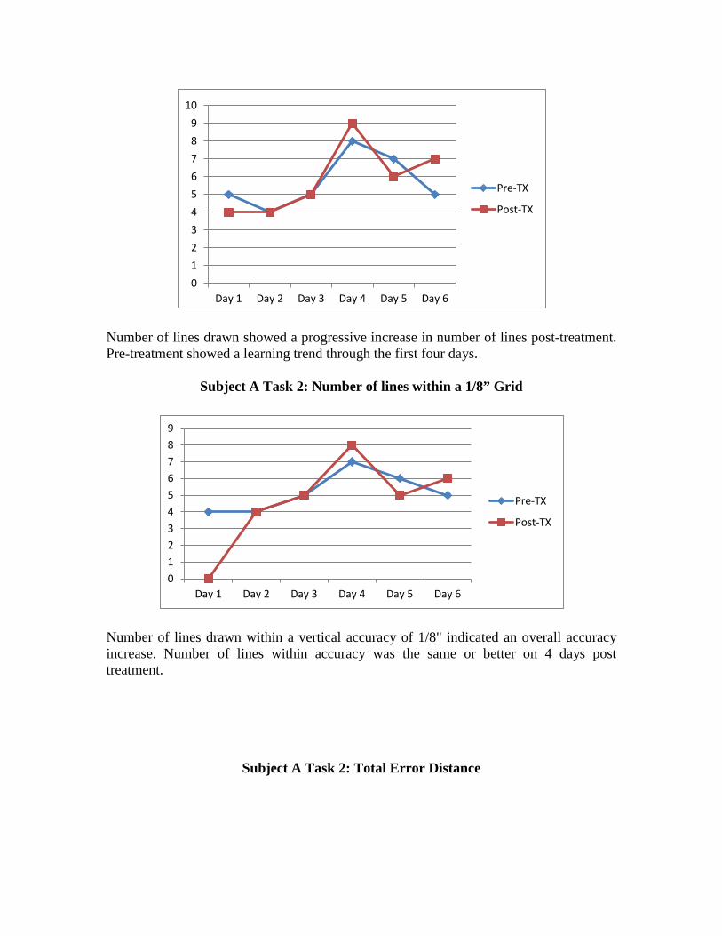
Subject A Task2: Number of Vertical Lines Drawn
0
1
2
3
4
5
6
7
8
9
10
Day 1 Day 2 Day 3 Day 4 Day 5 Day 6
Pre-TX
Post-TX
Number of lines drawn showed a progressive increase in number of lines post-treatment. Pre-treatment showed a learning trend through the first four days.
Subject A Task 2: Number of lines within a 1/8” Grid
0123456789
Day 1 Day 2 Day 3 Day 4 Day 5 Day 6
Pre-TX
Post-TX
Number of lines drawn within a vertical accuracy of 1/8" indicated an overall accuracy increase. Number of lines within accuracy was the same or better on 4 days post treatment.
Subject A Task 2: Total Error Distance
0
0.2
0.4
0.6
0.8
1
1.2
1.4
Day 1 Day 2 Day 3 Day 4 Day 5 Day 6
Pre-TX
Post-TX
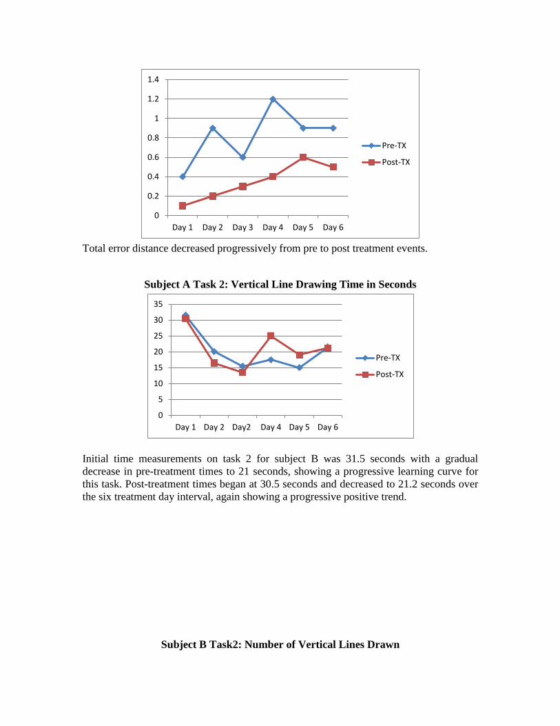
Total error distance decreased progressively from pre to post treatment events.
Subject A Task 2: Vertical Line Drawing Time in Seconds
0
5
10
15
20
25
30
35
Day 1 Day 2 Day2 Day 4 Day 5 Day 6
Pre-TX
Post-TX
Initial time measurements on task 2 for subject B was 31.5 seconds with a gradual decrease in pre-treatment times to 21 seconds, showing a progressive learning curve for this task. Post-treatment times began at 30.5 seconds and decreased to 21.2 seconds over the six treatment day interval, again showing a progressive positive trend.
Subject B Task2: Number of Vertical Lines Drawn
4.4
4.6
4.8
5
5.2
5.4
5.6
5.8
6
6.2
Day 1 Day 2 Day2 Day 4 Day 5 Day 6
Pre-TX
Post-TX
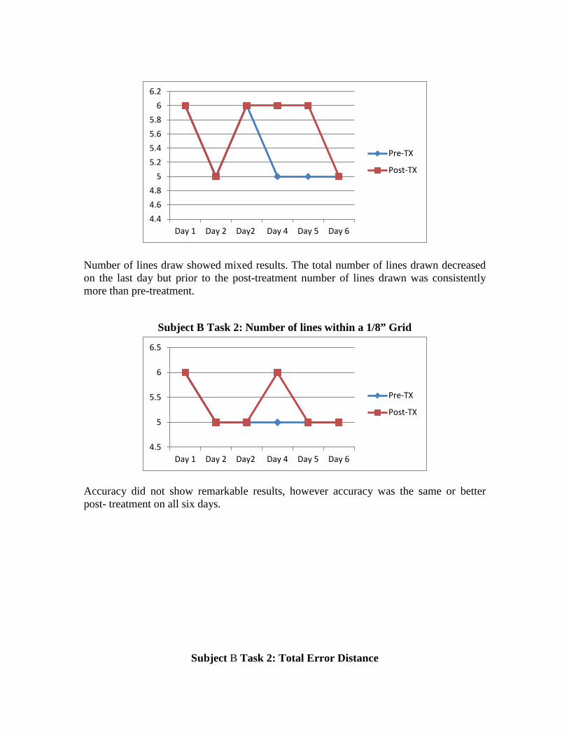
Number of lines draw showed mixed results. The total number of lines drawn decreased on the last day but prior to the post-treatment number of lines drawn was consistently more than pre-treatment.
Subject B Task 2: Number of lines within a 1/8” Grid
4.5
5
5.5
6
6.5
Day 1 Day 2 Day2 Day 4 Day 5 Day 6
Pre-TX
Post-TX
Accuracy did not show remarkable results, however accuracy was the same or better post- treatment on all six days.
Subject B Task 2: Total Error Distance
0
0.05
0.1
0.15
0.2
0.25
0.3
Day 1 Day 2 Day2 Day 4 Day 5 Day 6
Pre-TX
Post-TX
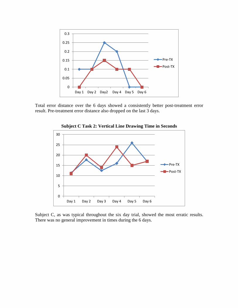
Total error distance over the 6 days showed a consistently better post-treatment error result. Pre-treatment error distance also dropped on the last 3 days.
Subject C Task 2: Vertical Line Drawing Time in Seconds
0
5
10
15
20
25
30
Day 1 Day 2 Day 3 Day 4 Day 5 Day 6
Pre-TX
Post-TX
Subject C, as was typical throughout the six day trial, showed the most erratic results. There was no general improvement in times during the 6 days.
Subject C Task 2: Number of Vertical Lines Drawn
0
1
2
3
4
5
6
7
8
9
10
Day 1 Day 2 Day 3 Day 4 Day 5 Day 6
Pre-TX
Post-TX
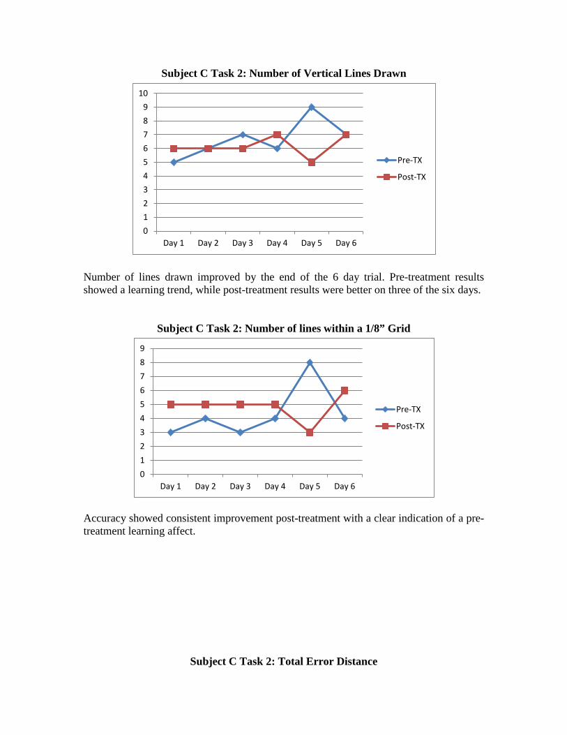
Number of lines drawn improved by the end of the 6 day trial. Pre-treatment results showed a learning trend, while post-treatment results were better on three of the six days.
Subject C Task 2: Number of lines within a 1/8” Grid
0
1
2
3
4
5
6
7
8
9
Day 1 Day 2 Day 3 Day 4 Day 5 Day 6
Pre-TX
Post-TX
Accuracy showed consistent improvement post-treatment with a clear indication of a pre-treatment learning affect.
Subject C Task 2: Total Error Distance
0
0.5
1
1.5
2
2.5
3
Day 1 Day 2 Day 3 Day 4 Day 5 Day 6
Pre-TX
Post-TX
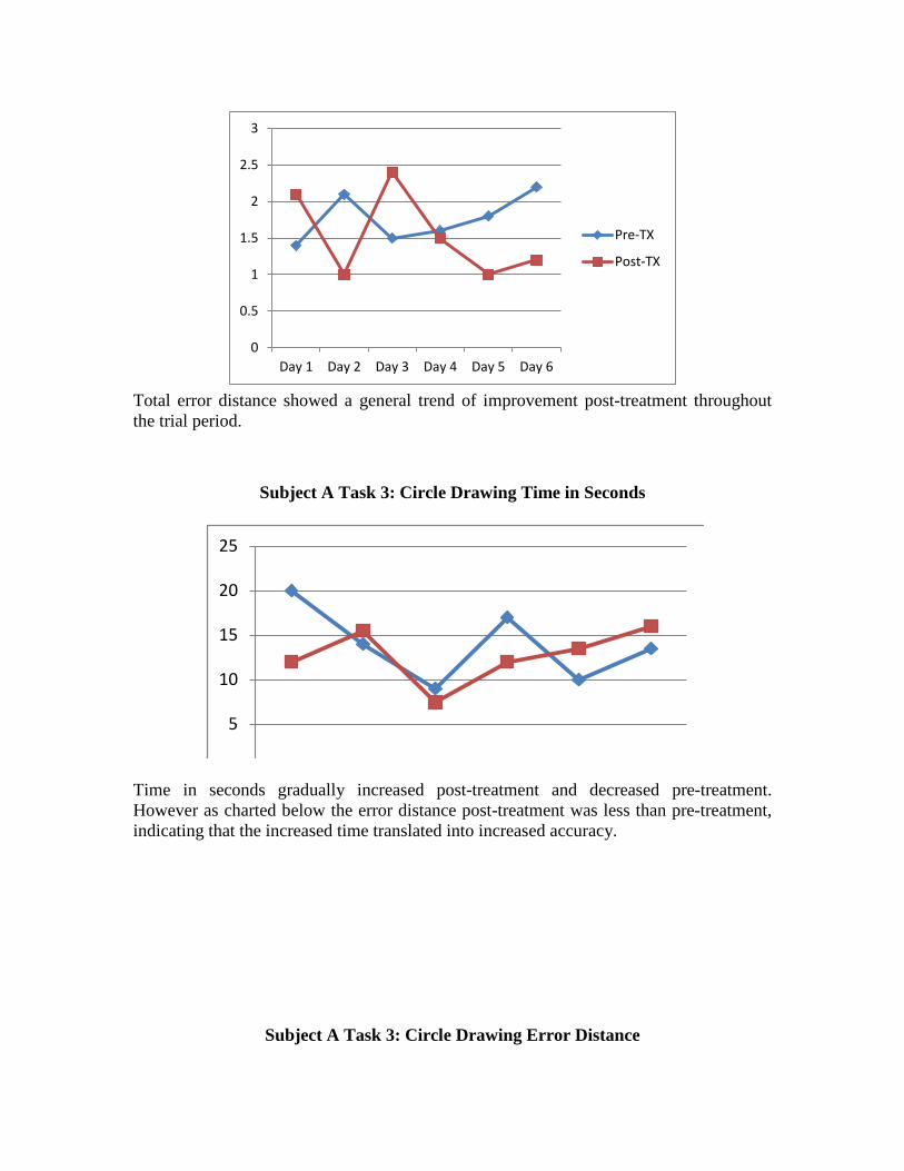
Total error distance showed a general trend of improvement post-treatment throughout the trial period.
Subject A Task 3: Circle Drawing Time in Seconds
5
10
15
20
25
Time in seconds gradually increased post-treatment and decreased pre-treatment. However as charted below the error distance post-treatment was less than pre-treatment, indicating that the increased time translated into increased accuracy.
Subject A Task 3: Circle Drawing Error Distance
0
0.05
0.1
0.15
0.2
0.25
0.3
Day 1 Day 2 Day 3 Day 4 Day 5 Day 6
Pre-TX
Post-TX
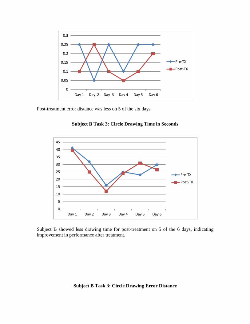
Post-treatment error distance was less on 5 of the six days.
Subject B Task 3: Circle Drawing Time in Seconds
0
5
10
15
20
25
30
35
40
45
Day 1 Day 2 Day 3 Day 4 Day 5 Day 6
Pre-TX
Post-TX
Subject B showed less drawing time for post-treatment on 5 of the 6 days, indicating improvement in performance after treatment.
Subject B Task 3: Circle Drawing Error Distance
0
0.05
0.1
0.15
0.2
0.25
Day 1 Day 2 Day 3 Day 4 Day 5 Day 6
Pre-TX
Post-TX
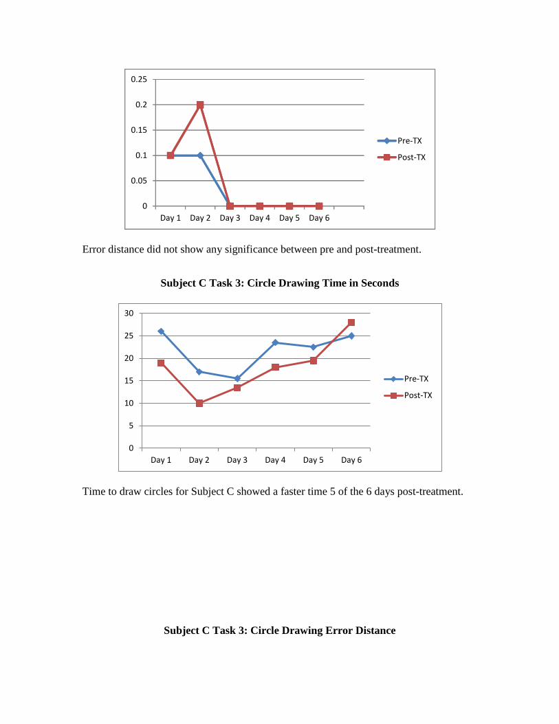
Error distance did not show any significance between pre and post-treatment.
Subject C Task 3: Circle Drawing Time in Seconds
0
5
10
15
20
25
30
Day 1 Day 2 Day 3 Day 4 Day 5 Day 6
Pre-TX
Post-TX
Time to draw circles for Subject C showed a faster time 5 of the 6 days post-treatment.
Subject C Task 3: Circle Drawing Error Distance
0
0.2
0.4
0.6
0.8
1
1.2
1.4
Day 1 Day 2 Day 3 Day 4 Day 5 Day 6
Pre-TX
Post-TX
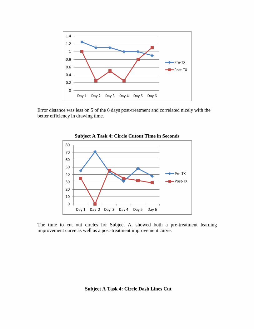
Error distance was less on 5 of the 6 days post-treatment and correlated nicely with the better efficiency in drawing time.
Subject A Task 4: Circle Cutout Time in Seconds
0
10
20
30
40
50
60
70
80
Day 1 Day 2 Day 3 Day 4 Day 5 Day 6
Pre-TX
Post-TX
The time to cut out circles for Subject A, showed both a pre-treatment learning improvement curve as well as a post-treatment improvement curve.
Subject A Task 4: Circle Dash Lines Cut
0
2
4
6
8
10
12
14
16
Day 1 Day 2 Day 3 Day 4 Day 5 Day 6
Pre-TX
Post-TX
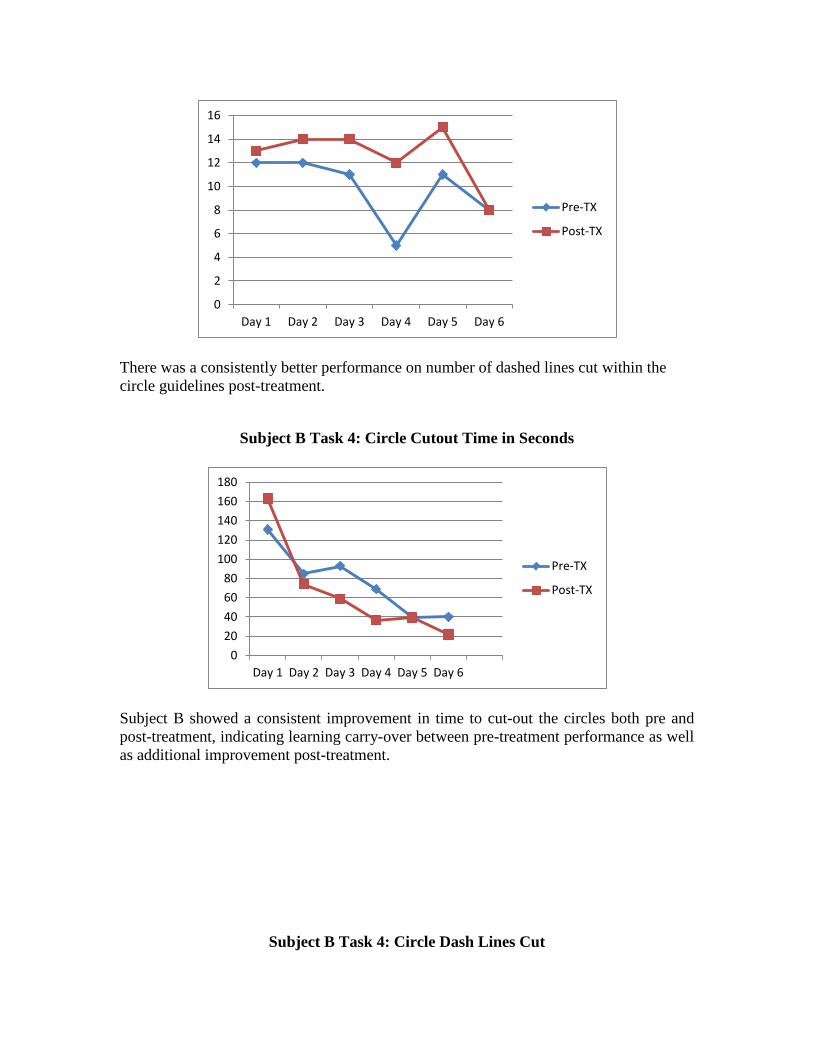
There was a consistently better performance on number of dashed lines cut within the circle guidelines post-treatment.
Subject B Task 4: Circle Cutout Time in Seconds
0
20
40
60
80
100
120
140
160
180
Day 1 Day 2 Day 3 Day 4 Day 5 Day 6
Pre-TX
Post-TX
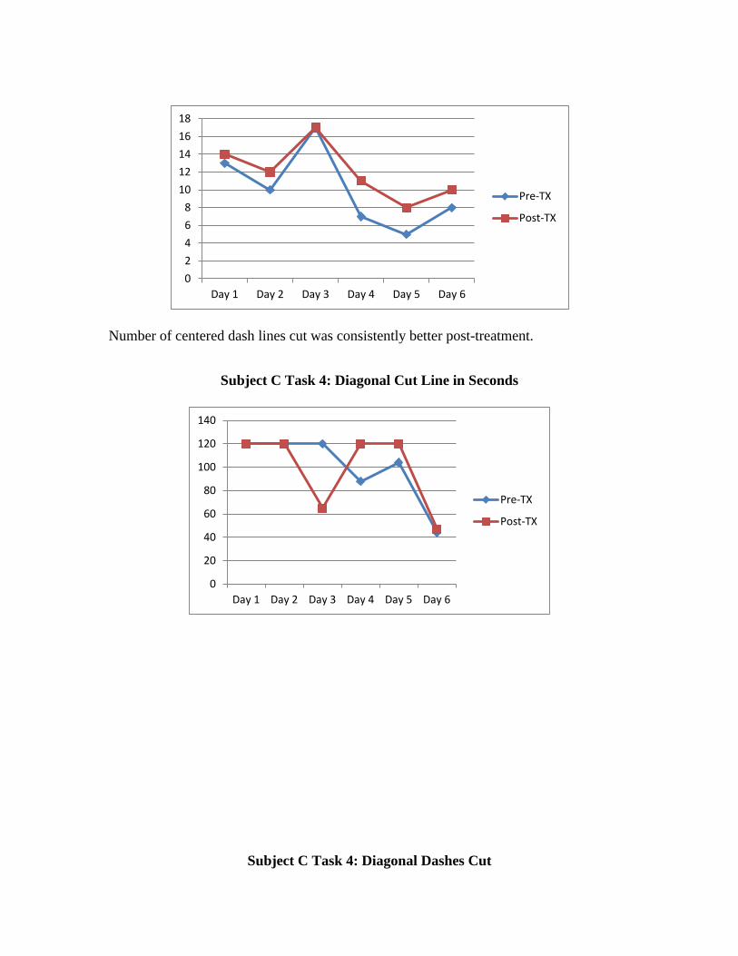
Subject B showed a consistent improvement in time to cut-out the circles both pre and post-treatment, indicating learning carry-over between pre-treatment performance as well as additional improvement post-treatment.
Subject B Task 4: Circle Dash Lines Cut
0
2
4
6
8
10
12
14
16
18
Day 1 Day 2 Day 3 Day 4 Day 5 Day 6
Pre-TX
Post-TX
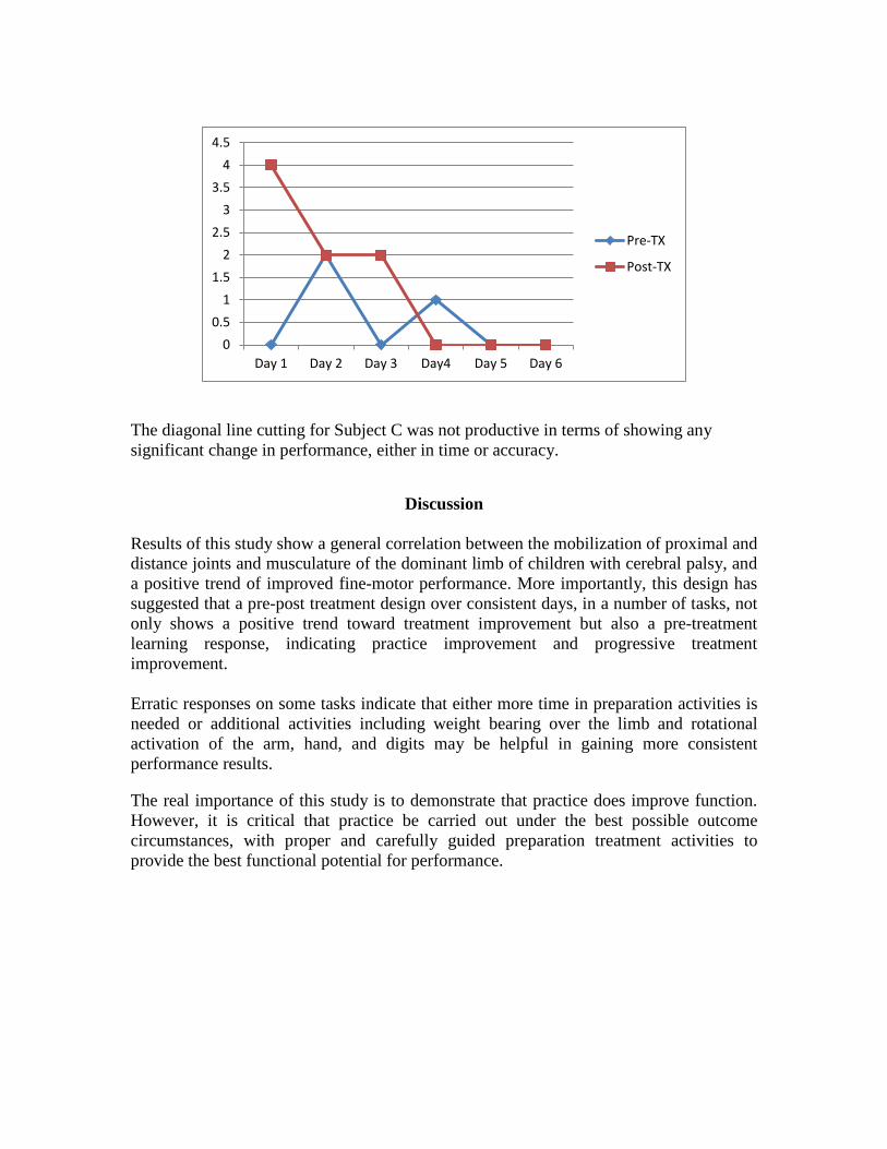
Number of centered dash lines cut was consistently better post-treatment.
Subject C Task 4: Diagonal Cut Line in Seconds
0
20
40
60
80
100
120
140
Day 1 Day 2 Day 3 Day 4 Day 5 Day 6
Pre-TX
Post-TX
Subject C Task 4: Diagonal Dashes Cut
0
0.5
1
1.5
2
2.5
3
3.5
4
4.5
Day 1 Day 2 Day 3 Day4 Day 5 Day 6
Pre-TX
Post-TX
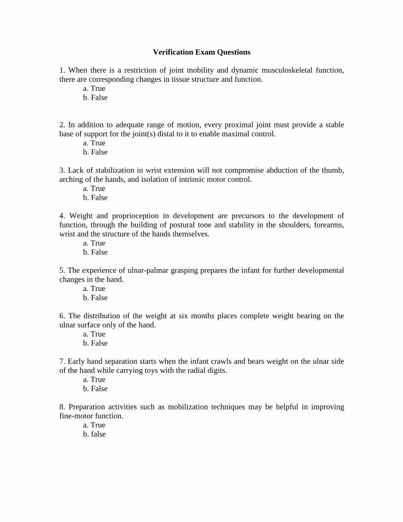
The diagonal line cutting for Subject C was not productive in terms of showing any significant change in performance, either in time or accuracy.
Discussion Results of this study show a general correlation between the mobilization of proximal and distance joints and musculature of the dominant limb of children with cerebral palsy, and a positive trend of improved fine-motor performance. More importantly, this design has suggested that a pre-post treatment design over consistent days, in a number of tasks, not only shows a positive trend toward treatment improvement but also a pre-treatment learning response, indicating practice improvement and progressive treatment improvement. Erratic responses on some tasks indicate that either more time in preparation activities is needed or additional activities including weight bearing over the limb and rotational activation of the arm, hand, and digits may be helpful in gaining more consistent performance results. The real importance of this study is to demonstrate that practice does improve function. However, it is critical that practice be carried out under the best possible outcome circumstances, with proper and carefully guided preparation treatment activities to provide the best functional potential for performance.
Verification Exam Questions 1. When there is a restriction of joint mobility and dynamic musculoskeletal function, there are corresponding changes in tissue structure and function. a. True
b. False
2. In addition to adequate range of motion, every proximal joint must provide a stable base of support for the joint(s) distal to it to enable maximal control. a. True b. False 3. Lack of stabilization in wrist extension will not compromise abduction of the thumb, arching of the hands, and isolation of intrinsic motor control. a. True b. False 4. Weight and proprioception in development are precursors to the development of function, through the building of postural tone and stability in the shoulders, forearms, wrist and the structure of the hands themselves. a. True b. False 5. The experience of ulnar-palmar grasping prepares the infant for further developmental changes in the hand. a. True b. False 6. The distribution of the weight at six months places complete weight bearing on the ulnar surface only of the hand. a. True b. False 7. Early hand separation starts when the infant crawls and bears weight on the ulnar side of the hand while carrying toys with the radial digits. a. True b. False 8. Preparation activities such as mobilization techniques may be helpful in improving fine-motor function. a. True b. false