Embed Size (px)
Citation preview
Vol. 115 No. 1 January 2013
Electrically conductive bacterial nanowires in bisphosphonate-related osteonecrosis of the jaw biofilmsGreg Wanger, PhD,a Yuri Gorby, PhD,b Mohamed Y. El-Naggar, PhD,c Thomas D. Yuzvinsky, PhD,c
Christoph Schaudinn, PhD,d Amita Gorur, MSc,e and Parish P. Sedghizadeh, DDS, MSf
J. Craig Venter Institute, San Diego, California; University of Southern California, Los Angeles, California; Robert Koch Institute, Berlin,Germany; Lawrence Berkeley National Laboratory, Berkeley, California
Objective. Bacterial biofilms play a role in the pathogenesis of bisphosphonate-related osteonecrosis of the jaw (BRONJ). Thepurpose of this preliminary study was to test the hypothesis that the extracellular filaments observed in biofilms associatedwith BRONJ contain electrically conductive nanowires.Study Design. Bone samples of patients affected by BRONJ were evaluated for conductive nanowires by scanning electronmicroscopy (SEM) and conductive probe atomic force microscopy (CP-AFM). We created nanofabricated electrodes tomeasure electrical transport along putative nanowires.Results. SEM revealed large-scale multispecies biofilms containing numerous filamentous structures throughout necrotic bone.CP-AFM analysis revealed that these structures were electrically conductive nanowires with resistivities on the order of 20��cm. Nanofabricated electrodes spaced along the nanowires confirmed their ability to transfer electrons over micron-scalelengths.Conclusions. Electrically conductive bacterial nanowires to date have been described only in environmental isolates. Thisstudy shows for the first time that these nanowires can also be found in clinically relevant biofilm-mediated diseases, such as
BRONJ, and may represent an important target for therapy. (Oral Surg Oral Med Oral Pathol Oral Radiol 2013;115:71-78)More than 40 million Americans and more than 200million patients worldwide are currently receiving an-tiresorptive drugs to treat common bone disorders suchas osteoporosis and skeletal complications associatedwith osseous metastasis and multiple myeloma.1 Osteo-necrosis of the jaw is a serious adverse effect associatedwith bisphosphonate (BP) antiresorptive therapy. Bis-phosphonate-related osteonecrosis of the jaw (BRONJ)is characterized by necrotic jaw bone in the oral cavity.The pathogenesis of BRONJ is considered to be mul-tifactorial and usually involves oral trauma, with sub-sequent delayed wound healing and a biofilm-mediatedinfection.2,3 Spontaneous cases of BRONJ have beenreported without mucosal breach, although in mostcases patients who develop BRONJ have a history ofinvasive dental procedures or complications from toothextractions or denture trauma that places them at riskfor BRONJ.4 Invasive oral surgical procedures ortrauma to jaw bone can expose bound BP from the
aElectromicrobiology Group, J. Craig Venter Institute.bDepartment of Biological Sciences, University of SouthernCalifornia.cDepartment of Physics, University of Southern California.dCenter for Biological Security, Robert Koch Institute.eLife Sciences Division, Lawrence Berkeley National Laboratory.fCenter for Biofilms, University of Southern California.Received for publication May 25, 2012; returned for revision Aug 14,2012; accepted for publication Aug 20, 2012.© 2013 Elsevier Inc. All rights reserved.2212-4403/$ - see front matter
http://dx.doi.org/10.1016/j.oooo.2012.08.446bone, releasing the drug into the local milieu, where ithas been shown to inhibit wound healing and increasethe binding affinity of oral bacteria to bone, culminat-ing in BRONJ.5 Sedghizadeh et al. described complexmicrobial communities that permeate the affected jawbone structure, implicating biofilms in the mineralogicdissolution of bone by yet unknown mechanisms.6 Thetypes of bacteria described in BRONJ biofilms aredifficult to treat and have known resistance to conven-tional antibiotics.7 Innovative approaches that destabi-lize the structural or metabolic integrity of biofilms areneeded to improve our ability to treat BRONJ and otherbiofilm-mediated infectious diseases. Such innovationsrequire a more complete understanding of the processesthat sustain microbial biofilms so that therapy couldtarget the disruption of these processes.
BRONJ biofilms, like many clinically relevant bio-films, produce extracellular filaments that represent a largefraction of the extracellular biofilm matrix. Filamentousextracellular structures in biofilms have commonly beenclassified as part of the extracellular polymeric substance(EPS), which includes polysaccharides, flagella, DNA,RNA, and a variety of dissolved small compounds.8-11
Recent evidence has revealed that a variety of micro-organisms produce electrically conductive filamentscalled bacterial nanowires that appear to be importantcontributors to extracellular electron transfer.12,13 Bac-terial nanowires were first described in 2005 as a phe-nomenon occurring in metal-reducing bacteria.14 Theywere implicated in the transfer of electrons to solid-
phase electron acceptors, such as iron and manganese71
Osteo
ORAL AND MAXILLOFACIAL PATHOLOGY OOOO72 Wanger et al. January 2013
oxides. Importantly, unique nanofabrication techniquescombined with imaging techniques such as conductiveprobe atomic force microscopy (CP-AFM) were neces-sary to prove that bacterial nanowires had actual elec-trical or conductive properties. Conductivity is the mea-sure of the ease at which an electric charge can passthrough a material. Before the demonstration of con-ductivity, structures could not be uniquely classified asbacterial nanowires, because they could merely repre-sent some known component of biofilm EPS. Confir-mation of conductive nanowires in a variety of otherenvironmental systems, including an oxygenic photo-synthetic cyanobacterium and a thermophilic methano-genic coculture, suggested early on that nanowiresmight be common structures in diverse microbial sys-tems; most published research describing the conduc-tive properties of bacterial nanowires has focused onthe environmental metal-reducing bacteria Geobactersulfurreducens and Shewanella oneidensis MR1.15,16
The complete composition and role of bacterial nano-wires remains largely unknown and unexplored, be-cause bacterial nanowires are a recent discovery andresearched by only a few laboratories. To date, thework on bacterial nanowires has focused on environ-mental microbes, and nanowires have not been identi-fied or characterized in human pathogens.
The exact physiologic role of bacterial nanowiresremains to be elucidated, but the nanofabrication andimaging techniques used for evaluating the conduc-tive properties of these biologic protein nanostruc-tures are now well established. These techniqueshave been applied to the conductive or electromicro-biologic characterization of environmental bacteriabut not to medically and clinically relevant microbes.Therefore, the purpose of this preliminary study wasto apply nanofabrication techniques along with CP-AFM imaging techniques to test our hypothesis thatthe extracellular filaments observed in microbial bio-films associated with BRONJ contain electricallyconductive nanowires.
MATERIALS AND METHODSPatient sample collectionSamples of necrotic bone were collected from pa-tients as part of an ongoing natural history study at
Table I. Clinical-pharmacological characteristics of stPatient #age/sex Ethnicity BP type/duration
1. 67/M Hispanic-American Pamidronate/36 months2. 68/M African-American Zoledronic acid/8 months3. 63/F Hispanic-American Alendronate/36 months4. 76/F Asian-American Alendronate/120 months
the University of Southern California. Before sample
collection, appropriate Institutional Review Boardapproval (no. HS-CG-05-00002) and written patientconsents were obtained. Samples were obtained from4 patients requiring routine clinical sequestrectomyprocedures as previously described,17 and study pa-tient characteristics are provided in Table I. Forstudy inclusion, all patients required a diagnosis of astage 1-3 BRONJ lesion, established by standardclinical and radiographic protocol per the AmericanAssociation of Oral and Maxillofacial Surgeons di-agnostic criteria.18 To minimize confounding vari-ables or effect modifiers, patients were excludedfrom the study if they had a history of head and neckradiation or concomitant steroid therapy, becausethese can cause osteonecrosis unrelated to BP ther-apy. One sample of bone from each patient affectedby BRONJ in our study was cut into subfragmentsand prepared for microscopic and electrical conduc-tivity analyses as described below.
Scanning electron microscopyScanning electron microscopy (SEM) allows for micro-scopic evaluation of specimens at magnifications over arange of up to 6 orders of magnitude (�10,000-500,000), about 250 times the magnification limit of thebest light microscopes. This makes SEM an ideal tech-nology for evaluation of microscopic biofilms whichcould be missed via routine histopathologic evaluation.For SEM analysis in our study, fragments of bonesamples from every study patient were fixed in a 4%formaldehyde/2% glutaraldehyde solution for 48 hoursat 4°C, washed with phosphate-buffered saline solutionbuffer, dehydrated in a graded ethanol series, criticalpoint dried, and mounted on an aluminum stub. Thesamples were then sputter coated with platinum (Pt)and imaged with the SEM operating at 5 kV in thesecondary electron mode (XL 30S; FEG, FEI Co.,Hillsboro, OR).
Conductive probe atomic force microscopyCP-AFM allows for current/voltage (I/V) measure-ments by applying voltage to putative electrical struc-tures and measuring the current along the wire as thevoltage changes. Typically, a linear to nonlinear re-sponse is expected for the resulting I/V data at some
atients
indicationLocation of
BRONJ Dental factorBRONJstage
te cancer Left maxilla Tooth extraction IIIle myeloma Right mandible Tooth extraction II
porosis Right maxilla Denture trauma IIporosis Left mandible Tooth extraction II
udy p
BP
ProstaMultipOsteo
negative to positive value of current if the structure has
OOOO ORIGINAL ARTICLEVolume 115, Number 1 Wanger et al. 73
conductive properties, otherwise a flat horizontal line atzero current results, which would indicate lack of con-ductivity. For CP-AFM analysis, fragments of eachbone sample were mechanically disrupted with sterile18-gauge needles, and 20 mL of the suspension wasspotted onto highly ordered pyrolytic graphite (HOPG)wafers (SPI Supplies, West Chester, PA). The HOPGwafers act as a flat and electrically conductive substratefor the biofilms, facilitating the conductive probe mea-surements with CP-AFM. For the purposes of ourstudy, CP-AFM was used in 2 common modes: tappingmode and contact mode. Tapping mode is used to maptopography of a surface (e.g., HOPG biofilm sample)by applying an oscillating cantilever probe tip to asurface via light tapping. The cantilever’s oscillationamplitude changes with sample surface topography,and a topographic image is obtained by monitoringthese changes. In contact mode, the probe tip is inperpetual contact with the sample, in contrast to tappingmode, and conductivity measurements are made in con-tact mode. The HOPG biofilm samples were allowed toair dry until all of the fluid had evaporated. Then, theywere gently washed with distilled H2O delivered drop-wise before air drying again. CP-AFM was performedwith the use of a Dimension V Scanning Probe Micro-scope (Veeco Instruments, Plainview, NY). The sam-ples were first imaged in tapping mode, using NSC36/AlBS probes (Mikromasch, San Jose, CA) to obtaintopographic images of the sample. Once areas withputative nanowires were located, the probe was re-placed with a Pt-Iridium–coated conductive contact tip(SCM-PIC; Veeco Instruments) for the conductivitymeasurements in contact mode. For control, an opencircuit was created by placing 2 Pt probes very closetogether (�150 nm without a bridging nanowire) onsubsamples that underwent the same SEM and CP-AFM protocol.
Nanofabrication technique for measuring electrontransport and resistivityFor electrical transport measurements, bone sampleswere deposited onto oxidized silicon wafers with pre-fabricated gold electrodes and loaded into a Zeiss 1540FIB/SEM electron microscope (Carl Zeiss Microscopy,Thornwood, NY). As described by our group previ-ously,12 putative nanowires in the uncoated sampleswere located by SEM in the proximity of the prefabri-cated gold contacts. Gold is used in this techniquebecause it ensures sufficient contact and is a well es-tablished conductor, like silver or copper, but does notreadily oxidize. Once a suitable nanowire was found ineach sample, Pt precursor gas was injected into thechamber in close proximity to the sample surface. Pt
leads that contacted the nanowires were deposited bypatterned exposure to the electron beam. Longer Ptleads (e.g., to bridge the connection to the gold elec-trodes) were established by exposure to the ion beam.I/V measurements were performed at room temperatureusing a probe station instrumented to an Agilent 4156C(Agilent Technologies, Santa Clara, CA) semiconduc-tor parameter analyzer. Nanowire resistivity (�) wascalculated as � � RA/L, where R is the measuredresistance, A is the cross sectional nanowire area (cal-culated with AFM height measurements describedabove), and L is the length of the nanowire segmentbetween the 2 probes measured by SEM and CP-AFMimaging.
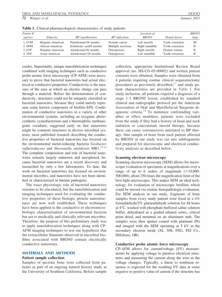
RESULTSScanning electron microscopySEM analysis of BRONJ-affected bone from all 4 pa-tients revealed thick multispecies biofilms permeating thebone tissue and composed of predominantly bacterialmorphotypes (Figure 1). All four of the study patients hadnecrotic bone colonized by biofilms with 15 distinguish-able bacterial morphotypes similarly observed in eachspecimen. Morphotypes included organisms from the ge-nuses Fusobacterium, Bacillus, Actinomyces, Staphylo-coccus, Streptococcus, and Selenomonas and 3 differentmorphotypes of treponemes. The bacteria identified inall 4 of the specimens were similar at the SEM level,and comprised gram-positive and gram-negative organ-isms with predominantly anaerobic and facultative an-aerobic organisms. The biofilms were associated withlarge portions of necrotic bone tissue and some areas ofthe samples appeared to contain more microbial biomassthan bone. Many thin filaments (e.g., putative nanowires)were seen connecting bacteria (Figure 1, B and C). Therewere no major differences that could be observed be-tween the nanowires among the various subsamples.Based on morphologic similarities between these struc-tures and nanowires produced by environmental organ-isms, we applied our nanofabrication approach withCP-AFM imaging for testing whether some of thesefilaments were conductive nanowires. Although it isquite likely that the EPS contains a combination of pili,flagella, polysaccharides, and DNA, nanofabricationand imaging techniques were applied to test our hy-pothesis that some of these filaments are truly conduc-tive bacterial nanowires.
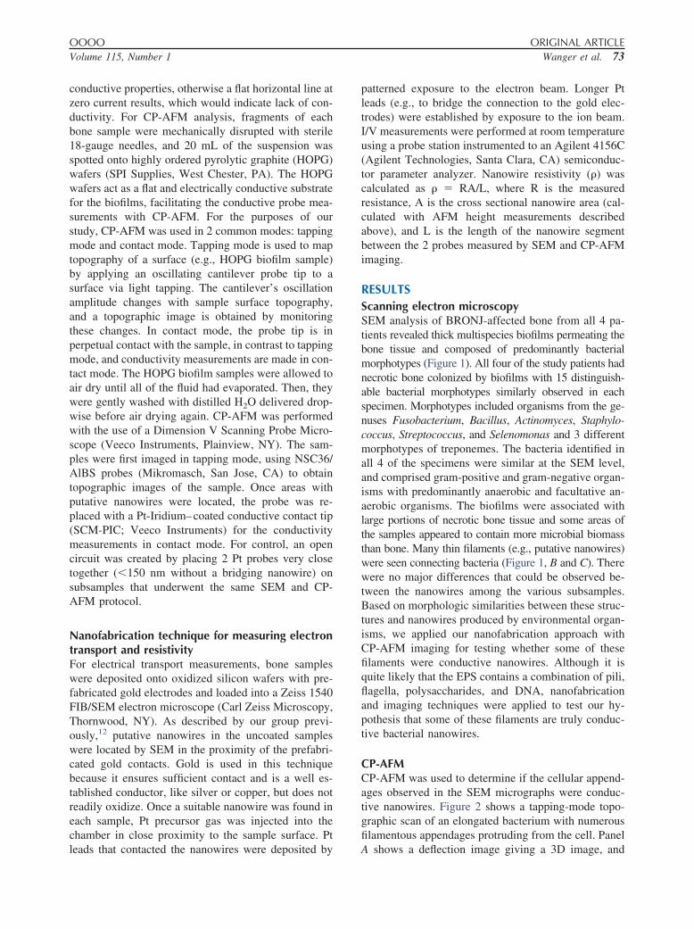
CP-AFMCP-AFM was used to determine if the cellular append-ages observed in the SEM micrographs were conduc-tive nanowires. Figure 2 shows a tapping-mode topo-graphic scan of an elongated bacterium with numerousfilamentous appendages protruding from the cell. Panel
A shows a deflection image giving a 3D image, and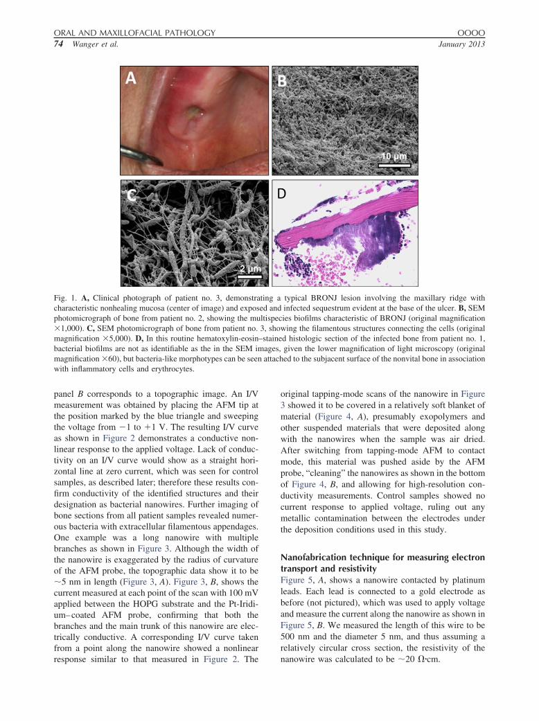
ORAL AND MAXILLOFACIAL PATHOLOGY OOOO74 Wanger et al. January 2013
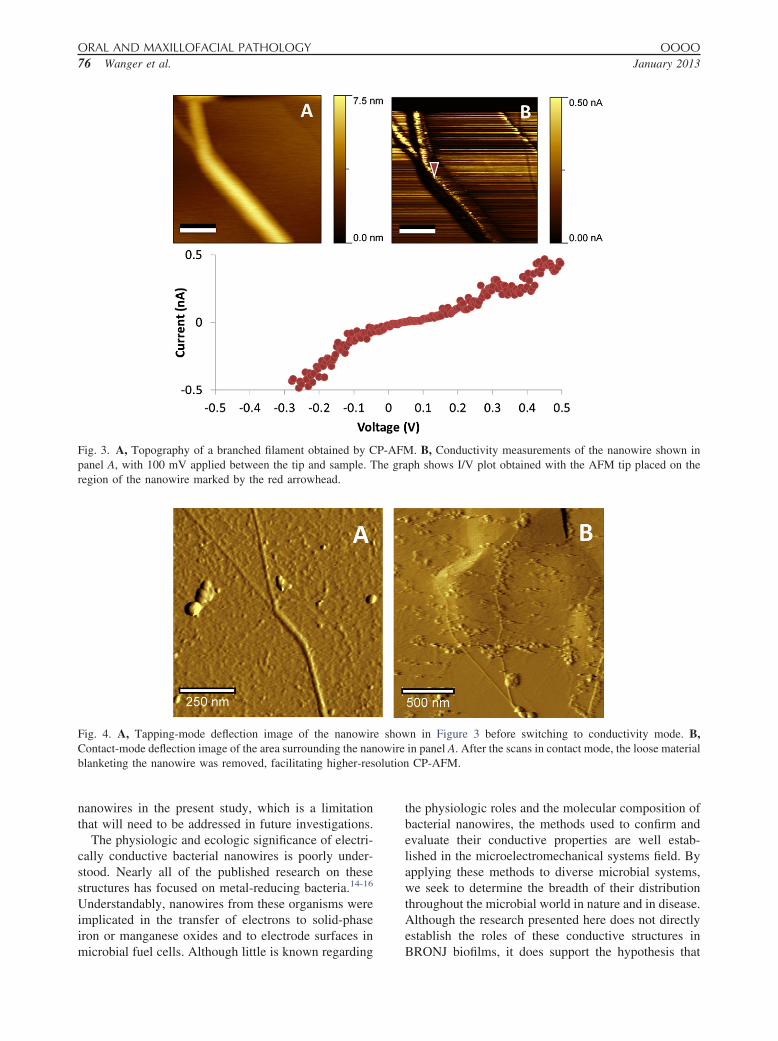
panel B corresponds to a topographic image. An I/Vmeasurement was obtained by placing the AFM tip atthe position marked by the blue triangle and sweepingthe voltage from �1 to �1 V. The resulting I/V curveas shown in Figure 2 demonstrates a conductive non-linear response to the applied voltage. Lack of conduc-tivity on an I/V curve would show as a straight hori-zontal line at zero current, which was seen for controlsamples, as described later; therefore these results con-firm conductivity of the identified structures and theirdesignation as bacterial nanowires. Further imaging ofbone sections from all patient samples revealed numer-ous bacteria with extracellular filamentous appendages.One example was a long nanowire with multiplebranches as shown in Figure 3. Although the width ofthe nanowire is exaggerated by the radius of curvatureof the AFM probe, the topographic data show it to be�5 nm in length (Figure 3, A). Figure 3, B, shows thecurrent measured at each point of the scan with 100 mVapplied between the HOPG substrate and the Pt-Iridi-um–coated AFM probe, confirming that both thebranches and the main trunk of this nanowire are elec-trically conductive. A corresponding I/V curve takenfrom a point along the nanowire showed a nonlinear
Fig. 1. A, Clinical photograph of patient no. 3, demonstracharacteristic nonhealing mucosa (center of image) and exposphotomicrograph of bone from patient no. 2, showing the mu�1,000). C, SEM photomicrograph of bone from patient no.magnification �5,000). D, In this routine hematoxylin-eosinbacterial biofilms are not as identifiable as the in the SEM immagnification �60), but bacteria-like morphotypes can be seenwith inflammatory cells and erythrocytes.
response similar to that measured in Figure 2. The
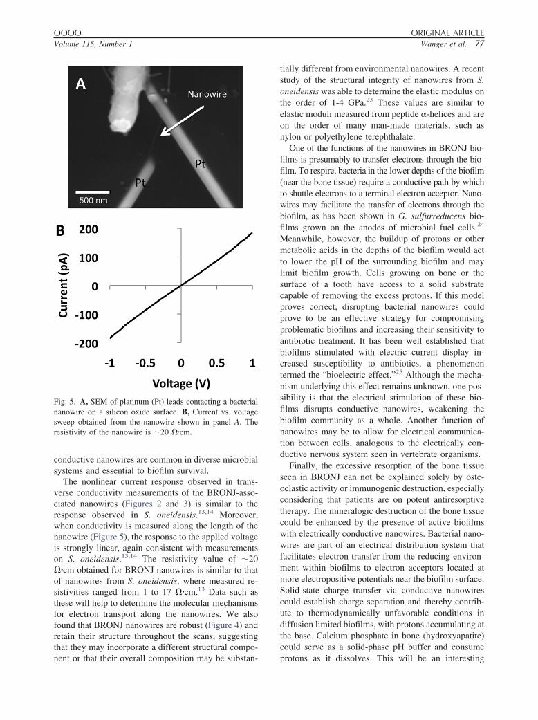
original tapping-mode scans of the nanowire in Figure3 showed it to be covered in a relatively soft blanket ofmaterial (Figure 4, A), presumably exopolymers andother suspended materials that were deposited alongwith the nanowires when the sample was air dried.After switching from tapping-mode AFM to contactmode, this material was pushed aside by the AFMprobe, “cleaning” the nanowires as shown in the bottomof Figure 4, B, and allowing for high-resolution con-ductivity measurements. Control samples showed nocurrent response to applied voltage, ruling out anymetallic contamination between the electrodes underthe deposition conditions used in this study.
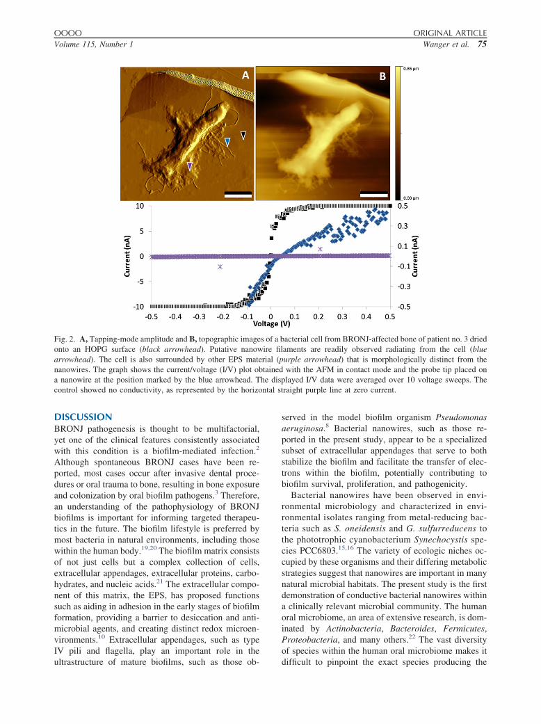
Nanofabrication technique for measuring electrontransport and resistivityFigure 5, A, shows a nanowire contacted by platinumleads. Each lead is connected to a gold electrode asbefore (not pictured), which was used to apply voltageand measure the current along the nanowire as shown inFigure 5, B. We measured the length of this wire to be500 nm and the diameter 5 nm, and thus assuming arelatively circular cross section, the resistivity of the
typical BRONJ lesion involving the maxillary ridge withinfected sequestrum evident at the base of the ulcer. B, SEMies biofilms characteristic of BRONJ (original magnificationwing the filamentous structures connecting the cells (originald histologic section of the infected bone from patient no. 1,given the lower magnification of light microscopy (originaled to the subjacent surface of the nonvital bone in association
ting aed andltispec3, sho–staine
ages,attach
nanowire was calculated to be �20 ��cm.
ntal st
OOOO ORIGINAL ARTICLEVolume 115, Number 1 Wanger et al. 75
DISCUSSIONBRONJ pathogenesis is thought to be multifactorial,yet one of the clinical features consistently associatedwith this condition is a biofilm-mediated infection.2
Although spontaneous BRONJ cases have been re-ported, most cases occur after invasive dental proce-dures or oral trauma to bone, resulting in bone exposureand colonization by oral biofilm pathogens.3 Therefore,an understanding of the pathophysiology of BRONJbiofilms is important for informing targeted therapeu-tics in the future. The biofilm lifestyle is preferred bymost bacteria in natural environments, including thosewithin the human body.19,20 The biofilm matrix consistsof not just cells but a complex collection of cells,extracellular appendages, extracellular proteins, carbo-hydrates, and nucleic acids.21 The extracellular compo-nent of this matrix, the EPS, has proposed functionssuch as aiding in adhesion in the early stages of biofilmformation, providing a barrier to desiccation and anti-microbial agents, and creating distinct redox microen-vironments.10 Extracellular appendages, such as typeIV pili and flagella, play an important role in the
Fig. 2. A, Tapping-mode amplitude and B, topographic imageonto an HOPG surface (black arrowhead). Putative nanowarrowhead). The cell is also surrounded by other EPS matenanowires. The graph shows the current/voltage (I/V) plot oba nanowire at the position marked by the blue arrowhead. Tcontrol showed no conductivity, as represented by the horizo
ultrastructure of mature biofilms, such as those ob-
served in the model biofilm organism Pseudomonasaeruginosa.8 Bacterial nanowires, such as those re-ported in the present study, appear to be a specializedsubset of extracellular appendages that serve to bothstabilize the biofilm and facilitate the transfer of elec-trons within the biofilm, potentially contributing tobiofilm survival, proliferation, and pathogenicity.
Bacterial nanowires have been observed in envi-ronmental microbiology and characterized in envi-ronmental isolates ranging from metal-reducing bac-teria such as S. oneidensis and G. sulfurreducens tothe phototrophic cyanobacterium Synechocystis spe-cies PCC6803.15,16 The variety of ecologic niches oc-cupied by these organisms and their differing metabolicstrategies suggest that nanowires are important in manynatural microbial habitats. The present study is the firstdemonstration of conductive bacterial nanowires withina clinically relevant microbial community. The humanoral microbiome, an area of extensive research, is dom-inated by Actinobacteria, Bacteroides, Fermicutes,Proteobacteria, and many others.22 The vast diversityof species within the human oral microbiome makes it
acterial cell from BRONJ-affected bone of patient no. 3 driedaments are readily observed radiating from the cell (blueurple arrowhead) that is morphologically distinct from thewith the AFM in contact mode and the probe tip placed onlayed I/V data were averaged over 10 voltage sweeps. The
raight purple line at zero current.
s of a bire filrial (ptained
he disp
difficult to pinpoint the exact species producing the
olution
ORAL AND MAXILLOFACIAL PATHOLOGY OOOO76 Wanger et al. January 2013
nanowires in the present study, which is a limitationthat will need to be addressed in future investigations.
The physiologic and ecologic significance of electri-cally conductive bacterial nanowires is poorly under-stood. Nearly all of the published research on thesestructures has focused on metal-reducing bacteria.14-16
Understandably, nanowires from these organisms wereimplicated in the transfer of electrons to solid-phaseiron or manganese oxides and to electrode surfaces in
Fig. 3. A, Topography of a branched filament obtained by Cpanel A, with 100 mV applied between the tip and sample. Tregion of the nanowire marked by the red arrowhead.
Fig. 4. A, Tapping-mode deflection image of the nanowirContact-mode deflection image of the area surrounding the nanblanketing the nanowire was removed, facilitating higher-res
microbial fuel cells. Although little is known regarding
the physiologic roles and the molecular composition ofbacterial nanowires, the methods used to confirm andevaluate their conductive properties are well estab-lished in the microelectromechanical systems field. Byapplying these methods to diverse microbial systems,we seek to determine the breadth of their distributionthroughout the microbial world in nature and in disease.Although the research presented here does not directlyestablish the roles of these conductive structures in
. B, Conductivity measurements of the nanowire shown inph shows I/V plot obtained with the AFM tip placed on the
n in Figure 3 before switching to conductivity mode. B,in panel A. After the scans in contact mode, the loose materialCP-AFM.
P-AFMhe gra
e showowire
BRONJ biofilms, it does support the hypothesis that
OOOO ORIGINAL ARTICLEVolume 115, Number 1 Wanger et al. 77
conductive nanowires are common in diverse microbialsystems and essential to biofilm survival.
The nonlinear current response observed in trans-verse conductivity measurements of the BRONJ-asso-ciated nanowires (Figures 2 and 3) is similar to theresponse observed in S. oneidensis.13,14 Moreover,when conductivity is measured along the length of thenanowire (Figure 5), the response to the applied voltageis strongly linear, again consistent with measurementson S. oneidensis.13,14 The resistivity value of �20��cm obtained for BRONJ nanowires is similar to thatof nanowires from S. oneidensis, where measured re-sistivities ranged from 1 to 17 ��cm.13 Data such asthese will help to determine the molecular mechanismsfor electron transport along the nanowires. We alsofound that BRONJ nanowires are robust (Figure 4) andretain their structure throughout the scans, suggestingthat they may incorporate a different structural compo-
Fig. 5. A, SEM of platinum (Pt) leads contacting a bacterialnanowire on a silicon oxide surface. B, Current vs. voltagesweep obtained from the nanowire shown in panel A. Theresistivity of the nanowire is �20 ��cm.
nent or that their overall composition may be substan-
tially different from environmental nanowires. A recentstudy of the structural integrity of nanowires from S.oneidensis was able to determine the elastic modulus onthe order of 1-4 GPa.23 These values are similar toelastic moduli measured from peptide -helices and areon the order of many man-made materials, such asnylon or polyethylene terephthalate.
One of the functions of the nanowires in BRONJ bio-films is presumably to transfer electrons through the bio-film. To respire, bacteria in the lower depths of the biofilm(near the bone tissue) require a conductive path by whichto shuttle electrons to a terminal electron acceptor. Nano-wires may facilitate the transfer of electrons through thebiofilm, as has been shown in G. sulfurreducens bio-films grown on the anodes of microbial fuel cells.24
Meanwhile, however, the buildup of protons or othermetabolic acids in the depths of the biofilm would actto lower the pH of the surrounding biofilm and maylimit biofilm growth. Cells growing on bone or thesurface of a tooth have access to a solid substratecapable of removing the excess protons. If this modelproves correct, disrupting bacterial nanowires couldprove to be an effective strategy for compromisingproblematic biofilms and increasing their sensitivity toantibiotic treatment. It has been well established thatbiofilms stimulated with electric current display in-creased susceptibility to antibiotics, a phenomenontermed the “bioelectric effect.”25 Although the mecha-nism underlying this effect remains unknown, one pos-sibility is that the electrical stimulation of these bio-films disrupts conductive nanowires, weakening thebiofilm community as a whole. Another function ofnanowires may be to allow for electrical communica-tion between cells, analogous to the electrically con-ductive nervous system seen in vertebrate organisms.
Finally, the excessive resorption of the bone tissueseen in BRONJ can not be explained solely by oste-oclastic activity or immunogenic destruction, especiallyconsidering that patients are on potent antiresorptivetherapy. The mineralogic destruction of the bone tissuecould be enhanced by the presence of active biofilmswith electrically conductive nanowires. Bacterial nano-wires are part of an electrical distribution system thatfacilitates electron transfer from the reducing environ-ment within biofilms to electron acceptors located atmore electropositive potentials near the biofilm surface.Solid-state charge transfer via conductive nanowirescould establish charge separation and thereby contrib-ute to thermodynamically unfavorable conditions indiffusion limited biofilms, with protons accumulating atthe base. Calcium phosphate in bone (hydroxyapatite)could serve as a solid-phase pH buffer and consume
protons as it dissolves. This will be an interesting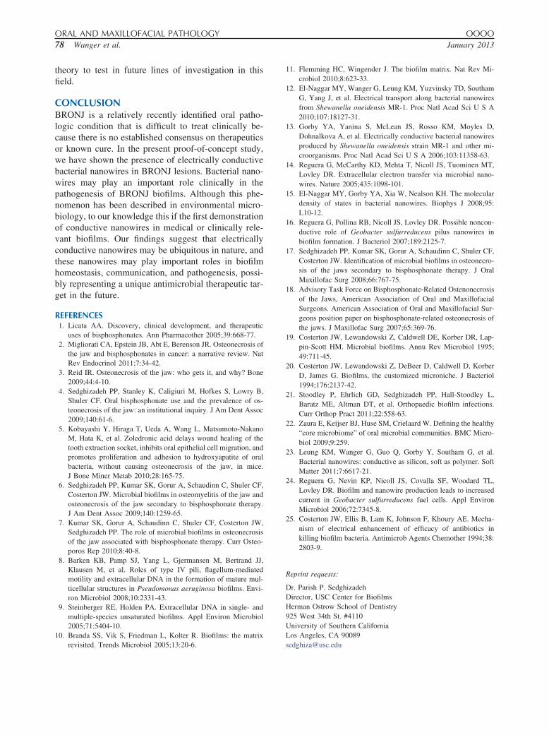
ORAL AND MAXILLOFACIAL PATHOLOGY OOOO78 Wanger et al. January 2013
theory to test in future lines of investigation in thisfield.
CONCLUSIONBRONJ is a relatively recently identified oral patho-logic condition that is difficult to treat clinically be-cause there is no established consensus on therapeuticsor known cure. In the present proof-of-concept study,we have shown the presence of electrically conductivebacterial nanowires in BRONJ lesions. Bacterial nano-wires may play an important role clinically in thepathogenesis of BRONJ biofilms. Although this phe-nomenon has been described in environmental micro-biology, to our knowledge this if the first demonstrationof conductive nanowires in medical or clinically rele-vant biofilms. Our findings suggest that electricallyconductive nanowires may be ubiquitous in nature, andthese nanowires may play important roles in biofilmhomeostasis, communication, and pathogenesis, possi-bly representing a unique antimicrobial therapeutic tar-get in the future.
REFERENCES1. Licata AA. Discovery, clinical development, and therapeutic
uses of bisphosphonates. Ann Pharmacother 2005;39:668-77.2. Migliorati CA, Epstein JB, Abt E, Berenson JR. Osteonecrosis of
the jaw and bisphosphonates in cancer: a narrative review. NatRev Endocrinol 2011;7:34-42.
3. Reid IR. Osteonecrosis of the jaw: who gets it, and why? Bone2009;44:4-10.
4. Sedghizadeh PP, Stanley K, Caligiuri M, Hofkes S, Lowry B,Shuler CF. Oral bisphosphonate use and the prevalence of os-teonecrosis of the jaw: an institutional inquiry. J Am Dent Assoc2009;140:61-6.
5. Kobayashi Y, Hiraga T, Ueda A, Wang L, Matsumoto-NakanoM, Hata K, et al. Zoledronic acid delays wound healing of thetooth extraction socket, inhibits oral epithelial cell migration, andpromotes proliferation and adhesion to hydroxyapatite of oralbacteria, without causing osteonecrosis of the jaw, in mice.J Bone Miner Metab 2010;28:165-75.
6. Sedghizadeh PP, Kumar SK, Gorur A, Schaudinn C, Shuler CF,Costerton JW. Microbial biofilms in osteomyelitis of the jaw andosteonecrosis of the jaw secondary to bisphosphonate therapy.J Am Dent Assoc 2009;140:1259-65.
7. Kumar SK, Gorur A, Schaudinn C, Shuler CF, Costerton JW,Sedghizadeh PP. The role of microbial biofilms in osteonecrosisof the jaw associated with bisphosphonate therapy. Curr Osteo-poros Rep 2010;8:40-8.
8. Barken KB, Pamp SJ, Yang L, Gjermansen M, Bertrand JJ,Klausen M, et al. Roles of type IV pili, flagellum-mediatedmotility and extracellular DNA in the formation of mature mul-ticellular structures in Pseudomonas aeruginosa biofilms. Envi-ron Microbiol 2008;10:2331-43.
9. Steinberger RE, Holden PA. Extracellular DNA in single- andmultiple-species unsaturated biofilms. Appl Environ Microbiol2005;71:5404-10.
10. Branda SS, Vik S, Friedman L, Kolter R. Biofilms: the matrix
revisited. Trends Microbiol 2005;13:20-6.11. Flemming HC, Wingender J. The biofilm matrix. Nat Rev Mi-crobiol 2010;8:623-33.
12. El-Naggar MY, Wanger G, Leung KM, Yuzvinsky TD, SouthamG, Yang J, et al. Electrical transport along bacterial nanowiresfrom Shewanella oneidensis MR-1. Proc Natl Acad Sci U S A2010;107:18127-31.
13. Gorby YA, Yanina S, McLean JS, Rosso KM, Moyles D,Dohnalkova A, et al. Electrically conductive bacterial nanowiresproduced by Shewanella oneidensis strain MR-1 and other mi-croorganisms. Proc Natl Acad Sci U S A 2006;103:11358-63.
14. Reguera G, McCarthy KD, Mehta T, Nicoll JS, Tuominen MT,Lovley DR. Extracellular electron transfer via microbial nano-wires. Nature 2005;435:1098-101.
15. El-Naggar MY, Gorby YA, Xia W, Nealson KH. The moleculardensity of states in bacterial nanowires. Biophys J 2008;95:L10-12.
16. Reguera G, Pollina RB, Nicoll JS, Lovley DR. Possible noncon-ductive role of Geobacter sulfurreducens pilus nanowires inbiofilm formation. J Bacteriol 2007;189:2125-7.
17. Sedghizadeh PP, Kumar SK, Gorur A, Schaudinn C, Shuler CF,Costerton JW. Identification of microbial biofilms in osteonecro-sis of the jaws secondary to bisphosphonate therapy. J OralMaxillofac Surg 2008;66:767-75.
18. Advisory Task Force on Bisphosphonate-Related Ostenonecrosisof the Jaws, American Association of Oral and MaxillofacialSurgeons. American Association of Oral and Maxillofacial Sur-geons position paper on bisphosphonate-related osteonecrosis ofthe jaws. J Maxillofac Surg 2007;65:369-76.
19. Costerton JW, Lewandowski Z, Caldwell DE, Korber DR, Lap-pin-Scott HM. Microbial biofilms. Annu Rev Microbiol 1995;49:711-45.
20. Costerton JW, Lewandowski Z, DeBeer D, Caldwell D, KorberD, James G. Biofilms, the customized microniche. J Bacteriol1994;176:2137-42.
21. Stoodley P, Ehrlich GD, Sedghizadeh PP, Hall-Stoodley L,Baratz ME, Altman DT, et al. Orthopaedic biofilm infections.Curr Orthop Pract 2011;22:558-63.
22. Zaura E, Keijser BJ, Huse SM, Crielaard W. Defining the healthy“core microbiome” of oral microbial communities. BMC Micro-biol 2009;9:259.
23. Leung KM, Wanger G, Guo Q, Gorby Y, Southam G, et al.Bacterial nanowires: conductive as silicon, soft as polymer. SoftMatter 2011;7:6617-21.
24. Reguera G, Nevin KP, Nicoll JS, Covalla SF, Woodard TL,Lovley DR. Biofilm and nanowire production leads to increasedcurrent in Geobacter sulfurreducens fuel cells. Appl EnvironMicrobiol 2006;72:7345-8.
25. Costerton JW, Ellis B, Lam K, Johnson F, Khoury AE. Mecha-nism of electrical enhancement of efficacy of antibiotics inkilling biofilm bacteria. Antimicrob Agents Chemother 1994;38:2803-9.
Reprint requests:
Dr. Parish P. SedghizadehDirector, USC Center for BiofilmsHerman Ostrow School of Dentistry925 West 34th St. #4110University of Southern CaliforniaLos Angeles, CA 90089
[email protected]