Embed Size (px)
Citation preview

Proper 12 Lead Electrode Placement
Paramedic Assistant Review

Let’s keep this brief. This will be review for most individuals. However, it is all to easy to fall into habitual inattention to this important detail. Take the next 6 or 7 minutes to hone your accuracy. Perhaps you may even learn something new…
Introduction

1. Review proper placement for limb and precordial leads.
2. Discuss special lead placement for right sided and posterior myocardial infarction.
3. Discuss troubleshooting methods for artifact.
Objectives

Proper site preparation can result in a clear reading and reduce artifact. ● Shave excess hair.● Avoid placement over major muscle
masses, tendons or bones.
Site Preparation
● For oily skin, clean with an alcohol swab.● Dry site with a brisk rub.● Ensure electrodes are not expired and
gel is intact.
Site Preparation (Continued)
● ECG monitors were first used in coronary care units.● Artifact from patient movement had an alarming
resemblance to life threatening arrhythmias. ● To reduce false alarms, CCU nurses moved the ECG
leads from the arms to the chest. ● This worked well and still identified arrhythmias
properly.● The placement of limb electrodes on the chest has
endured to today, however...
Limb Leads

Limb leads are designed to go on the LIMBS for accurate diagnosis of myocardial infarction and other pathologic conditions.
Limb Leads
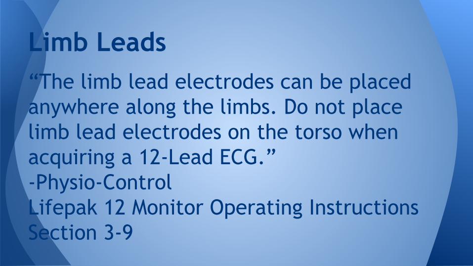
“The limb lead electrodes can be placed anywhere along the limbs. Do not place limb lead electrodes on the torso when acquiring a 12-Lead ECG.” -Physio-Control Lifepak 12 Monitor Operating Instructions Section 3-9
Limb Leads

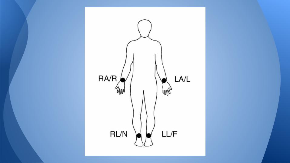
Limb leads can be placed anywhere distal to the patient’s shoulders and hips.● White: Right Arm● Black: Left Arm● Green: Right Leg● Red: Left Leg
Limb Leads

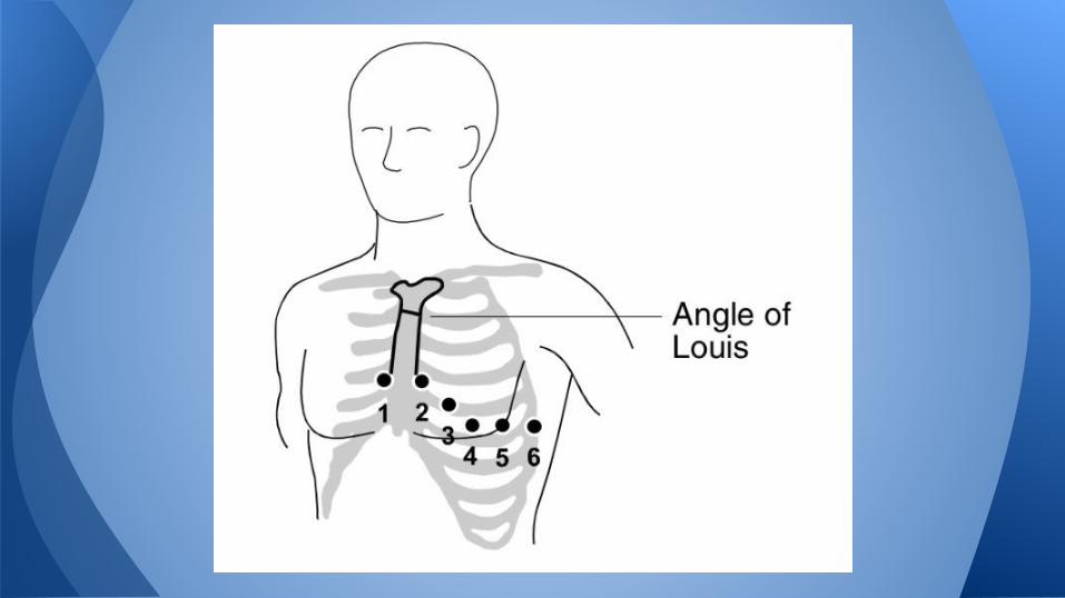
● Proper placement of the precordial leads requires identification of anatomical landmarks such as intercostal spaces.
● The most accurate way to begin this part of the procedure is to find the Angle of Louis (aka the Sternal Angle).
Precordial Leads

1. Place your finger at the notch at the top of the sternum
2. Move your finger slowly downward about 1.5 inches until you feel a slight horizontal ridge or elevation. This is the Angle of Louis where the manubrium joins the body of the sternum.
Precordial Leads: Finding V1
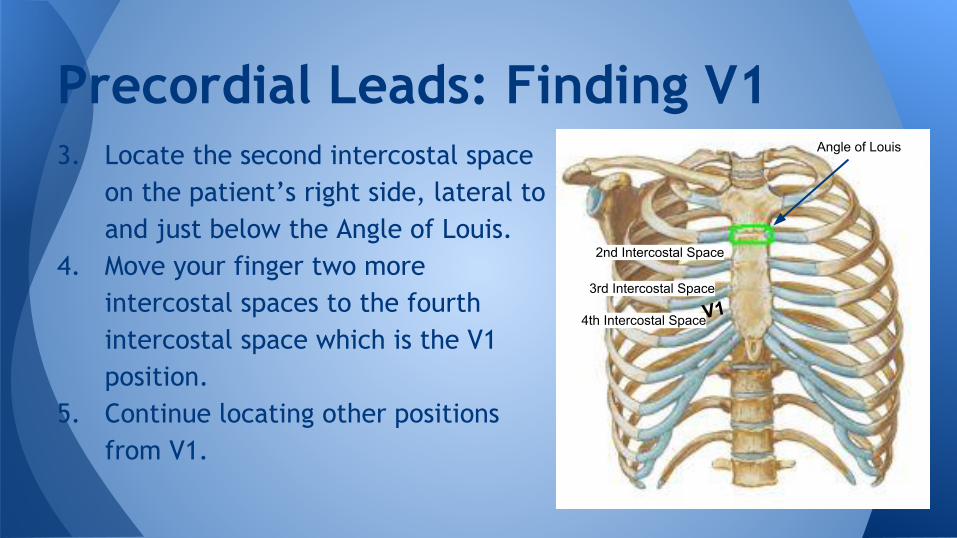
3. Locate the second intercostal space on the patient’s right side, lateral to and just below the Angle of Louis.
4. Move your finger two more intercostal spaces to the fourth intercostal space which is the V1 position.
5. Continue locating other positions from V1.
Precordial Leads: Finding V1
V1
Angle of Louis
2nd Intercostal Space
3rd Intercostal Space
4th Intercostal SpaceV1
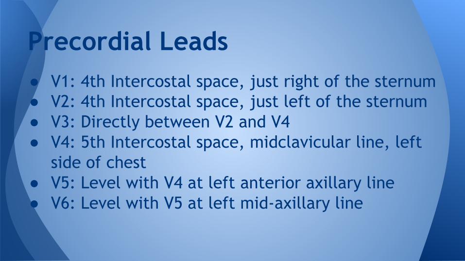
● V1: 4th Intercostal space, just right of the sternum● V2: 4th Intercostal space, just left of the sternum● V3: Directly between V2 and V4● V4: 5th Intercostal space, midclavicular line, left
side of chest● V5: Level with V4 at left anterior axillary line● V6: Level with V5 at left mid-axillary line
Precordial Leads

● In females, V3-V6 should be placed below the breast (Per Physio-Control).
● Never use the nipples as a reference point for locating the electrodes for men or women patients because nipple location can vary widely.
Precordial Leads

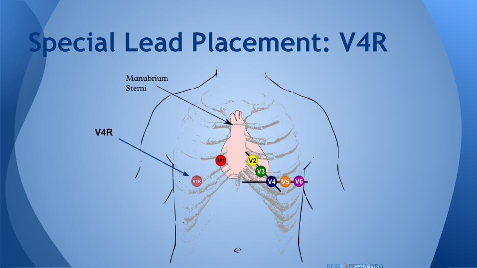
● Right Sided 12-Lead ECG● R sided 12-Lead is considered with an
inferior AMI pattern.● Identification of AMI on the R side of the
heart may significantly change ALS care.
Special Lead Placement: V4R
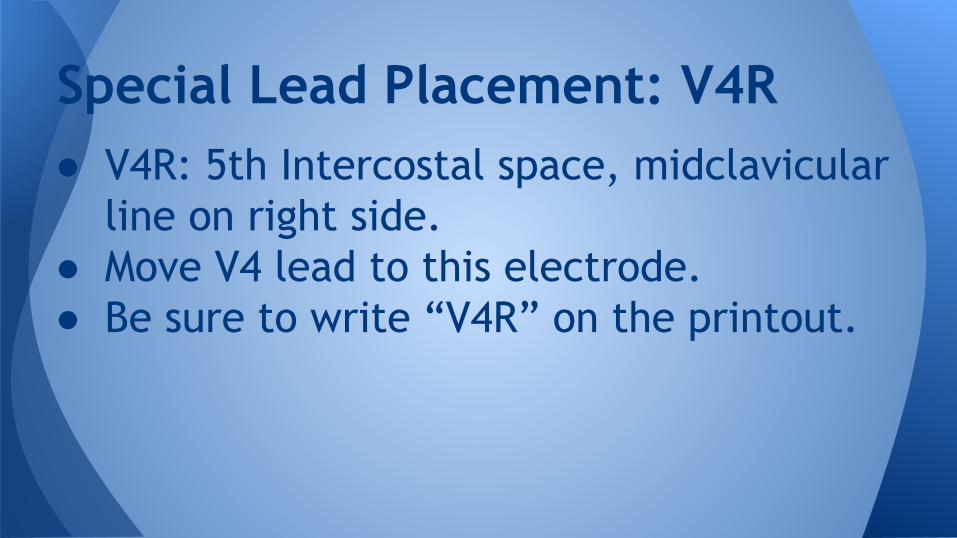
● V4R: 5th Intercostal space, midclavicular line on right side.
● Move V4 lead to this electrode. ● Be sure to write “V4R” on the printout.
Special Lead Placement: V4R
Special Lead Placement: V4R
V4R

● Posterior 12 Lead: Move V4, V5, V6 to posterior positions
● V7: Level with V6 at left posterior axillary line
● V8: Level with V7 at mid-scapular line● V9: Level with V8 just left of vertebral
line
Special Lead Placement: V7-V9

Special Lead Placement: V7-V9
● Ensure leads are properly connected.● Reposition cable to prevent electrodes
from pulling away from patient.
Troubleshooting Artifact

● Prepare the patient’s skin and apply new electrodes. Remember: ○ Shave excess hair.○ Dry diaphoretic skin.○ Cleaning the area with alcohol swab may
remove dead epidermal cells thus facilitating better electrode contact.
Troubleshooting Artifact

● Replace or reposition leads with artifact.○ Remember to avoid placement over major
muscle masses, tendons or bones.● Encourage patient to lie quietly.● Support the patient’s limbs.
Troubleshooting Artifact
● Consider stopping vehicle motion.● Check expiration date and proper seal on
electrode packaging- electrode may not function properly if the gel adhesive has dried out.
● Inspect all cables and replace if necessary.
Troubleshooting Artifact

● Proper site preparation will help avoid artifact.
● For accurate diagnosis of AMI, limb leads must be placed on the limbs.
● Place precordial leads according to anatomical landmarks.
Summary

This presentation was made with information gathered from: ● 12-Lead ECG for Acute and Critical Care Providers by Bob Page● 12-Lead ECG The Art of Interpretation by Garcia and Holtz● Lifepak 12 Operating Instructions by Physio-Control.
References

The End