Embed Size (px)
Citation preview
Electromagnetic Radiation and Autism: D.Klinghardt, Emerson College, 2018 Electrosmog and it’s Destructive Effect on the Brain, our Genome, Proteome and Microbiome of the Unborn Child Electrosmog refers to our exposure to the sum total of all man-made electric fields, magnetic fields, radiowaves, TV broadcasting, home lighting and all other sources of
radiation

Mediators of Inflammation Volume 2014 (2014), Article ID 924184, 14 pages http://dx.doi.org/10.1155/2014/924184
Clinical Study Metabolic and Genetic Screening of Electromagnetic Hypersensitive Subjects as a Feasible Tool for Diagnostics and Intervention Chiara De Luca,1,2 Jeffrey Chung Sheun Thai,3 Desanka Raskovic,4 Eleonora Cesareo,4 Daniela Caccamo,5 Arseny Trukhanov,2 and Liudmila Korkina1,2
Abstract
Growingnumbersof“electromagnetichypersensitive”(EHS)peopleworldwideself-reportseverelydisabling,multiorgan,non-specificsymptomswhenexposedtolow-doseelectromagneticradiations,oftenassociatedwithsymptomsofmultiplechemicalsensitivity(MCS)and/orotherenvironmental“sensitivity-relatedillnesses”(SRI).Thisclusterofchronicinflammatorydisordersstilllacksvalidatedpathogeneticmechanism,diagnosticbiomarkers,andmanagementguidelines.WehypothesizedthatSRI,notbeingmerelypsychogenic,mayshareorganicdeterminantsofimpaireddetoxificationofcommonphysic-chemicalstressors.BasedonourpreviousMCSstudies,wetestedapanelof12metabolicbloodredox-relatedparametersandofselecteddrug-metabolizing-enzymegenepolymorphisms,on153EHS,147MCS,and132controlItalians,confirmingMCSaltered(plessthan0.05–0.0001)glutathione-(GSH),GSH-peroxidase/S-transferase,andcatalaseerythrocyteactivities.Wefirstdescribedcomparable—thoughmilder—metabolicpro-oxidant/proinflammatoryalterationsinEHSwithdistinctivelyincreasedplasmacoenzyme-Q10oxidationratio.Severedepletionoferythrocytemembranepolyunsaturatedfattyacidswithincreasedω6/ω3ratiowasconfirmedinMCS,butnotinEHS.Wealsoidentifiedsignificantly(p=0.003)altereddistribution-versus-controloftheCYP2C19*1/*2SNPvariantsinEHS,anda9.7-foldincreasedrisk(OR:95%C.1=13–74.5)ofdevelopingEHSforthehaplotype(null)GSTT1+(null)GSTM1variants.Altogether,resultsonMCSandEHSstrengthenourproposaltoadoptthisbloodmetabolic/geneticbiomarkers’panelassuitablediagnostictoolforSRI.
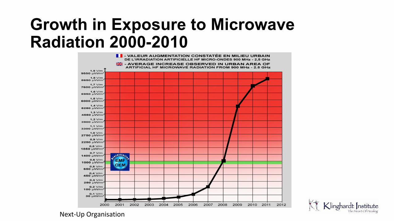
Growth in Exposure to Microwave Radiation 2000-2010
Next-UpOrganisation
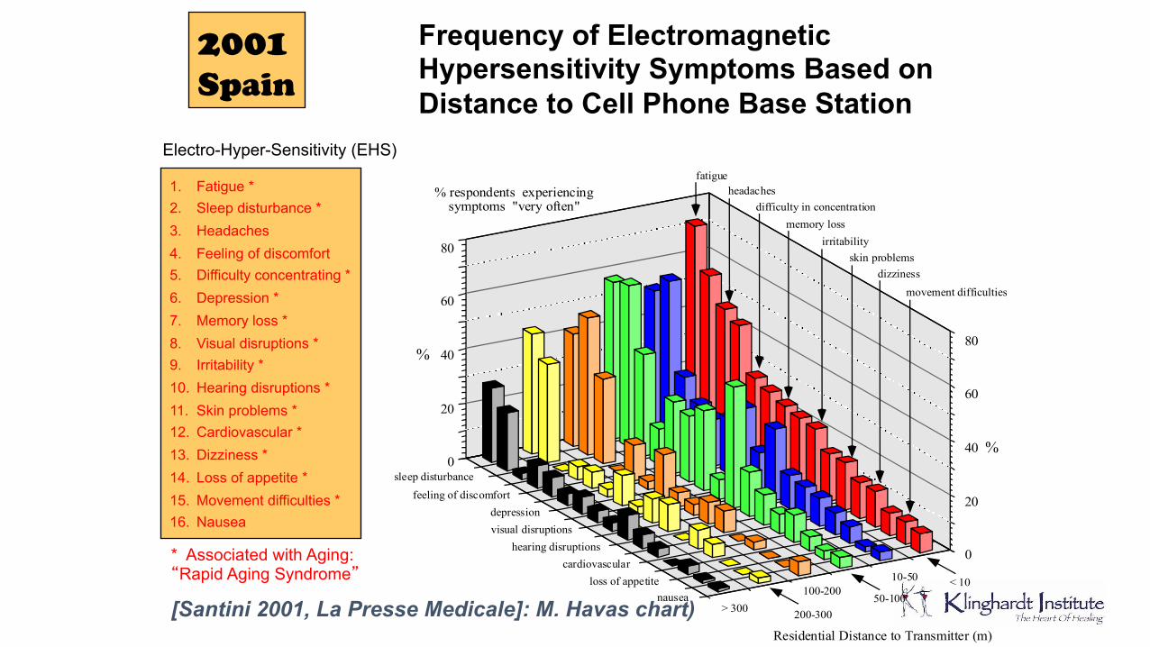
Frequency of Electromagnetic Hypersensitivity Symptoms Based on Distance to Cell Phone Base Station
2001 Spain
fatigue
sleep disturbance
headaches
feeling of discomfort
difficulty in concentration
depression
memory loss
visual disruptions
irritabilityskin problems
dizziness
movement difficulties
hearing disruptionscardiovascular
nausealoss of appetite < 1010-50
50-100100-200
200-300 > 300
% respondents experiencingsymptoms "very often"
80
60
40
20
0
80
60
40
20
0
%
%
Residential Distance to Transmitter (m)
Electro-Hyper-Sensitivity (EHS)
1. Fatigue * 2. Sleep disturbance * 3. Headaches 4. Feeling of discomfort 5. Difficulty concentrating * 6. Depression * 7. Memory loss * 8. Visual disruptions * 9. Irritability * 10. Hearing disruptions * 11. Skin problems * 12. Cardiovascular * 13. Dizziness * 14. Loss of appetite * 15. Movement difficulties * 16. Nausea
* Associated with Aging: “Rapid Aging Syndrome”
[Santini 2001, La Presse Medicale]: M. Havas chart)
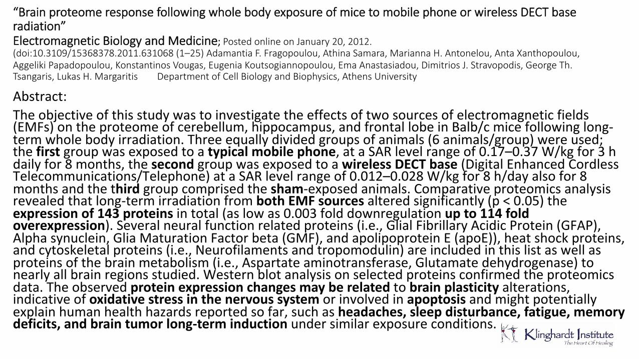
“Brain proteome response following whole body exposure of mice to mobile phone or wireless DECT base radiation” Electromagnetic Biology and Medicine; Posted online on January 20, 2012. (doi:10.3109/15368378.2011.631068 (1–25) Adamantia F. Fragopoulou, Athina Samara, Marianna H. Antonelou, Anta Xanthopoulou, Aggeliki Papadopoulou, Konstantinos Vougas, Eugenia Koutsogiannopoulou, Ema Anastasiadou, Dimitrios J. Stravopodis, George Th. Tsangaris, Lukas H. Margaritis Department of Cell Biology and Biophysics, Athens University Abstract:Theobjectiveofthisstudywastoinvestigatetheeffectsoftwosourcesofelectromagneticfields(EMFs)ontheproteomeofcerebellum,hippocampus,andfrontallobeinBalb/cmicefollowinglong-termwholebodyirradiation.Threeequallydividedgroupsofanimals(6animals/group)wereused;thefirstgroupwasexposedtoatypical mobile phone,ataSARlevelrangeof0.17–0.37W/kgfor3hdailyfor8months,thesecondgroupwasexposedtoawireless DECT base(DigitalEnhancedCordlessTelecommunications/Telephone)ataSARlevelrangeof0.012–0.028W/kgfor8h/dayalsofor8monthsandthethirdgroupcomprisedthesham-exposedanimals.Comparativeproteomicsanalysisrevealedthatlong-termirradiationfromboth EMF sources alteredsignificantly(p<0.05)theexpression of 143 proteinsintotal(aslowas0.003folddownregulationup to 114fold overexpression).Severalneuralfunctionrelatedproteins(i.e.,GlialFibrillaryAcidicProtein(GFAP),Alphasynuclein,GliaMaturationFactorbeta(GMF),andapolipoproteinE(apoE)),heatshockproteins,andcytoskeletalproteins(i.e.,Neurofilamentsandtropomodulin)areincludedinthislistaswellasproteinsofthebrainmetabolism(i.e.,Aspartateaminotransferase,Glutamatedehydrogenase)tonearlyallbrainregionsstudied.Westernblotanalysisonselectedproteinsconfirmedtheproteomicsdata.Theobservedproteinexpression changes may be relatedtobrain plasticityalterations,indicativeofoxidative stress in the nervoussystemorinvolvedinapoptosisandmightpotentiallyexplainhumanhealthhazardsreportedsofar,suchasheadaches, sleep disturbance, fatigue, memory deficits, and brain tumor long-term inductionundersimilarexposureconditions.
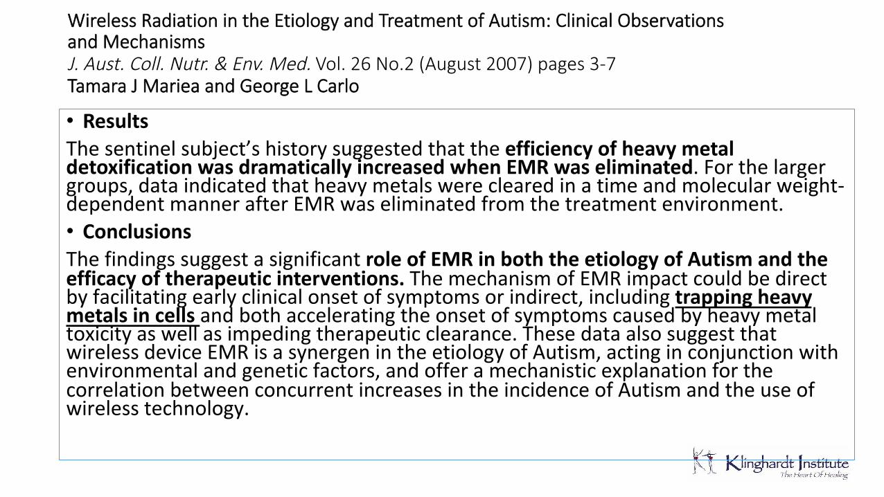
Wireless Radiation in the Etiology and Treatment of Autism: Clinical Observations and Mechanisms J. Aust. Coll. Nutr. & Env. Med. Vol. 26 No.2 (August 2007) pages 3-7 Tamara J Mariea and George L Carlo • ResultsThesentinelsubject’shistorysuggestedthattheefficiency of heavy metal detoxification was dramatically increased when EMR was eliminated.Forthelargergroups,dataindicatedthatheavymetalswereclearedinatimeandmolecularweight-dependentmannerafterEMRwaseliminatedfromthetreatmentenvironment.• ConclusionsThefindingssuggestasignificantrole of EMR in both the etiology of Autism and the efficacy of therapeutic interventions. ThemechanismofEMRimpactcouldbedirectbyfacilitatingearlyclinicalonsetofsymptomsorindirect,includingtrapping heavy metals in cells andbothacceleratingtheonsetofsymptomscausedbyheavymetaltoxicityaswellasimpedingtherapeuticclearance.ThesedataalsosuggestthatwirelessdeviceEMRisasynergenintheetiologyofAutism,actinginconjunctionwithenvironmentalandgeneticfactors,andofferamechanisticexplanationforthecorrelationbetweenconcurrentincreasesintheincidenceofAutismandtheuseofwirelesstechnology.
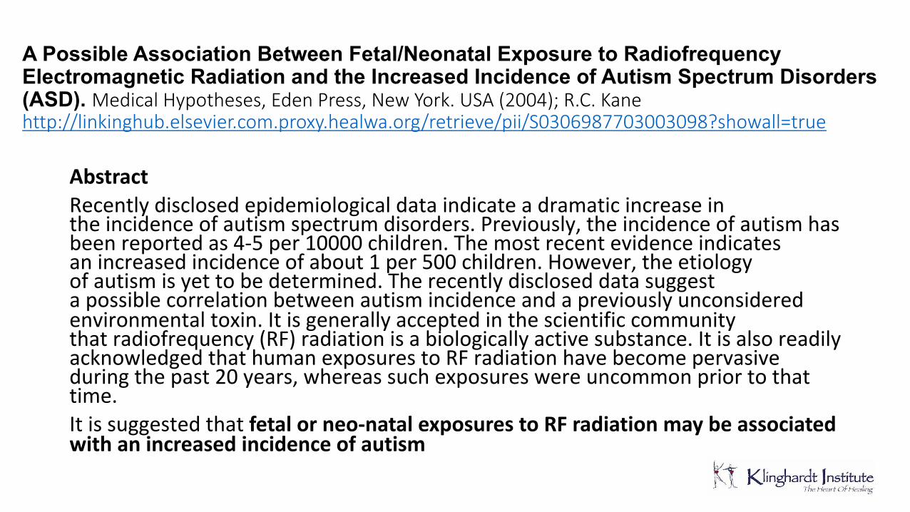
A Possible Association Between Fetal/Neonatal Exposure to Radiofrequency Electromagnetic Radiation and the Increased Incidence of Autism Spectrum Disorders (ASD). Medical Hypotheses, Eden Press, New York. USA (2004); R.C. Kane http://linkinghub.elsevier.com.proxy.healwa.org/retrieve/pii/S0306987703003098?showall=true
Abstract Recentlydisclosedepidemiologicaldataindicateadramaticincreaseintheincidenceofautismspectrumdisorders.Previously,theincidenceofautismhasbeenreportedas4-5per10000children.Themostrecentevidenceindicatesanincreasedincidenceofabout1per500children.However,theetiologyofautismisyettobedetermined.Therecentlydiscloseddatasuggestapossiblecorrelationbetweenautismincidenceandapreviouslyunconsideredenvironmentaltoxin.Itisgenerallyacceptedinthescientificcommunitythatradiofrequency(RF)radiationisabiologicallyactivesubstance.ItisalsoreadilyacknowledgedthathumanexposurestoRFradiationhavebecomepervasiveduringthepast20years,whereassuchexposureswereuncommonpriortothattime.Itissuggestedthatfetal or neo-natal exposures to RF radiation may be associated with an increased incidence of autism
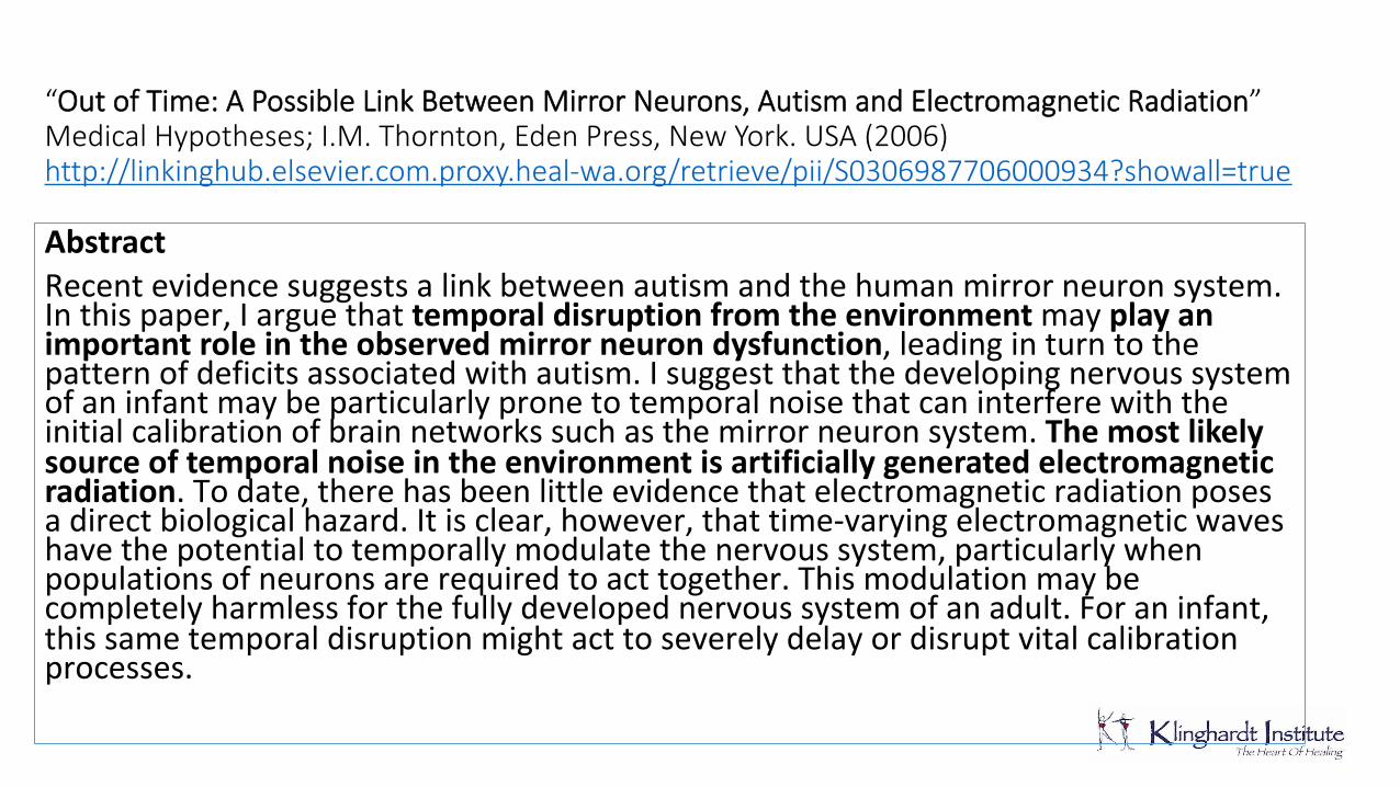
“Out of Time: A Possible Link Between Mirror Neurons, Autism and Electromagnetic Radiation” Medical Hypotheses; I.M. Thornton, Eden Press, New York. USA (2006) http://linkinghub.elsevier.com.proxy.heal-wa.org/retrieve/pii/S0306987706000934?showall=true Abstract Recentevidencesuggestsalinkbetweenautismandthehumanmirrorneuronsystem.Inthispaper,Iarguethattemporaldisruption from the environment mayplay an important role in the observed mirror neuron dysfunction,leadinginturntothepatternofdeficitsassociatedwithautism.Isuggestthatthedevelopingnervoussystemofaninfantmaybeparticularlypronetotemporalnoisethatcaninterferewiththeinitialcalibrationofbrainnetworkssuchasthemirrorneuronsystem.The most likely source of temporal noise in the environment is artificially generated electromagnetic radiation.Todate,therehasbeenlittleevidencethatelectromagneticradiationposesadirectbiologicalhazard.Itisclear,however,thattime-varyingelectromagneticwaveshavethepotentialtotemporallymodulatethenervoussystem,particularlywhenpopulationsofneuronsarerequiredtoacttogether.Thismodulationmaybecompletelyharmlessforthefullydevelopednervoussystemofanadult.Foraninfant,thissametemporaldisruptionmightacttoseverelydelayordisruptvitalcalibrationprocesses.
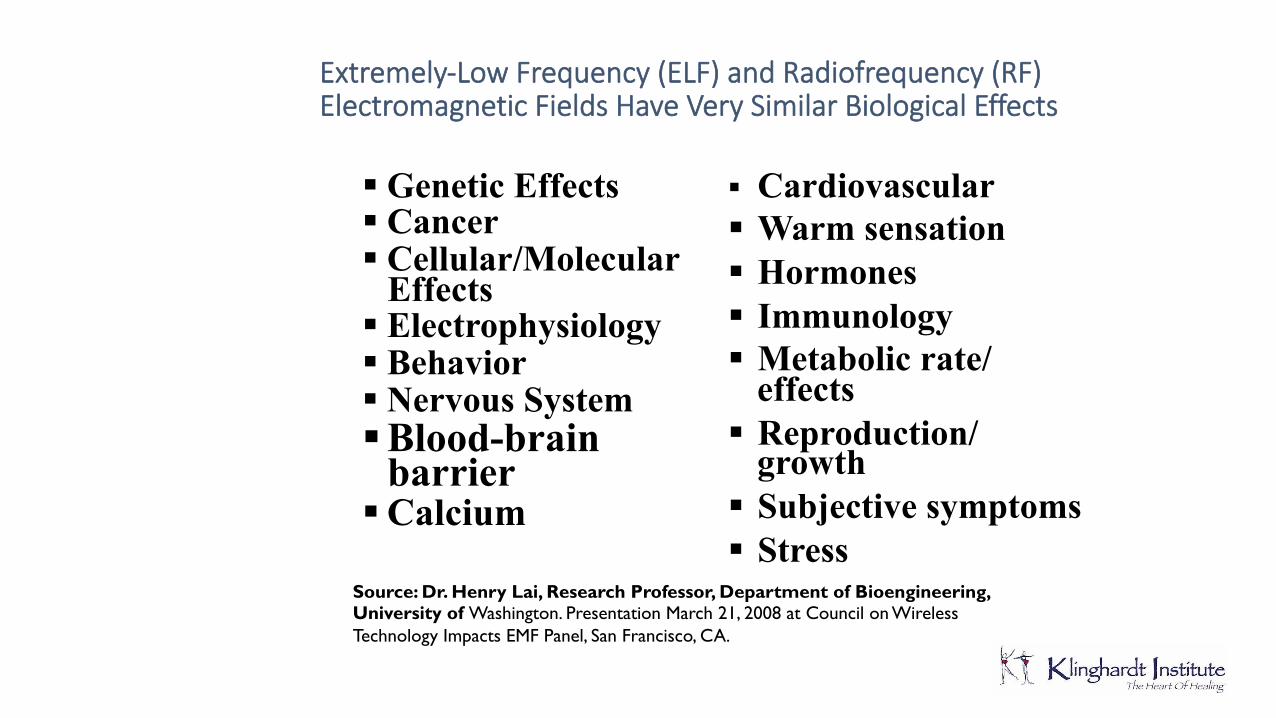
Extremely-Low Frequency (ELF) and Radiofrequency (RF) Electromagnetic Fields Have Very Similar Biological Effects
§ Genetic Effects § Cancer § Cellular/Molecular
Effects § Electrophysiology § Behavior § Nervous System § Blood-brain
barrier § Calcium
§ Cardiovascular § Warm sensation § Hormones § Immunology § Metabolic rate/
effects § Reproduction/
growth § Subjective symptoms § Stress
Source: Dr. Henry Lai, Research Professor, Department of Bioengineering, University of Washington. Presentation March 21, 2008 at Council on Wireless Technology Impacts EMF Panel, San Francisco, CA.
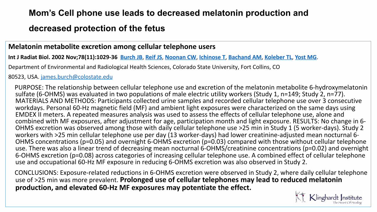
Mom’s Cell phone use leads to decreased melatonin production and
decreased protection of the fetus Melatonin metabolite excretion among cellular telephone users Int J Radiat Biol. 2002 Nov;78(11):1029-36 Burch JB,Reif JS,Noonan CW,Ichinose T,Bachand AM,Koleber TL,Yost MG.DepartmentofEnvironmentalandRadiologicalHealthSciences,ColoradoStateUniversity,FortCollins,CO80523,[email protected]
PURPOSE:Therelationshipbetweencellulartelephoneuseandexcretionofthemelatoninmetabolite6-hydroxymelatoninsulfate(6-OHMS)wasevaluatedintwopopulationsofmaleelectricutilityworkers(Study1,n=149;Study2,n=77).MATERIALSANDMETHODS:Participantscollectedurinesamplesandrecordedcellulartelephoneuseover3consecutiveworkdays.Personal60-Hzmagneticfield(MF)andambientlightexposureswerecharacterizedonthesamedaysusingEMDEXIImeters.Arepeatedmeasuresanalysiswasusedtoassesstheeffectsofcellulartelephoneuse,aloneandcombinedwithMFexposures,afteradjustmentforage,participationmonthandlightexposure.RESULTS:Nochangein6-OHMSexcretionwasobservedamongthosewithdailycellulartelephoneuse>25mininStudy1(5worker-days).Study2workerswith>25mincellulartelephoneuseperday(13worker-days)hadlowercreatinine-adjustedmeannocturnal6-OHMSconcentrations(p=0.05)andovernight6-OHMSexcretion(p=0.03)comparedwiththosewithoutcellulartelephoneuse.Therewasalsoalineartrendofdecreasingmeannocturnal6-OHMS/creatinineconcentrations(p=0.02)andovernight6-OHMSexcretion(p=0.08)acrosscategoriesofincreasingcellulartelephoneuse.Acombinedeffectofcellulartelephoneuseandoccupational60-HzMFexposureinreducing6-OHMSexcretionwasalsoobservedinStudy2.
CONCLUSIONS:Exposure-relatedreductionsin6-OHMSexcretionwereobservedinStudy2,wheredailycellulartelephoneuseof>25minwasmoreprevalent.Prolonged use of cellular telephones may lead to reduced melatonin production, and elevated 60-Hz MF exposures may potentiate the effect.
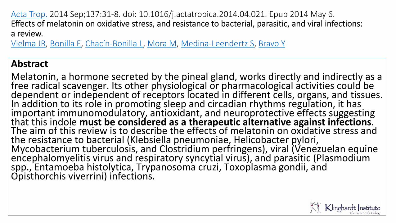
Acta Trop. 2014 Sep;137:31-8. doi: 10.1016/j.actatropica.2014.04.021. Epub 2014 May 6. Effects of melatonin on oxidative stress, and resistance to bacterial, parasitic, and viral infections: a review. Vielma JR, Bonilla E, Chacín-Bonilla L, Mora M, Medina-Leendertz S, Bravo Y Abstract Melatonin,ahormonesecretedbythepinealgland,worksdirectlyandindirectlyasafreeradicalscavenger.Itsotherphysiologicalorpharmacologicalactivitiescouldbedependentorindependentofreceptorslocatedindifferentcells,organs,andtissues.Inadditiontoitsroleinpromotingsleepandcircadianrhythmsregulation,ithasimportantimmunomodulatory,antioxidant,andneuroprotectiveeffectssuggestingthatthisindolemust be considered as a therapeutic alternative against infections.Theaimofthisreviewistodescribetheeffectsofmelatoninonoxidativestressandtheresistancetobacterial(Klebsiellapneumoniae,Helicobacterpylori,Mycobacteriumtuberculosis,andClostridiumperfringens),viral(Venezuelanequineencephalomyelitisvirusandrespiratorysyncytialvirus),andparasitic(Plasmodiumspp.,Entamoebahistolytica,Trypanosomacruzi,Toxoplasmagondii,andOpisthorchisviverrini)infections.
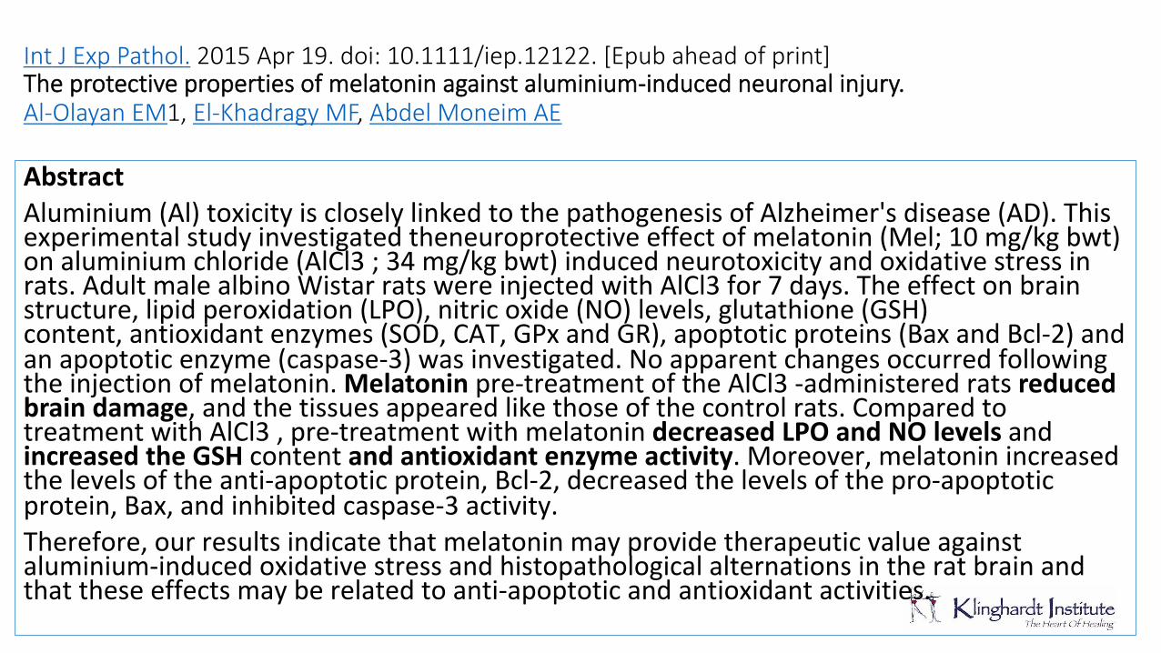
Int J Exp Pathol. 2015 Apr 19. doi: 10.1111/iep.12122. [Epub ahead of print] The protective properties of melatonin against aluminium-induced neuronal injury. Al-Olayan EM1, El-Khadragy MF, Abdel Moneim AE Abstract Aluminium(Al)toxicityiscloselylinkedtothepathogenesisofAlzheimer'sdisease(AD).Thisexperimentalstudyinvestigatedtheneuroprotectiveeffectofmelatonin(Mel;10mg/kgbwt)onaluminiumchloride(AlCl3;34mg/kgbwt)inducedneurotoxicityandoxidativestressinrats.AdultmalealbinoWistarratswereinjectedwithAlCl3for7days.Theeffectonbrainstructure,lipidperoxidation(LPO),nitricoxide(NO)levels,glutathione(GSH)content,antioxidantenzymes(SOD,CAT,GPxandGR),apoptoticproteins(BaxandBcl-2)andanapoptoticenzyme(caspase-3)wasinvestigated.Noapparentchangesoccurredfollowingtheinjectionofmelatonin.Melatoninpre-treatmentoftheAlCl3-administeredratsreduced brain damage,andthetissuesappearedlikethoseofthecontrolrats.ComparedtotreatmentwithAlCl3,pre-treatmentwithmelatonindecreased LPO and NO levels andincreased the GSH contentand antioxidant enzyme activity.Moreover,melatoninincreasedthelevelsoftheanti-apoptoticprotein,Bcl-2,decreasedthelevelsofthepro-apoptoticprotein,Bax,andinhibitedcaspase-3activity.Therefore,ourresultsindicatethatmelatoninmayprovidetherapeuticvalueagainstaluminium-inducedoxidativestressandhistopathologicalalternationsintheratbrainandthattheseeffectsmayberelatedtoanti-apoptoticandantioxidantactivities.
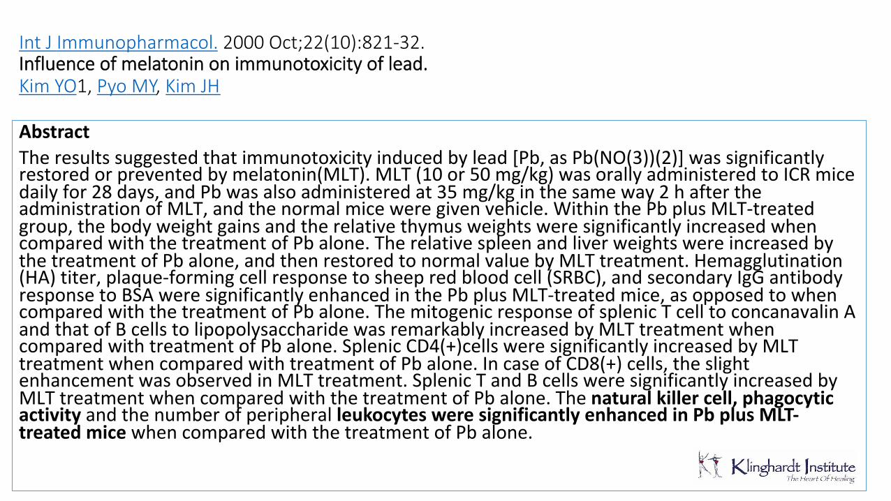
Int J Immunopharmacol. 2000 Oct;22(10):821-32. Influence of melatonin on immunotoxicity of lead. Kim YO1, Pyo MY, Kim JH
Abstract Theresultssuggestedthatimmunotoxicityinducedbylead[Pb,asPb(NO(3))(2)]wassignificantlyrestoredorpreventedbymelatonin(MLT).MLT(10or50mg/kg)wasorallyadministeredtoICRmicedailyfor28days,andPbwasalsoadministeredat35mg/kginthesameway2haftertheadministrationofMLT,andthenormalmiceweregivenvehicle.WithinthePbplusMLT-treatedgroup,thebodyweightgainsandtherelativethymusweightsweresignificantlyincreasedwhencomparedwiththetreatmentofPbalone.TherelativespleenandliverweightswereincreasedbythetreatmentofPbalone,andthenrestoredtonormalvaluebyMLTtreatment.Hemagglutination(HA)titer,plaque-formingcellresponsetosheepredbloodcell(SRBC),andsecondaryIgGantibodyresponsetoBSAweresignificantlyenhancedinthePbplusMLT-treatedmice,asopposedtowhencomparedwiththetreatmentofPbalone.ThemitogenicresponseofsplenicTcelltoconcanavalinAandthatofBcellstolipopolysaccharidewasremarkablyincreasedbyMLTtreatmentwhencomparedwithtreatmentofPbalone.SplenicCD4(+)cellsweresignificantlyincreasedbyMLTtreatmentwhencomparedwithtreatmentofPbalone.IncaseofCD8(+)cells,theslightenhancementwasobservedinMLTtreatment.SplenicTandBcellsweresignificantlyincreasedbyMLTtreatmentwhencomparedwiththetreatmentofPbalone.Thenatural killer cell, phagocytic activity andthenumberofperipheralleukocytes were significantly enhanced in Pb plus MLT-treated mice whencomparedwiththetreatmentofPbalone.
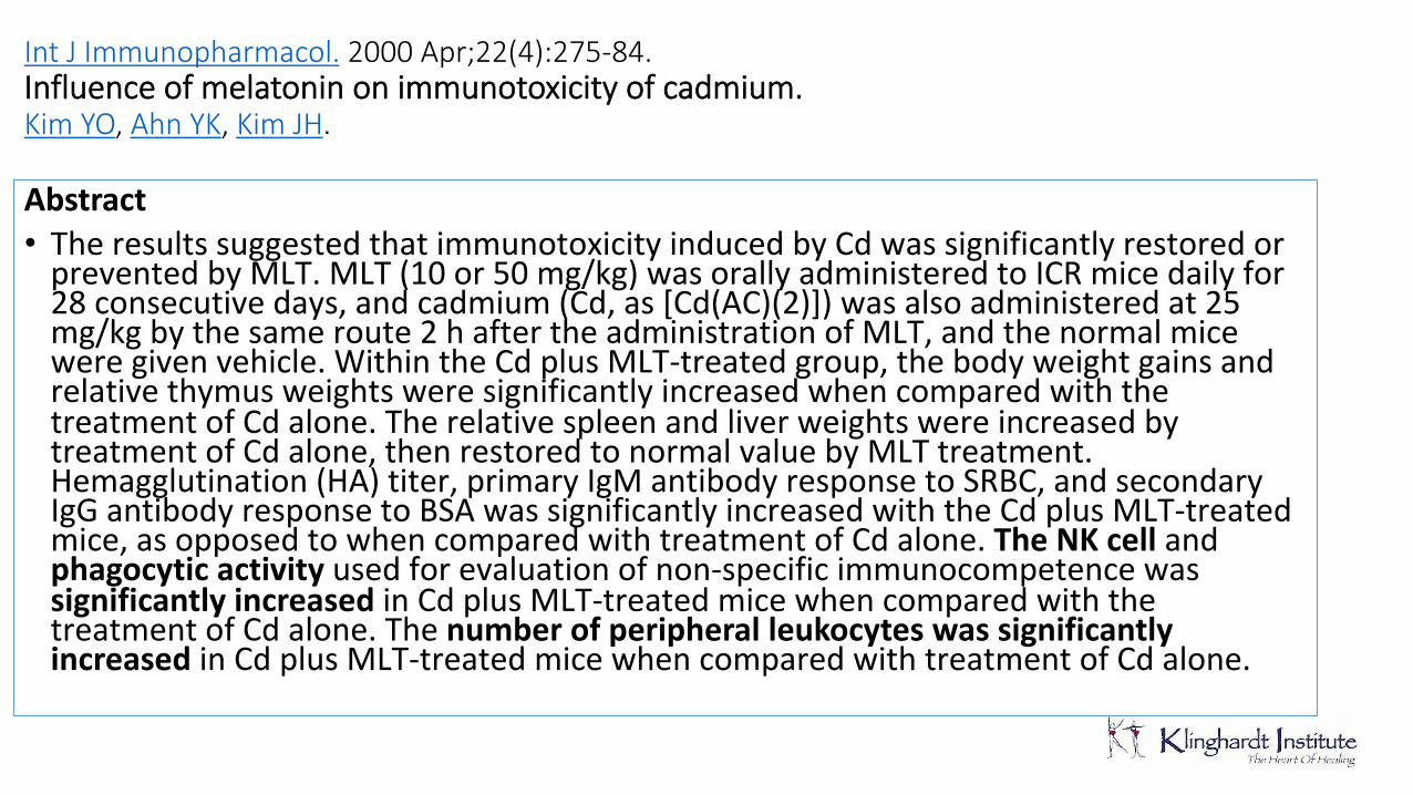
Int J Immunopharmacol. 2000 Apr;22(4):275-84. Influence of melatonin on immunotoxicity of cadmium. Kim YO, Ahn YK, Kim JH. Abstract • TheresultssuggestedthatimmunotoxicityinducedbyCdwassignificantlyrestoredorpreventedbyMLT.MLT(10or50mg/kg)wasorallyadministeredtoICRmicedailyfor28consecutivedays,andcadmium(Cd,as[Cd(AC)(2)])wasalsoadministeredat25mg/kgbythesameroute2haftertheadministrationofMLT,andthenormalmiceweregivenvehicle.WithintheCdplusMLT-treatedgroup,thebodyweightgainsandrelativethymusweightsweresignificantlyincreasedwhencomparedwiththetreatmentofCdalone.TherelativespleenandliverweightswereincreasedbytreatmentofCdalone,thenrestoredtonormalvaluebyMLTtreatment.Hemagglutination(HA)titer,primaryIgMantibodyresponsetoSRBC,andsecondaryIgGantibodyresponsetoBSAwassignificantlyincreasedwiththeCdplusMLT-treatedmice,asopposedtowhencomparedwithtreatmentofCdalone.The NK cell andphagocytic activity usedforevaluationofnon-specificimmunocompetencewassignificantly increased inCdplusMLT-treatedmicewhencomparedwiththetreatmentofCdalone.Thenumber of peripheral leukocytes was significantly increased inCdplusMLT-treatedmicewhencomparedwithtreatmentofCdalone.
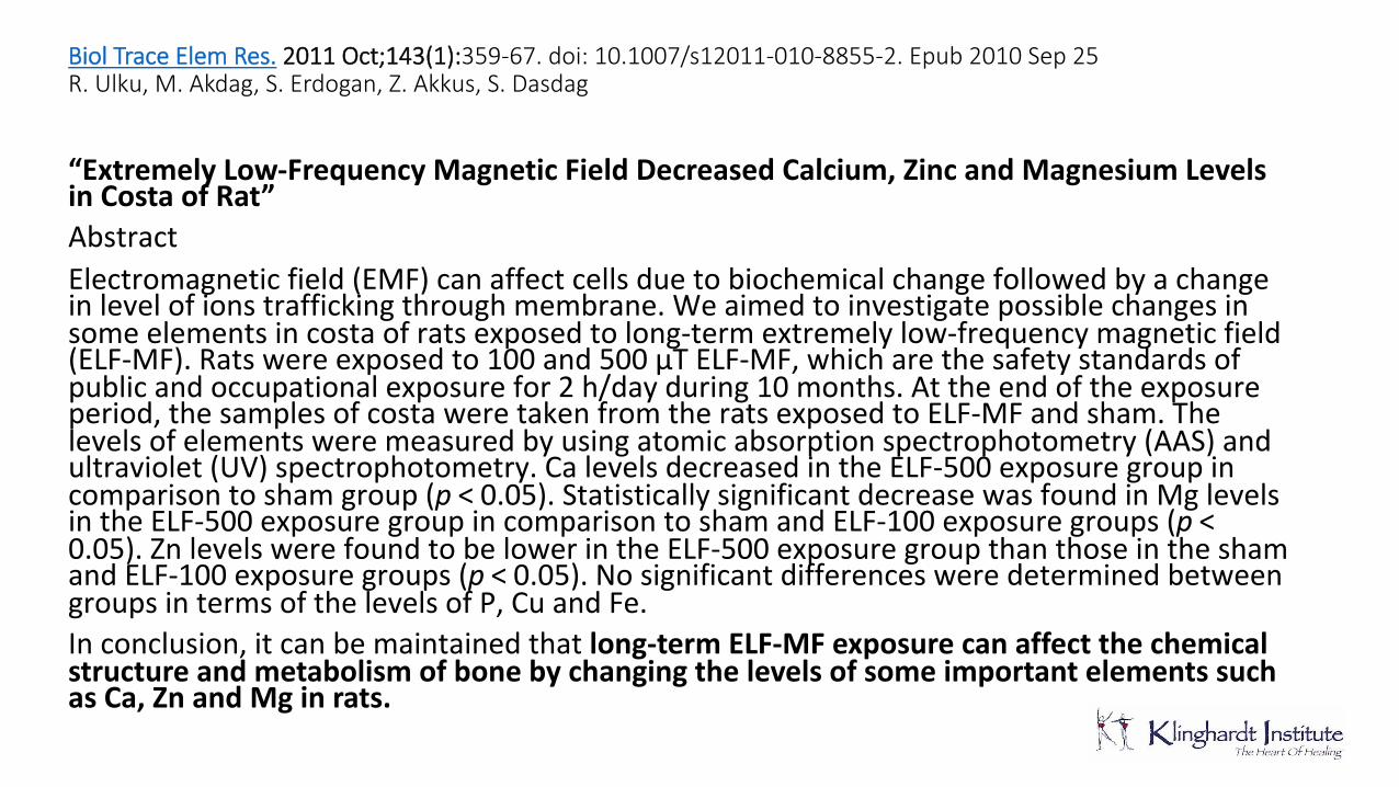
Biol Trace Elem Res. 2011 Oct;143(1):359-67. doi: 10.1007/s12011-010-8855-2. Epub 2010 Sep 25 R. Ulku, M. Akdag, S. Erdogan, Z. Akkus, S. Dasdag
“Extremely Low-Frequency Magnetic Field Decreased Calcium, Zinc and Magnesium Levels in Costa of Rat” AbstractElectromagneticfield(EMF)canaffectcellsduetobiochemicalchangefollowedbyachangeinlevelofionstraffickingthroughmembrane.Weaimedtoinvestigatepossiblechangesinsomeelementsincostaofratsexposedtolong-termextremelylow-frequencymagneticfield(ELF-MF).Ratswereexposedto100and500μTELF-MF,whicharethesafetystandardsofpublicandoccupationalexposurefor2h/dayduring10months.Attheendoftheexposureperiod,thesamplesofcostaweretakenfromtheratsexposedtoELF-MFandsham.Thelevelsofelementsweremeasuredbyusingatomicabsorptionspectrophotometry(AAS)andultraviolet(UV)spectrophotometry.CalevelsdecreasedintheELF-500exposuregroupincomparisontoshamgroup(p < 0.05).StatisticallysignificantdecreasewasfoundinMglevelsintheELF-500exposuregroupincomparisontoshamandELF-100exposuregroups(p < 0.05).ZnlevelswerefoundtobelowerintheELF-500exposuregroupthanthoseintheshamandELF-100exposuregroups(p < 0.05).NosignificantdifferencesweredeterminedbetweengroupsintermsofthelevelsofP,CuandFe.Inconclusion,itcanbemaintainedthatlong-term ELF-MF exposure can affect the chemical structure and metabolism of bone by changing the levels of some important elements such as Ca, Zn and Mg in rats.
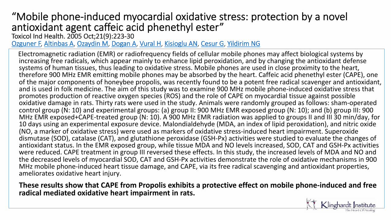
“Mobile phone-induced myocardial oxidative stress: protection by a novel antioxidant agent caffeic acid phenethyl ester” Toxicol Ind Health. 2005 Oct;21(9):223-30 Ozguner F, Altinbas A, Ozaydin M, Dogan A, Vural H, Kisioglu AN, Cesur G, Yildirim NG Electromagneticradiation(EMR)orradiofrequencyfieldsofcellularmobilephonesmayaffectbiologicalsystemsbyincreasingfreeradicals,whichappearmainlytoenhancelipidperoxidation,andbychangingtheantioxidantdefensesystemsofhumantissues,thusleadingtooxidativestress.Mobilephonesareusedincloseproximitytotheheart,therefore900MHzEMRemittingmobilephonesmaybeabsorbedbytheheart.Caffeicacidphenethylester(CAPE),oneofthemajorcomponentsofhoneybeepropolis,wasrecentlyfoundtobeapotentfreeradicalscavengerandantioxidant,andisusedinfolkmedicine.Theaimofthisstudywastoexamine900MHzmobilephone-inducedoxidativestressthatpromotesproductionofreactiveoxygenspecies(ROS)andtheroleofCAPEonmyocardialtissueagainstpossibleoxidativedamageinrats.Thirtyratswereusedinthestudy.Animalswererandomlygroupedasfollows:sham-operatedcontrolgroup(N:10)andexperimentalgroups:(a)groupII:900MHzEMRexposedgroup(N:10);and(b)groupIII:900MHzEMRexposed+CAPE-treatedgroup(N:10).A900MHzEMRradiationwasappliedtogroupsIIandIII30min/day,for10daysusinganexperimentalexposuredevice.Malondialdehyde(MDA,anindexoflipidperoxidation),andnitricoxide(NO,amarkerofoxidativestress)wereusedasmarkersofoxidativestress-inducedheartimpairment.Superoxidedismutase(SOD),catalase(CAT),andglutathioneperoxidase(GSH-Px)activitieswerestudiedtoevaluatethechangesofantioxidantstatus.IntheEMRexposedgroup,whiletissueMDAandNOlevelsincreased,SOD,CATandGSH-Pxactivitieswerereduced.CAPEtreatmentingroupIIIreversedtheseeffects.Inthisstudy,theincreasedlevelsofMDAandNOandthedecreasedlevelsofmyocardialSOD,CATandGSH-Pxactivitiesdemonstratetheroleofoxidativemechanismsin900MHzmobilephone-inducedhearttissuedamage,andCAPE,viaitsfreeradicalscavengingandantioxidantproperties,amelioratesoxidativeheartinjury.
These results show that CAPE from Propolis exhibits a protective effect on mobile phone-induced and free radical mediated oxidative heart impairment in rats.
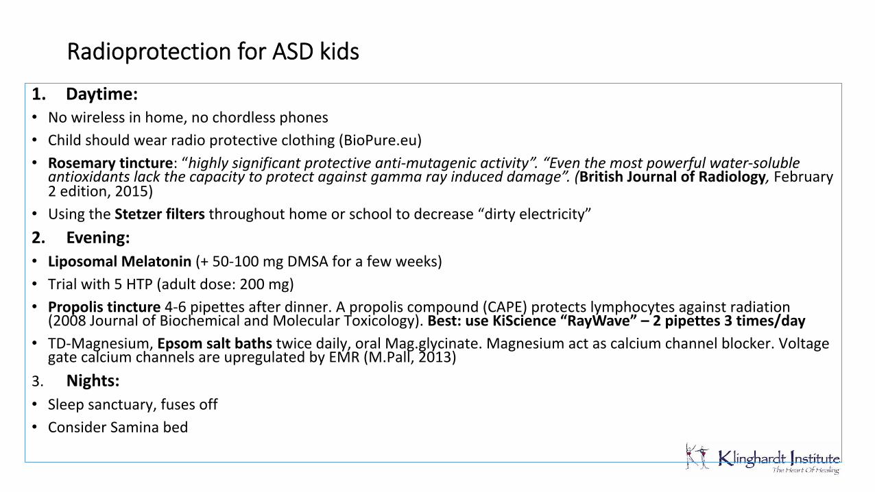
Radioprotection for ASD kids
1. Daytime: • Nowirelessinhome,nochordlessphones• Childshouldwearradioprotectiveclothing(BioPure.eu)• Rosemary tincture:“highly significant protective anti-mutagenic activity”. “Even the most powerful water-soluble
antioxidants lack the capacity to protect against gamma ray induced damage”. (British Journal of Radiology,February2edition,2015)
• UsingtheStetzer filters throughouthomeorschooltodecrease“dirtyelectricity”2. Evening: • Liposomal Melatonin (+50-100mgDMSAforafewweeks)• Trialwith5HTP(adultdose:200mg)• Propolis tincture 4-6pipettesafterdinner.Apropolis compound(CAPE)protectslymphocytesagainstradiation(2008JournalofBiochemicalandMolecularToxicology).Best: use KiScience “RayWave” – 2 pipettes 3 times/day
• TD-Magnesium,Epsom salt baths twicedaily,oralMag.glycinate.Magnesiumactascalciumchannelblocker.VoltagegatecalciumchannelsareupregulatedbyEMR(M.Pall,2013)
3.Nights: • Sleepsanctuary,fusesoff• ConsiderSaminabed
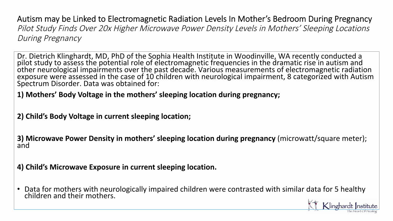
Autism may be Linked to Electromagnetic Radiation Levels In Mother’s Bedroom During Pregnancy Pilot Study Finds Over 20x Higher Microwave Power Density Levels in Mothers’ Sleeping Locations During Pregnancy Dr.DietrichKlinghardt,MD,PhDoftheSophiaHealthInstituteinWoodinville,WArecentlyconductedapilotstudytoassessthepotentialroleofelectromagneticfrequenciesinthedramaticriseinautismandotherneurologicalimpairmentsoverthepastdecade.Variousmeasurementsofelectromagneticradiationexposurewereassessedinthecaseof10childrenwithneurologicalimpairment,8categorizedwithAutismSpectrumDisorder.Datawasobtainedfor:1)Mothers’ Body Voltagein the mothers’ sleeping location during pregnancy;2)Child’s Body Voltage in current sleeping location; 3)Microwave Power Density in mothers’ sleeping location during pregnancy(microwatt/squaremeter);and4) Child’s Microwave Exposure in current sleeping location. • Dataformotherswithneurologicallyimpairedchildrenwerecontrastedwithsimilardatafor5healthychildrenandtheirmothers.
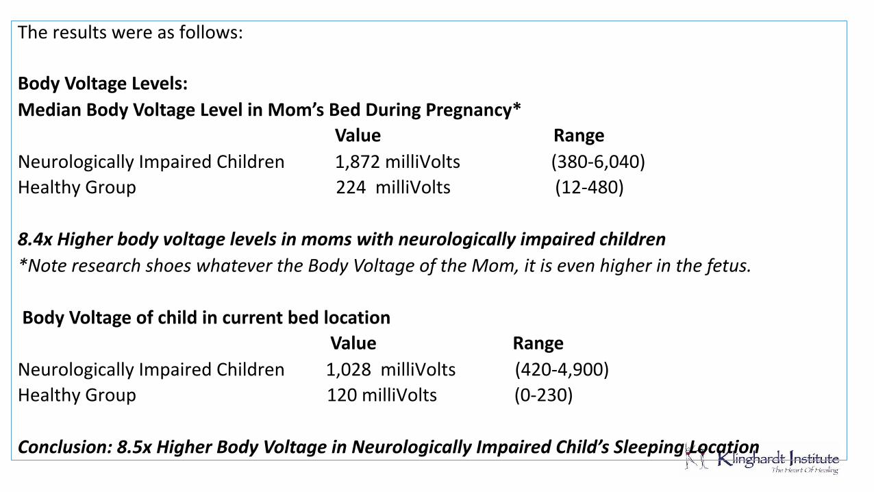
Theresultswereasfollows:Body Voltage Levels:Median Body Voltage Level in Mom’s Bed During Pregnancy* Value RangeNeurologicallyImpairedChildren1,872milliVolts(380-6,040)HealthyGroup224milliVolts(12-480)8.4x Higher body voltage levels in moms withneurologically impaired children*Note research shoes whatever the Body Voltage of the Mom, it is even higher in the fetus.Body Voltage of child in current bed location Value RangeNeurologicallyImpairedChildren1,028milliVolts(420-4,900)HealthyGroup120milliVolts(0-230)Conclusion: 8.5x Higher Body Voltage in Neurologically ImpairedChild’s Sleeping Location

Microwave Exposure:Microwave Power Density in Sleeping LocationNeurologicallyImpairedChildren-Mom’sBedmw/sq. meterRangeExposureInPregnancy290(110-1,710)HealthyGroup14(0-67)Conclusion: 20.7x higher microwave power density in moms sleepinglocation in cases where children were neurologically impairedThispilotdatastronglysuggeststhatelectromagneticradiationinthesleepingenvironmentofmothersduringpregnancy,aswellaselectromagneticradiationinthesleepingenvironmentofchildren,maybetheundiscoveredkeycontributing-ifnotcausative-factorinneurologicalimpairmentsinchildren,includingautism.Givenincreasinglevelsofambientelectromagneticradiationinmodernenvironmentsfromsociety’suseofelectronicequipment,wirelesstechnologies,suchascellphonesandwirelessnetworks,highfrequencytransientsonelectriclines,andbroadbandoverpowerlines(BPL),thisassociationneedsimmediatefurtherexploration