Embed Size (px)
Citation preview
Embryological Development of the
Tooth

Three Stages:
• Bud stage• Cap stage• Bell stage
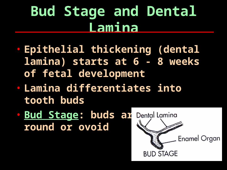
Bud Stage and Dental Lamina
• Epithelial thickening (dental lamina) starts at 6 - 8 weeks of fetal development
• Lamina differentiates into tooth buds
• Bud Stage: buds are round or ovoid
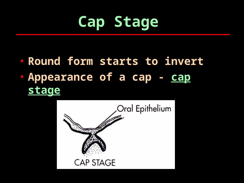
Cap Stage
• Round form starts to invert• Appearance of a cap - cap
stage
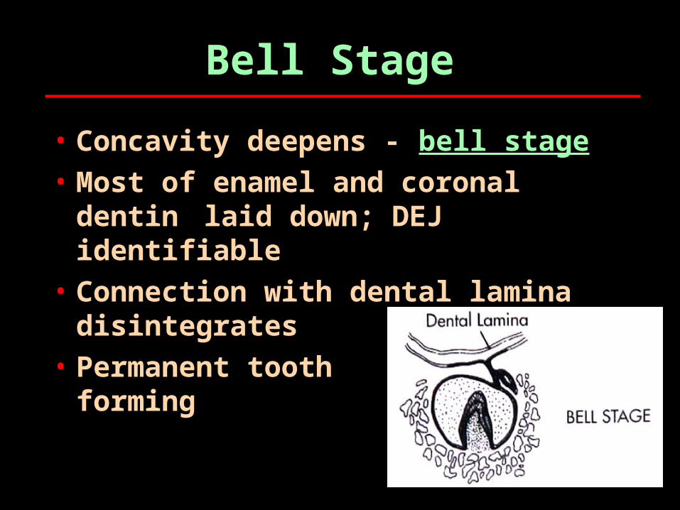
Bell Stage
• Concavity deepens - bell stage• Most of enamel and coronal
dentin laid down; DEJ identifiable
• Connection with dental lamina disintegrates
• Permanent tooth bud starts forming

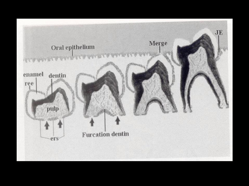
Root Development
• Bell stage ends with formation of CE line
• Enamel organ differentiates into Hertwig’s sheath, which forms the root structure
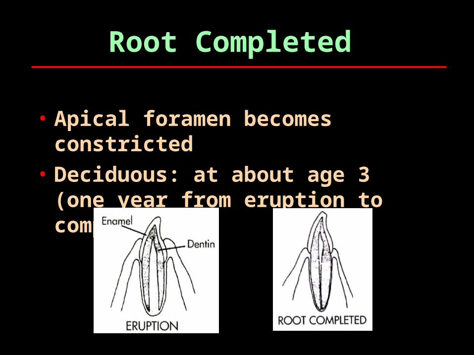
Root Completed
• Apical foramen becomes constricted
• Deciduous: at about age 3 (one year from eruption to completion)
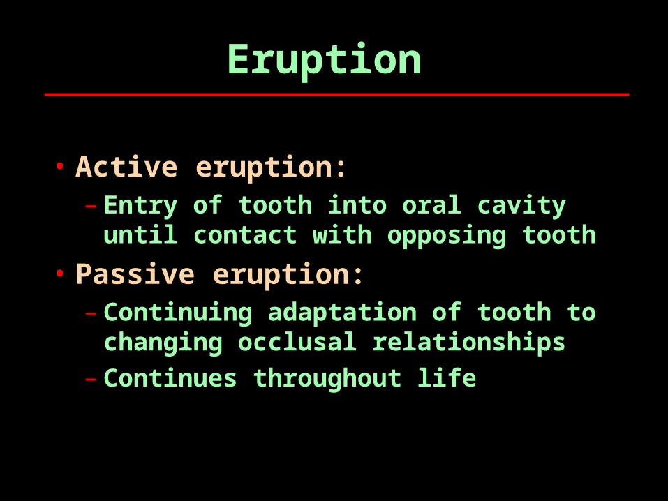
Eruption
• Active eruption:– Entry of tooth into oral cavity
until contact with opposing tooth
• Passive eruption:– Continuing adaptation of tooth to
changing occlusal relationships– Continues throughout life
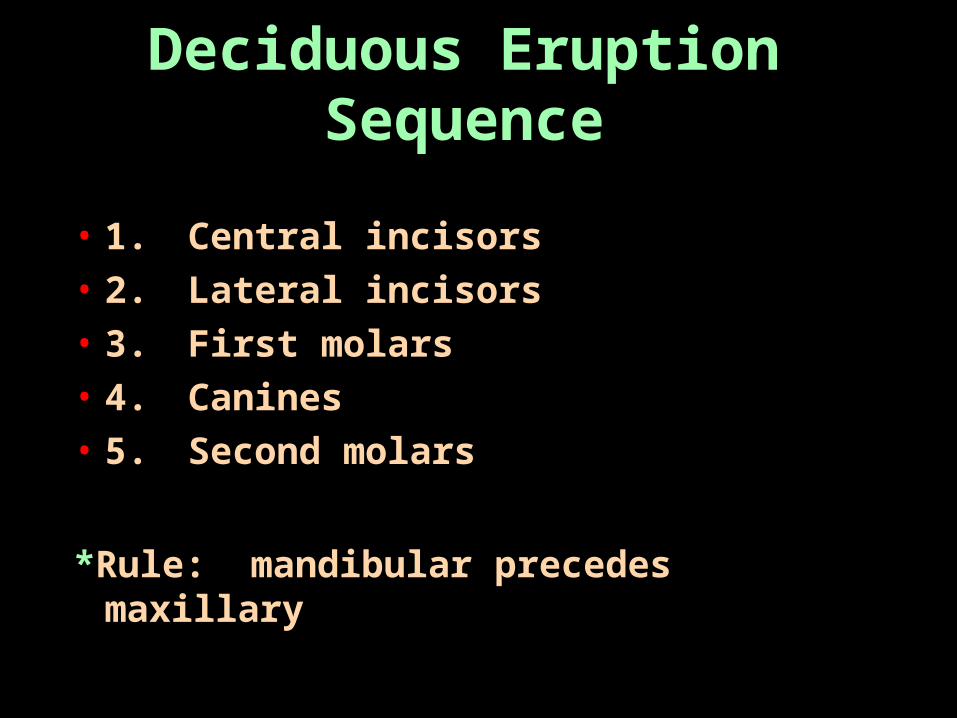
Deciduous Eruption Sequence
• 1. Central incisors• 2. Lateral incisors• 3. First molars• 4. Canines• 5. Second molars
*Rule: mandibular precedes maxillary
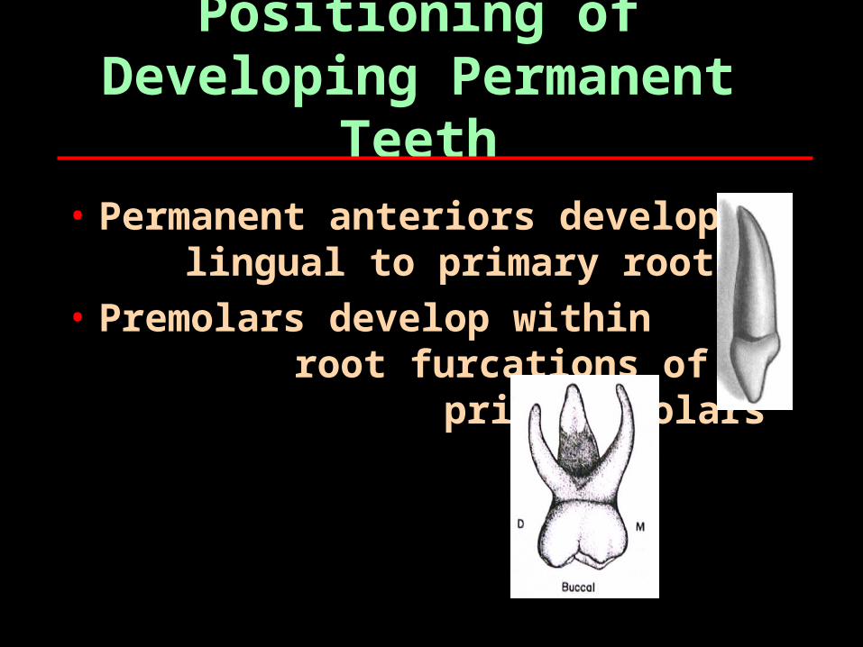
Positioning of Developing Permanent
Teeth• Permanent anteriors develop
lingual to primary roots• Premolars develop within
root furcations of primary molars
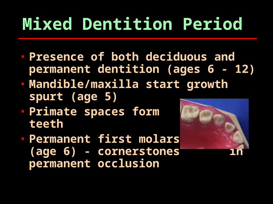
Mixed Dentition Period
• Presence of both deciduous and permanent dentition (ages 6 - 12)
• Mandible/maxilla start growth spurt (age 5)
• Primate spaces form between teeth
• Permanent first molars erupt (age 6) - cornerstones
in permanent occlusion
Resorption
• Permanent teeth start resorption of primary roots (osteoclastic process)
• Resorption starts at least one year prior to exfoliation
• Ankylosis: root fused to bone, disrupts resorption and exfoliation
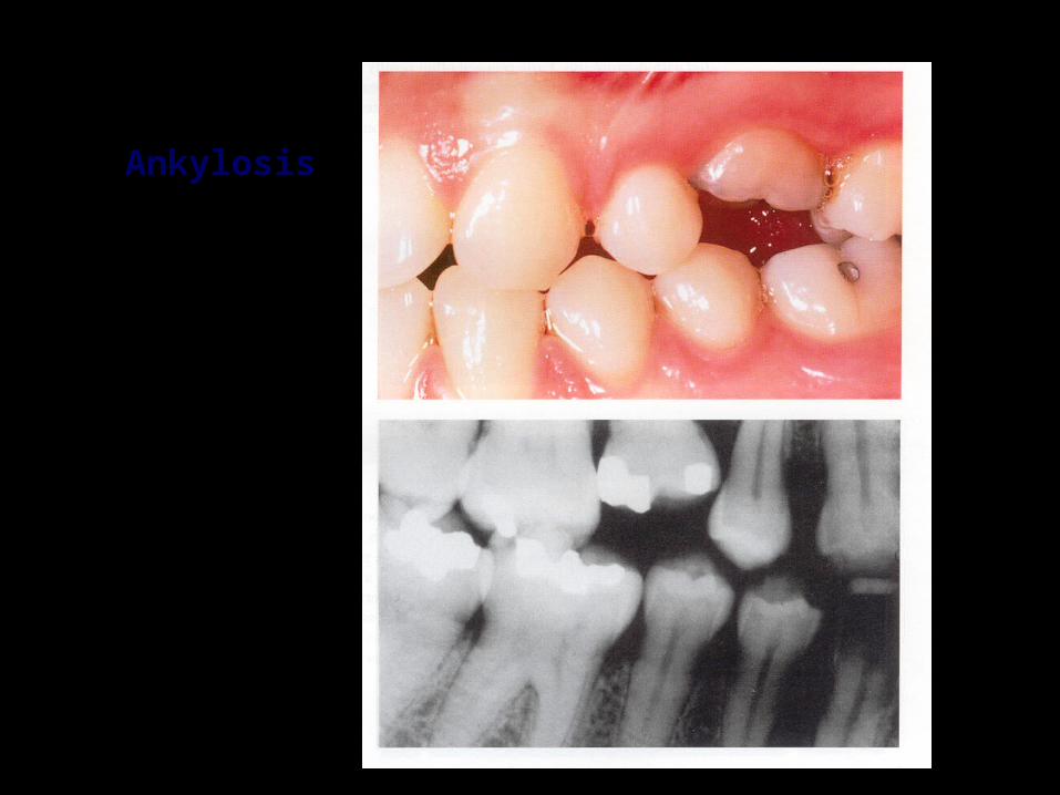
AnkylosisAnkylosis
Exfoliation
• Root structure mostly resorbed, tooth is loose
• Mandibular teeth precede maxillary in exfoliation except:– Primary second molars, all four
are lost about same time

Deciduous Dentition
General descriptions:
• Twenty primary teeth (A - T)• Dental formula: I C M• No premolars• Important in maintaining arch
space for permanent dentition
22
11
22
Comparisons With Permanent Dentition
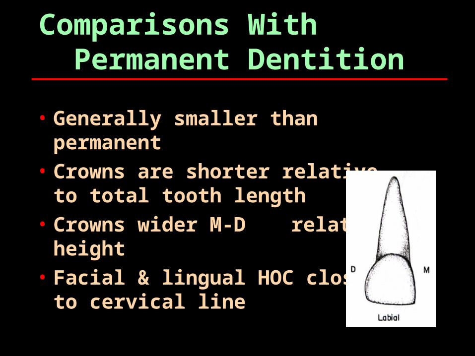
• Generally smaller than permanent
• Crowns are shorter relativeto total tooth length
• Crowns wider M-D relative to height
• Facial & lingual HOC closerto cervical line
More comparisons…
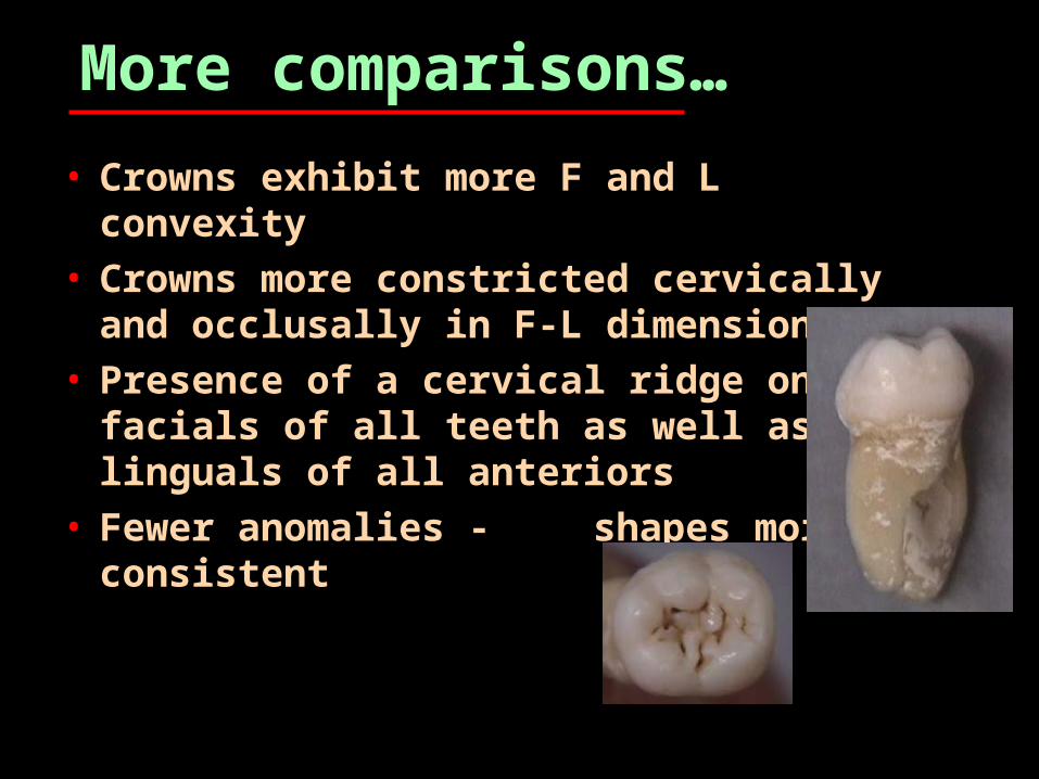
• Crowns exhibit more F and L convexity
• Crowns more constricted cervically and occlusally in F-L dimensions
• Presence of a cervical ridge on facials of all teeth as well as linguals of all anteriors
• Fewer anomalies - shapes more consistent
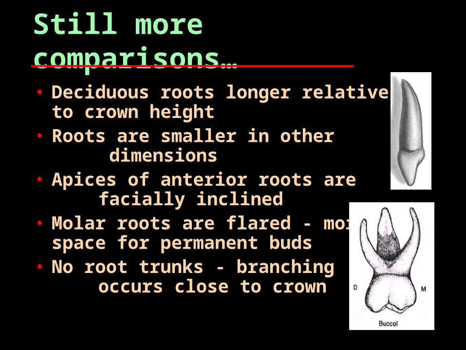
Still more comparisons…• Deciduous roots longer relative to
crown height• Roots are smaller in other
dimensions• Apices of anterior roots are
facially inclined• Molar roots are flared - more
space for permanent buds• No root trunks - branching
occurs close to crown
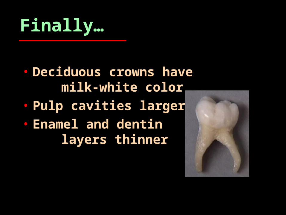
Finally…
• Deciduous crowns have milk-white color
• Pulp cavities larger• Enamel and dentin
layers thinner
Deciduous Dental Anatomy
Comparative DescriptionsTo Permanent Dentition

Maxillary Primary Teeth
Maxillary Central Incisor
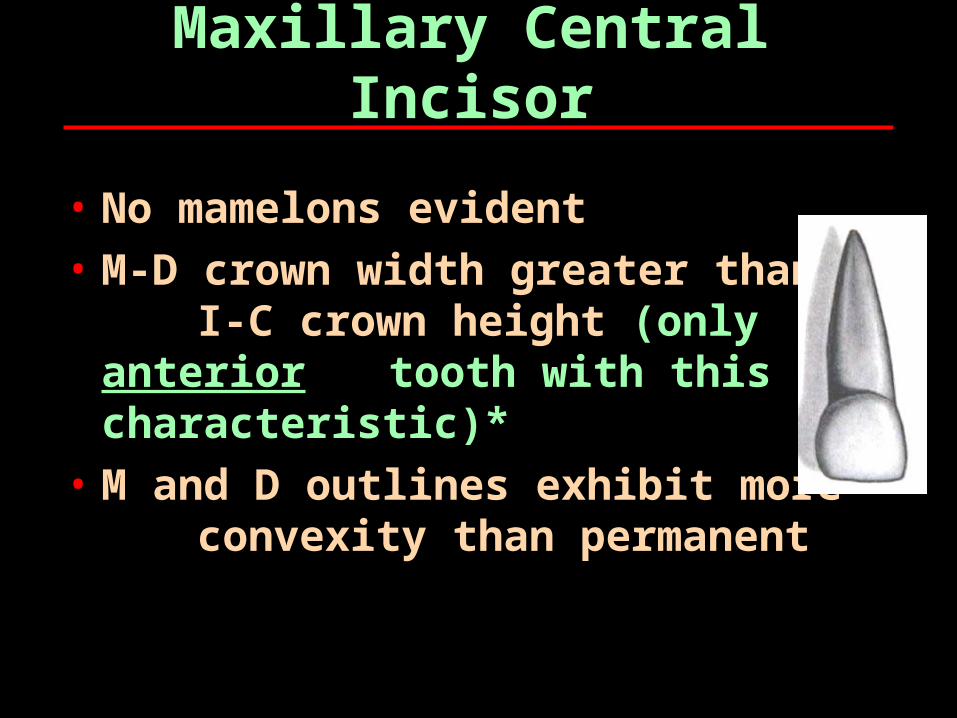
• No mamelons evident• M-D crown width greater than
I-C crown height (only anterior tooth with this characteristic)*
• M and D outlines exhibit more convexity than permanent
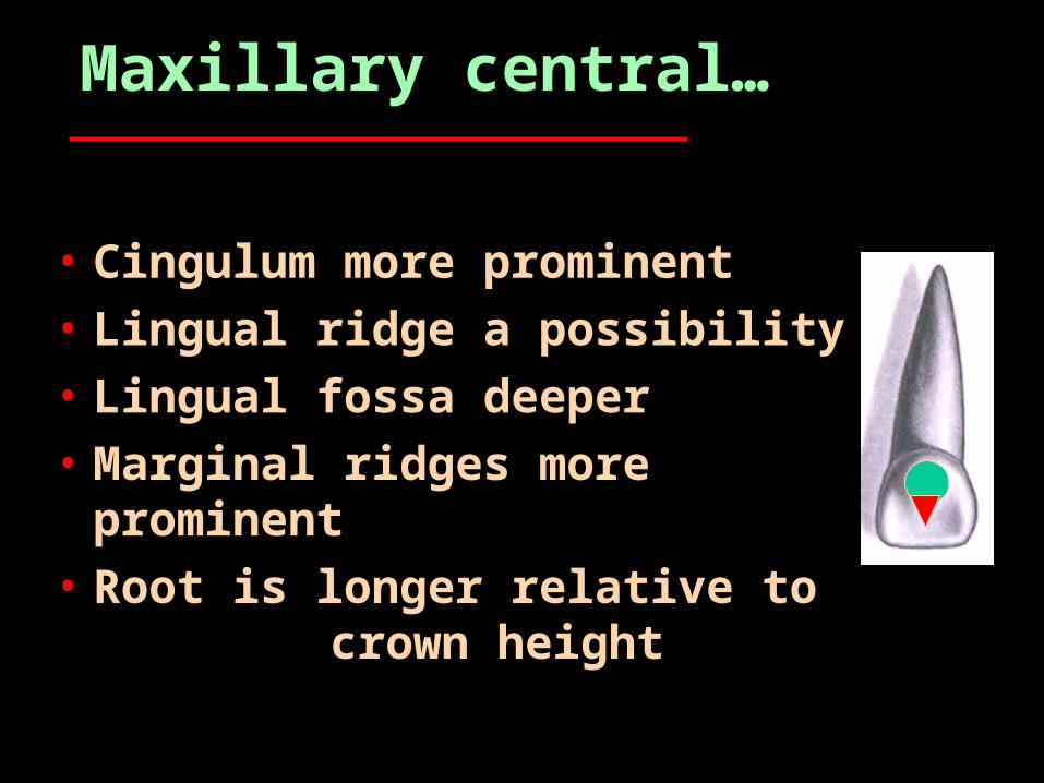
Maxillary central…
• Cingulum more prominent• Lingual ridge a possibility• Lingual fossa deeper• Marginal ridges more
prominent• Root is longer relative to
crown height

Maxillary central…
• Facial developmental depressions rare
• Incisal view: M-D width noticeably wider than F-L dimension
(MD>IC>FL)• Root deflected facially• Lingual CE line more
apically located• Incisal edge is slightly
facial to mid-root axis
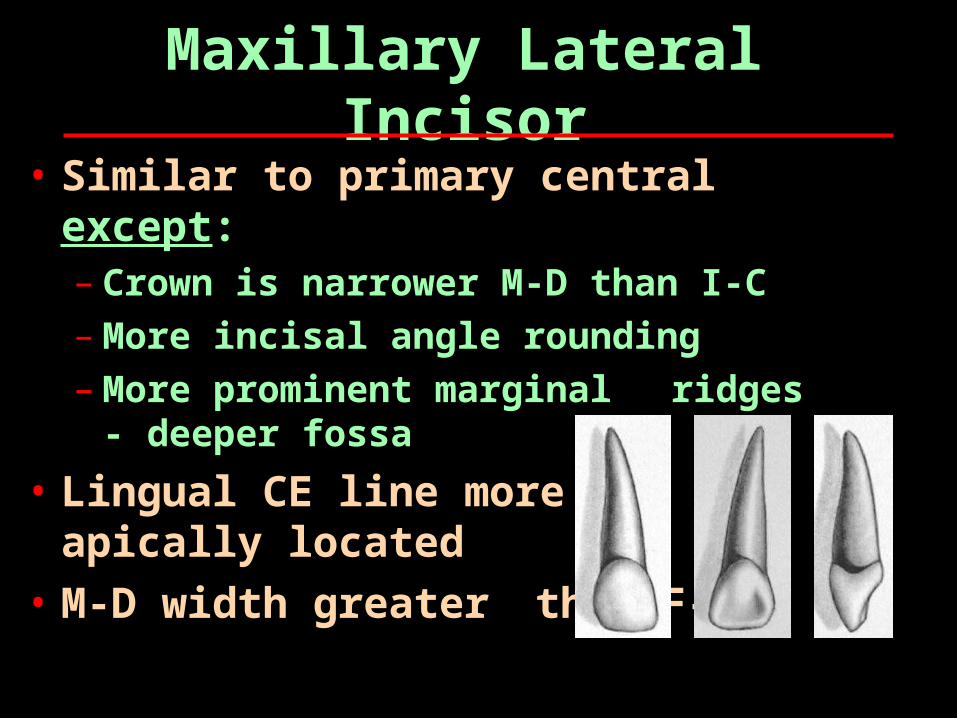
Maxillary Lateral Incisor
• Similar to primary central except:– Crown is narrower M-D than I-C– More incisal angle rounding– More prominent marginal
ridges - deeper fossa
• Lingual CE line more apically located
• M-D width greater than F-L
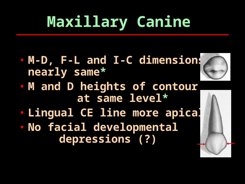
Maxillary Canine
• M-D, F-L and I-C dimensions nearly same*
• M and D heights of contour at same level*
• Lingual CE line more apical• No facial developmental
depressions (?)
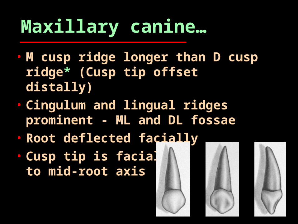
Maxillary canine…
• M cusp ridge longer than D cusp ridge* (Cusp tip offset distally)
• Cingulum and lingual ridges prominent - ML and DL fossae
• Root deflected facially• Cusp tip is facial to
mid-root axis
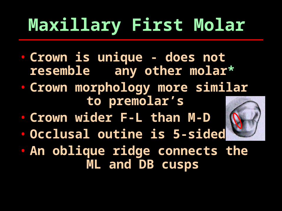
Maxillary First Molar
• Crown is unique - does not resemble any other molar*
• Crown morphology more similar to premolar’s
• Crown wider F-L than M-D• Occlusal outine is 5-sided• An oblique ridge connects the
ML and DB cusps
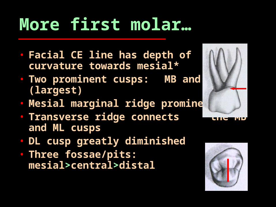
More first molar…
• Facial CE line has depth of curvature towards mesial*
• Two prominent cusps: MB and ML (largest)
• Mesial marginal ridge prominent• Transverse ridge connects
the MB and ML cusps• DL cusp greatly diminished• Three fossae/pits:
mesial>central>distal
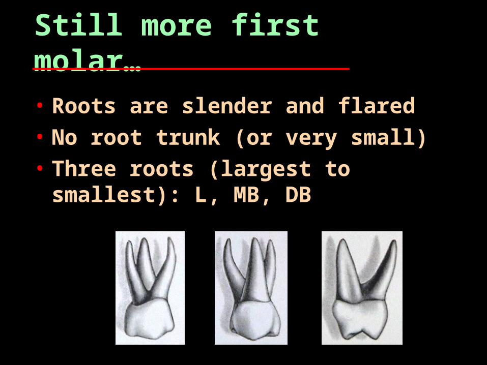
Still more first molar…
• Roots are slender and flared• No root trunk (or very small)• Three roots (largest to
smallest): L, MB, DB
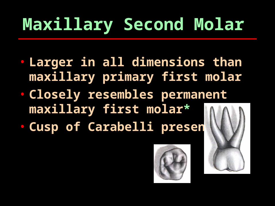
Maxillary Second Molar
• Larger in all dimensions than maxillary primary first molar
• Closely resembles permanent maxillary first molar*
• Cusp of Carabelli present

Mandibular Primary Teeth
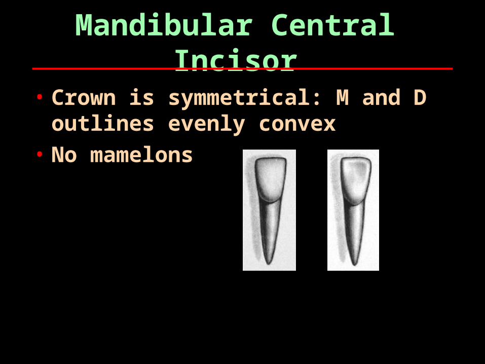
Mandibular Central Incisor
• Crown is symmetrical: M and D outlines evenly convex
• No mamelons
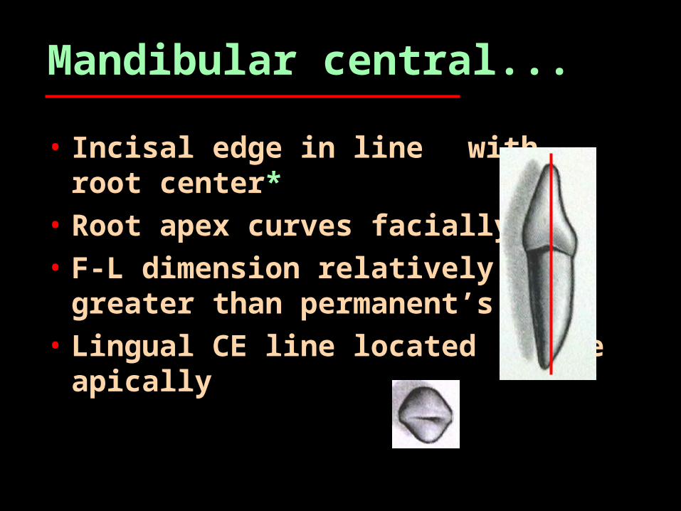
Mandibular central...
• Incisal edge in line with root center*
• Root apex curves facially• F-L dimension relatively
greater than permanent’s• Lingual CE line located
more apically
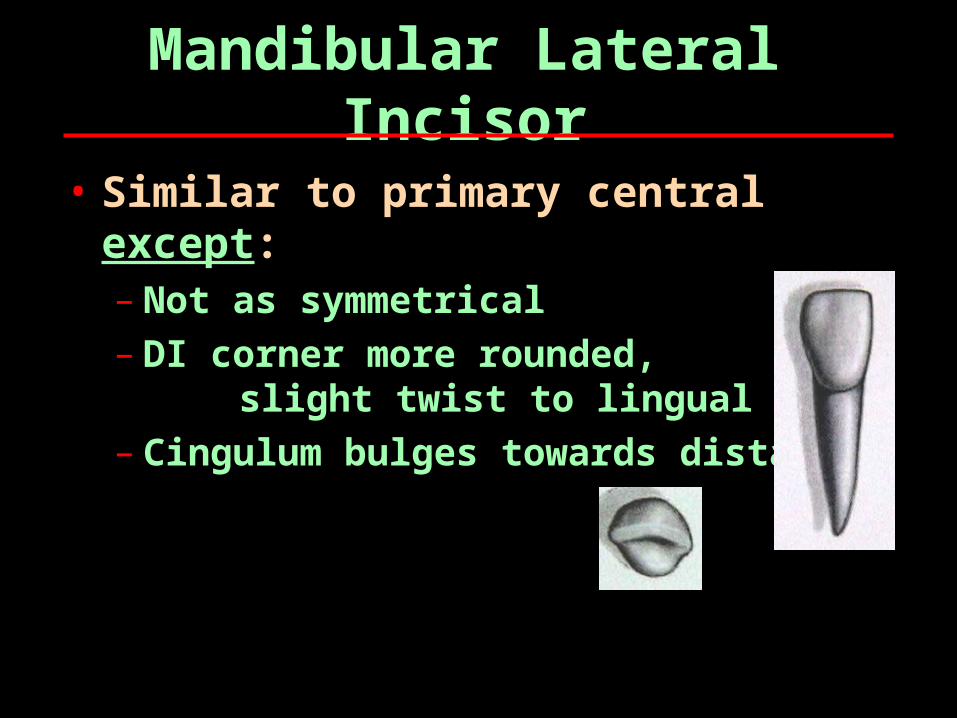
Mandibular Lateral Incisor
• Similar to primary central except:– Not as symmetrical– DI corner more rounded,
slight twist to lingual– Cingulum bulges towards distal
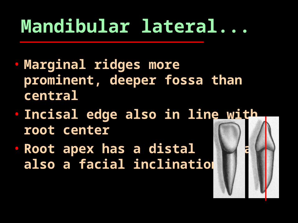
Mandibular lateral...
• Marginal ridges more prominent, deeper fossa than central
• Incisal edge also in line with root center
• Root apex has a distal and also a facial inclination
Mandibular Canine
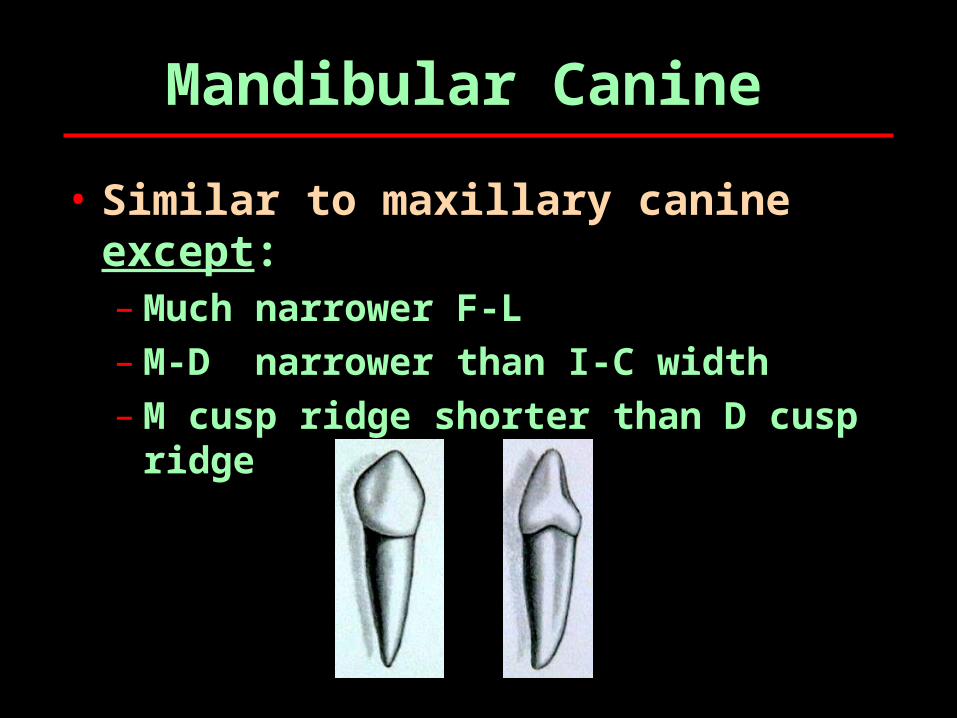
• Similar to maxillary canine except:– Much narrower F-L– M-D narrower than I-C width– M cusp ridge shorter than D cusp
ridge
Mandibular First Molar
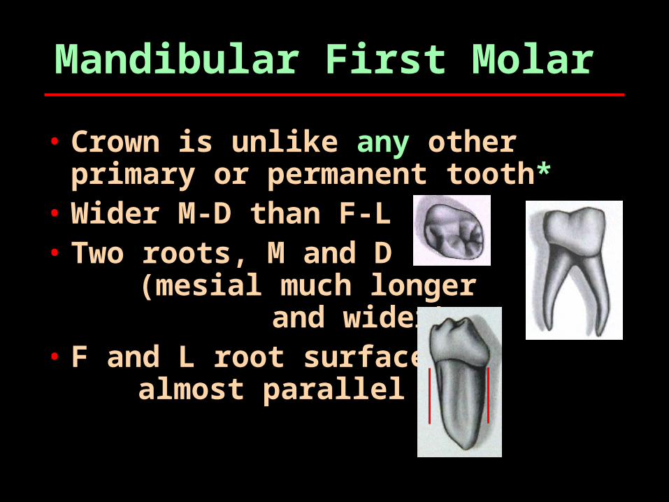
• Crown is unlike any other primary or permanent tooth*
• Wider M-D than F-L• Two roots, M and D
(mesial much longer and wider)
• F and L root surfaces almost parallel
More first molar…
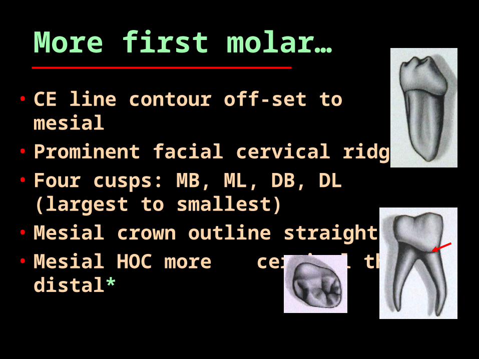
• CE line contour off-set to mesial• Prominent facial cervical ridge• Four cusps: MB, ML, DB, DL
(largest to smallest)• Mesial crown outline straight*• Mesial HOC more
cervical than distal*
Still more first molar…
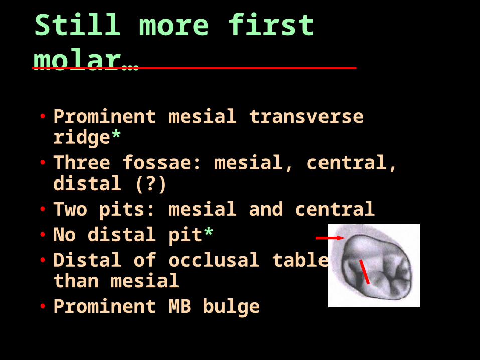
• Prominent mesial transverse ridge*
• Three fossae: mesial, central, distal (?)
• Two pits: mesial and central• No distal pit*• Distal of occlusal table
wider than mesial• Prominent MB bulge
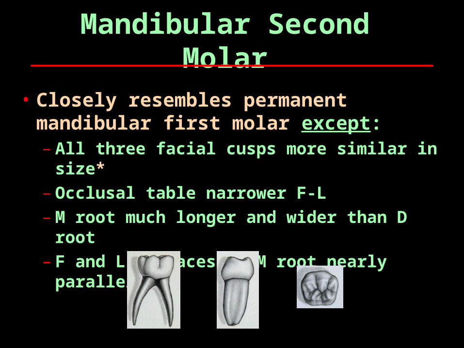
Mandibular Second Molar
• Closely resembles permanent mandibular first molar except:– All three facial cusps more similar in
size*– Occlusal table narrower F-L– M root much longer and wider than D
root– F and L surfaces of M root nearly
parallel

Lights!

Primary DentitionReview
Development and Eruption
• All 20 primaries begin calcifying 4-6 months in utero (2nd trimester)
• Crown completion in all primaries within first year after birth
• Eruption from 6 months to 2 years• Mixed dentition from age 6 to 12• Age 4-5 yrs, primate spaces
develop due to growth of maxilla and mandible
Eruption Sequence
• Mandibular teeth erupt prior to their maxillary counterparts
• Sequence:– Centrals– Laterals– 1st molars– Canines– 2nd molars
Exfoliation sequence:
1. Preceeds the eruption sequence of the permanent dentition.
2. Root resorption begins one year prior to exfoliation
3. All second molars are lost about the same time

Unique Characteristics of Primary Teeth
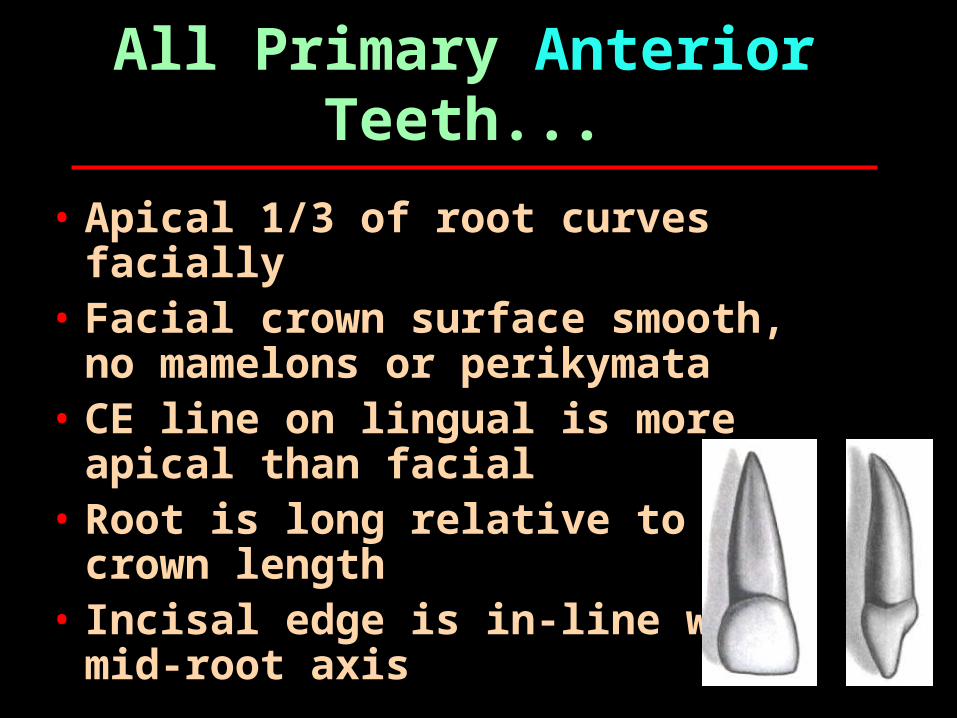
All Primary Anterior Teeth...
• Apical 1/3 of root curves facially
• Facial crown surface smooth, no mamelons or perikymata
• CE line on lingual is more apical than facial
• Root is long relative to crown length
• Incisal edge is in-line with mid-root axis
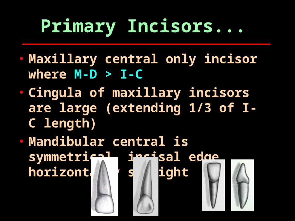
Primary Incisors...
• Maxillary central only incisor where M-D > I-C
• Cingula of maxillary incisors are large (extending 1/3 of I-C length)
• Mandibular central is symmetrical, incisal edge horizontally straight
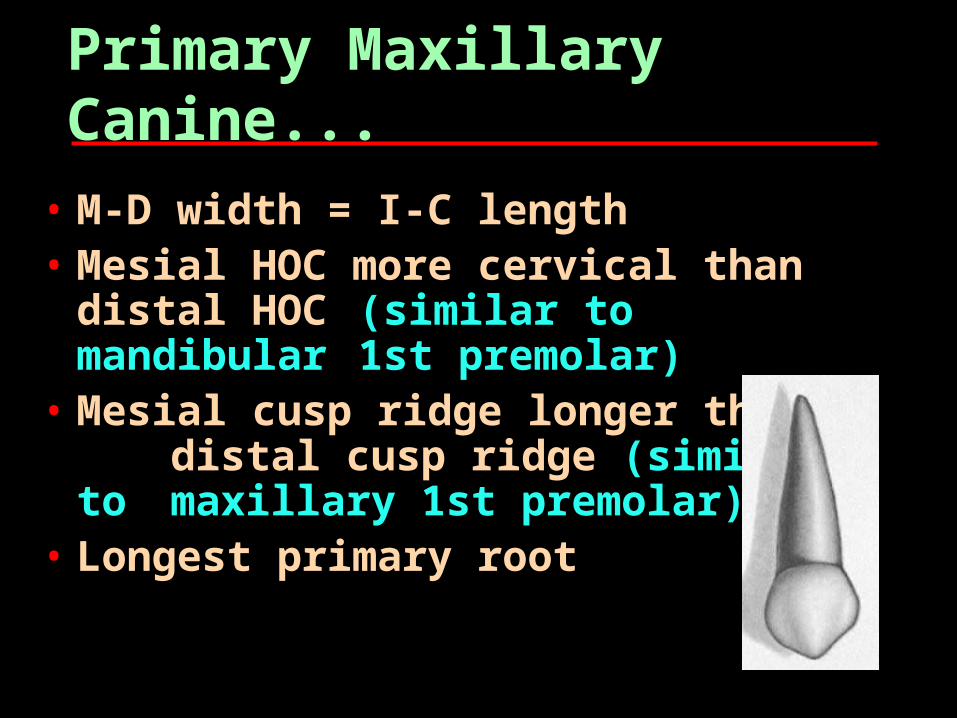
Primary Maxillary Canine...
• M-D width = I-C length• Mesial HOC more cervical than
distal HOC (similar to mandibular 1st premolar)
• Mesial cusp ridge longer than distal cusp ridge (similar to
maxillary 1st premolar)
• Longest primary root
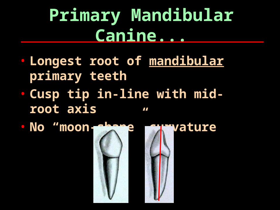
Primary Mandibular Canine...
• Longest root of mandibular primary teeth
• Cusp tip in-line with mid-root axis
• No “moon-shape” curvature
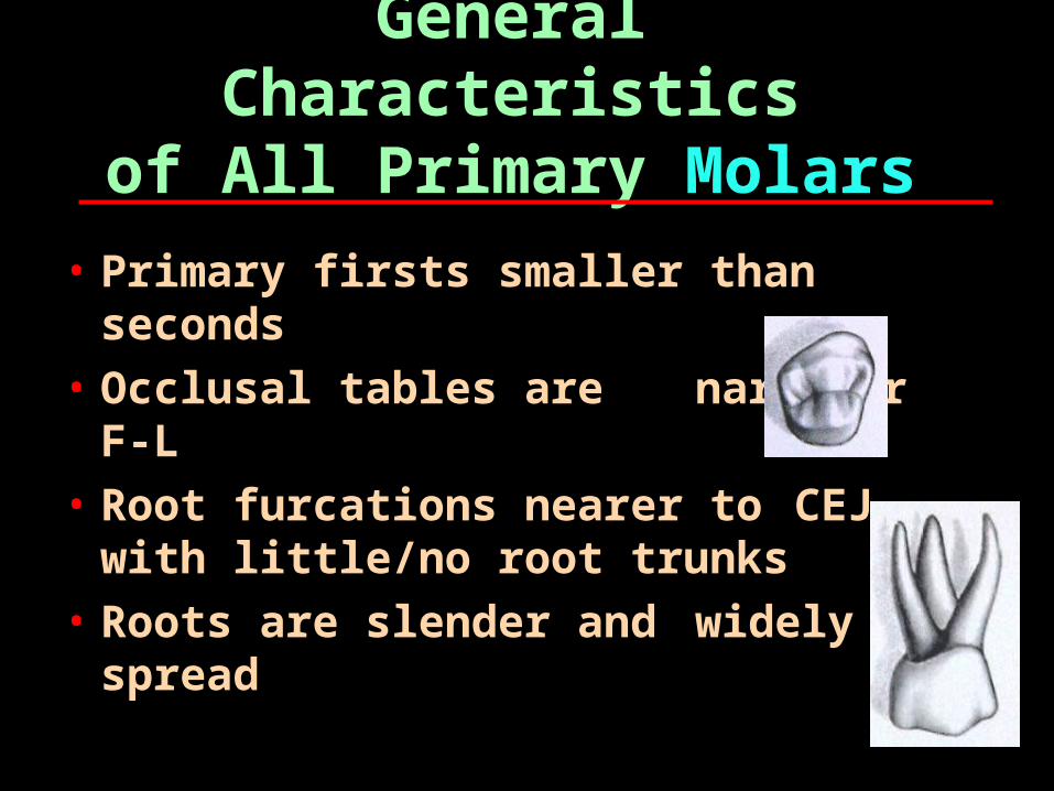
General Characteristicsof All Primary Molars
• Primary firsts smaller than seconds
• Occlusal tables are narrower F-L
• Root furcations nearer to CEJ with little/no root trunks
• Roots are slender and widely spread
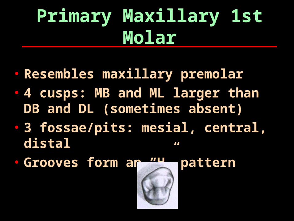
Primary Maxillary 1st Molar
• Resembles maxillary premolar• 4 cusps: MB and ML larger than
DB and DL (sometimes absent)• 3 fossae/pits: mesial, central,
distal• Grooves form an “H” pattern
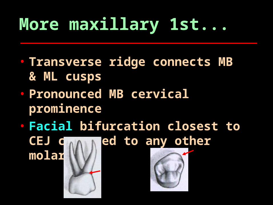
More maxillary 1st...
• Transverse ridge connects MB & ML cusps
• Pronounced MB cervical prominence
• Facial bifurcation closest to CEJ compared to any other molars*
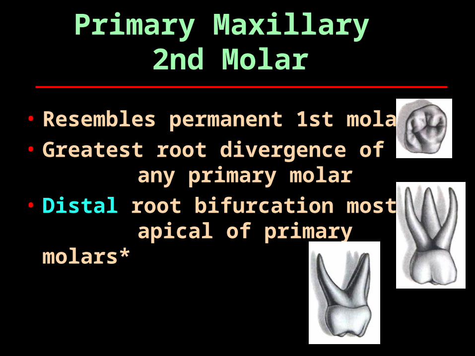
Primary Maxillary 2nd Molar
• Resembles permanent 1st molar
• Greatest root divergence of any primary molar
• Distal root bifurcation most apical of primary
molars*
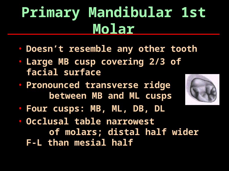
Primary Mandibular 1st Molar
• Doesn’t resemble any other tooth• Large MB cusp covering 2/3 of
facial surface• Pronounced transverse ridge
between MB and ML cusps• Four cusps: MB, ML, DB, DL• Occlusal table narrowest
of molars; distal half wider F-L than mesial half
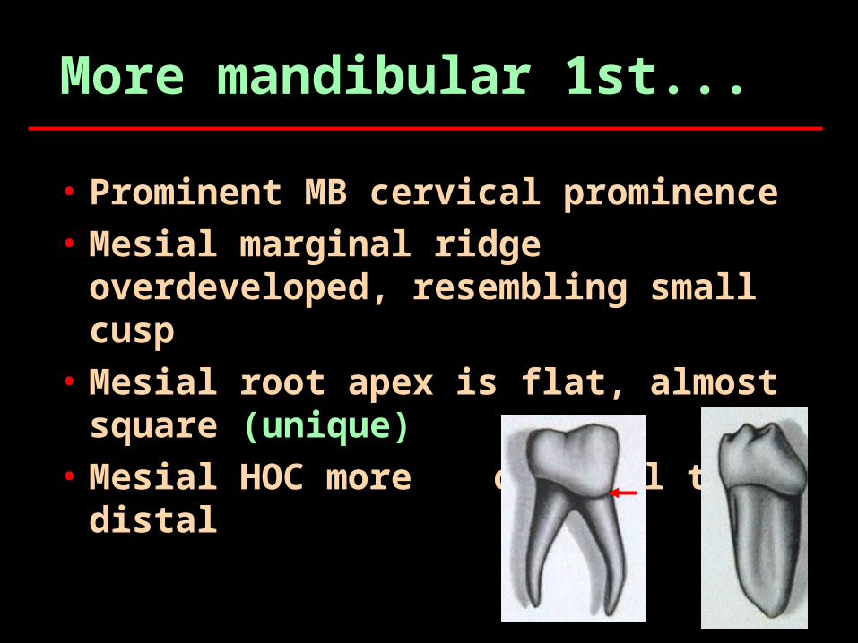
More mandibular 1st...
• Prominent MB cervical prominence• Mesial marginal ridge
overdeveloped, resembling small cusp
• Mesial root apex is flat, almost square (unique)
• Mesial HOC more cervical than distal
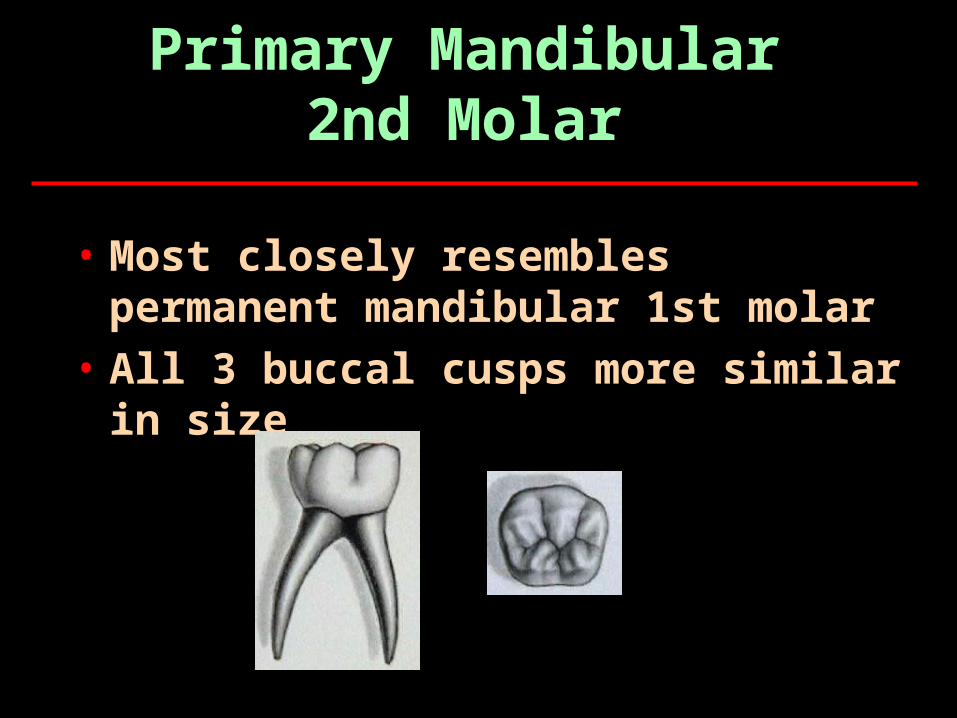
Primary Mandibular2nd Molar
• Most closely resembles permanent mandibular 1st molar
• All 3 buccal cusps more similar in size

All the Best on Your Exams
The Primary Dentition
• There are 20 primary teeth, 10 per dental arch.
• These teeth include incisors, canines, and molars.
• The primary teeth are smaller overall and have whiter enamel than the permanent teeth.
• The crown of any primary tooth is short in relation to its total length.
• The crowns are narrower at the CEJ.
The Primary Dentitioncont’d
• The pulp chambers and pulp horns in primary teeth are relatively large in proportion to those of the permanent teeth.
• There is a thick layer of dentin between the pulp chambers and the enamel, especially in the primary mandibular second molar.
• The enamel layer is relatively thin.
Clinical Considerations with Primary Teeth
• Often, parents do not understand the importance of the primary teeth.
• Primary teeth hold the eruption space for the permanent teeth that will replace the primary teeth.
• Because the enamel and dentin is thinner in primary teeth, decay can travel quickly through the enamel to the pulp, possibly causing loss of the tooth.
• Early dental health education and dental care are essential for keeping the primary dentition.
Primary Maxillary Incisors
• The crown of the primary maxillary central incisor (E and F) is wider mesiodistally than incisocervically.
• It is the only tooth of either dentition with this crown dimension.
• The primary maxillary incisors have no mamelons.
• The cingulum and marginal ridges are more prominent than on the permanent successor, and the lingual fossa is deeper.
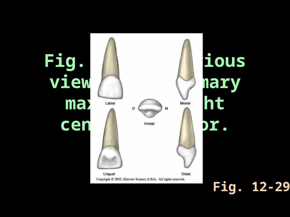
Fig. 12-29 Various views of a primary
maxillary right central incisor.
Fig. 12-29Fig. 12-29
Primary Maxillary Lateral Incisors
• The crown of the primary maxillary lateral incisor (D and G) is similar to that of the central incisor but is much smaller in all dimensions.
• The incisal angles on the lateral incisor are also more rounded than on the central incisor.
• The lateral root is longer in proportion to its crown, and its apex is sharper.
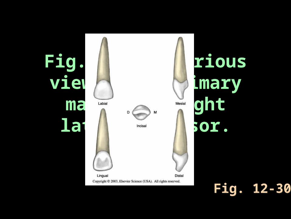
Fig. 12-30 Various views of a primary
maxillary right lateral incisor.
Fig. 12-30Fig. 12-30
Primary Mandibular Central Incisors
• The crown of the primary mandibular incisor (O and P) resembles the primary mandibular lateral incisor more than its permanent central successor.
• The mandibular central incisor is extremely symmetric.
• It is also not as constricted at the CEJ as the primary maxillary incisor.
• The lingual surface of the mandibular central incisors appears smooth and tapers toward the prominent cingulum.
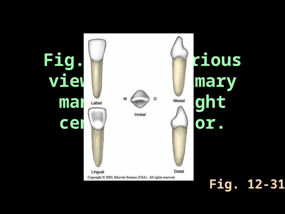
Fig. 12-31 Various views of a primary mandibular right central incisor.
Fig. 12-31Fig. 12-31
Primary Mandibular Lateral Incisors
• The crown of the primary lateral incisor (Q and N) is similar in form to that of the central incisor in the same arch but is wider and longer.
• The incisal edge of the mandibular lateral incisor slopes distally, and the distoincisal angle is more rounded.
• The root may have a distal curvature in its apical third and usually has a distal longitudinal groove.
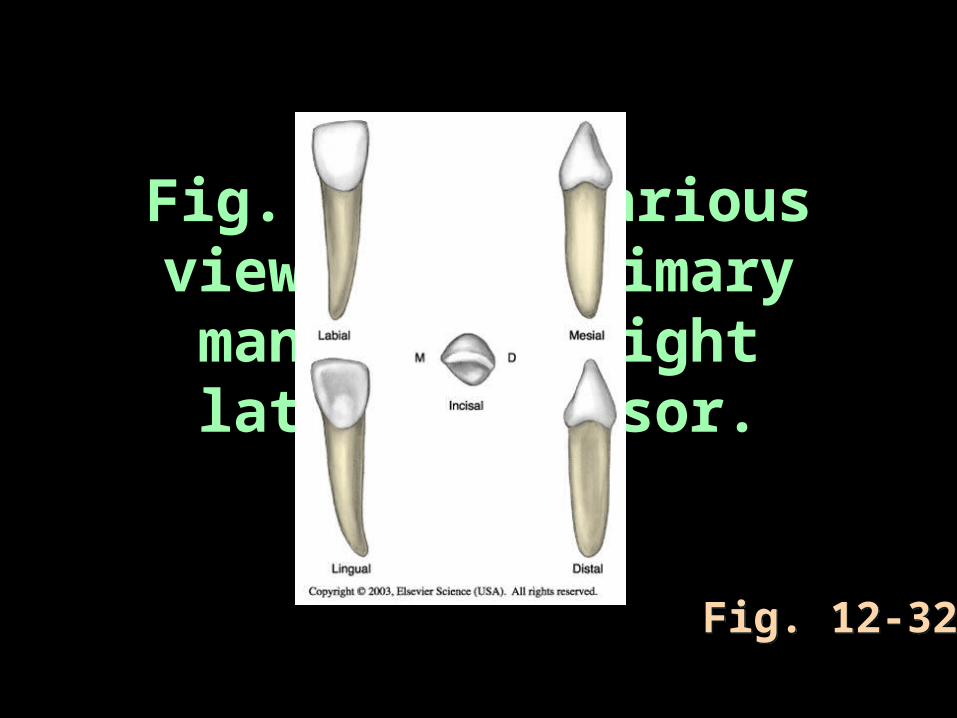
Fig. 12-32 Various views of a primary
mandibular right lateral incisor.
Fig. 12-32Fig. 12-32

Primary Canines
• There are four primary canines, two in each dental arch.
• These primary canines differ from the outline of their permanent successors in the following ways:•The crown of the primary maxillary
canine (C and H) has a relatively longer and sharper cusp than that of its permanent successor when first erupted.
•The mesial and distal outlines of the primary maxillary canine are rounder.
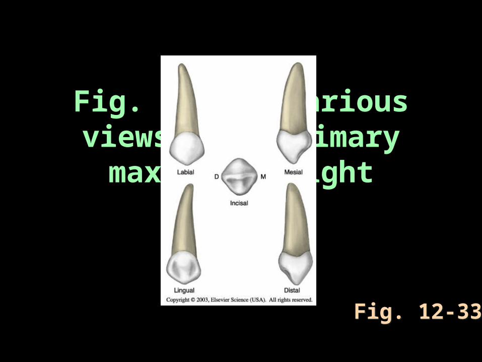
Fig. 12-33 Various views of a primary
maxillary right canine.
Fig. 12-33Fig. 12-33

Primary Mandibular Canines • The primary mandibular canine (M and R)
resembles the primary maxillary canine. • This tooth is much smaller labiolingually.• The distal cusp slope is much longer than
the mesial cusp slope.• The lingual surface of the primary
mandibular canine is marked by a shallow lingual fossa.
• The primary mandibular canine (M and R) resembles the primary maxillary canine, although some dimensions are different. This tooth is much smaller labiolingually.
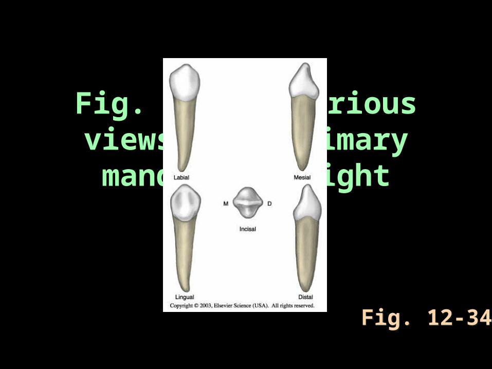
Fig. 12-34 Various views of a primary mandibular right
canine.
Fig. 12-34Fig. 12-34
Primary Molars• The crown of the primary maxillary first molar
(B and I) does not resemble any other crown of either dentition.
• The height of contour on the buccal surface is at the cervical one third of the tooth and on the lingual side is at the middle one third.
• The primary maxillary molars have three roots, which are thinner and have greater flare than the permanent maxillary first molar.
• The lingual root is the longest and most divergent.

Fig. 12-35 Various views of a primary maxillary right first
molar.
Fig. 12-35Fig. 12-35
Primary Maxillary Second Molars
• The primary maxillary second molar (A and J) is larger than the primary maxillary first molar.
• This tooth most closely resembles the form of the permanent maxillary first molar but is smaller in all dimensions.
• The second molar usually has a cusp of Carabelli, the minor fifth cusp.
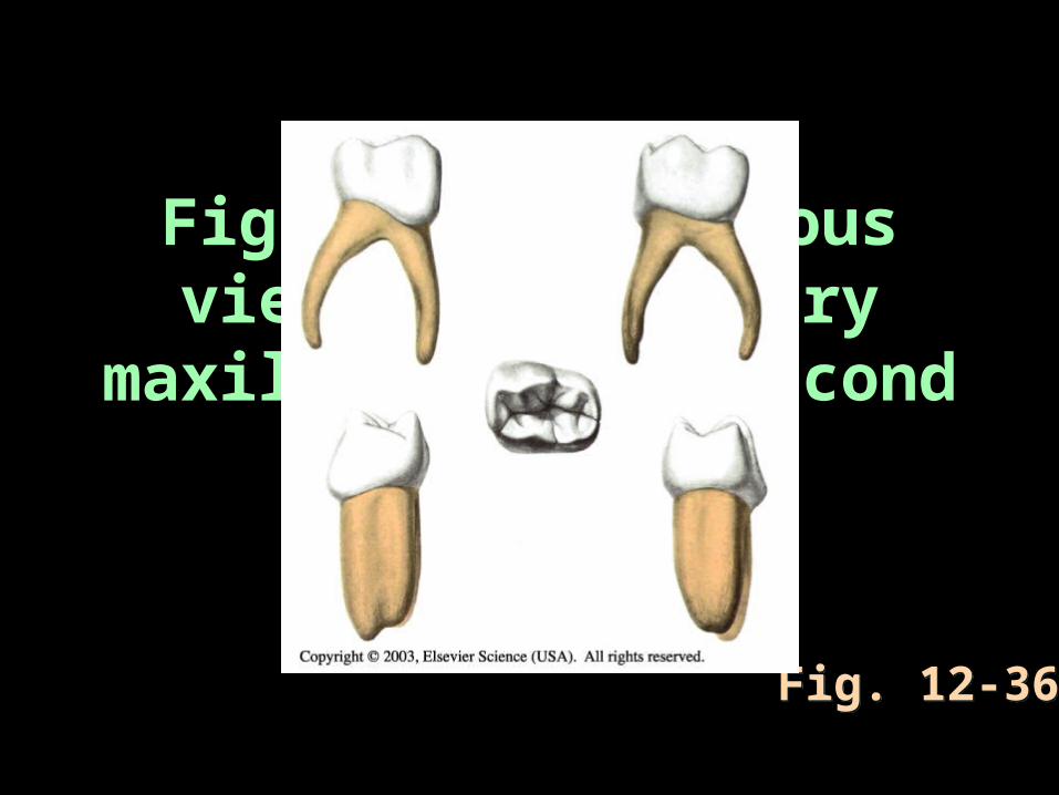
Fig. 12-36 Various views of a primary
maxillary right second molar.
Fig. 12-36Fig. 12-36
Primary Mandibular First Molars • The crown of the primary mandibular first
molar (L and S) is unlike any other tooth of either dentition.
• The height of contour on the buccal surface is at the cervical one third of the tooth and on the lingual side is at the middle one third.
• The primary mandibular first molar has four cusps; the mesial cusps are larger.
• The tooth has two roots, which are positioned similarly to those of other primary and permanent mandibular molars.
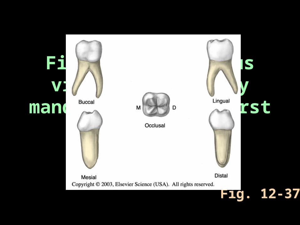
Fig. 12-37 Various views of a primary
mandibular right first molar.
Fig. 12-37Fig. 12-37
Primary Mandibular Second Molars
• The primary mandibular second molar (K and T) is larger than the primary mandibular first molar.
• It has five cusps; the second molar most closely resembles the form of the permanent mandibular first molar.
• The three buccal cusps are nearly equal in size.
• The primary mandibular second molar has an overall oval occlusal shape.
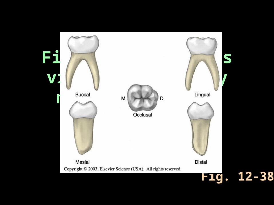
Fig. 12-38 Various views of a primary mandibular right
second molar.
Fig. 12-38Fig. 12-38