Embed Size (px)
Citation preview
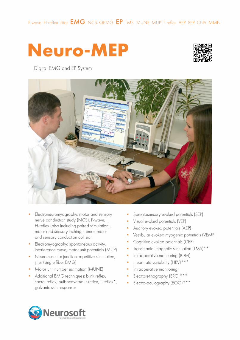
Neuro-MEPDigital EMG and EP System
• Electroneuromyography: motor and sensory nerve conduction study (NCS), F-wave, H-reflex (also including paired stimulation), motor and sensory inching, tremor, motor and sensory conduction collision
• Electromyography: spontaneous activity, interference curve, motor unit potentials (MUP)
• Neuromuscular junction: repetitive stimulation, jitter (single fiber EMG)
• Motor unit number estimation (MUNE)• Additional EMG techniques: blink reflex,
sacral reflex, bulbocavernous reflex, T-reflex*, galvanic skin responses
F-wave H-reflex Jitter EMG NCS QEMG EP TMS MUNE MUP T-reflex AEP SEP CNV MMN
• Somatosensory evoked potentials (SEP)• Visual evoked potentials (VEP) • Auditory evoked potentials (AEP)• Vestibular evoked myogenic potentials (VEMP) • Cognitive evoked potentials (CEP) • Transcranial magnetic stimulation (TMS)**• Intraoperative monitoring (IOM)• Heart rate variability (HRV)***• Intraoperative monitoring• Electroretinography (ERG)*** • Electro-oculography (EOG)***
•Neuro-MS/D – series of transcranial magnetic stimulators for diagnostics, therapy and research
•Neuro-ERG – software and equipment for electroretinography (ERG) and electro-oculography (EOG) study
•Poly-Spectrum-Rhythm – software and equipment for heart rate variability (HRV) analysis
According to safety standards all the computer equipment used with digital EMG/EP system should be connected via isolation transformer
Neuro-MEP Advantages
Modular Architecture with the Use of USB Technology
All the electronic units included in the device delivery set are connected to computer with the use of USB interface. It allows combining them flexibly to arrange a configuration corresponding to your own requirements. For example, if you connect one more 4-channel amplifier unit to Neuro-MEP-4, you will get 8-channel digital system. It is possible to connect up to 10 (!) different USB units.
Amplifiers and Stimulators with Specifications of Expert Class
Innovation technologies underlying module development provide expert class performance specifications. Neuro-MEP-4 digital EMG and EP system make it possible to obtain high quality traces even if other electroneuromyographs can not acquire responses. This becomes possible owing to the fact that Neuro-MEP-4 has high sampling rate, high resolution ADC, low noise level and wide range of stimulation current intensity.
New EMG TechniquesThe list of EMG techniques is enlarged by: • Motor and sensory inching • Jitter (single fiber EMG) • Sacral reflex • Bulbocavernous reflex • T-reflex* • Vestibular evoked myogenic potentials (VEMP) • Automatic detection of MUP • Motor unit number estimation (MUNE) • Conduction velocity combined test (motor/sensory response) • Registration and analysis of spontaneous activity and interference EMG in one test • Motor and sensory conduction collision
Neuro-MEP.NET update versionNow our digital EMG and EP systems are supplied with Neuro-MEP.NET update software. Specialists are already appreciating its main advantages:• New high performance graphic interface• Rapid examination execution• Built-in anatomy atlas (to help the beginners in EMG studies)• Combined EP tables (latency, intervals, amplitude, amplitude
ratio – all data in one table)• 32 and 64-bit program versions
Set of EMG Electrodes of New Generation
Now new EMG electrodes developed by our company are supplied with the digital EMG and EP system. They correspond to modern requirements. In the picture: stimulating bar electrode with replaceable steel and felt stimulation pads (adult). The set of replaceable stimulation pads from steel and felt of different types is available. It can be used as both stimulation and surface electrodes.
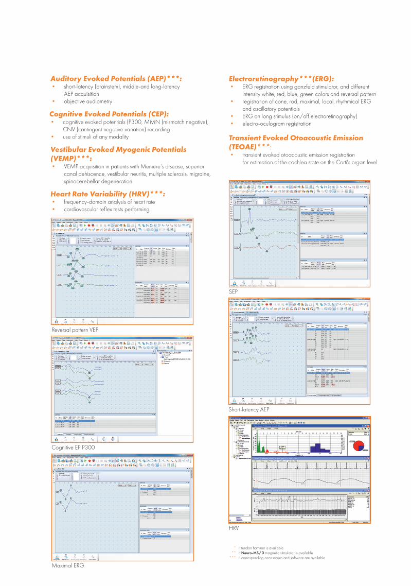
New high performance graphic interface
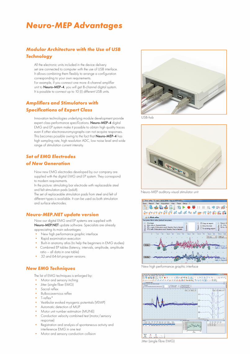
USB-hub
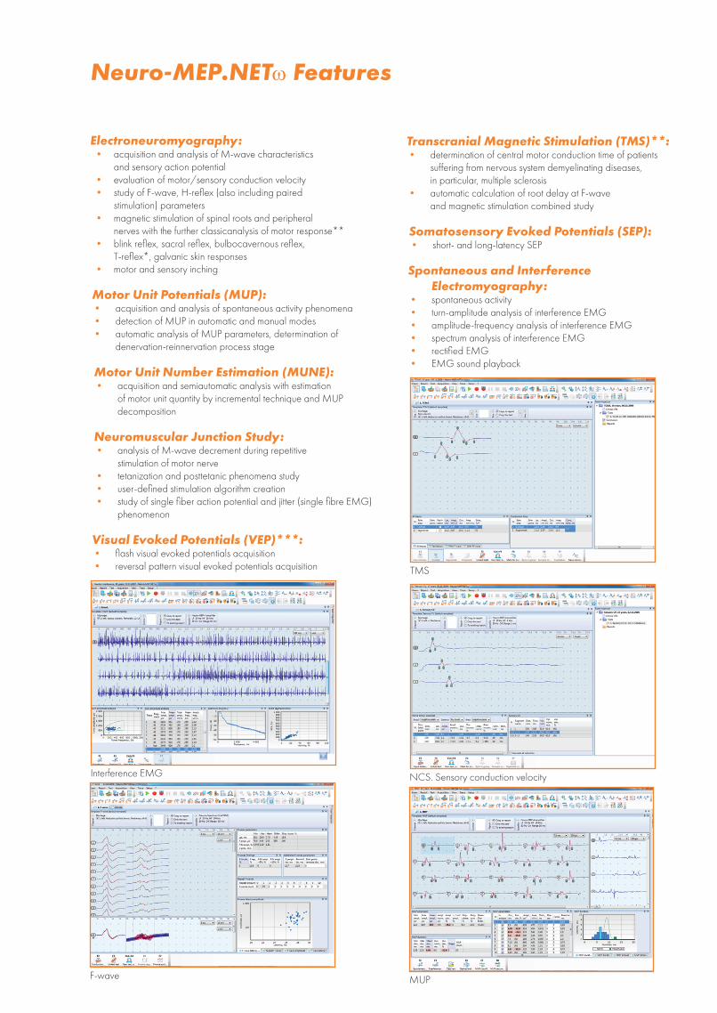
Neuro-MEP auditory-visual stimulator unit
Jitter (single fibre EMG)
MUP
Neuromuscular Junction Study:• analysis of M-wave decrement during repetitive
stimulation of motor nerve• tetanization and posttetanic phenomena study• user-defined stimulation algorithm creation• study of single fiber action potential and jitter (single fibre EMG)
phenomenon
Neuro-MEP.NETω Features
Interference EMG
F-wave
Transcranial Magnetic Stimulation (TMS)**:• determination of central motor conduction time of patients
suffering from nervous system demyelinating diseases, in particular, multiple sclerosis
• automatic calculation of root delay at F-wave and magnetic stimulation combined study
Visual Evoked Potentials (VEP)***:• flash visual evoked potentials acquisition• reversal pattern visual evoked potentials acquisition
Spontaneous and Interference Electromyography:
• spontaneous activity• turn-amplitude analysis of interference EMG• amplitude-frequency analysis of interference EMG• spectrum analysis of interference EMG• rectified EMG • EMG sound playback
Somatosensory Evoked Potentials (SEP):• short- and long-latency SEP
NCS. Sensory conduction velocity
TMS
Electroneuromyography:• acquisition and analysis of M-wave characteristics
and sensory action potential• evaluation of motor/sensory conduction velocity• study of F-wave, H-reflex (also including paired
stimulation) parameters• magnetic stimulation of spinal roots and peripheral
nerves with the further classicanalysis of motor response**• blink reflex, sacral reflex, bulbocavernous reflex,
T-reflex*, galvanic skin responses• motor and sensory inching
Motor Unit Potentials (MUP):• acquisition and analysis of spontaneous activity phenomena• detection of MUP in automatic and manual modes• automatic analysis of MUP parameters, determination of
denervation-reinnervation process stage
Motor Unit Number Estimation (MUNE):• acquisition and semiautomatic analysis with estimation
of motor unit quantity by incremental technique and MUP decomposition
if tendon hammer is available if Neuro-MS/D magnetic stimulator is available if corresponding accessories and software are available
***
***
Transient Evoked Otoacoustic Emission (TEOAE)***:• transient evoked otoacoustic emission registration
for estimation of the cochlea state on the Corti's organ level
Electroretinography***(ERG):• ERG registration using ganzfeld stimulator, and different
intensity white, red, blue, green colors and reversal pattern • registration of cone, rod, maximal, local, rhythmical ERG
and oscillatory potentials• ERG on long stimulus (on/off electroretinography)• electro-oculogram registration
Heart Rate Variability (HRV)***:• frequency-domain analysis of heart rate• cardiovascular reflex tests performing
Transcranial Magnetic Stimulation (TMS)**:• determination of central motor conduction time of patients
suffering from nervous system demyelinating diseases, in particular, multiple sclerosis
• automatic calculation of root delay at F-wave and magnetic stimulation combined study
Vestibular Evoked Myogenic Potentials (VEMP)***: • VEMP acquisition in patients with Meniere‘s disease, superior
canal dehiscence, vestibular neuritis, multiple sclerosis, migraine, spinocerebellar degeneration
Spontaneous and Interference Electromyography:
• spontaneous activity• turn-amplitude analysis of interference EMG• amplitude-frequency analysis of interference EMG• spectrum analysis of interference EMG• rectified EMG • EMG sound playback
Reversal pattern VEP
Cognitive EP P300
Maximal ERG
HRV
Short-latency AEP
SEP
Auditory Evoked Potentials (AEP)***:• short-latency (brainstem), middle-and long-latency
AEP acquisition• objective audiometry
Cognitive Evoked Potentials (CEP):• cognitive evoked potentials (P300, MMN (mismatch negative),
CNV (contingent negative variation) recording• use of stimuli of any modality
Additional Features
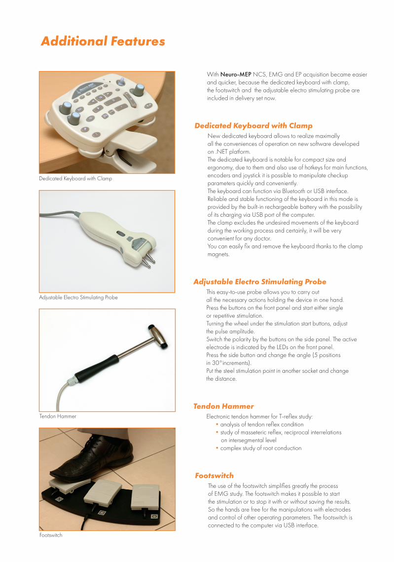
With Neuro-MEP NCS, EMG and EP acquisition became easier and quicker, because the dedicated keyboard with clamp, the footswitch and the adjustable electro stimulating probe are included in delivery set now.
Dedicated Keyboard with ClampNew dedicated keyboard allows to realize maximally all the conveniences of operation on new software developed on .NET platform. The dedicated keyboard is notable for сompact size and ergonomy, due to them and also use of hotkeys for main functions, encoders and joystick it is possible to manipulate checkup parameters quickly and conveniently. The keyboard can function via Bluetooth or USB interface. Reliable and stable functioning of the keyboard in this mode is provided by the built-in rechargeable battery with the possibility of its charging via USB port of the computer.The clamp excludes the undesired movements of the keyboard during the working process and certainly, it will be very convenient for any doctor.You can easily fix and remove the keyboard thanks to the clamp magnets.
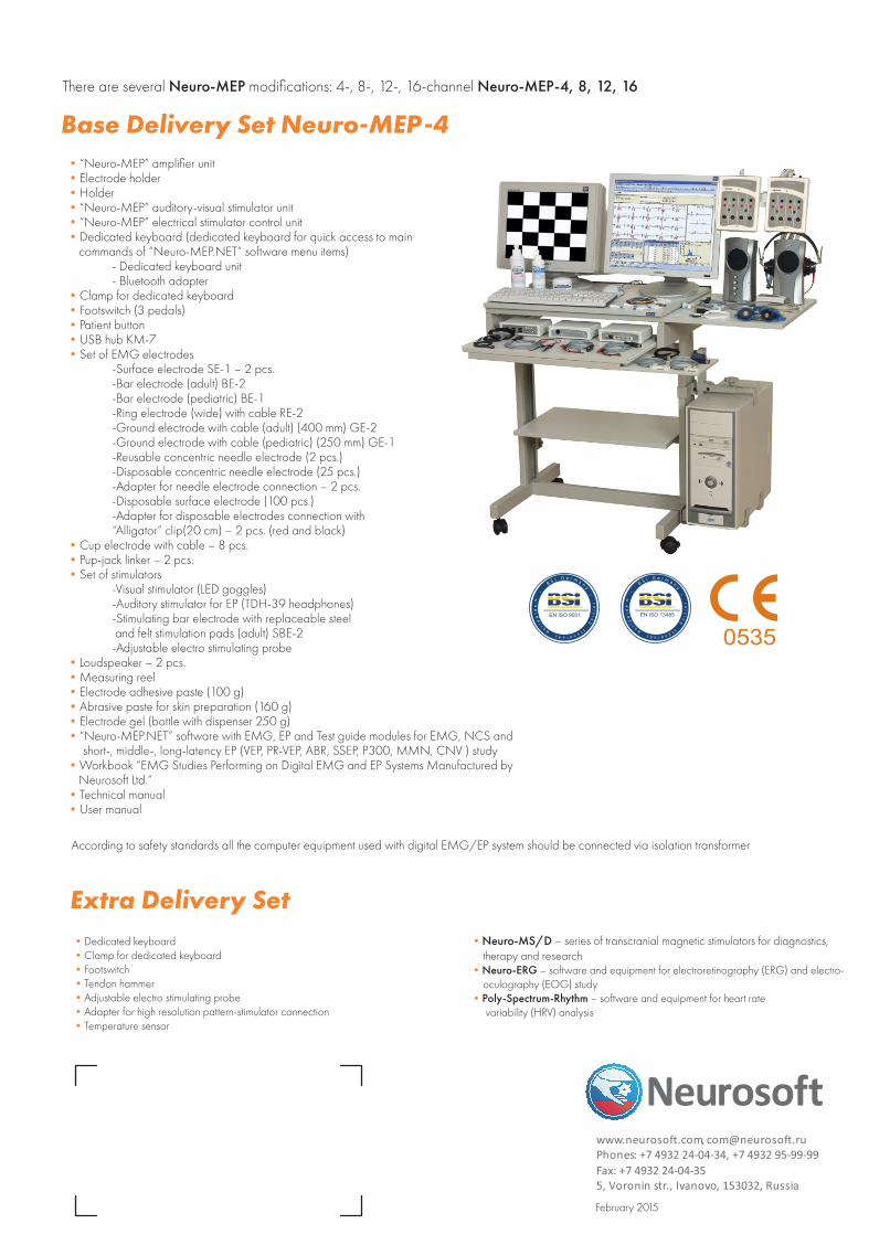
Adjustable Electro Stimulating ProbeThis easy-to-use probe allows you to carry out all the necessary actions holding the device in one hand. Press the buttons on the front panel and start either single or repetitive stimulation. Turning the wheel under the stimulation start buttons, adjust the pulse amplitude.Switch the polarity by the buttons on the side panel. The active electrode is indicated by the LEDs on the front panel.Press the side button and change the angle (5 positions in 30°increments).Put the steel stimulation point in another socket and change the distance.
Tendon HammerElectronic tendon hammer for T-reflex study:
•analysis of tendon reflex condition•study of masseteric reflex, reciprocal interrelations
on intersegmental level•complex study of root conduction
FootswitchThe use of the footswitch simplifies greatly the process of EMG study. The footswitch makes it possible to start the stimulation or to stop it with or without saving the results. So the hands are free for the manipulations with electrodes and control of other operating parameters. The footswitch is connected to the computer via USB interface.
Dedicated Keyboard with Clamp
Tendon Hammer
Footswitch
Adjustable Electro Stimulating Probe
Extra Delivery Set
Base Delivery Set Neuro-MEP-4
•Dedicated keyboard •Clamp for dedicated keyboard •Footswitch •Tendon hammer•Adjustable electro stimulating probe •Adapter for high resolution pattern-stimulator connection•Temperature sensor
•Neuro-MS/D – series of transcranial magnetic stimulators for diagnostics, therapy and research
•Neuro-ERG – software and equipment for electroretinography (ERG) and electro-oculography (EOG) study
•Poly-Spectrum-Rhythm – software and equipment for heart rate variability (HRV) analysis
Tendon HammerElectronic tendon hammer for T-reflex study:
•analysis of tendon reflex condition•study of masseteric reflex, reciprocal interrelations
on intersegmental level•complex study of root conduction
FootswitchThe use of the footswitch simplifies greatly the process of EMG study. The footswitch makes it possible to start the stimulation or to stop it with or without saving the results. So the hands are free for the manipulations with electrodes and control of other operating parameters. The footswitch is connected to the computer via USB interface.
•“Neuro-MEP” amplifier unit •Electrode holder •Holder•“Neuro-MEP” auditory-visual stimulator unit •“Neuro-MEP” electrical stimulator control unit •Dedicated keyboard (dedicated keyboard for quick access to main
commands of “Neuro-MEP.NET” software menu items) - Dedicated keyboard unit - Bluetooth adapter
•Clamp for dedicated keyboard •Footswitch (3 pedals) •Patient button •USB hub KM-7 •Set of EMG electrodes
-Surface electrode SE-1 – 2 pcs. -Bar electrode (adult) BE-2 -Bar electrode (pediatric) BE-1 -Ring electrode (wide) with cable RE-2 -Ground electrode with cable (adult) (400 mm) GE-2 -Ground electrode with cable (pediatric) (250 mm) GE-1 -Reusable concentric needle electrode (2 pcs.) -Disposable concentric needle electrode (25 pcs.) -Adapter for needle electrode connection – 2 pcs. -Disposable surface electrode (100 pcs.) -Adapter for disposable electrodes connection with “Alligator” clip(20 cm) – 2 pcs. (red and black)
•Cup electrode with cable – 8 pcs.•Pup-jack linker – 2 pcs. •Set of stimulators
-Visual stimulator (LED goggles) -Auditory stimulator for EP (TDH-39 headphones) -Stimulating bar electrode with replaceable steel and felt stimulation pads (adult) SBE-2 -Adjustable electro stimulating probe
•Loudspeaker – 2 pcs. •Measuring reel •Electrode adhesive paste (100 g) •Abrasive paste for skin preparation (160 g) •Electrode gel (bottle with dispenser 250 g) •“Neuro-MEP.NET” software with EMG, EP and Test guide modules for EMG, NCS and
short-, middle-, long-latency EP (VEP, PR-VEP, ABR, SSEP, P300, MMN, CNV ) study •Workbook “EMG Studies Performing on Digital EMG and EP Systems Manufactured by
Neurosoft Ltd.” •Technical manual •User manual
According to safety standards all the computer equipment used with digital EMG/EP system should be connected via isolation transformer
There are several Neuro-MEP modifications: 4-, 8-, 12-, 16-channel Neuro-MEP-4, 8, 12, 16
February 2015
,www.neurosoft.com [email protected]: +7 4932 24-04-34, +7 4932 95-99-99 Phones
Fax: +7 4932 24-04-355, Voronin str., Ivanovo, 153032, Russia

![Neuro Assessment for Scalp the Non-Neuro Nurse … · Neuro Assessment for the Non-Neuro Nurse Terry M. Foster, RN, ... Microsoft PowerPoint - Neuro Grand Forks ND [Read-Only] Author:](https://img.pdfslide.net/doc/110x75/5b88746b7f8b9a301e8d8c76/neuro-assessment-for-scalp-the-non-neuro-nurse-neuro-assessment-for-the-non-neuro.jpg)