Embed Size (px)
Citation preview
314 Gastroenterology & Hepatology Volume 14, Issue 5 May 2018
Endo
scop
y
ADVANCES IN ENDOSCOPY
Section Editor: Todd H. Baron, MD
C u r r e n t D e v e l o p m e n t s i n D i a g n o s t i c a n d T h e r a p e u t i c E n d o s c o p y
The Appropriate Use and Techniques of Tattooing in the Colon
G&H What are the reasons for endoscopically tattooing the colon?
DR Colon tattoos are used to mark lesions for subse-quent surgical resection or for later endoscopic resection, or to mark an endoscopic resection site for easy endo-scopic follow-up of the resection site. Alternatives to tattooing include taking an abdominal radiograph with the endoscope tip at the site of the lesion (for surgical location) or placing metal clips near a lesion followed by taking an abdominal radiograph. However, tattooing is widely considered the best method of marking the colon.
G&H Which lesions should be considered for marking?
DR Tattooing should be considered for obvious colorec-tal cancers and for lesions with suspected cancer, for pe-dunculated adenomas with endoscopic features of cancer or with sufficient size to have a substantial risk of cancer (≥2 cm in size is a reasonable guide), and for large flat or sessile lesions removed by piecemeal endoscopic mucosal resection (EMR) or by endoscopic submucosal dissection (ESD). Tattooing is appropriate for such lesions in all colon locations, except it is not required in the cecum because the cecum itself is a completely reliable landmark for both endoscopic and surgical follow-up. Similarly, some lesions in the proximal ascending colon do not need to be tattooed if the cecum is still visible. For the ce-cum and for some ascending colon lesions, a photograph
of the lesion with the appendiceal orifice or the ileocecal valve in view serves as an excellent record to guide surgi-cal resection or subsequent endoscopic follow-up.
G&H What types of ink or dye are available?
DR Carbon black is the only permanent tattoo ap-propriate for use in the colorectum. Carbon black is available in India ink, but pure carbon black is com-mercially available and approved by the US Food and Drug Administration in a formulation called Spot (GI Supply). A new formulation called Spot Ex is 50% darker than the original formulation; it is sold in 5-mL
Douglas K. Rex, MDDirector of EndoscopyIndiana University HospitalProfessor of MedicineDivision of Gastroenterology and HepatologyIndiana University School of MedicineIndianapolis, Indiana
... effective tattooing is the most reliable method of ensuring that the correct segment of colon is resected.
vials and is immediately ready for endoscopic injection. Other substances injected into the submucosa primarily for the purposes of facilitating EMR and ESD, such as indigo carmine, methylene blue, and indocyanine green, do not leave a permanent tattoo in the colon.
gh0518_endo_v1.indd 314 5/7/18 10:47 AM
Gastroenterology & Hepatology Volume 14, Issue 5 May 2018 315
Endo
scop
y
G&H How do the dyes compare in terms of safety, efficacy, and ease of use?
DR Carbon black in the form of India ink is a very ef-fective tattoo. However, because it contains several other substances that are immunologically active, injection of India ink has resulted in complications such as sterile abscess formation, focal peritonitis, and inflammatory pseudotumor, and has precipitated inflammatory bowel disease, although very rarely.
G&H What injection techniques are available, and how are they performed?
DR During tattooing, the goal is to place the entire injection volume into the submucosal space. Carbon black, when injected into the submucosa, creates a phys-ical black stain that can be seen endoscopically from the lumen and surgically from the peritoneal cavity. The most common error made during injection is to insert the needle through the colonic mucosa and simply begin the injection process. This action risks placing the tattoo in the peritoneal cavity or into extracolonic tissue such as the omentum, kidney, stomach, or small intestine.
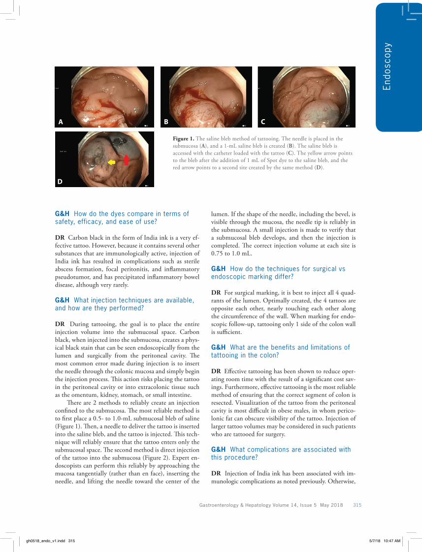
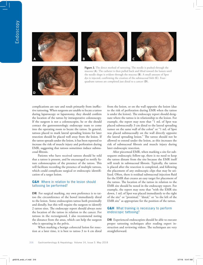
There are 2 methods to reliably create an injection confined to the submucosa. The most reliable method is to first place a 0.5- to 1.0-mL submucosal bleb of saline (Figure 1). Then, a needle to deliver the tattoo is inserted into the saline bleb, and the tattoo is injected. This tech-nique will reliably ensure that the tattoo enters only the submucosal space. The second method is direct injection of the tattoo into the submucosa (Figure 2). Expert en-doscopists can perform this reliably by approaching the mucosa tangentially (rather than en face), inserting the needle, and lifting the needle toward the center of the
lumen. If the shape of the needle, including the bevel, is visible through the mucosa, the needle tip is reliably in the submucosa. A small injection is made to verify that a submucosal bleb develops, and then the injection is completed. The correct injection volume at each site is 0.75 to 1.0 mL.
G&H How do the techniques for surgical vs endoscopic marking differ?
DR For surgical marking, it is best to inject all 4 quad-rants of the lumen. Optimally created, the 4 tattoos are opposite each other, nearly touching each other along the circumference of the wall. When marking for endo-scopic follow-up, tattooing only 1 side of the colon wall is sufficient.
G&H What are the benefits and limitations of tattooing in the colon?
DR Effective tattooing has been shown to reduce oper-ating room time with the result of a significant cost sav-ings. Furthermore, effective tattooing is the most reliable method of ensuring that the correct segment of colon is resected. Visualization of the tattoo from the peritoneal cavity is most difficult in obese males, in whom perico-lonic fat can obscure visibility of the tattoo. Injection of larger tattoo volumes may be considered in such patients who are tattooed for surgery.
G&H What complications are associated with this procedure?
DR Injection of India ink has been associated with im-munologic complications as noted previously. Otherwise,
Figure 1. The saline bleb method of tattooing. The needle is placed in the submucosa (A), and a 1-mL saline bleb is created (B). The saline bleb is accessed with the catheter loaded with the tattoo (C). The yellow arrow points to the bleb after the addition of 1 mL of Spot dye to the saline bleb, and the red arrow points to a second site created by the same method (D).
A B C
D
gh0518_endo_v1.indd 315 5/7/18 10:47 AM
316 Gastroenterology & Hepatology Volume 14, Issue 5 May 2018
Endo
scop
y
complications are rare and result primarily from ineffec-tive tattooing. When surgeons are unable to locate a tattoo during laparoscopy or laparotomy, they should confirm the location of the tattoo by intraoperative colonoscopy. If the surgeon is not a colonoscopist, he or she should contact the gastroenterologic endoscopy team to come into the operating room to locate the tattoo. In general, tattoos placed to mark lateral spreading lesions for later resection should be placed well away from the lesion. If the tattoo spreads under the lesion, it has been reported to increase the risk of muscle injury and perforation during EMR, suggesting that tattoos sometimes induce submu-cosal fibrosis.
Patients who have received tattoos should be told that a tattoo is present, and be encouraged to notify fu-ture colonoscopists of the presence of the tattoo. This will facilitate recording the presence of multiple tattoos, which could complicate surgical or endoscopic identifi-cation of a target lesion.
G&H Where in relation to the lesion should tattooing be performed?
DR For surgical marking, my own preference is to tat-too the circumference of the bowel immediately distal to the lesion. Some endoscopists tattoo both proximally and distally, but this will require the surgeon to identify 2 tattoo sites. The endoscopy report should always note the location of the tattoo in relation to the cancer. For tattoos in the rectosigmoid, I also recommend noting the distance from the anus, which can help the surgeon who is operating in the pelvis.
When marking a benign colorectal lesion for resec-tion at a later time, it is best to tattoo 3 to 4 cm distal
from the lesion, or on the wall opposite the lesion (due to the risk of perforation during EMR when the tattoo is under the lesion). The endoscopy report should desig-nate where the tattoo is in relationship to the lesion. For example, the report may note that “1 mL of Spot was placed submucosally 3 cm distal to the lateral spreading tumor on the same wall of the colon” or “1 mL of Spot was placed submucosally on the wall directly opposite the lateral spreading lesion.” The tattoo should not be allowed to extend under the lesion, as this increases the risk of submucosal fibrosis and muscle injury during later endoscopic resection.
After piecemeal EMR, when marking a site for sub-sequent endoscopic follow-up, there is no need to keep the tattoo distant from the site because the EMR itself will result in submucosal fibrosis. Typically, the tattoo is placed after the resection is completed, and following the placement of any endoscopic clips that may be uti-lized. Often, there is residual submucosal injection fluid for the EMR that creates an easy target for placement of the tattoo. The location of the tattoo in relation to the EMR site should be noted in the endoscopy report. For example, the report may note that “with the EMR site down, 1 mL of Spot was placed immediately to the right of the site” or “proximal,” “distal,” or “to the left of the EMR site” as appropriate for the position of the tattoo.
G&H What training is necessary to perform endoscopic tattooing?
DR Experienced endoscopists should be able to execute proper tattooing techniques after reading expert in-struction and reviewing videos. The techniques are very straightforward.
Figure 2. The direct method of tattooing. The needle is pushed through the mucosa (A). The catheter is then pulled back and lifted toward the lumen until the needle shape is evident through the mucosa (B). A small amount of Spot dye is injected, confirming the creation of the submucosal bleb (C). Four-quadrant tattoos are completed just distal to a cancer (D).
A B C
D
gh0518_endo_v1.indd 316 5/7/18 10:47 AM
Gastroenterology & Hepatology Volume 14, Issue 5 May 2018 317
Endo
scop
y
G&H Have any studies been conducted on the cost-effectiveness of the various dyes?
DR Cost analyses have demonstrated that tattooing is a cost-effective practice because it shortens operating room time significantly. The cost benefits of resecting the correct segment are obvious.
Dr Rex has no relevant conflicts of interest to disclose.
Suggested Reading
Kethu SR, Banerjee S, Desilets D, et al; ASGE Technology Committee. Endo-scopic tattooing. Gastrointest Endosc. 2010;72(4):681-685.
Letarte F, Webb M, Raval M, Karimuddin A, Brown CJ, Phang PT. Tattooing or not? A review of current practice and outcomes for laparoscopic colonic resection following endoscopy at a tertiary care centre. Can J Surg. 2017;60(6):394-398.
Trakarnsanga A, Akaraviputh T. Endoscopic tattooing of colorectal lesions: is it a risk-free procedure? World J Gastrointest Endosc. 2011;3(12):256-260.
Yang M, Pepe D, Schlachta CM, Alkhamesi NA. Endoscopic tattoo: the importance and need for standardised guidelines and protocol. J R Soc Med. 2017;110(7):287-291.
gh0518_endo_v1.indd 317 5/7/18 10:47 AM