Embed Size (px)
Citation preview
1
Engineered Soluble Monomeric IgG1 CH3 – Generation, Mechanisms of Function and
Implications for Design of Biological Therapeutics
Tianlei Ying1*
, Weizao Chen1, Yang Feng
1, Yanping Wang
1,2, Rui Gong
1, Dimiter S. Dimitrov
1
1Protein Interactions Group, Cancer and Inflammation Program, Center for Cancer Research,
National Cancer Institute, National Institutes of Health, and 2SAIC-Frederick, Inc., Frederick,
Maryland 21702
* Corresponding author. Mailing address:
Protein Interaction Group National Cancer Institute, NIH
Bldg 469, Rm 150B
Frederick, MD 21702
Tel: (301) 846-6275
Fax: (301) 846-5598
E-mail: [email protected]
Running Title: Monomeric IgG1 CH3
Background: The CH3 domain of an antibody is a homodimer.
Results: Soluble monomeric IgG1 CH3 (mCH3) exhibits pH-dependent binding to FcRn.
Conclusion: The mCH3 can be used as a new scaffold for generation of binders with potentially enhanced
half-life. Significance: The mCH3 is a promising fusion partner for therapeutic proteins with increased therapeutic
efficacy.
Most of the therapeutic antibodies approved for
clinical use are full-size IgG1 molecules. The
interaction of the IgG1 Fc with the neonatal Fc
receptor (FcRn) plays a critical role in
maintaining their long half-life. We have
hypothesized that isolated Fc domains could be
engineered to functionally mimic full-size IgG1
(nanoantibodies) but with decreased (10-fold)
size. Here, we report for the first time the
successful generation of a soluble, monomeric
CH3 domain (mCH3). In contrast to the wild-
type dimeric CH3, the mCH3 exhibited pH-
dependent binding to FcRn similar to that of Fc.
The binding free energy of mCH3 to FcRn was
higher than that of isolated CH2 but lower than
that of Fc. Therefore, CH3 may contribute a
larger portion of the free energy of binding to
FcRn than CH2. A fusion protein of mCH3
with an engineered antibody domain (m36.4)
also bound to FcRn in a pH-dependent fashion,
and exhibited significantly higher neutralizing
activity against HIV-1 than m36.4-Fc fusion
proteins. The m36.4-mCH3 fusion protein was
monomeric, stable, soluble and expressed at a
high level in E. coli. We also found that
engineering an additional disulfide bond in
mCH3 remarkably increased its thermal
stability while the FcRn binding was not
affected. These data suggest that mCH3 could
not only help in the exploration of the dual
mechanisms of the CH3 contribution to Fc
functions (dimerization and FcRn interactions)
but could also be used for the development of
candidate therapeutics with optimized half-life,
enhanced tissue penetration, access to sterically
restricted binding sites and increased
therapeutic efficacy.
http://www.jbc.org/cgi/doi/10.1074/jbc.M113.484154The latest version is at JBC Papers in Press. Published on July 18, 2013 as Manuscript M113.484154
Copyright 2013 by The American Society for Biochemistry and Molecular Biology, Inc.
by guest on September 8, 2018
http://ww
w.jbc.org/
Dow
nloaded from
2
INTRODUCTION
The vast majority of the monoclonal antibodies
(mAbs) approved for clinical use are full-size
antibodies in IgG1 format (1,2,3,4). The IgG1
CH3 domain has two important functions –
dimerization of the IgG1 Fc and interaction with
the neonatal Fc receptor (FcRn). It is well-
established that the Fc interaction with the FcRn
plays a critical role in maintaining the long half-
life of IgG1 (5,6,7). The antibody interacts with
FcRn by a pH-dependent mechanism that results
in the binding of the IgG1 Fc to FcRn in the acidic
environment of the endosomes, the recycling of
the IgG1 to the cell surface and the subsequent
release of IgG1 back into the circulation at
physiological pH. This process decreases IgG1
degradation, thereby extending its in vivo half-life.
The Fc-FcRn interaction has been the focus of a
number of engineering efforts seeking to modulate
the antibody pharmacokinetics, and fusion to IgG1
Fc (molecular mass ~55 kDa) has been developed
as an important strategy for extending the half-life
of therapeutic proteins (8,9). It is known that both
the CH2 and CH3 domains of the IgG1 Fc interact
with FcRn. Identification of the involved residues
has led to the development of Fc variants with
increased pH-dependent FcRn binding and in vivo
half-life (7,10,11,12). However, the individual
contribution of Fc domains to the pH-dependent
mechanism of FcRn binding is not known.
Identification of a domain that could best mimic
Fc in terms of binding to FcRn is also important
for the development of therapeutic proteins of both
optimized half-life and small size for enhanced
tissue penetration, access to sterically restricted
binding sites and lower production cost.
We have previously generated isolated
single CH2 domains and monomeric Fc (mFc) and
characterized their interactions with FcRn
(13,14,15). Here, we report for the first time the
successful generation of a soluble, monomeric
CH3 domain (mCH3). We found that the
engineering of CH3 by structure-based
mutagenesis, which resulted in soluble mFcs (15),
was not effective in the generation of soluble
mCH3. This was likely due to the absence of the
highly soluble CH2. In this current study we found
that a specific combination of four mutations is
essential in generating soluble mCH3. In contrast
to the wild-type dimeric CH3 (CH3), the mCH3
exhibited pH-dependent binding to a human
single-chain soluble FcRn (sFcRn) (15,16) which
resembled that of bacterially expressed Fc but with
lower affinity (KD = 940 nM) at pH 6. The free
energy of mCH3 binding to sFcRn was higher than
that of isolated CH2 and dimeric CH3 (which did
not bind FcRn) but lower than that of mFc. These
results indicate that CH3 in Fc may contribute a
larger portion of the free energy of binding to
sFcRn than CH2.
To increase the stability of isolated mCH3,
we engineered an additional disulfide bond which
resulted in a remarkable increase in the melting
temperature, Tm, from 40.6°C to 76.0°C, and a 5-
fold increase in protein expression with retained
binding to FcRn. These data suggest that a stable,
soluble mCH3 can be generated and used as a new
scaffold for the generation of binders with
potentially enhanced half-life.
We also demonstrated that a fusion protein
of mCH3 with an antibody heavy-chain variable
domain (VH), m36.4 (17), bound to FcRn in a pH-
dependent fashion and exhibited significantly
higher HIV-1 neutralizing activity than the large
size VH-Fc fusion proteins. This provides direct
evidence that the size of therapeutic proteins is
important for targeting sterically restricted
epitopes. In addition, we demonstrated that the
VH-mCH3 fusion protein was monomeric, stable,
and solubly expressed at a high level in E. coli.
Thus, the mCH3 is also a promising candidate for
a therapeutic protein fusion partner with
potentially better tissue penetration, reduced steric
hindrance and increased therapeutic efficacy.
EXPERIMENTAL PROCEDURES
Cloning of mCH3– The following primers
were used: Omp-F, 5'-
AAGACAGCTATCGCGATTGCAG-3'; gIII-
R, 5'-ATCACCGGAACCAGAGCCACCAC-
3’; CH3-F, 5'-
GTTGATGTAACGGCCCAGGCGGCCGGGCA
GCCCCGAGAAC-3'; dCH2-F, 5'-
GCCAAAGACAAAACTCACACAGCACCTGA
ACTCCTGGGGGGAC-3'; dCH2-R, 5'-
CAGGAGTTCAGGTGCTGTGTGAGTTTTGTC
TTTGGCTTTGGAGATGGTTTTC-3'. The wild-
type CH3 gene was amplified by PCR from an Fc-
expressing plasmid constructed in the pComb3x
by guest on September 8, 2018
http://ww
w.jbc.org/
Dow
nloaded from
3
vector (primer: CH3-F and gIII-R). Four residues
were mutated in mCH3 compared to wild-type
CH3 (residues 351, 366, 368 and 395). The
dimeric CH2 gene was generated by joining two
CH2 genes together with a human IgG1 hinge
(DKTHT) using an overlap-extension PCR
(primer: Omp-F, dCH2-R; dCH2-F, gIII-R).
Cloning of mCH3 fusion proteins– The m36.4
gene was amplified from the m36.4-encoding
plasmid pCom36.4, as described previously (17),
and was joined to the mCH3 gene to construct the
m36.4-mCH3 fusion protein. A (G4S)3 linker was
inserted between m36.4 and mCH3 by overlap-
extension PCR. The products were digested with
SfiI and cloned into a pComb3x
vector. m36.4h1Fc, the fusion protein with a
human IgG1 hinge region as a linker between
m36.4 and Fc, was generated as described
previously (17).
Generation of mCH3 P343C/A431C mutant
(mCH3cc)– The following primers were used:
P343C-F, 5'-
GTTGATGTAACGGCCCAGGCGGCCGGGCA
GTGCCGAGAACCACAGGTGTAC-3'; A431C-
F, 5'-
CTGCGTGTAGTGGTTGTGCAGACACTCATG
CATCACGGAGC-3'; A431C-R, 5'-
ATGCTCCGTGATGCATGAGTGTCTGCACAA
CCACTACACGCAG-3'. The mCH3 P343C
variant was generated by PCR using mCH3 as
template (primer: P343C-F and gIII-R). The
mCH3 P343C/A431C variant was generated by
the QuikChange Site-Directed Mutagenesis Kit
(Stratagene) using the primers A431C-F and
A431C-R.
Protein expression and purification– mCH3,
mCH3 fusion proteins, mCH3cc and other
antibody domains were expressed in E. coli
HB2151 by using a procedure similar to that
described previously (15). Protein purity was
judged by SDS-PAGE, and protein concentration
was measured spectrophotometrically (NanoVue,
GE Healthcare).
Size exclusion chromatography– Purified
antibody domains and fusion proteins were loaded
onto a Superdex 75 10/300 GL column running on
an FPLC AKTA BASIC pH/C system (GE
Healthcare). PBS (pH 7.4) was used as the running
buffer throughout (flow rate 0.5 mL/min), and
eluting proteins were monitored at 280 nm. The
molecular mass standards used were ribonuclease
A (13.7 kDa), chymotrypsinogen A (25
kDa), ovalbumin (44 kDa), bovine serum albumin
(67 kDa) and aldolase (158 kDa).
Circular dichroism (CD)– The CD
spectra were collected with an AVIV Model 202
spectropolarimeter (Aviv Biomedical). Purified
antibody domains and mCH3 fusion proteins were
dissolved in PBS, pH 7.4 at the final concentration
of 0.25 mg/mL. For native structure measurement,
spectra of mCH3 and wild-type CH3
were collected from 200 to 260 nm (0.1 cm path
length) at 25°C. For evaluation of thermal
stability, CD signals at 225 nm were recorded for
wild-type CH3, and signals at 216 nm were
recorded for all other antibody domains and fusion
proteins. The instrument was programmed to
acquire spectra at 1°C intervals over the range 25-
90°C.
Surface plasmon resonance binding
experiments– Surface plasmon resonance
measurements were performed using a BIAcore
X100 instrument (GE Healthcare). Purified human
soluble single-chain FcRn was immobilized on a
CM5 biosensor chip using a primary amine
coupling in 10 mM sodium acetate buffer (pH 5.0).
To test binding at pH 7.4, the proteins were diluted
in PBS plus 0.005% Tween 20. To test binding at
pH 6.0, the same running buffer was adjusted to
pH 6.0 with HCl. The running buffer was
allowed to flow through the cells at a rate of 30
μl/min. The analytes consisted of serial dilution of
proteins between 1 uM and 62.5 nM. The chip was
regenerated with pH 8.0 buffer (100 mM Tris, 50
mM NaCl, pH 8.0) after 10 min of dissociation.
ELISA– Recombinant protein A (Sigma-
Aldrich) and protein G (Invitrogen, Carlsbad, CA)
were coated on ELISA plate wells at 50 ng per
well in PBS overnight at 4°C, and blocked with
protein-free blocking buffer (Thermo Scientific) at
37°C for 2 hr. Two-fold serially diluted protein
was added and incubated at 37°C for 2 h. The
plates were washed with PBST,
and horseradish peroxidase (HRP)-conjugated
anti-FLAG tag antibody (Sigma-Aldrich) in PBS
by guest on September 8, 2018
http://ww
w.jbc.org/
Dow
nloaded from
4
was incubated in the wells for 1 h at 37°C. After
extensive washes with PBST, the binding was
detected by the addition of ABTS
substrate (Roche, Indianapolis, IN), and monitored
at 405 nm. For measurement of competition
between mCH3 and IgG1 ELISA plates were
coated with recombinant protein G and blocked as
described above. mCH3 at a concentration of 500
nM was premixed with two-fold serially diluted
competitor human IgG1 antibodies. Mixtures were
subsequently added to each ELISA well and
incubated. Bound mCH3 was detected with HRP-
conjugated anti-FLAG tag antibody, and the assay
was developed as described above.
Pseudovirus neutralization assay– Viruses
pseudotyped with HIV-1 Envs were prepared by
co-transfection of 70-80% confluent 293T cells
with pNL4-3.luc.E-R- and pSV7d constructs
encoding HIV-1 Envs by using the PolyFect
transfection reagent (Qiagen) according to the
manufacturer’s instructions. Pseudotyped viruses
were obtained after 48 hr by centrifugation and
filtration of cell culture through 0.45 μm filters.
Neutralization assays were performed as follows:
viruses were mixed with different concentrations
of antibodies for 1 h at 37°C, and then the mixture
was added to 1.5 × 10⁴ HOS-CD4-CXCR4 cells
grown in each well of 96-well plates. Luminesence
was measured after 48 hr by using the Bright-Glo
Luciferase Assay System (Promega, Madison, WI)
and a LumiCount microplate luminometer (Turner
Designs). Mean relative light units (RLU) for
duplicate wells were determined. Relative
infectivity (%) was calculated by the following
formula: (average RLU of antibody-containing
wells/average RLU of virus-only wells) × 100.
Simulation of molecular structures– The
initial coordinates of mCH3 and mCH3cc were
created based on the crystal structure of IgG1 Fc
(pdb entry: 2WAH). The m36.4-mCH3 and
m36.4h1Fc models were created with both VMD
program and Swiss-Model homology-modeling
server (http://swissmodel.expasy.org). The
structures were further simulated with CHARMM
force field by using NAMD program. Water
molecules were retained in the simulation. The
structures were first minimized for 5000 steps with
conjugate gradient method, and equilibrated for 10
ps with the time step of 1 fs, then were further
minimized for 100,000 steps for analysis with
VMD program.
RESULTS
Generation of soluble monomeric CH3
(mCH3). We previously reported the generation
of three monomeric Fc (mFc) proteins using a
novel multiple panning/screening procedure (15).
A combination of six or seven specific mutations
on the CH3 dimerization interface caused mFcs to
be highly soluble and monomeric. We
hypothesized that isolated single CH3 domains
from these mFcs would be also soluble and
monomeric. We therefore cloned the CH3 gene
from the mFcs into the pComb3x vector and tested
for soluble expression in E. coli. Unfortunately,
the isolated CH3 domains were not expressed
in soluble form in E. coli. To solve this problem
we performed structure-guided mutagenesis of
seven contact residues at the CH3 dimerization
interface. A specific combination of four
mutations (residues 351, 366, 368 and 395) (Fig. 1)
that is essential to generate mCH3 was identified.
Mutation of any of these residues in mCH3
eliminated the soluble expression. The expression
of mCH3 was relatively low (2 mg/L bacterial
culture).
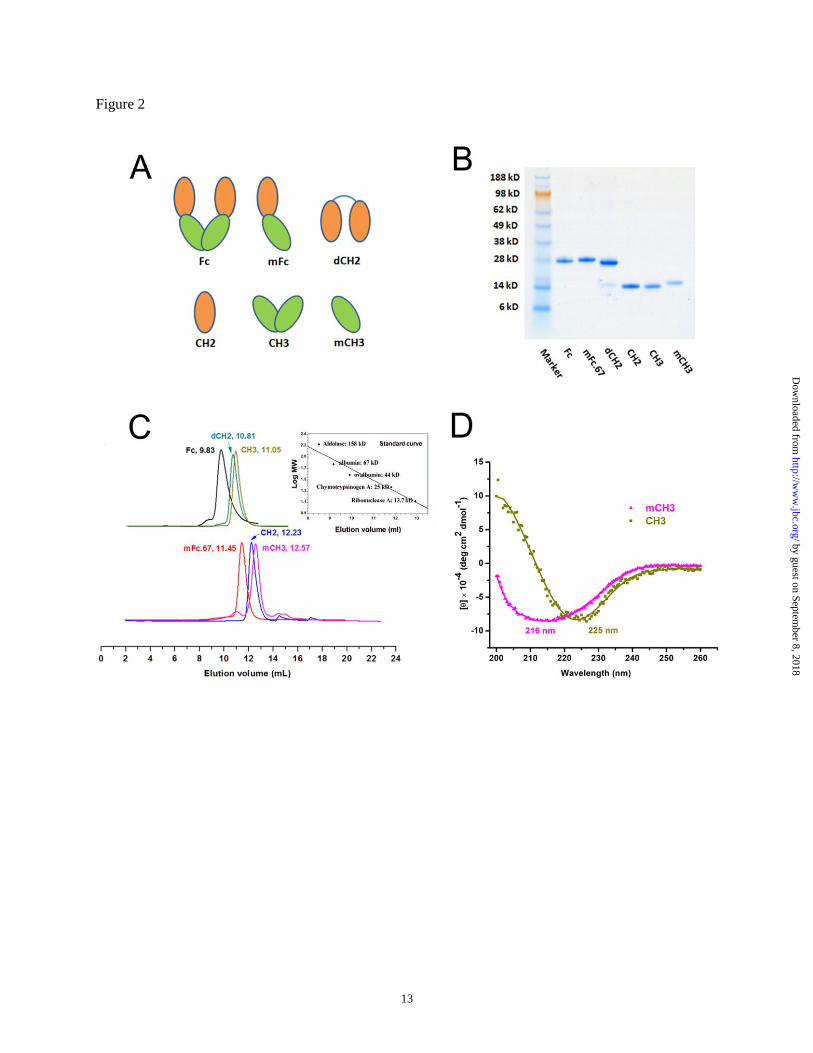
The isolation of the mCH3 provided an
opportunity for comparative analysis of IgG1 Fc
CH2 and CH3 domains in monomeric and dimeric
formats (Fig. 2A). The CH2 domain was
previously characterized and shown to have a
structure similar to that of the intact glycosylated
Fc (18). The dimeric CH2 (dCH2) was generated
by linking two CH2 molecules with a five residue
linker (DKTHT) from the human IgG1 hinge
region. The native dimeric CH3 was cloned from
the Fc with the same primers that were used to
generate mCH3. A monomeric Fc protein, mFc.67,
and the wild-type Fc were also expressed. All
domains and fragments were expressed and
purified as previously described (15).
There were no detectable impurities as shown
by SDS-PAGE (Fig. 2B). The mCH3 ran slightly
slower than the wild-type CH3 likely due to the
small difference in their molecular weights (MWs)
(14.4 kDa vs 14.3 kDa, respectively). The mCH3
was monomeric as demonstrated by size exclusion
by guest on September 8, 2018
http://ww
w.jbc.org/
Dow
nloaded from
5
chromatography (Fig. 2C). The CH2 was also
monomeric with a slightly larger size than the
mCH3 (Fig. 2C). The CH3 was dimeric (MW ~28
kDa) as expected, and the dCH2 which consists of
two CH2 molecules was of about the same size
(MW ~29 kDa).
Conformation and stability of mCH3. The
overall structure of CH2 and CH3 is a seven-
stranded antiparallel β-barrel with a buried
stabilizing disulfide bond (19). Despite the
topological similarity, the CH3 was found to be
much more stable than the CH2 (20,21). Whether
the high CH3 stability is due to the structure of the
CH3 molecule per se or to the homo-dimerization
was unknown. To investigate the influence of
dimerization on the structure of CH3, we first
compared the CD spectra of mCH3 and dimeric
CH3. Interestingly, we found significant
difference in their spectra. As shown in Fig. 2D,
the spectrum of mCH3 exhibited a local minimum
at 216 nm, which is very similar to that of an
isolated CH2, indicating that isolated mCH3 is
intact and well-folded. In contrast, the spectrum of
isolated wild-type (dimeric) CH3 exhibited a
minimum at 225 nm. These results suggest
that mCH3 has a conformational state different
from that of an isolated CH3 dimer.
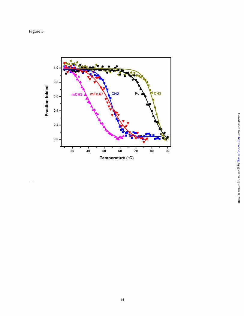
The thermal stability of mCH3 and other Fc
domains was then assessed by following changes
in the CD spectra in response to increasing
temperature (Fig. 3). The midpoint transition
(melting) temperature (Tm) of mCH3 was 40.6 ±
0.3°C. This value is similar to that of an isolated
murine CH2 domain, which was reported to have a
Tm of 41°C. The isolated human CH2 domain (Tm
= 54.5 ± 0.2°C) and mFc.67 (Tm = 51.0 ± 0.5°C)
are more stable than mCH3. Notably, the isolated
CH3 (Tm = 82.4 ± 0.6°C) was found to be more
stable than the wild-type Fc (Tm = 78.9 ± 0.5°C).
The dimeric CH2, in which two CH2 molecules
were joined by a linker, did not show enhanced
stability (Tm = 49.2 ± 0.5°C). Taken together,
these results indicate that strong intermolecular
hydrophobic interactions are critical for
stabilizing the CH3 homodimer although the four
mutations required for mCH3 generation could
also have destabilizing effects.
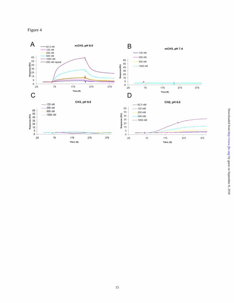
FcRn binding. We next explored whether
mCH3 could bind FcRn in a pH-dependent
manner. Binding to a human single-chain soluble
FcRn (sFcRn) immobilized on a BIAcore chip was
measured as described previously (15). The
measurements were carried out at pH 6.0 or pH
7.4, and the chips were regenerated after binding
by injection of a pH 8.0 buffer. While the isolated
wild-type dimeric CH3 did not show any FcRn
binding at either pH 6.0 or pH 7.4, significant
binding was observed at pH 6.0 for mCH3, but not
at pH 7.4 (Fig. 4A-C). Since mCH3 has a different
conformation than that of an isolated CH3, it is
likely that the conformational changes resulted in
a binding site on mCH3 more accessible to FcRn
than in the isolated CH3 dimer, thereby conferring
pH-dependent binding capability.
The calculated affinity (KD) of mCH3 was
940 nM. As expected, it was not as high as that of
the wild-type bacterially expressed Fc, which was
shown previously to bind with a KD of 126 nM
(15). Both CH2 and CH3 contribute to the Fc
interaction with FcRn. In an attempt to dissect
their relative contribution we also measured the
affinity of an isolated CH2 to FcRn. We found that
there was an unusual delay during the association
phase, and the dissociation of CH2 from the chip
was very slow (Fig. 4D). The calculated affinity
was 4 µM. We also noted that the CM5 chip could
be better regenerated by the pH 8.0 buffer after the
mCH3 binding measurement as compared to that
after the CH2 binding measurement, although
neither of the two proteins showed detectable
binding at pH 7.4. This behavior is not surprising
since most of the interface residues of the Fc/FcRn
complex in the CH2 domain are involved in
hydrophobic interactions, which are thought to be
inherently "sticky", as exemplified by residues
I253 and S254. In contrast, the FcRn binding
residues in the CH3 domain may participate in the
formation of titratable salt bridges, which are
likely to confer most of the pH dependence to the
Fc/FcRn interactions, as exemplified by residues
H433 and H435. These results indicate that the
CH2 and CH3 domain may have distinct binding
properties, and function differently when
participating in the pH-dependent interaction with
FcRn.
Protein A/G binding. It is well documented
that the Fc can bind a diverse set of proteins
(22,23). Examples include proteins A and G,
the bacterial cell wall proteins, which have
by guest on September 8, 2018
http://ww
w.jbc.org/
Dow
nloaded from
6
been widely used to detect and purify
immunoglobulins. Although they do not share
homology in sequence or structure, their binding
sites on the Fc were found to overlap at the inter-
CH2/CH3 domain region, which is also used by
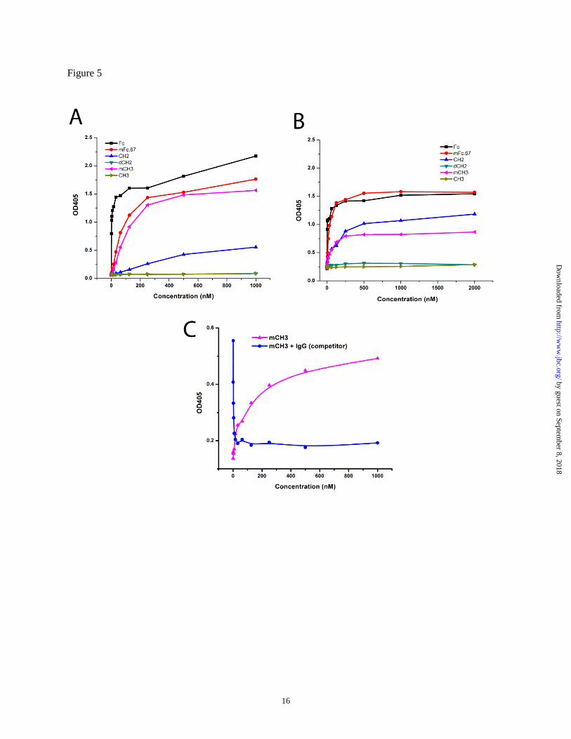
the Fc to interact with FcRn (23). We further
examined protein A and G binding to mCH3 and
CH2 using ELISA. As shown in Fig. 5, the wild-
type Fc binds to protein A or G with very low
EC50. The monomeric Fc, mFc.67, also has a high
binding capability to protein G but a weakened
binding to protein A. mCH3 can bind both protein
A and G, although its binding affinities for both
were decreased since it only possesses part of the
binding site of the wild-type Fc. The isolated CH2
domain can bind protein G but its binding to
protein A is very weak. Consistent with the FcRn
binding results described above, the isolated
dimeric CH3 did not bind to either protein A or G.
Additionally, the isolated CH2 monomer can bind
protein G but no detectable binding was observed
for the dimeric single-chain CH2 in which two
CH2 molecules were connected by a hinge. These
results suggest that the appropriate conformation
of an antibody domain is a key requirement for its
binding to protein A/G.
To analyze the binding site of protein G on
mCH3, we developed a competition ELISA assay.
IgG1 was shown to bind protein G more potently
than mCH3, and thus was used as a strong
competitor in this experiment. As shown in Fig.
5C, mCH3 binds well to protein G and was
competitively inhibited by IgG1, suggesting
that mCH3 and IgG1 share the same binding site
on protein G.
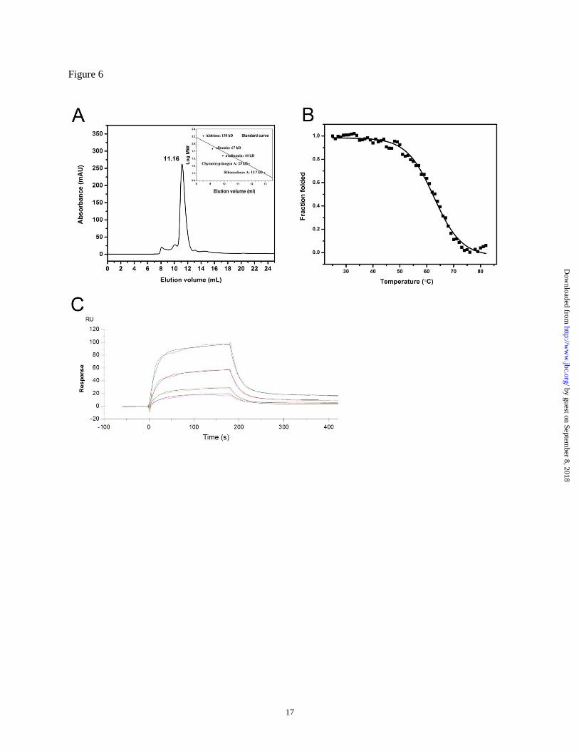
VH-mCH3 fusion protein is monomeric and
stable. We next examined whether mCH3 can be
used for generating monomeric and stable fusion
proteins. In this proof-of-concept study, m36.4,
a cross-reactive HIV-1 neutralizer (17), was used
to generate the VH-mCH3 fusion protein. m36.4
was joined to mCH3, resulting in a high
expression efficiency in E. coli (more than 10 mg
purified protein per liter culture). Size exclusion
chromatography analysis confirmed that the fusion
protein was monomeric, with a molecular size of
approximately 26 kDa (Fig. 6A). It was also found
to have a relatively high Tm of 62.9 ± 0.3°C (Fig.
6B).
Binding of the VH-mCH3 fusion protein to
FcRn. Fc fusion proteins have been found to
possess, in many cases, lower FcRn binding
affinities than those of mAbs, suggesting that the
FcRn binding region of the Fc fusion proteins may
exhibit some differences compared to native mAbs
(24). We investigated the FcRn binding of the VH-
mCH3 fusion protein. As shown in Fig. 6C, the KD
value calculated from this experiment was 685 nM
at pH 6.0, which is comparable to that of the
isolated mCH3 (940 nM). No binding was
observed at pH 7.4, indicating that the fusion
protein binds to sFcRn in a strictly pH-dependent
manner. These results confirmed that mCH3 could
be used as a fusion partner to confer pH-depedent
FcRn binding capability.
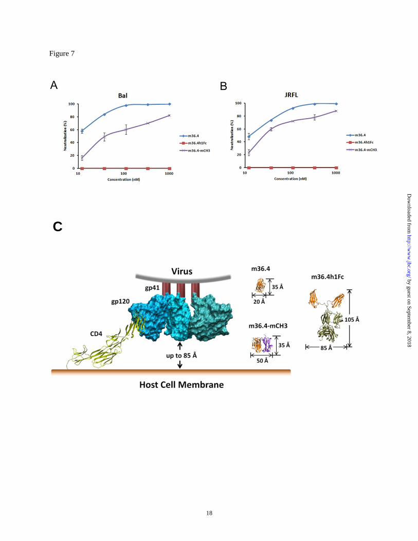
Potent neutralization of pseudotyped HIV-1
isolates by m36.4-mCH3. The engineered antibody
domain m36.4 targets highly conserved CD4-
induced structures on the HIV-1 envelope
glycoprotein (17). We have previously shown that
this epitope is sterically obstructed and fully
accessible only by relatively small-size
molecules during virus entry (17). To determine
the potency of HIV-1 neutralization by m36.4-
mCH3, viruses pseudotyped with Envs from the
HIV-1 isolates Bal and JRFL were used. The
dimeric CH3 fusion (MW ~60 kDa) and Fc fusion
(MW ~110 kDa) did not show any measurable
neutralizing activity against these two isolates. By
contrast, as shown in Fig. 7, m36.4 exhibited
particularly strong neutralization, with an IC50
(antibody concentration resulting in 50%
inhibition of virus infection) of 8 nM (Bal) and 25
nM (JRFL). The IC50 of m36.4-mCH3 was slightly
higher – 38 and 30 nM for Bal and JRFL
respectively. The slightly lower neutralizing
activity of m36.4-mCH3 compared to m36.4 is not
surprising, since its size (28 kDa) is twice that of
m36.4 (14 kDa). Although side-by-side
comparison was not done, it is noteworthy to point
out that m36.4-mCH3 appears to be more potent
than the gp41-derived peptide C34, which has an
IC50 of 270 nM against Bal and JRFL (17,25). The
C34 exhibits HIV-entry inhibitory activity
comparable to or higher than that of the FDA
approved peptide entry inhibitor T20 (brand name
Fuzeon) which is in clinical use. These results
suggest that m36.4-mCH3 could have potential as
by guest on September 8, 2018
http://ww
w.jbc.org/
Dow
nloaded from
7
an HIV entry inhibitor for therapeutic/prophylactic
applications.
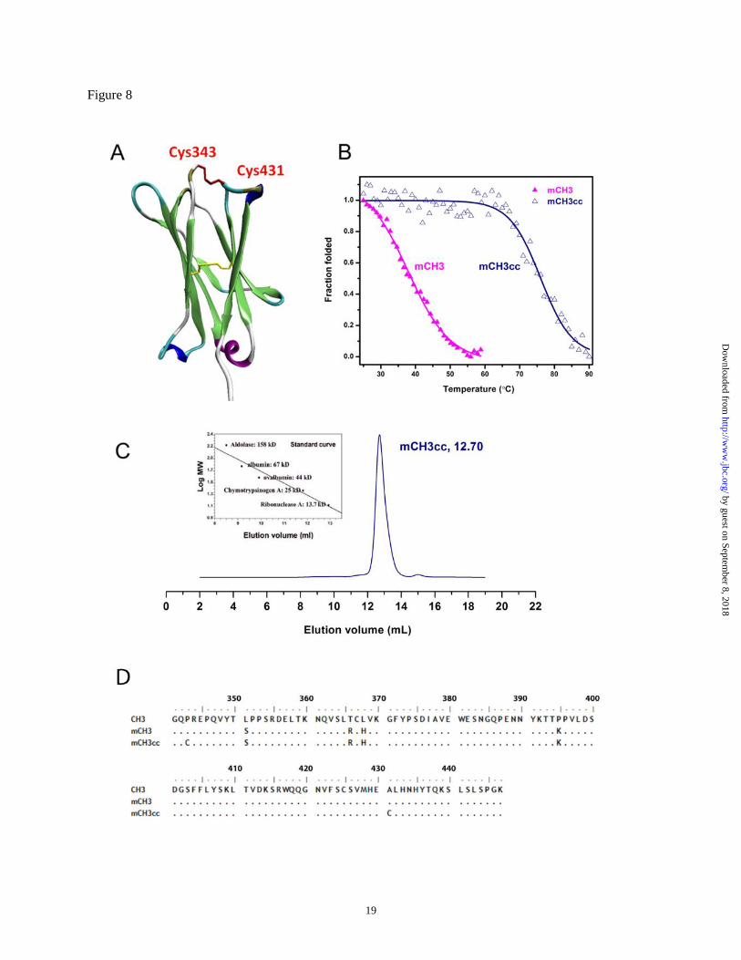
Increasing the mCH3 stability by an
engineered disulfide bond. The thermal stability of
mCH3 was relatively low (Tm = 40.6°C). We and
others have reported that the introduction of
additional disulfide bonds can enhance the
stability of CH2 and Fc (13,26). We used
structure-based design to choose specific residues
for introduction of additional cysteines and
generated the mCH3 P343C/A431C mutant
(mCH3cc) which has an additional disulfide bond
between the Cys343 and Cys431 which joins the
N-terminal A strand and the C-terminal G strand
of the mCH3 (Fig. 8A). We found that the
mCH3cc can be more efficiently produced (yield
of more than 10 mg/L bacterial culture) in E. coli
than mCH3 and is significantly more stable than
mCH3. The Tm of the mCH3cc was 76.0 ± 0.6°C,
35.4°C higher than that of the mCH3 (Fig. 8B).
The engineered CH3 monomer was also >99%
monomeric as indicated by size exclusion
chromatography (Fig. 8C). The affinity (KD) of
mCH3cc to sFcRn was calculated to be 1.1 uM at
pH 6.0, similar to that of the mCH3 (940 nM). No
binding was detected at pH 7.4, indicating that the
strictly pH-dependent FcRn binding capacity was
retained.
DISCUSSION
The biological function of CH3 is two-
fold: it provides bivalency through dimerization
and increases half-life in the circulation by
contributing to the Fc interaction with FcRn. To
understand the mechanisms of these functions we
generated an isolated IgG1 CH3
monomer, and expressed it in E. coli. This is, to
the best of our knowledge, the first report
describing the development of a soluble
monomeric antibody CH3 domain. We found that
the isolated dimeric CH3 was more stable than the
mCH3 and the wild-type dimeric Fc, while the
dimeric CH2 did not show enhanced stability
compared to an isolated CH2 monomer. This
result indicates that the strong intermolecular
hydrophobic interactions in the CH3 dimer are
important for stabilizing the Fc and the IgG1
homodimer. However, these results should be
taken with a note of caution, since we do not yet
know to what extent the four mutations required
for generation of the mCH3 contribute to its low
thermal stability.
The critical role of FcRn in
maintaining the long serum half-life of
IgG1 is well established, and the engineering of
the FcRn-Fc interaction holds promise for
modulating the pharmacokinetics of therapeutic
antibodies (7,10,12). The FcRn binds to a region at
the CH2-CH3 domain interface, and both domains
contribute to the interactions. For example,
positions 253 and 254 of the CH2 domain, and
positions 435 and 436 of the CH3 domain were
found to be critical for FcRn binding, since
mutation of any of these residues results in greatly
reduced binding affinity (27,28,29). In this study,
we found that the absolute value of the free energy
of binding (ΔG) of mCH3 to sFcRn (-34.6 kJ/mol)
was higher than that of isolated CH2 (-31 kJ/mol)
and that of dimeric CH3 (which did not bind FcRn)
but lower than that of mFc (-38.4 kJ/mol). These
results indicate that CH3 in Fc may contribute a
larger portion of the free energy of binding to
FcRn than CH2. It also appears that in relation to
binding to FcRn the native CH3 conformation as
part of Fc could be more similar to that of isolated
mCH3 than to that of isolated dimeric CH3.
It was previously suggested that local
conformational changes in the relative orientation
of two CH2 domains play a pivotal role in the
FcγR binding activity of IgG (30). It was also
noted that the CD spectra of mCH3 and CH2
resemble the spectrum of the reduced CH3, which
was shown to undergo a conformational change to
the oxidized CH3, but the structure of the reduced
CH3 remained intact and dimeric (31). We also
found that the mCH3 exhibited pH-dependent
binding to FcRn while the isolated dimeric CH3
did not bind, suggesting that the loop containing
residues 434-436 was more accessible to FcRn in
mCH3, or that mCH3 (but not CH3) underwent
other structural rearrangements resulting in
enhanced interactions with FcRn. These results
taken together suggest that structural mobility
could be an inherent property of CH2 and CH3
domains, and is likely to play a role in mediating
interactions of Fc with other biomolecules. We
expect that these results could provide a better
understanding of the mechanism of action of the
Fc and help in producing antibodies with
by guest on September 8, 2018
http://ww
w.jbc.org/
Dow
nloaded from
8
favorable pharmacokinetics and/or effector
functions.
Antibody fragments with binding activity
of full-size mAbs and engineered to possess other
unique and superior properties may constitute the
next wave of clinically useful antibody-based
therapeutics (32,33,34,35,36). Their small size
enables better tissue penetration compared to full-
size molecules, especially for penetration into
solid tumors. Moreover, the small size of antibody
domains and fragments provides better access to
sterically restricted epitopes. Antibody fragments
usually display greatly reduced half-life compared
to full-size mAbs; this has been a major issue for
clinical therapies. Finding optimized size and half-
life of antibody fragments is therefore of
paramount value. Importantly, the IgG1 mCH3 as
described in this study represents a very small
structural unit that possesses strictly pH-dependent
FcRn binding. We also proved that mCH3 can be
engineered to be more stable and more efficient in
soluble expression while the FcRn binding
remained unaffected.
We have also shown that the m36.4-
mCH3 fusion protein was highly expressed,
monomeric, soluble and stable. Furthermore, it
binds to FcRn in a strictly pH-dependent manner,
and possesses potent neutralization activity
against HIV pseudoviruses. Thus, VH-mCH3
represents a novel antibody fragment format. This
kind of antibody fragment is of fully human origin
with only four mutations in the wild-type CH3, so
the immunogenicity should be low if any. It is a
bispecific format, which can target antigens using
its VH domain and bind to FcRn using the mCH3
domain. The addition of mCH3 does not affect the
activity and stability of VH; therefore, a number of
engineered antibody domains targeting various
epitopes, as well as other therapeutic proteins (e.g.,
interferons, interleulins), can be fused to mCH3 to
acquire the additional pH-dependent FcRn binding
activity. Moreover, the size of the VH-mCH3 is
only 28 kDa, which is similar to that of a scFv.
The small size offers many advantages. For
instance, size is the determining factor for
efficiently targeting sterically restricted epitopes.
As shown in Fig. 7C, the distance between the
membrane-proximal portion of gp120 and the host
cell membrane is approximately 85 Å after CD4
attachment (37), making the CD4-induced gp120
epitopes accessible to small-size molecules (eg.,
m36.4 and m36.4-mCH3) but not to molecules
with relatively large size (Fc fusion protein).
Other advantages include good tissue penetration,
and higher molar quantities per gram of products
leading to a significant increase in potency per
dose and a reduction in overall manufacturing
cost.
In conclusion, we report here the first
successful development of a soluble monomeric
IgG1 CH3 that could be a valuable tool for
understanding the mechanisms of CH3
dimerization and FcRn binding as well as for
development of novel types of small biological
therapeutics.
REFERENCES
1. Carter, P. J. (2006) Nat Rev Immunol 6, 343-357
2. Dimitrov, D. S., and Marks, J. D. (2009) Methods Mol Biol 525, 1-27, xiii
3. Reichert, J. M. (2011) MAbs 3, 76-99
4. Dimitrov, D. S. (2012) Methods Mol Biol 899, 1-26
5. Simister, N. E., and Mostov, K. E. (1989) Nature 337, 184-187
6. Ober, R. J., Martinez, C., Vaccaro, C., Zhou, J., and Ward, E. S. (2004) J Immunol 172,
2021-2029
7. Roopenian, D. C., and Akilesh, S. (2007) Nat Rev Immunol 7, 715-725
8. Flanagan, M. L., Arias, R. S., Hu, P., Khawli, L. A., and Epstein, A. L. (2007) Methods
Mol Biol 378, 33-52
9. Jazayeri, J. A., and Carroll, G. J. (2008) BioDrugs 22, 11-26
10. Zalevsky, J., Chamberlain, A. K., Horton, H. M., Karki, S., Leung, I. W., Sproule, T. J.,
Lazar, G. A., Roopenian, D. C., and Desjarlais, J. R. (2010) Nat Biotechnol 28, 157-159
by guest on September 8, 2018
http://ww
w.jbc.org/
Dow
nloaded from
9
11. Hinton, P. R., Johlfs, M. G., Xiong, J. M., Hanestad, K., Ong, K. C., Bullock, C., Keller,
S., Tang, M. T., Tso, J. Y., Vasquez, M., and Tsurushita, N. (2004) J Biol Chem 279,
6213-6216
12. Vaccaro, C., Zhou, J., Ober, R. J., and Ward, E. S. (2005) Nat Biotechnol 23, 1283-1288
13. Gong, R., Vu, B. K., Feng, Y., Prieto, D. A., Dyba, M. A., Walsh, J. D., Prabakaran, P.,
Veenstra, T. D., Tarasov, S. G., Ishima, R., and Dimitrov, D. S. (2009) J Biol Chem 284,
14203-14210
14. Gong, R., Wang, Y., Feng, Y., Zhao, Q., and Dimitrov, D. S. (2011) J Biol Chem 286,
27288-27293
15. Ying, T., Chen, W., Gong, R., Feng, Y., and Dimitrov, D. S. (2012) J Biol Chem 287,
19399-19408
16. Feng, Y., Gong, R., and Dimitrov, D. S. (2011) Protein Expr Purif 79, 66-71
17. Chen, W., Zhu, Z., Feng, Y., and Dimitrov, D. S. (2008) Proc Natl Acad Sci U S A 105,
17121-17126
18. Prabakaran, P., Vu, B. K., Gan, J., Feng, Y., Dimitrov, D. S., and Ji, X. (2008) Acta
Crystallogr D Biol Crystallogr 64, 1062-1067
19. Bork, P., Holm, L., and Sander, C. (1994) J Mol Biol 242, 309-320
20. Feige, M. J., Walter, S., and Buchner, J. (2004) J Mol Biol 344, 107-118
21. Thies, M. J., Mayer, J., Augustine, J. G., Frederick, C. A., Lilie, H., and Buchner, J.
(1999) J Mol Biol 293, 67-79
22. Jefferis, R., Lund, J., and Pound, J. D. (1998) Immunol Rev 163, 59-76
23. DeLano, W. L., Ultsch, M. H., de Vos, A. M., and Wells, J. A. (2000) Science 287, 1279-
1283
24. Suzuki, T., Ishii-Watabe, A., Tada, M., Kobayashi, T., Kanayasu-Toyoda, T., Kawanishi,
T., and Yamaguchi, T. (2010) J Immunol 184, 1968-1976
25. Chan, D. C., Fass, D., Berger, J. M., and Kim, P. S. (1997) Cell 89, 263-273
26. Wozniak-Knopp, G., Stadlmann, J., and Ruker, F. (2012) PLoS One 7, e30083
27. Vaughn, D. E., Milburn, C. M., Penny, D. M., Martin, W. L., Johnson, J. L., and
Bjorkman, P. J. (1997) J Mol Biol 274, 597-607
28. Shields, R. L., Namenuk, A. K., Hong, K., Meng, Y. G., Rae, J., Briggs, J., Xie, D., Lai,
J., Stadlen, A., Li, B., Fox, J. A., and Presta, L. G. (2001) J Biol Chem 276, 6591-6604
29. Martin, W. L., West, A. P., Jr., Gan, L., and Bjorkman, P. J. (2001) Mol Cell 7, 867-877
30. Sondermann, P., Huber, R., Oosthuizen, V., and Jacob, U. (2000) Nature 406, 267-273
31. Thies, M. J., Talamo, F., Mayer, M., Bell, S., Ruoppolo, M., Marino, G., and Buchner, J.
(2002) J Mol Biol 319, 1267-1277
32. Holt, L. J., Herring, C., Jespers, L. S., Woolven, B. P., and Tomlinson, I. M. (2003)
Trends Biotechnol 21, 484-490
33. Holliger, P., and Hudson, P. J. (2005) Nat Biotechnol 23, 1126-1136
34. Saerens, D., Ghassabeh, G. H., and Muyldermans, S. (2008) Curr Opin Pharmacol 8,
600-608
35. Dimitrov, D. S. (2009) MAbs 1, 26-28
36. Nelson, A. L., and Reichert, J. M. (2009) Nat Biotechnol 27, 331-337
37. Labrijn, A. F., Poignard, P., Raja, A., Zwick, M. B., Delgado, K., Franti, M., Binley, J.,
Vivona, V., Grundner, C., Huang, C. C., Venturi, M., Petropoulos, C. J., Wrin, T.,
Dimitrov, D. S., Robinson, J., Kwong, P. D., Wyatt, R. T., Sodroski, J., and Burton, D. R.
(2003) J Virol 77, 10557-10565
by guest on September 8, 2018
http://ww
w.jbc.org/
Dow
nloaded from
10
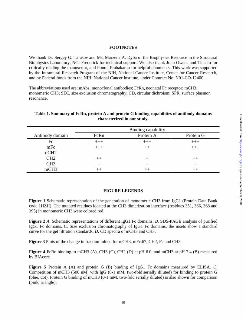
FOOTNOTES
We thank Dr. Sergey G. Tarasov and Ms. Marzena A. Dyba of the Biophysics Resource in the Structural
Biophysics Laboratory, NCI-Frederick for technical support. We also thank John Owens and Tina Ju for
critically reading the manuscript, and Ponraj Prabakaran for helpful comments. This work was supported
by the Intramural Research Program of the NIH, National Cancer Institute, Center for Cancer Research,
and by Federal funds from the NIH, National Cancer Institute, under Contract No. N01-CO-12400.
The abbreviations used are: mAbs, monoclonal antibodies; FcRn, neonatal Fc receptor; mCH3,
monomeric CH3; SEC, size exclusion chromatography; CD, circular dichroism; SPR, surface plasmon
resonance.
Table 1. Summary of FcRn, protein A and protein G binding capabilities of antibody domains
characterized in our study.
Binding capability
Antibody domain FcRn Protein A Protein G
Fc +++ +++ +++
mFc +++ ++ +++
dCH2 – – –
CH2 ++ + ++
CH3 – – –
mCH3 ++ ++ ++
FIGURE LEGENDS
Figure 1 Schematic representation of the generation of monomeric CH3 from IgG1 (Protein Data Bank
code 1HZH). The mutated residues located at the CH3 dimerization interface (residues 351, 366, 368 and
395) in monomeric CH3 were colored red.
Figure 2 A. Schematic representations of different IgG1 Fc domains. B. SDS-PAGE analysis of purified
IgG1 Fc domains. C. Size exclusion chromatography of IgG1 Fc domains, the insets show a standard
curve for the gel filtration standards. D. CD spectra of mCH3 and CH3.
Figure 3 Plots of the change in fraction folded for mCH3, mFc.67, CH2, Fc and CH3.
Figure 4 FcRn binding to mCH3 (A), CH3 (C), CH2 (D) at pH 6.0, and mCH3 at pH 7.4 (B) measured
by BIAcore.
Figure 5 Protein A (A) and protein G (B) binding of IgG1 Fc domains measured by ELISA. C.
Competition of mCH3 (500 nM) with IgG (0-1 mM, two-fold serially diluted) for binding to protein G
(blue, dot). Protein G binding of mCH3 (0-1 mM, two-fold serially diluted) is also shown for comparison
(pink, triangle).
by guest on September 8, 2018
http://ww
w.jbc.org/
Dow
nloaded from
11
Figure 6 A. Size exclusion chromatography of m36.4-mCH3, the insets show a standard curve for the gel
filtration standards. B. Plots of the change in fraction folded for m36.4-mCH3. C. sFcRn binding of
m36.4-mCH3 measured at pH 6.0 by BIAcore.
Figure 7 Neutralization of viruses pseudotyped with Envs of HIV-1 isolates Bal (A) and JRFL (B). C.
Schematic representations of the HIV-1 envelope glycoprotein gp120 interacts with the CD4 receptor at
the cell membrane. The predicted distance between the membrane-proximal portion of gp120 and the host
cell membrance is up to 85 Å. This sterically restricted epitope could be reached by the small-size m36.4
and m36.4-mCH3 molecules but could be blocked by the targeting of large-size m36.4h1Fc. Due to their
linker flexibilities, only the minimum hydrodynamic dimensions of mCH3-m36.4 and m36.4h1Fc were
shown.
Figure 8 A. Schematic representation of the mCH3 P343C/A431C variant (mCH3cc). Its native disulfide
bond was colored yellow. The introduced disulfide bond between Cys343 and Cys431 was colored red. B.
Plots of the change in fraction folded for mCH3cc. C. Size exclusion chromatography of mCH3cc. D.
amino acid sequence alignment of wide-type CH3, mCH3 and mCH3cc.
by guest on September 8, 2018
http://ww
w.jbc.org/
Dow
nloaded from
18
Figure 7
C
by guest on September 8, 2018
http://ww
w.jbc.org/
Dow
nloaded from
Tianlei Ying, Weizao Chen, Yang Feng, Yanping Wang, Rui Gong and Dimiter S. DimitrovImplications for Design of Biological Therapeutics
Engineered Soluble Monomeric IgG1 CH3 - Generation, Mechanisms of Function and
published online July 18, 2013J. Biol. Chem.
10.1074/jbc.M113.484154Access the most updated version of this article at doi:
Alerts:
When a correction for this article is posted•
When this article is cited•
to choose from all of JBC's e-mail alertsClick here
by guest on September 8, 2018
http://ww
w.jbc.org/
Dow
nloaded from