Embed Size (px)
Citation preview
ENTEROBACTERIACEAE 2 (LACTOSE FERMENTERS)
DR. AMADIN A. OLOTU
LECTURER/CONSULTANT MEDICAL MICROBIOLOGIST
BOWEN UNIVERSITY/BOWEN UNIVERSITY TEACHING HOSPITAL OGBOMOSO

Some Clinically Important Lactose Fermenting Enterobacteriaceae• Escherichia
Escherichia coli
• Klebsiella
Klebsiella pneumonia
Klebsiella oxytoca
• Enterobacter spp
Enterobacter aerogenes
Enterobacter agglomerans
Enterobacter cloacae
• Citrobacter
Citrobacter freundii
Citrobacter diversus
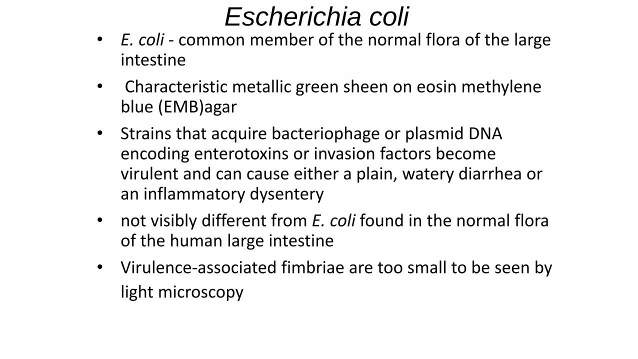
• E. coli - common member of the normal flora of the large intestine
• Characteristic metallic green sheen on eosin methylene blue (EMB)agar
• Strains that acquire bacteriophage or plasmid DNA encoding enterotoxins or invasion factors become virulent and can cause either a plain, watery diarrhea or an inflammatory dysentery
• not visibly different from E. coli found in the normal flora of the human large intestine
• Virulence-associated fimbriae are too small to be seen by
light microscopy
Escherichia coli
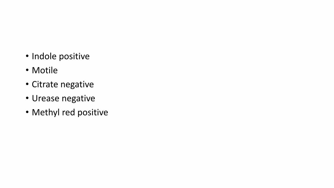
• Indole positive
• Motile
• Citrate negative
• Urease negative
• Methyl red positive
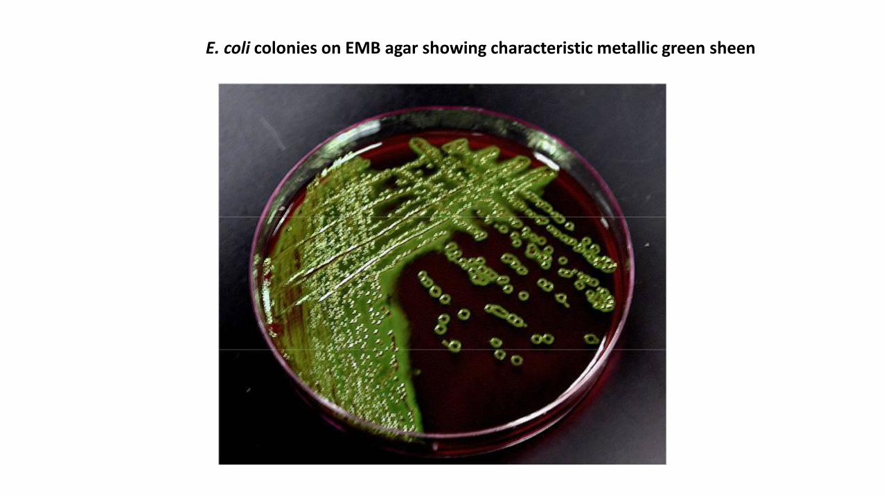
E. coli colonies on EMB agar showing characteristic metallic green sheen
• possess specific virulence-associated genes on plasmids
• Sero grouped according to the presence or absence of specific heat-stable somatic antigens (O antigens) composed of polysaccharide chains linked to the core lipopolysaccharide (LPS) complex common to all Gram-negative bacteria
• More than 170 different O-specific antigens have been defined since Kauffmann began this method of typing E coli in 1943
• Many O groups of E coli are cross-reactive or identical with specific O groups of--- Shigella, Salmonella, Klebsiella.
• H - flagellar antigens--- at least 56 types.
• E coli isolates may be nonmotile and nonflagellated and hence H negative (H).
• Serotyping is important for E. coli associated with diarrheal disease for two reasons.
• 1. a strain causing an outbreak or epidemic can be differentiated from the normal stool flora by its unique O:H antigenic makeup.
• 2. most virulent strains belong to specific serotypes
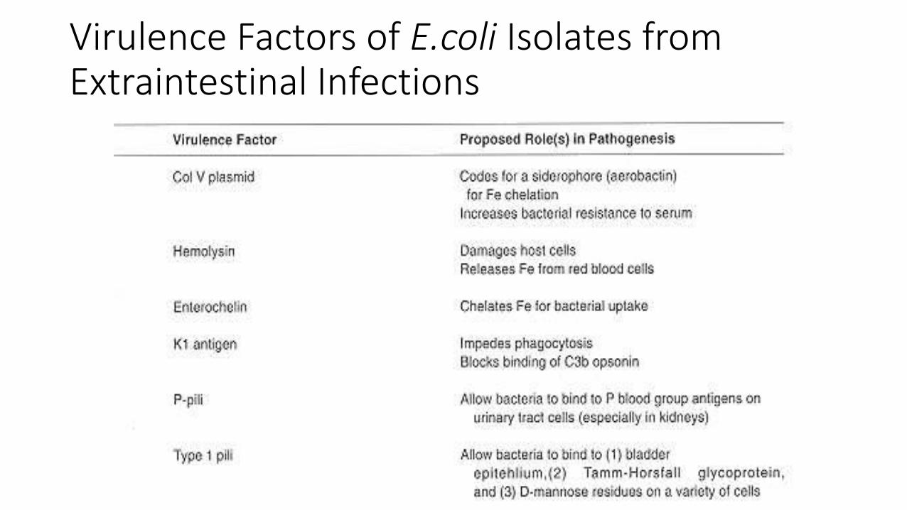
Virulence Factors of E.coli Isolates fromExtraintestinal Infections
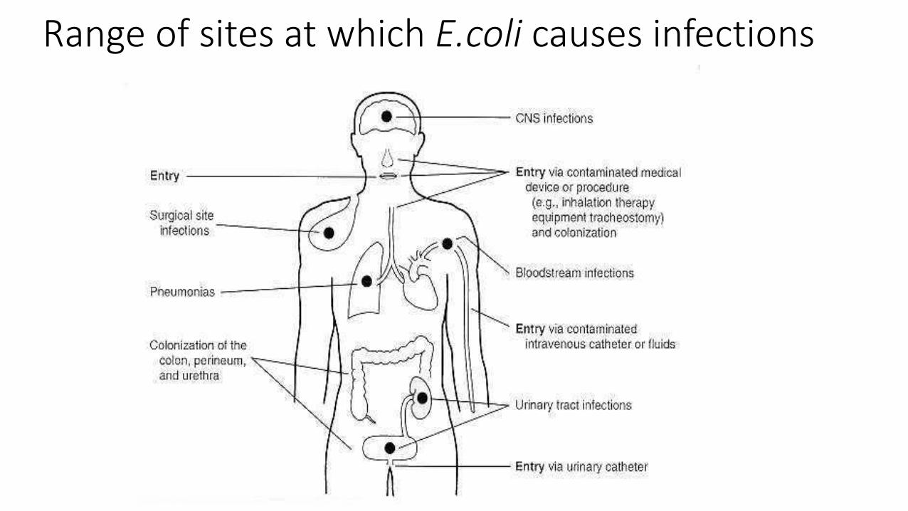
Range of sites at which E.coli causes infections
Diseases/Infections
• The infections caused by E. coli and the other enterobacteriaceaecannot be differentiated by symptoms or signs from infections caused by other bacteria.
• Clinical manifestations depend on the site of the infection.

Uropathogenic E. coli
• Urinary tract infection—E. coli is the most common cause of urinary tract infection and accounts for approximately 90% of first urinary tract infections in young women. The symptoms and signs include urinary frequency , dysuria, hematuria, and pyuria. Flank pain is associated with upper tract infection..
• Uropathogenic E. coli have specifically elaborated virulence factors that facilitate colonization and subsequent clinical infections.
• Typically these organisms produce hemolysin, which is cytotoxic and facilitates tissue invasion. Those strains that cause pyelonephritis express K antigen and elaborate a specific type of pilus, P fimbriae, which binds to the P blood group antigen.
• Meningitis—E. coli is a common cause of meningitis in infants. Approximately 75% of E. coli from meningitis cases have the K1 antigen.
• Sepsis—E. coli may reach the bloodstream and cause sepsis. Sepsis may occur secondary to other infections.
Diarrheogenic E coli
• E coli-associated diarrheal diseases—E coli that cause diarrhea are extremely common worldwide. These E coli are classified by the characteristics of their virulence properties (see below), and each group causes disease by a different mechanism. The small or large bowel epithelial cell adherence properties are encoded by genes on plasmids.
• Similarly , the toxins often are plasmid- or phage-mediated.

Enterotoxigenic E. coli
• Enterotoxigenic E. coli (ETEC) is a common cause of "traveler's diarrhea"
• a very important cause of diarrhea in infants in developing countries.
• ETEC colonization factors specific for humans promote adherence of ETEC to epithelial cells of the small bowel.
• Some strains of ETEC produce a heat-labile exotoxin (LT) (MW 80,000) that is under the genetic control of a plasmid.
• Its subunit B attaches to the GM1 ganglioside at the brush border of epithelial cells of the small intestine and facilitates the entry of subunit A (MW 26,000) into the cell
• Subunit A activates adenylyl cyclase. This markedly increases the local concentration of cyclic adenosine monophosphate (cAMP), which results in intense and prolonged hypersecretion of water and chlorides and inhibits the reabsorption of sodium.
.
• The gut lumen is distended with fluid, and hypermotility and diarrhea result, lasting for several days.
• LT is antigenic and cross-reacts with the enterotoxin of Vibrio cholerae to which it is similar although not identical.
• LT stimulates the production of neutralizing antibodies in the serum (and perhaps on the gut surface) of persons previously infected with enterotoxigenic E. coli.
• Persons residing in areas where such organisms are highly prevalent (eg, in some developing countries) are likely to possess antibodies and are less prone to develop diarrhea on reexposure to the LT –producing E. coli.
• Some strains of ETEC produce the heat-stable enterotoxin ST a (MW 1500–4000), which is under the genetic control of a heterogeneous group of plasmids.
• ST activates guanylyl cyclase in enteric epithelial cells and stimulates fluid secretion.
• Many ST-positive strains also produce LT . The strains with both toxins produce a more severe diarrhea.
• The plasmids carrying the genes for enterotoxins (LT , ST) also may carry genes for the colonization factors that facilitate the attachment of E. coli strains to intestinal epithelium.
• It is possible that virtually any E. coli may acquire a plasmid encoding for enterotoxins.
Enterohemorrhagic E. coli (EHEC) /Shiga toxin producing E coli (STEC)• Enterohemorrhagic E. coli (EHEC) /Shiga toxin producing E coli (STEC):
• They produce cytotoxic toxins.
• There are at least two antigenic forms of the toxin referred to as Shiga-like toxin 1 and Shiga-like toxin 2.
• STEC has been associated with two things
• hemorrhagic colitis, a severe form of bloody diarrhea,
• and with hemolytic uremic syndrome (HUS), a disease resulting in • acute renal failure,
• microangiopathic hemolytic anemia,
• and thrombocytopenia.
• The Shiga-like toxins have many properties that are similar to the Shiga toxin produced by some strains of Shigella dysenteriae type 1;
• the two toxins however,are antigenically and genetically distinct.
• Of the E. coli serotypes that produce Shiga toxin, O157:H7 is the most common and is the one that can be identified in clinical specimens.
• STEC O157:H7 does not use sorbitol, unlike most other E coli, and is negative on sorbitol MacConkey agar (sorbitol is used instead of lactose). Many of the non-O157 serotypes may be sorbitol positive, when grown in culture.
• Specific antisera are used to identify the O157:H7 strains.
• Assays for Shiga toxin using commercially available enzyme immunoassays are done in many laboratories.
• Other sensitive test methods include cell culture cytotoxin testing using Vero cells and polymerase chain reaction for the direct detection of toxin genes directly from stool samples.
• Many cases of hemorrhagic colitis and its associated complications can be prevented by thoroughly cooking ground beef .
Enteropathogenic E. coli (EPEC)
• Enteropathogenic E. coli (EPEC) is an important cause of diarrhea in infants, especially in developing countries. EPEC previously was associated with outbreaks of diarrhea in nurseries in developed countries.
• EPEC adhere to the mucosal cells of the small bowel. Chromosomally mediated factors promote tight adherence. There is loss of microvilli (effacement), formation of filamentous actin pedestals or cup-like structures, and, occasionally , entry of the EPEC into the mucosal cells.

• Characteristic lesions can be seen on electron micrographs of small bowel biopsy lesions.
• The result of EPEC infection is watery diarrhea, which is usually self -limited but can be chronic. EPEC diarrhea has been associated with multiple specific serotypes of E coli;
• strains are identified by O antigen and occasionally by H antigen typing. A two-stage infection model using HEp-2 cells also can be performed. T ests to identify EPEC are performed in reference laboratories.
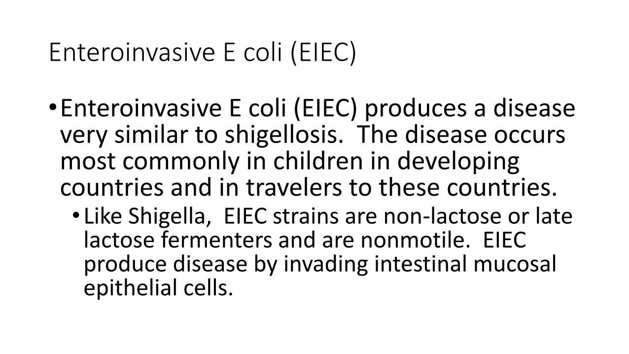
Enteroinvasive E coli (EIEC)
•Enteroinvasive E coli (EIEC) produces a disease very similar to shigellosis. The disease occurs most commonly in children in developing countries and in travelers to these countries. •Like Shigella, EIEC strains are non-lactose or late
lactose fermenters and are nonmotile. EIEC produce disease by invading intestinal mucosal epithelial cells.

Enteroaggregative E coli (EAEC)
•Enteroaggregative E coli (EAEC) causes acute and chronic diarrhea (>14 days in duration) in persons in developing countries. • These organisms also are the cause of food-borne
illnesses in industrialized countries. • They are characterized by their specific patterns of
adherence to human cells. • EAEC produce ST -like toxin and a hemolysin.
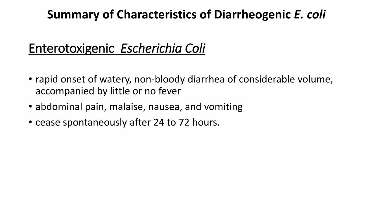
Enterotoxigenic Escherichia Coli
• rapid onset of watery, non-bloody diarrhea of considerable volume, accompanied by little or no fever
• abdominal pain, malaise, nausea, and vomiting
• cease spontaneously after 24 to 72 hours.
Summary of Characteristics of Diarrheogenic E. coli
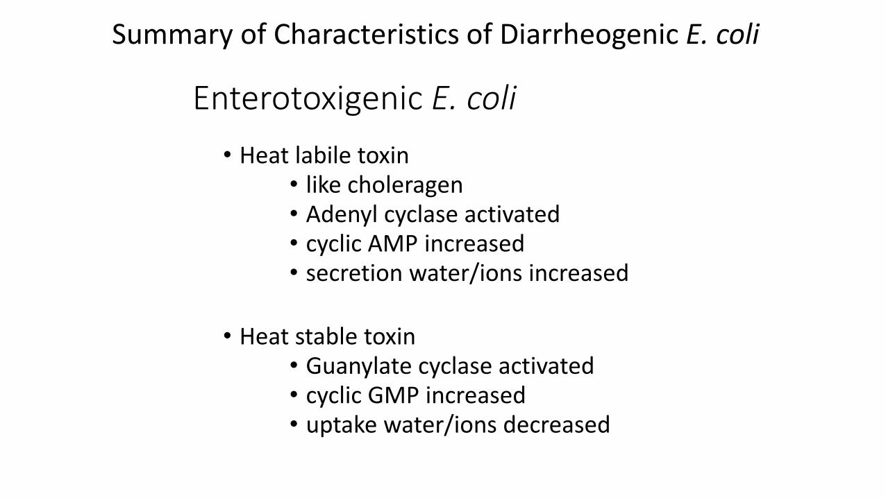
Enterotoxigenic E. coli
• Heat labile toxin• like choleragen• Adenyl cyclase activated • cyclic AMP increased• secretion water/ions increased
• Heat stable toxin• Guanylate cyclase activated • cyclic GMP increased• uptake water/ions decreased
Summary of Characteristics of Diarrheogenic E. coli
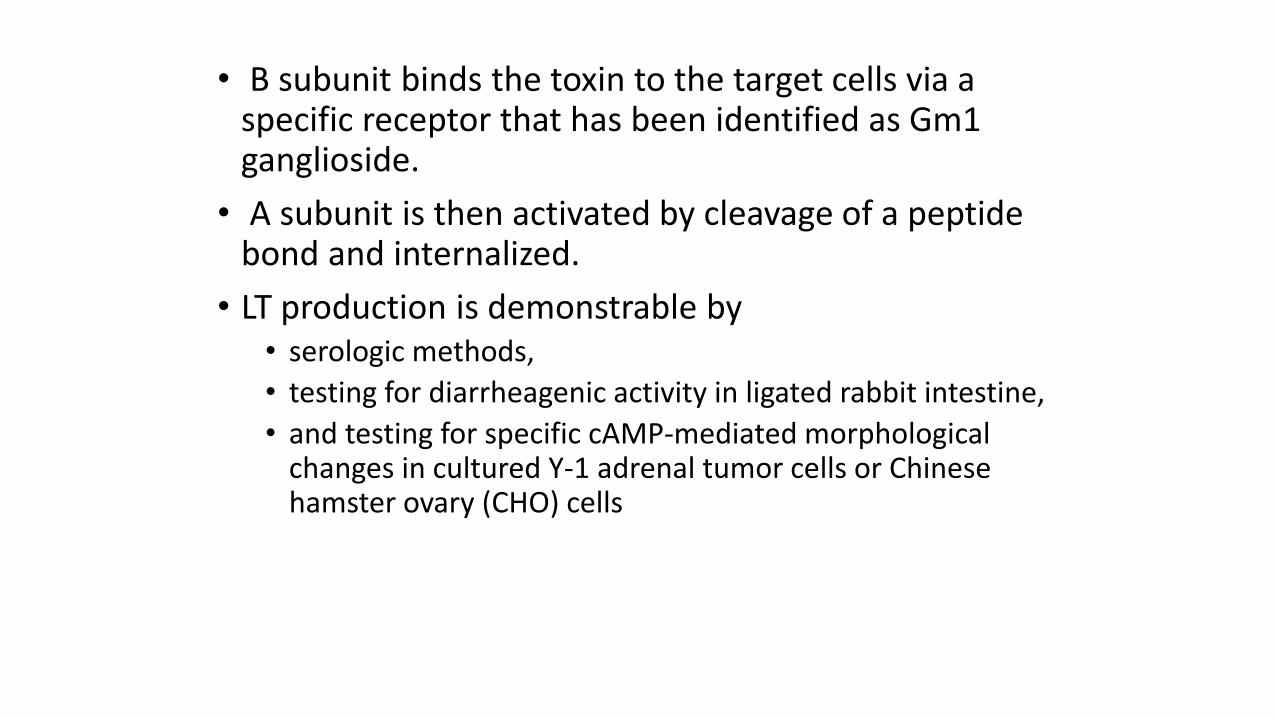
• B subunit binds the toxin to the target cells via a specific receptor that has been identified as Gm1 ganglioside.
• A subunit is then activated by cleavage of a peptide bond and internalized.
• LT production is demonstrable by• serologic methods,
• testing for diarrheagenic activity in ligated rabbit intestine,
• and testing for specific cAMP-mediated morphological changes in cultured Y-1 adrenal tumor cells or Chinese hamster ovary (CHO) cells
Host Defenses • First line of defense against ETEC diarrhea is
•gastric acidity
•small-intestinal motility and
•normal flora in the large intestine.
• (IgA) directed against surface antigens appears to be the key to immunity from ETEC diarrhea
• Passive immune protection of infants by colostral antibody is important
• Human breast milk also contains nonimmunoglobulin factors (receptor-containing molecules) that can neutralize E coli toxins and CFAs
Diagnosis
• Confirmation is achieved by
• serotyping,
• serologic identification of a specific CFA on isolates,
• demonstration of LT or ST production, and
• identification of genes encoding these virulence factors

Enteroinvasive E. coli (EIEC )
• Superficial tissue invasion with dysentery
• resembles shigellosis
• Affects children
• Non lactose or late lactose fermenters
• nonmotile
Enteropathogenic E. coli
• destruction of surface microvilli
• fever
• non-bloody watery diarrhea which is usually self limiting but can be chronic
• nausea
• vomiting
Enterohemorrhagic E. coli (Shiga toxin producing E. coli) O157:H7 most common
• Vero toxin or verocytotoxin
• “shiga-like”
• Hemolysins
• Associated with
• Hemorrhagic colitis
• Hemolytic uremic syndrome (HUS)• Microangiopathic hemolytic anemia• Thrombocytopenia• Anaemia
• negative on sorbitol MacConkey agar
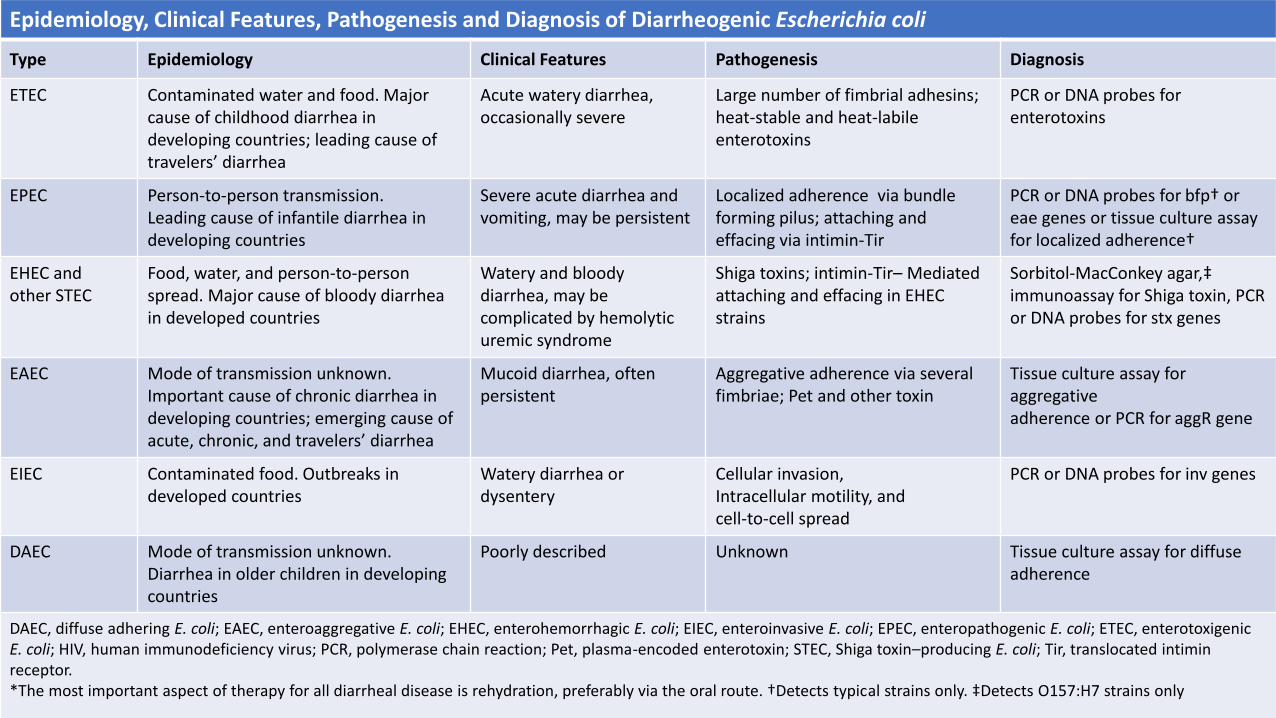
Epidemiology, Clinical Features, Pathogenesis and Diagnosis of Diarrheogenic Escherichia coli
Type Epidemiology Clinical Features Pathogenesis Diagnosis
ETEC Contaminated water and food. Major cause of childhood diarrhea in developing countries; leading cause of travelers’ diarrhea
Acute watery diarrhea, occasionally severe
Large number of fimbrial adhesins; heat-stable and heat-labile enterotoxins
PCR or DNA probes for enterotoxins
EPEC Person-to-person transmission.Leading cause of infantile diarrhea indeveloping countries
Severe acute diarrhea and vomiting, may be persistent
Localized adherence via bundle forming pilus; attaching and effacing via intimin-Tir
PCR or DNA probes for bfp† or eae genes or tissue culture assay for localized adherence†
EHEC andother STEC
Food, water, and person-to-personspread. Major cause of bloody diarrhea in developed countries
Watery and bloody diarrhea, may be complicated by hemolytic uremic syndrome
Shiga toxins; intimin-Tir– Mediated attaching and effacing in EHEC strains
Sorbitol-MacConkey agar,‡ immunoassay for Shiga toxin, PCR or DNA probes for stx genes
EAEC Mode of transmission unknown. Important cause of chronic diarrhea in developing countries; emerging cause of acute, chronic, and travelers’ diarrhea
Mucoid diarrhea, often persistent
Aggregative adherence via several fimbriae; Pet and other toxin
Tissue culture assay for aggregativeadherence or PCR for aggR gene
EIEC Contaminated food. Outbreaks indeveloped countries
Watery diarrhea ordysentery
Cellular invasion,Intracellular motility, andcell-to-cell spread
PCR or DNA probes for inv genes
DAEC Mode of transmission unknown. Diarrhea in older children in developing countries
Poorly described Unknown Tissue culture assay for diffuseadherence
DAEC, diffuse adhering E. coli; EAEC, enteroaggregative E. coli; EHEC, enterohemorrhagic E. coli; EIEC, enteroinvasive E. coli; EPEC, enteropathogenic E. coli; ETEC, enterotoxigenic E. coli; HIV, human immunodeficiency virus; PCR, polymerase chain reaction; Pet, plasma-encoded enterotoxin; STEC, Shiga toxin–producing E. coli; Tir, translocated intimin receptor.*The most important aspect of therapy for all diarrheal disease is rehydration, preferably via the oral route. †Detects typical strains only. ‡Detects O157:H7 strains only
ADAPTED FROM MANDELL, DOUGLAS, AND BENNETT’S PRINCIPLES AND PRACTICE OF INFECTIOUS DISEASES SEVENTH EDITION
Klebsiella• Also called “Friedlander’s Bacillus”
• Mucoid growth
• Large polysaccharide capsules
• Lack of motility
• Citrate positive, Voges-Proskauer positive, lysine decarboxylase positive
• K. pneumoniae causes 1% of bacterial pneumonias (Friedlander’s pneumonia)
• Can produce extensive hemorrhagic necrotizing consolidation of the lung
• Causes health care associated infections
• Urease is thought to play a major role in the production of infection-induced urinary stones
• K. pneumoniae is present in the respiratory tract and feces of about 5% of normal individuals.
• It causes a small proportion (about 1%) of bacterial pneumonias.
• K pneumoniae can produce extensive hemorrhagic necrotizing consolidation of the lung.
• It produces urinary tract infection and bacteremia with focal lesions in debilitated patients.
• Other enterics also may produce pneumonia.
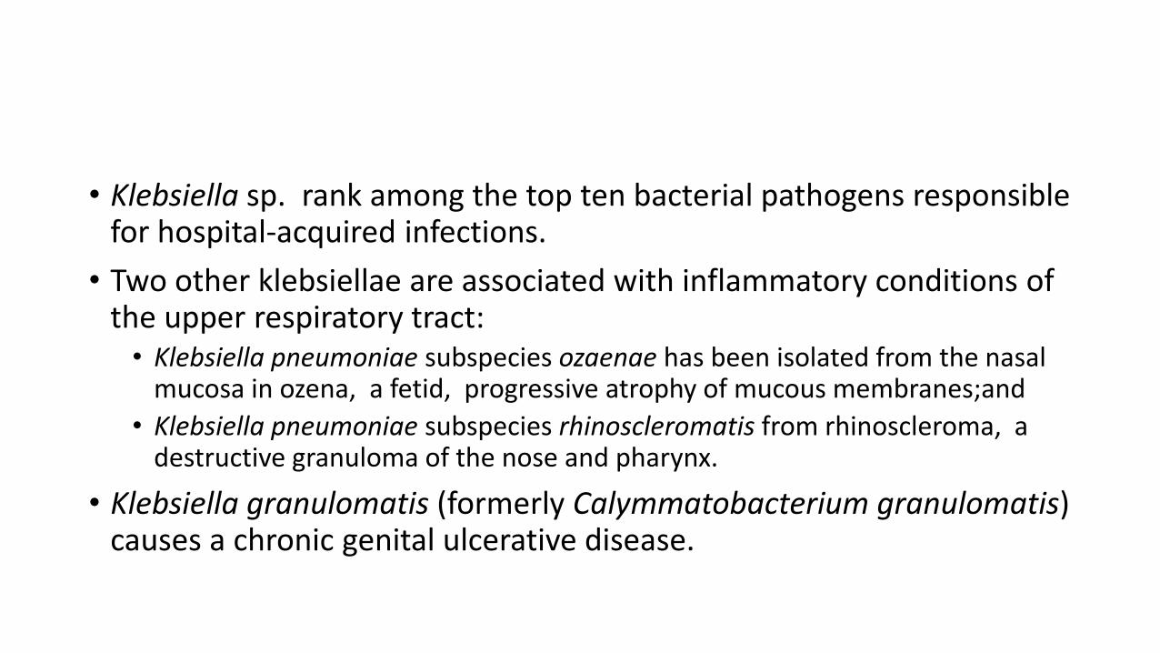
• Klebsiella sp. rank among the top ten bacterial pathogens responsible for hospital-acquired infections.
• Two other klebsiellae are associated with inflammatory conditions of the upper respiratory tract: • Klebsiella pneumoniae subspecies ozaenae has been isolated from the nasal
mucosa in ozena, a fetid, progressive atrophy of mucous membranes;and
• Klebsiella pneumoniae subspecies rhinoscleromatis from rhinoscleroma, a destructive granuloma of the nose and pharynx.
• Klebsiella granulomatis (formerly Calymmatobacterium granulomatis) causes a chronic genital ulcerative disease.
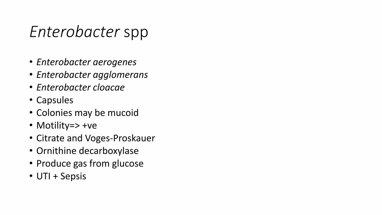
Enterobacter spp
• Enterobacter aerogenes• Enterobacter agglomerans• Enterobacter cloacae• Capsules• Colonies may be mucoid• Motility=> +ve• Citrate and Voges-Proskauer• Ornithine decarboxylase• Produce gas from glucose• UTI + Sepsis
• Enterobacter: These bacteria ferment lactose, may contain capsules that produce mucoid colonies and they are motile.
• These organisms cause a broad range of hospital acquired infections such as pneumonia, urinary tract infections, wound and device infections.
• Most strains possess a chromosomal -lactamase called ampC which renders them intrinsically resistant to ampicillin and first and second generation cephalosporins.
• Mutants may hyperproduce -lactamase conferring resistance to third generation cephalosporins.
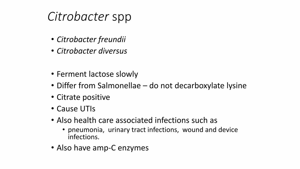
Citrobacter spp
• Citrobacter freundii
• Citrobacter diversus
• Ferment lactose slowly
• Differ from Salmonellae – do not decarboxylate lysine
• Citrate positive
• Cause UTIs
• Also health care associated infections such as • pneumonia, urinary tract infections, wound and device
infections.
• Also have amp-C enzymes
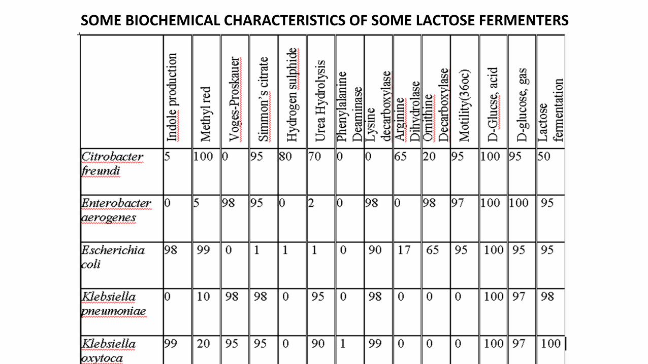
SOME BIOCHEMICAL CHARACTERISTICS OF SOME LACTOSE FERMENTERS

BIBLIOGRAPHY AND REFERENCES• Mandel, Douglas and Bennetts’ principles and practice of
infectious diseases. 9th ed.Philadelphia. ChurchhillLivingstone Elsevier
• Jawetz, Melnink and Adelberg’s medical microbiology. 28th
edition

THANK YOU FOR YOUR ATTENTION