Embed Size (px)
Citation preview
Enterovirus surveillance guidelinesGuidelines for enterovirus surveillance in support of the Polio Eradication Initiative
Enterovirus surveillance guidelines
Guidelines for enterovirus surveillance in support of the Polio Eradication Initiative.
iii
ENTEROVIRUS SURVEILLANCE GUIDELINES
These guidelines provide national decision-makers and their technical advisors with information on the principles and practices of adopting enterovirus surveillance in support of the Polio Eradication Initiative. The guidelines will help them decide if an enterovirus surveillance system could be established, or existing systems modified, that would meet both the disease control and programmatic requirements.
ABSTRACT
© World Health Organization 2015
All rights reserved. The Regional Office for Europe of the World Health Organization welcomes requests for permission to reproduce or translate its publications, in part or in full.
The designations employed and the presentation of the material in this publication do not imply the expression of any opinion whatsoever on the part of the World Health Organization concerning the legal status of any country, territory, city or area or of its authorities, or concerning the delimitation of its frontiers or boundaries. Dotted lines on maps represent approximate border lines for which there may not yet be full agreement.
The mention of specific companies or of certain manufacturers’ products does not imply that they are endorsed or recommended by the World Health Organization in preference to others of a similar nature that are not mentioned. Errors and omissions excepted, the names of proprietary products are distinguished by initial capital letters.
All reasonable precautions have been taken by the World Health Organization to verify the information contained in this publication. However, the published material is being distributed without warranty of any kind, either express or implied. The responsibility for the interpretation and use of the material lies with the reader. In no event shall the World Health Organization be liable for damages arising from its use. The views expressed by authors, editors, or expert groups do not necessarily represent the decisions or the stated policy of the World Health Organization.
Address
Address requests about publications of the WHO Regional Office for Europe to:
PublicationsWHO Regional Office for EuropeUN City, Marmorvej 51DK-2100 Copenhagen Ø Denmark
Alternatively, complete an online request form for documentation, health information, or for permission to quote or translate, on the Regional Office website (http://www.euro.who.int/pubrequest).
Keywords
COMMUNICABLE DISEASESDISEASE OUTBREAKSENTEROVIRUSGUIDELINESIMMUNIZATION PROGRAMSPOLIOMYELITIS
ISBN 978 92 890 5081 4 This publication was supported by Grant/Cooperative Agreement Number 1U66GH000968-01 from the Centers for Disease Control and Prevention (CDC), Atlanta, GA, USA and by the World Health Organization. Its contents are solely the responsibility of the authors and do not necessarily represent the official views of the CDC and WHO.
CONTENTS
Acknowledgements ivAbbreviations vPreface viRationale for enterovirus surveillance 1Current enterovirus taxonomy 1
Surveillance for wild polioviruses 1
Surveillance for vaccine-derived polioviruses 2
Enterovirus surveillance unrelated to polio eradication activities 2
Characteristics of an ideal enterovirus surveillance system in support of the PEI 4
Proportion of the population covered 4
Incidence of clinical illnesses covered 5
Proportion of clinical disease investigated 6
Virological testing and quality assurance 7
WHO-recommended procedures for specimen transport and enterovirus detection and characterization 8Specimen transport 9
Cell culture 10
Specimen collection and preparation for cell culture (adapted from Miller et al., 1999) 11
Virus isolation and identification 12
RT-PCR for detection and identification of enteroviruses 13
Documentation and reporting 14References 18Further reading 18Annex 1. RT-snPCR amplification and sequencing of the Enterovirus VP1 gene for serotype identification: protocols for clinical specimens
19Annex 2. Sequencing the EV VP1 gene for virus type identification: protocols for virus isolates 28Annex 3. Parechovirus VP1 sequencing from clinical specimens and isolates
40
iv v
ENTEROVIRUS SURVEILLANCE GUIDELINES
The development of this publication comprised a scientific literature search as well as individual and group consultations with recognized experts in the field, including of the WHO Global Specialized Polio Laboratory at the United States Centers for Disease Control and Prevention. The contributions and expertise of the following individuals are gratefully acknowledged:
Dr Sergei Deshevoi, Medical Officer, WHO Regional Office for Europe, Copenhagen, Denmark;
Dr Eugene Gavrilin, Scientist, WHO Regional Office for Europe, Copenhagen, Denmark;
Dr Nino Khetsuriani, Team lead for European Region, Disease Eradication and Elimination Branch, Global Immunization Division, Centers for Disease Control and Prevention, Atlanta, USA;
Dr Rebecca Martin, Director, Global Immunization Division, Centers for Disease Control and Prevention, Atlanta, USA;
Dr Allan Nix, Lead, Picornavirus Laboratory Team, Polio and Picornaivurs Laboratory Branch, Centers for Disease Control and Prevention, Atlanta, USA;
Dr Steve Oberste, Chief, Polio and Picornavirus Laboratory Branch, Centers for Disease Control and Prevention, Atlanta, USA;
Dr Mark Pallansch, Director, Division of Viral Diseases, Centers for Disease Control and Prevention, Atlanta, USA;
Ms Catharina de Kat Reynen, Consultant, WHO Regional Office for Europe, Copenhagen, Denmark;
Dr Raymond Sanders, Worcester, United Kingdom.
ACKNOWLEDGEMENTS
AFP acute flaccid paralysisAGMK primary African green monkey
kidney cell lineAHC acute hemorrhagic conjunctivitisAMV avian myeloblastosis virusBGMK African green monkey kidney cell
lineCDC United States Centers for Disease
Control and PreventionCMK cynomolgus monkey kidney cellscDNA complementary DNACNS central nervous systemCPE cytopathic effectCSF cerebrospinal fluidDNA deoxyribonucleic aciddNTP deoxynucleotide triphosphatesDTT dithiothreitolEB elution bufferEDTA ethylenediaminetetraacetic acidEQA external quality assessmentEtBr ethidium bromideEVs enterovirusesFS fast startGCC Global Commission for the
Certification of the Eradication of Poliomyelitis
GMK African green monkey kidney cell line
HEK human embryonic fibroblastHEp-2 human epithelial carcinomaHFMD hand, foot and mouth diseaseIg immunoglobulinICD International Classification of
DiseasesICTV International Committee on the
Taxonomy of VirusesIPV inactivated poliovirus vaccineISO International Organization for
StandardizationIQC internal quality controlLB lithium-boric acid conductive
electrophoresis mediumLBM Lim and Benyesh-MelnickLLC-MK2 a monkey kidney cell lineMDCK Madin Darby canine kidney cell lineMMuLV Moloney Murine Leukemia VirusMRC-5 human diploid cell lineOPV oral polio vaccinePCR polymerase chain reactionPEI Polio Eradication Initiativepolio poliomyelitisRD human rhabdomyosarcomaRMK rhesus monkey kidney cells RNA ribonucleic acid
RNases ribonucleasesRT-PCR reverse transcriptase polymerase
chain reactionRTsn reverse transcription seminestedRV rhinovirusSF human diploid cell linesnPCR seminested PCRSOP standard operating proceduresUV ultravioletVAPP vaccine-associated paralytic
poliomyelitisVDPV vaccine-derived poliovirusaVDPV ambiguous vaccine-derived
poliovirus, from an environmental sample or from a human source but with no genetic or epidemiologic link to another VDPV
cVDPV circulating vaccine-derived poliovirus
iVDPV vaccine-derived poliovirus from an immune deficient individual
Vero an African green monkey kidney cell line
VP1 viral capsid protein 1VTM viral transport mediumWI-38 human diploid cell lineWPV wild poliovirus
ABBREVIATIONS
vi 1
ENTEROVIRUS SURVEILLANCE GUIDELINES
The Polio Eradication Initiative (PEI) has made great progress since the World Health Assembly resolution to eradicate poliomyelitis (polio) was passed in 1988. Three WHO regions, the Americas (1994), the Western Pacific (2000) and Europe (2002), have been declared free of indigenous wild poliovirus (WPV), and by 2005 the number of polio-endemic countries in the remaining three WHO regions had been reduced to four. As of the end of 2014, only three countries, in two regions, remain endemic. Along with the success, however, came additional challenges. Among these is the problem of maintaining polio-free status in the face of importation of virus from remaining endemic foci. Since 2005 more than 20 countries have experienced importation and subsequent spread of imported wild polioviruses, with long-term transmission being re-established in some.
The strategy for achieving polio eradication and maintaining polio-free status until global certification of eradication is complete has two major components: establishing high population immunity to polioviruses through immunization, and detecting and rapidly responding to poliovirus cases through high-sensitivity surveillance. The Global Commission for the Certification of the Eradication of Poliomyelitis (GCC) established basic definitions, principles and criteria upon which global certification would be based, and considered acute flaccid paralysis (AFP) surveillance as the “gold standard” surveillance system for countries that were endemic or recently endemic for poliovirus. Other surveillance strategies and data have been accepted from countries with a long history of non-endemicity, high levels of sanitation and strong health systems. These strategies include combinations of the following: surveillance for “poliomyelitis cases” and for cases of vaccine-associated paralytic poliomyelitis (VAPP); environmental surveillance for polioviruses; and/or enterovirus surveillance. Many non-endemic countries have found it increasingly difficult to maintain high-sensitivity AFP surveillance in the long-term absence of circulating polioviruses, and are searching for alternative surveillance strategies that will both detect poliovirus importations and meet the certification requirements of the GCC.
The aim of these guidelines is to provide national decision-makers and their technical advisors with information on the principles and practices of adopting enterovirus surveillance in support of the PEI. The guidelines will also help them decide if an enterovirus surveillance system could be established, or existing systems modified, to meet both the disease control and programmatic requirements.
This publication is a new addition to a series of WHO practical guidelines to assist public health specialists in the organization, implementation and improvement of laboratory-based surveillance for polio. WHO previously published the Guidelines for environmental surveillance of poliovirus circulation (1).
PREFACE
Current enterovirus taxonomy Enteroviruses were originally classified into four groups: polioviruses, coxsackievirus A, coxsackievirus B and echoviruses. This was based largely on physical structure of the virus particles, cell or tissue culture growth capabilities, and pathogenesis in humans and other animals. This classification system has now been replaced by one based on virus genomic properties.
According to current taxonomy, the enterovirus genus has four species of human enteroviruses: enteroviruses A, B, C and D. Virus serotypes use their original names given before the current reclassification was adopted so that, although it appears strange at first glance, coxsackievirus A9, coxsackievirus B4, echovirus 6, and EV-69 are all members of species “Enterovirus B.” One of the challenges to enterovirus classification is that the viruses evolve relentlessly through mutation and recombination as they circulate in human and animal hosts, and viruses within a species can recombine to produce viable hybrids. This is reflected in an ever-increasing identification of variant enteroviruses.
For further information on the classification of enteroviruses, see the current taxonomy release of the International Committee on the Taxonomy of Viruses (ICTV) website. (http://ictvonline.org/virusTaxonomy.asp, search on “enteroviruses”), or consult the ICTV Picornaviridae Study Group website (www.picornastudygroup.com).
Surveillance for wild polioviruses The primary goal of the PEI is to stop transmission of WPV. High-sensitivity surveillance to detect the presence of WPV in a population has been a crucial component of this effort. The systematic examination of stool specimens from patients identified through the AFP surveillance system links poliovirus isolates to paralysis cases, permitting a focused investigation of that individual and the immediate community at risk. With well-defined performance criteria and a documented history of successful implementation on all continents over many years, AFP surveillance represents the gold standard surveillance system for the PEI.
AFP surveillance, however, has not been universally adopted. Several major industrialized countries that have been polio free for many years to decades have chosen to utilize alternative surveillance systems for the detection of WPV. These countries generally have a history of being polio free, and having high polio vaccine coverage and well-established systems for the detection and investigation of clinical poliomyelitis cases. Some developed countries that initially established AFP surveillance as a means of demonstrating their polio-free status have found it difficult to maintain surveillance quality at the level required by the PEI. These countries generally have surveillance systems in addition to AFP surveillance for the detection and investigation of possible poliovirus infections. These alternative, or supplementary, surveillance systems include environmental surveillance and non-polio enterovirus surveillance. Guidelines for the establishment and maintenance of environmental surveillance in support of the PEI have been published by WHO (Guidelines for environmental surveillance of poliovirus circulation (1).
In polio-free countries that have never established AFP surveillance, or in countries that cannot maintain AFP surveillance of a high enough quality to provide confidence that imported WPV would be rapidly detected and investigated, an appropriate enterovirus surveillance system could provide the information required for a country to document maintenance of its polio-free status. Surveillance for polioviruses will be required for some years after global eradication is achieved. However, with time it will become increasingly difficult to maintain high-quality AFP surveillance for countries that have been polio-free for years. Under these circumstances enterovirus surveillance
RATIONALE FOR ENTEROVIRUS SURVEILLANCE
2 3
ENTEROVIRUS SURVEILLANCE GUIDELINES RATIONALE FOR ENTEROVIRUS SURVEILLANCE
Using current AFP surveillance methods, an outbreak can only be detected when paralytic cases occur, by which time many thousands of infections could have taken place and the virus may be distributed over a wide geographic area. An enterovirus surveillance system could play an important role in the early detection of cVDPVs, through detection of virus in cases presenting with less severe, non-paralytic symptoms (e.g. upper respiratory tract infections, gastrointestinal disturbances, aseptic meningitis or meningoencephalitis). Effective enterovirus surveillance may also provide essential information on cVDPVs that will be required before all routine immunization with OPV can be stopped and for the period immediately after the cessation of OPV use.
Enterovirus surveillance unrelated to polio eradication activities There are three principal reasons for conducting enterovirus surveillance unrelated to polio eradication activities:
• outbreak detection and response
• virological investigation and research
• establishing disease burden data for long-term public health planning.
Outbreak detection and response Most human enterovirus infections are either asymptomatic or result in mild disease. Periodically, enteroviruses are associated with outbreaks of more serious disease, resulting in considerable morbidity and occasionally in significant mortality. These viruses cause a wide variety of diseases, and it is known that non-polio enteroviruses are the most common cause of aseptic meningitis in adults as well as children. Enteroviruses are responsible for approximately a quarter of adult aseptic meningitis cases with an identified causative agent. Individual enterovirus serotypes are not exclusively associated with particular disease syndromes, but sometimes have a propensity to cause particular symptoms. Echovirus serotypes are frequently reported to be responsible for meningitis, but also are responsible for most enterovirus infections. Among coxsackieviruses, the leading serotypes associated with central nervous system diseases are B1 to B6, A7 and A9.
Acute hemorrhagic conjunctivitis, a highly contagious infection characterized by eye pain, eyelid swelling, and subconjunctival haemorrhage has been associated with enterovirus 70 and coxsackievirus A24. Enterovirus 71 has been associated with major outbreaks of hand, foot and mouth disease (HFMD) with concurrent fatal encephalitis among very young children. This has been a particular problem in the Asia-Pacific region with major outbreaks reported from Malaysia in 1997; Taiwan, China in 1998, 2000 and 2001; Australia in 1999; Singapore in 2000; Japan in 1997 and 2000; and China in 2007 and 2008. In 2011, an epidemic with more than 25 000 reported cases and over 70 fatalities occurred in Vietnam, and to a subsequent outbreak with more than 60 fatalities occurred in Cambodia the following year.
Monitoring enterovirus infections, and providing laboratory confirmation of the serotypes associated with different presentations, is clearly of public health value. Although effective treatments for enterovirus infections are not yet available, antiviral drugs are being developed. Enterovirus surveillance may be useful in identifying the cause of a disease outbreak and can confirm that these outbreaks are not associated with preventable or currently treatable conditions, and not associated with any novel infectious agent. This can result in considerable savings if it prevents physicians and public health authorities from responding with inappropriate treatments or containment measures. In addition, the surveillance data provided to the public health community can be an effective communications tool to provide the public with timely information regarding the nature of the disease outbreak and an opportunity to reinforce basic prevention messages.
could play an increasingly important role in an early warning system for the possible re-introduction of WPV or emergence of vaccine-derived poliovirus (VDPV).
Surveillance for vaccine-derived polioviruses One of the characteristics of oral poliovirus vaccine (OPV) that made polio eradication feasible is the ability of vaccine viruses to spread to, infect and immunize unvaccinated contacts of vaccine recipients, as immunologically normal individuals usually excrete infectious vaccine viruses for 2 to 4 weeks after receiving OPV. However, similar to other enteroviruses, vaccine viruses undergo mutations during the period of their replication in the human intestines, which sometimes results in the loss of attenuating mutations. As a result, viruses excreted by healthy vaccine recipients tend to be less attenuated than the original OPV strains. The large majority of isolates from vaccine recipients and their contacts are closely related to the original OPV strains. These isolates, with >99% sequence identity in the VP1 encoding region of the virus genome, are described as “OPV-like” or “vaccine-like”. The average rate at which mutations are accumulated is reasonably regular, and the number of base changes indicates the approximate time since divergence from the original OPV strain. A 1% divergence in the VP1 region of the genome implies approximately one year of replication or circulation since the vaccine strain was given.
It must be remembered, however, that sequence divergence does not predict the biological properties of polioviruses, only their replication histories. Greater sequence divergence is considered indicative of prolonged virus replication, either in a single individual or in a succession of individuals. Poliovirus type 1 and 3 isolates with ≤ 99% VP1 region sequence identity to the corresponding OPV strain, and poliovirus type 2 isolates with < 99.4% VP1 region sequence identity are described as “vaccine-derived polioviruses” (VDPVs). The lower threshold for divergence of type 2 isolates resulted from the detection in Nigeria of numerous isolates with <1% divergence from the Sabin vaccine strain that were ancestral to circulating poliovirus type 2 VDPV lineages. As a result, the VDPV definition for type 2 was revised to include isolates with < 99.4% sequence identity to Sabin 2.
Three categories of VDPV are currently recognized: isolates obtained with evidence of person-to-person transmission (circulation) in the community (cVDPVs); isolates obtained from individuals with primary immunodeficiencies (immunodeficiency-associated) who have prolonged VDPV infections (iVDPVs); and clinical isolates from people with no known immunodeficiency or environmental sewage isolates whose source is unknown and therefore ambiguous (aVDPVs).
VDPVs are capable not only of circulating widely but can also cause cases of paralysis. The early cases and outbreaks caused by VDPV have been recognized retrospectively, through molecular evidence that was not available at the time. The first known polio case associated with VDPV was a VAPP case with prolonged excretion detected in the United States of America. Since the introduction of OPV in 1961, approximately 50 persons with B-cell immunodeficiencies have been found worldwide to be excreting iVDPVs, most of which were detected only after the onset of paralysis. The earliest known polio outbreak due to VDPV resulted from the widespread circulation of a type 2 cVDPV in Egypt in the 1980s and early 1990s. The first “real-time” recognition of a polio outbreak due to cVDPV occurred in late 2000/early 2001 when 31 cVDPV isolates associated with 21 confirmed polio cases were detected on the Caribbean island of Hispaniola. Since then the frequency of detection and diversity of VDPV isolates has increased substantially. This increased frequency is attributable partly to increased surveillance sensitivity and improved laboratory methods; but for cVDPVs the most important factor is the growth of type-specific immunity gaps in areas with low routine immunization (a summary of VDPV detections can be found in the Morbidity and Mortality Weekly Report of 21 September 2012 (2).
4 5
ENTEROVIRUS SURVEILLANCE GUIDELINES
• the proportion of patients with specific clinical illness that are tested for enteroviruses;
• the laboratory procedures used to test specimens and the quality management of those laboratories.
Proportion of the population covered To be able to assess the effectiveness of a surveillance system to detect polioviruses it is essential to know the size of the population that is being surveyed. In some cases the population under surveillance will be the entire country. Often, however, enterovirus surveillance systems are targeted to particular urban populations or specific subpopulation groups. When reporting enterovirus surveillance information there are two important factors to consider in determining denominators for coverage estimates:
• the total population of the geographical area under surveillance;
• estimated size of the specific subpopulation under surveillance, e.g., all infants under 5 years, or all children under 15 years, or all aseptic meningitis cases, etc.
It is also important to be aware of how the population data have been generated. Information on population size may come from an official census or registration system, or may be estimated using other data sources available. If the data comes from a census or registration system it is important to record the date on which the census or registration was carried out. If it is an estimate, it is important to document the statistical method, and the source data, used to generate the estimate.
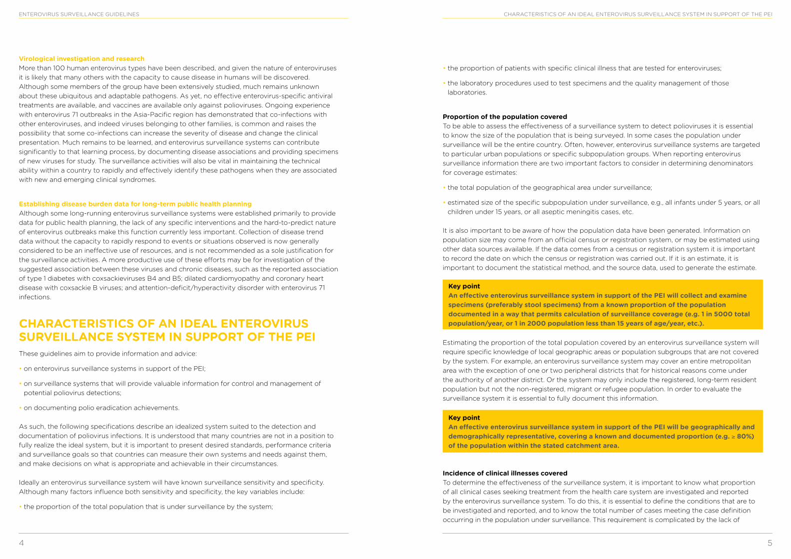
Key point An effective enterovirus surveillance system in support of the PEI will collect and examine specimens (preferably stool specimens) from a known proportion of the population documented in a way that permits calculation of surveillance coverage (e.g. 1 in 5000 total population/year, or 1 in 2000 population less than 15 years of age/year, etc.).
Estimating the proportion of the total population covered by an enterovirus surveillance system will require specific knowledge of local geographic areas or population subgroups that are not covered by the system. For example, an enterovirus surveillance system may cover an entire metropolitan area with the exception of one or two peripheral districts that for historical reasons come under the authority of another district. Or the system may only include the registered, long-term resident population but not the non-registered, migrant or refugee population. In order to evaluate the surveillance system it is essential to fully document this information.
Key point An effective enterovirus surveillance system in support of the PEI will be geographically and demographically representative, covering a known and documented proportion (e.g. ≥ 80%) of the population within the stated catchment area.
Incidence of clinical illnesses covered To determine the effectiveness of the surveillance system, it is important to know what proportion of all clinical cases seeking treatment from the health care system are investigated and reported by the enterovirus surveillance system. To do this, it is essential to define the conditions that are to be investigated and reported, and to know the total number of cases meeting the case definition occurring in the population under surveillance. This requirement is complicated by the lack of
Virological investigation and research More than 100 human enterovirus types have been described, and given the nature of enteroviruses it is likely that many others with the capacity to cause disease in humans will be discovered. Although some members of the group have been extensively studied, much remains unknown about these ubiquitous and adaptable pathogens. As yet, no effective enterovirus-specific antiviral treatments are available, and vaccines are available only against polioviruses. Ongoing experience with enterovirus 71 outbreaks in the Asia-Pacific region has demonstrated that co-infections with other enteroviruses, and indeed viruses belonging to other families, is common and raises the possibility that some co-infections can increase the severity of disease and change the clinical presentation. Much remains to be learned, and enterovirus surveillance systems can contribute significantly to that learning process, by documenting disease associations and providing specimens of new viruses for study. The surveillance activities will also be vital in maintaining the technical ability within a country to rapidly and effectively identify these pathogens when they are associated with new and emerging clinical syndromes.
Establishing disease burden data for long-term public health planning Although some long-running enterovirus surveillance systems were established primarily to provide data for public health planning, the lack of any specific interventions and the hard-to-predict nature of enterovirus outbreaks make this function currently less important. Collection of disease trend data without the capacity to rapidly respond to events or situations observed is now generally considered to be an ineffective use of resources, and is not recommended as a sole justification for the surveillance activities. A more productive use of these efforts may be for investigation of the suggested association between these viruses and chronic diseases, such as the reported association of type 1 diabetes with coxsackieviruses B4 and B5; dilated cardiomyopathy and coronary heart disease with coxsackie B viruses; and attention-deficit/hyperactivity disorder with enterovirus 71 infections.
CHARACTERISTICS OF AN IDEAL ENTEROVIRUS SURVEILLANCE SYSTEM IN SUPPORT OF THE PEIThese guidelines aim to provide information and advice:
• on enterovirus surveillance systems in support of the PEI;
• on surveillance systems that will provide valuable information for control and management of potential poliovirus detections;
• on documenting polio eradication achievements.
As such, the following specifications describe an idealized system suited to the detection and documentation of poliovirus infections. It is understood that many countries are not in a position to fully realize the ideal system, but it is important to present desired standards, performance criteria and surveillance goals so that countries can measure their own systems and needs against them, and make decisions on what is appropriate and achievable in their circumstances.
Ideally an enterovirus surveillance system will have known surveillance sensitivity and specificity. Although many factors influence both sensitivity and specificity, the key variables include:
• the proportion of the total population that is under surveillance by the system;
CHARACTERISTICS OF AN IDEAL ENTEROVIRUS SURVEILLANCE SYSTEM IN SUPPORT OF THE PEI
6 7
ENTEROVIRUS SURVEILLANCE GUIDELINES CHARACTERISTICS OF AN IDEAL ENTEROVIRUS SURVEILLANCE SYSTEM IN SUPPORT OF THE PEI
Key point An effective enterovirus surveillance system in support of the PEI will provide results of virological investigation of cases with a range of clearly defined presentations, including AFP, aseptic meningitis, encephalitis, and specified gastroenteric/enteric and respiratory diseases in a described high-risk population (e.g. children ≤ 15 years of age).
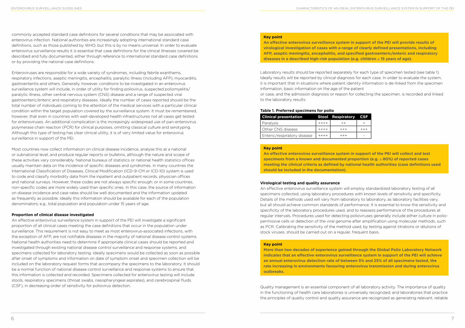
Laboratory results should be reported separately for each type of specimen tested (see table 1). Ideally results will be reported by clinical diagnosis for each case. In order to evaluate the system, it is important that in situations where patient identity information is de-linked from the specimen information, basic information on the age of the patient or case, and the admission diagnosis or reason for collecting the specimen, is recorded and linked to the laboratory results.
Table 1. Preferred specimens for polioClinical presentation Stool Respiratory CSFParalysis ++++ ++ +
Other CNS disease ++++ +++ +++
Enteric/respiratory disease ++++ +++ -
Key point An effective enterovirus surveillance system in support of the PEI will collect and test specimens from a known and documented proportion (e.g. ≥ 80%) of reported cases meeting the clinical criteria as defined by national health authorities (case definitions used should be included in the documentation).
Virological testing and quality assurance An effective enterovirus surveillance system will employ standardized laboratory testing of all specimens collected, using laboratory procedures with known levels of sensitivity and specificity. Details of the methods used will vary from laboratory to laboratory, as laboratory facilities vary, but all should achieve common standards of performance. It is essential to know the sensitivity and specificity of the laboratory procedures used, and to reassess performance of the procedures at regular intervals. Procedures used for detecting polioviruses generally include either culture in polio-permissive cells or detection of the viral genome after amplification using molecular methods, such as PCR. Calibrating the sensitivity of the method used, by testing against titrations or dilutions of stock viruses, should be carried out on a regular, frequent basis.
Key point More than two decades of experience gained through the Global Polio Laboratory Network indicates that an effective enterovirus surveillance system in support of the PEI will achieve an annual enterovirus detection rate of between 5% and 25% of all specimens tested, the rate increasing in environments favouring enterovirus transmission and during enterovirus outbreaks.
Quality management is an essential component of all laboratory activity. The importance of quality in the functioning of health care laboratories is universally recognized; and laboratories that practice the principles of quality control and quality assurance are recognized as generating relevant, reliable
commonly accepted standard case definitions for several conditions that may be associated with enterovirus infection. National authorities are increasingly adopting international standard case definitions, such as those published by WHO, but this is by no means universal. In order to evaluate enterovirus surveillance results it is essential that case definitions for the clinical illnesses covered be described and fully documented, either through reference to international standard case definitions or by providing the national case definitions.
Enteroviruses are responsible for a wide variety of syndromes, including febrile exanthems, respiratory infections, aseptic meningitis, encephalitis, paralytic illness (including AFP), myocarditis, gastroenteritis and others. Generally, however, conditions to be investigated in an enterovirus surveillance system will include, in order of utility for finding poliovirus, suspected poliomyelitis/paralytic illness, other central nervous system (CNS) disease and a range of suspected viral gastroenteric/enteric and respiratory diseases. Ideally the number of cases reported should be the total number of individuals coming to the attention of the medical services with a particular clinical condition within the target population covered by the surveillance system. It must be remembered, however, that even in countries with well-developed health infrastructures not all cases get tested for enteroviruses. An additional complication is the increasingly widespread use of pan-enterovirus polymerase chain reaction (PCR) for clinical purposes, omitting classical culture and serotyping. Although this type of testing has clear clinical utility, it is of very limited value for enterovirus surveillance in support of the PEI.
Most countries now collect information on clinical disease incidence, analyse this at a national or subnational level, and produce regular reports or bulletins, although the nature and scope of these activities vary considerably. National bureaus of statistics or national health statistics offices usually maintain data on the incidence of specific diseases and syndromes. In many countries the International Classification of Diseases, Clinical Modification (ICD-9-CM or ICD-10) system is used to code and classify morbidity data from the inpatient and outpatient records, physician offices and national surveys. However, these codes are not always specific enough; or in some countries, non-specific codes are more widely used than specific ones. In this case, the source of information on disease incidence and case rates should be well documented and the information updated as frequently as possible. Ideally this information should be available for each of the population denominators; e.g., total population and population under 15 years of age.
Proportion of clinical disease investigated An effective enterovirus surveillance system in support of the PEI will investigate a significant proportion of all clinical cases meeting the case definitions that occur in the population under surveillance. This requirement is not easy to meet as most enterovirus-associated infections, with the exception of AFP, are not notifiable diseases in the majority of national disease control systems. National health authorities need to determine if appropriate clinical cases should be reported and investigated through existing national disease control surveillance and response systems, and specimens collected for laboratory testing. Ideally specimens would be collected as soon as possible after onset of symptoms and information on date of symptom onset and specimen collection will be included on the laboratory request forms that accompany the specimens to the laboratory. It should be a normal function of national disease control surveillance and response systems to ensure that this information is collected and recorded. Specimens collected for enterovirus testing will include stools, respiratory specimens (throat swabs, nasopharyngeal aspirates), and cerebrospinal fluids (CSF), in decreasing order of sensitivity for poliovirus detection.
8 9
ENTEROVIRUS SURVEILLANCE GUIDELINES
enterovirus infections. In some instances serological assays can provide important supplementary information for confirming enterovirus-associated disease or determining the extent of enterovirus outbreaks. The two principal methods for laboratory confirmation of enterovirus infection, however, are cell culture and RT-PCR.
Traditionally, enteroviruses have been classified according to antigenic characteristics, revealed through serum neutralization with standardized type-specific reagent antisera. This method is time consuming and labour intensive, is sensitive to virus aggregation and antigenic variation, and requires a large number of antisera to identify all serotypes. Use of antiserum pools has reduced the number of separate tests that need to be carried out, but these antiserum pools were created many years ago and available volumes are now limited. In addition, continued evolution of enteroviruses, and the discovery of “new” enteroviruses, has resulted in an increasing number of virus isolates that cannot be successfully characterized using existing serological reagents.
Specimen transport Since enteroviruses are associated with rare fatal infections and more commonly severe disease (encephalitis and myocarditis), any specimen suspected of containing them should be treated as pathogenic and handled with caution. Appropriately labelled sealable containers should be used for all specimen transport. Diagnostic specimens other than stools and CSF should be transported to the laboratory in an appropriate virus transport medium. Stools and CSF specimens should be collected and transported in sterile, screw-capped containers, preferably plastic rather than glass. All specimens should be transported in sealed plastic bags or pouches, accompanied by completed laboratory request forms. The bags or pouches should carry appropriate hazard warning labels, according to the local-national policy in use.
Specimens should be transported to the laboratory as soon as possible after collection. Ideally, transportation systems should ensure that specimens arrive in the laboratory within 24 hours. Specimens other than stool and CSF should be placed in a suitable transport medium immediately after collection. When there is a delay in transporting the specimens they should be refrigerated at 4° to 8°C. Enteroviruses are relatively stable for several days at 4°C, and, depending on the pH and buffering capacity of the samples and on the level and nature of bacterial contaminants, can usually be detected in samples stored at 4°C for many days using molecular methods and by cell culture. If samples are stored frozen, repeated freezing and thawing should be avoided and every effort should be made to ensure that specimens stored frozen remain frozen until they can be processed in the laboratory.
Further information on safe handling of infectious and potentially infectious material can be found in the WHO Laboratory biosafety manual (5).
The transport of infectious substances is strictly regulated and training is required. WHO contributes to the development of United Nations Model Regulations for the transport of infectious substances. These Model Regulations are the basis for international and national regulations addressing transport by air, road, rail and sea. In order to ensure that this information is available to shippers of infectious substances, WHO has developed a training course which is divided into modules addressing the classification, documentation, marking, labelling and packaging of infectious substances, and the preparation of shipments requiring the use of dry ice. Training contributes to improving compliance with applicable regulations and ensuring protection of staff, the public and the environment. Compliance with applicable requirements also significantly increases access to courier and carrier services, and subsequent timely package delivery. More information about the course can be found online (www.who.int/ihr/i_s_shipping_training/en/index.html,accessed 9 September 2014).
and cost-effective results. Quality control and quality assurance cover a wide spectrum of activities which include:
• collection, storage and transportation of appropriate specimens;
• performance of testing using the correct techniques with suitable controls, according to standard operating procedures;
• data processing and management, including recording, interpretation, and reporting and feedback of results;
• maintaining a safe working environment through application of appropriate laboratory biosafety requirements;
• effective and safe use of laboratory equipment and facilities by following laboratory equipment guidelines, and
• providing in-service training to all staff to ensure that knowledge remains current.
Internal quality control (IQC) systems should monitor all of the above and provide the basis for laboratory quality assurance. External quality assessment (EQA) schemes should monitor whether internal quality control is in place. Proficiency testing exercises are often used as part of an EQA and demonstrate the technical ability of a laboratory. Laboratory accreditation or registration often includes an EQA component. WHO has published a Laboratory quality management system training toolkit available online (3).
All laboratories participating in an enterovirus surveillance system in support of the PEI should use standard operating procedures (SOPs) and maintain documentation on use of those procedures as described in the WHO Polio laboratory manual (4).
Key point All laboratories in an effective enterovirus surveillance system in support of the PEI will adhere to the principles of laboratory quality management as described by WHO, will maintain documentation on IQC procedures and EQA results and participate in accreditation and/or registration processes as is appropriate.
WHO-recommended laboratory procedures for the detection and characterization of enteroviruses are outlined in the following chapter.
WHO-RECOMMENDED PROCEDURES FOR SPECIMEN TRANSPORT AND ENTEROVIRUS DETECTION AND CHARACTERIZATIONVirus isolation in cell culture has for many years been the most useful method for detection of enteroviruses. Cell culture is relatively slow, but has the advantage that the isolate can be typed for epidemiological studies. However, culture is being progressively superseded by use of the reverse transcriptase polymerase chain reaction (RT-PCR) as the method of choice, particularly for examination of cerebrospinal fluid (CSF) and respiratory secretion specimens. RT-PCR is generally more sensitive, and certainly more rapid, and can detect infections with non-cultivatable enteroviruses. Many in-house and commercially available systems, however, are generic (i.e., pan-enterovirus) and provide no information on genotype or genotype-inferred serotypes. Without subsequent follow up, this molecular epidemiological data is lost.
Although specific IgM and IgG assays for enteroviruses have been described and are sometimes available, their use is limited by the heterotypic and anamnestic responses associated with
WHO-RECOMMENDED PROCEDURES FOR SPECIMEN TRANSPORT & ENTEROVIRUS DETECTION AND CHARACTERIZATION
10 11
ENTEROVIRUS SURVEILLANCE GUIDELINES WHO-RECOMMENDED PROCEDURES FOR SPECIMEN TRANSPORT & ENTEROVIRUS DETECTION AND CHARACTERIZATION
for the authenticity of new cell lines, together with identity testing, are therefore important means of avoiding wasted time and effort. All cell lines should be obtained from a reliable, and preferably authenticated, source. To avoid cross-contamination when handling and maintaining non-infected stock lines and during the preparation of the culture vessels for infection, only one cell line should be handled at a time, and the work area should be stringently cleaned and disinfected between culture sessions.
Stability Cell cultures serially passaged over an extended period of time will invariably show some signs of variation in genetic or phenotypic characteristics. The susceptibility to such variation will differ between cell lines. To minimize the effects of cell line deterioration it is strongly recommended that all cell lines used routinely for enterovirus isolation be discarded and replaced with fresh cells from the cell bank after a maximum of 15 sequential passages. If replacement with low-passage number cells is not possible, cells should be tested for continued sensitivity to viral infection and documentation of sensitivity testing results retained.
Detailed guidelines on correct handling of cell cultures used for virus diagnostic procedures can be found in Chapter 4 of the WHO Polio laboratory manual (4).
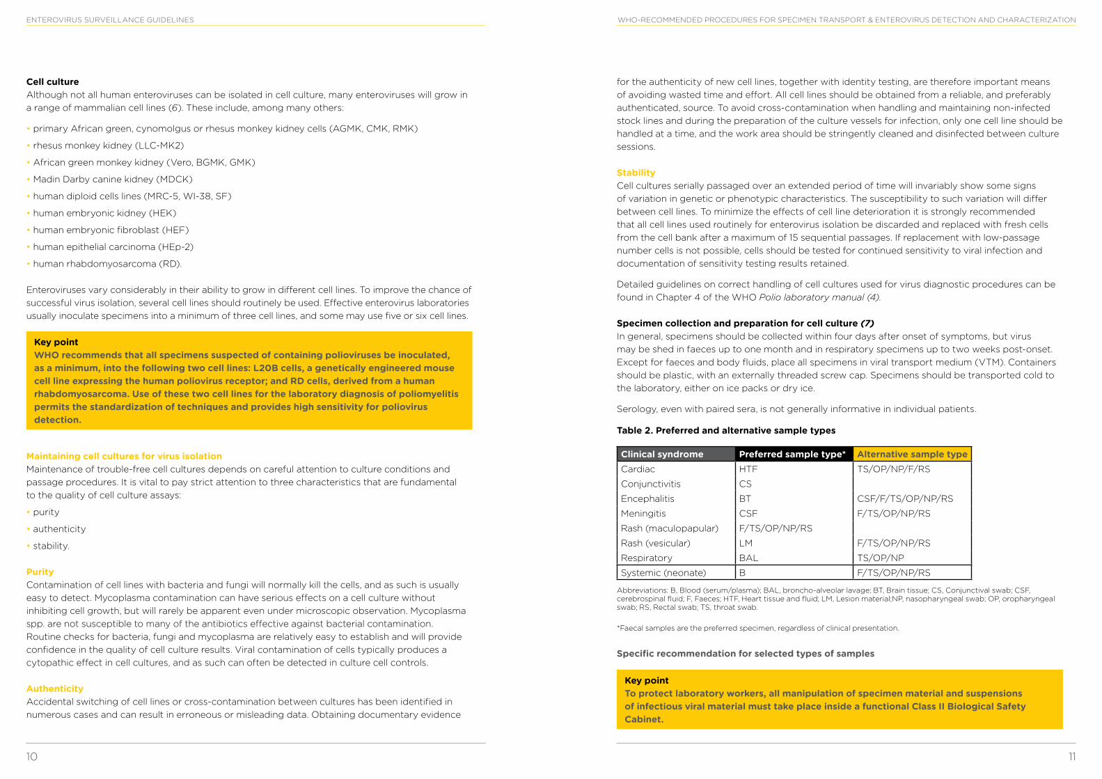
Specimen collection and preparation for cell culture (7) In general, specimens should be collected within four days after onset of symptoms, but virus may be shed in faeces up to one month and in respiratory specimens up to two weeks post-onset. Except for faeces and body fluids, place all specimens in viral transport medium (VTM). Containers should be plastic, with an externally threaded screw cap. Specimens should be transported cold to the laboratory, either on ice packs or dry ice.
Serology, even with paired sera, is not generally informative in individual patients.
Table 2. Preferred and alternative sample types
Clinical syndrome Preferred sample type* Alternative sample typeCardiac HTF TS/OP/NP/F/RS
Conjunctivitis CS
Encephalitis BT CSF/F/TS/OP/NP/RS
Meningitis CSF F/TS/OP/NP/RS
Rash (maculopapular) F/TS/OP/NP/RS
Rash (vesicular) LM F/TS/OP/NP/RS
Respiratory BAL TS/OP/NP
Systemic (neonate) B F/TS/OP/NP/RS
Abbreviations: B, Blood (serum/plasma); BAL, broncho-alveolar lavage; BT, Brain tissue; CS, Conjunctival swab; CSF, cerebrospinal fluid; F, Faeces; HTF, Heart tissue and fluid; LM, Lesion material;NP, nasopharyngeal swab; OP, oropharyngeal swab; RS, Rectal swab; TS, throat swab.
*Faecal samples are the preferred specimen, regardless of clinical presentation.
Specific recommendation for selected types of samples
Key point To protect laboratory workers, all manipulation of specimen material and suspensions of infectious viral material must take place inside a functional Class II Biological Safety Cabinet.
Cell culture Although not all human enteroviruses can be isolated in cell culture, many enteroviruses will grow in a range of mammalian cell lines (6). These include, among many others:
• primary African green, cynomolgus or rhesus monkey kidney cells (AGMK, CMK, RMK)
• rhesus monkey kidney (LLC-MK2)
• African green monkey kidney (Vero, BGMK, GMK)
• Madin Darby canine kidney (MDCK)
• human diploid cells lines (MRC-5, WI-38, SF)
• human embryonic kidney (HEK)
• human embryonic fibroblast (HEF)
• human epithelial carcinoma (HEp-2)
• human rhabdomyosarcoma (RD).
Enteroviruses vary considerably in their ability to grow in different cell lines. To improve the chance of successful virus isolation, several cell lines should routinely be used. Effective enterovirus laboratories usually inoculate specimens into a minimum of three cell lines, and some may use five or six cell lines.
Key point WHO recommends that all specimens suspected of containing polioviruses be inoculated, as a minimum, into the following two cell lines: L20B cells, a genetically engineered mouse cell line expressing the human poliovirus receptor; and RD cells, derived from a human rhabdomyosarcoma. Use of these two cell lines for the laboratory diagnosis of poliomyelitis permits the standardization of techniques and provides high sensitivity for poliovirus detection.
Maintaining cell cultures for virus isolation Maintenance of trouble-free cell cultures depends on careful attention to culture conditions and passage procedures. It is vital to pay strict attention to three characteristics that are fundamental to the quality of cell culture assays:
• purity
• authenticity
• stability.
Purity Contamination of cell lines with bacteria and fungi will normally kill the cells, and as such is usually easy to detect. Mycoplasma contamination can have serious effects on a cell culture without inhibiting cell growth, but will rarely be apparent even under microscopic observation. Mycoplasma spp. are not susceptible to many of the antibiotics effective against bacterial contamination. Routine checks for bacteria, fungi and mycoplasma are relatively easy to establish and will provide confidence in the quality of cell culture results. Viral contamination of cells typically produces a cytopathic effect in cell cultures, and as such can often be detected in culture cell controls.
Authenticity Accidental switching of cell lines or cross-contamination between cultures has been identified in numerous cases and can result in erroneous or misleading data. Obtaining documentary evidence
12 13
ENTEROVIRUS SURVEILLANCE GUIDELINES WHO-RECOMMENDED PROCEDURES FOR SPECIMEN TRANSPORT & ENTEROVIRUS DETECTION AND CHARACTERIZATION
An alternative approach is to extract RNA directly from the clinical sample for pan-enterovirus molecular identification (Annex 1) in parallel with inoculation into a limited number of cell lines. RT-PCR-positive, cell culture-negative samples can then be used to inoculate additional enterovirus-permissive cell lines or be subjected to VP1 genomic sequence analysis.
If characteristic enterovirus CPE appears, i.e. rounded, refractile cells detaching from the substrate surface, incubation should be allowed to develop until at least 75% of the cells are affected (3+ CPE), and they should be subjected to subsequent passages. If no CPE appears after seven days, a blind passage can be performed for an additional seven days. Contents of replicate cell cultures from an individual case should not be pooled for passage, i.e., individual cell cultures should be passaged separately. A description of the WHO-recommended process for isolation of polioviruses is provided in Chapter 7 of the WHO Polio laboratory manual (4).
Many enterovirus laboratories still employ antibody-based screening assays to determine the identity of viruses in CPE-positive cultures. Many of these assays include components for detecting poliovirus-positive cultures. For safety reasons, laboratories that do not belong to the WHO polio network should not attempt to type poliovirus isolates using antibody-based screening assays but should forward them to the designated WHO poliovirus laboratory.
The growth of enteroviruses in cell culture may be identified through the appearance of characteristic cytopathic changes and confirmed by immunofluorescence or neutralization. Antisera have been raised in animals against many enteroviruses, but the large number of viruses makes it impractical to routinely perform individual neutralization tests. To overcome this problem, antisera have been pooled in an overlapping scheme that allows many viruses to be identified using as few as nine tests. Interpretation of the results is carried out with the assistance of a list of the neutralization patterns of individual viruses. Type-specific monoclonal antibodies have also been prepared. These permit virus confirmation and differentiation by indirect immunofluorescence. Pooled horse antisera against the most frequently isolated echoviruses and coxsackieviruses have been prepared at the National Institute of Public Health and the Environment (RIVM), Bilthoven, the Netherlands, and are supplied free of charge to WHO Polio Laboratory Network laboratories. Details on the preparation and use of the enterovirus typing panels are provided in Chapter 7 of the WHO Polio laboratory manual (4). Other typing reagents have been prepared and include the Lim and Benyesh-Melnick (LBM) combination neutralizing serum pools, together with commercially available monoclonal antibody reagents.
RT-PCR for detection and identification of enteroviruses Use of RT-PCR offers several potential advantages for detection and diagnosis of enterovirus infections, but also presents some disadvantages. The main advantages lie in the increased sensitivity of detection and speed with which results can be reported. An additional advantage, which may become increasingly important, is the potential to identify and characterize enteroviruses that cannot be typed using existing serological systems. Although the technique can now be applied with reasonable reproducibility to CSF and some respiratory secretion specimens, direct application to faecal specimens continues to present problems on occasion. Many enterovirus laboratories using RT-PCR rely on an initial cell culture step before using the technique to detect and identify any enteroviruses present. Use of the PCR technique itself presents inherent problems, in the need to maintain physical separation of the preparation and amplification steps and prevent cross-contamination.
Choice of target sequence for RT-PCR A number of commercially available kits for the detection and primary characterization of
Stool specimens Faecal specimens should be made into a 10 to 20% suspension in a balanced salts solution with antibiotics. Stool specimens are usually treated with chloroform, to which enteroviruses are resistant (note: old or out-of-date [i.e., oxidized] chloroform is virucidal for enteroviruses). In addition to removing bacteria and fungi, this method removes potentially cytotoxic lipids and dissociates virus aggregates. After chloroform treatment with vigorous shaking or vortexing, the suspension is clarified by centrifugation at 1500 x g for 10 minutes. Clarified suspension is used as inoculum.
Throat swabs Swabs are agitated to release cellular material into the virus transport medium (VTM), taking care not to produce aerosols. This suspension can also be treated with chloroform and clarified if necessary and then is used as the inoculum.
CSF Clear CSF specimens are usually used directly as inoculum. Turbid or cloudy CSF specimens should be clarified by centrifugation at 1500 x g for 10 minutes prior to use.
Tissues Tissue samples are usually ground in a sterile mortar; occasionally the addition of sterile sand and a small amount of medium is required. After making a 10 to 20% suspension in VTM, the suspension is clarified by centrifugation at 1500 x g for 10 minutes.
Virus isolation and identification
Key point Any positive L20B culture should be considered potentially positive for poliovirus and referred to a WHO poliovirus laboratory immediately. Regardless of cell line used, culture materials from all specimens suspected of being positive for poliovirus should be sent, together with original specimen material or material prepared for inoculation to the designated WHO poliovirus laboratory. In WHO regions free of wild poliovirus circulation, or in countries without routine OPV immunization, the national health authorities should be notified immediately on isolation of poliovirus.
At least two, and preferably more, cell lines should be used for the isolation of enteroviruses. Laboratories providing supplementary enteroviral surveillance for polio eradication should, as a minimum, use L20B and RD cells. L20B is a mouse cell line (L-cells) genetically engineered to express the human poliovirus receptor. These cells are highly selective for polioviruses, which produce a characteristic enterovirus cytopathic effect (CPE). A small number of non-polio enteroviruses (e.g. some coxsackie A viruses) may also grow in L20B cells (occasionally only after prior growth in another cell line) and they can produce characteristic enterovirus CPE. RD cells, derived from a human rhabdomyosarcoma, are highly susceptible to polioviruses, many echoviruses and some other enteroviruses, all of which produce a characteristic enterovirus CPE. A third cell line, HEp-2 (Cincinnati), is also used extensively in enterovirus laboratories. Polio and coxsackie B viruses usually grow well on these cells, producing a characteristic CPE.
All cell lines chosen for enterovirus isolation should be inoculated on the same day, preferably during the same session. All specimens should be inoculated in duplicate in each cell line chosen, preferably in tube culture, or in microplate culture if cross-contamination of plate cultures can be avoided. Inoculated cells should be examined daily, using a standard inverted microscope, for signs of CPE. Observations on inoculated and control cell cultures should be made and recorded for 5–7 days after inoculation.
14 15
ENTEROVIRUS SURVEILLANCE GUIDELINES DOCUMENTATION AND REPORTING
• virological results from the tested specimens; and
• description of systems to assure quality of specimen collection and laboratory testing.
Interpretation of any virological data is dependent on understanding these elements. The structure and data elements described below represent an ideal collection of information and data. It is understood that not all information described will be readily available in all cases, but the more information that can be reported, the greater the confidence that the system is both sensitive and specific.
When an enterovirus surveillance system is used as a supplementary or alternative surveillance system to AFP surveillance, an essential element that increases the confidence that WPVs or VDPVs are not circulating in the population is the length of time during which surveillance has been conducted. While the global standard for the minimum length of observation with a system of AFP surveillance is three years, no equivalent standard is possible for supplementary surveillance systems. This is because of the wide range of possible sensitivities of different supplementary surveillance activities. Clearly, the longer the period for which high-quality data from supplementary surveillance systems can be reported, the stronger the documentation.
Documenting the system Countries using enterovirus surveillance as a supplementary or alternative surveillance system for polio may be requested by their Regional Certification Commission to submit an account of the system in use. This account will consist of a narrative, describing the nature and extent of the system, together with numerical information.
A narrative description of the surveillance system should include the following elements.
History and administrative responsibility A short review of the development of enterovirus surveillance provides a context for the current administrative structure. This should include the year when the elements of the current system were first established, when the current structure was established and any legal authority for reporting in the system.
If the system comprises many subnational reporting sites, some background information on how they are coordinated should be provided. This description does not need to be comprehensive, but should emphasize technical and administrative factors that would assure some uniformity in quality of surveillance data.
System structure and reporting sites Descriptions should be provided of:
• the number of reporting sites in the surveillance system, including whether they are directly from public health, hospital or other laboratories or through local public health officials;
• the national surveillance authority and its functional relationship to the other components and laboratories in the system; and
• the reporting system for laboratory results to the national authorities, and the frequency of reporting.
Based on the distribution of the laboratories and populations served, an estimate of the overall population served by the laboratory network should be made.
enteroviruses by RT-PCR target highly conserved sequences of the terminal 5’ non-coding region of the viral RNA. Other methods target sequences that code for viral structural proteins or the polymerase enzyme. Current information suggests the most useful target sequence for molecular typing of human enteroviruses may lie within the VP1 genomic region, coding for one of the capsid proteins. A database of sequence information for many of the prototype human enterovirus type strains has been established (www.picornaviridae.com/sequences/sequences.htm, accessed 9 September 2014). This can be used to identify unknown enteroviruses through comparison of partial VP1 sequence data with the published prototype sequences (8).
Outline of the method Detailed descriptions of several methods for detection and typing of enteroviruses by RT-PCR have been published. Although there are specific differences between methods, in general the procedure is as follows.
The targeted region of the viral RNA sequence is reverse transcribed to single stranded complementary DNA (cDNA) via primer extension with an RNA-dependent DNA polymerase.
After the reverse transcription step, the mixture is heated to denature the RNA-cDNA hybrid. Upon cooling, the upstream primer anneals to the cDNA strand, and a second polymerase catalyses the extension reaction for the synthesis of the second strand in the presence of deoxynucleotide triphosphates (dNTPs). The reaction mixture is heated again to denature the double-stranded molecule, exposing the primers’ target sequences. By this repeated process of heating, cooling, primers annealing and extension, there is a theoretical yield of more than a billion-fold amplification of the target.
Following amplification, the products (amplicons) are detected in an appropriate detection system. Certain amplicons (e.g., regions containing the VP1 gene) can be purified for detailed analysis and sequencing.
Details of the amplification and purification method currently used by the Picornavirus Laboratory, CDC, are provided for reference as Annex 1.
Please note that depending on the target sequence and the method used, some rhinoviruses could also be detected due to close genetic relatedness.
DOCUMENTATION AND REPORTINGThe ability to interpret the epidemiological significance and public health relevance of enterovirus surveillance results depends to a large extent on the level of documentation available. In turn, good documentation depends upon accurate recording of all appropriate information regarding the system. Relevant information extends far beyond the laboratory results, and includes data on the population being surveyed; the nature of the conditions triggering a surveillance response; and the measures taken to ensure the system is both sensitive and accurate. The five key areas to be documented are the:
• population that is covered by the system;
• rate of specific clinical illness in the covered population and its geographic and temporal distribution;
• rate that patients with specific clinical illness have specimens tested;
16 17
ENTEROVIRUS SURVEILLANCE GUIDELINES DOCUMENTATION AND REPORTING
– number of gastroenteritis cases with stool specimens tested;
– number of other specimens tested from gastroenteritis cases (type specified);
• virologic testing results:
– number of cases of suspected poliomyelitis from whom poliovirus was isolated;
– number of cases of CNS disease from whom poliovirus was isolated;
– number of cases of gastroenteritis from whom poliovirus was isolated;
– number of cases of other viral disease from whom poliovirus was isolated;
– number of cases of suspected poliomyelitis from whom non-polio enterovirus was isolated;
– number of cases of CNS disease from whom non-polio enterovirus was isolated;
– number of cases of gastroenteritis from whom non-polio enterovirus was isolated;
– number of cases of other viral disease from whom non-polio enterovirus was isolated.
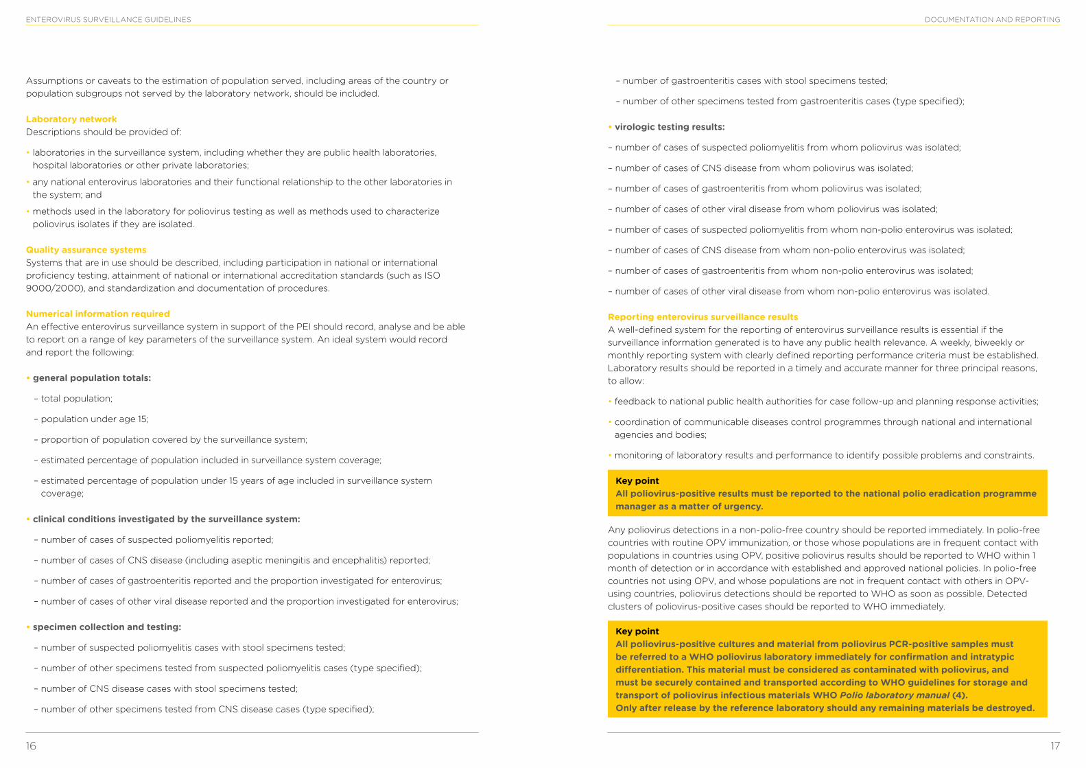
Reporting enterovirus surveillance results A well-defined system for the reporting of enterovirus surveillance results is essential if the surveillance information generated is to have any public health relevance. A weekly, biweekly or monthly reporting system with clearly defined reporting performance criteria must be established. Laboratory results should be reported in a timely and accurate manner for three principal reasons, to allow:
• feedback to national public health authorities for case follow-up and planning response activities;
• coordination of communicable diseases control programmes through national and international agencies and bodies;
• monitoring of laboratory results and performance to identify possible problems and constraints.
Key point All poliovirus-positive results must be reported to the national polio eradication programme manager as a matter of urgency.
Any poliovirus detections in a non-polio-free country should be reported immediately. In polio-free countries with routine OPV immunization, or those whose populations are in frequent contact with populations in countries using OPV, positive poliovirus results should be reported to WHO within 1 month of detection or in accordance with established and approved national policies. In polio-free countries not using OPV, and whose populations are not in frequent contact with others in OPV-using countries, poliovirus detections should be reported to WHO as soon as possible. Detected clusters of poliovirus-positive cases should be reported to WHO immediately.
Key point All poliovirus-positive cultures and material from poliovirus PCR-positive samples must be referred to a WHO poliovirus laboratory immediately for confirmation and intratypic differentiation. This material must be considered as contaminated with poliovirus, and must be securely contained and transported according to WHO guidelines for storage and transport of poliovirus infectious materials WHO Polio laboratory manual (4).Only after release by the reference laboratory should any remaining materials be destroyed.
Assumptions or caveats to the estimation of population served, including areas of the country or population subgroups not served by the laboratory network, should be included.
Laboratory network Descriptions should be provided of:
• laboratories in the surveillance system, including whether they are public health laboratories, hospital laboratories or other private laboratories;
• any national enterovirus laboratories and their functional relationship to the other laboratories in the system; and
• methods used in the laboratory for poliovirus testing as well as methods used to characterize poliovirus isolates if they are isolated.
Quality assurance systems Systems that are in use should be described, including participation in national or international proficiency testing, attainment of national or international accreditation standards (such as ISO 9000/2000), and standardization and documentation of procedures.
Numerical information required An effective enterovirus surveillance system in support of the PEI should record, analyse and be able to report on a range of key parameters of the surveillance system. An ideal system would record and report the following:
• general population totals:
– total population;
– population under age 15;
– proportion of population covered by the surveillance system;
– estimated percentage of population included in surveillance system coverage;
– estimated percentage of population under 15 years of age included in surveillance system coverage;
• clinical conditions investigated by the surveillance system:
– number of cases of suspected poliomyelitis reported;
– number of cases of CNS disease (including aseptic meningitis and encephalitis) reported;
– number of cases of gastroenteritis reported and the proportion investigated for enterovirus;
– number of cases of other viral disease reported and the proportion investigated for enterovirus;
• specimen collection and testing:
– number of suspected poliomyelitis cases with stool specimens tested;
– number of other specimens tested from suspected poliomyelitis cases (type specified);
– number of CNS disease cases with stool specimens tested;
– number of other specimens tested from CNS disease cases (type specified);
18 19
ENTEROVIRUS SURVEILLANCE GUIDELINES
1. Guidelines for environmental surveillance of poliovirus circulation. Geneva: World Health Organization, 2003. WHO/V&B/03.03 (http://whqlibdoc.who.int/hq/2003/WHO_V&B_03.03.pdf, accessed 9 September 2014).
2. Morbidity and Mortality Weekly Report, 2012; 61(37):741-746.
3. Laboratory quality management system – training toolkit. Geneva: World Health Organization; 2009 (www.who.int/ihr/training/laboratory_quality/en/index.html, accessed 9 September 2014).
4. Polio laboratory manual. Geneva: World Health Organization, 2004. WHO/IVB/04.10 (http://whqlibdoc.who.int/hq/2004/WHO_IVB_04.10.pdf, accessed 9 September 2014).
5. WHO Laboratory biosafety manual. Geneva: World Health Organization, 2004. (http://www.who.int/csr/resources/publications/biosafety/Biosafety7.pdf?ua=1, accessed 9 September 2014).
6. Schmidt, N.J. Ho HH, Lennette EH. Propagation and isolation of group A coxsackieviruses in RD cells. J Clin Microbiol 1975;2:183–185.
7. Adapted from: Miller JM. A Guide to Specimen Management in Clinical Microbiology, ASM Press, Washington, D.C. 1999.
8. Kroneman A, Vennema H, Deforche K, van der Avoort H, Peñaranda S, Oberste MS, Vinjé J, Koopmans M. An automated genotyping tool for enteroviruses and noroviruses. J Clin Virol 2011;51:121–125 (www.rivm.nl/mpf/enterovirus/typingtool, accessed 9 September 2014).
FURTHER READING 1. Nix WA, Maher K, Johansson ES, Niklasson B, Lindberg AM, Pallansch MA, Oberste MS. Detection
of all known parechoviruses by real-time PCR. J Clin Microbiol 2008; 46:2519–2524.
2. Nix WA, Maher K, Pallansch MA, Oberste MS. Parechovirus typing in clinical specimens by nested or semi-nested PCR coupled with sequencing. J Clin Virol 2010;48:202–207.
3. Nix WA, Oberste MS, Pallansch MA. Sensitive, seminested PCR amplification of VP1 sequences for direct identification of all enterovirus serotypes from original clinical specimens. J Clin Microbiol 2006; 44:2698–2704.
4. Oberste MS, Pallansch MA. Enterovirus molecular detection and typing. Rev Med Microbiol 2005;16:163-171.9.
5. Oberste MS, Maher K, Kilpatrick DR, Pallansch MA. Molecular evolution of human enteroviruses: Correlation of serotype with VP1 sequence and application to picornavirus classification. J Virol 1999;73:1941–1948.
6. Oberste MS, Kilpatrick DR, Maher K, Pallansch MA. Typing of human enteroviruses by partial sequencing of VP1. United States patent number 6,846,621, issued 25 January 2005.
7. Oberste MS, Maher K, Kilpatrick DR, Flemister MR, Brown BA, Pallansch MA. Typing of human enteroviruses by partial sequencing of VP1. J Clin Microbiol 1999;37:1288–1293.
8. Oberste MS, Maher K, Flemister MR, Marchetti G, Kilpatrick DR, Pallansch MA. Comparison of molecular and classic approaches for the identification of “untypeable” human enterovirus isolates. J Clin Microbiol 2000;38:1170–1174.
9. Oberste MS, Nix WA, Maher K, Pallansch MA. Improved molecular identification of enteroviruses by RT-PCR and amplicon sequencing. J Clin Virol 2003;26:375–377.
REFERENCES
Background Members of the family Picornaviridae are the most common viruses infecting humans and species in several genera also infect a wide variety of other mammalian and avian species. Picornaviruses have traditionally been classified by antigenic type, based on a serum neutralization assay. However, this method is time-consuming and labour-intensive, is sensitive to virus aggregation and antigenic variation, and requires a large number of antisera to identify all serotypes, even when antiserum pools are used. Additionally, no reference antisera are available for the approximately 45 new enterovirus (EV) types identified in the last 15 years. The VP1 gene has been shown to correlate with EV serotype, such that the virus can be identified by comparison of a partial VP1 sequence of an unknown to a database of prototype strain sequences. Generic RT-PCR primers have been developed to amplify all EV serotypes, as well as all rhinovirus (RV) types A and B and other picornaviruses. RT-PCR amplification of the VP1 gene and amplicon sequencing have been used to discriminate among the prototype strains of all EV serotypes, to identify EV isolated from human clinical specimens, to identify EV that were refractory to antigenic typing, and to identify potential new EV serotypes from collections of “untypeable” EV isolates. Adaptations of generic primer approaches that improve the sensitivity of these assays allow the analysis of clinical specimens in addition to EV isolates (Table A1). The RT-snPCR approach described here is intended for clinical specimens (Fig. A1).
RNA extraction This protocol assumes the user has a thorough knowledge of how to handle RNA and the hazard posed to RNA by ribonucleases (RNases). All reagents and disposable plasticware used in the procedure should be certified RNase free. This protocol lists the most commonly used commercial kits, which is not an endorsement of the kits. Other commercially available kits may work just as well or better. This disclaimer also holds true for the other commercial kits and equipment described in the following protocols.
Clinical specimens Materials QIAamp Viral RNA Mini Kit (QIAGEN; cat. # 52906)Sterile 1.5 ml microcentrifuge tubesProteinase K; PCR grade (Roche; cat. # 92643528)Water bath at 37°CVertrel XF (1,1,1,2,3,4,4,5,5,5-Decafluoropentane) (DuPont Fluorochemicals)Microcentrifuge (or suitable vacuum manifold)Sterile nuclease-free H2OVacuum dessicator
Methods 1. Method for cerebrospinal fluid (CSF), eye swabs, nasopharyngeal swabs and other respiratory
specimens.
a. Thaw and transfer the specimens in a Class II biological safety cabinet. Aliquot a maximum of 140 l of the specimen into a sterile 1.5 ml microcentrifuge tube. Add 20 g of proteinase K to each specimen, mix gently and incubate in a 37°C water bath for 30 minutes.
ANNEX 1. RT-snPCR AMPLIFICATION AND SEQUENCING OF THE ENTEROVIRUS VP1 GENE FOR SEROTYPE IDENTIFICATION: PROTOCOLS FOR CLINICAL SPECIMENS
20 21
ENTEROVIRUS SURVEILLANCE GUIDELINES ANNEX 1
b. Follow the manufacturer’s QIAamp Viral RNA Mini Spin Protocol. Elute the RNA off the column with 60 l sterile H2O.
c. For added sensitivity the RNA may be concentrated by partially or completely drying the RNA in a vacuum dessicator. If the RNA is completely dried, resuspend well in 20–40 l sterile nuclease-free H2O.
d. Store the RNA at –20°C or perferably at –70°C. Most viral RNAs are stable for many years at –70°C.
2. Method for stool and rectal swab specimens.
a. Stool suspensions should be prepared according to the WHO polio procedure (1). Stool suspensions and rectal swabs should be centrifuged at 10 000 x g for one minute in a microcentrifuge to remove suspended solids.
b. Take 200–300 l of the supernatant and extract with an equal volume of Vertrel XF. Shake vigorously. Centrifuge for one minute at 10 000 x g to break the emulsion. Transfer 140 l of the aqueous supernatant (upper layer) to a new microcentrifuge tube and add 20 g proteinase K. Mix gently and incubate in a 37°C water bath for 30 minutes.
c. Follow protocol 1 above beginning with step 1b.
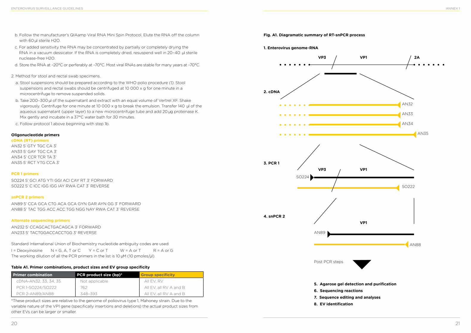
Oligonucleotide primers cDNA (RT) primers AN32 5’ GTY TGC CA 3’AN33 5’ GAY TGC CA 3’AN34 5’ CCR TCR TA 3’AN35 5’ RCT YTG CCA 3’
PCR 1 primersSO224 5’ GCI ATG YTI GGI ACI CAY RT 3’ FORWARDSO222 5’ C ICC IGG IGG IAY RWA CAT 3’ REVERSE
snPCR 2 primersAN89 5’ CCA GCA CTG ACA GCA GYN GAR AYN GG 3’ FORWARDAN88 5’ TAC TGG ACC ACC TGG NGG NAY RWA CAT 3’ REVERSE
Alternate sequencing primersAN232 5’ CCAGCACTGACAGCA 3’ FORWARDAN233 5’ TACTGGACCACCTGG 3’ REVERSE
Standard International Union of Biochemistry nucleotide ambiguity codes are used:
I = Deoxyinosine N = G, A, T or C Y = C or T W = A or T R = A or GThe working dilution of all the PCR primers in the list is 10 M (10 pmoles/ l).
Table A1. Primer combinations, product sizes and EV group specificity
Primer combination PCR product size (bp)* Group specificitycDNA-AN32, 33, 34, 35 Not applicable All EV, RV
PCR 1-SO224/SO222 762 All EV, all RV A and B
PCR 2-AN89/AN88 348–393 All EV, all RV A and B
*These product sizes are relative to the genome of poliovirus type 1, Mahoney strain. Due to the variable nature of the VP1 gene (specifically insertions and deletions) the actual product sizes from other EVs can be larger or smaller.
Fig. A1. Diagramatic summary of RT-snPCR process
VP3
VP3
1. Enterovirus genome-RNA
2. cDNA
3. PCR 1
4. snPCR 2
VP1
VP1
VP1
2A
AN32
AN33
AN34
SO222
SO224
AN89
5. Agarose gel detection and purification6. Sequencing reactions7. Sequence editing and analyses8. EV identification
Post PCR steps
AN88
AN35
22 23
ENTEROVIRUS SURVEILLANCE GUIDELINES ANNEX 1
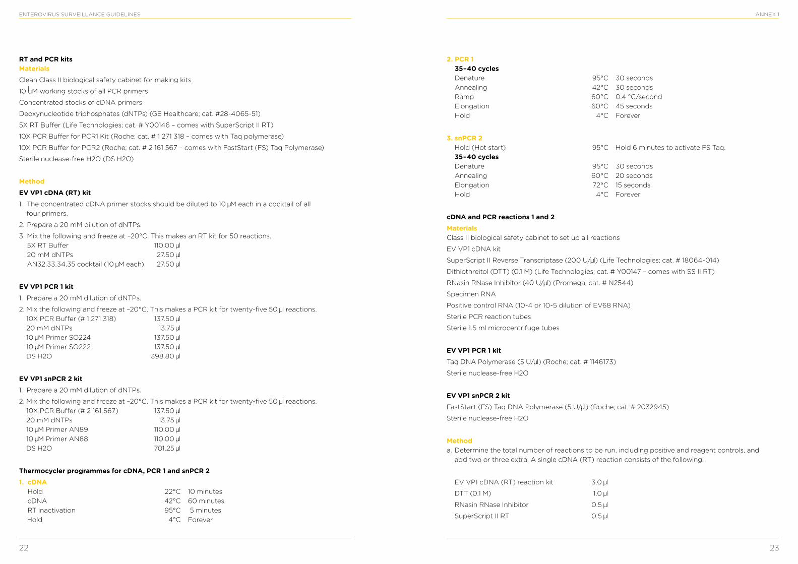
RT and PCR kits MaterialsClean Class II biological safety cabinet for making kits
10 uM working stocks of all PCR primers
Concentrated stocks of cDNA primers
Deoxynucleotide triphosphates (dNTPs) (GE Healthcare; cat. #28-4065-51)
5X RT Buffer (Life Technologies; cat. # Y00146 – comes with SuperScript II RT)
10X PCR Buffer for PCR1 Kit (Roche; cat. # 1 271 318 – comes with Taq polymerase)
10X PCR Buffer for PCR2 (Roche; cat. # 2 161 567 – comes with FastStart (FS) Taq Polymerase)
Sterile nuclease-free H2O (DS H2O)
MethodEV VP1 cDNA (RT) kit1. The concentrated cDNA primer stocks should be diluted to 10 M each in a cocktail of all
four primers.
2. Prepare a 20 mM dilution of dNTPs.
3. Mix the following and freeze at –20°C. This makes an RT kit for 50 reactions. 5X RT Buffer 110.00 l 20 mM dNTPs 27.50 l AN32,33,34,35 cocktail (10 M each) 27.50 l
EV VP1 PCR 1 kit1. Prepare a 20 mM dilution of dNTPs.
2. Mix the following and freeze at –20°C. This makes a PCR kit for twenty-five 50 l reactions. 10X PCR Buffer (# 1 271 318) 137.50 l 20 mM dNTPs 13.75 l 10 M Primer SO224 137.50 l 10 M Primer SO222 137.50 l DS H2O 398.80 l
EV VP1 snPCR 2 kit1. Prepare a 20 mM dilution of dNTPs.
2. Mix the following and freeze at –20°C. This makes a PCR kit for twenty-five 50 l reactions. 10X PCR Buffer (# 2 161 567) 137.50 l 20 mM dNTPs 13.75 l 10 M Primer AN89 110.00 l 10 M Primer AN88 110.00 l DS H2O 701.25 l
Thermocycler programmes for cDNA, PCR 1 and snPCR 21. cDNA
Hold 22°C 10 minutes cDNA 42°C 60 minutes RT inactivation 95°C 5 minutes
Hold 4°C Forever
l
2. PCR 1 35–40 cycles Denature 95°C 30 seconds Annealing 42°C 30 seconds Ramp 60°C 0.4 ºC/second Elongation 60°C 45 seconds
Hold 4°C Forever
3. snPCR 2 Hold (Hot start) 95°C Hold 6 minutes to activate FS Taq. 35–40 cycles Denature 95°C 30 seconds Annealing 60°C 20 seconds Elongation 72°C 15 seconds
Hold 4°C Forever
cDNA and PCR reactions 1 and 2Materials Class II biological safety cabinet to set up all reactions
EV VP1 cDNA kit
SuperScript II Reverse Transcriptase (200 U/ l) (Life Technologies; cat. # 18064-014)
Dithiothreitol (DTT) (0.1 M) (Life Technologies; cat. # Y00147 – comes with SS II RT)
RNasin RNase Inhibitor (40 U/ l) (Promega; cat. # N2544)
Specimen RNA
Positive control RNA (10-4 or 10-5 dilution of EV68 RNA)
Sterile PCR reaction tubes
Sterile 1.5 ml microcentrifuge tubes
EV VP1 PCR 1 kitTaq DNA Polymerase (5 U/ l) (Roche; cat. # 1146173)
Sterile nuclease-free H2O
EV VP1 snPCR 2 kitFastStart (FS) Taq DNA Polymerase (5 U/ l) (Roche; cat. # 2032945)
Sterile nuclease-free H2O
Method a. Determine the total number of reactions to be run, including positive and reagent controls, and
add two or three extra. A single cDNA (RT) reaction consists of the following:
EV VP1 cDNA (RT) reaction kit 3.0 l
DTT (0.1 M) 1.0 l
RNasin RNase Inhibitor 0.5 l
SuperScript II RT 0.5 l
24 25
ENTEROVIRUS SURVEILLANCE GUIDELINES ANNEX 1
b. Make a cDNA master mix by multiplying each of the reagents above by the total number of reactions to be run (plus two) and mixing the appropriate volumes. Aliquot 5 l of the cDNA master mix into each reaction tube.
c. Add 5 l of specimen RNA to each tube, giving a final reaction volume of 10 l. Add 5 l H2O to the reagent control. Place the tubes in a thermocycler and run the cDNA programme shown in the section entitled “EV VP1 cDNA (RT) kit” above.
d. Around 10 minutes before the cDNA reactions are complete, prepare a H2O plus Taq enzyme mix in a sterile 1.5 ml microcentrifuge tube. The enzyme mix consists of 261.3 l of sterile nuclease-free H2O plus 13.7 l of Taq polymerase. Prepare the PCR 1 master mix as described in step 1 for the cDNA master mix. A single PCR 1 reaction consists of the following:
EV VP1 PCR 1 kit 30 l
H2O + Taq 10 l
e. Remove the cDNA reactions from the thermocycler. Add 40 l of the PCR 1 master mix to each cDNA reaction for a total reaction volume of 50 l. Put the reactions back into the thermocycler and run the PCR 1 programme shown in the section entitled “EV VP1 PCR1 kit” above. Any remaining H2O plus Taq can be frozen at –20°C for up to six months.
f. Around 10 minutes before the PCR 1 reactions are complete, prepare a H2O plus Fast Start Taq enzyme mix in a sterile 1.5 ml microcentrifuge tube. The enzyme mix consists of 261.3 l of sterile nuclease-free H2O plus 13.7 l of FS Taq Polymerase. Prepare the snPCR 2 master mix as described in step 1 for the cDNA master mix. A single PCR 2 reaction consists of the following:
EV VP1 snPCR 2 kit 39 l
H2O + FS Taq 10 l
Aliquot 49 l of the PCR 2 master mix into a new set of reaction tubes and close.
g. Remove the PCR 1 reactions from the thermocycler. Transfer 1 l of a PCR 1 reaction into the corresponding snPCR 2 reaction tube. Put the snPCR 2 reactions into the thermocycler and run the snPCR 2 programme shown in the section entitled “EV VP1 snPCR2 kit” above.
Purification of snPCR 2 productsMaterialsQiaquick Gel Extraction Kit (QIAGEN; cat. # 28706)
Sterile 1.5 ml microcentrifuge tubes
Sterile nuclease-free H2O
Microcentrifuge (or suitable vacuum manifold)
Dry block heater or water bath at 50°C
Vacuum dessicator
lectrophoresis power supply and gel apparatus
Agarose
20X LB conductive medium (Faster Better Media, LLC; cat. # LB20-10)
Ethidium Bromide (EtBr) (10 g/ml stock) OR GelRed Nucleic Acid Gel Stain (Phenix Research; cat# 41003)
Ethanol, absolute
Isopropanol
Ultraviolet (UV) light box
Scalpels
Low DNA Mass Ladder (Life Technologies; cat. # 10068-013)
Gel loading dye
Method a. Prepare a medium sized (or larger) 1.5% agarose horizontal slab gel, using 1X LB buffer. For each
100 ml of agarose add 5 l of EtBr stock (or 4 l GelRed). Use a large comb with deep enough wells to hold 60 l of snPCR 2 reaction + gel loading buffer. After running the gel visualize the bands on a UV light box and cut the appropriate bands from the gel with a scalpel.
b. Follow the Qiaquick Gel Extraction Kit protocol. Elute the purified snPCR 2 product in 50.0 l of Buffer EB or sterile nuclease-free H2O.
c. If weak bands were present and cut from the gel, the purified PCR product may be dried in a dessicator to concentrate the DNA. A final volume of 15–20 l is good. If the sample is completely dry add 15–20 of water to rehydrate. Usually, visible PCR bands do not need to be concentrated.
d. Pour a 1.5% agarose gel, using a 20–24 well comb and add 5 l EtBr stock (or 4 l GelRed) per 100 ml of agarose. Run 4 l of the Low DNA Mass Ladder plus gel loading buffer on the gel and 4–5 l of each purified PCR 2 product plus gel loading buffer.
e. Visualize the purified DNA bands on a UV light box and photograph. Estimate the quantity of purified DNA for each specimen by comparing the fluorescence of each specimen with that of the Low DNA Mass Ladder.
f. Store the purified DNA at –20°C or at 4°C if sequencing reactions will be set up in the next four to eight hours.
Sequencing reactionsMaterialsBig Dye Terminator v1.1 Cycle (Life Technologies; cat. # 4337453)
5X Sequencing Buffer (Life Technologies; cat. # 4336699)
Sterile PCR reaction tubes
10 M Primer AN89 or AN232
10 M Primer AN88 or AN233
Purified specimen DNA templates
Centri-Sep 8 Strips (Princeton Separations, Inc.; cat. # CS-912)
MicroAmp Optical 96-Well Reaction Plate (Life Technologies; cat. # N801-056)
Sorvall centrifuge with plate carriers
Formamide
ABI Prism 3100 Genetic Analyser
Methoda. Sequencing reactions should be set up according to the ABI protocol for PCR products. The final
volume for each reaction is 10 l.
26 27
ENTEROVIRUS SURVEILLANCE GUIDELINES ANNEX 1
b. Purification of the sequencing reactions should be done with the Centri-Sep 8 strips, according to the supplied protocol. Add 10 l hi-di formamide to each sample and heat at 95oC for 2 minutes for further denaturation.
c. Analyse the sequencing reactions on the ABI Prism 3100 Genetic Analyser, according to the manufacturer’s instructions.
Sample mix:
Distilled H2O l1
Template (on ice) l1
Primer: AN89 or AN88 (10 M) 0.5 l
Big Dye Terminator v1.1 2.0 l
5X Sequencing Buffer 1.0 l
Total volume 10 l
NOTE: Make a master mix of Big Dye Terminator v1.1, 5X sequencing buffer, and one Primer. Add 3.5 l of this mix to tube and adjust with the template and water to 10 l.
Mix well with pipette tip.
d. Thermocycler programme:
96°C 10 seconds
50°C 5 seconds
60°C 4 minutes
For 25 cycles, it takes around 2.5 hours
Sequencing editing and analysis1. Sequence editing is done with the software package Sequencher, following the manufacturer’s
instructions. Alternative sequence assembly software packages may be used.
2. Analyses of the consensus sequences are done using the criteria of Oberste, et al. (2). Alternative sequence assembly software packages may be used.
References1. Polio laboratory manual. 4th edition, 2004. Geneva: World Health Organization; 2004 (http://
www.who.int/vaccines/en/poliolab/WHO-Polio-Manual-9.pdf, accessed 9 September 2014).
2. Oberste MS, Maher K, Kilpatrick DR, Pallansch MA. Molecular evolution of the human enteroviruses: correlation of serotype with VP1 sequence and application to picornavirus classification. J Virol 1999;73:1941–8.
Further reading1. BigDye® Terminator v1.1 cycle sequencing kit protocol. Part number 4337036 Rev. B. Applied
Biosystems. 2010 (http://tools.lifetechnologies.com/content/sfs/manuals/cms_041330.pdf, accessed 9 September 2014).
1 The volume of a template can vary. Use the volume of H20 to adjust final volume to 10 l.
2. Dilution of BigDye® Terminators, 1999 application note. Publication number 107PABA01–01 5X Sequencing Buffer. Applied Biosystems; 1999.
3. Nix WA, Berger MM, Oberste MS, Brooks BR, McKenna-Yasek DM, Brown RH Jr, et al. Failure to detect enterovirus in the spinal cord of ALS patients using a sensitive RT-PCR method. Neurology. 2004;62:1372–7.
4. Nix WA, Oberste MS, Pallansch MA. Sensitive, seminested PCR amplification of VP1 sequences for direct identification of all enterovirus serotypes from original clinical specimens. J Clin Microbil. 2006; 44(8):2698–704 (source of this lab protocol).
5. Oberste MS, Kilpatrick DR, Maher K, Pallansch MA. Typing of human enteroviruses by partial sequencing of VP1. United States provisional patent application number 60/127,464, 1999.
6. Oberste MS, Maher K, Flemister MR, Marchetti G, Kilpatrick DR, Pallansch MA. Comparison of classic and molecular approaches for the identification of untypeable enteroviruses. J Clin Microbiol. 2000;38:1170–4.
7. Oberste MS, Maher K, Kilpatrick DR, Flemister MR, Brown BA, Pallansch MA. Typing of human enteroviruses by partial sequencing of VP1. J Clin Microbiol. 1999;37:1288–93.
8. Oberste MS, Nix WA, Maher K, Pallansch MA. Improved molecular identification of enteroviruses by RT-PCR and amplicon sequencing. J Clin Virol. 2003;26:375–7.
9. Sambrook J, Russell D, editors. Molecular cloning. A laboratory manual, third edition. New York: Cold Spring Harbor Laboratory Press; 2001.
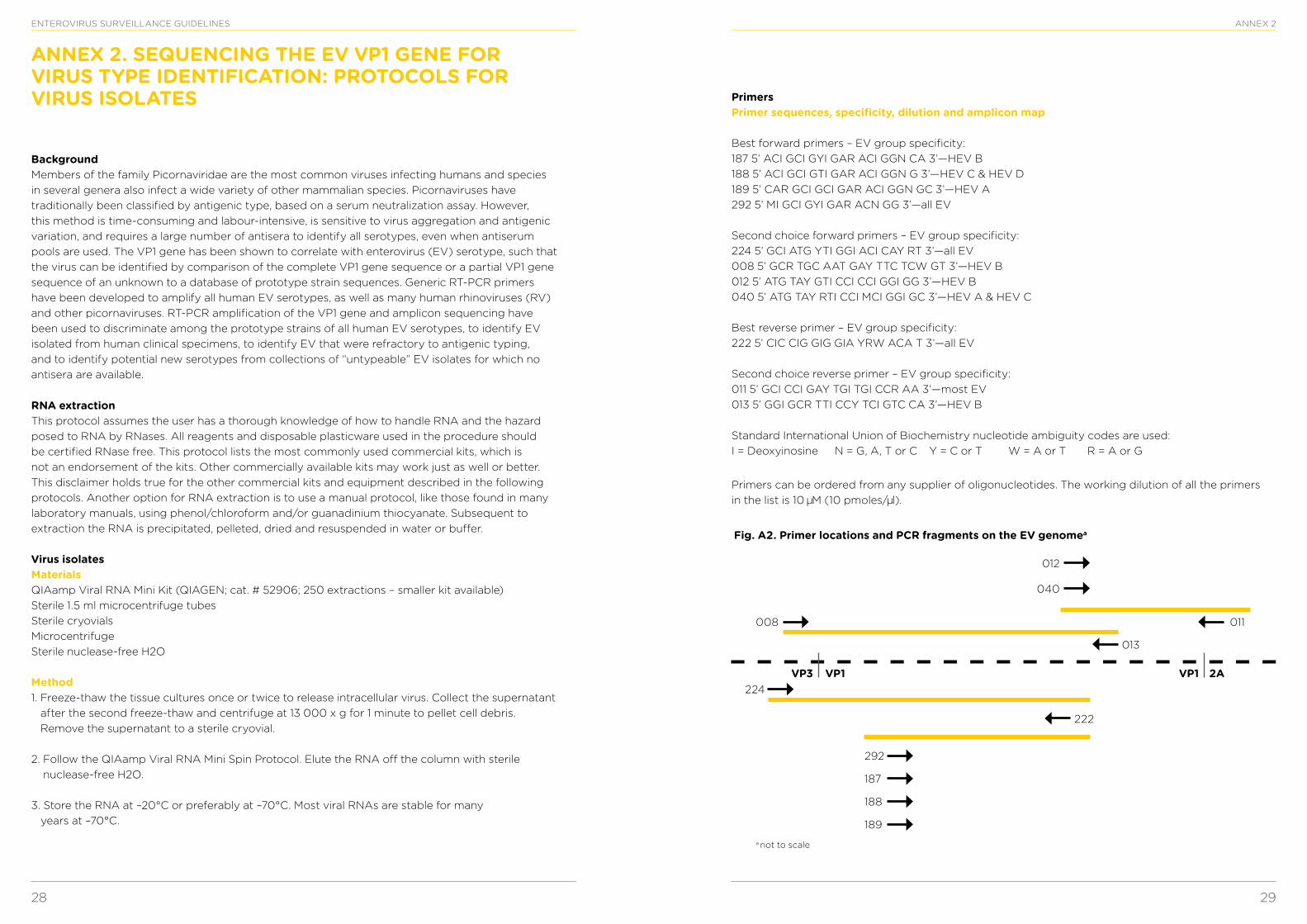
28 29
ENTEROVIRUS SURVEILLANCE GUIDELINES ANNEX 2
ANNEX 2. SEQUENCING THE EV VP1 GENE FOR VIRUS TYPE IDENTIFICATION: PROTOCOLS FOR VIRUS ISOLATES
BackgroundMembers of the family Picornaviridae are the most common viruses infecting humans and species in several genera also infect a wide variety of other mammalian species. Picornaviruses have traditionally been classified by antigenic type, based on a serum neutralization assay. However, this method is time-consuming and labour-intensive, is sensitive to virus aggregation and antigenic variation, and requires a large number of antisera to identify all serotypes, even when antiserum pools are used. The VP1 gene has been shown to correlate with enterovirus (EV) serotype, such that the virus can be identified by comparison of the complete VP1 gene sequence or a partial VP1 gene sequence of an unknown to a database of prototype strain sequences. Generic RT-PCR primers have been developed to amplify all human EV serotypes, as well as many human rhinoviruses (RV) and other picornaviruses. RT-PCR amplification of the VP1 gene and amplicon sequencing have been used to discriminate among the prototype strains of all human EV serotypes, to identify EV isolated from human clinical specimens, to identify EV that were refractory to antigenic typing, and to identify potential new serotypes from collections of “untypeable” EV isolates for which no antisera are available.
RNA extractionThis protocol assumes the user has a thorough knowledge of how to handle RNA and the hazard posed to RNA by RNases. All reagents and disposable plasticware used in the procedure should be certified RNase free. This protocol lists the most commonly used commercial kits, which is not an endorsement of the kits. Other commercially available kits may work just as well or better. This disclaimer holds true for the other commercial kits and equipment described in the following protocols. Another option for RNA extraction is to use a manual protocol, like those found in many laboratory manuals, using phenol/chloroform and/or guanadinium thiocyanate. Subsequent to extraction the RNA is precipitated, pelleted, dried and resuspended in water or buffer.
Virus isolatesMaterialsQIAamp Viral RNA Mini Kit (QIAGEN; cat. # 52906; 250 extractions – smaller kit available)Sterile 1.5 ml microcentrifuge tubesSterile cryovialsMicrocentrifuge Sterile nuclease-free H2O
Method1. Freeze-thaw the tissue cultures once or twice to release intracellular virus. Collect the supernatant
after the second freeze-thaw and centrifuge at 13 000 x g for 1 minute to pellet cell debris. Remove the supernatant to a sterile cryovial.
2. Follow the QIAamp Viral RNA Mini Spin Protocol. Elute the RNA off the column with sterile nuclease-free H2O.
3. Store the RNA at –20°C or preferably at –70°C. Most viral RNAs are stable for many years at –70°C.
PrimersPrimer sequences, specificity, dilution and amplicon map
Best forward primers – EV group specificity:187 5’ ACI GCI GYI GAR ACI GGN CA 3’—HEV B188 5’ ACI GCI GTI GAR ACI GGN G 3’—HEV C & HEV D189 5’ CAR GCI GCI GAR ACI GGN GC 3’—HEV A292 5’ MI GCI GYI GAR ACN GG 3’—all EV
Second choice forward primers – EV group specificity:224 5’ GCI ATG YTI GGI ACI CAY RT 3’—all EV008 5’ GCR TGC AAT GAY TTC TCW GT 3’—HEV B012 5’ ATG TAY GTI CCI CCI GGI GG 3’—HEV B040 5’ ATG TAY RTI CCI MCI GGI GC 3’—HEV A & HEV C
Best reverse primer – EV group specificity:222 5’ CIC CIG GIG GIA YRW ACA T 3’—all EV
Second choice reverse primer – EV group specificity:011 5’ GCI CCI GAY TGI TGI CCR AA 3’—most EV013 5’ GGI GCR TTI CCY TCI GTC CA 3’—HEV B
Standard International Union of Biochemistry nucleotide ambiguity codes are used:I = Deoxyinosine N = G, A, T or C Y = C or T W = A or T R = A or G
Primers can be ordered from any supplier of oligonucleotides. The working dilution of all the primers in the list is 10 M (10 pmoles/ l).
Fig. A2. Primer locations and PCR fragments on the EV genomea
a not to scale
VP3 VP1 VP1 2A
012
040
011
013
222
008
224
292
187
188
189
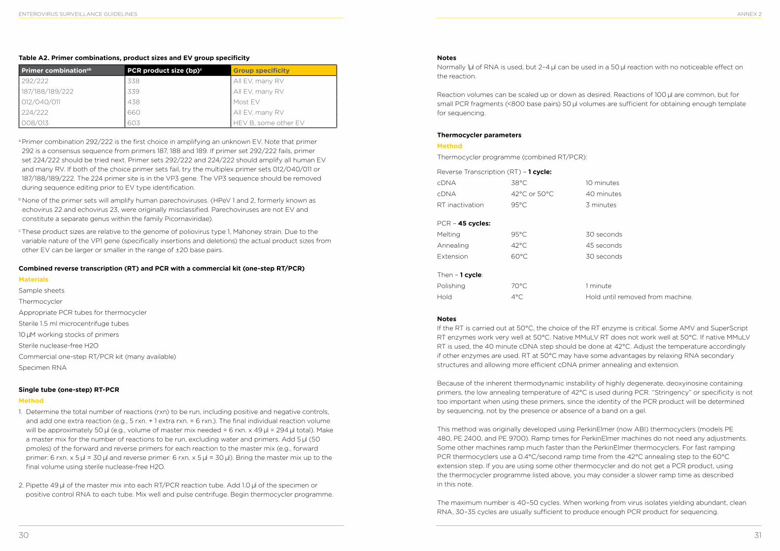
30 31
ENTEROVIRUS SURVEILLANCE GUIDELINES ANNEX 2
Table A2. Primer combinations, product sizes and EV group specificity
Primer combinationab PCR product size (bp)c Group specificity292/222 338 All EV, many RV
187/188/189/222 339 All EV, many RV
012/040/011 438 Most EV
224/222 660 All EV, many RV
008/013 603 HEV B, some other EV
a Primer combination 292/222 is the first choice in amplifying an unknown EV. Note that primer 292 is a consensus sequence from primers 187, 188 and 189. If primer set 292/222 fails, primer set 224/222 should be tried next. Primer sets 292/222 and 224/222 should amplify all human EV and many RV. If both of the choice primer sets fail, try the multiplex primer sets 012/040/011 or 187/188/189/222. The 224 primer site is in the VP3 gene. The VP3 sequence should be removed during sequence editing prior to EV type identification.
b None of the primer sets will amplify human parechoviruses. (HPeV 1 and 2, formerly known as echovirus 22 and echovirus 23, were originally misclassified. Parechoviruses are not EV and constitute a separate genus within the family Picornaviridae).
c These product sizes are relative to the genome of poliovirus type 1, Mahoney strain. Due to the variable nature of the VP1 gene (specifically insertions and deletions) the actual product sizes from other EV can be larger or smaller in the range of ±20 base pairs.
Combined reverse transcription (RT) and PCR with a commercial kit (one-step RT/PCR)MaterialsSample sheets
Thermocycler
Appropriate PCR tubes for thermocycler
Sterile 1.5 ml microcentrifuge tubes
10 M working stocks of primers
Sterile nuclease-free H2O
Commercial one-step RT/PCR kit (many available)
Specimen RNA
Single tube (one-step) RT-PCRMethod1. Determine the total number of reactions (rxn) to be run, including positive and negative controls,
and add one extra reaction (e.g., 5 rxn. + 1 extra rxn. = 6 rxn.). The final individual reaction volume will be approximately 50 l (e.g., volume of master mix needed = 6 rxn. x 49 l = 294 l total). Make a master mix for the number of reactions to be run, excluding water and primers. Add 5 l (50 pmoles) of the forward and reverse primers for each reaction to the master mix (e.g., forward primer: 6 rxn. x 5 l = 30 l and reverse primer: 6 rxn. x 5 l = 30 l). Bring the master mix up to the final volume using sterile nuclease-free H2O.
2. Pipette 49 l of the master mix into each RT/PCR reaction tube. Add 1.0 l of the specimen or positive control RNA to each tube. Mix well and pulse centrifuge. Begin thermocycler programme.
Notes Normally 1 l of RNA is used, but 2–4 l can be used in a 50 l reaction with no noticeable effect on the reaction.
Reaction volumes can be scaled up or down as desired. Reactions of 100 l are common, but for small PCR fragments (<800 base pairs) 50 l volumes are sufficient for obtaining enough template for sequencing.
Thermocycler parametersMethodThermocycler programme (combined RT/PCR):
Reverse Transcription (RT) – 1 cycle:cDNA 38°C 10 minutes
cDNA 42°C or 50°C 40 minutes
RT inactivation 95°C 3 minutes
PCR – 45 cycles:Melting 95°C 30 seconds
Annealing 42°C 45 seconds
Extension 60°C 30 seconds
Then – 1 cycle:
Polishing 70°C 1 minute
Hold 4°C Hold until removed from machine.
Notes If the RT is carried out at 50°C, the choice of the RT enzyme is critical. Some AMV and SuperScript RT enzymes work very well at 50°C. Native MMuLV RT does not work well at 50°C. If native MMuLV RT is used, the 40 minute cDNA step should be done at 42°C. Adjust the temperature accordingly if other enzymes are used. RT at 50°C may have some advantages by relaxing RNA secondary structures and allowing more efficient cDNA primer annealing and extension.
Because of the inherent thermodynamic instability of highly degenerate, deoxyinosine containing primers, the low annealing temperature of 42°C is used during PCR. “Stringency” or specificity is not too important when using these primers, since the identity of the PCR product will be determined by sequencing, not by the presence or absence of a band on a gel.
This method was originally developed using PerkinElmer (now ABI) thermocyclers (models PE 480, PE 2400, and PE 9700). Ramp times for PerkinElmer machines do not need any adjustments. Some other machines ramp much faster than the PerkinElmer thermocyclers. For fast ramping PCR thermocyclers use a 0.4°C/second ramp time from the 42°C annealing step to the 60°C extension step. If you are using some other thermocycler and do not get a PCR product, using the thermocycler programme listed above, you may consider a slower ramp time as described in this note.
The maximum number is 40–50 cycles. When working from virus isolates yielding abundant, clean RNA, 30–35 cycles are usually sufficient to produce enough PCR product for sequencing.
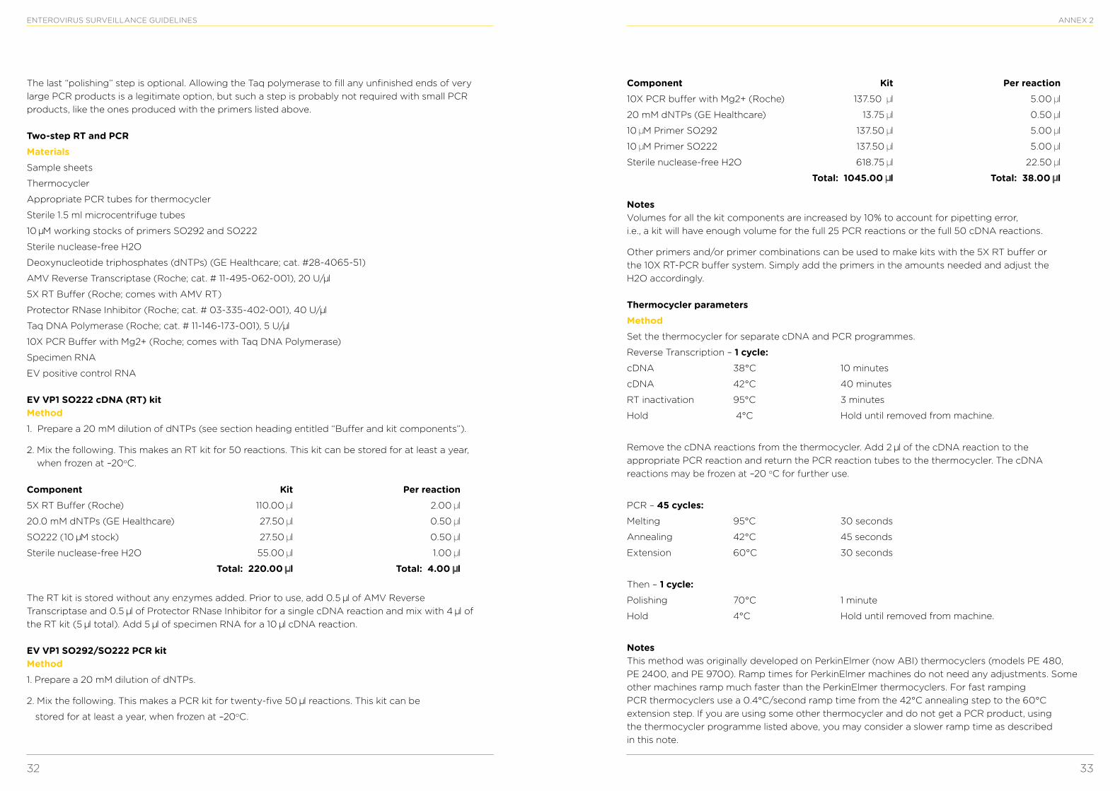
32 33
ENTEROVIRUS SURVEILLANCE GUIDELINES
The last “polishing” step is optional. Allowing the Taq polymerase to fill any unfinished ends of very large PCR products is a legitimate option, but such a step is probably not required with small PCR products, like the ones produced with the primers listed above.
Two-step RT and PCRMaterialsSample sheets
Thermocycler
Appropriate PCR tubes for thermocycler
Sterile 1.5 ml microcentrifuge tubes
10 M working stocks of primers SO292 and SO222
Sterile nuclease-free H2O
Deoxynucleotide triphosphates (dNTPs) (GE Healthcare; cat. #28-4065-51)
AMV Reverse Transcriptase (Roche; cat. # 11-495-062-001), 20 U/ l
5X RT Buffer (Roche; comes with AMV RT)
Protector RNase Inhibitor (Roche; cat. # 03-335-402-001), 40 U/ l
Taq DNA Polymerase (Roche; cat. # 11-146-173-001), 5 U/ l
10X PCR Buffer with Mg2+ (Roche; comes with Taq DNA Polymerase)
Specimen RNA
EV positive control RNA
EV VP1 SO222 cDNA (RT) kit Method1. Prepare a 20 mM dilution of dNTPs (see section heading entitled “Buffer and kit components”).
2. Mix the following. This makes an RT kit for 50 reactions. This kit can be stored for at least a year, when frozen at –20oC.
Component Kit Per reaction5X RT Buffer (Roche) 110.00 l 2.00 l
20.0 mM dNTPs (GE Healthcare) 27.50 l 0.50 l
SO222 (10 M stock) 27.50 l 0.50 l
Sterile nuclease-free H2O 55.00 l 1.00 l
Total: 220.00 l Total: 4.00 l
The RT kit is stored without any enzymes added. Prior to use, add 0.5 l of AMV Reverse Transcriptase and 0.5 l of Protector RNase Inhibitor for a single cDNA reaction and mix with 4 l of the RT kit (5 l total). Add 5 l of specimen RNA for a 10 l cDNA reaction.
EV VP1 SO292/SO222 PCR kit Method1. Prepare a 20 mM dilution of dNTPs.
2. Mix the following. This makes a PCR kit for twenty-five 50 l reactions. This kit can be
stored for at least a year, when frozen at –20oC.
ANNEX 2
Component Kit Per reaction10X PCR buffer with Mg2+ (Roche) 137.50 l 5.00 l
20 mM dNTPs (GE Healthcare) 13.75 l 0.50 l
10 M Primer SO292 137.50 l 5.00 l
10 M Primer SO222 137.50 l 5.00 l
Sterile nuclease-free H2O 618.75 l 22.50 l
Total: 1045.00 l Total: 38.00 l
Notes Volumes for all the kit components are increased by 10% to account for pipetting error, i.e., a kit will have enough volume for the full 25 PCR reactions or the full 50 cDNA reactions.
Other primers and/or primer combinations can be used to make kits with the 5X RT buffer or the 10X RT-PCR buffer system. Simply add the primers in the amounts needed and adjust the H2O accordingly.
Thermocycler parametersMethodSet the thermocycler for separate cDNA and PCR programmes.
Reverse Transcription – 1 cycle:cDNA 38°C 10 minutes
cDNA 42°C 40 minutes
RT inactivation 95°C 3 minutes
Hold 4°C Hold until removed from machine.
Remove the cDNA reactions from the thermocycler. Add 2 l of the cDNA reaction to the appropriate PCR reaction and return the PCR reaction tubes to the thermocycler. The cDNA reactions may be frozen at –20 oC for further use.
PCR – 45 cycles:Melting 95°C 30 seconds
Annealing 42°C 45 seconds
Extension 60°C 30 seconds
Then – 1 cycle:Polishing 70°C 1 minute
Hold 4°C Hold until removed from machine.
Notes This method was originally developed on PerkinElmer (now ABI) thermocyclers (models PE 480, PE 2400, and PE 9700). Ramp times for PerkinElmer machines do not need any adjustments. Some other machines ramp much faster than the PerkinElmer thermocyclers. For fast ramping PCR thermocyclers use a 0.4°C/second ramp time from the 42°C annealing step to the 60°C extension step. If you are using some other thermocycler and do not get a PCR product, using the thermocycler programme listed above, you may consider a slower ramp time as described in this note.
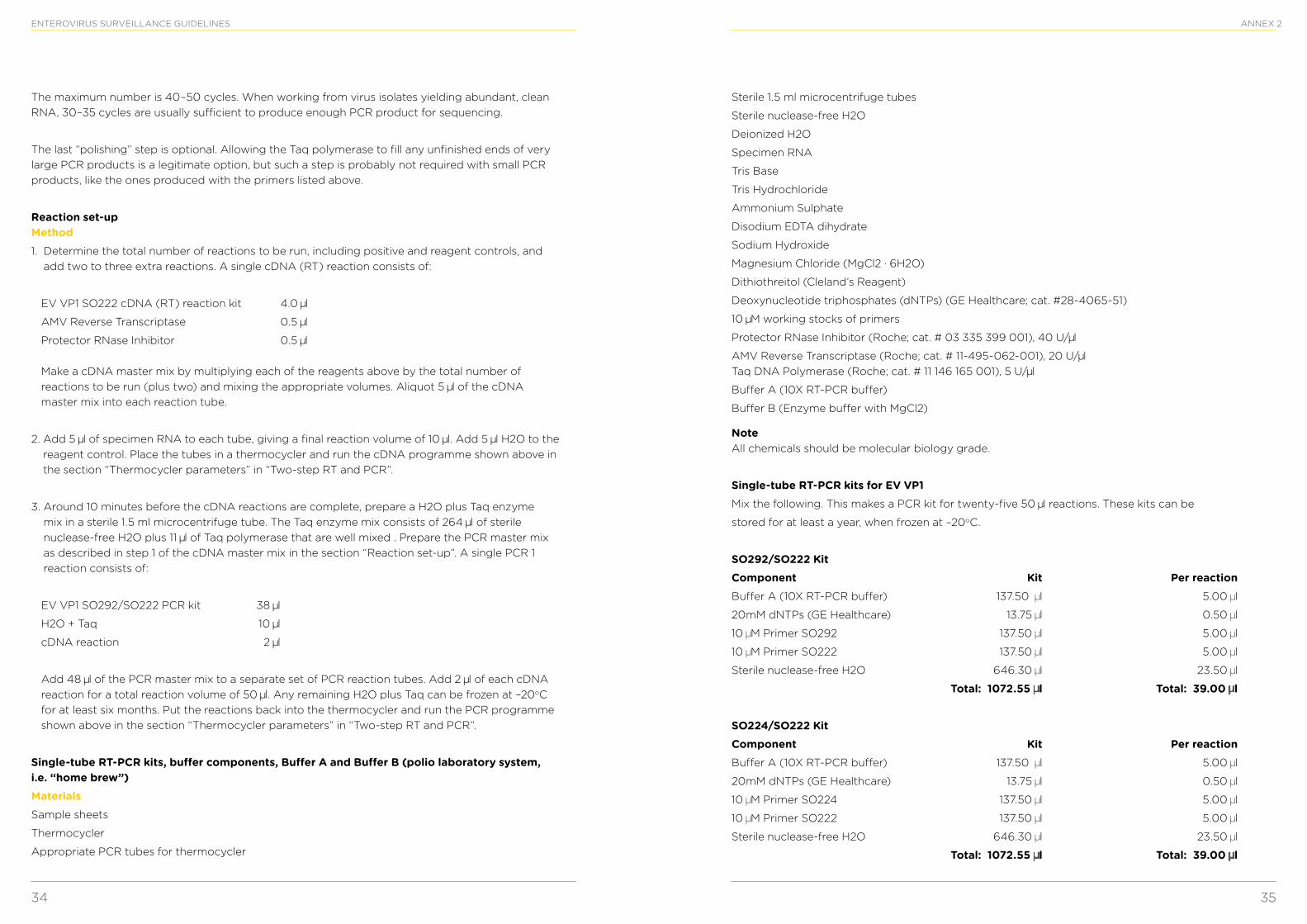
34 35
ENTEROVIRUS SURVEILLANCE GUIDELINES
The maximum number is 40–50 cycles. When working from virus isolates yielding abundant, clean RNA, 30–35 cycles are usually sufficient to produce enough PCR product for sequencing.
The last “polishing” step is optional. Allowing the Taq polymerase to fill any unfinished ends of very large PCR products is a legitimate option, but such a step is probably not required with small PCR products, like the ones produced with the primers listed above.
Reaction set-up Method1. Determine the total number of reactions to be run, including positive and reagent controls, and
add two to three extra reactions. A single cDNA (RT) reaction consists of:
EV VP1 SO222 cDNA (RT) reaction kit 4.0 l
AMV Reverse Transcriptase 0.5 l
Protector RNase Inhibitor 0.5 l
Make a cDNA master mix by multiplying each of the reagents above by the total number of reactions to be run (plus two) and mixing the appropriate volumes. Aliquot 5 l of the cDNA master mix into each reaction tube.
2. Add 5 l of specimen RNA to each tube, giving a final reaction volume of 10 l. Add 5 l H2O to the reagent control. Place the tubes in a thermocycler and run the cDNA programme shown above in the section “Thermocycler parameters” in “Two-step RT and PCR”.
3. Around 10 minutes before the cDNA reactions are complete, prepare a H2O plus Taq enzyme mix in a sterile 1.5 ml microcentrifuge tube. The Taq enzyme mix consists of 264 l of sterile nuclease-free H2O plus 11 l of Taq polymerase that are well mixed . Prepare the PCR master mix as described in step 1 of the cDNA master mix in the section “Reaction set-up”. A single PCR 1 reaction consists of:
EV VP1 SO292/SO222 PCR kit 38 l
H2O + Taq 10 l
cDNA reaction 2 l
Add 48 l of the PCR master mix to a separate set of PCR reaction tubes. Add 2 l of each cDNA reaction for a total reaction volume of 50 l. Any remaining H2O plus Taq can be frozen at –20oC for at least six months. Put the reactions back into the thermocycler and run the PCR programme shown above in the section “Thermocycler parameters” in “Two-step RT and PCR”.
Single-tube RT-PCR kits, buffer components, Buffer A and Buffer B (polio laboratory system, i.e. “home brew”)MaterialsSample sheets
Thermocycler
Appropriate PCR tubes for thermocycler
ANNEX 2
Sterile 1.5 ml microcentrifuge tubes
Sterile nuclease-free H2O
Deionized H2O
Specimen RNA
Tris Base
Tris Hydrochloride
Ammonium Sulphate
Disodium EDTA dihydrate
Sodium Hydroxide
Magnesium Chloride (MgCl2 · 6H2O)
Dithiothreitol (Cleland’s Reagent)
Deoxynucleotide triphosphates (dNTPs) (GE Healthcare; cat. #28-4065-51)
10 M working stocks of primers
Protector RNase Inhibitor (Roche; cat. # 03 335 399 001), 40 U/ l
AMV Reverse Transcriptase (Roche; cat. # 11-495-062-001), 20 U/ lTaq DNA Polymerase (Roche; cat. # 11 146 165 001), 5 U/ l
Buffer A (10X RT-PCR buffer)
Buffer B (Enzyme buffer with MgCl2)
Note All chemicals should be molecular biology grade.
Single-tube RT-PCR kits for EV VP1Mix the following. This makes a PCR kit for twenty-five 50 l reactions. These kits can be
stored for at least a year, when frozen at –20oC.
SO292/SO222 KitComponent Kit Per reactionBuffer A (10X RT-PCR buffer) 137.50 l 5.00 l
20mM dNTPs (GE Healthcare) 13.75 l 0.50 l
10 M Primer SO292 137.50 l 5.00 l
10 M Primer SO222 137.50 l 5.00 l
Sterile nuclease-free H2O 646.30 l 23.50 l
Total: 1072.55 l Total: 39.00 l
SO224/SO222 KitComponent Kit Per reactionBuffer A (10X RT-PCR buffer) 137.50 l 5.00 l
20mM dNTPs (GE Healthcare) 13.75 l 0.50 l
10 M Primer SO224 137.50 l 5.00 l
10 M Primer SO222 137.50 l 5.00 l
Sterile nuclease-free H2O 646.30 l 23.50 l
Total: 1072.55 l Total: 39.00 l
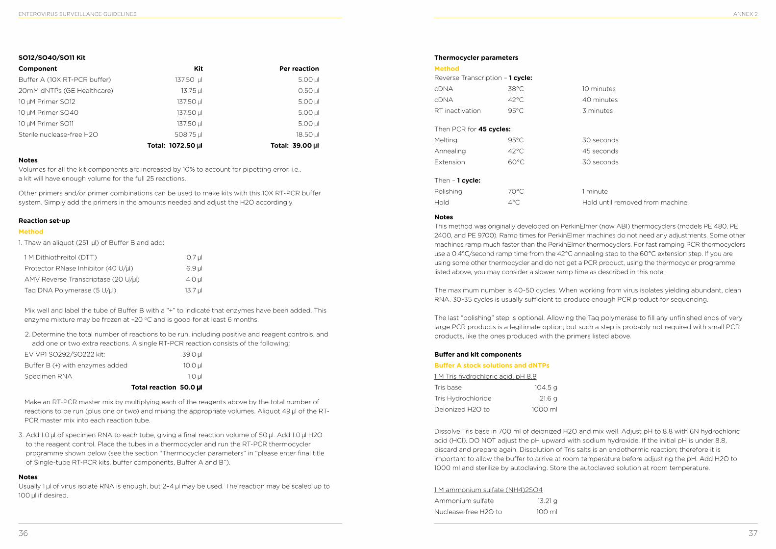
36 37
ENTEROVIRUS SURVEILLANCE GUIDELINES
SO12/SO40/SO11 KitComponent Kit Per reactionBuffer A (10X RT-PCR buffer) 137.50 l 5.00 l
20mM dNTPs (GE Healthcare) 13.75 l 0.50 l
10 M Primer SO12 137.50 l 5.00 l
10 M Primer SO40 137.50 l 5.00 l
10 M Primer SO11 137.50 l 5.00 l
Sterile nuclease-free H2O 508.75 l 18.50 l
Total: 1072.50 l Total: 39.00 l
Notes Volumes for all the kit components are increased by 10% to account for pipetting error, i.e., a kit will have enough volume for the full 25 reactions.
Other primers and/or primer combinations can be used to make kits with this 10X RT-PCR buffer system. Simply add the primers in the amounts needed and adjust the H2O accordingly.
Reaction set-upMethod1. Thaw an aliquot (251 l) of Buffer B and add:
1 M Dithiothreitol (DTT) 0.7 l
Protector RNase Inhibitor (40 U/ l) 6.9 l
AMV Reverse Transcriptase (20 U/ l) 4.0 l
Taq DNA Polymerase (5 U/ l) 13.7 l
Mix well and label the tube of Buffer B with a “+” to indicate that enzymes have been added. This enzyme mixture may be frozen at –20 oC and is good for at least 6 months.
2. Determine the total number of reactions to be run, including positive and reagent controls, and add one or two extra reactions. A single RT-PCR reaction consists of the following:
EV VP1 SO292/SO222 kit: 39.0 l
Buffer B (+) with enzymes added 10.0 l
Specimen RNA 1.0 l
Total reaction 50.0 l
Make an RT-PCR master mix by multiplying each of the reagents above by the total number of reactions to be run (plus one or two) and mixing the appropriate volumes. Aliquot 49 l of the RT-PCR master mix into each reaction tube.
3. Add 1.0 l of specimen RNA to each tube, giving a final reaction volume of 50 l. Add 1.0 l H2O to the reagent control. Place the tubes in a thermocycler and run the RT-PCR thermocycler programme shown below (see the section “Thermocycler parameters” in “please enter final title of Single-tube RT-PCR kits, buffer components, Buffer A and B”).
Notes Usually 1 l of virus isolate RNA is enough, but 2–4 l may be used. The reaction may be scaled up to 100 l if desired.
ANNEX 2
Thermocycler parametersMethod Reverse Transcription – 1 cycle:cDNA 38°C 10 minutes
cDNA 42°C 40 minutes
RT inactivation 95°C 3 minutes
Then PCR for 45 cycles:Melting 95°C 30 seconds
Annealing 42°C 45 seconds
Extension 60°C 30 seconds
Then – 1 cycle:Polishing 70°C 1 minute
Hold 4°C Hold until removed from machine.
Notes This method was originally developed on PerkinElmer (now ABI) thermocyclers (models PE 480, PE 2400, and PE 9700). Ramp times for PerkinElmer machines do not need any adjustments. Some other machines ramp much faster than the PerkinElmer thermocyclers. For fast ramping PCR thermocyclers use a 0.4°C/second ramp time from the 42°C annealing step to the 60°C extension step. If you are using some other thermocycler and do not get a PCR product, using the thermocycler programme listed above, you may consider a slower ramp time as described in this note.
The maximum number is 40-50 cycles. When working from virus isolates yielding abundant, clean RNA, 30-35 cycles is usually sufficient to produce enough PCR product for sequencing.
The last “polishing” step is optional. Allowing the Taq polymerase to fill any unfinished ends of very large PCR products is a legitimate option, but such a step is probably not required with small PCR products, like the ones produced with the primers listed above.
Buffer and kit componentsBuffer A stock solutions and dNTPs1 M Tris hydrochloric acid, pH 8.8
Tris base 104.5 g
Tris Hydrochloride 21.6 g
Deionized H2O to 1000 ml
Dissolve Tris base in 700 ml of deionized H2O and mix well. Adjust pH to 8.8 with 6N hydrochloric acid (HCl). DO NOT adjust the pH upward with sodium hydroxide. If the initial pH is under 8.8, discard and prepare again. Dissolution of Tris salts is an endothermic reaction; therefore it is important to allow the buffer to arrive at room temperature before adjusting the pH. Add H2O to 1000 ml and sterilize by autoclaving. Store the autoclaved solution at room temperature.
1 M ammonium sulfate (NH4)2SO4
Ammonium sulfate 13.21 g
Nuclease-free H2O to 100 ml
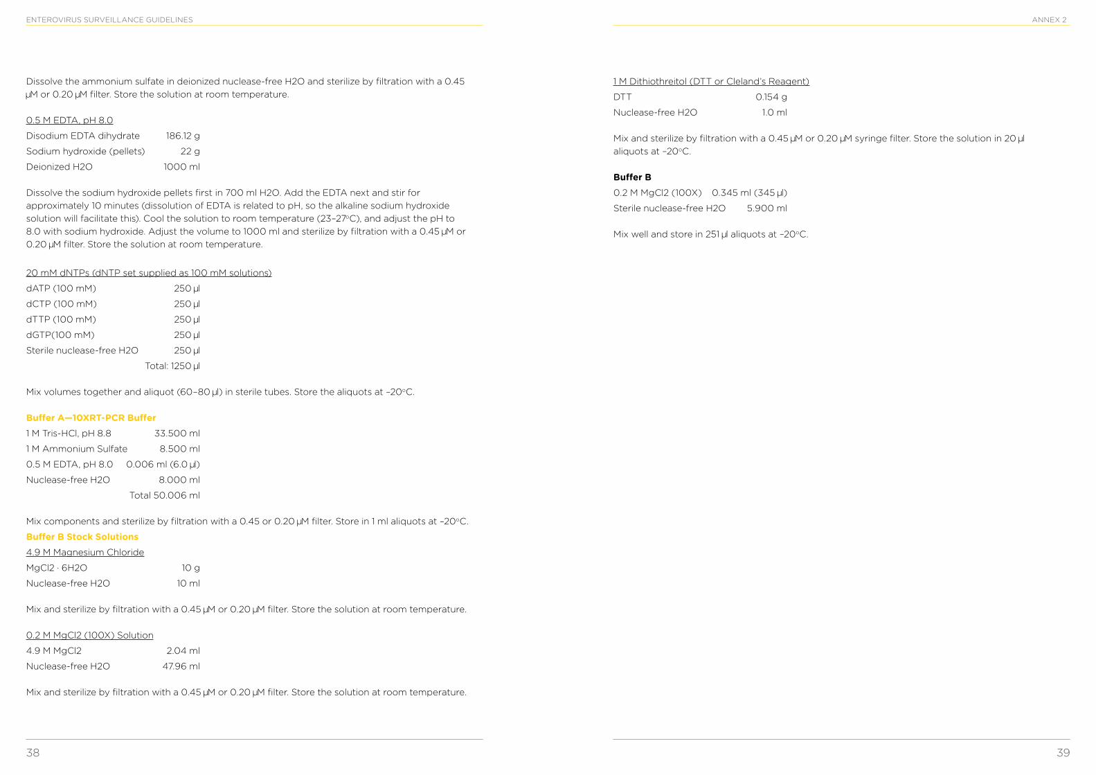
38 39
ENTEROVIRUS SURVEILLANCE GUIDELINES
Dissolve the ammonium sulfate in deionized nuclease-free H2O and sterilize by filtration with a 0.45 M or 0.20 M filter. Store the solution at room temperature.
0.5 M EDTA, pH 8.0
Disodium EDTA dihydrate 186.12 g
Sodium hydroxide (pellets) 22 g
Deionized H2O 1000 ml
Dissolve the sodium hydroxide pellets first in 700 ml H2O. Add the EDTA next and stir for approximately 10 minutes (dissolution of EDTA is related to pH, so the alkaline sodium hydroxide solution will facilitate this). Cool the solution to room temperature (23–27oC), and adjust the pH to 8.0 with sodium hydroxide. Adjust the volume to 1000 ml and sterilize by filtration with a 0.45 M or 0.20 M filter. Store the solution at room temperature.
20 mM dNTPs (dNTP set supplied as 100 mM solutions)
dATP (100 mM) 250 l
dCTP (100 mM) 250 l
dTTP (100 mM) 250 l
dGTP(100 mM) 250 l
Sterile nuclease-free H2O 250 l
Total: 1250 l
Mix volumes together and aliquot (60–80 l) in sterile tubes. Store the aliquots at –20oC.
Buffer A—10XRT-PCR Buffer1 M Tris-HCl, pH 8.8 33.500 ml
1 M Ammonium Sulfate 8.500 ml
0.5 M EDTA, pH 8.0 0.006 ml (6.0 l)
Nuclease-free H2O 8.000 ml
Total 50.006 ml
Mix components and sterilize by filtration with a 0.45 or 0.20 M filter. Store in 1 ml aliquots at –20oC.
Buffer B Stock Solutions4.9 M Magnesium Chloride
MgCl2 · 6H2O 10 g
Nuclease-free H2O 10 ml
Mix and sterilize by filtration with a 0.45 M or 0.20 M filter. Store the solution at room temperature.
0.2 M MgCl2 (100X) Solution
4.9 M MgCl2 2.04 ml
Nuclease-free H2O 47.96 ml
Mix and sterilize by filtration with a 0.45 M or 0.20 M filter. Store the solution at room temperature.
ANNEX 2
1 M Dithiothreitol (DTT or Cleland’s Reagent)
DTT 0.154 g
Nuclease-free H2O 1.0 ml
Mix and sterilize by filtration with a 0.45 M or 0.20 M syringe filter. Store the solution in 20 l aliquots at –20oC.
Buffer B0.2 M MgCl2 (100X) 0.345 ml (345 l)
Sterile nuclease-free H2O 5.900 ml
Mix well and store in 251 l aliquots at –20oC.
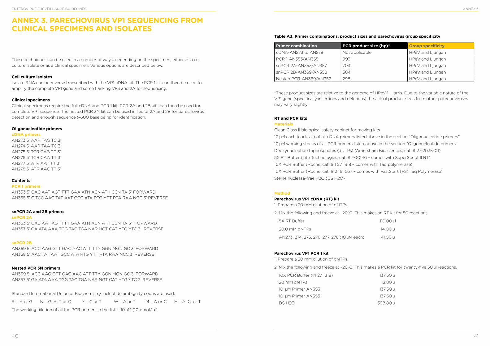
40 41
ENTEROVIRUS SURVEILLANCE GUIDELINES ANNEX 3
These techniques can be used in a number of ways, depending on the specimen, either as a cell culture isolate or as a clinical specimen. Various options are described below.
Cell culture isolates Isolate RNA can be reverse transcribed with the VP1 cDNA kit. The PCR 1 kit can then be used to amplify the complete VP1 gene and some flanking VP3 and 2A for sequencing.
Clinical specimens Clinical specimens require the full cDNA and PCR 1 kit. PCR 2A and 2B kits can then be used for complete VP1 sequence. The nested PCR 3N kit can be used in lieu of 2A and 2B for parechovirus detection and enough sequence (≈300 base pairs) for identification.
Oligonucleotide primers cDNA primersAN273 5’ AAR TAG TC 3’AN274 5’ AAR TAA TC 3’AN275 5’ TCR CAG TT 3’AN276 5’ TCR CAA TT 3’AN277 5’ ATR AAT TT 3’AN278 5’ ATR AAC TT 3’
ContentsPCR 1 primersAN353 5’ GAC AAT AGT TTT GAA ATN ACN ATH CCN TA 3’ FORWARDAN355 5’ C TCC AAC TAT AAT GCC ATA RTG YTT RTA RAA NCC 3’ REVERSE
snPCR 2A and 2B primers snPCR 2A AN353 5’ GAC AAT AGT TTT GAA ATN ACN ATH CCN TA 3’ FORWARDAN357 5’ GA ATA AAA TGG TAC TGA NAR NGT CAT YTG YTC 3’ REVERSE
snPCR 2B AN369 5’ ACC AAG GTT GAC AAC ATT TTY GGN MGN GC 3’ FORWARDAN358 5’ AAC TAT AAT GCC ATA RTG YTT RTA RAA NCC 3’ REVERSE
Nested PCR 3N primers AN369 5’ ACC AAG GTT GAC AAC ATT TTY GGN MGN GC 3’ FORWARDAN357 5’ GA ATA AAA TGG TAC TGA NAR NGT CAT YTG YTC 3’ REVERSE
Standard International Union of Biochemistry ucleotide ambiguity codes are used:
R = A or G N = G, A, T or C Y = C or T W = A or T M = A or C H = A, C, or T
The working dilution of all the PCR primers in the list is 10 M (10 pmol/ l).
ANNEX 3. PARECHOVIRUS VP1 SEQUENCING FROM CLINICAL SPECIMENS AND ISOLATES
Table A3. Primer combinations, product sizes and parechovirus group specificity
Primer combination PCR product size (bp)* Group specificitycDNA-AN273 to AN278 Not applicable HPeV and Ljungan
PCR 1-AN353/AN355 993 HPeV and Ljungan
snPCR 2A-AN353/AN357 703 HPeV and Ljungan
snPCR 2B-AN369/AN358 584 HPeV and Ljungan
Nested PCR-AN369/AN357 298 HPeV and Ljungan
*These product sizes are relative to the genome of HPeV 1, Harris. Due to the variable nature of the VP1 gene (specifically insertions and deletions) the actual product sizes from other parechoviruses may vary slightly.
RT and PCR kitsMaterials Clean Class II biological safety cabinet for making kits
10 M each (cocktail) of all cDNA primers listed above in the section “Oligonucleotide primers”
10 M working stocks of all PCR primers listed above in the section “Oligonucleotide primers”
Deoxynucleotide triphosphates (dNTPs) (Amersham Biosciences; cat. # 27-2035-01)
5X RT Buffer (Life Technologies; cat. # Y00146 – comes with SuperScript II RT)
10X PCR Buffer (Roche; cat. # 1 271 318 – comes with Taq polymerase)
10X PCR Buffer (Roche; cat. # 2 161 567 – comes with FastStart (FS) Taq Polymerase)
Sterile nuclease-free H2O (DS H2O)
Method Parechovirus VP1 cDNA (RT) kit 1. Prepare a 20 mM dilution of dNTPs.
2. Mix the following and freeze at –20oC. This makes an RT kit for 50 reactions.
5X RT Buffer 110.00 l
20.0 mM dNTPs 14.00 l
AN273, 274, 275, 276, 277, 278 (10 M each) 41.00 l
Parechovirus VP1 PCR 1 kit 1. Prepare a 20 mM dilution of dNTPs.
2. Mix the following and freeze at –20oC. This makes a PCR kit for twenty-five 50 l reactions.
10X PCR Buffer (#1 271 318) 137.50 l
20 mM dNTPs 13.80 l
10 M Primer AN353 137.50 l
10 M Primer AN355 137.50 l
DS H2O 398.80 l
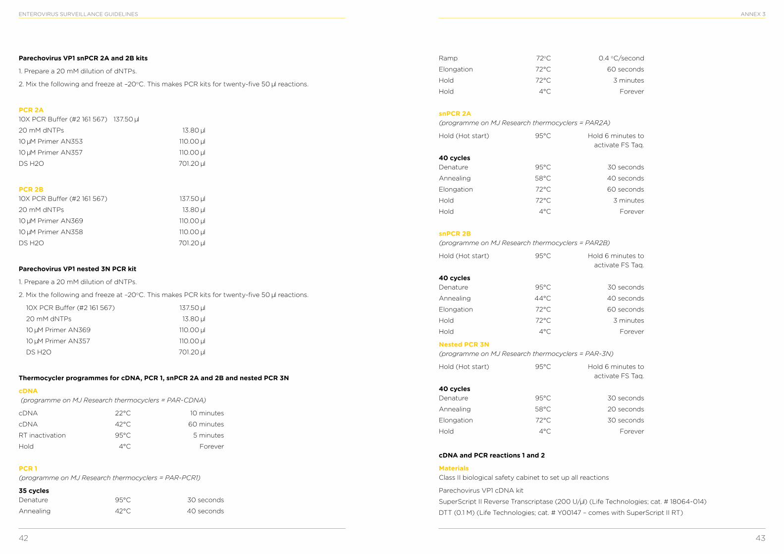
42 43
ENTEROVIRUS SURVEILLANCE GUIDELINES
Parechovirus VP1 snPCR 2A and 2B kits
1. Prepare a 20 mM dilution of dNTPs.
2. Mix the following and freeze at –20oC. This makes PCR kits for twenty-five 50 l reactions.
PCR 2A 10X PCR Buffer (#2 161 567) 137.50 l
20 mM dNTPs 13.80 l
10 M Primer AN353 110.00 l
10 M Primer AN357 110.00 l
DS H2O 701.20 l
PCR 2B 10X PCR Buffer (#2 161 567) 137.50 l
20 mM dNTPs 13.80 l
10 M Primer AN369 110.00 l
10 M Primer AN358 110.00 l
DS H2O 701.20 l
Parechovirus VP1 nested 3N PCR kit
1. Prepare a 20 mM dilution of dNTPs.
2. Mix the following and freeze at –20oC. This makes PCR kits for twenty-five 50 l reactions.
10X PCR Buffer (#2 161 567) 137.50 l
20 mM dNTPs 13.80 l
10 M Primer AN369 110.00 l
10 M Primer AN357 110.00 l
DS H2O 701.20 l
Thermocycler programmes for cDNA, PCR 1, snPCR 2A and 2B and nested PCR 3N
cDNA (programme on MJ Research thermocyclers = PAR-CDNA)
cDNA 22°C 10 minutes
cDNA 42°C 60 minutes
RT inactivation 95°C 5 minutes
Hold 4°C Forever
PCR 1 (programme on MJ Research thermocyclers = PAR-PCR1)
35 cycles Denature 95°C 30 seconds
Annealing 42°C 40 seconds
ANNEX 3
Ramp 72oC 0.4 oC/second
Elongation 72°C 60 seconds
Hold 72°C 3 minutes
Hold 4°C Forever
snPCR 2A (programme on MJ Research thermocyclers = PAR2A)
Hold (Hot start) 95°C Hold 6 minutes to activate FS Taq.
40 cycles Denature 95°C 30 seconds
Annealing 58°C 40 seconds
Elongation 72°C 60 seconds
Hold 72°C 3 minutes
Hold 4°C Forever
snPCR 2B (programme on MJ Research thermocyclers = PAR2B)
Hold (Hot start) 95°C Hold 6 minutes to activate FS Taq.
40 cycles Denature 95°C 30 seconds
Annealing 44°C 40 seconds
Elongation 72°C 60 seconds
Hold 72°C 3 minutes
Hold 4°C Forever
Nested PCR 3N (programme on MJ Research thermocyclers = PAR-3N)
Hold (Hot start) 95°C Hold 6 minutes to activate FS Taq.
40 cycles Denature 95°C 30 seconds
Annealing 58°C 20 seconds
Elongation 72°C 30 seconds
Hold 4°C Forever
cDNA and PCR reactions 1 and 2
Materials Class II biological safety cabinet to set up all reactions
Parechovirus VP1 cDNA kit
SuperScript II Reverse Transcriptase (200 U/ l) (Life Technologies; cat. # 18064-014)
DTT (0.1 M) (Life Technologies; cat. # Y00147 – comes with SuperScript II RT)
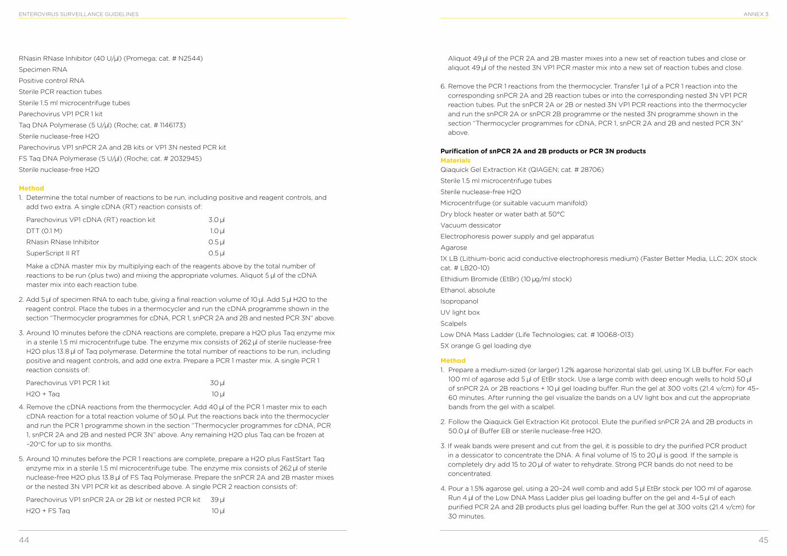
44 45
ENTEROVIRUS SURVEILLANCE GUIDELINES
RNasin RNase Inhibitor (40 U/ l) (Promega; cat. # N2544)
Specimen RNA
Positive control RNA
Sterile PCR reaction tubes
Sterile 1.5 ml microcentrifuge tubes
Parechovirus VP1 PCR 1 kit
Taq DNA Polymerase (5 U/ l) (Roche; cat. # 1146173)
Sterile nuclease-free H2O
Parechovirus VP1 snPCR 2A and 2B kits or VP1 3N nested PCR kit
FS Taq DNA Polymerase (5 U/ l) (Roche; cat. # 2032945)
Sterile nuclease-free H2O
Method 1. Determine the total number of reactions to be run, including positive and reagent controls, and
add two extra. A single cDNA (RT) reaction consists of:
Parechovirus VP1 cDNA (RT) reaction kit 3.0 l
DTT (0.1 M) 1.0 l
RNasin RNase Inhibitor 0.5 l
SuperScript II RT 0.5 l
Make a cDNA master mix by multiplying each of the reagents above by the total number of reactions to be run (plus two) and mixing the appropriate volumes. Aliquot 5 l of the cDNA master mix into each reaction tube.
2. Add 5 l of specimen RNA to each tube, giving a final reaction volume of 10 l. Add 5 l H2O to the reagent control. Place the tubes in a thermocycler and run the cDNA programme shown in the section “Thermocycler programmes for cDNA, PCR 1, snPCR 2A and 2B and nested PCR 3N” above.
3. Around 10 minutes before the cDNA reactions are complete, prepare a H2O plus Taq enzyme mix in a sterile 1.5 ml microcentrifuge tube. The enzyme mix consists of 262 l of sterile nuclease-free H2O plus 13.8 l of Taq polymerase. Determine the total number of reactions to be run, including positive and reagent controls, and add one extra. Prepare a PCR 1 master mix. A single PCR 1 reaction consists of:
Parechovirus VP1 PCR 1 kit 30 l
H2O + Taq 10 l
4. Remove the cDNA reactions from the thermocycler. Add 40 l of the PCR 1 master mix to each cDNA reaction for a total reaction volume of 50 l. Put the reactions back into the thermocycler and run the PCR 1 programme shown in the section “Thermocycler programmes for cDNA, PCR 1, snPCR 2A and 2B and nested PCR 3N” above. Any remaining H2O plus Taq can be frozen at –20oC for up to six months.
5. Around 10 minutes before the PCR 1 reactions are complete, prepare a H2O plus FastStart Taq enzyme mix in a sterile 1.5 ml microcentrifuge tube. The enzyme mix consists of 262 l of sterile nuclease-free H2O plus 13.8 l of FS Taq Polymerase. Prepare the snPCR 2A and 2B master mixes or the nested 3N VP1 PCR kit as described above. A single PCR 2 reaction consists of:
Parechovirus VP1 snPCR 2A or 2B kit or nested PCR kit 39 l
H2O + FS Taq 10 l
ANNEX 3
Aliquot 49 l of the PCR 2A and 2B master mixes into a new set of reaction tubes and close or aliquot 49 l of the nested 3N VP1 PCR master mix into a new set of reaction tubes and close.
6. Remove the PCR 1 reactions from the thermocycler. Transfer 1 l of a PCR 1 reaction into the corresponding snPCR 2A and 2B reaction tubes or into the corresponding nested 3N VP1 PCR reaction tubes. Put the snPCR 2A or 2B or nested 3N VP1 PCR reactions into the thermocycler and run the snPCR 2A or snPCR 2B programme or the nested 3N programme shown in the section “Thermocycler programmes for cDNA, PCR 1, snPCR 2A and 2B and nested PCR 3N” above.
Purification of snPCR 2A and 2B products or PCR 3N products Materials Qiaquick Gel Extraction Kit (QIAGEN; cat. # 28706)
Sterile 1.5 ml microcentrifuge tubes
Sterile nuclease-free H2O
Microcentrifuge (or suitable vacuum manifold)
Dry block heater or water bath at 50°C
Vacuum dessicator
Electrophoresis power supply and gel apparatus
Agarose
1X LB (Lithium-boric acid conductive electrophoresis medium) (Faster Better Media, LLC; 20X stock cat. # LB20-10)
Ethidium Bromide (EtBr) (10 g/ml stock)
Ethanol, absolute
Isopropanol
UV light box
Scalpels
Low DNA Mass Ladder (Life Technologies; cat. # 10068-013)
5X orange G gel loading dye
Method 1. Prepare a medium-sized (or larger) 1.2% agarose horizontal slab gel, using 1X LB buffer. For each
100 ml of agarose add 5 l of EtBr stock. Use a large comb with deep enough wells to hold 50 l of snPCR 2A or 2B reactions + 10 l gel loading buffer. Run the gel at 300 volts (21.4 v/cm) for 45–60 minutes. After running the gel visualize the bands on a UV light box and cut the appropriate bands from the gel with a scalpel.
2. Follow the Qiaquick Gel Extraction Kit protocol. Elute the purified snPCR 2A and 2B products in 50.0 l of Buffer EB or sterile nuclease-free H2O.
3. If weak bands were present and cut from the gel, it is possible to dry the purified PCR product in a dessicator to concentrate the DNA. A final volume of 15 to 20 l is good. If the sample is completely dry add 15 to 20 l of water to rehydrate. Strong PCR bands do not need to be concentrated.
4. Pour a 1.5% agarose gel, using a 20–24 well comb and add 5 l EtBr stock per 100 ml of agarose. Run 4 l of the Low DNA Mass Ladder plus gel loading buffer on the gel and 4–5 l of each purified PCR 2A and 2B products plus gel loading buffer. Run the gel at 300 volts (21.4 v/cm) for 30 minutes.
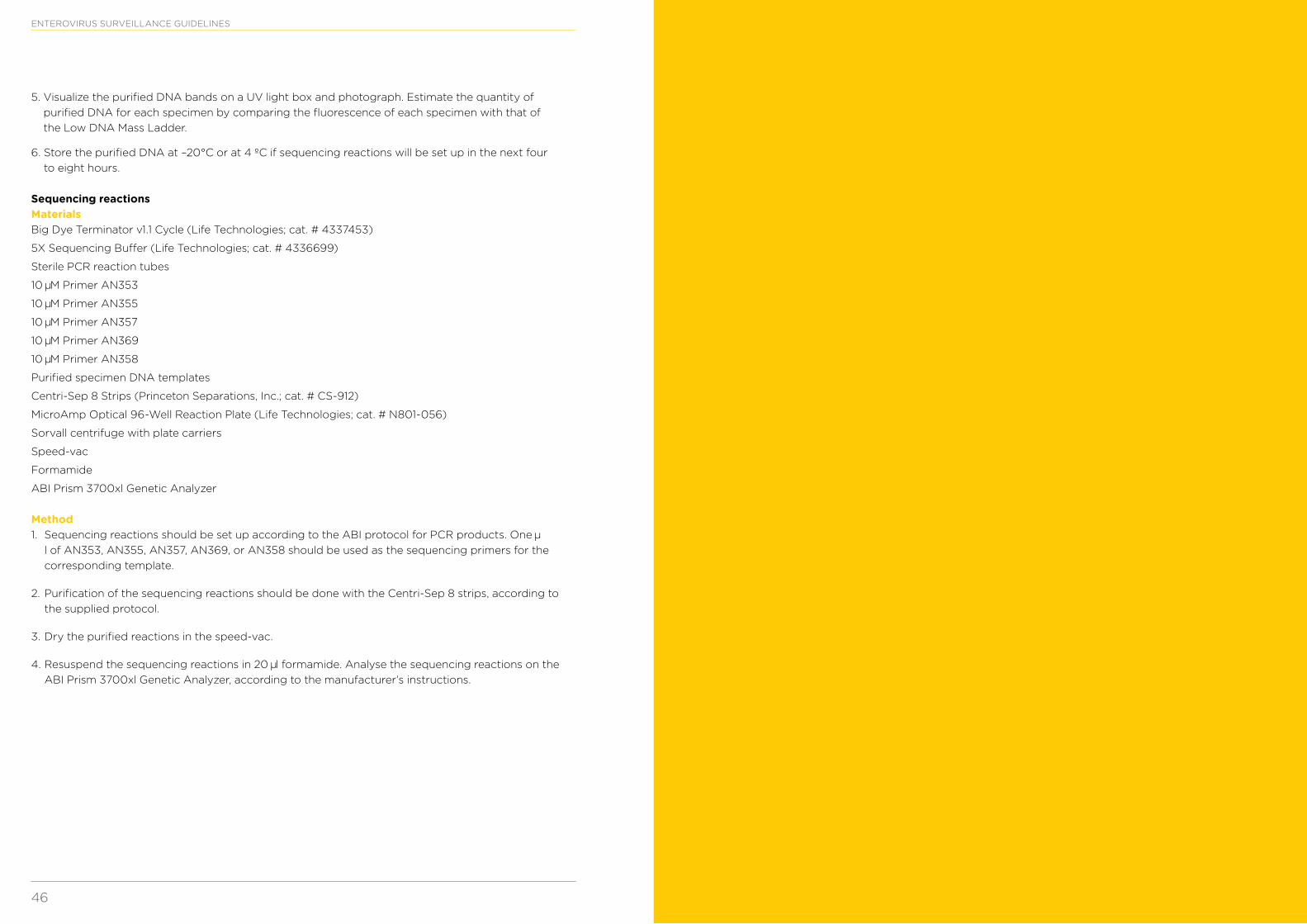
46
ENTEROVIRUS SURVEILLANCE GUIDELINES
5. Visualize the purified DNA bands on a UV light box and photograph. Estimate the quantity of purified DNA for each specimen by comparing the fluorescence of each specimen with that of the Low DNA Mass Ladder.
6. Store the purified DNA at –20°C or at 4 ºC if sequencing reactions will be set up in the next four to eight hours.
Sequencing reactions Materials Big Dye Terminator v1.1 Cycle (Life Technologies; cat. # 4337453)
5X Sequencing Buffer (Life Technologies; cat. # 4336699)
Sterile PCR reaction tubes
10 M Primer AN353
10 M Primer AN355
10 M Primer AN357
10 M Primer AN369
10 M Primer AN358
Purified specimen DNA templates
Centri-Sep 8 Strips (Princeton Separations, Inc.; cat. # CS-912)
MicroAmp Optical 96-Well Reaction Plate (Life Technologies; cat. # N801-056)
Sorvall centrifuge with plate carriers
Speed-vac
Formamide
ABI Prism 3700xl Genetic Analyzer
Method 1. Sequencing reactions should be set up according to the ABI protocol for PCR products. One
l of AN353, AN355, AN357, AN369, or AN358 should be used as the sequencing primers for the corresponding template.
2. Purification of the sequencing reactions should be done with the Centri-Sep 8 strips, according to the supplied protocol.
3. Dry the purified reactions in the speed-vac.
4. Resuspend the sequencing reactions in 20 l formamide. Analyse the sequencing reactions on the ABI Prism 3700xl Genetic Analyzer, according to the manufacturer’s instructions.
World Health Organization Regional Office for EuropeUN City, Marmorvej 51, DK-2100 Copenhagen Ø, DenmarkTel.: +45 45 33 70 00 Fax: +45 45 33 70 01Email: [email protected] Website: www.euro.who.int
Member StatesAlbania
Andorra
Armenia
Austria
Azerbaijan
Belarus
Belgium
Bosnia and Herzegovina
Bulgaria
Croatia
Cyprus
Czech Republic
Denmark
Estonia
Finland
France
Georgia
Germany
Greece
Hungary
Iceland
Ireland
Israel
Italy
Kazakhstan
Kyrgyzstan
Latvia
Lithuania
Luxembourg
Malta
Monaco
Montenegro
Netherlands
Norway
Poland
Portugal
Republic of Moldova
Romania
Russian Federation
San Marino
Serbia
Slovakia
Slovenia
Spain
Sweden
Switzerland
Tajikistan
The former Yugoslav Republic of Macedonia
Turkey
Turkmenistan
Ukraine
United Kingdom
Uzbekistan
WHO Regional Office for EuropeThe World Health Organization (WHO) is a specialized agency of the United Nations created in 1948 with the primary responsibility for international health matters and public health. The WHO Regional Office for Europe is one of six regional offices throughout the world, each with its own programme geared to the particular health conditions of the countries it serves.
9 789289 050814 >
ISBN 978-92-890-5081-4


![2do ex enterovirus[1]](https://img.pdfslide.net/doc/110x75/556e2432d8b42a6a698b456f/2do-ex-enterovirus1.jpg)