Embed Size (px)
Citation preview
4Research Branch
Technical Bulletin 1993-8E
Enumerationand
identification
of meatspoilage
bacteria
Canada
?*'?
Cover illustration
The images represent the Research Branch's objective: .
•
to improve the long-term competitiveness of the Canadian
agri-food sector through the development and transfer of new
technologies.
Designed by Research Program Service.
Illustration de la couverture
Les dessins illustrent l'objectif de la Direction generale de la
recherche : ameliorer la competitivite a long ferme du secteur
agro-alimentaire canadien grace a la tnise au point et au transfert
de nouvelles technologies.
Conception par le Service au.x programmes de recherchcs.
©
Enumeration andidentification of
meat spoilage
bacteria
CO. GILL and G.G. GREERResearch Station
Lacombe, Alberta
Technical Bulletin 1993-8ELacombe Technical Bulletin No. 6
Research BranchAgriculture Canada
1993
Copies of this publication are available from
The Information Officer
Research Station
Research Branch, Agriculture Canada
Bag Service 5000
Lacombe, Alberta
TOC ISO
© Minister of Supply and Services Canada 1993
Cat. No. 54-8/1993-8E
ISBN 0-662-20963-X
Printed 1993
CONTENTSPage
SUMMARY/RESUME iv
INTRODUCTION 1
INITIAL FLORA ON MEAT 1
SPOILAGE FLORA 2
SPOILAGE MICROORGANISMS 3
Pseudomonas 3
Acinetobacter I Moraxella (Psychrobacter) 3
Enterobacteriaceae 4
Lactic acid bacteria 4
Brochothrix thermosphacta 5
Shewanella (Alteromonas) putrefaciens 5
OTHER ORGANISMS ENCOUNTERED IN THE INITIAL FLORA 6
SPOILAGE DEVELOPMENT 7
IDENTIFICATION OF SPOILAGE BACTERIA 7
TESTS USED FOR DIFFERENTIATION OF SPOILAGE BACTERIA 7
Gram reaction 7
Morphology 1
1
Catalase 1
1
Oxidase 1
1
Arginine 12
Glucose utilization 13
Motility 13
SELECTIVE/DIFFERENTIAL MEDIA FOR SPOILAGE BACTERIA 14
Introduction 14
Total counts: Plate Count Agar 15
Pseudomonas: Cephaloridine Fucidin Cetrimide Agar (CFC) 16
Enterobacteriaceae: Violet Red Bile Glucose Agar (VRBG) 17
Lactic acid bacteria: deMan, Rogosa and Sharpe Agar (MRS) 18
Carnobacterium: Cresol Red Thallium Acetate Sucrose Agar (CTAS) 19
Brochothrix thermosphacta: Streptomycin Thallous Acetate
Actidione Agar (STAA) 20Brochothrix thermosphacta: Streptomycin Sulphate Inositol
Neutral Red Agar (SIN) 21
Shewanella putrefaciens: Peptone Iron Agar 22
REFERENCES 22

SUMMARY
During prolonged storage of meat at chill temperatures, only a few types
of organisms from the microbial flora originally present on meat are able to grow,
and so appear in the spoilage flora. Therefore, spoilage flora of chilled meatusually contain only a limited number of bacterial types. Depending on the initial
flora and the growth environment, only Brochothrix thermosphacta, Shewanella
(Alteromonas) putrefaciens, a few species of Pseudomonas, Acinetobacter,
Moraxella, (Psychrobacter), lactic acid bacteria, and some members of the family
Enterobacteriaceae are significantly represented in most spoilage flora of chilled
meats. These seven principal groups of spoilage bacteria can be readily
differentiated from one another using the following seven properties: Gram-reaction, morphology, motility, the manner in which glucose is utilized, and the
presence of catalase, oxidase and arginine dehydrolase/decarboxylase.
Spoilage bacteria can also be enumerated using selective and differential
culture media, to suppress the growth of unwanted bacteria and assist the
detection of the desired organisms. Those media have the disadvantage of
commonly underestimating the bacterial population, but they can be used to
conveniently identify changes in the numbers of specific groups of spoilage
bacteria.
RESUME
Lorsque la viande est entreposee a une temperature froide pour une
periode prolongee, peu d'especes de la flore microbienne peuvent y crottre et
celles qui y reussissent font partie de la flore contaminante, responsable de la
deterioration de la qualite de la viande. Dependamment de la nature de la flore
pionniere et du milieu de croissance, on retrouve les groupes suivants:
Brochothrix thermosphacta , Shewanella (Alteromonas) putrefaciens, quelques
especes de Pseudomonas, Acinetobacter, Moraxella, (Psychrobacter), des
bacteries lactiques et quelques membres de la famille des Enterobacteriaceae,
sont aussi representees de fagon significative On peut differencier ces sept
groupes principaux a I'aide de la reaction a gram, la morphologie ou motilite, la
facon d'utiliser le glucose, la presence de catalase, d'oxydase et d'arginine
dehydrolase/decarboxylase.
Ces bacteries peuvent egalement etre denombrees en utilisant des
milieux de culture differentiels et selectifs pour soit, supprimer la croissance des
bacteries indesirables ou pour aider a identifier celles qui pourraient etre
utilisees favorablement. Si ces milieux ont le desavantage de sous-estimer la
population bacterienne, ils peuvent par contre etre employes pour identifier defagon appropriee les changements dans le nombre de certains groupes de
bacteries.
INTRODUCTION
Many species of microorganisms are able to grow on fresh meat. Their
growth will eventually make the meat unacceptable for human consumption
because of changes in the appearance, odour and flavour of the food (Dainty et
ai, 1983; Gill, 1986). The changes caused by microbial activity will not occur if
fresh meat is frozen to temperatures that are too low for microbial growth to
occur. At chiller temperatures, -1.5° to +5°C, microbial growth is slowed, but is
not prevented (McMeekin, 1981). However, spoilage is delayed by the slower
rates of microbial growth. The rate of microbial growth can be further reduced
by changing the atmosphere to which the bacteria are exposed. Both restricting
the oxygen available to the bacteria and increasing the concentration of carbon
dioxide tends to reduce the rate of growth of most spoilage bacteria. Chiller
storage alone prevents the growth of mesophilic (cold intolerant) species,
ensuring that only the few psychrotrophic (cold tolerant) organisms that were
initially present on the meat are able to grow and form a spoilage flora.
Consequently, floras developing on meat stored at chiller temperatures generally
have a low species diversity (Ingram and Simonsen, 1980; Dainty and Mackey,
1992).
INITIAL FLORA ON MEAT
The bacteria found on skinned carcasses immediately after dressing are
skin commensals, and soil and faecal organisms that were present on the hide.
Most of those bacteria are Gram-positive mesophiles such as Micrococcus,
Staphylococcus, and Bacillus, but a few Gram-negative psychrotrophic
organisms will also be present. Unskinned pig carcasses that have beenscalded, singed and polished will carry similar numbers of Gram-positive
mesophiles and Gram-negative psychrotrophs, because the thermoduric (heat-
tolerant) organisms associated with the skin that survive the scalding operation
are augmented by psychrotrophic species derived from the dehairing andpolishing equipment.
Growth of the Gram-positive mesophiles will be insignificant during
carcass cooling, but substantial growth of the psychrotrophic species can occur
when carcass cooling processes are poorly controlled.
When carcasses are broken down, bacteria are transferred from the
contaminated carcass surfaces to the freshly cut meat surfaces. Additional
mesophiles will be transferred to the meat from the hands of workers. Additional
psychrotrophs will also be transferred from equipment when proper attention is
not given to the cleaning of equipment.
The final hygienic condition of the meat when it leaves the fabrication line
will reflect the overall hygiene of the dressing, cooling and fabrication processes
(Egan and Roberts, 1987). When good hygienic practices are maintained in all
three processes, the average numbers of bacteria on beef and lamb are likely to
be less than 103/cm2 , with less than 5% of the flora being psychrotrophic,
spoilage types. When hygiene practices are lacking in some respects, beef andlamb will carry bacteria at numbers greater than 103/cm2 , with 10% or more of
the flora being spoilage types. With pork, the initial contamination of the scalded
carcasses will usually ensure that the average numbers of bacteria usually
exceed 103/cm2 , with about 50% of the flora being spoilage types.
SPOILAGE FLORA
The spoilage flora of chilled meat are usually dominated by the bacteria
that grow most rapidly under the conditions experienced by the meat. The mostrapidly growing species will fail to dominate only when heavy contamination with
a slower growing competitor leaves insufficient time before spoilage for the
growth rate advantage to be fully expressed. Fresh meat that is held in air at
chill temperatures will develop a flora that is usually dominated by species of
Pseudomonas, but with substantial fractions of the other strictly aerobic genera,
Acinetobacter and Moraxella. Enterobacteria, and Brochothrix thermosphacta
may also be present, if either formed a substantial fraction of the initial flora.
The bacteria forming an aerobic flora will interact only when the maximum cell
numbers are reached, at which time the bacteria apparently compete for the
available oxygen. Aerobic spoilage bacteria grow by using soluble, low
molecular weight compounds present at the meat surface. The bacteria that are
primarily responsible for aerobic spoilage, the pseudomonads, use glucose
preferentially. However, the bacteria eventually attain numbers so great that
glucose cannot diffuse from the underlying meat to the surface sufficiently fast to
satisfy the demands of the flora. The pseudomonads maintain their growth by
switching to alternate substrates, notably the amino acids. When amino acids
are used, malodorous compounds are generated as by-products. The putrid
smells of those by-products indicate the onset of aerobic spoilage (Gill, 1986).
The rate of growth of the aerobic spoilage flora is slowed by high
concentrations of carbon dioxide. However, the genera that dominate the
spoilage flora of meat stored in air will still predomonate in aerobic atmospheres
modified by the addition of carbon dioxide. The only means of precluding that
dominance is to exclude oxygen from the atmosphere around the meat (Gill and
Molin, 1991). The most common means of achieving anaerobic conditions is to
vacuum package the meat in a film of low oxygen permeability. Biochemical
activities of the muscle tissue then scavange any residual oxygen in the pack,
and consume the small quantities of oxygen that permeate through the film. Thespoilage flora of meat stored under anaerobic conditions is usually dominated by
lactic acid bacteria of the genera Lactobacillus, Leuconostoc and
Carnobacterium. Such lactobacilli will usually be the only bacteria
distinguishable in anaerobic spoilage flora from muscle tissue of normal pH (5.5-
5.8). A flora of that type will produce mild acid-dairy flavours in meat only sometime after the flora has attained its maximum numbers. However, other types of
bacteria can grow on high pH (>5.8) muscle, and fat tissue that is not bathed in
normal-pH exudate. Those bacteria are the facultative anaerobes of the family
Enterobacteriaceae, and the species Shewanella putrefaciens and Brochothrix
thermosphacta. Although the facultative anaerobes grow at slower rates than
the lactobacilli, they produce by-products that are highly offensive. Thus, they
will dominate the spoilage process even when they are only a minor fraction of
the total bacterial population.
Meat flora may contain very different fractions of spoilage bacteria, and
the spoilage activities of some types are disproportional to their relative
numbers. Therefore, knowledge of the flora composition, as well as the total
number, is required to understand and gain control over the spoilage process.
Chill temperature spoilage flora are confined to a few bacterial types, so a short
identification scheme that differentiates between the principal groups of spoilage
organisms can be used for all but the most specialized investigations.
SPOILAGE MICROORGANISMS
Pseudomonas
Working definition: Pseudomonas species consist of Gram-negative,
motile rods. They are strictly aerobic, catalase-positive, oxidase-positive andarginine-positive. They attack sugars by oxidation and do not produce gas.
Pseudomonads dominate the aerobic spoilage of chilled meat. The main
species associated with raw meat are P. tragi and P. fluorescens. Thepseudomonads preferentially use glucose. While using that substrate they donot produce malodorous compounds. However, when glucose is insufficient,
they attack amino acids, forming sulphides, amines and esters that are
organoleptically detected as putrid odours and flavours. Spoilage of normal pH(5.5-5.8) muscle tissue by Pseudomonas occurs when bacterial numbers exceed
108/cm2 . However, muscle of dark, firm, dry (DFD) condition can be devoid of
glucose and fat tissue is always glucose-deficient, unless it is bathed by muscle
exudate. Spoilage of those glucose-deficient tissues occurs when bacterial
numbers are only about 106/cm2 . When bacterial numbers approach 109/cm2 ,
slime becomes visible on the meat surface. When bacterial numbers are greater
than 109/cm2 the aerobic bacteria stop growing, apparently because oxygencannot diffuse fast enough into the bacterial slime to support further growth.
Acinetobacter/Moraxella (Psychrobacter)
Working definition: Acinetobacter species consist of Gram-negative, non-
motile cocci or coccobacilli, which sometimes occur as pairs (diplococci). Theyare strictly aerobic, catalase-positive, oxidase-negative and arginine-negative.
They attack sugars by oxidation or not at all.
Working definition: Moraxella species consist of Gram-negative, non-
motile cocci or coccobacilli, which sometimes occur as pairs (diplococci). Theyare strictly aerobic, catalase-positive, oxidase-positive and arginine-negative.
They do not attack sugars. It has been suggested that the family Moraxella
should comprise the non-motile organisms previously assigned to the P. tragi
group and Psychrobacter immobilis.
These strict aerobes commonly form a significant fraction of aerobic
spoilage flora. Most do not utilize glucose, but degrade amino acids for all their
growth on meat. However, they do not produce the highly offensive by-products
that are associated with the degradation of amino acids by the pseudomonads.Therefore, bacteria of the Acinetobacter/Moraxella group have only a low
spoilage potential
Enterobacteriaceae
Working difinition: The Enterobacteriaceae consist or Gram-negative,
motile or non-motile rods. They are facultatively anaerobic, catalase-positive,
oxidase-negative and arginine-negative or -positive. They ferment sugars,
usually with gas production.
The family Enterobacteriaceae is comprised of a wide range of
facultatively anaerobic organisms, that includes some pathogenic species.
Enterobacteriaceae preferentially use glucose, then glucose-6-phosphate,
before amino acids are attacked, with the release of malodorous by-products.
Therefore, spoilage by organisms of this group is analogous to spoilage by the
pseudomonads. They rarely contribute significantly to aerobic spoilage flora,
because their aerobic growth rates are slow relative of those of the
pseudomonads. However, they are important in the spoilage of vacuum-packedchilled meat, when the meat provides an environment of pH above 5.8. Thegenera of psychrotrophic enterobacteria commonly associated with chilled meatinclude Serratia, Hafnia and Enterobacter. Also, much raw meat is contaminated
with small numbers of the mesophilic Escherichia coli.
Lactic acid bacteria
Working definition: This group of bacteria consists of Gram-positive,
typically non-motile, non-sporing, non-acid-fast rods and cocci. They are
facultatively anaerobic, catalase-negative, oxidase-negative and arginine-
negative. They attack sugars fermentatively with the production of lactic acid.
These strictly fermentative organisms usually dominate the flora of meat
stored anaerobically. Glucose, and perhaps the amino acid arginine, are the
only substrates in meat that these organisms can use for growth. Maximumnumbers are determined by the availability of those fermentable substrates, and
do not exceed 108/cm 2. The amino acids valine and leucine are also
metabolized by the lactobacilli. Although those substances do not support
growth, they are degraded, with the slow production of volatile fatty acids that
ultimately impart an acid-dairy flavour to the meat. That form of spoilage
becomes apparent only well after the maximum numbers have been attained.
The most prevalent genera in meat spoilage flora are Lactobacillus, Leuconostoc
and Carnobacterium. The genus Lactococcus may appear in the initial flora, and
can replace the predominant bacillary types in anaerobic spoilage flora at
maximum numbers. The lactic acid bacteria can be homofermentative
(producing lactic acid as the main product of glucose fermentation) or
heterofermentative (producing a mixture of lactate, carbon dioxide and ethanol
from glucose). The heterofermenters are the more important in the acid-dairy
spoilage of meat.
Brochothrix thermosphacta
Working definition: Brochothrix thermosphacta is a Gram-positive, non-
motile, non-sporing, non-acid-fast rod. It is facultatively anaerobic, catalase-
positive, oxidase-negative and arginine-negative. It attacks sugars
fermentatively.
This facultative anaerobe can occur in the flora of meat stored either in air
or in vacuum packages. It is of greater importance in anaerobic than in aerobic
spoilage flora, but only if the meat pH is above 5.8, as B. thermosphacta will not
grow anaerobically at pH values below 5.8. It grows primarily by using glucose,
producing acetic acid and acetoin under aerobic conditions, and lactic acid under
anaerobic conditions. In addition, it can metabolize leucine and valine to
produce isovaleric and isobutyric acids, which impart a strong aromatic odour to
meat. Production of those odorous compounds gives this organism a high
spoilage potential in both aerobic and anaerobic flora.
Shewanella putrefaciens
Working definition: Shewanella (Alteromonas) putrefaciens is a Gram-negative, motile rod. It is facultatively anaerobic, catalase-positive, oxidase-
positive and arginine-negative. It attacks sugars oxidatively.
S. putrefaciens does not grow below pH 6.0, but on high-pH meat it can
be the critical spoilage organism. Under aerobic conditions the spoilage
behaviour of this organism is very similar to that of the pseudomonads, although
it uses the amino acids cysteine and serine even when glucose is abundant.
Under anaerobic conditions, the organism produces large amounts of hydrogen
sulphide and organic sulphides from cysteine before the spoilage flora depletes
the available glucose. It is therefore a potent spoilage organism and, if
conditions allow its growth, it will promote early spoilage even when it is not
numerically dominant in the spoilage microflora.
OTHER ORGANISMS ENCOUNTERED IN THE INITIAL FLORA
Micrococcus
Working definition: Species of Micrococcus are strictly aerobic, Gram-positive, non-motile cocci that are catalase-positive, oxidase-negative andarginine-negative.
These mesophilic organisms are major components of the normal flora of
the mammalian skin, both human and animal. Consequently, they are usually
found in substantial numbers in the initial flora on meat. However, as they are
mesophiles they do not contribute to the spoilage flora.
Bacillus
Working definition: Species of Bacillus are aerobic, Gram-positive rods
that form spores. Species may be motile or non-motile. All are catalase-
positive, oxidase-negative and arginine-negative.
These organisms are common components of the flora on animals' skins,
probably being derived from the animals' environment. Being mesophiles, they
do not contribute to the spoilage flora of chilled meat.
Staphylococcus
Working definition: Staphylococci are facultatively anaerobic, Gram-positive cocci. They ferment glucose, and are catalase-positive, oxidase-
negative and arginine-positive.
These organisms are part of the normal flora of mammalian skin and
mucous membranes. Some species are opportunistic pathogens, and can occur
in high numbers in skin lesions or the oro-nasal discharges from infected
individuals. Enterotoxins produced during the growth of S. aureus and someother species in prepared foods are a major cause of food poisoning. Thestaphylococci characteristically divide in more than one plane, to appear under
the microscope as tetrads and irregular clusters as well as single or paired cells.
Being mesophiles, they do not contribute to the spoilage flora of chilled meat.
Vibrio
Working definition: Facultative anaerobic, motile, Gram-negative, straight
or curved rods that ferment glucose, and are catalase-positive, oxidase-positive
and arginine-positive.
This group of organisms includes psychrotrophic species that are
important in the spoilage of some cured meats, to which they impart fishy-putrid
odours and flavours. Their occurance in the spoilage flora of cured meatsderives from their advantaged growth in brines and salted products. They are
found only occassionally in the initial flora on beef and lamb, and cannot be
expected in the spoilage flora from those meats. However, pork is morecommonly contaminated with vibrios, no doubt because the slaughtering of pigs
and pork curing are often carried out within the same plant. When pork has
been heavily contaminated with vibrios, those organisms may be found as a
minor fraction of the spoilage flora.
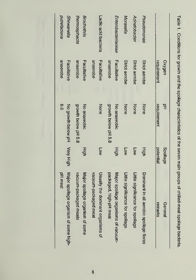
SPOILAGE DEVELOPMENT
The conditions required by the principal groups of spoilage bacteria for
growth on chilled meat are summarized in Table 1. The availability of oxygen
and the pH of the meat are the most important factors that determine which
types of bacteria will be represented in the final spoilage flora.
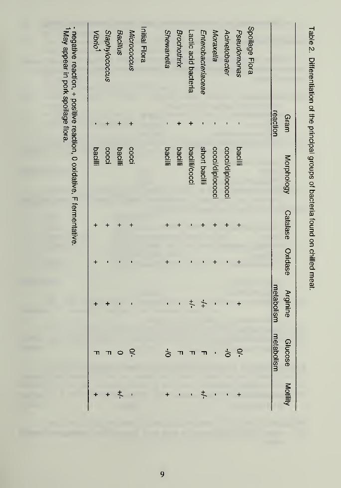
IDENTIFICATION OF SPOILAGE BACTERIA
Only five genera {Pseudomonas, Acinetobacter, Moraxella, Brochothrix
and Shewanella), one family {Enterobacteriaceae) and the lactic acid bacteria
are likely to be present in significant numbers in the flora developing on chilled
meat. Those groups, and others likely to be found in the initial flora, can be
differentiated using the properties of: Gram-reaction, morphology, presence of
catalase, presence of oxidase, presence of arginine dehydrolase/decarboxylase,
mode of glucose utilization, and motility (Table 2). That identification scheme is
derived from the general scheme proposed by Cowan and Steel (Cowan, 1974).
If bacteria other than those listed in Tables 2 are present in a flora, their
identification will require the use of that or other general identification schemes,
(Bergey, 1984; Blazevic and Ederes, 1975; McFaddin, 1980).

T3C
CD
CO
CO
CD
1CD
a?
CD
1OI"3-
o5J
D) Tl D)3 03 303 O 05CD C CD3 35 —
»
O Q) Oa- & crCD < CD
CD
03 r~ mO
1CD_
}* Tj
33-o
03Oo03
3CD
C35'
CD
3
CoCDC
*'
Oq"
a0)oCD3.03
03o903'
03CD
oT 03o
3osCO
ti 03 T| 0) Tl CO CO CO03 3 03 3 03o 0} O 03 o —
.
3. 3.c_ CD C_ CD c_ O o o5T o oT O oT 03 03 03
ct ^^ cr CD CD CD<'
CDCD <"
CDCD <
CDSCTCD
3CTCD
3CTCD
0> z CO Z Z CQ Z z Z zo o o o O o O o O o
cq 033
3CD % 03
33CD
3CD
3CD
o CT 03 CT 03
CT CD CT CD
ct CD"3
o CD sCTCD
O CTo"
O CTo'
O o o$ X ID Ol CJ1
I bo bo
< I I
-t. I
-r~ I
CD
<2cq'CT
Ocq'CT
o ocq'CT
n:cq'CT
oJ"
<03O
<03O
cCO 03
o J-9.
r; Oo33
CDCD
o' c o" c9L
7T o' CD CD-1
COT3
c3
l
•"1
COT3
c3
03CQCD
COoCO
cq'
CO
co'
3'
033o o o •6 5 Q. g.
3 3oT
03o oT
03o
CD
Q. CT oT o' o'5'
CO 7? CQ 7T cq"CT
i
CQ 03 0391CD 03 CD 03 O CD 3 3
o•1
CQCD
oT3
CQCD
33033
o1OCD
OCD 03
CD"1
cCO033
Q.
3CO033
Q.
3 3
CQ033
Oco'
3
COo3
CD03
CO
CO
3g^COo3
CD03.
Oa033to"
3CO
CD03.
co'
3CO
o<03o
COog.
oTCQCD
COog.oTCQCD
0"
COTJg.oTCQCD
CD CD o c -HCT ccq' 3 03CT 1
CO
CDLQ
—
^
CD
3CD3
CD
-
^
CD
3CD3
oxCQCD3
oI
re cog -ocd g.
03 CQ= CD
3 3i Ic^ S
03CTCD
oo3Q.
5'
3CO
CQ-^
o$31
033Q.
CTCD
COCTg.oTCQCD
oCT03™J03OCD
co'
o'CO
CTCD
COCD<CD33033*
CQ
SCoCO
g^o
CDQ.
3CD
03.
CO•og.
oTCQCD
CT03
g.CD303"

m CDHJ*<
CDCO-o <"O CDCD ^CO CD^ CO5" O"O oO 3a?r +COag.
-ooCO
03^^
CO <CD CD
i
.
-i
o CDCO
03 Oo"3
*•
OoXo!CO1-^<<D
-n_kCDa3CD3CO-<*
CD
CO 03CO
1
o'
3oo
g_
K-n
o
Co3-CD
1
CD
39-o
03Oo'
CO3C3-
5O
1
o3"CD
Oo
O COCD 3 Co
O Co" Coo c sr * Oooc:
CO CTCOo
CD
Co*
c5"
COCD
co'
oCDCoCD
3?CDC§-
3o§CO
COog,5TcqCD
oco
CJ o CT o CT CT CT CO o o CTCo o CO o CO CO Co
3- o o COo. o o. o o O o o o o oo o 3. o. o. —-• — •
o~ CT cl Q""~
*
coo •o' o'oo =j oo
oo
o oo oo o
ti o
OCDCOo ja
5- 33
oT3
ooCO
OCo
Co.
CoCOCD
oX
coCOCD
CD >5T <BCT =rg_ 3.
g' CD
CD OCoCTo
cooco
CO Q
coC[CD
CD
3D£.'
o'Do
CD
TJ
oo"CO.
co
scoCO
g^crcooCD
CO'
—%oca.
o
Og;
cd"
q.
CDco
TESTS USED FOR DIFFERENTIATION OF SPOILAGE BACTERIA
GRAM REACTION
Preston and Morrell's modification of Gram's method is recommended,because it gives reliable results while allowing some variation in the duration of
decolourization.
Solutions required:
Ammonium oxalate-crystal violet
Crystal violet 20 gMethanol 200 ml
Ammonium oxalate 800 ml
(1% w/v aqueous solution)
Lugol's iodine:
Iodine 10 gPotassium iodide 20 gDistilled water 1000 ml
Dissolve the potassium iodide in a small volume of water, add and
dissolve the iodine, then make up to 1000 ml.
lodine-Methanol:
Iodine 10gPotassium iodide egMethanol 90 ml
Distilled water 10ml
Iodine-acetone:
lodine-methanol 35 mAcetone 965 m
Ziehl-Neelsen's (strong) carbol fuchsin:
Basic fuchsin 10 gAbsolute ethanol 1 00 m
I
Phenol solution, 5% in water 1000 ml
Dissolve the dye in the alcohol and add to the phenol solution.
Dilute carbol fuchsin:
Ziehl-Neelsen's carbol fuchsin 50 ml
Distilled water 950 ml
Procedure:
(1) Place a light suspension of cells on a slide, air-dry and heat-fix over a
flame.
10
(2) Cover the slide with ammonium oxalate-crystal violet solution and leave
for 30 seconds.
(3) Pour off the solution and wash the slide freely with Lugol's iodine. Coverwith fresh Lugol's iodine solution and leave for 30 seconds.
(4) Pour off the Lugol's iodine solution and wash the slide freely with iodine-
acetone solution. Cover with fresh iodine-acetone solution and leave for
30 seconds.
(5) Wash the slide thoroughly with water.
(6) Cover the slide with dilute carbol fuchsin solution and leave for 30seconds.
(7) Wash the slide with water, blot off excess moisture and allow the slide to
dry before it is examined.
It is essential that the whole slide is flooded with each solution in turn, and
that the previous solution is thoroughly removed at each step.
MORPHOLOGY
The shape (morphology) of bacterial cells is determined by microscopic
examination of the Gram-stained preparations.
Term Description
Cocci Spherical or nearly spherical cells.
Coccobacillus Slightly elongate spherical cells.
Diplococci Pairs of cocci, with the cells slightly elongated along
the axis of the pair.
Bacilli Straight, or slightly curved, rod-shaped cells.
CATALASE
This test demonstrates the presence of catalase, an enzyme that
catalyses the release of oxygen from hydrogen peroxide.
Pour 1 ml of 3% (10 vol) hydrogen peroxide solution (H2 2 ) over a 24-hr
nutrient agar slope culture of the test organism, holding the tube in a slanting
position.
Alternatively, pick a small amount of the culture to be tested from a
nutrient agar slope or from a colony on a plate, using a clean glass rod. Asuitable rod can be formed by sealing the tip of a Pasteur pipette. Place the
sample on a clean microscope slide, they add a drop of 3% hydrogen peroxide
solution.
The immediate production of gas bubbles from the surface of the culture
material is a positive reaction.
11
Note. Do not pick material for this test with an inoculating loop, as platinum can
catalyse the release of oxygen from H2C>2.
OXIDASE
This test detects the presence of enzymes that catalyse the transport of
electrons between electron donors in the bacterial cell and a redox dye,
tetramethyl-p-phenylene-diamine. If an oxidase is present, the dye is reduced to
a deep purple colour.
Reagent: 1% (w/v) aqueous solution of tetramethyl-p-phenylene-diamine
dihydrochloride. This solution is unstable and must be stored in a refrigerator in
a glass-stoppered bottle, protected from light. N.B. The reagent should not beused if it has become deep blue . Autoxidation can be retarded by the addition
of ascorbic acid to a final concentration of 1% (w/v).
Soak a strip of filter paper with the reagent solution and immediately pick
a sample of culture with a platinum loop, then rub the inoculated loop on the
soaked paper. Development of an intense deep-purple colour within 60 seconds
is a positive reaction If the purple colour developes after 60 seconds, or fails to
develop, the reaction is negative. The test can also be carried out by adding the
reagent to colonies on a Petri plate.
Note: The growth must be transferred to the test paper with a clean instrument
made of platinum or glass, as traces or iron will catalyse the reaction to give a
false positive result. If the colonies of a particular type are small, several
colonies may have to be picked to obtain enough material to give a strong
reaction. A positive reaction is often discernible within 10 seconds, but delayed
reactions may require the full 60 seconds stipulated for the test.
ARGININE
This test detects the presence in bacteria of the arginine
dehydrolase/decarboxylase enzyme system, which decarboxylates the amino
acid arginine to give basic (alkaline) end products. Accumulation of those basic
products causes the pH of the test medium to rise.
Arginine dehydrolase/decarboxylase media:
Basal medium:
Glucose 0.5 gSodium chloride 10 gTrypticase (Tryptone) 10 gPhenol red 0.05 gDistilled water 1000 ml
The pH of the basal medium should be 7.2.
12
Test medium:The test medium is prepared by adding 10 g of L(+) arginine
monohydrochloride, or 20 g of LD (±) arginine monohydrochloride, to 1 litre of
the basal medium. After addition of arginine, the pH must be readjusted to 7.2.
Basal medium without the addition or arginine is used as a control.
Control and test media are dispensed into small screw-capped tubes
(usually 13 x 100-mm tubes) to a depth of 15 to 20 mm. The filled tubes are
sterilized at 121°C for 10 minutes. Tubes of both control and test media can be
stored for up to 6 weeks at 4°C.
The control and test media are stab inoculated with a straight wire, then
incubated at 25°C. The tubes are examined after 3, 5 and 7 days. At any of
those times, a changed colour of the test medium, from orange to red, is a
positive reaction.
GLUCOSE UTILIZATION
This test determines whether or not an organism utilizes glucose with the
production of acid and, if glucose is so utilized, whether it is utilized oxidatively
(O) or fermentatively (F). The O/F reaction of meat isolates is determined in
Hugh and Leifson's medium using the single-tube procedure.
Hugh and Leifson's semi-solid medium:
Trypticase (Tryptone) 10 g
Yeast extract 1 g
Glucose 10 g
Bromocresol purple 0.04 gAgar 3 g
Distilled water 1000 ml
Dissolve the ingredients in the distilled water and adjust the pH to 7.0.
The medium is dispensed, in 10-ml amounts, into screw-capped tubes (13
x 100-mm), then sterilized at 1 15°C for 20 minutes. Hugh and Leifson's mediumwill store for several weeks at 4°C. Before tubes of stored medium are used,
they should be steamed for 10 minutes, then cooled rapidly in a bath of iced
water. This procedure removes any oxygen that may have dissolved in the
medium during storage. Failure to remove dissolved oxygen could allow strictly
aerobic organisms to grow throughout the medium to produce the colour
changes typical of glucose fermentation.
The test medium is stab inoculated with a straight wire, then incubated at
25°C. Tubes are examined after 3, 5 and 7 days. At any of those times, a
change in the colour of the top third of the tube, from purple to yellow, is a
positive oxidative reaction. The same colour change progressing from the
botton of the tube is a positive fermentative reaction.
13
MOTILITY
The motility of bacteria can be determined from the O/F reaction tubes.
Motile organisms show spreading growth from the initial line of the stab
inoculation, whereas non-motile organisms are confined to the line of the stab.
Motility can also be observed microscopically by moist mount, or by using
commercially available motility test media.
SELECTIVE / DIFFERENTIAL MEDIA FOR SPOILAGE BACTERIA
INTRODUCTION
General purpose (enumeration) media such as Plate Count Agar
(Standard Methods Agar) are nutritionally complete media formulated for the
primary isolation and enumeration of bacteria from water and foods. As they are
not selective, they support the luxuriant growth of most aerobic and facultatively
anaerobic spoilage bacteria associated with meat.
The disadvantage of that type of medium is that is does not readily permit
the differentiation of bacterial types, except on the uncertain basis of colonial
morphology. Further biochemical characterization is necessary to properly
identify the various types of colony that can be distinguished. Also, there are
some organisms, such as lactic acid bacteria, which have complex nutritional
requirements and grow poorly on the general purpose media.
It is possible, through the use of selective and differential agents and/or
conditions, to suppress the growth of most groups of bacteria and so allow only
one or a few specific groups to form colonies on the plate. A disadvantage with
the use of selective cultural conditions is that often the growth of some strains of
the targeted group is inhibited, with consequent underestimation of the numbersof the targeted group. However, a selective medium can be used to advantage
when the objective is to detect the presence, or follow shifts in the numbers, of a
spoilage organism or group of particular concern.
A medium is usually rendered selective by the incorporation of
substances, such as antibiotics, that are inhibitory to many bacteria but to which
the targeted organisms are relatively resistant. Appropriate choices of carbon
and nitrogen sources, medium pH, and/or salt concentrations can also favour the
growth of the targeted group. Further, incubation conditions favourable to the
targeted group, but unfavourable to others, can be used.
It is usually not possible to formulate a medium that severely inhibits all
competitors with the targeted group of organisms. However, colonies of the
targeted group can be distinguished from others if a differentiating system is
incorporated in the medium. A differentiating system may involve a pH and/or
14
specific chemical indicator and/or a substrate characteristically utilized by the
targeted group. Then, a distinctive change in the appearance of colonies and/or
the surrounding medium, because of the reaction with the differentiating system,
will allow colonies of the targeted group to be discriminated.
In contrast to the use of selective/differential media, the cultivation of
some spoilage bacteria requires the use of complex culture media which have
been formulated to support the growth of specific organisms that are fastidious in
their nutritional requirements. Such media have been termed elective.
A number of the media described herein are commercially available,
making preparation under industrial conditions relatively simple. The recipes
and description have been modified from "Pharmacopoeia of Culture Media for
Food Microbiology" (Baird et al., 1987; 1989) which is an excellent source of
media formulations that are appropriate to meat microbiology.
TOTAL COUNTS: PLATE COUNT AGAR (PCA)
Description
A nutritionally rich culture medium initially developed for enumerating
bacteria from dairy products (Marth, 1978). It is now accepted as a standard
medium for enumerating total bacteria from food in general. The medium is
commercially available, being designated by some manufacturers as Standard
Methods Agar.
Composition
(9/L)
Tryptone
Yeast Extract
Glucose
Agar
5
2.5
1
15
Preparation
Suspend all the ingredients in 1 L of water. Boil the suspension to
dissolve the ingredients, then autoclave for 15 min at 121 °C. Cool in a water
bath to 47°C. Dispense 15 ml portions into Petri dishes.
Properties
The medium has a pH of 7.0 and is light amber in colour,
can be stored for 2 weeks at 4°C.
Prepared plates
15
Plate inoculation and colonial appearance
Spread a 0.1 ml portion of each suitable sample over the entire plate.
Incubate the plates in air; at 35°C for 48 h, for total mesophiles; or 5°C for 10 d,
for total psychrotrophs.
The pour plate method can also be used, with the incorporation of 1 ml
sample portions into 15 ml of tempered agar.
As this medium is not selective, the sizes, morphologies and colours of
the recovered colonies will vary.
Plates having 30-300 bacterial colonies are counted, and the Standard
Plate Count (SPC) or Total Viable Count (TVC) is reported.
Total anaerobic counts
To estimate the total anaerobic population, plates are inoculated as for
the aerobic count but are then incubated in an anaerobic jar or a disposable
anaerobic system.
Pseudomonas: CEPHALORIDINE FUCIDIN CETRIMIDE AGAR (CFC)
Description
This medium was initially developed for the selective enumeration of
pseudomonads from poultry (Mead and Adams, 1977). It has since been used
extensively in the isolation and enumeration of pseudomonads from red meats
and other foods. The antimicrobial agents, cephaloridine, fucidin and cetrimide
suppress the growth of most meatborne organisms other than Pseudomonasspecies.
Composition
(g/L)
Heart Infusion Agar 40
Cephaloridine 0.05
Sodium fusidate 0.01
Cetyltrimethyl ammonium bromide 0.01
Preparation
Suspend the heart infusion agar in 1 L of distilled water, then boil to
dissolve the ingredients. Sterilize the basal medium by autoclaving for 15 min a
121°C. Cool the sterilized medium to 47°C in a water bath. Prepare the
selective agents as 1% solutions in distilled water and sterilize each by
16
membrane filtration. Aseptically, add 5 ml of the cephaloridine, 1 ml of the
fucidin and 1 ml of the cetrimide solution to the cooled basal medium. Mix the
medium by swirling before pouring plates.
Properties
The medium has a pH of 7.4 and is straw coloured. Prepared plates can
be stored for 1 month at 4°C.
Plate inoculation and colonial appearance
Incubate spread plates, at 25°C for 48 h. Pseudomonads appear as
round, cream-coloured colonies. The identity of colonies can be confirmed by
flooding the plates with oxidase reagent. The Pseudomonas colonies then
develop a deep purple colour.
Enterobacteriaceae: VIOLET RED BILE GLUCOSE AGAR (VRBG)
Description
The medium is useful for performing total counts of Enterobacteriaceae
(Mosselefa/. 1978).
Composition
(g/L)
Peptone 7.0
Yeast extract 3.0
Sodium chloride 5.0
Glucose 10.0
Bile salts 1.5
Crystal violet 0.002
Neutral red 0.03
Agar 15gs medium is commercially available.
Preparation
Suspend the ingredients in distilled water. Boil the suspension to dissolve
the ingredients. Do not autoclave the medium. Cool to 47°C in a water bath.
Properties
This medium has a final pH of 7.4. It is a purple-violet colour. It must beprepared immediately before it is used.
17
Plate inoculation and colonial appearance
Add 10 ml of molten agar to 1.0 ml of the inoculum in a Petri dish. Swirl
the dish to disperse the inoculum throughout the agar. Allow the basal layer to
hardened, then overlay it with 5 ml of the same medium. Incubate in air; at 35°Cfor 18-24 h, for total enterics; or at 5°C for 10 d, for psych rotrophic
enterobacteria. Enterobacteria appear as red-purple colonies that are
surrounded by a red-purple halo.
LACTIC ACID BACTERIA: deMAN, ROGOSA AND SHARPE AGAR (MRS)
Description
MRS agar was developed for the cultivation of lactobacilli (de Man,Rogosa and Sharpe, 1960). The medium is not selective, but some selectivity
can be gained by adjusting the pH to 5.7, by addition of 1 N HCI, and/or using
anaerobic incubation conditions. Unfortunately, reducing the pH of the mediummay restrict the growth of some meatborne lactics, such as Carnobacterium.
Composition
(g/L)
Peptone 10
Beef extract 8.0
Yeast extract 4.0
Glucose 20.0
Sorbitan monoleate 1.0
Dipotassium phosphate 2.0
Magnesium sulphate.7H2 0.2
Manganese sulphate.7H2 0.05
Ammonium citrate 2.0
Sodium acetate 5.0
Agar 15.0
This medium is commercially available.
Preparation
Suspend the ingredients in 1 L of distilled water. Boil, to dissolve the
ingredients, then autoclave the solution for 15 min at 121°C. Cool the sterilized
solution to 47°C in a water bath, then dispense 15 ml portions into Petri dishes.
Properties
The medium has a final pH of 6.2, and is amber in colour. Prepared
plates can be stored for 2 weeks at 4°C.
18
Plate Inoculation and Colonial Appearance
Incubate spread plates in an anaerobic jar, for 72 h at 25°C. Lactic acid
bacteria appear as small, white or grey colonies. Colony diameters range from
0.5 to 2.0 mm. Colonies of typical lactic morphology can be confirmed by the
catalase test, as lactics are catalase negative, while most likely contaminants are
catalase positive.
Carnobacterium: CRESOL RED THALLIUM ACETATE SUCROSE AGAR(CTAS)
Description
MRS medium, particularly the acidified version, does not favour the
growth of carnobacteria (Lactobacillus divergens), an important group of the
meatbome lactic acid bacteria. The medium described is selective for
Carnobacterium
Composition
Peptone
Yeast extract
(g/L)
10
10
Sucrose 20Tween 80 1
Sodium nitrate 15
Manganese sulphate. 7H2Dipotassium hydrogen sulphate
Thallium acetate
4
2
1
Nalidixic acid 0.04
Cresol red 0.004
Triphenyltetrazolium chloride
Agar0.01
15
Preparation
Suspend all the ingredients, except the triphenyltetrazolium chloride in 1 L
of distilled water. Boil the suspension to dissolve the ingredients. Cool the
solution in a water bath to 55°C, then adjust the pH to 9.1 using I N NaOH.Sterilize the solution by autoclaving, for 10 min. at 121°C, then again cool the
solution to 55°C. Add triphenyltetrazolium chloride, as 10 ml of a 10%, filter-
sterilized solution. Mix the completed medium by swirling before dispensing it
into Petri dishes.
19
Properties
The prepared medium appears purple-red and contains precipitated
material. The pH is 9.0. It can be stored for 1 week at 4°C.
Inoculation and colonial appearance
Spread plates are incubated in air, for 4 d at 25°C. C. piscicola producebronze-metallic, yellow to pink colonies that are surrounded by yellow mediumthat is cleared of the precipitate. C. divergens produces pin-point colonies with a
bronze-metallic shine. It may or may not produce a colour change in the
medium.
Brochothrix thermosphacta: STREPTOMYCIN THALLOUS ACETATEACITIDIONE AGAR (STAA)
Description
This medium was originally formulated for the selective isolation andenumeration of Brochothrix thermosphacta from meat (Gardner 1966). Thecombination of antimicrobial agents excludes most other meatbome bacteria.
Composition
Peptone
Yeast extract
9/L
202
Glycerol
Dipotassium hydrogen phosphate
Magnesium sulphate.7 H2Agar
15
1
1
13
Selective AgentsStreptomycin sulphate 0.5
Actidione (Cycloheximide) 0.05
Thallous acetate 0.05
Preparation
Prepare the basal medium by dispersing the non-selective ingredients in
water. Boil the suspensions, to dissolve the ingredients, then autoclave the
solution for 15 min at 121°C. Cool the sterile solution to 47°C in a water bath.
Dissolve the selective agents in distilled water to give stock solutions, that each
contain 50 mg/ml of the agent. Sterilize each stock solution by membranefiltration. Add 10 ml of the streptomycin sulphate solution and 1 ml each of the
actidione (cycloheximide) and thallous acetate solutions to the cooled basal
20
medium. Mix the complete medium by swirling before dispensing it into Petri
dishes.
Properties
The medium has a final pH of 7.0 and is a pale straw colour. Prepared
plates can be stored for 1 week at 4°C and the filter-sterilized selective agents
for 1 month at 4°C.
Plate inoculation and colonial appearance
Spread plates are incubated in air, at 25°C for 48 h. B. thermosphacta
appear as white colonies 0.5 to 1 mm in diameter.
Brochothrix thermosphacta: STREPTOMYCIN SULPHATE INOSITOLNEUTRAL RED AGAR (SIN)
Description
This is a medium selective for Brochothrix thermosphacta fSchillinger and
Lucke 1987). It allows the recovery and differentiation of B. thermosphacta in
the presence of lactics, pseudomonads and enterics.
This medium is not widely used but it has the advantages over STAA of
improving the recovery of B. thermosphacta and enabling detection andenumeration using Hydrophobic Grid Membrane Filtration.
Composition
(9/L)
Blood agar base 40Yeast extract 2
Dipotassium hydrogen phosphate 1
Magnesium sulphate.7H2 0.8
Sodium carbonate 0.35
Myo-lnositol 10
Neutral red (0.3%) 10 ml
Preparation
Suspend all the ingredients in 1 L distilled water. Boil the suspension to
dissolve the ingredients, then autoclave the medium for 15 min at 121°C. Cool
the sterilized medium to 47°C in a water bath. Prepare a solution containing 50mg/ml of streptomycin sulphate. Sterilize the solution by membrane filtration,
and add 10 ml of the sterile solution to the cooled medium. Mix the completed
medium by swirling before dispensing it into Petri dishes.
21
Properties
The medium has a pH of 7.0 and is pale red in colour. Prepared plates
can be stored for 1 week at 4°C.
Plate inoculation and colonial appearance
Incubate spread plates in air, at 25°C for 48 h. B. thermosphacta appear
as pink colonies, 0.5 to 1 mm in diameter.
Shewanella putrefaciens: PEPTONE IRON AGAR
Description
This differential medium was designed to detect hydrogen sulphide
production by bacteria (Levin, 1968). The medium is useful for the isolation of
Shewanella putrefaciens, when the condition of the meat suggests that the
organism may be present as a substantial fraction of the spoilage flora.
Peptone
Proteose peptone
Ferric ammonium citrate
Compositiong/L
15
5
0.5
Sodium glycerophosphate
Sodium thiosulphate
1
0.08
Agar 15
The medium is commercially available.
Preparation
Dissolve the ingredients in 1 L of water, then autoclave for 15 min at
121 °C. Cool the solution to 47°C in a water bath.
Properties
This medium has a final pH of 6.7 and is light amber in colour.
Plate inoculation and colonial appearance
Add 10 ml of molten agar to 1.0 ml of the inoculum in a Petri dish. Swirl
the dish to dispense the inoculum throughout the agar. Allow the basal layer to
harden then overlay it with 5 ml of the same medium. Incubate in air at 25°C for
22
72 h. Colonies producing hydrogen sulphide will be grey-black and 1-2 mm in
diameter. In some instances, blackening of the entire plate will occur.
REFERENCES
Baird, R.M., Corry, F.E.L, and Curtis, G.D.W. (Eds). 1987. Pharmacopeia of
culture media for food microbiology. Int. J. Food Microbiol 5: 187-299.
Baird, R.M., Cary, J.E.L, Curtis, G.D.W., Mossel, D.A.A. and Skovgaard, N.
(Eds). 1989. Pharmacopeia of culture media for food microbiology -
additional monographs. Int. J. Food Microbiol 9: 85-144.
Bergey, D.F.M. 1984. Bergey's Manual of Determinative Bacteriology. TheWilliams and Wilkins Co., Baltimore, MD, U.S.A.
Blazevic, D., and Ederer, G. 1975. Principles of Biochemical Tests in Diagnostic
Microbiology. John Wiley and Sons, Toronto, Canada.
Cowan, ST. 1974. Cowan and Steel's Manual for the Identification of Medical
Bacteria, 2nd Edition. Cambridge University Press, Cambridge, UK.
Dainty, R.H. and Mackey, B.M. 1992. The relationship between the phenotypic
properties of bacteria from chill-stored meat and spoilage processes. J.
App. Bacteriol. 73: 103s-114s.
Dainty, R.H., Shaw, B.G. and Roberts, T.A. 1983. Microbial and chemical
changes in chill-stored red meats. Pages 151-178 in Food Microbiology:
Advances and Prospects, ed. Roberts, T.A. Academic Press, London,
UK.
de Man, J.D., Rogosa, M., and Sharpe, M.E. 1960. A medium for the cultivation
of lactobacilli. J. Appl. Bacteriol. 23: 130-135.
Egan, A.F. and Roberts, T.A. 1987. Microbiology of meat and meat products.
Pages 167-197 in Essays in Agricultural and Food Microbiology, eds.
Norris, J.R. and Pettipher, G.L John Wiley and Sons Ltd., New York,
U.S.A.
Gardner, G.A. 1966. A selective medium for the enumeration of Microbacterium
thermosphactum in meat and meat products. J. Appl. Bacteriol. 29: 455-
460.
Gill, CO. 1986. The control of microbial spoilage in fresh meats. Pages 49-88
in Advances in Meat Research, Vol. 2, eds. Pearson A.M. and Dutson
T.R. AVI Publishing, Westport, CT, U.S.A.
23
Gill, CO. and Molin, G. 1991. Modified atmospheres and vacuum packaging
Pages. 172-199 in Food Preservatives, eds. Russel, N.J. and Gould,
G.W. Blackie and Son Ltd., Glasgow, U.K.
Holzapfel, W.H., and Gerber, E.S. 1983. Lactobacillus divergens sp. nov., a
new heterofermentative Lactobacillus species producing L(+) lactate.
System. Appl. Microbiol. 4: 522-534.
Ingram, M. and Simonsen, B. 1980. Meats and meat products. Pages 333-409
in Microbiol Ecology of Foods, Vol. 2., International Commission onMicrobiological Specifications for Foods (ICMSF). Academic Press, NewYork, U.S.A.
McFaddin, J.F. 1980. Biochemical Tests for Identification of Medical Bacteria,
2nd edn. The William and Wilkins Co., Baltimore, NY, U.S.A.
McMeekin, T.A. 1981. Microbial spoilage of meats. Pages 1-40 in
Developments in Food Microbiology, Vol. 1, ed. Davies R. Applied
Science Publishers, London, U.K.
Marth, E.H., 1978. Standard Methods for the Examination of Dairy Products.
14th edn. American Public Health Association.
Mead, G.C., and Adams, B.W. 1977. A selective medium for the rapid isolation
of pseudomonads associated with poultry meat spoilage. Br. Poult. Sci.
18: 661-670.
Mossel, D.A.A., Elderink, I., Koopmans, M., and van Rossem, F. 1978.
Optimalisation of a MacConkey-type medium for the enumeration of
Enteivbacteriaceae. Lab Pract. 27: 1049-1050.
Schillinger, U. and Lucke, F.-K. 1987. Lactic acid bacteria on vacuum-packaged
meat and their influence on shelf life. Fleishwirtsch. 67: 1244-1248.
24

CANADIAN AGRI
lillillBIBLIOTHEQUE CANAOIENNE OE I'AGRICULTURE
3 T073 00101bT3 2