Embed Size (px)
Citation preview
Cr
CSa
b
a
ARRAA
KBEiqMA
1
cubt(
ottaabf
c
1d
Environmental Toxicology and Pharmacology 30 (2010) 5–10
Contents lists available at ScienceDirect
Environmental Toxicology and Pharmacology
journa l homepage: www.e lsev ier .com/ locate /e tap
haracteristics of Escherichia coli biofilm production, genetic typing, drugesistance pattern and gene expression under aminoglycoside pressures
haoxi Chena,b,1, Xiaoping Liaoa,1, Hongxia Jianga, Hengqian Zhua, Lei Yuea,hujuan Lia, Binghu Fanga, Yahong Liua,∗
Guangdong Provincial Key Laboratory of Veterinary Pharmaceutics Development and Safety Evaluation, Guangzhou, ChinaCollege of Life Science and Technology, Southwest University for Nationalities, Chengdu, China
r t i c l e i n f o
rticle history:eceived 8 September 2009eceived in revised form 28 February 2010ccepted 2 March 2010vailable online 9 March 2010
eywords:iofilm-forming ability
a b s t r a c t
In our studies, qualitative (scanning electron microscope) and semi-quantitative (modified crystal vio-let staining method) methods had been used to evaluate Escherichia coli biofilm-forming ability. Brothmicrodilution method and enterobacterial repetitive intergenic consensus-based PCR (ERIC-PCR) wereperformed to study E. coli drug resistance pattern and genetic typing. Based on the results above, westudied the correlation between biofilm-forming ability phenotype, drug resistance pattern and genetictyping in E. coli. Real-time qPCR (qRT-PCR) was used to reveal mRNA expression level of E. coli biofilmrelated multiple antibiotics resistance genes (acrA, agn43, csgA, csgD, ompF and pgaA) under differentconcentrations of four aminoglycoside pressures. Our results showed that: (i) forty-nine out of 64 strains
RIC-PCR (enterobacterial repetitiventergenic consensus-based PCR)RT-PCRultiple antibiotics resistance
minoglycosides
of E. coli (76.56%) showed significant production of biofilm and most of them performed weak biofilm-forming ability; (ii) ERIC-PCR showed that there was significant correlation between biofilm-formingability and genotype; while there was weak correlation between biofilm-forming ability and drug resis-tance patterns based upon the results of semi-quantitative method and antibiotics susceptibility test; (iii)qRT-PCR revealed mRNA expression of acrA, agn43, csgA, csgD, ompF and pgaA genes changed accordingly
t con
by stimulation of differen. Introduction
Several types of infection caused by Escherichia coli (e.g., urethralatheter, biliary tract prosthesis, biliary tract infection, lithangiuria,rinary tract infection and many other diseases) are associated withiofilm formation, which leads to an inability to eradicate the infec-ion due to its intrinsic nature to resist high levels of antibioticsFontaine and Smith, 2006; Melchior et al., 2006).
In addition, under extensive and persistent pressure of antibi-tics, in order to survive, the E. coli forms biofilm to evadehe immune clearance and results in multiple antibiotics resis-ance. Hoffman et al. (2005) found that subinhibitory levels of theminoglycoside antibiotics could induce E. coli and Pseudomonaseruginosa biofilm formation. Some other studies also revealed that
iofilm formation by E. coli in vitro correlated with the virulenceactors (Naves et al., 2008; Rijavec et al., 2008).The primary aims of our study were to investigate: (i) The E.oli biofilm-forming ability based on the qualitative (scanning elec-
∗ Corresponding author. Tel.: +86 20 85287189; fax: +86 20 85284896.E-mail address: [email protected] (Y. Liu).
1 These authors contributed equally to this work.
382-6689/$ – see front matter © 2010 Elsevier B.V. All rights reserved.oi:10.1016/j.etap.2010.03.004
centrations of four aminoglycosides.© 2010 Elsevier B.V. All rights reserved.
tron microscope) and quantitative (modified crystal violet stainingmethod) methods. (ii) The correlations between E. coli genotype,drug resistance pattern and biofilm-forming ability phenotype. (iii)Changes of mRNA level of acrA, agn43, csgA, csgD, ompF and pgaAunder persistent and extensive antibiotic pressures using qRT-PCRmethod.
2. Materials and methods
2.1. Bacterial strains
A total of 64 E. coli strains were chosen due to their varying sources, isolatedfrom poultry farms and swine farms in Guangdong Province and pet hospital ofSouth China Agricultural University (SCAU), and all of the isolates were collectedfrom sick animals between 2003 and 2007.
All cultures were stored at −80 ◦C in Luria–Bertani broth with 30% glycerol andwere freshly streaked on MacConkey agar before each assay. For biofilm formationassays, single colonies were grown in tryptic soy broth statically and all experimentswere performed at 37 ◦C.
2.2. Antimicrobial agents and molecular biology reagents
All the 10 antimicrobial agents (enrofloxacin, amikacin, gentamycin, doxycy-cline, florfenicol, apramycin, amoxicillin, streptomycin, rifampin and ceftiofur) werekindly provided by veterinary medicine plant of SCAU, and were prepared for sus-ceptibility assays as described in the Clinical and Laboratory Standards Institute(CLSI) susceptibility testing guidelines.
6 C. Chen et al. / Environmental Toxicology and Pharmacology 30 (2010) 5–10
Table 1Primers for acrA, agn43, csgA, csgD, ompF and pgaA amplification.
Gene name Sequence Product length (bp) Entry no. or reference
acrA-F 5′-AAGATAGCGCGTAGGGTGATAGAC-3′ 143 ThisstudyacrA-R 5′-GGCACGCTGAAACAAGAGAAC-3′
agn43-F 5′-TTCCGGGAAGACGGTGAA-3′ 144 AF233271agn43-R 5′-TTCTGGGTGAGTGTGGTGTTG-3′
csgA-F 5′-TGGCAGGTGTTGTTCCTCAGT-3′ 146 L04979csgA-R 5′-GTCAGAGTTACGGGCATCAGTTT-3′
csgD-F 5′-CCGCTTGTGTCCGGTTTT-3′ 97 ThisstudycsgD-R 5′-GAGATCGCTCGTTCGTTGTTC-3′
ompF-F 5′-TAAAGGGGAAACTCAAATCAA-3′ 240 M74489ompF-R 5′-CTGTATGCAGTATCACCACCA-3′
pgaA-F 5′-AGGGACTGCGCATTGATTAC-3′ 164 This
of
2
2
pB3rfwaw1sTs
2
G1bicm
2
iwa
instruction. qRT-PCR was performed using SYBR Prime Script RT-PCR Kit followingthe instruction described by the manufacturer. The amplification courses of all thesix genes included three steps as follows: (1) 1 cycle of an initial denaturation for
◦ ◦
pgaA-R 5′-GTTCAGGTTCGACAACATCG-3′
16sRNA-F 5′-AAGTTAATACCTTTGCTCATTGAC-3′
16sRNA-R 5′-GCTTTACGCCCAGTAATTCC-3′
TRIzol reagent Kit, SYBR Prime Script RT-PCR Kit, Prime Script RT reagent Kit andther molecular biology reagents for qRT-PCR and universal PCR were purchasedrom Takara (Japan).
.3. Study design and experimental approach
.3.1. Semi-quantitative biofilm formation experimentSemi-quantitative biofilm formation experiment was performed as described
reviously (Stepanovic et al., 2000; Cerca et al., 2007) with some modifications.riefly, all E. coli isolates were grown in 96-well polystyrene plates for 24 h at7 ◦C and were washed twice by submerging the plates in deionized water. Theemaining attached bacteria were fixed with 0.2 mL of absolute methanol per wellor 20 min before plates were emptied and left to dry. The biofilm were stainedith 0.2 mL of 2.5% Crystal Violet (CV) for 10 min. Excess stain was rinsed off
nd the plates were washed with distilled water for three times. After the platesere air-dried overnight, the dye bound to the adherent cells was dissolved with
60 �L of 33% (v/v) glacial acetic acid per well. The OD570 of each well was mea-ured at 570 nm using an automated ELX800 Universal Spectrophotometer Reader.hree separate experiments with duplicates were performed for each conditiontudied.
.3.2. Scanning electron microscope (SEM)Sample preparation for scanning electron microscope was described by
anderton et al. (1992). Briefly, medical silica gel sheets were sectioned (1 cm bycm) and fixed in 4% buffered glutaraldehyde and rinsed three times with phosphateuffered saline (PBS, pH 7.4). Critical drying was performed with 50–100% ethanol
n double distilled water and the samples were dried in a critical point dryer andoated with a gold layer. Three different points were examined by SEM at properagnification.
.3.3. Correlation between drug resistance pattern and biofilm-forming abilityBroth microdilution method was used for antimicrobial drug susceptibility test-
ng of 10 antimicrobial agents to 64 clinical isolates and the experimental dataas statistically analyzed by WHONET5.3 software. E. coli ATCC25922 was used
s reference strain for quality controls.
Fig. 1. Biofilm-forming ability of 64 E. coli.
study
117 Thisstudy
Based on the results of semi-quantitative biofilm formation experiment in Sec-tion 2.3.1, we focused on whether biofilm formation was related to drug resistancepatterns (Moskowitz et al., 2004; Frank et al., 2007; Suman et al., 2007).
2.3.4. Correlation between genotype and biofilm-forming abilityTo analyze the correlation between the biofilm-forming ability and genetic
typing of clinical isolates, ERIC-PCR was used according to related references(Coquet et al., 2002; Yang et al., 2005; Wilson and Sharp, 2006; Silva etal., 2009). The ERIC region was amplified with the ERIC primers ERIC-F (5′-ATGTAAGCTCCTGGGGATTCAC-3′) and ERIC-R (5′-AAGTAAGTGACTGGGGTGAGCG-3′) in 25 �L standard reaction system consisting of 2 pmol of ERIC primers, 10 nmolof each dNTP, 50 ng of DNA template and 1 U rTaq DNA polymerase. PCR was done ina DNA thermal cycler with the following settings: an initial denaturation for 5 minat 94 ◦C, followed by 35 cycles of denaturation for 1 min at 94 ◦C, annealing for 1 minat 53 ◦C and extension for 4 min at 72 ◦C and a final extension for 10 min at 72 ◦C.Electrophoresis was performed in 1.5% agarose gel, the ERIC-PCR fingerprint wasconverted to a binary matrix (1 = presence; 0 = absence of amplicons) and was ana-lyzed with NTSYSpc2.1 software. The unweighed pair-group method with averagelinkage was used for the cluster analysis.
2.3.5. RNA extraction and real-time qRT-PCRIn Section 2.3.3, we selected strains E53 which was sensitive to all the 10 antimi-
crobial agents and was positive for genes acrA, agn43, csgA, csgD, ompF and pgaA usinguniversal PCR method. And E53 was induced with increase of gradient concentrationof four aminoglycosides until it was resistant to the aminoglycoside drugs.
Bacterial RNA was extracted using the TRIzol reagent and cDNA was synthe-sized from RNA by using Prime Script RT reagent Kit according to the manufacturer’s
10 min at 95 C, (2) 40 cycles of an initial denaturation for 15 s at 95 C, annealing for15 s at 58 ◦C and extension for 40 s at 72 ◦C and (3) 1 cycle of an initial denaturationfor 15 s at 95 ◦C, annealing for 1 min at 60 ◦C a final extension for 15 s at 95 ◦C. The
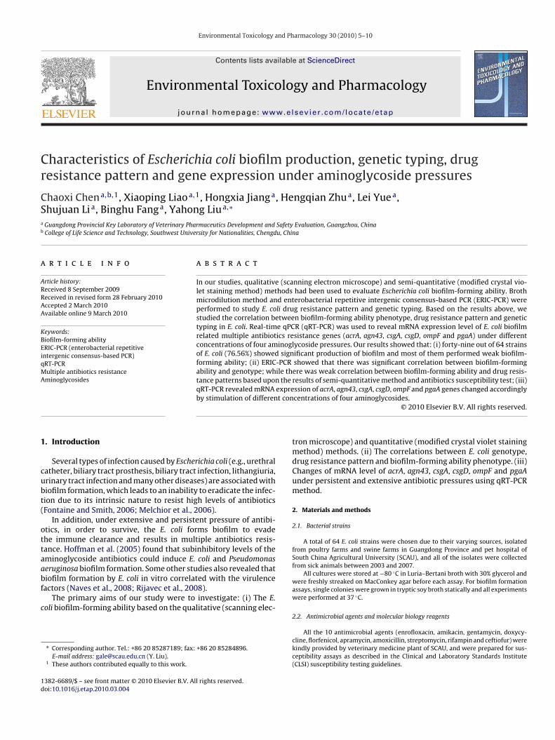
Fig. 2. SEM photograph of strain E53 (6400× magnification).
C. Chen et al. / Environmental Toxicology and Pharmacology 30 (2010) 5–10 7
yping cluster analysis of 64 E. coli.
pita
3
3
tb(nb
3
b
Table 2Mean (%) of strains with different biofilm-forming ability.
Biofilm-forming ability Mean (%) of strains resistant
Strong 3.7 (61.67%)
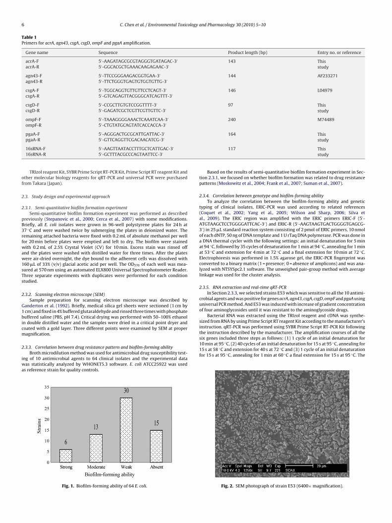
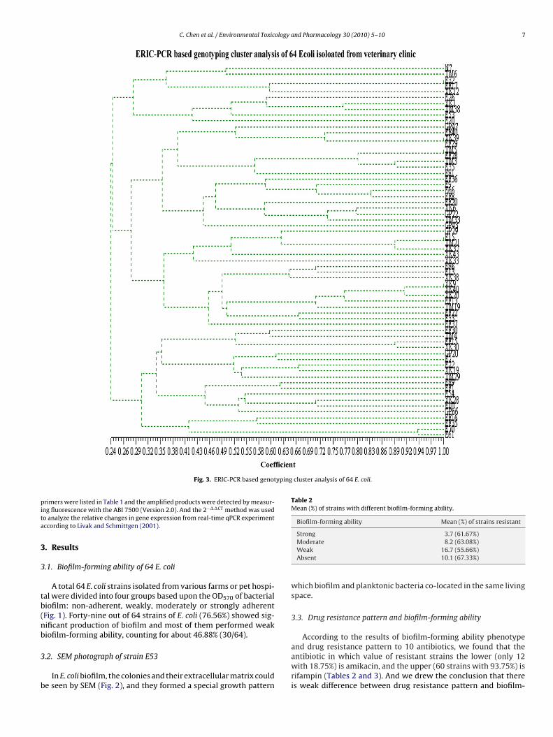
Fig. 3. ERIC-PCR based genot
rimers were listed in Table 1 and the amplified products were detected by measur-ng fluorescence with the ABI 7500 (Version 2.0). And the 2−��CT method was usedo analyze the relative changes in gene expression from real-time qPCR experimentccording to Livak and Schmittgen (2001).
. Results
.1. Biofilm-forming ability of 64 E. coli
A total 64 E. coli strains isolated from various farms or pet hospi-al were divided into four groups based upon the OD570 of bacterialiofilm: non-adherent, weakly, moderately or strongly adherentFig. 1). Forty-nine out of 64 strains of E. coli (76.56%) showed sig-ificant production of biofilm and most of them performed weakiofilm-forming ability, counting for about 46.88% (30/64).
.2. SEM photograph of strain E53
In E. coli biofilm, the colonies and their extracellular matrix coulde seen by SEM (Fig. 2), and they formed a special growth pattern
Moderate 8.2 (63.08%)Weak 16.7 (55.66%)Absent 10.1 (67.33%)
which biofilm and planktonic bacteria co-located in the same livingspace.
3.3. Drug resistance pattern and biofilm-forming ability
According to the results of biofilm-forming ability phenotype
and drug resistance pattern to 10 antibiotics, we found that theantibiotic in which value of resistant strains the lower (only 12with 18.75%) is amikacin, and the upper (60 strains with 93.75%) isrifampin (Tables 2 and 3). And we drew the conclusion that thereis weak difference between drug resistance pattern and biofilm-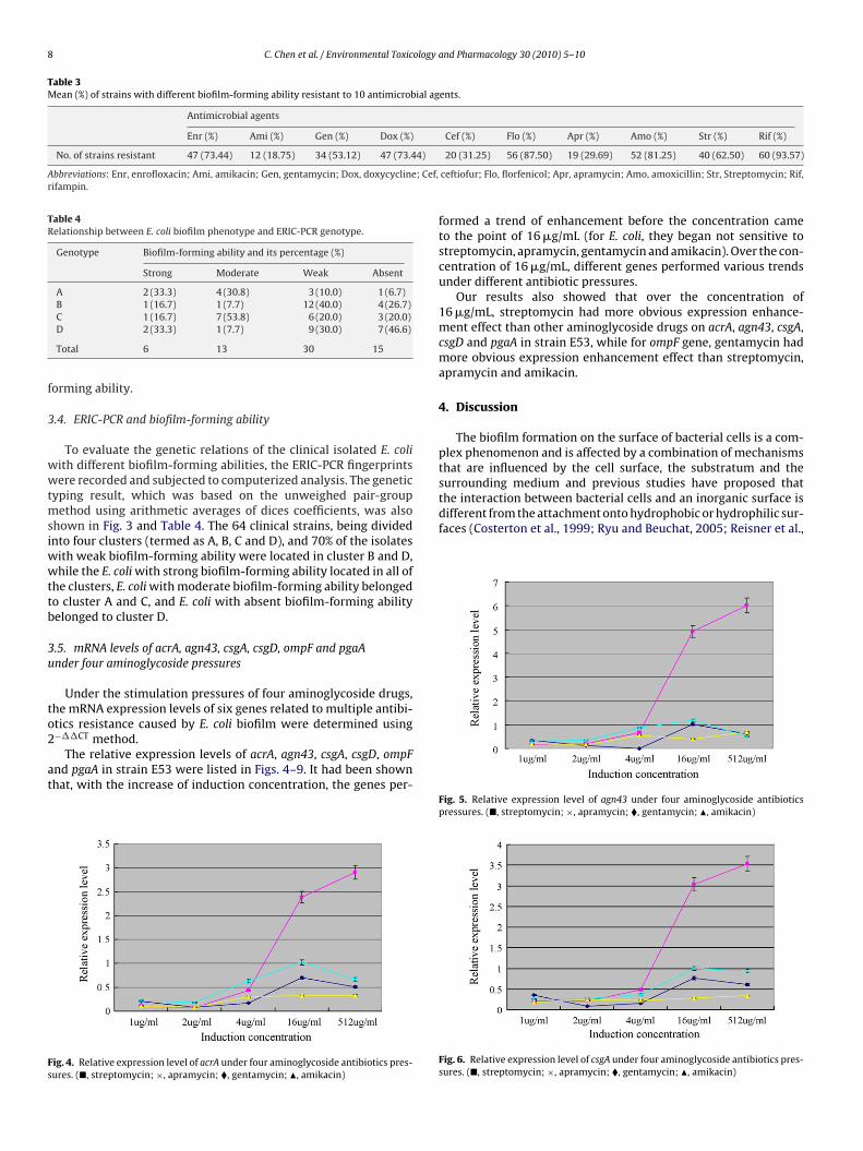
8 C. Chen et al. / Environmental Toxicology and Pharmacology 30 (2010) 5–10
Table 3Mean (%) of strains with different biofilm-forming ability resistant to 10 antimicrobial agents.
Antimicrobial agents
Enr (%) Ami (%) Gen (%) Dox (%) Cef (%) Flo (%) Apr (%) Amo (%) Str (%) Rif (%)
No. of strains resistant 47 (73.44) 12 (18.75) 34 (53.12) 47 (73.44)
Abbreviations: Enr, enrofloxacin; Ami, amikacin; Gen, gentamycin; Dox, doxycycline; Cef,rifampin.
Table 4Relationship between E. coli biofilm phenotype and ERIC-PCR genotype.
Genotype Biofilm-forming ability and its percentage (%)
Strong Moderate Weak Absent
A 2 (33.3) 4 (30.8) 3 (10.0) 1 (6.7)B 1 (16.7) 1 (7.7) 12 (40.0) 4 (26.7)
f
3
wwtmsiwwttb
3u
to2
at
Fs
surrounding medium and previous studies have proposed thatthe interaction between bacterial cells and an inorganic surface isdifferent from the attachment onto hydrophobic or hydrophilic sur-faces (Costerton et al., 1999; Ryu and Beuchat, 2005; Reisner et al.,
C 1 (16.7) 7 (53.8) 6 (20.0) 3 (20.0)D 2 (33.3) 1 (7.7) 9 (30.0) 7 (46.6)
Total 6 13 30 15
orming ability.
.4. ERIC-PCR and biofilm-forming ability
To evaluate the genetic relations of the clinical isolated E. coliith different biofilm-forming abilities, the ERIC-PCR fingerprintsere recorded and subjected to computerized analysis. The genetic
yping result, which was based on the unweighed pair-groupethod using arithmetic averages of dices coefficients, was also
hown in Fig. 3 and Table 4. The 64 clinical strains, being dividednto four clusters (termed as A, B, C and D), and 70% of the isolates
ith weak biofilm-forming ability were located in cluster B and D,hile the E. coli with strong biofilm-forming ability located in all of
he clusters, E. coli with moderate biofilm-forming ability belongedo cluster A and C, and E. coli with absent biofilm-forming abilityelonged to cluster D.
.5. mRNA levels of acrA, agn43, csgA, csgD, ompF and pgaAnder four aminoglycoside pressures
Under the stimulation pressures of four aminoglycoside drugs,he mRNA expression levels of six genes related to multiple antibi-
tics resistance caused by E. coli biofilm were determined using−��CT method.The relative expression levels of acrA, agn43, csgA, csgD, ompFnd pgaA in strain E53 were listed in Figs. 4–9. It had been shownhat, with the increase of induction concentration, the genes per-
ig. 4. Relative expression level of acrA under four aminoglycoside antibiotics pres-ures. (�, streptomycin; ×, apramycin; �, gentamycin; �, amikacin)
20 (31.25) 56 (87.50) 19 (29.69) 52 (81.25) 40 (62.50) 60 (93.57)
ceftiofur; Flo, florfenicol; Apr, apramycin; Amo, amoxicillin; Str, Streptomycin; Rif,
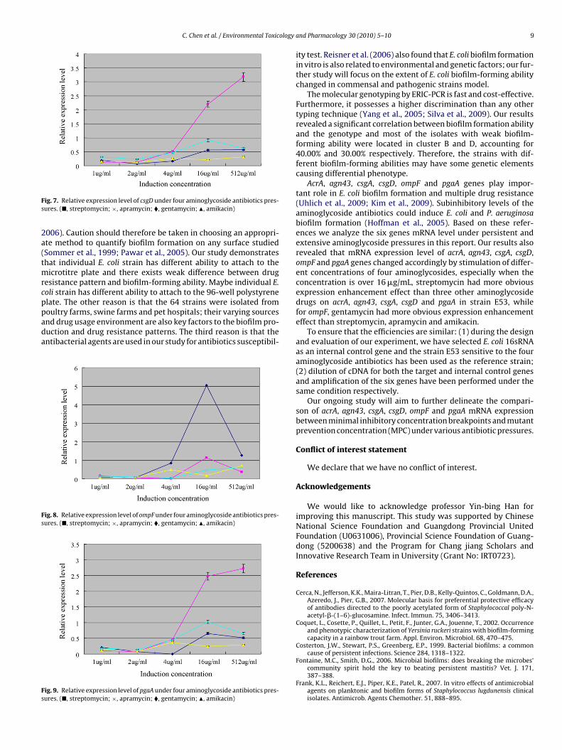
formed a trend of enhancement before the concentration cameto the point of 16 �g/mL (for E. coli, they began not sensitive tostreptomycin, apramycin, gentamycin and amikacin). Over the con-centration of 16 �g/mL, different genes performed various trendsunder different antibiotic pressures.
Our results also showed that over the concentration of16 �g/mL, streptomycin had more obvious expression enhance-ment effect than other aminoglycoside drugs on acrA, agn43, csgA,csgD and pgaA in strain E53, while for ompF gene, gentamycin hadmore obvious expression enhancement effect than streptomycin,apramycin and amikacin.
4. Discussion
The biofilm formation on the surface of bacterial cells is a com-plex phenomenon and is affected by a combination of mechanismsthat are influenced by the cell surface, the substratum and the
Fig. 5. Relative expression level of agn43 under four aminoglycoside antibioticspressures. (�, streptomycin; ×, apramycin; �, gentamycin; �, amikacin)
Fig. 6. Relative expression level of csgA under four aminoglycoside antibiotics pres-sures. (�, streptomycin; ×, apramycin; �, gentamycin; �, amikacin)
C. Chen et al. / Environmental Toxicology a
Fs
2a(tmrcppada
Fs
Fs
ig. 7. Relative expression level of csgD under four aminoglycoside antibiotics pres-ures. (�, streptomycin; ×, apramycin; �, gentamycin; �, amikacin)
006). Caution should therefore be taken in choosing an appropri-te method to quantify biofilm formation on any surface studiedSommer et al., 1999; Pawar et al., 2005). Our study demonstrateshat individual E. coli strain has different ability to attach to the
icrotitre plate and there exists weak difference between drugesistance pattern and biofilm-forming ability. Maybe individual E.oli strain has different ability to attach to the 96-well polystyrenelate. The other reason is that the 64 strains were isolated from
oultry farms, swine farms and pet hospitals; their varying sourcesnd drug usage environment are also key factors to the biofilm pro-uction and drug resistance patterns. The third reason is that thentibacterial agents are used in our study for antibiotics susceptibil-ig. 8. Relative expression level of ompF under four aminoglycoside antibiotics pres-ures. (�, streptomycin; ×, apramycin; �, gentamycin; �, amikacin)
ig. 9. Relative expression level of pgaA under four aminoglycoside antibiotics pres-ures. (�, streptomycin; ×, apramycin; �, gentamycin; �, amikacin)
nd Pharmacology 30 (2010) 5–10 9
ity test. Reisner et al. (2006) also found that E. coli biofilm formationin vitro is also related to environmental and genetic factors; our fur-ther study will focus on the extent of E. coli biofilm-forming abilitychanged in commensal and pathogenic strains model.
The molecular genotyping by ERIC-PCR is fast and cost-effective.Furthermore, it possesses a higher discrimination than any othertyping technique (Yang et al., 2005; Silva et al., 2009). Our resultsrevealed a significant correlation between biofilm formation abilityand the genotype and most of the isolates with weak biofilm-forming ability were located in cluster B and D, accounting for40.00% and 30.00% respectively. Therefore, the strains with dif-ferent biofilm-forming abilities may have some genetic elementscausing differential phenotype.
AcrA, agn43, csgA, csgD, ompF and pgaA genes play impor-tant role in E. coli biofilm formation and multiple drug resistance(Uhlich et al., 2009; Kim et al., 2009). Subinhibitory levels of theaminoglycoside antibiotics could induce E. coli and P. aeruginosabiofilm formation (Hoffman et al., 2005). Based on these refer-ences we analyze the six genes mRNA level under persistent andextensive aminoglycoside pressures in this report. Our results alsorevealed that mRNA expression level of acrA, agn43, csgA, csgD,ompF and pgaA genes changed accordingly by stimulation of differ-ent concentrations of four aminoglycosides, especially when theconcentration is over 16 �g/mL, streptomycin had more obviousexpression enhancement effect than three other aminoglycosidedrugs on acrA, agn43, csgA, csgD and pgaA in strain E53, whilefor ompF, gentamycin had more obvious expression enhancementeffect than streptomycin, apramycin and amikacin.
To ensure that the efficiencies are similar: (1) during the designand evaluation of our experiment, we have selected E. coli 16sRNAas an internal control gene and the strain E53 sensitive to the fouraminoglycoside antibiotics has been used as the reference strain;(2) dilution of cDNA for both the target and internal control genesand amplification of the six genes have been performed under thesame condition respectively.
Our ongoing study will aim to further delineate the compari-son of acrA, agn43, csgA, csgD, ompF and pgaA mRNA expressionbetween minimal inhibitory concentration breakpoints and mutantprevention concentration (MPC) under various antibiotic pressures.
Conflict of interest statement
We declare that we have no conflict of interest.
Acknowledgements
We would like to acknowledge professor Yin-bing Han forimproving this manuscript. This study was supported by ChineseNational Science Foundation and Guangdong Provincial UnitedFoundation (U0631006), Provincial Science Foundation of Guang-dong (5200638) and the Program for Chang jiang Scholars andInnovative Research Team in University (Grant No: IRT0723).
References
Cerca, N., Jefferson, K.K., Maira-Litran, T., Pier, D.B., Kelly-Quintos, C., Goldmann, D.A.,Azeredo, J., Pier, G.B., 2007. Molecular basis for preferential protective efficacyof antibodies directed to the poorly acetylated form of Staphylococcal poly-N-acetyl-�-(1–6)-glucosamine. Infect. Immun. 75, 3406–3413.
Coquet, L., Cosette, P., Quillet, L., Petit, F., Junter, G.A., Jouenne, T., 2002. Occurrenceand phenotypic characterization of Yersinia ruckeri strains with biofilm-formingcapacity in a rainbow trout farm. Appl. Environ. Microbiol. 68, 470–475.
Costerton, J.W., Stewart, P.S., Greenberg, E.P., 1999. Bacterial biofilms: a commoncause of persistent infections. Science 284, 1318–1322.
Fontaine, M.C., Smith, D.G., 2006. Microbial biofilms: does breaking the microbes’community spirit hold the key to beating persistent mastitis? Vet. J. 171,387–388.
Frank, K.L., Reichert, E.J., Piper, K.E., Patel, R., 2007. In vitro effects of antimicrobialagents on planktonic and biofilm forms of Staphylococcus lugdunensis clinicalisolates. Antimicrob. Agents Chemother. 51, 888–895.
1 ology
G
H
K
L
M
M
N
P
R
R
(ERIC) sequences in Escherichia coli: evolution and implications for ERIC-PCR.Mol. Biol. Evol. 23 (6), 1156–1168.
Yang, W.Q., Shi, L., Jia, W.X., Yin, X.L., Su, J.Y., Kou, Y.L., Xie, Y., Shinodo, S., 2005.
0 C. Chen et al. / Environmental Toxic
anderton, L., Chawla, J., Winters, C., Wimpenny, J., Stickler, D., 1992. Scanning elec-tron microscopy of bacterial biofilms on indwelling bladder catheters. Eur. J.Clin. Microbiol. Infect. Dis. 11, 789–796.
offman, L.R., Dargenio, D.A., Maccoss, M.J., Zhang, Z.Y., Jones, R.A., Miller, S.I.,2005. Aminoglycoside antibiotics induce bacterial biofilm formation. Nature436, 1171–1175.
im, Y., Wang, X., Ma, Q., Zhang, X.S., Wood, T.K., 2009. Toxin–antitoxin systems inEscherichia coli influence biofilm formation through YjgK (TabA) and fimbriae. J.Bacteriol. 191, 1258–1267.
ivak, K.J., Schmittgen, T.D., 2001. Analysis of relative gene expression data usingreal-time quantitative PCR and the 2−��CT method. Methods 25, 402–408.
elchior, M.B., Vaarkamp, H., Fink-Gremmels, J., 2006. Biofilms: a role in recurrentmastitis infections? Vet. J. 171, 398–407.
oskowitz, S.M., Foster, J.M., Emerson, J., Burns, J.L., 2004. Clinically feasible biofilmsusceptibility assay for isolates of Pseudomonas aeruginosa from patients withcystic fibrosis. J. Clin. Microbiol. 42, 1915–1922.
aves, P., Del Huelves, L., Gracia, M., Ruiz, V., Blanco, J., Dahbi, G., Blanco, M., Pon-temdel, C., Soriano, F., 2008. Correlation between virulence factors and in vitrobiofilm formation by Escherichia coli strains. Microb. Pathog. 45, 86–91.
awar, D.M., Rossman, M.L., Chen, J., 2005. Role of curli fimbriae in mediating thecells of enterohaemorrhagic Escherichia coli to attach to abiotic surfaces. J. Appl.Microbiol. 99, 418–425.
eisner, A., Krogfelt, K.A., Klein, B.M., Zechner, E.L., Molin, S., 2006. In vitro biofilmformation of commensal and pathogenic Escherichia coli strains: impact of envi-ronmental and genetic factors. J. Bacteriol. 188, 3572–3581.
ijavec, M., Muller-Premru, M., Zakotnik, B., Zgur-Bertok, D., 2008. Virulence factorsand biofilm production among Escherichia coli strains causing bacteraemia ofurinary tract origin. J. Med. Microbiol. 57, 1329–1334.
and Pharmacology 30 (2010) 5–10
Ryu, J.H., Beuchat, L.R., 2005. Biofilm formation by Escherichia coli O157:H7 on stain-less steel: effect of exopolysaccharide and curli production on its resistance tochlorine. Appl. Environ. Microbiol. 71, 247–255.
Silva, E., Leitao, S., Tenreiro, T., Pomba, C., Nunes, T., Lopes da Costa, L., Mateus,L., 2009. Genomic and phenotypic characterization of Escherichia coli isolatesrecovered from the uterus of puerperal dairy cows. J. Dairy Sci. 92, 6000–6010.
Sommer, P., Marin-Rouas, C., Mettler, E., 1999. Influence of the adherent popula-tion level on biofilm population, structure and resistance to chlorination. FoodMicrobiol. 16, 503–515.
Stepanovic, S., Vukovic, D., Dakic, I., Sacic, B., Svabic-Vihovic, M., 2000. A modifiedmicrotiter-plate test for quantification of staphylococcal biofilm formation. J.Microbiol. Methods 40, 175–179.
Suman, E., Jose, J., Varghese, S., Kotian, M.S., 2007. Study of biofilm production inEscherichia coli causing urinary tract infection. Indian J. Med. Microbiol. 25,305–306.
Uhlich, G.A., Gunther, N.W., Bayles, D.O., Mosier, D.A., 2009. The CsgA and Lpp pro-teins of an Escherichia coli O157:H7 strain affect HEp-2 cell invasion, motility,and biofilm formation. Infect. Immun. 77 (4), 1543–1552.
Wilson, L.A., Sharp, P.M., 2006. Enterobacterial repetitive intergenic consensus
Evaluation of the biofilm-forming ability and genetic typing for clinical isolatesof Pseudomonas aeruginosa by enterobacterial repetitive intergenic consensus-based PCR. Microbiol. Immunol. 49, 1057–1061.