Embed Size (px)
Citation preview
Epidemiological and clinical features of respiratory viralinfections in hospitalized children during the circulationof influenza virus A(H1N1) 2009
Gianvincenzo Zuccotti,a Dario Dilillo,a Alessandra Zappa,b Erica Galli,a Antonella Amendola,b,c,d
Marianna Martinelli,b Elena Pariani,b Filippo Salvini,c Elisabetta Tanzi,b,c,d Enrica Riva,c Marcello
Giovannini,c
aDepartment of Pediatrics, Sacco Hospital, University of Milan, Milan, Italy. bDepartment of Public Health-Microbiology-Virology, University of
Milan, Milan, Italy. cDepartment of Pediatrics, San Paolo Hospital, University of Milan, Milan, Italy. dDepartment of Health Sciences, University of
Genoa, Genoa, Italy.
Correspondence: Gianvincenzo Zuccotti, Department of Pediatrics, Sacco Hospital, University of Milan, via GB Grassi 74, 20157 Milan, Italy.
E-mail: [email protected]
Setting: Two Pediatric Clinics at the University of Study of Milan, in Milan, Italy (‘L. Sacco’ and ‘S. Paolo’ Hospitals); Department of Public Health-
Microbiology-Virology, University of Milan.
Accepted 27 April 2011. Published Online 25 May 2011.
Background Seasonal influenza viruses and respiratory syncytial
virus (RSV) are primary causes of acute respiratory tract
infections (ARTIs) in children. New respiratory viruses including
human metapneumovirus (hMPV), human bocavirus (hBoV), and
influenza 2009 A(H1N1) virus have a strong impact on the
pediatric population.
Objectives To evaluate epidemiological and clinical features of
ARTIs in hospitalized children.
Methods From December 1, 2008, to December 31, 2009, all
children under age fifteen (n = 575) hospitalized for ARTIs were
investigated for influenza A (subtype H1N1, H3N2, and 2009
H1N1) and B, RSV A and B, hMPV, and hBoV by PCR.
Results Fifty-one percent of samples were positive for these
respiratory viruses. The frequencies of virus detection were RSV
34Æ1%, hBoV 6Æ8%, hMPV 5%, seasonal influenza A 5%, and
seasonal influenza B 0%. From April 2009, 11Æ6% of collected
samples were influenza 2009 A(H1N1) positive. Respiratory
syncytial virus activity peaked in January, hBoV in February, and
hMPV in April. Seasonal influenza A was detected only between
January and April 2009, while influenza 2009 A(H1N1) peaked in
November. Respiratory syncytial virus and hMPV were mainly
associated with lower respiratory tract infections (LRTIs) and with
necessity of O2 administration. The 2009 pandemic influenza was
more frequently detected in elder children (P < 0Æ001) and was
associated with higher, longer-lasting fevers compared with other
viral infections (P < 0Æ05).
Conclusions All considered viruses were involved in LRTIs. The
primary clinical relevance of RSV and a similar involvement of
both seasonal influenza and emerging viruses investigated were
observed on the pediatric population.
Keywords Acute respiratory tract infections, emerging viruses,
influenza virus A(H1N1) 2009, pediatric hospitalizations, viral
respiratory infections.
Please cite this paper as: Zuccotti et al. (2011) Epidemiological and clinical features of respiratory viral infections in hospitalized children during the circula-
tion of influenza virus A(H1N1) 2009. Influenza and Other Respiratory Viruses 5(6), e528–e534.
Introduction
Acute respiratory tract infections (ARTIs) are associated
with significant morbidity worldwide; viruses are by far
the most common causes of ARTIs, especially among
young children.1 In particular, seasonal influenza viruses
(type A and B) and respiratory syncytial virus (RSV) are
the main etiological agents during the epidemic period
(between October and April in the northern hemi-
sphere).2,3 RSV infection is usually much more frequently
identified than influenza in children; indeed, about 77%
of infants have had an RSV infection before 5 years of
age. This virus is the main cause of bronchiolitis, which is
one of the major reasons of hospitalization in children
under 2 years of age.4 However, recent data show the
important clinical role of seasonal influenza in children.
In fact, the rate of hospitalization for seasonal influenza is
not <3Æ6 per 10 000 child ⁄ year, and hospitalization can
also regard previously healthy children and children older
than 2 years.4,5 The recent epidemiological scenario has
DOI:10.1111/j.1750-2659.2011.00264.x
www.influenzajournal.comOriginal Article
e528 ª 2011 Blackwell Publishing Ltd, Influenza and Other Respiratory Viruses, 5, e528–e534
been enlivened by the identification and emergence of sev-
eral pathogens with an airborne transmission pathway
such as human metapneumovirus (hMPV), first isolated
in 2001,6 human bocavirus (hBoV), discovered in 2005,7
and influenza virus 2009 A(H1N1), identified in 2009, and
responsible for the first pandemic of the new millennium.8
HMPV and hBoV are isolated in 3Æ9–16% of children hos-
pitalized for ARTIs. The first is associated with a large
spectrum of clinical manifestations that range from mild
upper respiratory tract disease to severe bronchiolitis and
pneumonia,9,10 and thus, it is considered to be one of the
most important respiratory emerging viruses, while the
second seems to have a marginal role as it causes mainly
upper respiratory tract infections (URTIs), when detected
alone.11
The 2009 pandemic influenza affected the pediatric pop-
ulation in 60% of cases8 causing a significant number of
recovery also in children aged over 5 years. It seems to
cause generally mild disease, similar to those of seasonal
influenza12, while severe manifestations seem concentrated
in patients with risk factors.
This study aimed to evaluate the frequency and the
demographic and clinical features of ARTIs caused by
known viruses (i.e., RSV and seasonal influenza) and newly
identified viruses (i.e., influenza 2009 A(H1N1), hMPV,
and hBoV) in children hospitalized for ARTI in Milan
(Italy) from December 2008 to December 2009.
Materials and methods
From December 1, 2008, to December 31, 2009, we
enrolled 575 children aged between 0 and 15 years hospi-
talized for an ARTI in two Pediatric Clinics at the Univer-
sity of Milan (‘L. Sacco’ and ‘S. Paolo’ Hospitals). After
informed consent was obtained from the parents, an oro-
pharyngeal swab (Plain Swabs; Copan, Brescia, Italy) was
collected from each child within 24 hour after hospital
admission and tested at the Department of Public Health-
Microbiology-Virology, University of Milan, using PCR
assays to detect viral respiratory pathogens (i.e., influenza
type A, subtype H1N1, H3N2, and 2009 H1N1; influenza
type B; RSV type A and B; hMPV; and hBoV).
A standardized datasheet was used to record socio-
demographic data (age, gender, risk factors) obtained
through parents interview and clinical information (dura-
tion of hospitalization, detailed disease signs and symptoms
before and during hospitalization, prescribed drug therapy)
by medical chart abstraction.
Clinical data interpretationFor this study, children <15 years of age hospitalized with
symptoms of ARTI such as cough, rhinitis, sore throat,
wheezing, panting, dyspnea, or apnea were enrolled.
Patients were assessed and categorized according to diag-
nosis of upper respiratory tract infections, wheezy bronchi-
tis, bronchitis, bronchiolitis, and pneumonia on the basis
of clinical and roentgenographic findings. The criteria pro-
posed by Ruuskanen and Ogra13 were used for definitions
of bronchiolitis, pneumonia, and wheezy bronchitis.
Acute illnesses indicated as upper respiratory infections
(URTIs) were characterized by cough, rhinorrhea, sore
throat, and ⁄ or otitis media, with normal thoracic objectiv-
ity and X-ray. An acute illness characterized by cough,
rhonchi, and diffuse expiratory wheezing upon thoracic
auscultation was diagnosed as wheezy bronchitis, while a
illness with the same characteristic but without expiratory
wheezing in any phase of its course was diagnosed as bron-
chitis. Bronchiolitis was diagnosed when dyspnea, tachyp-
nea, diffuse small crackles upon thoracic auscultation, and
roentgenographic evidence of hyperinflation of the lung
with or without areas of collapse were present in children
younger than 2 years of age. Pneumonia diagnosis was
based on auscultation of pathological breath sounds, such
as small crackles or decrease ⁄ absence of vesicular sound, in
a zone of the chest, and on radiographic findings of lung
parenchymal involvement with interstitial-alveolar infil-
trates and ⁄ or consolidation.
The term lower respiratory tract infections (LRTIs) was
used to indicate acute illness with the presence of signs of
lower airway involvement (tachypnea, dyspnea, wheezing,
rhonchi, or rales) and ⁄ or a positive chest X-ray, so it
includes bronchitis, wheezy bronchitis, bronchiolitis, and
pneumonia.
Nucleic acid extraction and amplificationNucleic acid extraction was conducted using a commercial
method (NucliSENS�, miniMAG�; Biomerieux, Marcy
L’Etoile, France). For RNA virus detection, cDNA was syn-
thesized with pd(N)6 random hexamer primers (Amersham
Biosciences, Little Chalfont, UK) using an MMLV reverse
transcriptase (Invitrogen Tech-Line, Carlsbad, CA, USA).
Viral detection was performed by PCR assays. To simulta-
neously detect and type seasonal A and B influenza viruses,
a one-step real-time RT-multiplex-PCR assay was per-
formed using primer ⁄ probe sets for two different genome
regions: the matrix region of influenza type A virus and
the nucleoprotein region of influenza type B virus.14 Influ-
enza A-positive samples were subtyped using an RT-multi-
plex-PCR assay with specific primers for the hemagglutinin
gene of influenza A ⁄ H1 and A ⁄ H3 viruses.15 Pandemic
2009 A(H1N1) influenza virus was detected using a one-
step real-time RT-PCR, in accordance with the Centers for
Disease Control and Prevention guidelines.16 RSV A and
RSV B were identified by multiplex nested PCR (fusion
gene, 336 and 582 bp, respectively) using specific primer
sets.17 Two nested PCR assays were performed to detect a
Respiratory viral infections in children
ª 2011 Blackwell Publishing Ltd, Influenza and Other Respiratory Viruses, 5, e528–e534 e529
151-bp (nt. 44–195) fragment of the matrix gene of hMPV
and a 354-bp (nt. 2351–2704) fragment of the nucleopro-
tein gene of hBoV,18 respectively. Appropriate positive and
negative controls were included in any PCR assay.
Statistical analysisData were expressed as median (interquartile range, IQR)
and percentages (95% confidence intervals, 95% CI) as
appropriate. Comparisons between groups were performed
using the chi-square test or Fisher’s exact test. A
P-value < 0Æ05 was considered statistically significant (two-
tailed test). All statistical analyses were performed using
OpenEPI software, version 2.2.1.19
Results
Study populationFrom December 2008 to December 2009, 575 children hos-
pitalized for ARTIs (338 boys and 237 girls; median age
9Æ0 months, IQR 3Æ0–24Æ0 months) were enrolled. Twenty-
four children were not enrolled because guardian consent
was lacking.
Children were divided into five age-groups: <6 months
(n = 242; 42Æ1%, 95% CI: 38Æ1–46Æ2), 6–11 months (n = 77;
13Æ4%, 95% CI: 10Æ8–16Æ4), 12–23 months (n = 106; 18Æ4%,
95% CI: 15Æ4–21Æ8), 2–5 years (n = 99; 17Æ2%, 95% CI: 14Æ3–
20Æ5), and 6–15 years (n = 51; 8Æ9%, 95% CI: 6Æ7–11Æ4).
Patients were more frequently affected by LRTIs than
URTIs (67Æ8%, 95% CI: 63Æ9–71Æ5 versus 32Æ2%, 95% CI:
28Æ4–36Æ1; P < 0Æ0001). Bronchiolitis was diagnosed in 145
children (25Æ2%, 95% CI: 21Æ8–28Æ9), pneumonia in 151
(26Æ3%, 95% CI: 22Æ8–30Æ0), wheezy bronchitis in 57 (9Æ9%,
95% CI: 7Æ7–12Æ6), and bronchitis in 37 (6Æ4%, 95% CI:
4Æ6–8Æ7).
Frequency of viral respiratory infectionsMolecular investigations revealed viral gene sequences in
293 (51Æ0%, 95% CI: 46Æ9–55Æ0) samples. Respiratory syn-
cytial virus was detected in 196 (34Æ1%, 95% CI: 30Æ3–38Æ0)
of collected samples. Of these, 67 (34Æ2%, 95% CI: 27Æ8–
41Æ0) were RSV A positive and 129 (65Æ8%, 95% CI: 59Æ0–
72Æ2) were RSV B positive (P < 0Æ05). HBoV was detected
in 39 (6Æ8%, 95% CI: 5Æ0–9Æ1) and hMPV and seasonal
influenza A in 29 (5Æ0%, 95% CI: 3Æ5–7Æ1) samples. Regard-
ing seasonal influenza A-positive samples, 25 (86Æ2%, 95%
CI: 70Æ0–95Æ5) were subtype H3 and 4 (13Æ8%, 95% CI:
4Æ5–30Æ0) were subtype H1 (P < 0Æ05). No influenza B virus
cases were identified.
From April 2009 to December 2009, 224 samples were
also tested for 2009 A(H1N1) viral sequences: 26 (11Æ6%,
95% CI: 7Æ9–16Æ3%) were 2009 A(H1N1) positive.
Mixed infections were detected in 8Æ2% of ARTIs
(Table 1), and in particular, in 41Æ0% of samples positive
for hBoV, 9Æ2% of samples positive for RSV, and 7Æ7% of
samples positive for influenza 2009 A(H1N1) (hBoV co-
infections versus RSV and influenza 2009 A(H1N1);
P < 0Æ003). Single and co-infections were associated with
LRTIs with similar percentage (83Æ3% versus 78Æ8%,
P = 0Æ3).
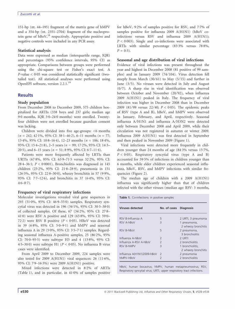
Seasonal and age distribution of viral infectionsEvidence of viral infections was present throughout the
year and highest in December 2008 (81 positive of 99 sam-
ples) and in January 2009 (74 ⁄ 104). Virus detection fell
steeply from March (30 ⁄ 61) to May (5 ⁄ 15) and further in
June (3 ⁄ 5). No viruses were detected in July and August
(0 ⁄ 7). A sharp rise in viral identification was observed
between October and November (20 ⁄ 92), when influenza
2009 A(H1N1) peaked in Italy. The frequency of viral
infection was higher in December 2008 than in December
2009 (81 ⁄ 99 versus 22 ⁄ 40; P < 0Æ05). The epidemic peaks
of RSV (type A and B), hBoV, and hMPV were observed
in January, February, and April, respectively. Seasonal
influenza A ⁄ H1N1 and influenza A ⁄ H3N2 were detected
only between December 2008 and April 2009, while their
circulation was not registered in autumn or winter 2009.
Influenza 2009 A(H1N1) was first detected in September
and then peaked in November 2009 (Figure 1).
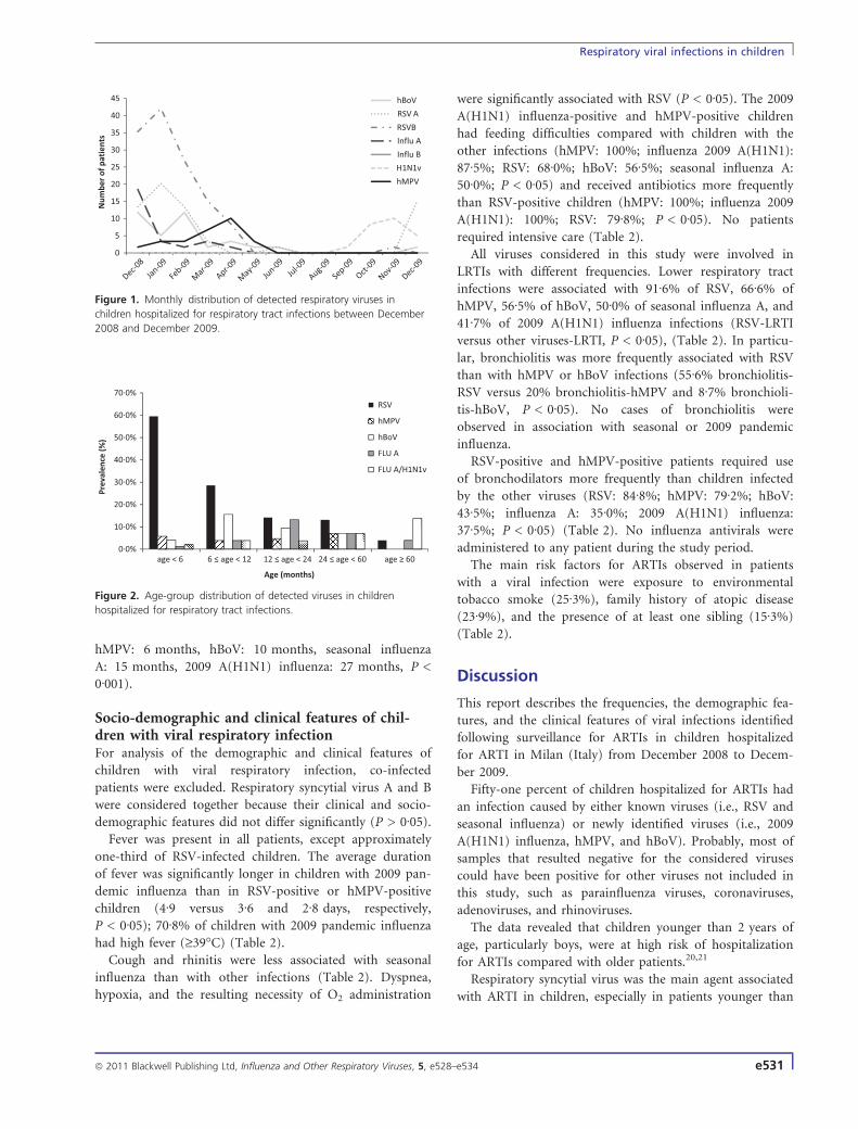
Viral infections were detected more frequently in chil-
dren younger than 24 months of age (84Æ3% versus 15Æ7%,
P < 0Æ05). Respiratory syncytial virus (type A and B)
accounted for 59Æ5% of infections in children younger than
6 months, while elder children experienced seasonal influ-
enza, hBoV, RSV, and hMPV infections with similar fre-
quencies (Figure 2).
The median age of children with a 2009 A(H1N1)
influenza was significantly higher than that of children
infected with the other viruses (median age: RSV: 3 months,
Table 1. Co-infections in positive samples
Viruses detected No. of cases Diagnosis
RSV B-Influenza A 5 2 URTI, 3 pneumonia
RSV A-hBoV 3 1 pneumonia,
2 wheezy bronchitis
RSV B-hBoV 5 2 pneumonia,
3 bronchiolitis
Influenza A-hBoV 2 2 URTI
Influenza A-RSV A-hBoV 2 2 bronchiolitis
RSV B-hMPV 3 1 bronchiolitis,
2 wheezy bronchitis
Influenza A(H1N1)2009-hBoV 2 2 pneumonia
hMPV–hBoV 2 2 bronchiolitis
hBoV, human bocavirus; hMPV, human metapneumovirus; RSV,
Respiratory syncytial virus; URTI, upper respiratory tract infections.
Zuccotti et al.
e530 ª 2011 Blackwell Publishing Ltd, Influenza and Other Respiratory Viruses, 5, e528–e534
hMPV: 6 months, hBoV: 10 months, seasonal influenza
A: 15 months, 2009 A(H1N1) influenza: 27 months, P <
0Æ001).
Socio-demographic and clinical features of chil-dren with viral respiratory infectionFor analysis of the demographic and clinical features of
children with viral respiratory infection, co-infected
patients were excluded. Respiratory syncytial virus A and B
were considered together because their clinical and socio-
demographic features did not differ significantly (P > 0Æ05).
Fever was present in all patients, except approximately
one-third of RSV-infected children. The average duration
of fever was significantly longer in children with 2009 pan-
demic influenza than in RSV-positive or hMPV-positive
children (4Æ9 versus 3Æ6 and 2Æ8 days, respectively,
P < 0Æ05); 70Æ8% of children with 2009 pandemic influenza
had high fever (‡39�C) (Table 2).
Cough and rhinitis were less associated with seasonal
influenza than with other infections (Table 2). Dyspnea,
hypoxia, and the resulting necessity of O2 administration
were significantly associated with RSV (P < 0Æ05). The 2009
A(H1N1) influenza-positive and hMPV-positive children
had feeding difficulties compared with children with the
other infections (hMPV: 100%; influenza 2009 A(H1N1):
87Æ5%; RSV: 68Æ0%; hBoV: 56Æ5%; seasonal influenza A:
50Æ0%; P < 0Æ05) and received antibiotics more frequently
than RSV-positive children (hMPV: 100%; influenza 2009
A(H1N1): 100%; RSV: 79Æ8%; P < 0Æ05). No patients
required intensive care (Table 2).
All viruses considered in this study were involved in
LRTIs with different frequencies. Lower respiratory tract
infections were associated with 91Æ6% of RSV, 66Æ6% of
hMPV, 56Æ5% of hBoV, 50Æ0% of seasonal influenza A, and
41Æ7% of 2009 A(H1N1) influenza infections (RSV-LRTI
versus other viruses-LRTI, P < 0Æ05), (Table 2). In particu-
lar, bronchiolitis was more frequently associated with RSV
than with hMPV or hBoV infections (55Æ6% bronchiolitis-
RSV versus 20% bronchiolitis-hMPV and 8Æ7% bronchioli-
tis-hBoV, P < 0Æ05). No cases of bronchiolitis were
observed in association with seasonal or 2009 pandemic
influenza.
RSV-positive and hMPV-positive patients required use
of bronchodilators more frequently than children infected
by the other viruses (RSV: 84Æ8%; hMPV: 79Æ2%; hBoV:
43Æ5%; influenza A: 35Æ0%; 2009 A(H1N1) influenza:
37Æ5%; P < 0Æ05) (Table 2). No influenza antivirals were
administered to any patient during the study period.
The main risk factors for ARTIs observed in patients
with a viral infection were exposure to environmental
tobacco smoke (25Æ3%), family history of atopic disease
(23Æ9%), and the presence of at least one sibling (15Æ3%)
(Table 2).
Discussion
This report describes the frequencies, the demographic fea-
tures, and the clinical features of viral infections identified
following surveillance for ARTIs in children hospitalized
for ARTI in Milan (Italy) from December 2008 to Decem-
ber 2009.
Fifty-one percent of children hospitalized for ARTIs had
an infection caused by either known viruses (i.e., RSV and
seasonal influenza) or newly identified viruses (i.e., 2009
A(H1N1) influenza, hMPV, and hBoV). Probably, most of
samples that resulted negative for the considered viruses
could have been positive for other viruses not included in
this study, such as parainfluenza viruses, coronaviruses,
adenoviruses, and rhinoviruses.
The data revealed that children younger than 2 years of
age, particularly boys, were at high risk of hospitalization
for ARTIs compared with older patients.20,21
Respiratory syncytial virus was the main agent associated
with ARTI in children, especially in patients younger than
0
5
10
15
20
25
30
35
40
45
Dec-08
Jan-0
9
Feb-0
9
Mar
-09
Apr-09
May
-09
Jun-0
9Ju
l-09
Aug-09
Sep-0
9
Oct-09
Nov-09
Dec-09
Num
ber
of p
atie
nts
hBoV
RSV A
RSVB
Influ A
Influ B
H1N1v
hMPV
Figure 1. Monthly distribution of detected respiratory viruses in
children hospitalized for respiratory tract infections between December
2008 and December 2009.
0·0%
10·0%
20·0%
30·0%
40·0%
50·0%
60·0%
70·0%
age < 6 6 ≤ age < 12 12 ≤ age < 24 24 ≤ age < 60 age ≥ 60
Prev
alen
ce (%
)
Age (months)
RSV
hMPV
hBoV
FLU A
FLU A/H1N1v
Figure 2. Age-group distribution of detected viruses in children
hospitalized for respiratory tract infections.
Respiratory viral infections in children
ª 2011 Blackwell Publishing Ltd, Influenza and Other Respiratory Viruses, 5, e528–e534 e531
6 months, and was responsible for almost all LRTIs. The
primary clinical relevance of RSV in children is well charac-
terized. Serologic evidence indicates that nearly all children
have been infected by RSV within the first 2 years of life.22
Hospitalization is required in approximately 0Æ5–2% of
cases and 80% of these occur in the first year of life.23 In
Europe, RSV accounts for 42–45% of hospital admission
for LRTIs in children younger than 2 years; in particular,
in studies on hospitalized children, RSV is associated with
bronchiolitis in 60–90% of cases and with pneumonia in
25–50% of cases.24 No vaccines against this virus are avail-
able so far, but prophylaxis with palivizumab (a human,
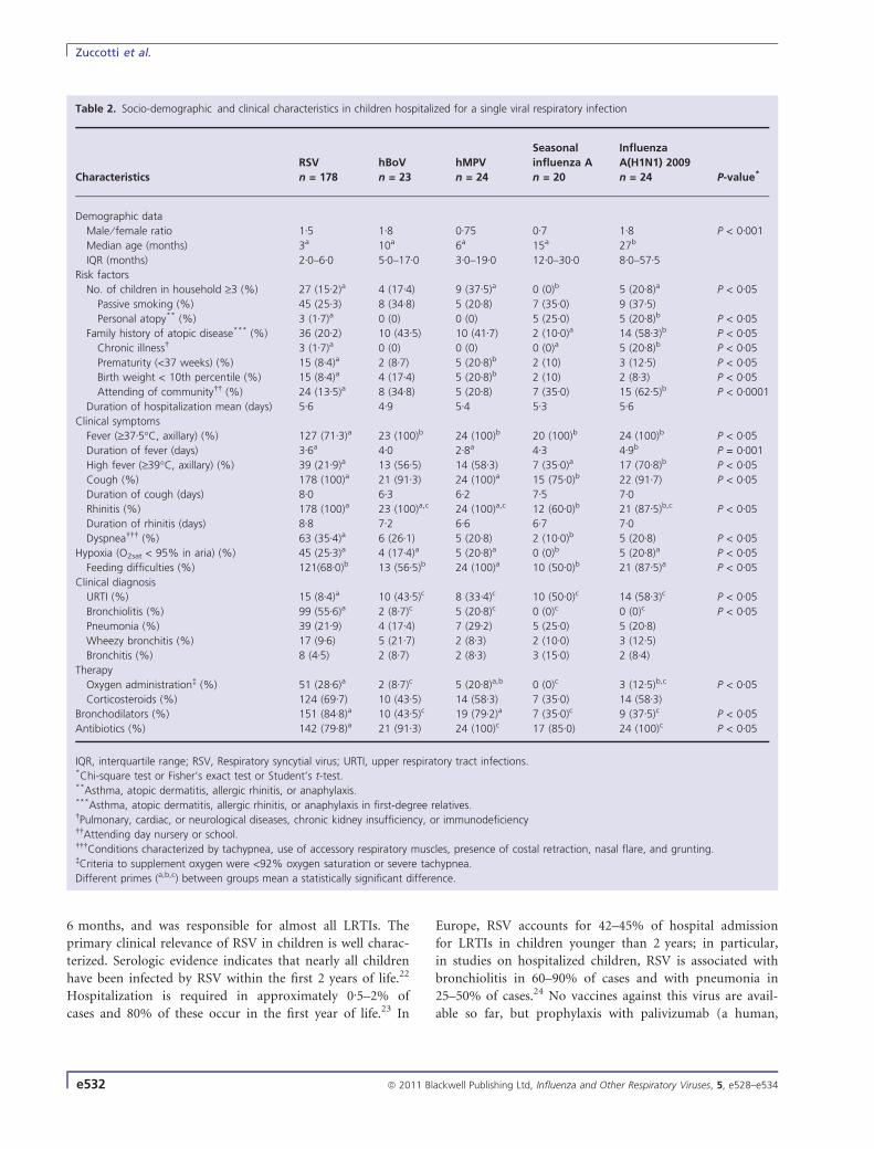
Table 2. Socio-demographic and clinical characteristics in children hospitalized for a single viral respiratory infection
Characteristics
RSV
n = 178
hBoV
n = 23
hMPV
n = 24
Seasonal
influenza A
n = 20
Influenza
A(H1N1) 2009
n = 24 P-value*
Demographic data
Male ⁄ female ratio 1Æ5 1Æ8 0Æ75 0Æ7 1Æ8 P < 0Æ001
Median age (months) 3a 10a 6a 15a 27b
IQR (months) 2Æ0–6Æ0 5Æ0–17Æ0 3Æ0–19Æ0 12Æ0–30Æ0 8Æ0–57Æ5Risk factors
No. of children in household ‡3 (%) 27 (15Æ2)a 4 (17Æ4) 9 (37Æ5)a 0 (0)b 5 (20Æ8)a P < 0Æ05
Passive smoking (%) 45 (25Æ3) 8 (34Æ8) 5 (20Æ8) 7 (35Æ0) 9 (37Æ5)
Personal atopy** (%) 3 (1Æ7)a 0 (0) 0 (0) 5 (25Æ0) 5 (20Æ8)b P < 0Æ05
Family history of atopic disease*** (%) 36 (20Æ2) 10 (43Æ5) 10 (41Æ7) 2 (10Æ0)a 14 (58Æ3)b P < 0Æ05
Chronic illness� 3 (1Æ7)a 0 (0) 0 (0) 0 (0)a 5 (20Æ8)b P < 0Æ05
Prematurity (<37 weeks) (%) 15 (8Æ4)a 2 (8Æ7) 5 (20Æ8)b 2 (10) 3 (12Æ5) P < 0Æ05
Birth weight < 10th percentile (%) 15 (8Æ4)a 4 (17Æ4) 5 (20Æ8)b 2 (10) 2 (8Æ3) P < 0Æ05
Attending of community�� (%) 24 (13Æ5)a 8 (34Æ8) 5 (20Æ8) 7 (35Æ0) 15 (62Æ5)b P < 0Æ0001
Duration of hospitalization mean (days) 5Æ6 4Æ9 5Æ4 5Æ3 5Æ6Clinical symptoms
Fever (‡37Æ5�C, axillary) (%) 127 (71Æ3)a 23 (100)b 24 (100)b 20 (100)b 24 (100)b P < 0Æ05
Duration of fever (days) 3Æ6a 4Æ0 2Æ8a 4Æ3 4Æ9b P = 0Æ001
High fever (‡39�C, axillary) (%) 39 (21Æ9)a 13 (56Æ5) 14 (58Æ3) 7 (35Æ0)a 17 (70Æ8)b P < 0Æ05
Cough (%) 178 (100)a 21 (91Æ3) 24 (100)a 15 (75Æ0)b 22 (91Æ7) P < 0Æ05
Duration of cough (days) 8Æ0 6Æ3 6Æ2 7Æ5 7Æ0Rhinitis (%) 178 (100)a 23 (100)a,c 24 (100)a,c 12 (60Æ0)b 21 (87Æ5)b,c P < 0Æ05
Duration of rhinitis (days) 8Æ8 7Æ2 6Æ6 6Æ7 7Æ0Dyspnea��� (%) 63 (35Æ4)a 6 (26Æ1) 5 (20Æ8) 2 (10Æ0)b 5 (20Æ8) P < 0Æ05
Hypoxia (O2sat < 95% in aria) (%) 45 (25Æ3)a 4 (17Æ4)a 5 (20Æ8)a 0 (0)b 5 (20Æ8)a P < 0Æ05
Feeding difficulties (%) 121(68Æ0)b 13 (56Æ5)b 24 (100)a 10 (50Æ0)b 21 (87Æ5)a P < 0Æ05
Clinical diagnosis
URTI (%) 15 (8Æ4)a 10 (43Æ5)c 8 (33Æ4)c 10 (50Æ0)c 14 (58Æ3)c P < 0Æ05
Bronchiolitis (%) 99 (55Æ6)a 2 (8Æ7)c 5 (20Æ8)c 0 (0)c 0 (0)c P < 0Æ05
Pneumonia (%) 39 (21Æ9) 4 (17Æ4) 7 (29Æ2) 5 (25Æ0) 5 (20Æ8)
Wheezy bronchitis (%) 17 (9Æ6) 5 (21Æ7) 2 (8Æ3) 2 (10Æ0) 3 (12Æ5)
Bronchitis (%) 8 (4Æ5) 2 (8Æ7) 2 (8Æ3) 3 (15Æ0) 2 (8Æ4)
Therapy
Oxygen administration� (%) 51 (28Æ6)a 2 (8Æ7)c 5 (20Æ8)a,b 0 (0)c 3 (12Æ5)b,c P < 0Æ05
Corticosteroids (%) 124 (69Æ7) 10 (43Æ5) 14 (58Æ3) 7 (35Æ0) 14 (58Æ3)
Bronchodilators (%) 151 (84Æ8)a 10 (43Æ5)c 19 (79Æ2)a 7 (35Æ0)c 9 (37Æ5)c P < 0Æ05
Antibiotics (%) 142 (79Æ8)a 21 (91Æ3) 24 (100)c 17 (85Æ0) 24 (100)c P < 0Æ05
IQR, interquartile range; RSV, Respiratory syncytial virus; URTI, upper respiratory tract infections.*Chi-square test or Fisher’s exact test or Student’s t-test.**Asthma, atopic dermatitis, allergic rhinitis, or anaphylaxis.***Asthma, atopic dermatitis, allergic rhinitis, or anaphylaxis in first-degree relatives.�Pulmonary, cardiac, or neurological diseases, chronic kidney insufficiency, or immunodeficiency��Attending day nursery or school.���Conditions characterized by tachypnea, use of accessory respiratory muscles, presence of costal retraction, nasal flare, and grunting.�Criteria to supplement oxygen were <92% oxygen saturation or severe tachypnea.
Different primes (a,b,c) between groups mean a statistically significant difference.
Zuccotti et al.
e532 ª 2011 Blackwell Publishing Ltd, Influenza and Other Respiratory Viruses, 5, e528–e534
mouse monoclonal antibody directed against the F protein
of RSV) is available for vulnerable infants, in whom the
course of RSV infection could be worse, with a 1–5% mor-
tality rate.25
The two most important studies on RSV epidemiology
in Italy have shown that the epidemic starts in October–
November and ends in April–May, with peak incidence in
February.3,26–29 In this study, RSV activity peaked in Janu-
ary; however, in contrast with previous studies, it rose
gradually from October 2009.
Seasonal influenza viruses were identified in 5% of sam-
ples and between December 2008 and April 2009 only. No
influenza B viruses were detected during the study period.
The absence of seasonal influenza viruses at the end of
2009 in our study is in agreement with data from both the
Italian Influenza Surveillance Network (INFLUNET)30 and
the European Centre for Disease Prevention and Control.31
In this study, hMPV was detected in 5% of patients.
These data are in agreement with published literature, in
which frequencies of hMPV infections ranged from 3Æ9%
to 16%. Some authors have claimed a clinical similarity
between hMPV and RSV infections.32,33 In the population
analyzed, hMPV was associated with LRTIs, dyspnea, and
hypoxia less frequently than RSV.
HBoV infection was involved in 6% of ARTIs and in
41% of all co-infections identified. These data are in agree-
ment with studies investigating hBoV infection in ARTIs,
which have found a prevalence of 1Æ5–18Æ3%,11,34 with a
42Æ5% mean percentage of co-infections with other viral
pathogens.34 In most cases, hBoV had a marginal clinical
importance when identified alone, causing an URTI in
43Æ5% and requiring oxygen in 8Æ7% of cases. However,
according to other studies, this study raises the possibility
that hBoV is also associated with severe ARTIs such as
bronchiolitis,35,36 asthma exacerbations,37,38 and pneumo-
nia.39 The seasonal peak of hBoV varies among studies, but
is usually described in the early winter.34 In this study,
hBoV peak was observed in February, and its circulation
was low at the end of 2009. In agreement with data regis-
tered in European countries,30,31,40 the present study dem-
onstrates that the 2009 pandemic influenza virus peaked in
November 2009; In agreement with published litera-
ture,41,42 our data indicate that children with high hospital-
ization risk for pandemic influenza were not only those
younger than 2 years of age or with chronic disease (as
usually observed for seasonal influenza), but also children
older than 5 years of age, accounting for more of 25% of
recovery from 2009 pandemic influenza. Finally, 2009 pan-
demic influenza caused a clinical manifestation similar to
seasonal influenza with mild severity, although these chil-
dren had bad general conditions because of fever.
In conclusion, all viruses considered in this study circu-
lated in Italy and were involved in LRTIs in children. These
findings confirm the primary clinical relevance of RSV, and
a similar involvement of both the seasonal influenza and
the emerging viruses investigated in ARTIs among hospital-
ized children.
Acknowledgements
Funding: This research was supported by MIUR (Ministry
of Education, University and Research); Grant numbers:
2005067255_003 and 2007 LPAF42_003. Competing inter-
ests: None declared. Ethical approval: Luigi Sacco Hospital
Ethics Committee.
Author contributions
Study concept and design: G Zuccotti, E Tanzi, M Giovan-
nini, E Riva, A Amendola. Acquisition of data: D Dilillo,
E Galli, F Salvini. Analysis and interpretation of data: A Zap-
pa, M Martinelli, E Galli, A Amendola, E Pariani. Drafting of
the manuscript: E Galli, A Zappa. Critical revision of the
manuscript for important intellectual content: D Dilillo,
A Amendola, A Zappa. Statistical analysis: E Galli, A Zappa,
M Martinelli. Final approval of the version to be published:
G Zuccotti, E Tanzi, M Giovannini, E Riva.
References
1 Henrickson KJ, Hoover S, Kehl KS, Fan J. National disease burden of
respiratory viruses detected in children by polymerase chain reac-
tion. Pediatr Infect Dis J 2004; 23:211–218.
2 Esposito S, Marchisio P, Principi N. The global state of influenza in
children. Pediatr Infect Dis J 2008; 27:S149–S153.
3 Lanari M, Giovannini M, Giuffre L et al. Prevalence of respiratory
syncytial virus infection in Italian infants hospitalized for acute lower
respiratory tract infections, and association between respiratory syn-
cytial virus infection risk factors and disease severity. Pediatr Pul-
monol 2002; 33:458–465.
4 Coffin SE, Zaoutis TE, Rosenquist AB et al. Incidence, complications
and risk factors for prolonged stay in children hospitalized with
community-acquired influenza. Pediatrics 2007; 119:740–748.
5 Schrag SJ, Shay DK, Gershman K et al. Multistate surveillance for
laboratory-confirmed, influenza-associated hospitalizations in chil-
dren 2003–2004. Pediatr Infect Dis J 2006; 25:395–400.
6 Van den Hoogen BV, de Jong J, Green J et al. A newly discovered
human pneumovirus isolated from young children with respiratory
tract disease. Nat Med 2001; 7:719–724.
7 Schildgen O, Muller A, Allander T et al. Human bocavirus: passenge
or pathogen in acute respiratory tract infections? Clin Microbiol Rev
2008; 21:291–304.
8 Dawood FS, Jain S, Finelli L, Shaw MW, Lindstrom S, Garten RJ.
Emergence of novel swine-origin influenza A (H1N1) virus in
humans. N Engl J Med 2009; 360:2605–2615.
9 Williams JV, Harris PA, Tollefson SJ et al. Human metapneumovirus
and lower respiratory tract disease in otherwise healthy infants and
children. N Engl J Med 2004; 350:443–450.
10 Williams JV, wang CK, Yang CF et al. The role of human meta-
pneumovirus in upper respiratory tract infections in children: a 20-
year experience. J Infect Dis 2006; 193:387–395.
Respiratory viral infections in children
ª 2011 Blackwell Publishing Ltd, Influenza and Other Respiratory Viruses, 5, e528–e534 e533
11 Esposito S, Bosis S, Niesters HG. Impact of human bocavirus on chil-
dren and their families. J Clin Microbiol 2008; 46:1337–1342.
12 Halasa NB. Update on the 2009 pandemic influenza A H1N1 in chil-
dren. Curr Opin Pediatr 2010; 22:83–87.
13 Ruuskanen O, Ogra PL. Respiratory syncytial virus. Curr Probl Pediatr
1993; 23:50–79.
14 Valle L, Amicizia D, Bacilieri S et al. Performance testing of two
new one-step real time PCR assay for detection of human influenza
and avian influenza viruses isolated in humans and respiratory syn-
cytial virus. J Prev Med Hyg 2006; 47:127–133.
15 Stockton J, Ellis JS, Saville M, Clewley JP, Zambon MC. Multiplex
PCR for typing and subtyping influenza and respiratory syncytial
viruses. J Clin Microbiol 1998; 36:2990–2995.
16 Centers for Disease Control and Prevention. CDC protocol of real-
time RT-PCR for influenza A (H1N1). 2009. Available at http://
www.who.int/csr/resources/publications/swineflu/realtimeptpcr/en/
index.html (Accessed 9 August 2009).
17 Coiras MT, Perez-Brena P, Garcia ML, Casas I. Simultaneous detec-
tion of influenza A, B, and C viruses, respiratory syncytial virus, and
adenoviruses in clinical samples by multiplex reverse transcription
nested-PCR assay. J Med Virol 2003; 69:132–144.
18 Manning A, Russel V, Eastick K et al. Epidemiological profile and
clinical associations of human bocavirus and other human parvovi-
ruses. J Infect Dis 2006; 194:1283–1290.
19 Dean AG, Sullivan KM, Soe MM. OpenEpi: Open Source Epidemio-
logic Statistics for Public Health, Version 2.3. Available at http://
www.openepi.com (Accessed 5 May 2010).
20 Monto AS. Occurence of respiratory virus: time, place and person.
Pediatr Infect Dis J 2004; 23:S58–S64.
21 Schnabel E, Sausenthaler S, Liese J et al. Hospital admission in chil-
dren up to the age of 2 years. Eur J Pediatr 2009; 168:925–931.
22 Hall CB, Walsh EE, Schnabel KC et al. Occurrence of groups A and
B RSV over 15 years: associated epidemiologic and clinical charac-
teristics in hospitalized and ambulatory children. J Infect Dis 1990;
162:1283–1290.
23 Kim HW, Arrobio JO, Brandt CD et al. Epidemiology of RSV in
Washington, DC. I. Importance in different respiratory tract disease
syndromes and temporal distribution. Am J Epidemiol 1973;
98:216–225.
24 Simoes E, Carbonell-Estrany X. Impact of severe disease caused by
respiratory syncytial virus in children living in developed countries.
Pediatr Infect Dis J 2003; 22:S13–S20.
25 Fitzgerald DA. Preventing RSV bronchiolitis in vulnerable infants: the
role of palivizumab. Paediatr Respir Rev 2009; 10:143–147.
26 Centers for disease control and prevention (CDC). Respiratory syn-
cytial virus activity-United States, 2003–2004. MMWR Morb Mortal
Wkly Rep 2004;53:1159–1160.
27 Stensballe LG, Devasundaram JK, Simoes E. Respiratory syncytial
virus epidemics: the ups and downs of a seasonal virus. Pediatr
Infect Dis J 2003; 22:S21–S32.
28 Medici MC, Arcangeletti MC, Merolla R, Chezzi C, ‘‘Osservatorio
VRS’’ Study Group. Incidence of respiratory syncytial virus infection
in infants and young children reffered to the emergency departe-
ments for lower respiratory tract disease in Italy. Acta Biomed
2004; 75:26–33.
29 Medici MC, Arcangeletti MC, Rossi GA et al. Four year incidence of
respiratory syncytial virus infection in infants and young children
referred to emergency departments for lower respiratory tract dis-
eases in Italy: the ‘‘Osservatorio VRS’’ Study (2000–2004). New
Microbiol 2006; 29:35–43.
30 Istituto Superiore di Sanita. Sorveglianza virologica dell’influenza:
aggiornamento settimana 23 ⁄ 2010. Available at http: ⁄ ⁄ www.salute.
gov.it ⁄ influenza ⁄ documenti ⁄ virologia ⁄ AggVir23_10.pdf (Accessed
30 June 2010).
31 European Centre for Disease Prevention and Control. Weekly influ-
enza surveillance overview. Available at http: ⁄ ⁄ www.ecdc.
europa.eu ⁄ en ⁄ publications ⁄ Publications ⁄ 100430_SUR_Weekly_Influ-
enza_Surveillance_Overview.pdf (Accessed 28 March 2011).
32 Wilkesmann A, Schildgen O, Eis-Hubinger AM, Geikowski T, Glatzel
T, lentze MJ. Human metapneumovirus infections cause similar
symptoms and clinical severity as respiratory syncytial virus infec-
tions. Eur J Pediatr 2006; 165:467–475.
33 Foulongne V, Olejnik Y, Perez V et al. Human bocavirus in French
children. Emerg Infect Dis 2006; 12:1251–1253.
34 Chow BDW, Esper FP. The human bocaviruses: a review and discus-
sion of their role in infection. Clin Lab Med 2009; 29:695–713.
35 Maggi F, Andreoli E, Pifferi M, Meschi S, Rocchi J, Bendinelli M.
Human bocavirus in Italian patients with respiratory disease. J Clin
Virol 2007; 38:321–325.
36 Jacques J, Moret H, Renois F, Leveque N, Motte J, Andreoletti L.
Human bocavirus quantitative DNA detection in French children
hospitalized for acute bronchiolitis. J Clin Virol 2008; 43:142–
147.
37 Allander T, Jartti T, Gupta S et al. Human bocavirus and acute
wheezing in children. Clin Infect Dis 2007; 44:904–910.
38 Gendrel D, Guedj R, Pons-Catalano C et al. Human bocavirus in
children with acute asthma. Clin Infect Dis 2007; 45:404–405.
39 Fry AM, Lu X, Chittaganpitch M et al. Human bocavirus: a novel
parvovirus epidemiologically associated with pneumonia requiring
hospitalization in Thailand. J Infect Dis 2007; 195:1038–1045.
40 Casalegno JS, Ottmann M, Bouscambert-Duchamp M, Valette M,
Morfin F, Lina B. Impact of the 2009 influenza A(H1N1) pandemic
wave on the pattern of hibernal respiratory virus epidemics, France,
2009. Euro Surveill 2010;15:pii=19485.
41 O’Riordan S, Barton M, Yau Y, Read SE, Allen U, Tran D. Risk fac-
tors and outcomes among children admitted to hospital with pan-
demic H1N1 influenza. CMAJ 2010; 182:39–44.
42 Centers for Disease Control and Prevention. Update: influenza activ-
ity-United States, August 30, 2009–March 27,2010, and composi-
tion of the 2010–11 influenza vaccine. MMWR Morb Mortal Wkly
Rep 2010; 59:423–429.
Zuccotti et al.
e534 ª 2011 Blackwell Publishing Ltd, Influenza and Other Respiratory Viruses, 5, e528–e534