Embed Size (px)
Citation preview
205
Review
ISSN 1750-191110.2217/EPI.13.5 © 2013 Future Medicine Ltd Epigenomics (2013) 5(2), 205–227
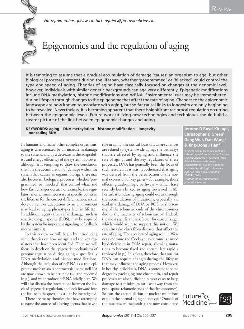
Epigenomics and the regulation of aging
In humans and many other complex organisms, aging is characterized by an increase in damage to the system, and by a decrease in the adaptabil-ity and energy efficiency of the system. However, although it is tempting to draw the conclusion that it is the accumulation of damage within the system that ‘causes’ an organism to age, there may also be certain biological processes, whether ‘pro-grammed’ or ‘hijacked’, that control what, and how fast, changes occur. For example, the regu-latory mechanisms necessary at specific points in the lifespan for the correct differentiation, sexual development or adaptation to an environment may lead to aging phenotypes later in life [1,2]. In addition, agents that cause damage, such as reactive oxygen species (ROS), may be required by the system for important signaling or feedback mechanisms [3].
In this review we will begin by introducing some theories on how we age, and the key reg-ulators that have been identified. Then we will focus in depth on the epigenetic mechanisms of genome regulation during aging – specifically DNA methylation and histone modifications. Although the inclusion of ncRNA as a true epi-genetic mechanism is controversial, some ncRNA are now known to be heritable ([4], and reviewed in [1]), and we introduce ncRNA briefly here. We will also discuss the interactions between the lev-els of epigenetic regulation, and look forward into the future to the questions still to be investigated.
There are many theories that have attempted to name the sources of altering agents that have a
role in aging, the critical locations where changes are related to system-wide aging, the pathways that are affected by aging and influence the rate of aging, and the key regulators of these processes. DNA has generally been the focus of such research as it was hypothesized that aging was derived from the perturbation of the nor-mal expression of key genes – for example, those effecting authophagic pathways – which have recently been linked to aging (reviewed in [5]). Perturbation during aging could occur through the accumulation of mutations, especially via oxidative damage of DNA by ROS, or shorten-ing of the telomeric ends of the chromosomes due to the inactivity of telomerase [6]. Indeed, the most significant risk factor for cancer is age, which would seem to support this notion. We can also take clues from diseases that affect the rate of aging. The accelerated aging seen in Wer-ner syndrome and Cockayne syndrome is caused by deficiencies in DNA repair, allowing muta-tions to become fixed and accumulate rapidly (reviewed in [7]). It is clear, therefore, that nuclear DNA can acquire changes during the lifespan that may influence the aging process. However, in healthy individuals, DNA is protected to some degree by packaging into chromatin, and repair processes are also sufficient in most cases to keep damage to a minimum (at least away from the gene-sparse telomeric ends of the chromosomes).So can the accumulation of nuclear mutations explain the normal aging phenotype? Outside of the nucleus, mitochondria are now considered
It is tempting to assume that a gradual accumulation of damage ‘causes’ an organism to age, but other biological processes present during the lifespan, whether ‘programmed’ or ‘hijacked’, could control the type and speed of aging. Theories of aging have classically focused on changes at the genomic level; however, individuals with similar genetic backgrounds can age very differently. Epigenetic modifications include DNA methylation, histone modifications and ncRNA. Environmental cues may be ‘remembered’ during lifespan through changes to the epigenome that affect the rate of aging. Changes to the epigenomic landscape are now known to associate with aging, but so far causal links to longevity are only beginning to be revealed. Nevertheless, it is becoming apparent that there is significant reciprocal regulation occurring between the epigenomic levels. Future work utilizing new technologies and techniques should build a clearer picture of the link between epigenomic changes and aging.
KEYWORDS: aging n DNA methylation n histone modification n longevity n noncoding RNA
Jerome D Boyd-Kirkup1, Christopher D Green1, Gang Wu1, Dan Wang1 & Jing-Dong J Han*1
1Chinese Academy of Sciences Key Laboratory of Computational Biology, Chinese Academy of Sciences–Max Planck Partner Institute for Computational Biology, Shanghai Institutes for Biological Sciences, 320 Yue Yang Road, Shanghai, 200031, China *Author for correspondence: [email protected]
part of
For reprint orders, please contact: [email protected]
Epigenomics (2013) 5(2)206 future science group
Review Boyd-Kirkup, Green, Wu, Wang & Han
to be important to the aging process as they are the major site of ROS production, and possess a vulnerable genome (mtDNA) that lacks chroma-tin (reviewed in [8]). DNA repair deficiencies in the mitochondria also caused accelerated aging when modeled in the mouse [9], but it is still not clear how important mtDNA mutations are to the ‘normal’ aging phenotype. In addition, the role of ROS as a mutator is being superseded by an understanding of ROS as a small signaling molecule, especially as stimulation by low levels of ROS (known as hormesis) may actually be beneficial to aging, through adaptation to stress and increased stress resistance ([10], and reviewed in [11]). This would suggest that the aging process may be more complicated than simply the accu-mulation of genetic mutations. When changes to the DNA code occur, there should be an imme-diate, localized change in gene expression. How-ever, aging is characterized by gradual changes to the whole system, implying a coordinated global process and possibly a ‘program’ for aging. Stud-ies investigating twins, populations or animal models have also shown that organisms with largely similar genetic backgrounds can age very differently (reviewed in [12]). Therefore, it would appear that, in addition to any genetic changes, environmental or other stimuli experienced dur-ing the lifespan may modify the system directly, or stimulate adaptations in the system, perturb-ing gene expression. The possibilities that this raises are attractive; a better understanding of genome–environment interactions could pro-pose lifestyle interventions or drug targets that may improve the aging process (reviewed in [13]).
DNA methylation was the first true epigenetic (from the greek epi or ‘above’ genetics) mecha-nism of variation identified in the genome [14]. Subsequently, a plethora of histone modifications has been identified (reviewed in [15]). These two epigenomic levels are known to be linked to dis-tinct gene expression and replication pheno types, and to be modified by environmental factors. It has been hypothesized that epi genetic marks at key genomic regions, such as transcription start sites, splice sites, and especially in the coding or control regions of known aging regulators, could be an important way that environmental cues are ‘remembered’ by a system, effecting the wider aging process. The question is, where are the effects most important; to what genes, pro-teins, processes, and in which cells and tissues? In the epigenomics field we choose to focus not on individual modifications to specific gene loci, but instead on an understanding of epigenetics in a whole genome context.
The most widely applicable way to increase lifespan is through dietary restriction (DR) (reviewed in [16]). DR has been shown to increase the lifespan of yeast by 25% [17], Caenorhabditis elegans by 17–25% [18], Drosophila by 100% [19] and rodents by 30–40% [20]. However, it should be noted that two studies on primates that are presently underway disagree on whether or not there is reduction in age-related mortality with DR [21,22]. Researchers have sought to tease apart the system-wide changes brought on by DR to identify the key changes that are occurring at the cellular and tissue scale, and then attempt to mimic these effects specifically. DR has been postulated to increase lifespan by decreasing ROS production (or by stimulating horme-sis), increasing autophagy and mitophagy, and stimulating expression of some key aging regu-lators. The known lifespan regulators all reside in nutrient-sensing or stress-response pathways (reviewed in [23]):
�� Foxo/Daf-16 and IGF1R/Daf-2, in the insulin/IGF signaling pathway;
�� AMPK/AAK-2, in the ROS signaling path-way;
�� mTOR/LET-363, in the autophagy and translation pathway;
�� SIRT1/2, in the genome stability pathway (histone deacetylation).
One important route for the effects of DR could be via changes to the epigenome, which in turn effect lifespan. In support of this hypo-thesis, SIRT1/2 are epigenetic modifiers, and there is much data that suggests they have some involvement in healthy aging ([24], and reviewed in [25]). However, it should be noted that recent evidence suggests that their involvement in lifes-pan regulation may not be as dramatic as was once thought [26]. On the other hand, a recent systems biology study of the DR interactome gene network has shown that genes involved in chromatin organization and modification are significantly enriched in DR-stimulated network interactions, emphasizing chromatin regulation as an important mediator for DR effects on aging [27].
Approaches to study the epigenomics of agingEpigenomics, like other ‘omics’, is benefit-ing from the accessibility of affordable high-throughput techniques such as the rapidly developing second-generation sequencing plat-forms of 454 Pyrosequencing® [28], Illumina®
www.futuremedicine.com 207future science group
Epigenomics & the regulation of aging Review
and SOLiD™ [29]. The ana lysis of raw data from these technologies requires the availability of significant but affordable computer power, and complex computational methodologies. Epigenomics utilizes these approaches to evalu-ate the genome as a whole from specific cells or tissues and in specific situations (e.g., samples of different ages). This is also the basis of so-called epigenome-wide association studies which, in a similar way to genome-wide association stud-ies, link phenotypes to the epigenome (reviewed in [30]). As biases exist for different platforms, library construction and experimental proto-cols, consideration must be taken into account when designing and analyzing next-generation sequencing data. This is particularly important for avoiding false-positive sequence changes between samples, especially for epigenomic analyses of DNA methylation [31]. Epigenomic studies must also be cautious when assuming the universality of epigenetic changes observed in a heterogenous tissue. It is possible that apparent changes to the epigenome may only represent changes in cell-type ratios within a tissue, and should be confirmed across different tissues and ideally on the level of individual cell types. In addition to human tissues and cell lines, there are several model organisms that have been used extensively in epigenomics. Plant methylation is well studied, but it would now appear that there are significant differences between the mecha-nisms in plants compared with animals, which prevent us from drawing parallels. C. elegans has been widely used for histone epigenomics but is a poor model for DNA methylation, which appears to be absent in the C. elegans genome [32]. On the other hand, insect models such as honey bees for DNA methylation, and Dro‑sophila for a variety of epigenetic modifications, are widely used, and increasingly mice and rats. There have been suggestions that epigenetic modifications may be more speciated than the DNA sequence itself, possibly even more so than histone modifications, and comparative stud-ies between humans and primates could be an interesting new approach to epigenomics [33]. The major hurdle of current epigenomic studies has been the requirement of large quantities of starting material. Many of the techniques used in the preparation of samples before sequenc-ing (see Boxes 1 & 2), such as bisulfite treatment and chromatin immunoprecipitation (ChIP), have poor yields. Third-generation sequencing technologies have great potential to streamline analyses, including single-molecule sequencers, such as the HeliScope™ system, that remove the
need for library construction and PCR amplifi-cation [34]. Research continues towards minia-turization and/or automization of techniques, allowing for the processing of smaller quantities of precious samples with the eventual goal of single-cell epigenomics.
There are now databases and projects designed to curate different layers of epigenetic data for future examination of epigenetic associations with phenotypes, such as the rate of aging for an organism. For example, MethylomeDB and NGSmethDB curate DNA methylation profiles for human and mouse tissues and cell lines [35], the Human Epigenome Project analyzes DNA methylation in each tissue, the ENCODE and modENCODE projects curate epigenomic data for human cell types and model organisms (C. elegans and D. melanogaster), respectively, and expression databases can allow for func-tional comparisons to epigenetic patterns. To interpret this data and predict which changes, or artificial perturbations, are the most important, requires us to consider the system as a whole. Therefore, many epigenomics studies choose a systems biology approach, which computation-ally integrates the data from various levels of regulation, for example, ChIP-seq and RNA-seq data, to predict the key regulators of aging.
Regulation of epigenomic marks�n DNA methylation
DNA methylation occurs at the 5́ position of a cytosine to form 5-methylcytosine (5mC), through the donation of a methionine group from S-adenosylmethionine (SAM), predomi-nantly at cytosines in a CpG context [36]. It can also occur in rarer cases at non-CpG cytosines, especially, but not exclusively, in embryonic stem cells, the function of which remains unclear [36,37]. 60–70% of all CpGs are methylated; however, CpG-rich regions of up to 1 kb (known as CpG islands or CGI) are much more likely to be present in an unmethylated state [36]. Outside of CGIs the genome is depleted of CpGs, and studies have therefore mainly focused on the functional implications of the methylation state of CGIs, and in particular, CGIs at transcriptional start sites (TSSs) and promoters. The role of methylation in the gene body, and other regulatory regions such as enhancers and insulators, is only now beginning to be appreciated.
Although methylation can exist as a relatively stable and inheritable modification, it is widely accepted that methylation and demethylation is a dynamic process that is ongoing through-out an organism’s lifespan. DNA methylation
Epigenomics (2013) 5(2)208 future science group
Review Boyd-Kirkup, Green, Wu, Wang & Han
is catalyzed by the DNA methyltransferase enzymes (DNMTs). In humans there are three DNMTs: DNMT1, DNMT3a and DNMT3b. DNMT1 is generally thought of as the ‘main-tenance’ methylase, due to its preference for hemi-methylated CpG sequences, and preserves methylation during DNA replication [38]. In vitro studies suggested DNMT3a and 3b had no such preference, and thus they were considered de novo methylases [39]. However, it is likely that this is an oversimplification. Accumulating evidence dem-onstrates that in specialized circumstances, such as single copy sequences and certain repetitive elements [40], DNMT1 can also de novo methyl-ate and DNMT3a and DNMT3b have a role in maintenance methylation [41,42].
Demethylation of 5mC is not fully under-stood. The most important finding in recent years was the presence of hydroxymethylated cytosines (5hmC) in the genome, albeit present at much lower levels than 5mC [43–45]. The Tet (ten eleven translocases) family of enzymes cata-lyze the conversion of 5mC to 5hmC [43]. They may also convert 5hmC further to 5-formyl-cytosine and 5-carboxylcytosine, which can then be further reconstituted to unmethylated
cytosine through the base excision repair actions of TDG [46,47]. It should be noted that the dis-tribution of 5hmC does not necessarily relate to the distribution of 5mC, which suggests that 5hmC may have other unknown functional roles, a hypothesis now being explored [44,48].
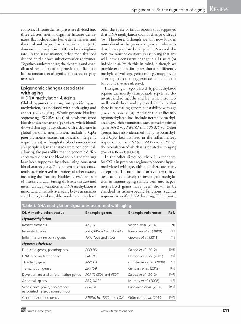
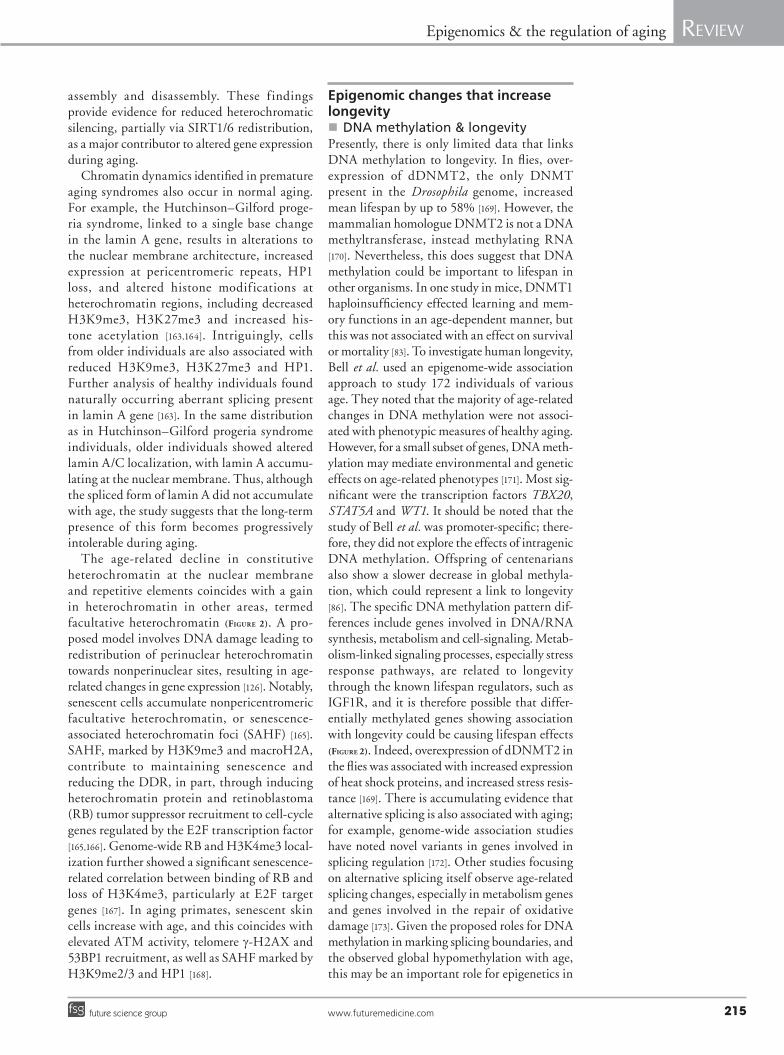
�n Epigenomic patterns of DNA methylationDistinct patterns of DNA methylation exist within the human genome (Figure 1). Changes to these patterns have been proposed as one of the major mechanisms for differential gene expres-sion between tissues [49], and are critical for correct differentiation of stem cells [50]. Aging-associated pattern changes may therefore have a significant role in the aging process.
Methylation of CGIs in promoter regions (CGI-rich promoters) is inversely associated with transcription (Figure 1). Methylation can inhibit transcription factor binding or recruit methyl-CpG-binding proteins that help block transcrip-tion [51,52]. Conversely, hydroxymethylation at promoter regions is associated with a release of methyl-binding proteins and an increase in tran-scription [48,53]. Methylation in the gene body is
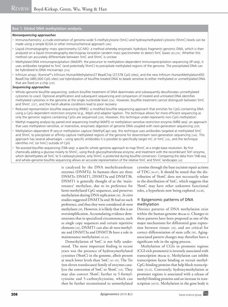
Box 1. Global DNA methylation ana lysis.
Nonsequencing approaches
� Immunochemistry: a crude estimation of genome-wide 5-methylcytosine (5mC) and hydroxymethylated cytosine (5hmC) levels can be made using a simple ELISA or other immunochemical approach [106]
� Liquid chromatography–mass spectrometry (LC-MS): a method whereby enzymatic hydrolysis fragments genomic DNA, which is then analyzed on a liquid chromatography electrospray ionization tandem mass spectrometer to detect 5mC bases [83,233]. Whether this method can accurately differentiate between 5mC and 5hmC is unclear
� Methylated DNA immunoprecipitation (MeDIP): the precursor to methylation-dependent immunoprecipitation sequencing (IP-seq), it uses antibodies targeted to 5mC (and potentially 5hmC) to precipitate methylated regions of the genome. The precipitated DNA can be hybridized to DNA microarrays [234]
� Infinium arrays: Illumina®’s Infinium HumanMethylation27 BeadChip (27,578 CpG sites), and the new Infinium HumanMethylation450 BeadChip (485,000 CpG sites) use hybridization of bisulfite-treated DNA to beads sensitive to either methylated or unmethylated DNA that are fixed on a chip [235]
Sequencing approaches
� Whole-genome bisulfite sequencing: sodium bisulfite treatment of DNA deaminates and subsequently desulfonates unmethylated cytosines to uracil. Optional amplification and subsequent sequencing and comparison of treated and untreated DNA identifies methylated cytosines in the genome at the single nucleotide level [236]. However, bisulfite treatment cannot distinguish between 5mC and 5hmC [237], and the harsh alkaline conditions lead to poor recovery
� Reduced representation bisulfite sequencing (RRBS): a modified bisulfite sequencing approach that enriches for CpG-containing DNA using a CpG-dependent restriction enzyme (e.g., MspI) and adaptor ligation. The technique allows for more efficient sequencing as only the genomic regions containing CpGs are sequenced [238]. However, this technique under-represents non-CpG methylation
� Methyl-mapping ana lysis by paired end sequencing (methyl-MAPS) or methylation-sensitive restriction enzyme (MRE-seq): an approach that uses methylation-sensitive, or insensitive, enzymatic digestion of genome DNA coupled with next-generation sequencing [239]
� Methylation-dependent IP-seq or methylation capture (MethylCap)-seq: this technique uses antibodies targeted at methylated 5mC and 5hmC to precipitate or affinity capture methylated regions of the genome for downstream next-generation sequencing [240]. This approach has several advantages – using specific antibodies it is possible to specifically target mC or hmC [44], and the technique identifies mC (or hmC) outside of CpG
� Tet-assisted bisulfite-sequencing (TAB-seq): a specific whole-genome approach to map 5hmC at a single-base resolution. By first chemically adding a glucose moiety to 5hmC, using the b-glucosyltransferase enzyme, and treatment with the recombinant Tet1 enzyme, which demethylates all 5mC to 5-carboxylcytosine, only 5hmC is protected during bisulfite conversion. Comparing the data from TAB-seq and whole-genome bisulfite sequencing allows an accurate representation of the relative 5mC and 5hmC landscapes [45]
www.futuremedicine.com 209future science group
Epigenomics & the regulation of aging Review
not associated with transcriptional repression, but is a feature of actively transcribed genes [54,55] (Figure 1). Methylation may act to prevent initiation outside of the TSS, and regulate the use of alternative promoters [56]. Whole-genome studies have also shown that 5mC and 5hmC levels in gene bodies are not uniform; exons are more highly methylated (and hydroxymethyl-ated) than introns, and there is a clear transi-tion at exon–intron boundaries suggesting 5mC and 5hmC may help define splicing boundaries (Figure 1) [44,57,58]. Specifically, methylation spikes at 5́ splice sites, and dips at 3´ splice sites [58,59]. This has recently been supported by observations in the honeybee, where highly methylated exons were more likely to be included in transcipts than poorly methylated exons [60].
Methylation of repeat elements such as Alu and L1, endogenous retrotransposons and proto-genes is necessary to prevent aberrant transcrip-tion, and methylation at centromeres is important for chromosomal stability (Figure 1) [61]. Enhancers are characterized by the fact that they are CpG sparse [36,55]. Although the function of methyla-tion at enhancers is unclear, most enhancers are neither 100% methylated nor unmethylated, which suggests extensive dynamic regulation [62]. Methylation could also decrease enhancer activity, or prevent promoter–enhancer interac-tions by modifying insulator regions (e.g., IGF2 enhancer, an aging regulator) [63,64].
DNMTs themselves have very little specificity for target sequences. Although trans-acting cofactors
of the DNMTs do exist, such as DNMT3L, a noncatalytic cofactor of DNMT3a/3b [65], and Uhrf1 (or Np95), which interacts with DNMTs [66,67], the distinct patterns of DNA methylation across the genomic landscape must be established and maintained by additional mechanisms. Sev-eral groups have reported that they can recreate or ‘seed’ methylation patterns in the genome using artificially inserted sequences, such as promoter-derived sequences [68] or repeat elements proximal to the region of interest [69,70]. In addition, there is a significant overlap between transcription fac-tor binding sites in loci with similar methylation levels [71]. This implies that methylation patterns can be regulated in cis.
�n Histone modificationChromatin is composed of nucleosome units containing an octamer of histone proteins, with two copies of each histone H2A, H2B, H3 and H4, wrapped by DNA. As a whole, chroma-tin can be divided into regions of euchroma-tin; more open, accessible and transcriptionally active, and heterochromatin, which is stably closed and repressed. The ability to maintain chromatin stability and regulate gene expres-sion requires post-translational modifications of histone tails, including acetylation, meth-ylation, phosphorylation and ubiquitylation, as well as novel modifications such as crotonylation [72]. Studies using ChIP for single genes and more in-depth analyses, combining ChIP with microarray (ChIP-on-chip) or deep sequencing
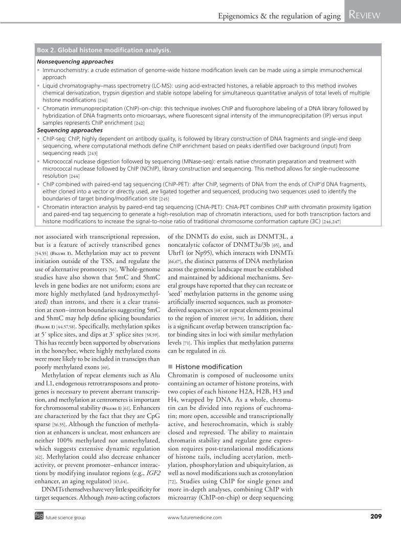
Box 2. Global histone modification ana lysis.
Nonsequencing approaches
� Immunochemistry: a crude estimation of genome-wide histone modification levels can be made using a simple immunochemical approach
� Liquid chromatography–mass spectrometry (LC-MS): using acid-extracted histones, a reliable approach to this method involves chemical derivatization, trypsin digestion and stable isotope labeling for simultaneous quantitative ana lysis of total levels of multiple histone modifications [241]
� Chromatin immunoprecipitation (ChIP)-on-chip: this technique involves ChIP and fluorophore labeling of a DNA library followed by hybridization of DNA fragments onto microarrays, where fluorescent signal intensity of the immunoprecipitation (IP) versus input samples represents ChIP enrichment [242]
Sequencing approaches
� ChIP-seq: ChIP, highly dependent on antibody quality, is followed by library construction of DNA fragments and single-end deep sequencing, where computational methods define ChIP enrichment based on peaks identified over background (input) from sequencing reads [243]
� Micrococcal nuclease digestion followed by sequencing (MNase-seq): entails native chromatin preparation and treatment with micrococcal nuclease followed by ChIP (NChIP), library construction and sequencing. This method allows for single-nucleosome resolution [244]
� ChIP combined with paired-end tag sequencing (ChIP-PET): after ChIP, segments of DNA from the ends of ChIP’d DNA fragments, either cloned into a vector or directly used, are ligated together and sequenced, producing two sequences used to identify the boundaries of target binding/modification site [245]
� Chromatin interaction ana lysis by paired-end tag sequencing (ChIA-PET): ChIA-PET combines ChIP with chromatin proximity ligation and paired-end tag sequencing to generate a high-resolution map of chromatin interactions, used for both transcription factors and histone modifications to increase the signal-to-noise ratio of traditional chromosome conformation capture (3C) [246,247]

Epigenomics (2013) 5(2)210 future science group
Review Boyd-Kirkup, Green, Wu, Wang & Han
(ChIP-seq) and other modified approaches (Box 2), has revealed that different modifications mark different transcriptional states. Histone acetylation is generally associated with an open chromatin structure, marking sites of active transcription at the gene locus and at distal enhancer regions bound by active transcription factors [73,74]. Methylation marks many differ-ent states, with active gene expression exhibiting trimethylated histone 3 on lysine 4 (H3K4me3) at TSSs, active enhancers and TSSs displaying H3K4me2, and repressed genes associated with H3K27me3 [75,76]. A recent ana lysis comparing ENCODE ChIP-seq data from seven cell lines illustrates the generality of some histone marks, such as H3K9ac and H3K4me3, for detecting active genes, while H3K79me2 and H3K36me3 are more useful for determining the level of gene expression [77,78]. Different regions of chroma-tin are also characterized by different histone marks. H3K79me is enriched in euchromatin, whereas H3K9me3 and H4K20me3 largely occur in heterochromatin [79]. In addition, other marks associate with different events, such as phosphorylation of H2AX (g-H2AX) at sites of DNA damage. Mechanistically, histone acetyla-tion often functions by creating access to DNA by neutralizing positive lysine charges, while
histone methylation and sometimes acetylation and other marks function by providing docking sites for other proteins. For example, bromodo-main-containing proteins, such as the SWI/SNF chromatin remodelers, bind acetylated lysines, and HP1 binds to methylated H3K9 in regions of heterochromatin (reviewed in [80]).
Importantly, histone modif ications are dynamic in that they are actively added and removed by histone-modifying enzymes in a site-specific manner, which is essential for coordi-nated transcriptional control. Illustrating speci-ficity of histone acetylation, the histone acetyl-transferases GCN5/PCAF and CBP/P300 medi-ate, respectively, H3K9ac and H3K18/K27ac [81]. Histone lysine deacetylases (HDACs) are grouped into four classes based on homology to yeast genes, such as the class III NAD+-dependent sirtuin family that have important roles in metabolism and aging. Thus far, meth-ylation is known to occur through arginine- and lysine-specific methyltransferases. For example, histone H3K4 trimethylation at transcriptionally active genes is mediated by enzymes containing the general lysine methyltransferase SET domain, which reside in one of three complexes: complex proteins associated with SET1 (COMPASS); the trithorax complex; or the trithorax-related
Active gene
Repressed gene
Relative 5mC level
CGI rich
Gene body (exons)
Enhancer/ CGI-richpromoter
High
Low
5´
5´
3´
3´
3´
5´
Genomic methylation landscape
Low
High
Relative 5mC level
elementsRepeat
Insulator
Figure 1. Model of the genomic methylation landscape at enhancers/insulators, repeat elements, CGI-rich promoters and gene exons. Enhancers/insulators are neither completely unmethylated nor methylated, but active enhancers may be less methylated. Repeat elements are generally methylated. CGI-rich promoters are methylated in repressed genes and unmethylated in active genes. Apart from the 5´ exon, actively transcribed exons will be methylated, and methylation will spike near 5´ splice sites and dip near 3´ splice sites. 5mC: 5-methylcytosine; CGI: CpG islands.
www.futuremedicine.com 211future science group
Epigenomics & the regulation of aging Review
complex. Histone demethylases are divided into three classes: methyl-arginine histone deimi-nases; flavin- dependent lysine demethylases; and the third and largest class that contains a JmjC domain requiring iron Fe(II) and a-ketogluta-rate. In the same manner, other modifications depend on their own subset of various enzymes. Together, understanding the dynamic and coor-dinated regulation of epigenetic modifications has become an area of significant interest in aging research.
Epigenomic changes associated with aging�n DNA methylation & aging
Global hypomethylation, but specific hyper-methylation, is associated with both aging and cancer (TaBle 1) [82,83]. Whole-genome bisulfite sequencing (WGBS; Box 1) of newborns (cord blood) and centenarians (peripheral whole blood) showed that age is associated with a decrease in global genomic methylation, including CpG poor promoters, exonic, intronic and intergenic sequences [84]. Although the blood sources (cord and peripheral) in that study were not identical, allowing the possibility that epigenomic differ-ences were due to the blood source, the findings have been supported by others using consistent blood sources [85,86]. This pattern has also consis-tently been observed in a variety of other tissues, including the heart and bladder [87–89]. The issue of intraindividual (using different tissues) and interindividual variation in DNA methylation is important, as naïvely averaging between samples could abrogate observable trends, and may have
been the cause of initial reports that suggested that DNA methylation did not change with age [90]. Therefore, although we will now look in more detail at the genes and genomic elements that show age-related changes in DNA methyla-tion, we must be cautious in assuming that any will show a consistent change in all tissues (or individuals). With this in mind, although we provide examples for genes that are differently methylated with age, gene ontology may provide a better picture of the types of cellular and tissue functions that are affected.
Intriguingly, age-related hypomethylated regions are mostly transposable repetitive ele-ments, including Alu and L1, which are nor-mally methylated and repressed, implying that there is increasing genomic instability with age (TaBle 1 & Figure 2) [91]. Additional significantly hypo methylated loci include normally methyl-ated CpG-rich promoters, such as the imprinted genes IGF2 [92], PWCR1 and TRPM5 [85]. Other groups have also identified many hypomethyl-ated CpG loci involved in the inflammatory response, such as TNF [93], iNOS and TLR2 [85], the modulation of which is associated with aging (TaBle 1 & Figure 2) [86,94,95].
In the other direction, there is a tendency for CGIs in promoter regions to become hyper-methylated with age, although there are many exceptions. Illumina bead arrays (Box 1) have been used extensively to investigate methyla-tion in human aging sample sets, and hyper-methylated genes have been shown to be enriched in tissue- specific functions, such as sequence-specific DNA binding, TF activity,
Table 1. DNA methylation signatures associated with aging.
DNA methylation status Example genes Example reference Ref.
Hypomethylation
Repeat elements Alu, L1 Wilson et al. (2007) [91]
Imprinted genes IGF2, PWCR1 and TRPM5 Bjornsson et al. (2008) [85]
Inflammatory response genes TNF, iNOS and TLR2 Gowers et al. (2011) [95]
Hypermethylation
Duplicate genes, pseudogenes ECEL1P2 Salpea et al. (2012) [105]
DNA-binding factor genes GAS2L3 Hernandez et al. (2011) [98]
TF activity genes MYOD1 Christensen et al. (2009) [97]
Transcription genes ZNF169 Gentilini et al. (2012) [86]
Development and differentiation genes FGF17, FZD1 and FZD7 Salpea et al. (2012) [105]
Apoptosis genes FAS, XAF1 Murphy et al. (2008) [99]
Senescence genes, senescence-associated heterochromatin foci
ECRG4 Funayama et al. (2007) [100]
Cancer-associated genes P16INK4a, TET2 and LOX Grönniger et al. (2010) [103]
Epigenomics (2013) 5(2)212 future science group
Review Boyd-Kirkup, Green, Wu, Wang & Han
transcription regulation and genes involved in development and differentiation (TaBle 1) [86,96–
98]. Some groups have also found enrichment in DNA hypermethylation in genes involved in apoptosis, and domains associated with senes-cence (TaBle 1) [99–101]. Bocklandt et al. used this approach in a twins study that sought to derive a regression model that could accurately predict age. From the 88 age-related loci that they identified in saliva, including ten identi-fied previously in whole blood, they found that the methylation state of the promoters of just three genes (EDARDD, TOM1L1 and NPTX2) could predict age with an accuracy of 5.2 years [102]. Overall, it is becoming increasingly appar-ent that there are similarities between those genes that are hypermethylated in aging and hypermethylated in cancer, including P16INK4a, TET2, LOX and NPTX2 [102–104].
There have been very few studies on the asso-ciation of methylation outside of CGIs with age. Salpea et al. used methylation-dependent-immuno precipitation-sequencing (Box 1) to iden-tify regions of the genome that show differential methylation between newborns and adults [105]. They observed that the proto-cadherin gene cluster shows extensive age-related increases in
DNA methylation. These changes were in the gene body and not the regulatory regions, and could therefore be affecting splicing.
Finally, as a recently identified component of the epigenomic landscape, studies on 5hmC pattern change with age are also lacking. How-ever, an age-related increase in 5hmC content has been observed in the mouse hippocampus, independent of changes in the expression of TET enzymes, and without a corresponding decrease in 5mC [106]. Enrichment is at discrete locations, primarily intragenic, with the highest levels on the exons [107]. This underlines the hypothesis that 5hmC may be regulated as an epigenetic mark in its own right, not only as an intermediate of 5mC demethylation.
�n Causes of age-related DNA methylationInitially, reports suggested DNMT1 and DNMT3a decreased with age in some somatic cells, but DNMT3b increased [108,109]. Oliveira et al. noted a decreased expression of DNMT3a2 in mice during aging, which was associated with decreased cognitive function, and rescue of DNMT3a2 levels restored these functions [110]. Interestingly, neuronal activity itself has been
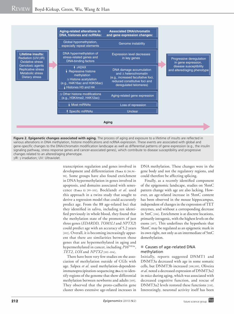
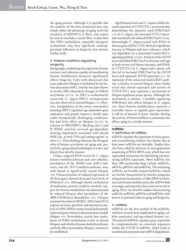
Lifetime insults:Radiation (UV/γIR)
Oxidative stressGenotoxic agentsReplicative stressMetabolic stressDietary stress
Aging-related alterations in DNA, histones and ncRNAs:
Global hypomethylation,especially repeat elements
DNA hypermethylation ofstress-related genes and
DNA-binding factors
γH2AXRepressive histone
methylation∆ Histone acetylation
(e.g., H4K16ac and H3K56ac)
∆ Other histone modifications(e.g., H3K4me2, H4K12ac)
Histones H3 and H4
Most miRNAs
Specific miRNAs
Associated DNA/chromatinand gene expression changes:
Genome instability
Expression level decreasesin key genes
DNA damage accumulationand ∆ heterochromatin
(e.g., increased facultative foci,reduced constitutive foci and
deregulated telomeres)
Aging-related gene expression
Loss of repression
Unclear
Progressive deregulationin gene expression, disease susceptibility
and altered/aging phenotype
Aging
Figure 2. Epigenetic changes associated with aging. The process of aging and exposure to a lifetime of insults are reflected in various alterations in DNA methylation, histone modifications and ncRNA expression. These events are associated with global and gene-specific changes to the DNA/chromatin modification landscape as well as differential patterns of gene expression (e.g., the insulin signaling pathway, stress response genes and cancer-associated genes), which contribute to disease susceptibility and progressive changes related to an altered/aging phenotype. gIR: g irradiation; UV: Ultraviolet.
www.futuremedicine.com 213future science group
Epigenomics & the regulation of aging Review
linked to modification of the DNA methyla-tion landscape, both dynamic demethylation and de novo methylation, in some cases on the scale of hours [111]. However, others suggest that there is commonly no significant differences in DNMTs in mice that could explain the global decrease in DNA methylation, implying other influences are at work [112].
Genome-wide DNA methylation stud-ies have revealed that the greatest differences occur between populations, and there is little overlap in genetic associations of methylation patterns. This implies that there are complex gene–environment interactions (Figure 2) [113]. Data from twin studies appears to support this; DNA methylation patterns in twins are concor-dant in early life, but gradually diverge during lifespan, implying a large environmental influ-ence on genome methylation [114–116]. There is a clear link between metabolism and DNA meth-ylation. SAM, which provides the methyl group for DNA methylation by DNMTs, produces S-adenosyl homocysteine during the reaction. Diet is known to modulate SAM levels, although the rate of S-adenosyl homocysteine clearance, which inhibits DNMTs, is also important [117,118]. Regulating dietary folate and other methyl donors can also alter the level of DNA methylation [119]. Although the molecular mech-anisms responsible are opaque, it has also been shown that moderate exercise is associated with a delay in the aging-related hypomethylation of genes involved in the inflammatory response, such as ASC [120]. However, are these changes to methylation important to aging, or simply a phenotype? If we return to dietary restriction, DR attenuates the DNMT3a changes seen in the mouse brain and increases DNMT1 expression [121,122]. DR is also associated with an increase in methylation of proto-oncogenes, such as Ras in rats, and P16INK4a in mammalian cell cul-ture [122,123]. DR has also been observed to cause DNA methylation changes in genes linked to aging including TNF‑a, and TFs such as WT1 (Figure 2) [124,125].
�n Age-related histone modification dynamics: DNA damage responseAging is coupled with a gradual deregulation of chromatin stability and gene expression. Pro-posed theories for why this deregulation occurs with age include DNA damage (e.g., via repli-cation errors, radiation and genotoxic agents), telomere maintenance and redistribution of het-erochromatin foci (Figure 2) [126]. Interestingly, histone modifications have an integral role in
responding to these events, maintaining chroma-tin stability, and as a consequence of prolonged chronic stress, contributing to functional decline in later life. Although genome-wide histone modification analyses during aging are scarce, a recent study identified Rhesus macaque brain to exhibit an age-related global increase in dimeth-ylated H3K4me2, associated with transcription-ally active sites, at both promoter and enhancer regions [127]. Elevated H3K4me2-marked genes enriched for response to DNA damage stimuli and genes highly associated with stress, reflecting the close connection between histone modifica-tion and stress response during aging. Here, we describe histone modification changes associated with aging in the context of mechanisms pro-posed for deregulation of chromatin and gene expression (TaBle 2).
Unrepaired DNA damage can cause various adverse effects including accelerated aging [128]. The DNA damage response (DDR) utilizes mul-tiple pathways to remove damaged nucleotides and repair single-stranded breaks and double-stranded breaks. After DNA damage is sensed by various DDR complexes, transducer kinases ATM, DNA-dependent protein kinase catalytic subunit (DNA-PKcs) and ATR are subsequently brought in to initiate the DDR [129–131]. This is followed by recruitment of mediator proteins, such as MDC1, 53BP1, BRCA1 (early onset), TopBP1 and Claspin, activation of effector kinases, and propagation of the DDR to the rest of the cell.
Coordinated recruitment of DDR factors requires interactions with a number of his-tone modifications. Phosphorylation of H2AX (g-H2AX) by transducer kinases ATM and DNA-PKcs [132,133] is bound by MDC1, which aids in recruiting other factors to enhance the DDR [134]. On a megabase scale, g-H2AX spreads outward from the damage site [135], and longer-lasting g-H2AX foci typically exist outside of heterochromatin [136]. Interestingly, g-H2AX foci accumulate with age and in patients with accelerated aging [137,138]. Another DNA damage-induced mark is ubiquitinylation (Ub), accumulating at foci particularly on H2A and H2B. As Ub-H2B is associated with tran-scription elongation, it is proposed that Ub-H2B promotes chromatin relaxation and mediator binding at the damaged site, whereas Ub-H2A creates a repressed environment that, along with g-H2AX, spreads outward from the site [139–141]. The importance of histone modifications to the DDR is emphasized in studies of DDR mediator recruitment. Mediator protein 53BP1 contains a
Epigenomics (2013) 5(2)214 future science group
Review Boyd-Kirkup, Green, Wu, Wang & Han
tudor domain with which the yeast orthologs bind H4K20me2 (Crb2) and H3K79me3 (Rad9), and human 53BP1 binds both H4K20me1/2 and H3K79me3 [142–146]. In fibroblasts recov-ered from individuals, old cells displayed higher levels of H4K20me2 and H3K79me2 compared with young cells [147]. Studies have shown that the H4 methyl transferases PR-Set7/Set8 and MMSET are involved in DNA damage-induced H4K20me1/2, which is then bound by 53BP1 [146,148]. While total H3K79 methylation did not change with DNA damage, the placement of this modification by the only identified H3K79 methyltransferase DOT1L, conserved across species, allows proper 53BP1 binding in human cells and activation of the DDR in yeast [142]. Besides methylation, recruitment of MDC1, 53BP1 and BRCA1 was demonstrated to require MOF-mediated H4K16ac, as well as general H4 acetylation via TIP60, albeit including K16ac, around sites of DNA damage [149–151].
�n Age-related histone modification dynamics: heterochromatin alterationsFor over 20 years, aging has been known to be associated with altered heterochromatin abun-dance [152]. Accumulating evidence links the DDR with maintenance of constitutive hetero-chromatin and the aging process (TaBle 2 & Figure 2). Constitutive heterochromatin regions reside in repetitive DNA elements such as centromeres, telomeres, rDNA and transposable elements. A prime example of the DDR/heterochromatin link is the activity of the deacetylase SIRT1, which acts on acetylated H1K26, H3K9 and H4K16 [153]. Upon oxidative stress, repression of pericentromere heterochromatic repetitive DNA
transcripts by SIRT1 and the yeast homolog SIR2 is lost [154]. Genome-wide ana lysis fur-ther showed oxidative stress caused a dramatic SIRT1 redistribution away from repetitive DNA elements, and other diverse genes, to a more ran-dom localization and increased H1K26ac at sites previously repressed by SIRT1 [154]. Similar to yeast SIR protein relocation from telomeres [155], SIRT1 is recruited to sites of DNA damage and required for the DDR [154]. Interestingly, genes repressed by SIRT1 prior to redistribution are also deregulated in aged mouse brain, demon-strating a link between the age-related DDR and altered gene expression [154]. In addition, SIRT1 affects H3K9me3 by activating the H3K9 meth-yltransferase SUV39H1 by direct deacetylation [156]. Another sirtuin member, SIRT6, was also shown to be required for the normal DDR, and its deficiency reduces lifespan and causes premature aging [157]. SIRT6, which deacety-lates H3K9 and H3K56, locates to telomeres and regulates their stability, in part, through modulating telomere association with WRN, which is mutated in the premature aging dis-order Werner syndrome [158,159]. Genome-wide SIRT6 promoter binding ana lysis revealed a close association with NFkB-regulated genes, particularly those related to cell senescence and aging [160]. In aging yeast and late-passage cells, reduced telomere silencing is associated with decreased H3K56ac and increased H4K16ac, a SIRT1 substrate that opposes higher order chromatin structure by preventing interactions between neighboring nucleosomes [147,161,162]. Acetylated H3K56 resides in the globular core at the point where DNA interacts with the histone, and modulating H3K56ac effects nucleosome
Table 2. Histone modification signatures associated with aging.
Modification Change Location/function Example reference(s) Ref.
gH2AX Up DNA damage foci/DNA damage response signaling
Sedelnikova et al. (2004) [137]
H3K4me2 Up Enhancer and promoter/transcription activation
Han et al. (2012) [127]
H3K9me3 Up/down Heterochromatin maintenance Scaffidi et al. (2006) [163]
H3K27me3 Down Heterochromatin and gene regulation (e.g., IGF1R)
Jin et al. (2011); Maures et al. (2011) [179,180]
H3K56ac Down Telomere and DNA damage O’Sullivan et al. (2010); Dang et al. (2009) [147,162]
H4K12ac Loss of induction Memory impairment Peleg et al. (2010) [177]
H4K16ac Up Heterochromatin, telomere and DNA damage
O’Sullivan et al. (2010); Dang et al. (2009) [147,162]
H4K20me2 Up Heterochromatin and DNA damage O’Sullivan et al. (2010) [147]
H4K20me3 Up/down Heterochromatin maintenance O’Sullivan et al. (2010) [147]
www.futuremedicine.com 215future science group
Epigenomics & the regulation of aging Review
assembly and disassembly. These f indings provide evidence for reduced heterochromatic silencing, partially via SIRT1/6 redistribution, as a major contributor to altered gene expression during aging.
Chromatin dynamics identified in premature aging syndromes also occur in normal aging. For example, the Hutchinson–Gilford proge-ria syndrome, linked to a single base change in the lamin A gene, results in alterations to the nuclear membrane architecture, increased expression at pericentromeric repeats, HP1 loss, and altered histone modif ications at hetero chromatin regions, including decreased H3K9me3, H3K27me3 and increased his-tone acetylation [163,164]. Intriguingly, cells from older individuals are also associated with reduced H3K9me3, H3K27me3 and HP1. Further ana lysis of healthy individuals found naturally occurring aberrant splicing present in lamin A gene [163]. In the same distribution as in Hutchinson–Gilford progeria syndrome individuals, older individuals showed altered lamin A/C localization, with lamin A accumu-lating at the nuclear membrane. Thus, although the spliced form of lamin A did not accumulate with age, the study suggests that the long-term presence of this form becomes progressively intolerable during aging.
The age-related decline in constitutive hetero chromatin at the nuclear membrane and repetitive elements coincides with a gain in hetero chromatin in other areas, termed facultative heterochromatin (Figure 2). A pro-posed model involves DNA damage leading to redistribution of perinuclear heterochromatin towards nonperinuclear sites, resulting in age-related changes in gene expression [126]. Notably, senescent cells accumulate nonpericentromeric facultative heterochromatin, or senescence-associated heterochromatin foci (SAHF) [165]. SAHF, marked by H3K9me3 and macroH2A, contribute to maintaining senescence and reducing the DDR, in part, through inducing hetero chromatin protein and retinoblastoma (RB) tumor suppressor recruitment to cell-cycle genes regulated by the E2F transcription factor [165,166]. Genome-wide RB and H3K4me3 local-ization further showed a significant senescence-related correlation between binding of RB and loss of H3K4me3, particularly at E2F target genes [167]. In aging primates, senescent skin cells increase with age, and this coincides with elevated ATM activity, telomere g-H2AX and 53BP1 recruitment, as well as SAHF marked by H3K9me2/3 and HP1 [168].
Epigenomic changes that increase longevity�n DNA methylation & longevity
Presently, there is only limited data that links DNA methylation to longevity. In flies, over-expression of dDNMT2, the only DNMT present in the Drosophila genome, increased mean lifespan by up to 58% [169]. However, the mammalian homologue DNMT2 is not a DNA methyltransferase, instead methylating RNA [170]. Nevertheless, this does suggest that DNA methylation could be important to lifespan in other organisms. In one study in mice, DNMT1 haploinsufficiency effected learning and mem-ory functions in an age-dependent manner, but this was not associated with an effect on survival or mortality [83]. To investigate human longevity, Bell et al. used an epigenome-wide association approach to study 172 individuals of various age. They noted that the majority of age-related changes in DNA methylation were not associ-ated with phenotypic measures of healthy aging. However, for a small subset of genes, DNA meth-ylation may mediate environmental and genetic effects on age-related phenotypes [171]. Most sig-nificant were the transcription factors TBX20, STAT5A and WT1. It should be noted that the study of Bell et al. was promoter- specific; there-fore, they did not explore the effects of intragenic DNA methylation. Offspring of centenarians also show a slower decrease in global methyla-tion, which could represent a link to longevity [86]. The specific DNA methylation pattern dif-ferences include genes involved in DNA/RNA synthesis, metabolism and cell-signaling. Metab-olism-linked signaling processes, especially stress response pathways, are related to longevity through the known lifespan regulators, such as IGF1R, and it is therefore possible that differ-entially methylated genes showing association with longevity could be causing lifespan effects (Figure 2). Indeed, overexpression of dDNMT2 in the flies was associated with increased expression of heat shock proteins, and increased stress resis-tance [169]. There is accumulating evidence that alternative splicing is also associated with aging; for example, genome-wide association studies have noted novel variants in genes involved in splicing regulation [172]. Other studies focusing on alternative splicing itself observe age-related splicing changes, especially in metabolism genes and genes involved in the repair of oxidative damage [173]. Given the proposed roles for DNA methylation in marking splicing boundaries, and the observed global hypomethylation with age, this may be an important role for epigenetics in
Epigenomics (2013) 5(2)216 future science group
Review Boyd-Kirkup, Green, Wu, Wang & Han
the aging process. Although it is possible that the majority of the data mentioned here may simply reflect the phenotype of aging (with the exception of dDNMT2 in flies), and cannot be used to conclude a causal effect, it does hint that DNA methylation, especially intragenic methylation, may have significant underap-preciated influences on longevity that warrant further study.
�n Histone modifiers regulating longevityIntriguingly, modulating the expression of some enzymes and cofactors capable of coordinating histone modification dynamics significantly affects longevity. Early work discovered that yeast replicative lifespan is modulated by the his-tone deacetylase SIR2, and this was later shown to involve SIR2-dependent changes in H4K16 acetylation [162,174]. As SIR2 is evolutionarily conserved, C. elegans SIR2.1 overexpression was also observed to extend lifespan [175]. How-ever, manipulation of the mouse mammalian homolog SIRT1 regulated age-dependent gene expression and greatly improves health span under metabolically challenging conditions, but had little effect on lifespan [154,176]. In contrast to SIR2/SIRT1, blocking class I and II HDAC activities reversed age-dependent learning impairment associated with altered H4K12ac, at both TSSs and coding regions, in mice [177]. These findings illustrate the divergent roles of histone acetylation on aging and, par-ticularly, aging-related pathologies in a site- and deacetylase-specific manner.
Using a targeted RNAi screen for C. elegans histone methyltransferases and core subunits, knockdown of the WDR‑5 and ASH‑2 sub-units, and the SET‑2 methyltransferase, were each found to significantly extend lifespan [178]. Characterization of reduced expression of all three genes showed decreased total levels of H3K4me3 [178]. Although altered methylation of nonhistone proteins could be involved, sup-port for histone methylation was demonstrated by reduced lifespan after knockdown of the RBR‑2 H3K4me3 demethylase [178]. Lifespan extension by reduced WDR‑5, ASH‑2 and SET‑2 requires an intact germline, and microarray ana-lysis of ASH‑2 RNAi worms found deferentially expressed genes related to determination of adult lifespan [178]. Nevertheless, exactly how modu-lation of H3K4 methylation results in altered lifespan, and whether histone methyltransferases similarly affect mammalian lifespan, remains to be established.
Aged human brain and C. elegans exhibit ele-vated expression of UTX/UTX‑1, an enzyme that demethylates the repressive mark H3K27me3 [179]. In C. elegans, this increased UTX‑1 expres-sion correlated with reduced H3K27me3 [179,180]. Interestingly, C. elegans treated with RNAi or heterozygous null for UTX‑1 showed a significant increase in lifespan and stress tolerance, which was dependent on a functional insulin/IGF-1 signaling pathway [179,180]. Analysis of the IGF1R locus identified H3K27me3 to decrease with age in both worms and rhesus macaque, and RNAi of UTX/UTX‑1 in C. elegans and a mouse cell line led to increased H3K27me3 at the IGF1R locus and repressed IGF1R expression [179]. As repression of the conserved insulin/IGF1 path-way is known to extend lifespan, these results reveal that altered expression and activity of UTX/UTX-1 may represent a mechanism of lifespan regulation across species. Although the mechanism is unclear, a modifier regulating H3K36me3 also affects lifespan in C. elegans [180]. Since histone modifications represent a reversible means to regulate gene expression, it will be of interest to know whether altering the activity of these modifiers in mammals also affects aging in a similar manner.
ncRNAs�n Definition of ncRNAs
ncRNAs regulate the expression of many genes, and accumulating evidence has shown that at least some ncRNAs are heritable. Studies have also been aided by advances in next-generation sequencing of RNA (RNA-seq), which has now superseded microarrays for identifying and mea-suring ncRNA expression. Short ncRNAs less than 200 nucleotides long include miRNAs, siRNAs, piRNAs and snoRNAs. The remaining ncRNAs are broadly termed lncRNAs, which are further characterized by distinct properties and genome localization. Unlike DNA methyla-tion and histone modifications, the ncRNA field is younger, and much less data exists on its role in aging. Here, we describe studies characterizing ncRNAs, miRNAs, piRNAs and lncRNAs, with known or potential roles in aging and longevity.
�n miRNAsmiRNAs are the best studied of the ncRNAs, and have recently been implicated in aging, cel-lular senescence, and age-related diseases [181]. The pool of miRNAs regulates gene expression by complementarity to consensus sequences within the 3´UTR of mRNAs, which leads to translational repression and mRNA degradation.
www.futuremedicine.com 217future science group
Epigenomics & the regulation of aging Review
miRNAs (18–25 nucleotides) are transcribed by RNA polymerase II or III and regulate several cell processes, including proliferation, develop-ment, stress responses and apoptosis.
Although miRNAs are essential for normal development, they are also important to health and lifespan in adults [182]. Global miRNA sequencing ana lysis of C. elegans identified expression changes associated with age, and modulation of some of these miRNAs could regulate longevity [183,184]. In addition, three miRNA biomarkers have been found that can predict up to 47% of lifespan differences, which further suggests that miRNAs could con-trol longevity [185]. In support of this, the first miRNA to be shown to regulate longevity in C. elegans, lin-4, also targets the insulin signal-ing pathway, which is well known to modify lifespan [186]. In higher organisms, miRNAs have been associated with tissue specificity and differentiation and, therefore, their effects on aging may also be tissue-specific. To date, stud-ies on miRNA’s role in mammalian aging have predominantly focused on the brain or blood. Genome-wide studies of miRNAs in mamma-lian aging have shown that, in general, miRNA levels gradually decline. Interestingly, declining miRNAs in human blood could be associated with the loss of repression of cancer-associated genes as they target p53 signaling, the citrate cycle and other cancer processes, while target genes of miRNAs declining in mouse brain have known roles in aging, such as miR-145 and miR-375 that target the insulin signaling path-way [187–189]. Some miRNAs that were observed to increase with age include miR-144 in chim-panzees and humans, and let-7 and miR-34a in mouse [189–191]. In mice, miR34-a has now been proposed as a biomarker of aging [191]. Multiple miRNAs are differentially expressed in C. elegans aging including miR-71, miR-238, miR-239 and miR-246 [184]. These miRNAs both positively and negatively influence lifespan in C. elegans by interacting with genes involved in the DDR and insulin signaling pathways. Boulias et al. screened 81 miRNA mutants in C. elegans and found that miR-58, miR-61, miR-71 and miR-238 knockdown significantly shorten lifespan [192].
There appears to be clustering of miRNAs that are differentially expressed with age, pos-sibly due to shared promoter sequences, or genomic location [187]. Lanceta et al. propose that miRNAs act in concert and are therefore regulated as ‘packs’ [193]. Overall, miRNAs implicated in aging show many target pathways
that have been previously implicated in regulat-ing longevity. However, despite growing con-sensus of their importance to aging, until new data arises, an understanding of the roles and regulation of miRNAs and their targets remains elusive.
�n Piwi-interacting RNAsIt was recently shown that RNAi can be herita-ble in C. elegans, implying that small ncRNAs may also have transgenerational effects [194,195]. A conserved role is now emerging from work in C. elegans and Drosophila, and the ncRNA responsible for this appears to be maternal inheritance of piRNAs [4,196,197]. piRNAs are slightly longer than miRNAs and are known to associate into RNA–protein complexes, mediated by their binding to Piwi proteins. Although less is understood about piRNAs, they represent a much bigger class of ncRNA than miRNAs. piRNAs are also associated with gene silencing, most especially at repeat elements such as transposons [196–198]. Thus far, piRNAs seem to be most important in the germline, where they can aid in establishing other epigenetic patterns, such as DNA meth-ylation and histone modifications. The expres-sion of piRNA was also found to be downregu-lated during aging in C. elegans [183]. Never-theless, specific roles of piRNAs in longevity are not known.
�n lncRNAslncRNAs are essential for multiple aspects of gene regulation, such as recruitment of histone and chromatin-modifying complexes, transcrip-tion factor recruitment and RNA processing [199]. To date, the roles of lncRNAs in aging are only speculative. RNA-seq identified 14 novel puta-tive lncRNAs that are differentially expressed in rat brain during aging, although their functional roles are unknown [200]. lncRNAs are required for genomic imprinting. However, one study showed that altered imprinting of IGF2 did not affect longevity [201]. Healthy aging and pro-tection against age-related diseases requires an adequate response to a variety of stresses. Inter-estingly, activation of the heat shock response through the HSF1 requires a lncRNA termed HSR1 [202]. In a study of glioblastoma brain tis-sue, lncRNA profiling found changes in expres-sion between glioblastoma and healthy control samples, suggesting a role of lncRNAs in dis-ease [203]. More genome-wide and RNA-specific lncRNA studies are imperative for elucidating the functional roles of these ncRNAs.
Epigenomics (2013) 5(2)218 future science group
Review Boyd-Kirkup, Green, Wu, Wang & Han
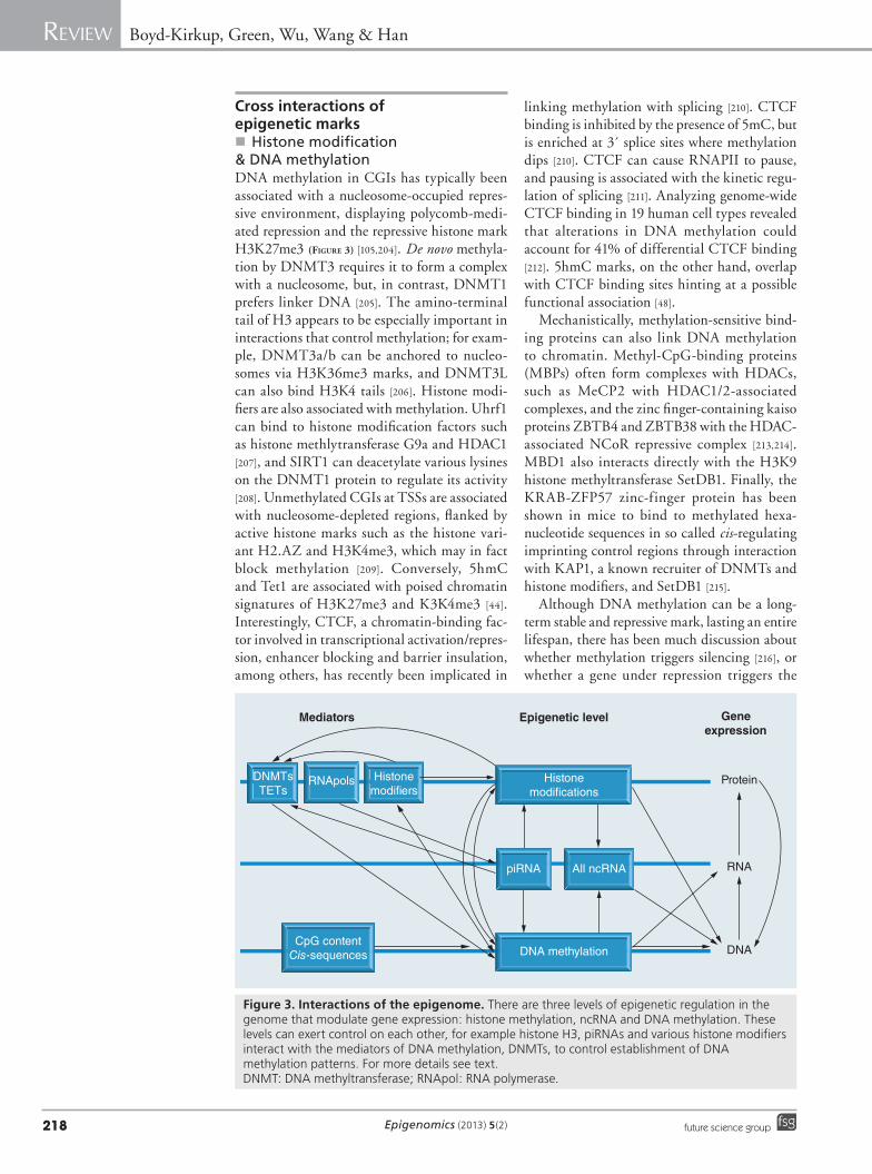
Cross interactions of epigenetic marks�n Histone modification
& DNA methylationDNA methylation in CGIs has typically been associated with a nucleosome-occupied repres-sive environment, displaying polycomb-medi-ated repression and the repressive histone mark H3K27me3 (Figure 3) [105,204]. De novo methyla-tion by DNMT3 requires it to form a complex with a nucleosome, but, in contrast, DNMT1 prefers linker DNA [205]. The amino-terminal tail of H3 appears to be especially important in interactions that control methylation; for exam-ple, DNMT3a/b can be anchored to nucleo-somes via H3K36me3 marks, and DNMT3L can also bind H3K4 tails [206]. Histone modi-fiers are also associated with methylation. Uhrf1 can bind to histone modification factors such as histone methlytransferase G9a and HDAC1 [207], and SIRT1 can deacetylate various lysines on the DNMT1 protein to regulate its activity [208]. Unmethylated CGIs at TSSs are associated with nucleosome-depleted regions, flanked by active histone marks such as the histone vari-ant H2.AZ and H3K4me3, which may in fact block methylation [209]. Conversely, 5hmC and Tet1 are associated with poised chromatin signatures of H3K27me3 and K3K4me3 [44]. Interestingly, CTCF, a chromatin-binding fac-tor involved in transcriptional activation/repres-sion, enhancer blocking and barrier insulation, among others, has recently been implicated in
linking methylation with splicing [210]. CTCF binding is inhibited by the presence of 5mC, but is enriched at 3´ splice sites where methylation dips [210]. CTCF can cause RNAPII to pause, and pausing is associated with the kinetic regu-lation of splicing [211]. Analyzing genome-wide CTCF binding in 19 human cell types revealed that alterations in DNA methylation could account for 41% of differential CTCF binding [212]. 5hmC marks, on the other hand, overlap with CTCF binding sites hinting at a possible functional association [48].
Mechanistically, methylation-sensitive bind-ing proteins can also link DNA methylation to chromatin. Methyl-CpG-binding proteins (MBPs) often form complexes with HDACs, such as MeCP2 with HDAC1/2-associated complexes, and the zinc finger-containing kaiso proteins ZBTB4 and ZBTB38 with the HDAC-associated NCoR repressive complex [213,214]. MBD1 also interacts directly with the H3K9 histone methyltransferase SetDB1. Finally, the KRAB-ZFP57 zinc-finger protein has been shown in mice to bind to methylated hexa-nucleotide sequences in so called cis-regulating imprinting control regions through interaction with KAP1, a known recruiter of DNMTs and histone modifiers, and SetDB1 [215].
Although DNA methylation can be a long-term stable and repressive mark, lasting an entire lifespan, there has been much discussion about whether methylation triggers silencing [216], or whether a gene under repression triggers the
DNMTsTETs
RNApols Histonemodi ers
Histonemodi cations
piRNA All ncRNA
DNA methylationCpG contentCis-sequences
Mediators Geneexpression
Epigenetic level
Protein
RNA
DNA
Figure 3. Interactions of the epigenome. There are three levels of epigenetic regulation in the genome that modulate gene expression: histone methylation, ncRNA and DNA methylation. These levels can exert control on each other, for example histone H3, piRNAs and various histone modifiers interact with the mediators of DNA methylation, DNMTs, to control establishment of DNA methylation patterns. For more details see text. DNMT: DNA methyltransferase; RNApol: RNA polymerase.
www.futuremedicine.com 219future science group
Epigenomics & the regulation of aging Review
promoter to become methylated (Figure 3) [217]. It is now believed that methylation itself does not cause repression, but rather it recruits com-plexes that apply repressive histone marks and package chromatin [218,219]. Acetylated his-tones, associated with active genes, can stimu-late expression even if the DNA is methylated [220]. This hypothesis was recently supported by a study using a GFP reporter controlled by a hypermethylated CMV promoter [221]. Using a histone deacetylase inhibitor, both the GFP reporter and other endogenously ‘silenced’ genes could be reactivated, and importantly, the reacti-vated genes were marked by active histone modi-fications without any change to the underlying methylation state. After 2 weeks the loci had reverted back to a silenced state, demonstrating that DNA methylation can recruit repressive histones. In addition, DNA methylation inhibi-tion can stably activate genes for much longer periods [221].
�n ncRNAIn yeast and flies, ncRNAs are known to direct de novo DNMTs to homologous target sequences for methylation (Figure 3) [222]. Using DICER knockout mouse embryonic stem cells, defects in the RNAi machinery showed defective meth-ylation patterns [223]. More recently, ncRNAs were found to target mammalian DNMT3b to rRNA genes, and RNA was also found to bind in the catalytic domain of human DNMT3a to modulate DNMT3a activity in vitro [224,225]. Interestingly, a methylation-determining region identified by Park et al. produced a specialized form of RNA called pit-RNA (a form of piRNA) that was essential for methylation in cis [70]. This adds further support to the involvement of ncRNA in cis-regulation of methylation. Trans-generational inheritance of ncRNA, including piRNAs and other small RNAs, has been iden-tified in C. elegans [4,194] and Drosophila [197], where it is associated with heritable gene and transposon silencing [195,196]. Heritable piRNA also appears to direct methylation occurring in mammalian germ cells, and mice lacking cer-tain piRNAs cells fail to remethylate properly after the loss of methylation during fertilization (Figure 3) [198,223]. Small RNA-seq profiles show that piRNAs cluster at L1 promoter elements, supporting an involvement of the establishment of DNA methylation at least at this feature [196]. siRNA was responsible for inheritance of histone marks in C. elegans, which lack DNA methylation, implying further important inter-actions with histone modifiers [195]. Therefore,
inheritance of histone modifications in humans could be at least partially mediated by inheri-tance of ncRNA and the underlying DNA methylation state, representing a coordinated effect across the epigenetic levels ncRNA–DNA methylation–histone modification. However, ncRNA involvement with methylation patterns in somatic cells is still unsupported at this time.
Conclusion & future perspectiveTheories of aging that focus on the genome can-not explain the gradual decline in the system that is the phenotype of aging. Changes at all epigenomic levels – DNA methylation, histone modifications and circulating ncRNA levels – are associated with aging, and potentially represent an important way that environmental effects may be linked to the rate of aging (Figure 2). It is also important to recognize the close interplay of the various epigenetic marks when considering system-wide implications (Figure 3). For example, ncRNA expression influences patterns of DNA methylation, which in turn affect histone mod-ification. Nevertheless, to date, evidence for a direct link between epigenetic modifications and longevity in humans remains lacking and should be a focus of future research.
A systematic approach for combining mul-tiple high-throughput data to produce a mean-ingful and testable hypothesis is now becom-ing a priority as more data sets are produced. Work has begun on collating the available data for the interactions of various epigenetic marks and deciphering how this affects epig-enome patterns and system- wide phenotypes. Beyond combining DNA methylation and histone modifications for general characteriza-tion of enhancer, promoter and coding regions [36,226,227], Wrzodek et al. presented a compu-tational synthesis of epigenomic data and used it to predict patterns in cell types with incom-plete data sets [228]. This has added support to several of the hypotheses outlined here, such as the interaction between DNA methylation and H3K4me3. Xiao et al. have proposed a similar approach, comparing the epigenomes of humans and other model species and revealing regulatory regions of the genome that cannot be identified from sequence alone [229]. These attempts at synthesizing all the data will be bol-stered by further data on intragenic methyla-tion, CpG and non-CpG methylation in other regulatory regions outside of promoters, and 5hmC patterns during aging. The first high-resolution genomic maps of 5hmC patterns are now becoming available [230], and we expect a
Epigenomics (2013) 5(2)220 future science group
Review Boyd-Kirkup, Green, Wu, Wang & Han
clearer picture of 5hmC’s role in epigenetics to be forthcoming.
The links between ncRNA, DNA methyla-tion and histone modification is, in our opinion, one of the most interesting future directions for epigenomics research, especially aging research. Changes in alternative splicing could have an important role in aging that is regulated by intragenic epigenetic patterns. In addition, a better understanding of the regulation of DNA methylation and histone modifications, directly and indirectly, through ncRNA both in the inheritance of patterns, and establishment and maintenance of patterns, will be enlightening. Finally, however the epigenome changes during aging, approaches for targeted manipulation of the epigenome, including targeted application of specific histone marks [231] and modification of DNA methylation patterns (reviewed in [232]), will be essential for further establishing caus-ative roles of epigenomic changes in aging and lifespan regulation.
AcknowledgementsThe authors thank H Cheng for critical reading of the manuscript.
Financial & competing interests disclosureThe authors acknowledge the support of the China Natu‑ral National Science Foundation research fund for Young International Scientists (Grant # 31150110469 to JD Boyd‑Kirkup and grant #31250110212 to CD Green). G Wu is supported by grants from Shanghai Institutes of Biological Sciences (2011KIP202 to G Wu) and Chinese Ministry of Science and Technology (Grant # 2012AA020406). G Wu acknowledges the support of SA‑SIBS Scholarship Program. This work was also sup‑ported by grants from the China Natural National Sci‑ence Foundation (Grant #30890033, 31210103916 and 91019019), Chinese Ministry of Science and Technology (Grant #2011CB504206) and Chinese Academy of Sci‑ences (Grant #KSCX2‑EW‑R‑02 and KSCX2‑EW‑J‑15) and stem cell leading project XDA01010303 to J‑DJ Han. JD Boyd‑Kirkup and CD Green hold Chinese Academy of Science Fellowships for Young International Scientists (#2011Y1SB05 to JD Boyd‑Kirkup and #2010Y2SB06 to CD Green). The authors have no other relevant affiliations or financial involvement with any organization or entity with a financial interest in or financial conflict with the subject matter or materials discussed in the manuscript apart from those disclosed.
No writing assistance was utilized in the production of this manuscript.
Executive summary
Approaches to study the epigenomics of aging
� Advances in sequencing and sample preparation techniques have allowed for genome-wide analyses of the epigenome.
� Databases developed to curate high-throughput data from humans and model organisms can link the epigenome to diverse phenotypes.
Regulation of epigenomic marks
� DNA methylation is established and maintained by DNA methyltransferases, and demethylated by TET proteins.
� A plethora of histone modifications are controlled by the actions of a variety of modifiers including methylases, acetylases, demethylases and deacteylases.
� Patterns of DNA methylation and histone modifications are associated with defined genomic elements.
Epigenomic changes associated with aging
� Aging is characterized by global DNA hypomethylation, and specific DNA hypermethylation on key genes.
� Aging is characterized by a gradual deregulation of chromatin stability.
� Aging-related histone modification patterns are associated with the DNA damage response and loss of heterochromatin.
Epigenomic changes that increase longevity
� Modulating several histone modifiers has been shown to affect lifespan in model organisms, but data for a link between DNA methylation and longevity are limited.
ncRNAs
� ncRNAs, and their effect on aging, is a young field that is quickly gaining interest. Thus far, miRNAs are the most well studied, with evidence for lifespan effects in model organisms.
Cross-interactions of epigenetic marks
� Histone modification and DNA methylation complexes interact to regulate the epigenome.
� Emerging data show that ncRNAs are involved in the establishment of DNA methylation and histone modification patterns.
Conclusion & future perspective
� The development of new methods for integrating high-throughput epigenomic data will aid in-depth study of the association of epigenomic patterns with the regulation of aging and longevity.
www.futuremedicine.com 221future science group
Epigenomics & the regulation of aging Review
ReferencesPapers of special note have been highlighted as:n of interestnn of considerable interest
1 Kirkwood TBL. Understanding the odd science of aging. Cell 120(4), 437–447 (2005).
2 Partridge L, Gems D, Withers DJ. Sex and death: what is the connection? Cell 120(4), 461–472 (2005).
3 D’Autréaux B, Toledano MB. ROS as signalling molecules: mechanisms that generate specificity in ROS homeostasis. Nat. Rev. Mol. Cell Biol. 8(10), 813–824 (2007).
4 Ashe A, Sapetschnig A, Weick EM et al. piRNAs can trigger a multigenerational epigenetic memory in the germline of C. elegans. Cell 150(1), 88–99 (2012).
5 Rubinsztein DC, Mariño G, Kroemer G. Autophagy and aging. Cell 146(5), 682–695 (2011).
6 Martin GM. Genetic modulation of senescent phenotypes in Homo sapiens. Cell 120(4), 523–532 (2005).
7 Hoeijmakers JH. DNA damage, aging, and cancer. N. Engl. J. Med. 361(15), 1475–1485 (2009).
8 Balaban RS, Nemoto S, Finkel T. Mitochondria, oxidants, and aging. Cell 120(4), 483–495 (2005).
9 Trifunovic A, Wredenberg A, Falkenberg M et al. Premature ageing in mice expressing defective mitochondrial DNA polymerase. Nature 429(6990), 417–423 (2004).
10 Ristow M, Zarse K. How increased oxidative stress promotes longevity and metabolic health: the concept of mitochondrial hormesis (mitohormesis). Exp. Gerontol. 45(6), 410–418 (2010).
11 Hekimi S, Lapointe J, Wen Y. Taking a “good” look at free radicals in the aging process. Trends Cell Biol. 21(10), 569–576 (2011).
12 Steves CJ, Spector TD, Jackson SHD. Ageing, genes, environment and epigenetics: what twin studies tell us now, and in the future. Age Ageing 41(5), 581–586 (2012).
n� Summarizes twin studies that point towards the interaction between the environment, the genome and epigenome during aging.
13 de Magalhães JP, Wuttke D, Wood SH, Plank M, Vora C. Genome–environment interactions that modulate aging: powerful targets for drug discovery. Pharmacol. Rev. 64(1), 88–101 (2012).
14 Holliday R, Pugh JE. DNA modification mechanisms and gene activity during development. Science 187(4173), 226–232 (1975).
15 Zhou VW, Goren A, Bernstein BE. Charting histone modifications and the functional organization of mammalian genomes. Nat. Publ. Group 12(1), 7–18 (2011).
16 Guarente L, Picard F. Calorie restriction – the SIR2 connection. Cell 120(4), 473–482 (2005).
17 Lin SJ, Kaeberlein M, Andalis AA et al. Calorie restriction extends Saccharomyces cerevisiae lifespan by increasing respiration. Nature 418(6895), 344–348 (2002).
18 Schulz TJ, Zarse K, Voigt A, Urban N, Birringer M, Ristow M. Glucose restriction extends Caenorhabditis elegans life span by inducing mitochondrial respiration and increasing oxidative stress. Cell Metab. 6(4), 280–293 (2007).
19 Pletcher SD, Macdonald SJ, Marguerie R et al. Genome-wide transcript profiles in aging and calorically restricted Drosophila melanogaster. Curr. Biol. 12(9), 712–723 (2002).
20 Yu BP, Masoro EJ, McMahan CA. Nutritional influences on aging of Fischer 344 rats: I. Physical, metabolic, and longevity characteristics. J. Gerontol. 40(6), 657–670 (1985).
21 Colman RJ, Anderson RM, Johnson SC et al. Caloric restriction delays disease onset and mortality in rhesus monkeys. Science 325(5937), 201–204 (2009).
22 Mattison JA, Roth GS, Beasley TM et al. Impact of caloric restriction on health and survival in rhesus monkeys from the NIA study. Nature 489(7415), 318–321 (2012).
23 Kenyon C. The plasticity of aging: insights from long-lived mutants. Cell 120(4), 449–460 (2005).
24 Herranz D, Muñoz-Martin M, Cañamero M et al. Sirt1 improves healthy ageing and protects from metabolic syndrome-associated cancer. Nat. Commun. 1, 3 (2010).
25 Naiman S, Kanfi Y, Cohen HY. Sirtuins as regulators of mammalian aging. Aging (Albany NY) 4(8), 521–522 (2012).
26 Burnett C, Valentini S, Cabreiro F et al. Absence of effects of Sir2 overexpression on lifespan in C. elegans and Drosophila. Nature 477(7365), 482–485 (2011).
27 Wuttke D, Connor R, Vora C et al. Dissecting the gene network of dietary restriction to identify evolutionarily conserved pathways and new functional genes. PLoS Genet. 8(8), e1002834 (2012).
28 Margulies M, Egholm M, Altman WE et al. Genome sequencing in microfabricated high-density picolitre reactors. Nature 437(7057), 376–380 (2005).
29 Valouev A, Ichikawa J, Tonthat T et al. A high-resolution, nucleosome position map of
C. elegans reveals a lack of universal sequence-dictated positioning. Genome Res. 18(7), 1051–1063 (2008).
30 Rakyan VK, Down TA, Balding DJ, Beck S. Epigenome-wide association studies for common human diseases. Nat. Publ. Group 12(8), 529–541 (2011).
31 de Magalhães JP, Finch CE, Janssens G. Next-generation sequencing in aging research: emerging applications, problems, pitfalls and possible solutions. Ageing Res. Rev. 9(3), 315–323 (2010).
32 Simpson VJ, Johnson TE, Hammen RF. Caenorhabditis elegans DNA does not contain 5-methylcytosine at any time during development or aging. Nucleic Acids Res. 14(16), 6711–6719 (1986).
33 Pai AA, Bell JT, Marioni JC, Pritchard JK, Gilad Y. A genome-wide study of DNA methylation patterns and gene expression levels in multiple human and chimpanzee tissues. PLoS Genet. 7(2), e1001316 (2011).
34 Thompson JF, Steinmann KE. Single molecule sequencing with a HeliScope genetic analysis system. Curr. Protoc. Mol. Biol. Chapter 7, Unit 7.10 (2010).
35 Xin Y, Chanrion B, O’Donnell AH et al. MethylomeDB: a database of DNA methylation profiles of the brain. Nucleic Acids Res. 40, D1245–D1249 (2011).
36 Lister R, Pelizzola M, Dowen RH et al. Human DNA methylomes at base resolution show widespread epigenomic differences. Nature 462(7271), 315–322 (2009).
n� Publication of the first full reference epigenome that describes CpG and non-CpG methylation across the genomic landscape.
37 Yan J, Zierath JR, Barrès R. Evidence for non-CpG methylation in mammals. Exp. Cell Res. 317(18), 2555–2561 (2011).
38 Okano M, Xie S, Li E. Cloning and characterization of a family of novel mammalian DNA (cytosine-5) methyltransferases. Nat. Genet. 19(3), 219–220 (1998).
39 Gowher H, Jeltsch A. Enzymatic properties of recombinant Dnmt3a DNA methyltransferase from mouse: the enzyme modifies DNA in a non-processive manner and also methylates non-CpG [correction of non-CpA] sites. J. Mol. Biol. 309(5), 1201–1208 (2001).
40 Arand J, Spieler D, Karius T et al. In vivo control of CpG and non-CpG DNA methylation by DNA methyltransferases. PLoS Genet. 8(6), e1002750 (2012).
41 Fatemi M, Hermann A, Gowher H, Jeltsch A. Dnmt3a and Dnmt1 functionally cooperate during de novo methylation of DNA. Eur. J. Biochem. 269(20), 4981–4984 (2002).
Epigenomics (2013) 5(2)222 future science group
Review Boyd-Kirkup, Green, Wu, Wang & Han
42 Liang G, Chan MF, Tomigahara Y et al. Cooperativity between DNA methyltransferases in the maintenance methylation of repetitive elements. Mol. Cell Biol. 22(2), 480–491 (2002).
43 Tahiliani M, Koh KP, Shen Y et al. Conversion of 5-methylcytosine to 5-hydroxymethylcytosine in mammalian DNA by MLL partner TET1. Science 324(5929), 930–935 (2009).
44 Pastor WA, Pape UJ, Huang Y et al. Genome-wide mapping of 5-hydroxymethylcytosine in embryonic stem cells. Nature 473(7347), 394–397 (2011).
45 Yu M, Hon GC, Szulwach KE et al. Base-resolution analysis of 5-hydroxymethylcytosine in the mammalian genome. Cell 149(6), 1368–1380 (2012).
46 Ito S, Shen L, Dai Q et al. Tet proteins can convert 5-methylcytosine to 5-formylcytosine and 5-carboxylcytosine. Science 333(6047), 1300–1303 (2011).
47 He YF, Li BZ, Li Z et al. Tet-mediated formation of 5-carboxylcytosine and its excision by TDG in mammalian DNA. Science 333(6047), 1303–1307 (2011).
48 Ficz G, Branco MR, Seisenberger S et al. Dynamic regulation of 5-hydroxymethylcytosine in mouse ES cells and during differentiation. Nature 473(7347), 398–402 (2011).
49 Davies MN, Volta M, Pidsley R et al. Functional annotation of the human brain methylome identifies tissue-specific epigenetic variation across brain and blood. Genome Biol. 13(6), R43 (2012).
50 Ma DK, Marchetto MC, Guo JU, Ming GL, Gage FH, Song H. Epigenetic choreographers of neurogenesis in the adult mammalian brain. Nat. Neurosci. 13(11), 1338–1344 (2010).
51 Meehan RR, Lewis JD, Bird AP. Characterization of MeCP2, a vertebrate DNA binding protein with affinity for methylated DNA. Nucleic Acids Res. 20(19), 5085–5092 (1992).
52 Prendergast GC, Ziff EB. Methylation-sensitive sequence-specific DNA binding by the c-Myc basic region. Science 251(4990), 186–189 (1991).
53 Jin SG, Kadam S, Pfeifer GP. Examination of the specificity of DNA methylation profiling techniques towards 5-methylcytosine and 5-hydroxymethylcytosine. Nucleic Acids Res. 38(11), e125 (2010).
54 Wolf SF, Jolly DJ, Lunnen KD, Friedmann T, Migeon BR. Methylation of the hypoxanthine phosphoribosyltransferase locus on the human X chromosome: implications for X-chromosome inactivation. Proc. Natl Acad. Sci. USA 81(9), 2806–2810 (1984).
55 Cokus SJ, Feng S, Zhang X et al. Shotgun bisulphite sequencing of the Arabidopsis genome reveals DNA methylation patterning. Nature 452(7184), 215–219 (2008).
56 Maunakea AK, Nagarajan RP, Bilenky M et al. Conserved role of intragenic DNA methylation in regulating alternative promoters. Nature 466(7303), 253–257 (2010).
57 Laurent L, Wong E, Li G et al. Dynamic changes in the human methylome during differentiation. Genome Res. 20(3), 320–331 (2010).
58 Sati S, Tanwar VS, Kumar KA et al. High resolution methylome map of rat indicates role of intragenic DNA methylation in identification of coding region. PLoS ONE 7(2), e31621 (2012).
59 Choi JK. Contrasting chromatin organization of CpG islands and exons in the human genome. Genome Biol. 11(7), R70 (2010).
60 Flores KB, Amdam GV. Deciphering a methylome: what can we read into patterns of DNA methylation? J. Exp. Biol. 214(Pt 19), 3155–3163 (2011).
61 Moarefi AH, Chédin F. ICF syndrome mutations cause a broad spectrum of biochemical defects in DNMT3B-mediated de novo DNA methylation. J. Mol. Biol. 409(5), 758–772 (2011).
62 Stadler MB, Murr R, Burger L et al. DNA-binding factors shape the mouse methylome at distal regulatory regions. Nature 480(7378), 490–495 (2011).
63 Schmidl C, Klug M, Boeld TJ et al. Lineage-specific DNA methylation in T cells correlates with histone methylation and enhancer activity. Genome Res. 19(7), 1165–1174 (2009).
64 Bell AC, Felsenfeld G. Methylation of a CTCF-dependent boundary controls imprinted expression of the Igf2 gene. Nature 405(6785), 482–485 (2000).
65 Gowher H, Liebert K, Hermann A, Xu G, Jeltsch A. Mechanism of stimulation of catalytic activity of Dnmt3A and Dnmt3B DNA-(cytosine-C5)-methyltransferases by Dnmt3L. J. Biol. Chem. 280(14), 13341–13348 (2005).
66 Sharif J, Muto M, Takebayashi SI et al. The SRA protein Np95 mediates epigenetic inheritance by recruiting Dnmt1 to methylated DNA. Nature 450(7171), 908–912 (2007).
67 Meilinger D, Fellinger K, Bultmann S et al. Np95 interacts with de novo DNA methyltransferases, Dnmt3a and Dnmt3b, and mediates epigenetic silencing of the viral CMV promoter in embryonic stem cells. EMBO Rep. 10(11), 1259–1264 (2009).
68 Lienert F, Wirbelauer C, Som I, Dean A, Mohn F, Schübeler D. Identification of genetic elements that autonomously determine DNA methylation states. Nat. Genet. 43(11), 1091–1097 (2011).
69 Zhang Y, Shu J, Si J, Shen L, Estecio MR, Issa JP. Repetitive elements and enforced transcriptional repression co-operate to enhance DNA methylation spreading into a promoter CpG-island. Nucleic Acids Res. 40(15), 7257–7268 (2012).
70 Park YJ, Herman H, Gao Y et al. Sequences sufficient for programming imprinted germline DNA methylation defined. PLoS ONE 7(3), e33024 (2012).
71 Teng M, Balch C, Liu Y et al. The influence of cis-regulatory elements on DNA methylation fidelity. PLoS ONE 7(3), e32928 (2012).
72 Tan M, Luo H, Lee S et al. Identification of 67 histone marks and histone lysine crotonylation as a new type of histone modification. Cell 146(6), 1016–1028 (2011).
73 Kim TH, Barrera LO, Zheng M et al. A high-resolution map of active promoters in the human genome. Nature 436(7052), 876–880 (2005).
74 Roh TY, Cuddapah S, Zhao K. Active chromatin domains are defined by acetylation islands revealed by genome-wide mapping. Genes Dev. 19(5), 542–552 (2005).
75 Schübeler D, MacAlpine DM, Scalzo D et al. The histone modification pattern of active genes revealed through genome-wide chromatin analysis of a higher eukaryote. Genes Dev. 18(11), 1263–1271 (2004).
76 Wang Z, Zang C, Rosenfeld JA et al. Combinatorial patterns of histone acetylations and methylations in the human genome. Nat. Genet. 40(7), 897–903 (2008).
77 Dong X, Greven MC, Kundaje A et al. Modeling gene expression using chromatin features in various cellular contexts. Genome Biol. 13(9), R53 (2012).
78 ENCODE Project Consortium, Dunham I, Kundaje A, Aldred SF et al. An integrated encyclopedia of DNA elements in the human genome. Nature 489(7414), 57–74 (2012).
79 Schotta G, Lachner M, Sarma K et al. A silencing pathway to induce H3-K9 and H4-K20 trimethylation at constitutive heterochromatin. Genes Dev. 18(11), 1251–1262 (2004).
80 Suganuma T, Workman JL. Signals and combinatorial functions of histone modifications. Annu. Rev. Biochem. 80, 473–499 (2011).
81 Jin Q, Yu LR, Wang L et al. Distinct roles of GCN5/PCAF-mediated H3K9ac and
www.futuremedicine.com 223future science group
Epigenomics & the regulation of aging Review
223www.futuremedicine.com
CBP/p300-mediated H3K18/27ac in nuclear receptor transactivation. EMBO J. 30(2), 249–262 (2011).
82 Liu L, Wylie RC, Andrews LG, Tollefsbol TO. Aging, cancer and nutrition: the DNA methylation connection. Mech. Ageing Dev. 124(10–12), 989–998 (2003).
83 Liu L, van Groen T, Kadish I et al. Insufficient DNA methylation affects healthy aging and promotes age-related health problems. Clin. Epigenetics 2(2), 349–360 (2011).
84 Heyn H, Li N, Ferreira HJ et al. Distinct DNA methylomes of newborns and centenarians. Proc. Natl Acad. Sci. USA 109(26), 10522–10527 (2012).
85 Bjornsson HT, Sigurdsson MI, Fallin MD et al. Intra-individual change over time in DNA methylation with familial clustering. JAMA 299(24), 2877–2883 (2008).
86 Gentilini D, Mari D, Castaldi D et al. Role of epigenetics in human aging and longevity: genome-wide DNA methylation profile in centenarians and centenarians’ offspring. Age (Dordr.) doi:10.1007/s11357-012-9463-1 (2012) (Epub ahead of print).
87 Bollati V, Schwartz J, Wright R et al. Decline in genomic DNA methylation through aging in a cohort of elderly subjects. Mech. Ageing Dev. 130(4), 234–239 (2009).
88 Kim M, Long TI, Arakawa K, Wang R, Yu MC, Laird PW. DNA methylation as a biomarker for cardiovascular disease risk. PLoS ONE 5(3), e9692 (2010).
89 Moore LE, Pfeiffer RM, Poscablo C et al. Genomic DNA hypomethylation as a biomarker for bladder cancer susceptibility in the Spanish Bladder Cancer Study: a case–control study. Lancet Oncol. 9(4), 359–366 (2008).
90 Eckhardt F, Lewin J, Cortese R et al. DNA methylation profiling of human chromosomes 6, 20 and 22. Nat. Genet. 38(12), 1378–1385 (2006).
91 Wilson AS, Power BE, Molloy PL. DNA hypomethylation and human diseases. Biochim. Biophys. Acta 1775(1), 138–162 (2007).
92 Bennett-Baker PE, Wilkowski J, Burke DT. Age-associated activation of epigenetically repressed genes in the mouse. Genetics 165(4), 2055–2062 (2003).
93 Madrigano J, Baccarelli A, Mittleman MA et al. Aging and epigenetics: longitudinal changes in gene-specific DNA methylation. Epigenetics 7(1), 63–70 (2012).
94 Salvioli S, Capri M, Valensin S et al. Inflamm-aging, cytokines and aging: state of the art, new hypotheses on the role of mitochondria and new perspectives from
systems biology. Curr. Pharm. Des. 12(24), 3161–3171 (2006).
95 Gowers IR, Walters K, Kiss-Toth E, Read RC, Duff GW, Wilson AG. Age-related loss of CpG methylation in the tumour necrosis factor promoter. Cytokine 56(3), 792–797 (2011).
96 Rakyan VK, Down TA, Maslau S et al. Human aging-associated DNA hypermethylation occurs preferentially at bivalent chromatin domains. Genome Res. 20(4), 434–439 (2010).
97 Christensen BC, Houseman EA, Marsit CJ et al. Aging and environmental exposures alter tissue-specific DNA methylation dependent upon CpG island context. PLoS Genet. 5(8), e1000602 (2009).
98 Hernandez DG, Nalls MA, Gibbs JR et al. Distinct DNA methylation changes highly correlated with chronological age in the human brain. Hum. Mol. Genet. 20(6), 1164–1172 (2011).
99 Murphy TM, Perry AS, Lawler M. The emergence of DNA methylation as a key modulator of aberrant cell death in prostate cancer. Endocr. Relat. Cancer 15(1), 11–25 (2008).
100 Funayama R, Ishikawa F. Cellular senescence and chromatin structure. Chromosoma 116(5), 431–440 (2007).
101 Salminen A, Ojala J, Kaarniranta K. Apoptosis and aging: increased resistance to apoptosis enhances the aging process. Cell Mol. Life Sci. 68(6), 1021–1031 (2011).
102 Bocklandt S, Lin W, Sehl ME et al. Epigenetic predictor of age. PLoS ONE 6(6), e14821 (2011).
103 Grönniger E, Weber B, Heil O et al. Aging and chronic sun exposure cause distinct epigenetic changes in human skin. PLoS Genet. 6(5), e1000971 (2010).
104 So K, Tamura G, Honda T et al. Multiple tumor suppressor genes are increasingly methylated with age in non-neoplastic gastric epithelia. Cancer Sci. 97(11), 1155–1158 (2006).
105 Salpea P, Russanova VR, Hirai TH et al. Postnatal development- and age-related changes in DNA-methylation patterns in the human genome. Nucleic Acids Res. 40(14), 6477–6494 (2012).
nn� Excellent investigation of whole-genome methylation signatures, including intragenic methylation, that associate with human aging, from newborns to adults.
106 Chen H, Dzitoyeva S, Manev H. Effect of aging on 5-hydroxymethylcytosine in the mouse hippocampus. Restor. Neurol. Neurosci. 30(3), 237–245 (2012).
107 Szulwach KE, Li X, Li Y et al. 5-hmC-mediated epigenetic dynamics during
postnatal neurodevelopment and aging. Nat. Neurosci. 14(12), 1607–1616 (2011).
108 Casillas MA, Lopatina N, Andrews LG, Tollefsbol TO. Transcriptional control of the DNA methyltransferases is altered in aging and neoplastically-transformed human fibroblasts. Mol. Cell Biochem. 252(1–2), 33–43 (2003).
109 Li Y, Liu Y, Strickland FM, Richardson B. Age-dependent decreases in DNA methyltransferase levels and low transmethylation micronutrient levels synergize to promote overexpression of genes implicated in autoimmunity and acute coronary syndromes. Exp. Gerontol. 45(4), 312–322 (2010).
110 Oliveira AMM, Hemstedt TJ, Bading H. Rescue of aging-associated decline in Dnmt3a2 expression restores cognitive abilities. Nat. Neurosci. 15(8), 1111–1113 (2012).
111 Guo JU, Ma DK, Mo H et al. Neuronal activity modifies the DNA methylation landscape in the adult brain. Nat. Neurosci. 14(10), 1345–1351 (2011).
112 Maegawa S, Hinkal G, Kim HS et al. Widespread and tissue specific age-related DNA methylation changes in mice. Genome Res. 20(3), 332–340 (2010).
113 Fraser HB, Lam L, Neumann S, Kobor MS. Population-specificity of human DNA methylation. Genome Biol. 13(2), R8 (2012).
114 Martin GM. Epigenetic drift in aging identical twins. Proc. Natl Acad. Sci. USA 102(30), 10413–10414 (2005).
115 Fraga MF, Ballestar E, Paz MF et al. Epigenetic differences arise during the lifetime of monozygotic twins. Proc. Natl Acad. Sci. USA 102(30), 10604–10609 (2005).
116 Talens RP, Christensen K, Putter H et al. Epigenetic variation during the adult lifespan: cross-sectional and longitudinal data on monozygotic twin pairs. Aging Cell 11(4), 694–703 (2012).
117 Poirier LA, Wise CK, Delongchamp RR, Sinha R. Blood determinations of S-adenosylmethionine, S-adenosylhomocysteine, and homocysteine: correlations with diet. Cancer Epidemiol. Biomarkers Prev. 10(6), 649–655 (2001).
118 Selhub J, Miller JW. The pathogenesis of homocysteinemia: interruption of the coordinate regulation by S-adenosylmethionine of the remethylation and transsulfuration of homocysteine. Am. J. Clin. Nutr. 55(1), 131–138 (1992).
119 Feil R, Fraga MF. Epigenetics and the environment: emerging patterns and implications. Nat. Publ. Group 13(2), 97–109 (2012).
Epigenomics (2013) 5(2)224 future science group
Review Boyd-Kirkup, Green, Wu, Wang & Han
120 Nakajima K, Takeoka M, Mori M et al. Exercise effects on methylation of ASC gene. Int. J. Sports Med. 31(9), 671–675 (2010).
121 Chouliaras L, van den Hove DLA, Kenis G et al. Age-related increase in levels of 5-hydroxymethylcytosine in mouse hippocampus is prevented by caloric restriction. Curr. Alzheimer Res. 9(5), 536–544 (2012).
122 Li Y, Liu L, Tollefsbol TO. Glucose restriction can extend normal cell lifespan and impair precancerous cell growth through epigenetic control of hTERT and p16 expression. FASEB J. 24(5), 1442–1453 (2010).
123 Hass BS, Hart RW, Lu MH, Lyn-Cook BD. Effects of caloric restriction in animals on cellular function, oncogene expression, and DNA methylation in vitro. Mutat. Res. 295(4–6), 281–289 (1993).
124 Milagro FI, Campión J, Cordero P et al. A dual epigenomic approach for the search of obesity biomarkers: DNA methylation in relation to diet-induced weight loss. FASEB J. 25(4), 1378–1389 (2011).
125 Campión J, Milagro FI, Goyenechea E, Martínez JA. TNF-a promoter methylation as a predictive biomarker for weight-loss response. Obesity (Silver Spring) 17(6), 1293–1297 (2009).
126 Sinclair DA, Oberdoerffer P. The ageing epigenome: damaged beyond repair? Ageing Res. Rev. 8(3), 189–198 (2009).
127 Han Y, Han D, Yan Z et al. Stress-associated H3K4 methylation accumulates during postnatal development and aging of Rhesus macaque brain. Aging Cell 11(6), 1055–1064 (2012).
128 Jackson SP, Bartek J. The DNA-damage response in human biology and disease. Nature 461(7267), 1071–1078 (2009).
129 Cortez D, Guntuku S, Qin J, Elledge SJ. ATR and ATRIP: partners in checkpoint signaling. Science 294(5547), 1713–1716 (2001).
130 Uziel T, Lerenthal Y, Moyal L, Andegeko Y, Mittelman L, Shiloh Y. Requirement of the MRN complex for ATM activation by DNA damage. EMBO J. 22(20), 5612–5621 (2003).
131 Falck J, Coates J, Jackson SP. Conserved modes of recruitment of ATM, ATR and DNA-PKcs to sites of DNA damage. Nature 434(7033), 605–611 (2005).
132 Burma S, Chen BP, Murphy M, Kurimasa A, Chen DJ. ATM phosphorylates histone H2AX in response to DNA double-strand breaks. J. Biol. Chem. 276(45), 42462–42467 (2001).
133 Stiff T, O’Driscoll M, Rief N, Iwabuchi K, Lobrich M, Jeggo PA. ATM and DNA-PK function redundantly to phosphorylate H2AX
after exposure to ionizing radiation. Cancer Res. 64(7), 2390–2396 (2004).
134 Polo SE, Jackson SP. Dynamics of DNA damage response proteins at DNA breaks: a focus on protein modifications. Genes Dev. 25(5), 409–433 (2011).
135 Rogakou EP, Boon C, Redon C, Bonner WM. Megabase chromatin domains involved in DNA double-strand breaks in vivo. J. Cell Biol. 146(5), 905–916 (1999).
136 Cowell IG, Sunter NJ, Singh PB, Austin CA, Durkacz BW, Tilby MJ. gH2AX foci form preferentially in euchromatin after ionising-radiation. PLoS ONE 2(10), e1057 (2007).
137 Sedelnikova OA, Horikawa I, Zimonjic DB, Popescu NC, Bonner WM, Barrett JC. Senescing human cells and ageing mice accumulate DNA lesions with unrepairable double-strand breaks. Nat. Cell Biol. 6(2), 168–170 (2004).
138 Sedelnikova OA, Horikawa I, Redon C et al. Delayed kinetics of DNA double-strand break processing in normal and pathological aging. Aging Cell 7(1), 89–100 (2008).
139 Weake VM, Workman JL. Histone ubiquitination: triggering gene activity. Mol. Cell 29(6), 653–663 (2008).
140 Shanbhag NM, Rafalska-Metcalf IU, Balane-Bolivar C, Janicki SM, Greenberg RA. ATM-dependent chromatin changes silence transcription in cis to DNA double-strand breaks. Cell 141(6), 970–981 (2010).
141 Shiloh Y, Shema E, Moyal L, Oren M. RNF20-RNF40: a ubiquitin-driven link between gene expression and the DNA damage response. FEBS Lett. 585(18), 2795–2802 (2011).
142 Huyen Y, Zgheib O, Ditullio RAJ et al. Methylated lysine 79 of histone H3 targets 53BP1 to DNA double-strand breaks. Nature 432(7015), 406–411 (2004).
143 Botuyan MV, Lee J, Ward IM et al. Structural basis for the methylation state-specific recognition of histone H4-K20 by 53BP1 and Crb2 in DNA repair. Cell 127(7), 1361–1373 (2006).
144 Grenon M, Costelloe T, Jimeno S et al. Docking onto chromatin via the Saccharomyces cerevisiae Rad9 Tudor domain. Yeast 24(2), 105–119 (2007).
145 Greeson NT, Sengupta R, Arida AR, Jenuwein T, Sanders SL. Di-methyl H4 lysine 20 targets the checkpoint protein Crb2 to sites of DNA damage. J. Biol. Chem. 283(48), 33168–33174 (2008).
146 Oda H, Hubner MR, Beck DB et al. Regulation of the histone H4 monomethylase PR-Set7 by CRL4(Cdt2)-mediated PCNA-dependent degradation during DNA damage. Mol. Cell 40(3), 364–376 (2010).
147 O’Sullivan RJ, Kubicek S, Schreiber SL, Karlseder J. Reduced histone biosynthesis and chromatin changes arising from a damage signal at telomeres. Nat. Struct. Mol. Biol. 17(10), 1218–1225 (2010).
148 Pei H, Zhang L, Luo K et al. MMSET regulates histone H4K20 methylation and 53BP1 accumulation at DNA damage sites. Nature 470(7332), 124–128 (2011).
149 Murr R, Loizou JI, Yang YG et al. Histone acetylation by Trrap-Tip60 modulates loading of repair proteins and repair of DNA double-strand breaks. Nat. Cell Biol. 8(1), 91–99 (2006).
150 Li L, Greer C, Eisenman RN, Secombe J. Essential functions of the histone demethylase lid. PLoS Genet. 6(11), e1001221 (2010).
151 Sharma GG, So S, Gupta A et al. MOF and histone H4 acetylation at lysine 16 are critical for DNA damage response and double-strand break repair. Mol. Cell Biol. 30(14), 3582–3595 (2010).
152 Ishimi Y, Kojima M, Takeuchi F, Miyamoto T, Yamada M, Hanaoka F. Changes in chromatin structure during aging of human skin fibroblasts. Exp. Cell Res. 169(2), 458–467 (1987).
153 Vaquero A, Scher M, Lee D, Erdjument-Bromage H, Tempst P, Reinberg D. Human SirT1 interacts with histone H1 and promotes formation of facultative heterochromatin. Mol. Cell 16(1), 93–105 (2004).
154 Oberdoerffer P, Michan S, McVay M et al. SIRT1 redistribution on chromatin promotes genomic stability but alters gene expression during aging. Cell 135(5), 907–918 (2008).
n� Demonstrates SIRT1 redistribution away from repetitive DNA elements by oxidative stress, and genes repressed by SIRT1 prior to redistribution are also deregulated in aged mouse brain.
155 Martin SG, Laroche T, Suka N, Grunstein M, Gasser S M. Relocalization of telomeric Ku and SIR proteins in response to DNA strand breaks in yeast. Cell 97, 621–633 (1999).
156 Vaquero A, Scher M, Erdjument-Bromage H, Tempst P, Serrano L, Reinberg D. SIRT1 regulates the histone methyl-transferase SUV39H1 during heterochromatin formation. Nature 450(7168), 440–444 (2007).
157 Mostoslavsky R, Chua KF, Lombard DB et al. Genomic instability and aging-like phenotype in the absence of mammalian SIRT6. Cell 124(2), 315–329 (2006).
158 Michishita E, McCord RA, Berber E et al. SIRT6 is a histone H3 lysine 9 deacetylase that modulates telomeric chromatin. Nature 452(7186), 492–496 (2008).
159 Michishita E, McCord RA, Boxer LD et al. Cell cycle-dependent deacetylation of
www.futuremedicine.com 225future science group
Epigenomics & the regulation of aging Review
225www.futuremedicine.com
telomeric histone H3 lysine K56 by human SIRT6. Cell Cycle 8(16), 2664–2666 (2009).
160 Kawahara TL, Rapicavoli NA, Wu AR, Qu K, Quake SR, Chang HY. Dynamic chromatin localization of Sirt6 shapes stress- and aging-related transcriptional networks. PLoS Genet. 7(6), e1002153 (2011).
161 Shogren-Knaak M, Ishii H, Sun JM, Pazin MJ, Davie JR, Peterson CL. Histone H4-K16 acetylation controls chromatin structure and protein interactions. Science 311(5762), 844–847 (2006).
162 Dang W, Steffen KK, Perry R et al. Histone H4 lysine 16 acetylation regulates cellular lifespan. Nature 459(7248), 802–807 (2009).
163 Scaffidi P, Misteli T. Lamin A-dependent nuclear defects in human aging. Science 312(5776), 1059–1063 (2006).
164 Shumaker DK, Dechat T, Kohlmaier A et al. Mutant nuclear lamin A leads to progressive alterations of epigenetic control in premature aging. Proc. Natl Acad. Sci. USA 103(23), 8703–8708 (2006).
165 Narita M, Nunez S, Heard E et al. Rb-mediated heterochromatin formation and silencing of E2F target genes during cellular senescence. Cell 113(6), 703–716 (2003).
166 Zhang R, Poustovoitov MV, Ye X et al. Formation of MacroH2A-containing senescence-associated heterochromatin foci and senescence driven by ASF1a and HIRA. Dev. Cell 8(1), 19–30 (2005).
167 Chicas A, Kapoor A, Wang X et al. H3K4 demethylation by Jarid1a and Jarid1b contributes to retinoblastoma-mediated gene silencing during cellular senescence. Proc. Natl Acad. Sci. USA 109(23), 8971–8976 (2012).
168 Herbig U, Ferreira M, Condel L, Carey D, Sedivy JM. Cellular senescence in aging primates. Science 311(5765), 1257 (2006).
169 Lin MJ, Tang LY, Reddy MN, Shen CK. DNA methyltransferase gene dDnmt2 and longevity of Drosophila. J. Biol. Chem. 280(2), 861–864 (2005).
170 Goll MG, Kirpekar F, Maggert KA et al. Methylation of tRNAAsp by the DNA methyltransferase homolog Dnmt2. Science 311(5759), 395–398 (2006).
171 Bell JT, Tsai PC, Yang TP et al. Epigenome-wide scans identify differentially methylated regions for age and age-related phenotypes in a healthy ageing population. PLoS Genet. 8(4), e1002629 (2012).
nn� Used epigenome-wide association studies to associate DNA methylation and aging phenotypes across 172 individuals of different ages.
172 Sebastiani P, Riva A, Montano M et al. Whole genome sequences of a male and
female supercentenarian, ages greater than 114 years. Front. Genet. 2, 90 (2011).
173 Tollervey JR, Wang Z, Hortobágyi T et al. Analysis of alternative splicing associated with aging and neurodegeneration in the human brain. Genome Res. 21(10), 1572–1582 (2011).
174 Kaeberlein M, McVey M, Guarente L. The SIR2/3/4 complex and SIR2 alone promote longevity in Saccharomyces cerevisiae by two different mechanisms. Genes Dev. 13(19), 2570–2580 (1999).
175 Viswanathan M, Guarente L. Regulation of Caenorhabditis elegans lifespan by sir-2.1 transgenes. Nature 477(7365), E1–E2 (2011).
176 Pearson KJ, Baur JA, Lewis KN et al. Resveratrol delays age-related deterioration and mimics transcriptional aspects of dietary restriction without extending life span. Cell Metab. 8(2), 157–168 (2008).
177 Peleg S, Sananbenesi F, Zovoilis A et al. Altered histone acetylation is associated with age-dependent memory impairment in mice. Science 328(5979), 753–756 (2010).
178 Greer EL, Maures TJ, Hauswirth AG et al. Members of the H3K4 trimethylation complex regulate lifespan in a germline-dependent manner in C. elegans. Nature 466(7304), 383–387 (2010).
nn� Targeted RNAi screen for Caenorhabditis elegans histone methyltransferases, and core subunits showed knockdown of the WDR-5 and ASH-2 subunits and the SET-2 methyltransferase each significantly extended lifespan.
179 Jin C, Li J, Green CD et al. Histone demethylase UTX-1 regulates C. elegans life span by targeting the insulin/IGF-1 signaling pathway. Cell Metab. 14(2), 161–172 (2011).
nn� C. elegans treated with RNAi or heterozygous null for UTX-1 showed a significant increase in lifespan and stress tolerance, which was dependent on a functional insulin/IGF-1 signaling pathway.
180 Maures TJ, Greer EL, Hauswirth AG, Brunet A. The H3K27 demethylase UTX-1 regulates C. elegans lifespan in a germline-independent, insulin-dependent manner. Aging Cell 10(6), 980–990 (2011).
181 Cogswell JP, Ward J, Taylor IA et al. Identification of miRNA changes in Alzheimer’s disease brain and CSF yields putative biomarkers and insights into disease pathways. J. Alzheimers Dis. 14(1), 27–41 (2008).
182 Lehrbach NJ, Castro C, Murfitt KJ, Abreu-Goodger C, Griffin JL, Miska EA. Post-developmental microRNA expression is required for normal physiology, and regulates aging in parallel to insulin/IGF-1 signaling in C. elegans. RNA 18(12), 2220–2235 (2012).
183 Kato M, Chen X, Inukai S, Zhao H, Slack FJ. Age-associated changes in expression of small, noncoding RNAs, including microRNAs, in C. elegans. RNA 17(10), 1804–1820 (2011).
184 de Lencastre A, Pincus Z, Zhou K, Kato M, Lee SS, Slack FJ. MicroRNAs both promote and antagonize longevity in C. elegans. Curr. Biol. 20(24), 2159–2168 (2010).
nn� Thorough investigation of miRNAs that increase and decrease lifespan in C. elegans.
185 Pincus Z, Smith-Vikos T, Slack FJ. MicroRNA Predictors of longevity in Caenorhabditis elegans. PLoS Genet. 7(9), e1002306 (2011).
186 Boehm M, Slack F. A developmental timing microRNA and its target regulate life span in C. elegans. Science 310(5756), 1954–1957 (2005).
187 Elsharawy A, Keller A, Flachsbart F et al. Genome-wide miRNA signatures of human longevity. Aging Cell 11(4), 607–616 (2012).
188 Noren Hooten N, Abdelmohsen K, Gorospe M, Ejiogu N, Zonderman AB, Evans MK. microRNA expression patterns reveal differential expression of target genes with age. PLoS ONE 5(5), e10724 (2010).
189 Inukai S, de Lencastre A, Turner M, Slack F. Novel microRNAs differentially expressed during aging in the mouse brain. PLoS ONE 7(7), e40028 (2012).
190 Persengiev S, Kondova I, Otting N, Koeppen AH, Bontrop RE. Genome-wide analysis of miRNA expression reveals a potential role for miR-144 in brain aging and spinocerebellar ataxia pathogenesis. Neurobiol. Aging 32(12), 2316.e17–2316.e27 (2011).
191 Li X, Khanna A, Li N, Wang E. Circulatory miR34a as an RNAbased, noninvasive biomarker for brain aging. Aging (Albany NY). 3(10), 985–1002 (2011).
192 Boulias K, Horvitz HR. The C. elegans microRNA mir-71 acts in neurons to promote germline-mediated longevity through regulation of DAF-16/FOXO. Cell Metab. 15(4), 439–450 (2012).
193 Lanceta J, Prough RA, Liang R, Wang E. MicroRNA group disorganization in aging. Exp. Gerontol. 45(4), 269–278 (2010).
194 Rechavi O, Minevich G, Hobert O. Transgenerational inheritance of an acquired small RNA-based antiviral response in C. elegans. Cell 147(6), 1248–1256 (2011).
195 Burton NO, Burkhart KB, Kennedy S. Nuclear RNAi maintains heritable gene silencing in Caenorhabditis elegans. Proc. Natl Acad. Sci. USA 108(49), 19683–19688 (2011).
196 Brennecke J, Malone CD, Aravin AA, Sachidanandam R, Stark A, Hannon GJ.
Epigenomics (2013) 5(2)226 future science group
Review Boyd-Kirkup, Green, Wu, Wang & Han
An epigenetic role for maternally inherited piRNAs in transposon silencing. Science 322(5906), 1387–1392 (2008).
nn� Demonstrates that maternal Piwi-interacting RNAs mediate epigenetic inheritance of transposon silencing in Drosophila.
197 Grentzinger T, Armenise C, Brun C et al. piRNA-mediated transgenerational inheritance of an acquired trait. Genome Res. 22(10), 1877–1888 (2012).
198 Aravin AA, Sachidanandam R, Bourc’his D et al. A piRNA pathway primed by individual transposons is linked to de novo DNA methylation in mice. Mol. Cell 31(6), 785–799 (2008).
199 Qureshi IA, Mattick JS, Mehler MF. Long non-coding RNAs in nervous system function and disease. Brain Res. 1338, 20–35 (2010).
200 Wood SH, Craig T, Li Y, Merry B, de Magalhaes JP. Whole transcriptome sequencing of the aging rat brain reveals dynamic RNA changes in the dark matter of the genome. Age (Dordr.) doi:10.1007/s11357-012-9410-1 (2012) (Epub ahead of print).
201 Conover CA, Bale LK, Grell JA, Mader JR, Mason MA. Longevity is not influenced by prenatal programming of body size. Aging Cell 9(4), 647–649 (2010).
202 Shamovsky I, Ivannikov M, Kandel ES, Gershon D, Nudler E. RNA-mediated response to heat shock in mammalian cells. Nature 440(7083), 556–560 (2006).
203 Han L, Zhang K, Shi Z et al. LncRNA profile of glioblastoma reveals the potential role of lncRNAs in contributing to glioblastoma pathogenesis. Int. J. Oncol. 40(6), 2004–2012 (2012).
204 Taberlay PC, Kelly TK, Liu CC et al. Polycomb-repressed genes have permissive enhancers that initiate reprogramming. Cell 147(6), 1283–1294 (2011).
205 Jeong S, Liang G, Sharma S et al. Selective anchoring of DNA methyltransferases 3A and 3B to nucleosomes containing methylated DNA. Mol. Cell Biol. 29(19), 5366–5376 (2009).
206 Ooi SK, Qiu C, Bernstein E et al. DNMT3L connects unmethylated lysine 4 of histone H3 to de novo methylation of DNA. Nature 448(7154), 714–717 (2007).
207 Kim JK, Estève PO, Jacobsen SE, Pradhan S. UHRF1 binds G9a and participates in p21 transcriptional regulation in mammalian cells. Nucleic Acids Res. 37(2), 493–505 (2009).
208 Peng L, Yuan Z, Ling H et al. SIRT1 Deacetylates the DNA methyltransferase 1 (DNMT1) protein and alters its activities. Mol. Cell Biol. 31(23), 4720–4734 (2011).
209 Kelly TK, Miranda TB, Liang G et al. H2A.Z maintenance during mitosis reveals nucleosome shifting on mitotically silenced genes. Mol. Cell 39(6), 901–911 (2010).
210 Phillips JE, Corces VG. CTCF: master weaver of the genome. Cell 137(7), 1194–1211 (2009).
211 Shukla S, Kavak E, Gregory M et al. CTCF-promoted RNA polymerase II pausing links DNA methylation to splicing. Nature 479(7371), 74–79 (2011).
212 Wang H, Maurano MT, Qu H et al. Widespread plasticity in CTCF occupancy linked to DNA methylation. Genome Res. 22(9), 1680–1688 (2012).
213 Nan X, Ng HH, Johnson CA et al. Transcriptional repression by the methyl-CpG-binding protein MeCP2 involves a histone deacetylase complex. Nature 393(6683), 386–389 (1998).
214 Filion GJ, Zhenilo S, Salozhin S, Yamada D, Prokhortchouk E, Defossez PA. A family of human zinc finger proteins that bind methylated DNA and repress transcription. Mol. Cell Biol. 26(1), 169–181 (2006).
215 Quenneville S, Verde G, Corsinotti A et al. In embryonic stem cells, ZFP57/KAP1 recognize a methylated hexanucleotide to affect chromatin and DNA methylation of imprinting control regions. Mol. Cell 44(3), 361–372 (2011).
216 Lock LF, Takagi N, Martin GR. Methylation of the Hprt gene on the inactive X occurs after chromosome inactivation. Cell 48(1), 39–46 (1987).
217 Challen GA, Sun D, Jeong M et al. Dnmt3a is essential for hematopoietic stem cell differentiation. Nat. Genet. 44(1), 23–31 (2012).
218 Gal-Yam EN, Egger G, Iniguez L et al. Frequent switching of Polycomb repressive marks and DNA hypermethylation in the PC3 prostate cancer cell line. Proc. Natl Acad. Sci. USA 105(35), 12979–12984 (2008).
219 Schlesinger Y, Straussman R, Keshet I et al. Polycomb-mediated methylation on Lys27 of histone H3 pre-marks genes for de novo methylation in cancer. Nat. Genet. 39(2), 232–236 (2007).
220 Pruitt K, Zinn RL, Ohm JE et al. Inhibition of SIRT1 reactivates silenced cancer genes without loss of promoter DNA hypermethylation. PLoS Genet. 2(3), e40 (2006).
221 Raynal NJ, Si J, Taby RF et al. DNA methylation does not stably lock gene expression but instead serves as a molecular mark for gene silencing memory. Cancer Res. 72(5), 1170–1181 (2012).
222 Matzke MA, Birchler JA. RNAi-mediated pathways in the nucleus. Nat. Rev. Genet. 6(1), 24–35 (2005).
223 Aravin AA, Bourc’his D. Small RNA guides for de novo DNA methylation in mammalian germ cells. Genes Dev. 22(8), 970–975 (2008).
224 Schmitz KM, Mayer C, Postepska A, Grummt I. Interaction of noncoding RNA with the rDNA promoter mediates recruitment of DNMT3b and silencing of rRNA genes. Genes Dev. 24(20), 2264–2269 (2010).
225 Holz-Schietinger C, Matje DM, Flexer Harrison M, Reich NO. Oligomerization of DNMT3A controls the mechanism of de novo DNA methylation. J. Biol. Chem. 286(48), 41479–41488 (2011).
226 Ball MP, Li JB, Gao Y et al. Targeted and genome-scale strategies reveal gene-body methylation signatures in human cells. Nat. Biotechnol. 27(4), 361–368 (2009).
227 Koch F, Fenouil R, Gut M et al. Transcription initiation platforms and GTF recruitment at tissue-specific enhancers and promoters. Nat. Struct. Mol. Biol. 18(8), 956–963 (2011).
228 Wrzodek C, Büchel F, Hinselmann G, Eichner J, Mittag F, Zell A. Linking the epigenome to the genome: correlation of different features to DNA methylation of CpG islands. PLoS ONE 7(4), e35327 (2012).
229 Xiao S, Xie D, Cao X et al. Comparative epigenomic annotation of regulatory DNA. Cell 149(6), 1381–1392 (2012).
230 Song CX, Clark TA, Lu XY et al. Sensitive and specific single-molecule sequencing of 5-hydroxymethylcytosine. Nat. Methods 9(1), 75–77 (2012).
231 Carone BR, Rando OJ. Rewriting the epigenome. Cell 149(7), 1422–1423 (2012).
232 Johnson AA, Akman K, Calimport SRG, Wuttke D, Stolzing A, de Magalhães JP. The role of DNA methylation in aging, rejuvenation, and age-related disease. Rejuvenation Res. 15(5), 483–494 (2012).
233 Song L, James SR, Kazim L, Karpf AR. Specific method for the determination of genomic DNA methylation by liquid chromatography-electrospray ionization tandem mass spectrometry. Anal. Chem. 77(2), 504–510 (2005).
234 Weber M, Davies JJ, Wittig D et al. Chromosome-wide and promoter-specific analyses identify sites of differential DNA methylation in normal and transformed human cells. Nat. Genet. 37(8), 853–862 (2005).
235 Bibikova M, Le J, Barnes B et al. Genome-wide DNA methylation profiling using Infinium® assay. Epigenomics 1(1), 177–200 (2009).
www.futuremedicine.com 227future science group
Epigenomics & the regulation of aging Review
227www.futuremedicine.com
236 Frommer M, McDonald LE, Millar DS et al. A genomic sequencing protocol that yields a positive display of 5-methylcytosine residues in individual DNA strands. Proc. Natl Acad. Sci. USA 89(5), 1827–1831 (1992).
237 Huang Y, Pastor WA, Shen Y, Tahiliani M, Liu DR, Rao A. The behaviour of 5-hydroxymethylcytosine in bisulfite sequencing. PLoS ONE 5(1), e8888 (2010).
238 Gu H, Smith ZD, Bock C, Boyle P, Gnirke A, Meissner A. Preparation of reduced representation bisulfite sequencing libraries for genome-scale DNA methylation profiling. Nat. Protocols 6(4), 468–481 (2011).
239 Edwards JR, O’Donnell AH, Rollins RA et al. Chromatin and sequence features that define the fine and gross structure of genomic
methylation patterns. Genome Res. 20(7), 972–980 (2010).
240 Down TA, Rakyan VK, Turner DJ et al. A Bayesian deconvolution strategy for immunoprecipitation-based DNA methylome analysis. Nat. Biotechnol. 26(7), 779–785 (2008).
241 Plazas-Mayorca MD, Zee BM, Young NL et al. One-pot shotgun quantitative mass spectrometry characterization of histones. J. Proteome Res. 8(11), 5367–5374 (2009).
242 Bernstein BE, Humphrey EL, Erlich RL et al. Methylation of histone H3 Lys 4 in coding regions of active genes. Proc. Natl Acad. Sci. USA 99(13), 8695–8700 (2002).
243 Johnson DS, Mortazavi A, Myers RM, Wold B. Genome-wide mapping of in vivo
protein–DNA interactions. Science 316(5830), 1497–1502 (2007).
244 O’Neill LP, Turner BM. Immunoprecipitation of native chromatin: NChIP. Methods 31(1), 76–82 (2003).
245 Fullwood MJ, Wei CL, Liu ET, Ruan Y. Next-generation DNA sequencing of paired-end tags (PET) for transcriptome and genome analyses. Genome Res. 19(4), 521–532 (2009).
246 Fullwood MJ, Liu MH, Pan YF et al. An oestrogen-receptor-a-bound human chromatin interactome. Nature 462(7269), 58–64 (2009).
247 Chepelev I, Wei G, Wangsa D, Tang Q, Zhao K. Characterization of genome-wide enhancer-promoter interactions reveals co-expression of interacting genes and modes of higher order chromatin organization. Cell Res. 22(3), 490–503 (2012).