Embed Size (px)
Citation preview
Epithelial Cells Augment Barrier Function via Activation of the Toll-Like Receptor 2/Phosphatidylinositol 3-Kinase Pathway uponRecognition of Salmonella enterica Serovar Typhimurium Curli Fibrilsin the Gut
Gertrude O. Oppong, Glenn J. Rapsinski, Tiffanny N. Newman, Jessalyn H. Nishimori, Steven G. Biesecker, Çagla Tükel
Department of Microbiology and Immunology, School of Medicine, Temple University, Philadelphia, Pennsylvania, USA
Curli fibrils, the best-characterized functional bacterial amyloids, are an important component of enterobacterial biofilms. Wehave previously shown that curli fibrils are recognized by the Toll-like receptor 2 (TLR2)/TLR1 heterodimer complex. Utilizingpolarized T-84 cells, an intestinal epithelial cell line derived from colon carcinoma grown on semipermeable tissue culture in-serts, we determined that infection with a Salmonella enterica serovar Typhimurium csgBA mutant, which does not expresscurli, resulted in an increase in intestinal barrier permeability and an increase in bacterial translocation compared to infectionwith curliated wild-type S. Typhimurium. When the TLR2 downstream signaling molecule phosphatidylinositol 3-kinase (PI3K)was blocked using wortmannin or LY294002, the difference in disruption of the intestinal epithelium and bacterial translocationwas no longer observed. Additionally, disruption of polarized T-84 cells treated basolaterally with the TLR5 ligand flagellin wasprevented when the polarized cells were simultaneously treated with the synthetic TLR2/TLR1 ligand Pam3CSK4 or with purifiedcurli fibrils in the apical compartment. Similar to in vitro observations, C57BL/6 mice infected with the csgBA mutant sufferedincreased disruption of the intestinal epithelium and therefore greater dissemination of the bacteria to the mesenteric lymphnodes than mice infected with wild-type S. Typhimurium. The differences in disruption of the intestinal epithelium and bacte-rial dissemination in the mice infected with csgBA mutant or wild-type S. Typhimurium were not apparent in TLR2-deficientmice. Overall, these studies report for the first time that activation of the TLR2/PI3K pathway by microbial amyloids plays a crit-ical role in regulating the intestinal epithelial barrier as well as monitoring bacterial translocation during infection.
The intestinal epithelium represents a physical as well as animmunological barrier which is in constant contact with ap-
proximately 1013 to 1014 microorganisms (1–3). Bacteria com-prise the vast majority of the intestinal organisms with at least1,000 different species present within the community (4–6).Therefore, there is a critical need for mechanisms for protectingthe host from hyperresponsive inflammatory processes due to thepresence of an unprecedented amount of antigens while still sup-porting the growth of commensal bacteria which are beneficial tohost health and function. As a first line of innate immune re-sponse, the intestinal epithelium has been found to play an impor-tant role in the maintenance and regulation of gastrointestinalhomeostasis. For instance, it is currently known that the produc-tion of antimicrobial peptides and lectins by enterocytes and Pan-eth cells (7–11), the production of mucins by goblet cells (12, 13),and modulation of epithelial barrier integrity (14) all act in con-cert to regulate and maintain intestinal immune homeostasis.
Toll-like receptors (TLRs) comprise a family of innate patternrecognition receptors (PRRs) that sense conserved microbial struc-tures known as pathogen-associated molecular patterns (PAMPs)and endogenous danger molecules (15–17). In the gut mucosa, vari-ous TLRs are involved in the recognition of microbial signature mol-ecules. TLR5, expressed on the basolateral side of the epithelial cells,recognizes flagella of invading microbes and consequently activatesnuclear factor kappa B (NF-�B), leading to the production of proin-flammatory cytokines, including interleukin 8 (IL-8) (18). TLR2, amember of this family, recognizes a number of conserved molecularpatterns, including lipopeptides, lipoteichoic acid, and zymosan,through the formation of heterodimers with TLR1 or with TLR6 (19–
25). MyD88 (myeloid differentiation primary response gene 88) andMal/TIRAP are both required for TLR2-dependent signaling whereNF-�B is activated. While Mal/TIRAP is involved in bridging MyD88to the TLR2 receptor complex and directing the recruitment ofTRAF6, which is necessary for NF-�B activation, Mal binds to thep85� subunit of phosphatidylinositol 3-kinase (PI3K) upon activa-tion of the TLR2/TLR6 heterodimer, resulting in Akt phosphoryla-tion, which consequently leads to macrophage polarization and cellsurvival by inhibiting apoptosis. In contrast, TLR2/TLR1-mediatedactivation of PI3K occurs in the absence of Mal and MyD88, suggest-ing the presence of another adaptor molecule (26–29). Activation ofthe PI3K pathway as a downstream effect of TLR2 activation has alsobeen shown to augment the tight-junction-associated epithelial bar-rier integrity, possibly by acting as a surveillance receptor which mon-itors luminal bacteria and translocation of pathogens (30–32).
Amyloids, which possess a fibrillar cross-�-sheet quaternarystructure, are produced by both humans and bacteria. While amy-loids in humans are associated mostly with complex diseases,functional amyloids that serve a role in physiological processes
Received 9 May 2012 Returned for modification 30 May 2012Accepted 18 November 2012
Published ahead of print 3 December 2012
Editor: J. B. Bliska
Address correspondence to Çagla Tükel, [email protected].
Copyright © 2013, American Society for Microbiology. All Rights Reserved.
doi:10.1128/IAI.00453-12
478 iai.asm.org Infection and Immunity p. 478–486 February 2013 Volume 81 Number 2
on February 17, 2019 by guest
http://iai.asm.org/
Dow
nloaded from
such as melanin production and blood clotting have been re-ported (33–38). In bacteria, amyloids function as a component ofthe extracellular matrix in biofilms of commensal organisms, suchas spore-forming Bacillus subtilis and Pseudomonas fluorescens, orhuman pathogens, such as Mycobacterium tuberculosis, Salmonellaenterica serovar Typhimurium, Citrobacter freundii, Enterobactersakazakii, and Escherichia coli (39–46). Curli fibrils produced byenteric bacteria, including Salmonella spp. and E. coli, are the best-characterized bacterial amyloid to date. Earlier studies have shownthat curli fibrils activate the immune system, inducing the produc-tion of inflammatory cytokines in a mouse model of sepsis as wellas urinary tract infection induced by E. coli (47–52). Curli fibrilsare indeed a pathogen-associated molecular pattern (PAMP) thatis recognized by the TLR2/TLR1 heterodimer (48–50). Interest-ingly, TLR2 not only responds to curli fibrils but also recognizeshost amyloids such as �-amyloid 1-40 and �-amyloid 1-42 ofAlzheimer’s plaques as well as serum amyloid A, an acute-phaseprotein (48, 53–57). In fact, TLR2 recognizes the conserved qua-ternary �-sheet structure that is common to amyloids of all dis-tinct origins (48).
Amyloids have also been reported to be present in the biofilmsof members of Bacteriodetes and Firmicutes, the predominantphyla found in the gastrointestinal tract (44, 58). In this study, weinvestigated whether recognition of amyloid fibrils could induce aTLR2-dependent response in intestinal epithelia contributing tothe regulation of intestinal barrier integrity by using S. Typhimu-rium as a model.
MATERIALS AND METHODSBacterial strains and culture conditions. S. Typhimurium strain IR715(wild type) is a fully virulent, nalidixic acid-resistant strain derived fromstrain ATCC 14028 (59). CT16 is a mutant strain derived from IR715 andcontains an unmarked csgBA deletion (60). To induce the expression ofcurli fibrils, the bacterial strains were grown on tryptone agar (T-me-dium) plates at 28°C for 48 h (61). For in vivo experiments, bacterialstrains were grown overnight with shaking at 37°C in Luria-Bertani (LB)broth (Fisher Bioreagents) supplemented with nalidixic acid (FisherBioreagents) at a final concentration of 0.05 mg/ml.
Cell culture. The human intestinal epithelial cell (IEC) lines fromcolon carcinoma (T-84) and cervical carcinoma (HeLa) were obtainedfrom the American Type Culture Collection. T-84 cells were grown inDulbecco modified Eagle medium (DMEM)/F-12 (GIBCO) supple-mented with 10% heat-inactivated fetal bovine serum (FBS) (GIBCO).T-84 cells were grown to confluence on 0.4-�m semipermeable tissueculture inserts (Transwell; Corning) in a humidified incubator at 37°Cand 5% CO2. T-84 cells achieved a polarized and differentiated statewithin 5 to 10 days and were used when the transepithelial resistance(TER) had reached �1,500 � cm2 (62).
Invasion assay. The invasion assay was carried out as described pre-viously (49). Briefly, T-84 monolayers were infected with 3.5 �105 ofwild-type IR715 and csgBA mutant CT16 bacterial strains (multiplicity ofinfection [MOI] of 7) grown under conditions optimal for curli expres-sion or type III secretion system 1 (T3SS-1) expression. Bacteria wereallowed to invade cells for an hour. This was then followed by replacementof the medium containing 1 mg/ml gentamicin (Invitrogen) to eliminateextracellular bacteria and incubation for 1.5 h. Epithelial cells were thenlysed with 1% Triton-X (Sigma). Cell lysates were then plated on LB agarplates supplemented with nalidixic acid at a final concentration of 0.05mg/ml. Invasion assays were repeated three times.
IL-8 production. Polarized T-84 cells were infected with wild-typeIR715 and the csgBA mutant CT16 as described above. At 24 h postinfec-tion, 100 �l of the supernatant was removed from the basolateral com-partment of the Transwell. The IL-8 concentration was determined by
enzyme-linked immunosorbent assay (ELISA) according to the manufac-turer’s instructions (Biolegend).
Translocation studies. For translocation studies, polarized T-84 epi-thelial layers grown on 3.0-�m semipermeable tissue culture inserts(Transwell; Corning) were infected apically with S. Typhimurium (wildtype) or its csgBA mutant (CT16) for 1 h as described above. One-hun-dred-microliter samples from the basolateral medium were taken at 1 hpostinfection, and appropriate dilutions were plated on LB agar platescontaining nalidixic acid. For studies involving the PI3K inhibitorLY294002, the polarized epithelial layer was incubated with 50 �MLY294002 for 1 h prior to bacterial infection.
Epithelial integrity. Polarized T-84 cells were infected with wild-typeIR715 and the csgBA mutant CT16 as described above. At 5 h or 24 hpostinfection, 5 �l of 10-mg/ml fluorescein isothiocyanate-labeled dex-tran (FITC-dextran) (average molecular weight, 3,000 to 5,000; Sigma)was added to the apical side of the Transwell chamber. Two hours after theaddition of FITC-dextran, medium from the basolateral side of the Trans-well chamber was collected and fluorescence intensity was measured usingan Omega plate reader (BMG Labtech) at 485-nm excitation and 520-nmemission wavelengths (63). To study the role played by flagellin and curlifibrils in intestinal epithelial integrity, flagellin (FLA-ST; Invivogen) wasadded to the Transwells basolaterally at a final concentration of 0.01 �g/ml. Purification of curli fibrils from the S. Typhimurium msbB mutant(RPW3) was performed according to an established protocol (61). Briefly,bacterial cells were removed from T-medium plates and lysed by sonica-tion. This was followed by enzymatic digestion and preparative sodiumdodecyl sulfate-polyacrylamide gel electrophoresis (SDS-PAGE). Insolu-ble material (curli fibrils) retained in the well of the SDS-polyacrylamidegel was collected after the electrophoresis was complete. The protein con-centration of curli fibrils was determined with the bicinchoninic acid(BCA) protein assay (Calbiochem). Curli fibrils (10 �g/ml) or the triacy-lated synthetic TLR2/TLR1 ligand Pam3CSK4 (0.1 �g/ml; Invivogen) wasadded either alone or simultaneously with basolateral flagellin treatmentto the apical chamber of the Transwell. To block PI3K, polarized epithelialcells were incubated either with 20 �M wortmannin (Calbiochem) for 30min or with 50 �M LY294002 (Cell Signal) for 1 h prior to bacterialinfection. Experiments were repeated three times.
Mouse experiments. Six- to 8-week-old female C57BL/6 mice wereobtained from Jackson Laboratory. TLR2-deficient mice (B6.129-TLR2tm1Kir/J) were purchased from Jackson Laboratory and weremaintained and bred in Temple University’s animal facility. The Tem-ple University Animal Care and Use Committee approved all animalstudies.
The use of FITC-dextran to assess intestinal permeability in vivo hasbeen previously described (12). Briefly, groups of 3 or 4 mice were eitherorally inoculated with 1 � 109 bacteria in LB or mock infected with sterileLB. At 72 h postinfection, 150 �l of 80-mg/ml FITC-dextran was admin-istered orally. Mice were sacrificed 4 h later, and blood was collected viacardiac puncture. Blood was collected into microcentrifuge tubes coatedwith a mixture of the anticoagulant heparin (15 mg/ml) and acid citrate-dextrose (20 mM citric acid, 100 mM sodium citrate, 5 mM dextrose).Blood was then spun at 1,000 rpm for 20 min to separate serum fromwhole blood cells. Fluorescence intensity in the serum was then deter-mined using the Omega plate reader (BMG Labtech) at 485-nm excitationand 520-nm emission wavelengths.
To assess bacterial numbers, oral inoculation of bacteria as describedabove was performed. Mice were sacrificed 72 h later, and tissue samplesfrom the cecum, liver, spleen, mesenteric lymph nodes, and Peyer’spatches were collected. Colonic content was collected in 1 ml of sterilephosphate-buffered saline (PBS). Organ samples were homogenized insterile PBS, and appropriate serial dilutions were plated on LB-nalidixicacid agar plates. All the animal experiments were repeated 3 times.
PCR. To examine the expression of TLR1 and TLR2 by epithelial cells,T-84 cells were grown to confluence on permeable tissue culture inserts asdescribed above. RNA was extracted in 0.5 ml of TriReagent. Following
Gut Epithelium Promotes Barrier by Detecting Amyloids
February 2013 Volume 81 Number 2 iai.asm.org 479
on February 17, 2019 by guest
http://iai.asm.org/
Dow
nloaded from
RNA isolation, 2 �g of total RNA was reverse transcribed using murineleukemia virus (MuLV) reverse transcriptase. Two microliters of cDNAwas subjected to PCR amplification using a high-fidelity PCR Supermix(Invitrogen) and the primers listed in Table 1. The following program wasused for PCR amplification: 95°C for 120 s, followed by 35 cycles of 95°Cfor 60s, 55 to 58°C for 45 s (annealing temperatures were optimized foreach TLR primer pair used), and 72°C for 60 s. As a positive control, HeLacells were stably transfected with a plasmid expressing either TLR1 orTLR2 as described previously (48). HeLa cells transfected with an emptyvector were employed as a negative control. The resultant PCR productswere then analyzed on a 1.5% agarose gel.
Statistical analysis. The Student t test was used to calculate statisticallysignificant differences (P � 0.05). For analysis of bacterial numbers, val-ues were logarithmically converted prior to statistical analysis.
RESULTSTLR2 and TLR1 are expressed by polarized T-84 epithelial cells.In humans, TLR2 is expressed at the apical pole of the intestinalepithelium (64–66). Likewise, mouse intestinal epithelial cells ex-press TLR2 (67). Earlier studies, using germfree mice, demon-strated that expression of TLR2 is increased by stimuli derivedfrom commensal bacteria (14). Since many bacteria belonging toFirmicutes and Bacteriodetes, two predominant phyla in the gut,produce amyloids as a component of their extracellular matrix
(44), we hypothesized that detection of amyloids via TLR2 mayaffect immune responses in the intestinal epithelium in the gut. Tounravel the immune responses generated against amyloid fibrils,we used a human colon carcinoma cell line, T-84. When grown onsemipermeable tissue culture inserts, T-84 epithelial cells are ableto differentiate and polarize to take on functional and morpho-logical characteristics that are specific to the intestinal epithelium,with apical microvilli and a basolateral surface that can be likened tothe cellular surface in contact with the subepithelial lamina propria(62, 68, 69). To ensure that the T-84 cell lines indeed expressed TLR2,RNA from these epithelial cell lines was extracted and subjected toreverse transcription and PCR amplification. As controls, HeLa cellswere transfected with an empty human expression vector (negativecontrol) or a vector containing the human TLR2 gene. T84 cells werefound to express TLR2. Since curli amyloid fibrils have been reportedto signal through TLR2 complexed with TLR1 (48), TLR1 expressionwas also confirmed via PCR (Fig. 1A).
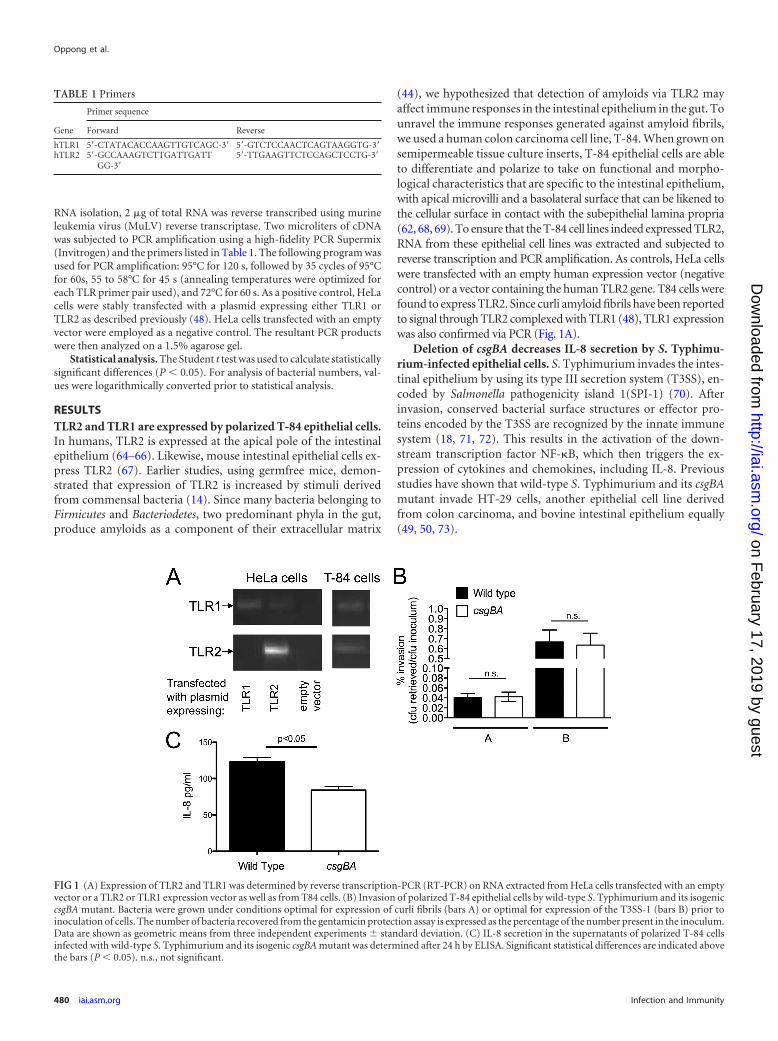
Deletion of csgBA decreases IL-8 secretion by S. Typhimu-rium-infected epithelial cells. S. Typhimurium invades the intes-tinal epithelium by using its type III secretion system (T3SS), en-coded by Salmonella pathogenicity island 1(SPI-1) (70). Afterinvasion, conserved bacterial surface structures or effector pro-teins encoded by the T3SS are recognized by the innate immunesystem (18, 71, 72). This results in the activation of the down-stream transcription factor NF-�B, which then triggers the ex-pression of cytokines and chemokines, including IL-8. Previousstudies have shown that wild-type S. Typhimurium and its csgBAmutant invade HT-29 cells, another epithelial cell line derivedfrom colon carcinoma, and bovine intestinal epithelium equally(49, 50, 73).
TABLE 1 Primers
Gene
Primer sequence
Forward Reverse
hTLR1 5=-CTATACACCAAGTTGTCAGC-3= 5=-GTCTCCAACTCAGTAAGGTG-3=hTLR2 5=-GCCAAAGTCTTGATTGATT
GG-3=5=-TTGAAGTTCTCCAGCTCCTG-3=
FIG 1 (A) Expression of TLR2 and TLR1 was determined by reverse transcription-PCR (RT-PCR) on RNA extracted from HeLa cells transfected with an emptyvector or a TLR2 or TLR1 expression vector as well as from T84 cells. (B) Invasion of polarized T-84 epithelial cells by wild-type S. Typhimurium and its isogeniccsgBA mutant. Bacteria were grown under conditions optimal for expression of curli fibrils (bars A) or optimal for expression of the T3SS-1 (bars B) prior toinoculation of cells. The number of bacteria recovered from the gentamicin protection assay is expressed as the percentage of the number present in the inoculum.Data are shown as geometric means from three independent experiments standard deviation. (C) IL-8 secretion in the supernatants of polarized T-84 cellsinfected with wild-type S. Typhimurium and its isogenic csgBA mutant was determined after 24 h by ELISA. Significant statistical differences are indicated abovethe bars (P � 0.05). n.s., not significant.
Oppong et al.
480 iai.asm.org Infection and Immunity
on February 17, 2019 by guest
http://iai.asm.org/
Dow
nloaded from
To determine whether differences in host responses were dueto differences in invasiveness between bacterial strains, we in-fected polarized T-84 epithelial cells with wild-type S. Typhimu-rium or its isogenic csgBA mutant, grown under optimal condi-tions for curli expression or T3SS-1 expression, and performed agentamicin protection assay. We did not observe any difference ininvasiveness between the wild-type S. Typhimurium and thecsgBA mutant under both conditions (Fig. 1B). However, in-creased levels of IL-8 were observed in the basolateral compart-ment of cells infected by wild-type S. Typhimurium compared tothose infected by the csgBA mutant grown under curli-inducingconditions (Fig. 1C).
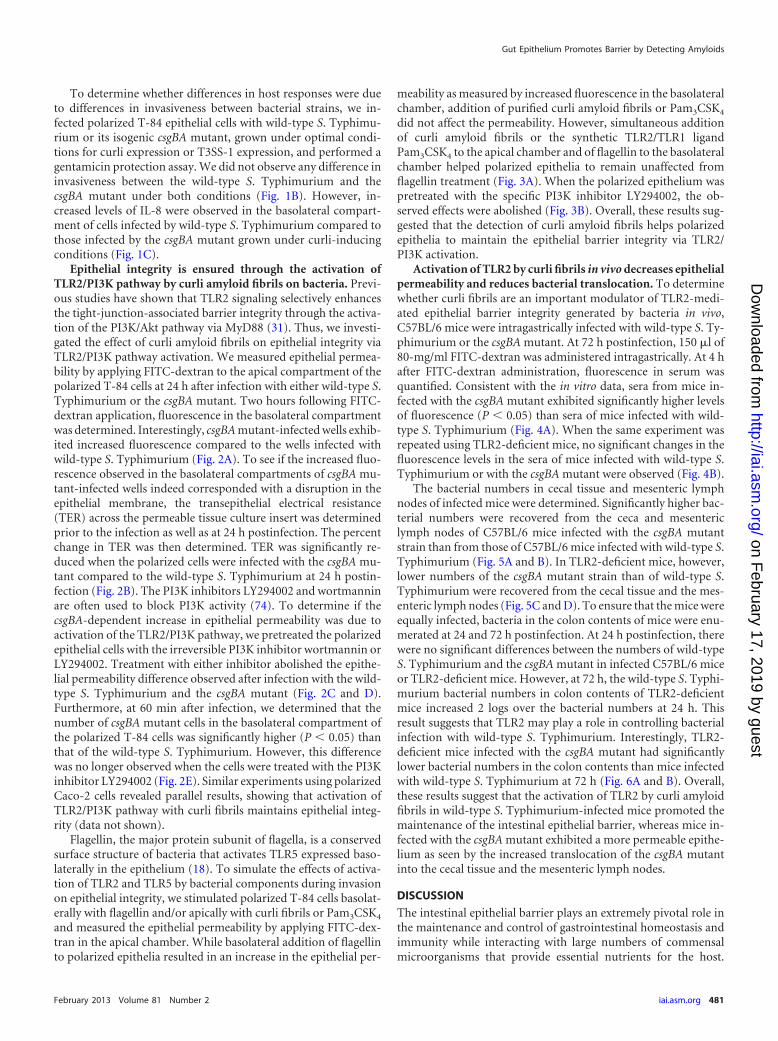
Epithelial integrity is ensured through the activation ofTLR2/PI3K pathway by curli amyloid fibrils on bacteria. Previ-ous studies have shown that TLR2 signaling selectively enhancesthe tight-junction-associated barrier integrity through the activa-tion of the PI3K/Akt pathway via MyD88 (31). Thus, we investi-gated the effect of curli amyloid fibrils on epithelial integrity viaTLR2/PI3K pathway activation. We measured epithelial permea-bility by applying FITC-dextran to the apical compartment of thepolarized T-84 cells at 24 h after infection with either wild-type S.Typhimurium or the csgBA mutant. Two hours following FITC-dextran application, fluorescence in the basolateral compartmentwas determined. Interestingly, csgBA mutant-infected wells exhib-ited increased fluorescence compared to the wells infected withwild-type S. Typhimurium (Fig. 2A). To see if the increased fluo-rescence observed in the basolateral compartments of csgBA mu-tant-infected wells indeed corresponded with a disruption in theepithelial membrane, the transepithelial electrical resistance(TER) across the permeable tissue culture insert was determinedprior to the infection as well as at 24 h postinfection. The percentchange in TER was then determined. TER was significantly re-duced when the polarized cells were infected with the csgBA mu-tant compared to the wild-type S. Typhimurium at 24 h postin-fection (Fig. 2B). The PI3K inhibitors LY294002 and wortmanninare often used to block PI3K activity (74). To determine if thecsgBA-dependent increase in epithelial permeability was due toactivation of the TLR2/PI3K pathway, we pretreated the polarizedepithelial cells with the irreversible PI3K inhibitor wortmannin orLY294002. Treatment with either inhibitor abolished the epithe-lial permeability difference observed after infection with the wild-type S. Typhimurium and the csgBA mutant (Fig. 2C and D).Furthermore, at 60 min after infection, we determined that thenumber of csgBA mutant cells in the basolateral compartment ofthe polarized T-84 cells was significantly higher (P � 0.05) thanthat of the wild-type S. Typhimurium. However, this differencewas no longer observed when the cells were treated with the PI3Kinhibitor LY294002 (Fig. 2E). Similar experiments using polarizedCaco-2 cells revealed parallel results, showing that activation ofTLR2/PI3K pathway with curli fibrils maintains epithelial integ-rity (data not shown).
Flagellin, the major protein subunit of flagella, is a conservedsurface structure of bacteria that activates TLR5 expressed baso-laterally in the epithelium (18). To simulate the effects of activa-tion of TLR2 and TLR5 by bacterial components during invasionon epithelial integrity, we stimulated polarized T-84 cells basolat-erally with flagellin and/or apically with curli fibrils or Pam3CSK4
and measured the epithelial permeability by applying FITC-dex-tran in the apical chamber. While basolateral addition of flagellinto polarized epithelia resulted in an increase in the epithelial per-
meability as measured by increased fluorescence in the basolateralchamber, addition of purified curli amyloid fibrils or Pam3CSK4
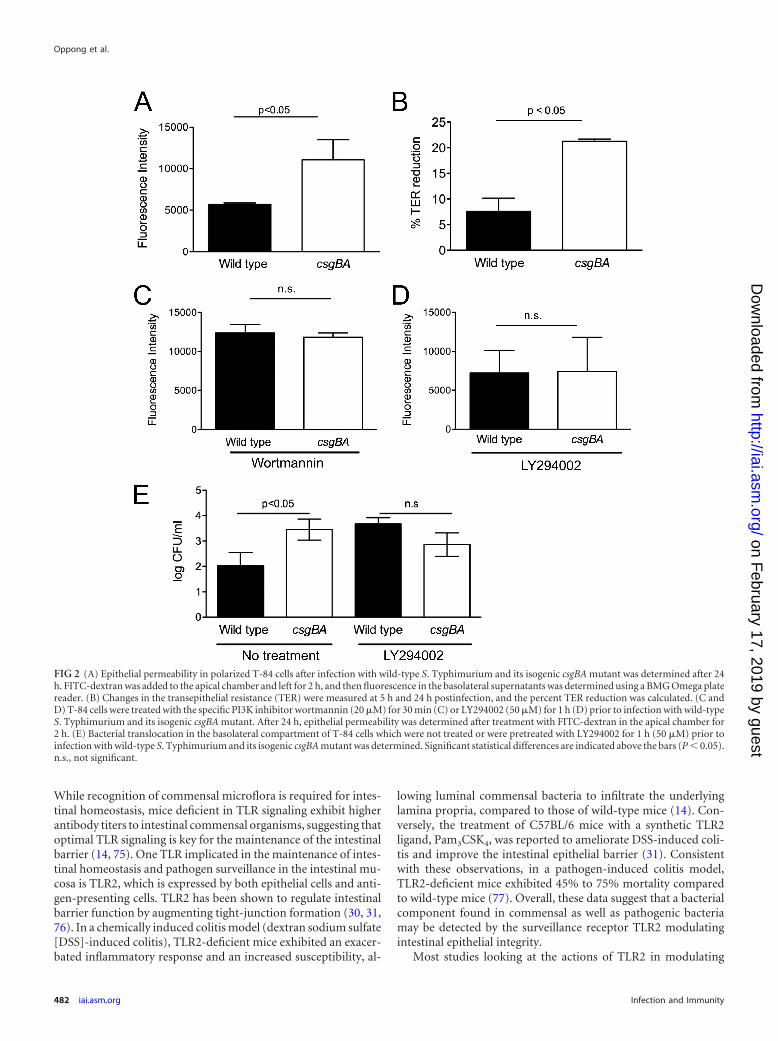
did not affect the permeability. However, simultaneous additionof curli amyloid fibrils or the synthetic TLR2/TLR1 ligandPam3CSK4 to the apical chamber and of flagellin to the basolateralchamber helped polarized epithelia to remain unaffected fromflagellin treatment (Fig. 3A). When the polarized epithelium waspretreated with the specific PI3K inhibitor LY294002, the ob-served effects were abolished (Fig. 3B). Overall, these results sug-gested that the detection of curli amyloid fibrils helps polarizedepithelia to maintain the epithelial barrier integrity via TLR2/PI3K activation.
Activation of TLR2 by curli fibrils in vivo decreases epithelialpermeability and reduces bacterial translocation. To determinewhether curli fibrils are an important modulator of TLR2-medi-ated epithelial barrier integrity generated by bacteria in vivo,C57BL/6 mice were intragastrically infected with wild-type S. Ty-phimurium or the csgBA mutant. At 72 h postinfection, 150 �l of80-mg/ml FITC-dextran was administered intragastrically. At 4 hafter FITC-dextran administration, fluorescence in serum wasquantified. Consistent with the in vitro data, sera from mice in-fected with the csgBA mutant exhibited significantly higher levelsof fluorescence (P � 0.05) than sera of mice infected with wild-type S. Typhimurium (Fig. 4A). When the same experiment wasrepeated using TLR2-deficient mice, no significant changes in thefluorescence levels in the sera of mice infected with wild-type S.Typhimurium or with the csgBA mutant were observed (Fig. 4B).
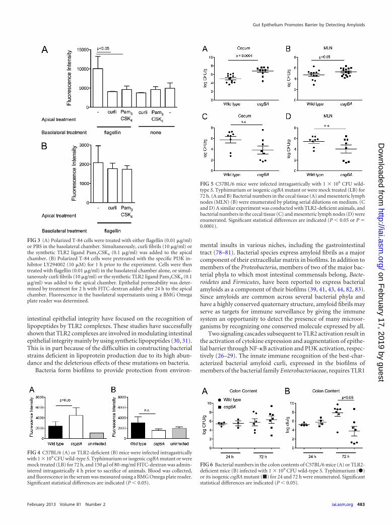
The bacterial numbers in cecal tissue and mesenteric lymphnodes of infected mice were determined. Significantly higher bac-terial numbers were recovered from the ceca and mesentericlymph nodes of C57BL/6 mice infected with the csgBA mutantstrain than from those of C57BL/6 mice infected with wild-type S.Typhimurium (Fig. 5A and B). In TLR2-deficient mice, however,lower numbers of the csgBA mutant strain than of wild-type S.Typhimurium were recovered from the cecal tissue and the mes-enteric lymph nodes (Fig. 5C and D). To ensure that the mice wereequally infected, bacteria in the colon contents of mice were enu-merated at 24 and 72 h postinfection. At 24 h postinfection, therewere no significant differences between the numbers of wild-typeS. Typhimurium and the csgBA mutant in infected C57BL/6 miceor TLR2-deficient mice. However, at 72 h, the wild-type S. Typhi-murium bacterial numbers in colon contents of TLR2-deficientmice increased 2 logs over the bacterial numbers at 24 h. Thisresult suggests that TLR2 may play a role in controlling bacterialinfection with wild-type S. Typhimurium. Interestingly, TLR2-deficient mice infected with the csgBA mutant had significantlylower bacterial numbers in the colon contents than mice infectedwith wild-type S. Typhimurium at 72 h (Fig. 6A and B). Overall,these results suggest that the activation of TLR2 by curli amyloidfibrils in wild-type S. Typhimurium-infected mice promoted themaintenance of the intestinal epithelial barrier, whereas mice in-fected with the csgBA mutant exhibited a more permeable epithe-lium as seen by the increased translocation of the csgBA mutantinto the cecal tissue and the mesenteric lymph nodes.
DISCUSSION
The intestinal epithelial barrier plays an extremely pivotal role inthe maintenance and control of gastrointestinal homeostasis andimmunity while interacting with large numbers of commensalmicroorganisms that provide essential nutrients for the host.
Gut Epithelium Promotes Barrier by Detecting Amyloids
February 2013 Volume 81 Number 2 iai.asm.org 481
on February 17, 2019 by guest
http://iai.asm.org/
Dow
nloaded from
While recognition of commensal microflora is required for intes-tinal homeostasis, mice deficient in TLR signaling exhibit higherantibody titers to intestinal commensal organisms, suggesting thatoptimal TLR signaling is key for the maintenance of the intestinalbarrier (14, 75). One TLR implicated in the maintenance of intes-tinal homeostasis and pathogen surveillance in the intestinal mu-cosa is TLR2, which is expressed by both epithelial cells and anti-gen-presenting cells. TLR2 has been shown to regulate intestinalbarrier function by augmenting tight-junction formation (30, 31,76). In a chemically induced colitis model (dextran sodium sulfate[DSS]-induced colitis), TLR2-deficient mice exhibited an exacer-bated inflammatory response and an increased susceptibility, al-
lowing luminal commensal bacteria to infiltrate the underlyinglamina propria, compared to those of wild-type mice (14). Con-versely, the treatment of C57BL/6 mice with a synthetic TLR2ligand, Pam3CSK4, was reported to ameliorate DSS-induced coli-tis and improve the intestinal epithelial barrier (31). Consistentwith these observations, in a pathogen-induced colitis model,TLR2-deficient mice exhibited 45% to 75% mortality comparedto wild-type mice (77). Overall, these data suggest that a bacterialcomponent found in commensal as well as pathogenic bacteriamay be detected by the surveillance receptor TLR2 modulatingintestinal epithelial integrity.
Most studies looking at the actions of TLR2 in modulating
FIG 2 (A) Epithelial permeability in polarized T-84 cells after infection with wild-type S. Typhimurium and its isogenic csgBA mutant was determined after 24h. FITC-dextran was added to the apical chamber and left for 2 h, and then fluorescence in the basolateral supernatants was determined using a BMG Omega platereader. (B) Changes in the transepithelial resistance (TER) were measured at 5 h and 24 h postinfection, and the percent TER reduction was calculated. (C andD) T-84 cells were treated with the specific PI3K inhibitor wortmannin (20 �M) for 30 min (C) or LY294002 (50 �M) for 1 h (D) prior to infection with wild-typeS. Typhimurium and its isogenic csgBA mutant. After 24 h, epithelial permeability was determined after treatment with FITC-dextran in the apical chamber for2 h. (E) Bacterial translocation in the basolateral compartment of T-84 cells which were not treated or were pretreated with LY294002 for 1 h (50 �M) prior toinfection with wild-type S. Typhimurium and its isogenic csgBA mutant was determined. Significant statistical differences are indicated above the bars (P � 0.05).n.s., not significant.
Oppong et al.
482 iai.asm.org Infection and Immunity
on February 17, 2019 by guest
http://iai.asm.org/
Dow
nloaded from
intestinal epithelial integrity have focused on the recognition oflipopeptides by TLR2 complexes. These studies have successfullyshown that TLR2 complexes are involved in modulating intestinalepithelial integrity mainly by using synthetic lipopeptides (30, 31).This is in part because of the difficulties in constructing bacterialstrains deficient in lipoprotein production due to its high abun-dance and the deleterious effects of these mutations on bacteria.
Bacteria form biofilms to provide protection from environ-
mental insults in various niches, including the gastrointestinaltract (78–81). Bacterial species express amyloid fibrils as a majorcomponent of their extracellular matrix in biofilms. In addition tomembers of the Proteobacteria, members of two of the major bac-terial phyla to which most intestinal commensals belong, Bacte-roidetes and Firmicutes, have been reported to express bacterialamyloids as a component of their biofilms (39, 41, 43, 44, 82, 83).Since amyloids are common across several bacterial phyla andhave a highly conserved quaternary structure, amyloid fibrils mayserve as targets for immune surveillance by giving the immunesystem an opportunity to detect the presence of many microor-ganisms by recognizing one conserved molecule expressed by all.
Two signaling cascades subsequent to TLR2 activation result inthe activation of cytokine expression and augmentation of epithe-lial barrier through NF-�B activation and PI3K activation, respec-tively (26–29). The innate immune recognition of the best-char-acterized bacterial amyloid curli, expressed in the biofilms ofmembers of the bacterial family Enterobacteriaceae, requires TLR1
FIG 3 (A) Polarized T-84 cells were treated with either flagellin (0.01 �g/ml)or PBS in the basolateral chamber. Simultaneously, curli fibrils (10 �g/ml) orthe synthetic TLR2 ligand Pam3CSK4 (0.1 �g/ml) was added to the apicalchamber. (B) Polarized T-84 cells were pretreated with the specific PI3K in-hibitor LY294002 (10 �M) for 1 h prior to the experiment. Cells were thentreated with flagellin (0.01 �g/ml) in the basolateral chamber alone, or simul-taneously curli fibrils (10 �g/ml) or the synthetic TLR2 ligand Pam3CSK4 (0.1�g/ml) was added to the apical chamber. Epithelial permeability was deter-mined by treatment for 2 h with FITC-dextran added after 24 h to the apicalchamber. Fluorescence in the basolateral supernatants using a BMG Omegaplate reader was determined.
FIG 4 C57BL/6 (A) or TLR2-deficient (B) mice were infected intragastricallywith 1 � 109 CFU wild-type S. Typhimurium or isogenic csgBA mutant or weremock treated (LB) for 72 h, and 150 �l of 80-mg/ml FITC-dextran was admin-istered intragastrically 4 h prior to sacrifice of animals. Blood was collected,and fluorescence in the serum was measured using a BMG Omega plate reader.Significant statistical differences are indicated (P � 0.05).
FIG 5 C57BL/6 mice were infected intragastrically with 1 � 109 CFU wild-type S. Typhimurium or isogenic csgBA mutant or were mock treated (LB) for72 h. (A and B) Bacterial numbers in the cecal tissue (A) and mesenteric lymphnodes (MLN) (B) were enumerated by plating serial dilutions on medium. (Cand D) A similar experiment was conducted with TLR2-deficient animals, andbacterial numbers in the cecal tissue (C) and mesenteric lymph nodes (D) wereenumerated. Significant statistical differences are indicated (P � 0.05 or P 0.0001).
FIG 6 Bacterial numbers in the colon contents of C57BL/6 mice (A) or TLR2-deficient mice (B) infected with 1 � 109 CFU wild-type S. Typhimurium (�)or its isogenic csgBA mutant (�) for 24 and 72 h were enumerated. Significantstatistical differences are indicated (P � 0.05).
Gut Epithelium Promotes Barrier by Detecting Amyloids
February 2013 Volume 81 Number 2 iai.asm.org 483
on February 17, 2019 by guest
http://iai.asm.org/
Dow
nloaded from
dimerization with TLR2 (48). Our recent investigations haveshown that the expression of curli amyloid fibrils by S. Typhimu-rium resulted in the expression of interleukin 17 (IL-17)/IL-22,produced by direct or indirect activation of T cells, via TLR2 acti-vation in the gastrointestinal tract of mice (60). Here, we havefocused on determining the effect of TLR2 activation by amyloidfibrils on epithelial cell function in the gastrointestinal tract. Al-though epithelial damage caused by both curliated wild-type S.Typhimurium and the csgBA mutant was evident in vitro and invivo, infection with the csgBA mutant caused more pronounceddamage to the epithelium, allowing more bacteria to translocate tothe basolateral side of the epithelium, and this effect was abolishedin the presence of PI3K inhibitors (Fig. 2) or in the absence ofTLR2 (Fig. 4), suggesting that the curli amyloid fibrils on bacteriaactivate the TLR2/PI3K pathway in intestinal epithelial cells, re-sulting in the reinforcement of the epithelial barrier.
During infection, multiple TLRs, including TLR2 and TLR5,found in the apical and basolateral sides of the epithelium, respec-tively, are activated (18, 71, 84). In this study, we mimicked theconditions that the epithelium encounters during bacterial infec-tion using purified ligands, curli amyloid fibrils, and flagellin. Ourin vitro results using polarized epithelial cells suggested that apicalactivation of TLR2 by curli amyloid fibrils restored the epithelialdamage introduced by basolateral flagellin treatment (Fig. 3).Therefore, the greater permeability seen in the in vitro experi-ments (Fig. 2) as well as in the gastrointestinal tracts of mice in-fected with the csgBA mutant in vivo (Fig. 4) is possibly due to thelack of TLR2 activation while typical TLR5 activation occurs,damaging the epithelium and allowing increased translocation ofbacteria.
Previous studies have shown that MyD88 and TLR2 deficiencyimpaired intestinal barrier repair during infection with anotherenteric pathogen, Citrobacter rodentium (77, 85). Although therewere no differences in the bacterial numbers in the colon contentsof both wild-type C57BL/6 and TLR2-deficient mice at 24 hpostinfection, consistent with these reports, we determined thatwild-type S. Typhimurium numbers were increased (P � 0.05),whereas csgBA mutant bacterial counts were significantly de-creased (P � 0.05), in the colon contents of the TLR2-deficientmice at 72 h postinfection compared to the bacterial numbers inC57BL/6 mice (Fig. 6A and B). Interestingly, curli fibrils are re-quired for efficient colonization of the intestinal epithelia by E. colistrains, while this phenotype is not observed with S. Typhimu-rium (47, 86–88). Recently, we determined that TLR2-deficientanimals acquire less inflammation during S. Typhimurium infec-tion, which could be measured by a decreased expression of IL-17A and IL-22 (60). Thus, under conditions of low or no inflam-mation, the presence of curli fibrils may provide the wild-type S.Typhimurium a colonization and growth advantage similar towhat is observed with E. coli. Nonetheless, we are currently inves-tigating the mechanism underlying this phenotype.
In the studies of chronic Citrobacter infection, the activation ofTLR2 was attributed to the recognition of bacterial lipoproteins(77, 89). To our interest, similar to the case for E. coli and S.Typhimurium, Citrobacter spp. also express curli fibrils (45). Eventhough, diacylated and triacylated bacterial lipoproteins havebeen shown to trigger TLR2 activation, when bacterial amyloidsare present, they exist as the predominant TLR2 ligand on bacte-ria, due to lipopeptides being buried in the outer membrane orbacterial cell wall while bacterial amyloids are being secreted to the
cell surface (48). In conclusion, our data point to bacterial amy-loids as a TLR2 ligand that enables epithelial cells to monitor bac-terial translocation from the gut.
ACKNOWLEDGMENTS
Work in C.T.’s laboratory was supported by Scientist Development grant0835248N from the American Heart Association and by Mid-AtlanticRegional Center for Excellence for Biodefense and Emerging InfectiousDiseases Research grant U54 AI57168 from the National Institutes ofHealth. G.J.R. was supported by a grant from the Pennsylvania Depart-ment of Health.
The Pennsylvania Department of Health specifically disclaims respon-sibility for any analyses, interpretations, and conclusions.
REFERENCES1. Xu J, Gordon JI. 2003. Honor thy symbionts. Proc. Natl. Acad. Sci.
U. S. A. 100:10452–10459.2. Turnbaugh PJ, Ley RE, Hamady M, Fraser-Liggett CM, Knight R,
Gordon JI. 2007. The human microbiome project. Nature 449:804 – 810.3. Gill SR, Pop M, Deboy RT, Eckburg PB, Turnbaugh PJ, Samuel BS,
Gordon JI, Relman DA, Fraser-Liggett CM, Nelson KE. 2006. Meta-genomic analysis of the human distal gut microbiome. Science 312:1355–1359.
4. Qin J, Li R, Raes J, Arumugam M, Burgdorf KS, Manichanh C, NielsenT, Pons N, Levenez F, Yamada T, Mende DR, Li J, Xu J, Li S, Li D, CaoJ, Wang B, Liang H, Zheng H, Xie Y, Tap J, Lepage P, Bertalan M, BattoJM, Hansen T, Le Paslier D, Linneberg A, Nielsen HB, Pelletier E,Renault P, Sicheritz-Ponten T, Turner K, Zhu H, Yu C, Jian M, ZhouY, Li Y, Zhang X, Qin N, Yang H, Wang J, Brunak S, Dore J, GuarnerF, Kristiansen K, Pedersen O, Parkhill J, Weissenbach J, Bork P, EhrlichSD. 2010. A human gut microbial gene catalogue established by meta-genomic sequencing. Nature 464:59 – 65.
5. Zhang T, Breitbart M, Lee WH, Run JQ, Wei CL, Soh SW, Hibberd ML,Liu ET, Rohwer F, Ruan Y. 2006. RNA viral community in human feces:prevalence of plant pathogenic viruses. PLoS Biol. 4:e3. doi:10.1371/journal.pbio.0040003.
6. Hooper LV. 2009. Do symbiotic bacteria subvert host immunity? Nat.Rev. Microbiol. 7:367–374.
7. Ayabe T, Satchell DP, Wilson CL, Parks WC, Selsted ME, Ouellette AJ.2000. Secretion of microbicidal alpha-defensins by intestinal Paneth cellsin response to bacteria. Nat. Immunol. 1:113–118.
8. Satchell DP, Sheynis T, Shirafuji Y, Kolusheva S, Ouellette AJ, JelinekR. 2003. Interactions of mouse Paneth cell alpha-defensins and alpha-defensin precursors with membranes. Prosegment inhibition of peptideassociation with biomimetic membranes. J. Biol. Chem. 278:13838 –13846.
9. Cunliffe RN, Rose FR, Keyte J, Abberley L, Chan WC, Mahida YR.2001. Human defensin 5 is stored in precursor form in normal Paneth cellsand is expressed by some villous epithelial cells and by metaplastic Panethcells in the colon in inflammatory bowel disease. Gut 48:176 –185.
10. Cash HL, Whitham CV, Behrendt CL, Hooper LV. 2006. Symbioticbacteria direct expression of an intestinal bactericidal lectin. Science 313:1126 –1130.
11. Christa L, Carnot F, Simon MT, Levavasseur F, Stinnakre MG, LasserreC, Thepot D, Clement B, Devinoy E, Brechot C. 1996. HIP/PAP is anadhesive protein expressed in hepatocarcinoma, normal Paneth, and pan-creatic cells. Am. J. Physiol. 271:G993–G1002.
12. Bergstrom KS, Kissoon-Singh V, Gibson DL, Ma C, Montero M, ShamHP, Ryz N, Huang T, Velcich A, Finlay BB, Chadee K, Vallance BA.2010. Muc2 protects against lethal infectious colitis by disassociatingpathogenic and commensal bacteria from the colonic mucosa. PLoS Pat-hog. 6:e1000902. doi:10.1371/journal.ppat.1000902.
13. Van der Sluis M, De Koning BA, De Bruijn AC, Velcich A, Meijerink JP,Van Goudoever JB, Buller HA, Dekker J, Van Seuningen I, Renes IB,Einerhand AW. 2006. Muc2-deficient mice spontaneously develop colitis,indicating that MUC2 is critical for colonic protection. Gastroenterology131:117–129.
14. Rakoff-Nahoum S, Paglino J, Eslami-Varzaneh F, Edberg S, MedzhitovR. 2004. Recognition of commensal microflora by Toll-like receptors isrequired for intestinal homeostasis. Cell 118:229 –241.
Oppong et al.
484 iai.asm.org Infection and Immunity
on February 17, 2019 by guest
http://iai.asm.org/
Dow
nloaded from
15. Akira S, Takeda K. 2004. Toll-like receptor signalling. Nat. Rev. Immu-nol. 4:499 –511.
16. Medzhitov R. 2007. Recognition of microorganisms and activation of theimmune response. Nature 449:819 – 826.
17. Takeda K, Akira S. 2007. Toll-like receptors. Curr. Protoc. Immunol.Chapter 14:Unit 14.12.
18. Gewirtz AT, Navas TA, Lyons S, Godowski PJ, Madara JL. 2001.Bacterial flagellin activates basolaterally expressed TLR5 to induce epithe-lial proinflammatory gene expression. J. Immunol. 167:1882–1885.
19. Takeuchi O, Hoshino K, Kawai T, Sanjo H, Takada H, Ogawa T,Takeda K, Akira S. 1999. Differential roles of TLR2 and TLR4 in recog-nition of gram-negative and gram-positive bacterial cell wall components.Immunity 11:443– 451.
20. Takeuchi O, Sato S, Horiuchi T, Hoshino K, Takeda K, Dong Z, ModlinRL, Akira S. 2002. Cutting edge: role of Toll-like receptor 1 in mediatingimmune response to microbial lipoproteins. J. Immunol. 169:10 –14.
21. Aliprantis AO, Yang RB, Mark MR, Suggett S, Devaux B, Radolf JD,Klimpel GR, Godowski P, Zychlinsky A. 1999. Cell activation and apop-tosis by bacterial lipoproteins through toll-like receptor-2. Science 285:736 –739.
22. Brightbill HD, Libraty DH, Krutzik SR, Yang RB, Belisle JT, BleharskiJR, Maitland M, Norgard MV, Plevy SE, Smale ST, Brennan PJ, BloomBR, Godowski PJ, Modlin RL. 1999. Host defense mechanisms triggeredby microbial lipoproteins through toll-like receptors. Science 285:732–736.
23. Takeda K, Akira S. 2004. TLR signaling pathways. Semin. Immunol.16:3–9.
24. Takeuchi O, Hoshino K, Akira S. 2000. TLR2-deficient and MyD88-deficient mice are highly susceptible to Staphylococcus aureus infection. J.Immunol. 165:5392–5396.
25. Takeuchi O, Kawai T, Muhlradt PF, Morr M, Radolf JD, Zychlinsky A,Takeda K, Akira S. 2001. Discrimination of bacterial lipoproteins byToll-like receptor 6. Int. Immunol. 13:933–940.
26. Franke TF, Kaplan DR, Cantley LC. 1997. PI3K: downstream AKTionblocks apoptosis. Cell 88:435– 437.
27. Santos-Sierra S, Deshmukh SD, Kalnitski J, Kuenzi P, Wymann MP,Golenbock DT, Henneke P. 2009. Mal connects TLR2 to PI3Kinaseactivation and phagocyte polarization. EMBO J. 28:2018 –2027.
28. Mansell A, Brint E, Gould JA, O’Neill LA, Hertzog PJ. 2004. Malinteracts with tumor necrosis factor receptor-associated factor (TRAF)-6to mediate NF-kappaB activation by Toll-like receptor (TLR)-2 and TLR4.J. Biol. Chem. 279:37227–37230.
29. Verstak B, Nagpal K, Bottomley SP, Golenbock DT, Hertzog PJ, Man-sell A. 2009. MyD88 adapter-like (Mal)/TIRAP interaction with TRAF6 iscritical for TLR2- and TLR4-mediated NF-kappaB proinflammatory re-sponses. J. Biol. Chem. 284:24192–24203.
30. Cario E, Gerken G, Podolsky DK. 2004. Toll-like receptor 2 enhancesZO-1-associated intestinal epithelial barrier integrity via protein kinase C.Gastroenterology 127:224 –238.
31. Cario E, Gerken G, Podolsky DK. 2007. Toll-like receptor 2 controlsmucosal inflammation by regulating epithelial barrier function. Gastro-enterology 132:1359 –1374.
32. Podolsky DK, Gerken G, Eyking A, Cario E. 2009. Colitis-associatedvariant of TLR2 causes impaired mucosal repair because of TFF3 defi-ciency. Gastroenterology 137:209 –220.
33. Leonhardt RM, Vigneron N, Rahner C, Van den Eynde BJ, Cresswell P.2010. Endoplasmic reticulum export, subcellular distribution, and fibrilformation by Pmel17 require an intact N-terminal domain junction. J.Biol. Chem. 285:16166 –16183.
34. Pfefferkorn CM, McGlinchey RP, Lee JC. 2010. Effects of pH on aggre-gation kinetics of the repeat domain of a functional amyloid, Pmel17.Proc. Natl. Acad. Sci. U. S. A. 107:21447–21452.
35. Theos AC, Truschel ST, Raposo G, Marks MS. 2005. The Silver locusproduct Pmel17/gp100/Silv/ME20: controversial in name and in function.Pigment Cell Res. 18:322–336.
36. Aigelsreiter A, Janig E, Stumptner C, Fuchsbichler A, Zatloukal K,Denk H. 2007. How a cell deals with abnormal proteins. Pathogeneticmechanisms in protein aggregation diseases. Pathobiology 74:145–158.
37. Hull RL, Westermark GT, Westermark P, Kahn SE. 2004. Islet amyloid:a critical entity in the pathogenesis of type 2 diabetes. J. Clin. Endocrinol.Metab. 89:3629 –3643.
38. Brandan E, Inestrosa NC. 1993. Extracellular matrix components and
amyloid in neuritic plaques of Alzheimer’s disease. Gen. Pharmacol. 24:1063–1068.
39. Romero D, Aguilar C, Losick R, Kolter R. 2010. Amyloid fibers providestructural integrity to Bacillus subtilis biofilms. Proc. Natl. Acad. Sci.U. S. A. 107:2230 –2234.
40. Alteri CJ, Xicohtencatl-Cortes J, Hess S, Caballero-Olin G, Giron JA,Friedman RL. 2007. Mycobacterium tuberculosis produces pili duringhuman infection. Proc. Natl. Acad. Sci. U. S. A. 104:5145–5150.
41. Dueholm MS, Petersen SV, Sonderkaer M, Larsen P, Christiansen G,Hein KL, Enghild JJ, Nielsen JL, Nielsen KL, Nielsen PH, Otzen DE.2010. Functional amyloid in Pseudomonas. Mol. Microbiol. 77:1009 –1020.
42. Blanco LP, Evans ML, Smith DR, Badtke MP, Chapman MR. 2012.Diversity, biogenesis and function of microbial amyloids. Trends Micro-biol. 20:66 –73.
43. Chapman MR, Robinson LS, Pinkner JS, Roth R, Heuser J, Hammar M,Normark S, Hultgren SJ. 2002. Role of Escherichia coli curli operons indirecting amyloid fiber formation. Science 295:851– 855.
44. Larsen P, Nielsen JL, Dueholm MS, Wetzel R, Otzen D, Nielsen PH.2007. Amyloid adhesins are abundant in natural biofilms. Environ. Mi-crobiol. 9:3077–3090.
45. Zogaj X, Bokranz W, Nimtz M, Romling U. 2003. Production of cellu-lose and curli fimbriae by members of the family Enterobacteriaceae iso-lated from the human gastrointestinal tract. Infect. Immun. 71:4151–4158.
46. Collinson SK, Clouthier SC, Doran JL, Banser PA, Kay WW. 1996.Salmonella enteritidis agfBAC operon encoding thin, aggregative fim-briae. J. Bacteriol. 178:662– 667.
47. Kai-Larsen Y, Luthje P, Chromek M, Peters V, Wang X, Holm A, KadasL, Hedlund KO, Johansson J, Chapman MR, Jacobson SH, Romling U,Agerberth B, Brauner A. 2010. Uropathogenic Escherichia coli modu-lates immune responses and its curli fimbriae interact with the antimicro-bial peptide LL-37. PLoS Pathog. 6:e1001010. doi:10.1371/journal.ppat.1001010.
48. Tukel C, Nishimori JH, Wilson RP, Winter MG, Keestra AM, vanPutten JP, Baumler AJ. 2010. Toll-like receptors 1 and 2 cooperativelymediate immune responses to curli, a common amyloid from enterobac-terial biofilms. Cell. Microbiol. 12:1495–1505.
49. Tukel C, Raffatellu M, Humphries AD, Wilson RP, Andrews-PolymenisHL, Gull T, Figueiredo JF, Wong MH, Michelsen KS, Akcelik M, AdamsLG, Baumler AJ. 2005. CsgA is a pathogen-associated molecular patternof Salmonella enterica serotype Typhimurium that is recognized by Toll-like receptor 2. Mol. Microbiol. 58:289 –304.
50. Tukel C, Wilson RP, Nishimori JH, Pezeshki M, Chromy BA, BaumlerAJ. 2009. Responses to amyloids of microbial and host origin are mediatedthrough Toll-like receptor 2. Cell Host Microbe 6:45–53.
51. Bian Z, Brauner A, Li Y, Normark S. 2000. Expression of and cytokineactivation by Escherichia coli curli fibers in human sepsis. J. Infect. Dis.181:602– 612.
52. Bian Z, Yan ZQ, Hansson GK, Thoren P, Normark S. 2001. Activationof inducible nitric oxide synthase/nitric oxide by curli fibers leads to a fallin blood pressure during systemic Escherichia coli infection in mice. J.Infect. Dis. 183:612– 619.
53. Cheng N, He R, Tian J, Ye PP, Ye RD. 2008. TLR2 is a functionalreceptor for acute-phase serum amyloid A. J. Immunol. 181:22–26.
54. He RL, Zhou J, Hanson CZ, Chen J, Cheng N, Ye RD. 2009. Serumamyloid A induces G-CSF expression and neutrophilia via Toll-like recep-tor 2. Blood 113:429 – 437.
55. Jana M, Palencia CA, Pahan K. 2008. Fibrillar amyloid-beta peptidesactivate microglia via TLR2: implications for Alzheimer’s disease. J. Im-munol. 181:7254 –7262.
56. Reed-Geaghan EG, Savage JC, Hise AG, Landreth GE. 2009. CD14 andToll-like receptors 2 and 4 are required for fibrillar A�-stimulated micro-glial activation. J. Neurosci. 29:11982–11992.
57. Udan ML, Ajit D, Crouse NR, Nichols MR. 2008. Toll-like receptors 2and 4 mediate Abeta(1-42) activation of the innate immune response in ahuman monocytic cell line. J. Neurochem. 104:524 –533.
58. Lay C, Sutren M, Rochet V, Saunier K, Dore J, Rigottier-Gois L. 2005.Design and validation of 16S rRNA probes to enumerate members of theClostridium leptum subgroup in human faecal microbiota. Environ. Mi-crobiol. 7:933–946.
59. Stojiljkovic I, Baumler AJ, Heffron F. 1995. Ethanolamine utilization inSalmonella typhimurium: nucleotide sequence, protein expression, and
Gut Epithelium Promotes Barrier by Detecting Amyloids
February 2013 Volume 81 Number 2 iai.asm.org 485
on February 17, 2019 by guest
http://iai.asm.org/
Dow
nloaded from
mutational analysis of the cchA cchB eutE eutJ eutG eutH gene cluster. J.Bacteriol. 177:1357–1366.
60. Nishimori JH, Newman TN, Oppong GO, Rapsinski GJ, Yen JH,Biesecker SG, Wilson RP, Butler BP, Winter MG, Tsolis RM, Ganea D,Tukel C. 2012. Microbial amyloids induce interleukin 17A (IL-17A) andIL-22 responses via Toll-like receptor 2 activation in the intestinal mucosa.Infect. Immun. 80:4398 – 4408.
61. Collinson SK, Emody L, Muller KH, Trust TJ, Kay WW. 1991. Purifi-cation and characterization of thin, aggregative fimbriae from Salmonellaenteritidis. J. Bacteriol. 173:4773– 4781.
62. Madara JL, Dharmsathaphorn K. 1985. Occluding junction structure-function relationships in a cultured epithelial monolayer. J. Cell Biol. 101:2124 –2133.
63. Lambert D, O’Neill CA, Padfield PJ. 2005. Depletion of Caco-2 cellcholesterol disrupts barrier function by altering the detergent solubilityand distribution of specific tight-junction proteins. Biochem. J. 387:553–560.
64. Cario E, Podolsky DK. 2000. Differential alteration in intestinal epithelialcell expression of Toll-like receptor 3 (TLR3) and TLR4 in inflammatorybowel disease. Infect. Immun. 68:7010 –7017.
65. Abreu MT, Vora P, Faure E, Thomas LS, Arnold ET, Arditi M. 2001.Decreased expression of Toll-like receptor-4 and MD-2 correlates withintestinal epithelial cell protection against dysregulated proinflammatorygene expression in response to bacterial lipopolysaccharide. J. Immunol.167:1609 –1616.
66. Abreu MT. 2010. Toll-like receptor signalling in the intestinal epithelium:how bacterial recognition shapes intestinal function. Nat. Rev. Immunol.10:131–144.
67. Chabot S, Wagner JS, Farrant S, Neutra MR. 2006. TLRs regulate thegatekeeping functions of the intestinal follicle-associated epithelium. J.Immunol. 176:4275– 4283.
68. Chantret I, Barbat A, Dussaulx E, Brattain MG, Zweibaum A. 1988.Epithelial polarity, villin expression, and enterocytic differentiation of cul-tured human colon carcinoma cells: a survey of twenty cell lines. CancerRes. 48:1936 –1942.
69. Hidalgo IJ, Raub TJ, Borchardt RT. 1989. Characterization of the humancolon carcinoma cell line (Caco-2) as a model system for intestinal epithe-lial permeability. Gastroenterology 96:736 –749.
70. Galan JE, Curtiss R, III. 1989. Cloning and molecular characterization ofgenes whose products allow Salmonella typhimurium to penetrate tissueculture cells. Proc. Natl. Acad. Sci. U. S. A. 86:6383– 6387.
71. Gewirtz AT, Simon PO, Jr, Schmitt CK, Taylor LJ, Hagedorn CH,O’Brien AD, Neish AS, Madara JL. 2001. Salmonella typhimuriumtranslocates flagellin across intestinal epithelia, inducing a proinflamma-tory response. J. Clin. Invest. 107:99 –109.
72. Bruno VM, Hannemann S, Lara-Tejero M, Flavell RA, Kleinstein SH,Galan JE. 2009. Salmonella Typhimurium type III secretion effectorsstimulate innate immune responses in cultured epithelial cells. PLoS Pat-hog. 5:e1000538. doi:10.1371/journal.ppat.1000538.
73. Takatori H, Kanno Y, Watford WT, Tato CM, Weiss G, Ivanov II,Littman DR, O’Shea JJ. 2009. Lymphoid tissue inducer-like cells are aninnate source of IL-17 and IL-22. J. Exp. Med. 206:35– 41.
74. Walker EH, Pacold ME, Perisic O, Stephens L, Hawkins PT, WymannMP, Williams RL. 2000. Structural determinants of phosphoinositide3-kinase inhibition by wortmannin, LY294002, quercetin, myricetin, andstaurosporine. Mol. Cell 6:909 –919.
75. Slack E, Hapfelmeier S, Stecher B, Velykoredko Y, Stoel M, Lawson MA,Geuking MB, Beutler B, Tedder TF, Hardt WD, Bercik P, Verdu EF,
McCoy KD, Macpherson AJ. 2009. Innate and adaptive immunity coop-erate flexibly to maintain host-microbiota mutualism. Science 325:617–620.
76. Ey B, Eyking A, Gerken G, Podolsky DK, Cario E. 2009. TLR2 mediatesgap junctional intercellular communication through connexin-43 in in-testinal epithelial barrier injury. J. Biol. Chem. 284:22332–22343.
77. Gibson DL, MA C, Rosenberger CM, Bergstrom KS, Valdez Y, HuangJT, Khan MA, Vallance BA. 2008. Toll-like receptor 2 plays a critical rolein maintaining mucosal integrity during Citrobacter rodentium-inducedcolitis. Cell. Microbiol. 10:388 – 403.
78. Bollinger RR, Everett ML, Wahl SD, Lee YH, Orndorff PE, Parker W.2006. Secretory IgA and mucin-mediated biofilm formation by environ-mental strains of Escherichia coli: role of type 1 pili. Mol. Immunol. 43:378 –387.
79. Swidsinski A, Loening-Baucke V, Lochs H, Hale LP. 2005. Spatialorganization of bacterial flora in normal and inflamed intestine: a fluores-cence in situ hybridization study in mice. World J. Gastroenterol. 11:1131–1140.
80. Swidsinski A, Schlien P, Pernthaler A, Gottschalk U, Barlehner E,Decker G, Swidsinski S, Strassburg J, Loening-Baucke V, Hoffmann U,Seehofer D, Hale LP, Lochs H. 2005. Bacterial biofilm within diseasedpancreatic and biliary tracts. Gut 54:388 –395.
81. Swidsinski A, Weber J, Loening-Baucke V, Hale LP, Lochs H. 2005.Spatial organization and composition of the mucosal flora in patients withinflammatory bowel disease. J. Clin. Microbiol. 43:3380 –3389.
82. Jordal PB, Dueholm MS, Larsen P, Petersen SV, Enghild JJ, Christian-sen G, Hojrup P, Nielsen PH, Otzen DE. 2009. Widespread abundanceof functional bacterial amyloid in mycolata and other gram-positive bac-teria. Appl. Environ. Microbiol. 75:4101– 4110.
83. Larsen P, Nielsen JL, Otzen D, Nielsen PH. 2008. Amyloid-like adhesinsproduced by floc-forming and filamentous bacteria in activated sludge.Appl. Environ. Microbiol. 74:1517–1526.
84. Cario E, Brown D, McKee M, Lynch-Devaney K, Gerken G, PodolskyDK. 2002. Commensal-associated molecular patterns induce selectiveToll-like receptor-trafficking from apical membrane to cytoplasmic com-partments in polarized intestinal epithelium. Am. J. Pathol. 160:165–173.
85. Gibson DL, Ma C, Bergstrom KS, Huang JT, Man C, Vallance BA. 2008.MyD88 signalling plays a critical role in host defence by controlling patho-gen burden and promoting epithelial cell homeostasis during Citrobacterrodentium-induced colitis. Cell. Microbiol. 10:618 – 631.
86. Torres AG, Cieza RJ, Rojas-Lopez M, Blumentritt CA, Souza CS,Johnston RK, Strockbine N, Kaper JB, Sbrana E, Popov VL. 2012. Invivo bioluminescence imaging of Escherichia coli O104:H4 and role ofaerobactin during colonization of a mouse model of infection. BMC Mi-crobiol. 12:112.
87. Wang X, Rochon M, Lamprokostopoulou A, Lunsdorf H, Nimtz M,Romling U. 2006. Impact of biofilm matrix components on interaction ofcommensal Escherichia coli with the gastrointestinal cell line HT-29. Cell.Mol. Life Sci. 63:2352–2363.
88. Weening EH, Barker JD, Laarakker MC, Humphries AD, Tsolis RM,Baumler AJ. 2005. The Salmonella enterica serotype Typhimurium lpf,bcf, stb, stc, std, and sth fimbrial operons are required for intestinal per-sistence in mice. Infect. Immun. 73:3358 –3366.
89. Gibson DL, Montero M, Ropeleski MJ, Bergstrom KS, Ma C, Ghosh S,Merkens H, Huang J, Mansson LE, Sham HP, McNagny KM, VallanceBA. 2010. Interleukin-11 reduces TLR4-induced colitis in TLR2-deficientmice and restores intestinal STAT3 signaling. Gastroenterology 139:1277–1288.
Oppong et al.
486 iai.asm.org Infection and Immunity
on February 17, 2019 by guest
http://iai.asm.org/
Dow
nloaded from


![MaXingShiGanDecoctionProtectsagainstPM2.5-InducedLung ... · 2020. 6. 17. · pathogens [13]. e epithelial barrier function largely de-pends on the tight junction surrounding epithelial](https://img.pdfslide.net/doc/110x75/611bf06c9b84416f1939a63f/maxingshigandecoctionprotectsagainstpm25-inducedlung-2020-6-17-pathogens.jpg)