Embed Size (px)
Citation preview
MEDICINE
EPSTEIN-BARR VIRUS AND CHLAMYDIA TRACHOMATIS AS COFACTORS IN CERVICAL CARCINOGENESIS
CRISTINA DANIELA GOIAa, IULIA IANCUa, IRINA HUICAa, ANCA BOTEZATUa, DEMETRA SOCOLOVb, SERGIU TELEMANb, ANDA DRAGODANa, CONSTANŢA ANTIPAa and GABRIELA ANTONa
a “Stefan S. Nicolau” Institute of Virology, 285, Mihai Bravu Ave., 030304, Bucharest, Romania b “Grigore T. Popa” University of Medicine and Pharmacy, Iassy, Romania
Corresponding Author: Cristina Daniela Goia, [email protected]
Received Mars 11, 2009
High risk human papillomaviruses (HPV) are known to be the etiological agents of cervical cancer disease, but other cofactors are considered to be important in cervix carcinogenesis. In order to evaluate the role of Epstein-Barr Virus (EBV) and Chlamydia trachomatis infections in cervical carcinogenesis, cervical smears collected in liquid cytology medium from 32 women (29–55 years old) were investigated. Total genomic DNAs (High Pure PCR Template, Roche) and RNA (RNAEasy Mini Kit, Qiagen) were isolated from scrape specimens from patients with different citology (normal cervical epithelium, ASCUS, LGSIL and HGSIL). Total DNAs were tested for HPV DNA presence (Linear Array HPV Genotyping Test, Roche) and C. trachomatis DNA presence (Chlamydia trachomatis Real-TM kit, Sacace Biotechnologies). To elucidate a causative role of EBV in cervical lesions, EBNA1, LMP1 and BZLF1 mRNA gene expression were performed using TaqMan Real Time PCR assays (Applied Biosystems). Our results showed a low incidence of C. trachomatis infection, only 2 cases of NILM presenting C. trachomatis DNA. EBNA1 and BZLF1 expression were relatively high in most cases suggesting a possible role of EBV in cervical lesion progression. HPV DNA was detected in 22.3% of NILM, 33.3% of ASCUS, 75% of LGSIL and 77.8% of HGSIL cases, demonstating the high frequency of HPV infection. Therefore, we suggest that EBV may play a role in cervical precursor lesions but the mechanism of carcinogenesis remains to be further investigated.
Key words: Cervical cancer; HPV; EBV; Chlamydia trachomatis.
INTRODUCTION
Cervical carcinoma is one of the most common types of cancer worldwide and one of the leading causes of death from cancer among women. High risk human papillomaviruses (hrHPV) are recognized as significant sexually transmitted etiological agents of cervical neoplastic lesions and subsequently cervical cancer1. Apart from HPV, other cofactors are implicated in cervical carcinogenesis, including Chlamydia trachomatis (CT) and Epstein-Barr virus (EBV) infections which reportedly facilitate the development of invasive squamous cell carcinoma.
Proc. Rom. Acad., Series B, 2009, 1, p. 9–14
Although CT infection was described as an independent risk factor for the development and progression of cervical lesions towards invasive cervical carcinoma, past infection with CT was implicated as surrogate marker for exposure to HPV, and according to some authors synergized with HPV infection in the development and acceleration of invasive cervical carcinoma. Other authors disputed any association between HPV and CT infection, and rather suggested that HPV and CT are independently occurring agents2.
On the other hand, EBV is considered as a causative agent for Burrkitt’s lymphoma and malignant B lymphocytes proliferation3. Recent
Cristina Daniela Goia et al. 10
studies revealed the striking association of EBV infection with the development of cervical tumors. The immediate–early gene products of EBV can transactivate other viral and cellular genes and it has been suggested that concurrent genital
infection with EBV and HPV might increase the risk for cervical cancer development4.
Here we investigated whether the two cofactors play an independent or synergic role in cervical lesions progression.
MATERIALS AND METHODS
Study group and Biological Samples
32 women (29–55 years old) referred to the Gynecology Clinic, Iassy for the management of abnormal cervical cytology were recruited with written informed consent. Patients with normal colposcopy had follow up cytology performed at three to six month intervals. For the purpose of our study, women with normal colposcopy who had normal cytology results throughout the subsequent 12 month follow up period were classified as normal. The study protocol was approved by the local institution ethics committee.
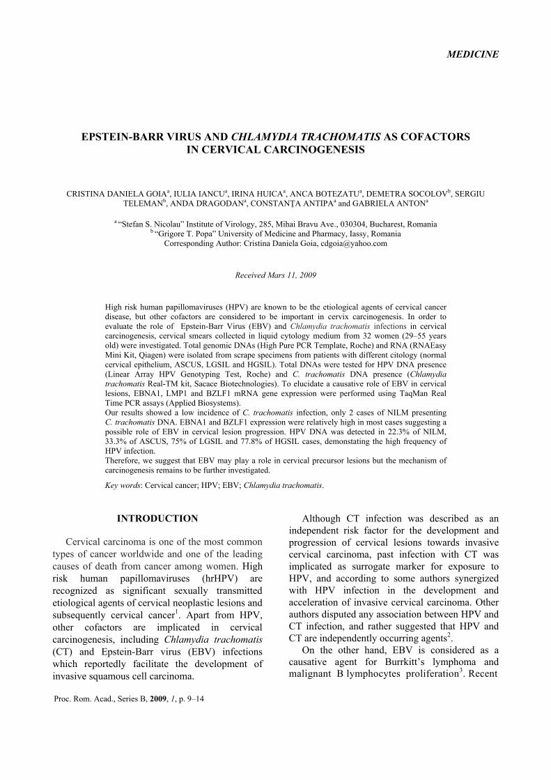
In order to evaluate the role of EBV and C. trachomatis infection in cervical lesions progression, cervical smears collected in liquid cytology medium from patients were investigated. Total genomic DNA (High Pure PCR Template, Roche) and RNA (RNAEasy Mini Kit, Qiagen) were isolated from scrape specimens from patients with normal cervical epithelium and ASCUS, LGSIL and HGSIL cytology (Fig. 1).
DNA was released by lysing cervical cell specimens under denaturating conditions at elevated temperatures. Lysis was performed in the presence of proteinase K, chaotropic agent and detergent and was followed by isolation and purification of DNA over a column and elution with elution reagent. The quality of isolated RNAs and DNAs was assessed by determining the concentration and purity using Nanodrop spectrophotometer.
Fig. 1. Cytology distribution in the study group. NILM – Negative for Intraepithelial Lesion and Malignancy; ASCUS – Atypical Squamous Cells of Undetermined Significance, LGSIL – Low Grade Squamous Intraepithelial Lesion, HGSIL –
High Grade Squamous Intraepithelial Lesion.
HPV DNA Detection and Genotyping
HPV genotyping was performed with Linear Array HPV Genotyping Test, Roche according to the manufacturer’s instructions. This test uses biotinylated primers to define a
sequence of nucleotides within the polymorphic L1 region of the HPV genome of approximately 450 base pairs long. A pool of HPV primers present in the Master Mix is designed to amplify HPV DNA from 37 HPV genotypes including 13 high risk genotypes (16, 18, 31, 33, 35, 39, 45, 51, 52, 56, 58, 59 and 68). An additional primer pair targets the human β-globin gene to provide a control for cell adequacy, extraction and amplification.
Reverse-transcription
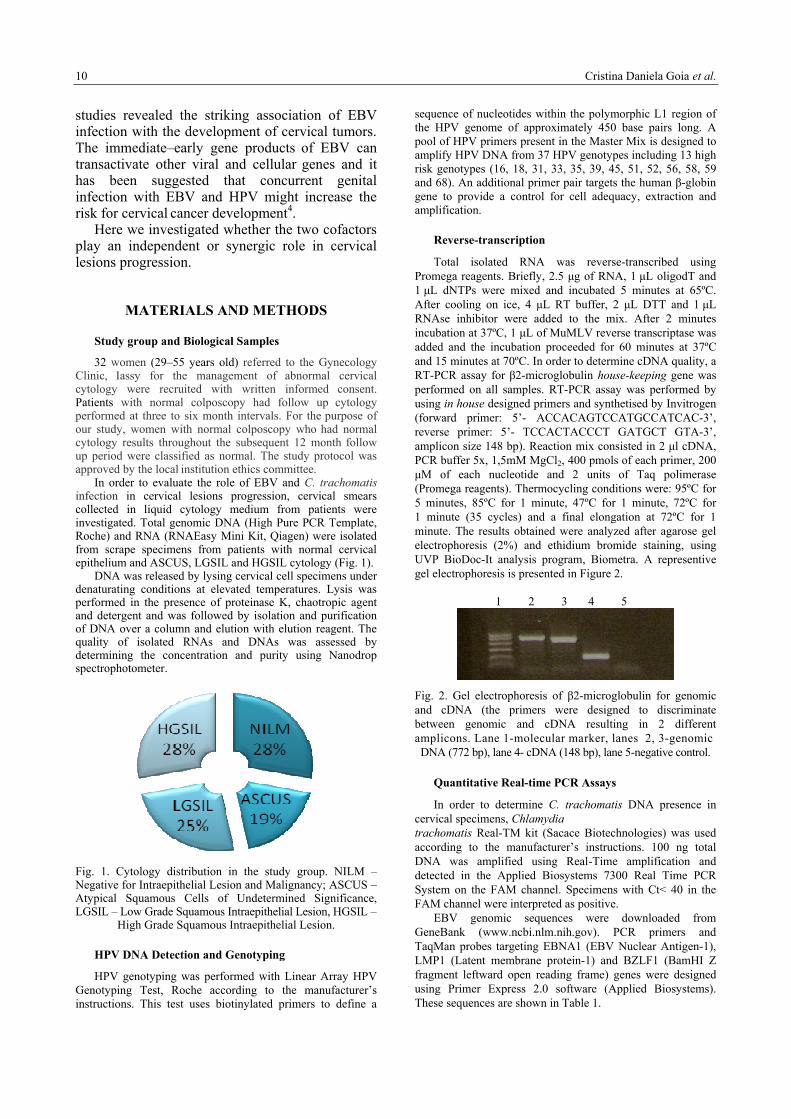
Total isolated RNA was reverse-transcribed using Promega reagents. Briefly, 2.5 µg of RNA, 1 µL oligodT and 1 µL dNTPs were mixed and incubated 5 minutes at 65ºC. After cooling on ice, 4 µL RT buffer, 2 µL DTT and 1 µL RNAse inhibitor were added to the mix. After 2 minutes incubation at 37ºC, 1 µL of MuMLV reverse transcriptase was added and the incubation proceeded for 60 minutes at 37ºC and 15 minutes at 70ºC. In order to determine cDNA quality, a RT-PCR assay for β2-microglobulin house-keeping gene was performed on all samples. RT-PCR assay was performed by using in house designed primers and synthetised by Invitrogen (forward primer: 5’- ACCACAGTCCATGCCATCAC-3’, reverse primer: 5’- TCCACTACCCT GATGCT GTA-3’, amplicon size 148 bp). Reaction mix consisted in 2 µl cDNA, PCR buffer 5x, 1,5mM MgCl2, 400 pmols of each primer, 200 µM of each nucleotide and 2 units of Taq polimerase (Promega reagents). Thermocycling conditions were: 95ºC for 5 minutes, 85ºC for 1 minute, 47ºC for 1 minute, 72ºC for 1 minute (35 cycles) and a final elongation at 72ºC for 1 minute. The results obtained were analyzed after agarose gel electrophoresis (2%) and ethidium bromide staining, using UVP BioDoc-It analysis program, Biometra. A representive gel electrophoresis is presented in Figure 2.
1 2 3 4 5
Fig. 2. Gel electrophoresis of β2-microglobulin for genomic and cDNA (the primers were designed to discriminate between genomic and cDNA resulting in 2 different amplicons. Lane 1-molecular marker, lanes 2, 3-genomic DNA (772 bp), lane 4- cDNA (148 bp), lane 5-negative control.
Quantitative Real-time PCR Assays
In order to determine C. trachomatis DNA presence in cervical specimens, Chlamydia trachomatis Real-TM kit (Sacace Biotechnologies) was used according to the manufacturer’s instructions. 100 ng total DNA was amplified using Real-Time amplification and detected in the Applied Biosystems 7300 Real Time PCR System on the FAM channel. Specimens with Ct< 40 in the FAM channel were interpreted as positive.
EBV genomic sequences were downloaded from GeneBank (www.ncbi.nlm.nih.gov). PCR primers and TaqMan probes targeting EBNA1 (EBV Nuclear Antigen-1), LMP1 (Latent membrane protein-1) and BZLF1 (BamHI Z fragment leftward open reading frame) genes were designed using Primer Express 2.0 software (Applied Biosystems). These sequences are shown in Table 1.
Epstein-Barr virus and Chlamydia trachomatis as cofactors in cervical carcinogenesis 11
Table 1
Sequences of primers and TaqMan probes used for real-time PCR
LMP1 Forward 5'-CAG TCA GGC AAG CCT ATG A-3'
Reverse 5'-CTG GTT CCG GTG GAG ATG A-3'
Probe 5'-(6FAM)GTC ATA GTA GCT TAG CTG AAC(TAMRA)-3'
Amplicon
size
104 bp
BZLF1 Forward 5'-AAA TTT AAG AGA TCC TCG TGT AAA ACA TC-3'
Reverse 5'-CGC CTC CTG TTG AAG CAG AT-3'
Probe 5'-(6FAM)ATA ATG GAG TCA ACA TCC AGG CTT GGG C(TAMRA)-3'
Amplicon
size
91 bp
EBNA1 Forward 5'-TAC AGG ACC TGG AAA TGG CC-3'
Reverse 5'-TCT TTG AGG TCC ACT GCC G-3'
Probe 5'-(6FAM)AGG GAG ACA CAT CTG GAC CAG AAG GC(TAMRA)-3'
Amplicon
size
78 bp
PCR was performed and products were detected using
ABI prism 7300 Real-Time PCR instrument and Sequence Detection System software. Thermocycling conditions were: 50ºC for 2 minutes, 95ºC for 10 minutes, 95ºC for 15 seconds and 60ºC for 1 minute for 40 cycles. Each 25-µL reaction contained: 1X TaqMan Universal MasterMix, forward and reverse primer (15 pmol each) and TaqMan probe (10 pmol). DNA template volume was of 2 µL.
To check for amplicon contamination, every run contained at least “no template” controls in which nuclease-free water was substituted for template. As positive controls, cDNA from B95 and Namalwa cell lines were used. Samples were tested in duplicate in each Q-PCR assay and specimens with Ct< 36 were interpreted as positive.
RESULTS AND DISCUSSIONS
There is strong evidence that the uterine cervix is the site of Epstein-Barr virus (EBV) shedding and some authors have proposed that EBV could play a role in the carcinogenesis of cervical tumors5. On the other hand, Chlamydia trachomatis is the most common sexually transmitted bacterial infection in the world and it may also be involved in cervical lesions progression towards malignancy.
The aim of cervical cancer prevention is the early diagnosis of its etiological agent and the identification of specific markers of cervical lesions progression. Infection of epithelial squamous cells with high-risk HPV represents an
essential request for cervical cancer development6. HPV E6 and E7 proteins can act as oncogenes. By binding p53, HPV oncoprotein E6 can prevent DNA repair and p53-mediated apoptosis. HPV oncoprotein E7 binds and inactivates pRB thereby promoting uncontrolled cell division7.
Studies demonstrated that HPV DNA presence is necessary for the development and persistency of cervical neoplasia and DNA disappearance anticipates the regression of neoplastic cells, even in an advanced cervical lesion (HGSIL)8. Even though cervical lesions may be detected on cervical smears several years before the onset of invasive squamous carcinoma, there is no morphologic criterion that may predict if a cervical lesion will disappear or will progress toward cancer. High risk DNA HPV detection in such lesions selects women with high risk for cervical cancer development9.
Regarding HPV detection and genotyping, the present study revealed the fact that HPV was widely spread in our study group (Fig. 3). HPV DNA was detected in 22.3% of NILM, 33.3% of ASCUS, 75% of LGSIL and 77.8% of HGSIL cases, demonstrating the high frequency of HPV infection. Our data also showed a rather high rate of single infections in LGSIL and HGSIL cases (Table 2). In HGSIL cases, single infections were dominant with the prevalent HPV16 genotype.
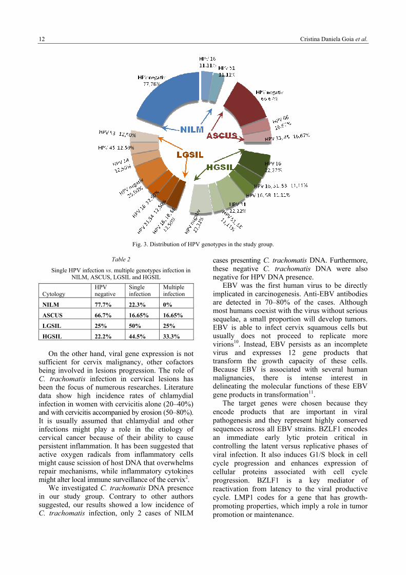
Cristina Daniela Goia et al. 12
Fig. 3. Distribution of HPV genotypes in the study group.
Table 2
Single HPV infection vs. multiple genotypes infection in NILM, ASCUS, LGSIL and HGSIL
Cytology HPV negative
Single infection
Multiple infection
NILM 77.7% 22.3% 0%
ASCUS 66.7% 16.65% 16.65%
LGSIL 25% 50% 25%
HGSIL 22.2% 44.5% 33.3%
On the other hand, viral gene expression is not sufficient for cervix malignancy, other cofactors being involved in lesions progression. The role of C. trachomatis infection in cervical lesions has been the focus of numerous researches. Literature data show high incidence rates of chlamydial infection in women with cervicitis alone (20–40%) and with cervicitis accompanied by erosion (50–80%). It is usually assumed that chlamydial and other infections might play a role in the etiology of cervical cancer because of their ability to cause persistent inflammation. It has been suggested that active oxygen radicals from inflammatory cells might cause scission of host DNA that overwhelms repair mechanisms, while inflammatory cytokines might alter local immune surveillance of the cervix2.
We investigated C. trachomatis DNA presence in our study group. Contrary to other authors suggested, our results showed a low incidence of C. trachomatis infection, only 2 cases of NILM
cases presenting C. trachomatis DNA. Furthermore, these negative C. trachomatis DNA were also negative for HPV DNA presence.
EBV was the first human virus to be directly implicated in carcinogenesis. Anti-EBV antibodies are detected in 70–80% of the cases. Although most humans coexist with the virus without serious sequelae, a small proportion will develop tumors. EBV is able to infect cervix squamous cells but usually does not proceed to replicate more virions10. Instead, EBV persists as an incomplete virus and expresses 12 gene products that transform the growth capacity of these cells. Because EBV is associated with several human malignancies, there is intense interest in delineating the molecular functions of these EBV gene products in transformation11.
The target genes were chosen because they encode products that are important in viral pathogenesis and they represent highly conserved sequences across all EBV strains. BZLF1 encodes an immediate early lytic protein critical in controlling the latent versus replicative phases of viral infection. It also induces G1/S block in cell cycle progression and enhances expression of cellular proteins associated with cell cycle progression. BZLF1 is a key mediator of reactivation from latency to the viral productive cycle. LMP1 codes for a gene that has growth-promoting properties, which imply a role in tumor promotion or maintenance.
Epstein-Barr virus and Chlamydia trachomatis as cofactors in cervical carcinogenesis 13
EBNA-1 gene codes for a protein that binds to a replication origin (oriP) within the viral genome and mediates replication and partitioning of the episome during division of the host cell. It is the only viral protein expressed during group I latency. EBNA1 also acts as a transcriptional activator and enhances the expression of several viral genes from the viral Cp-promoter. EBNA1 was the first viral protein found to be expressed in EBV-associated human tumors and postulated to contribute to the growth-transformed state of EBV-infected cells directly.
Table 3
EBV genes expression correlated with the cytology
Cytology EBNA1+ LMP1+ BZLF1+
NILM 57.0% 14.3% 85.7%
ASCUS 60.0% 20.0% 80.0%
LGSIL 40.0% 20.0% 100%
HGSIL 57.2% 28.6% 85.8%
Firstly, our study revealed that a certain percent of samples are negative for all 3 EBV genes, 22.2% (NILM), 16.7% (ASCUS), 37.5% (LGSIL) and 22.2% (HGSIL). On the other hand, EBNA1, LMP1 and BZLF1 mRNA expression varied widely (Table 3). Regardless of cytology, BZLF1 mRNA expression was present in over 85% of the cases suggesting a reactivation form latency of EBV. This fact may be possibly due to a mixture of RNA from lymphocytes and RNA from epithelial squamous cells. EBNA1 mRNA expression is present in virtually the same percent in various cytological cases. This gene is required for the maintenance of EBV genome, suggesting the existence of a latent EBV infection in cervical cells.
CONCLUSIONS
The results of the study suggested that EBV may play a role in cervical precursor lesions. LMP1, EBV gene with growth-promoting properties,
seems to correlate with lesions progression from NILM to HGSIL, but the mechanism of carcinogenesis remains to be further investigated. On the other hand, we failed to find any association between HPV infection and C. trachomatis infection. This might be due to the small number of cases and further work must be done on larger series of cases to confirm the results of the present study.
REFERENCES
1. Tjiong, M.Y., Out, T.A., Schegget, J.T., Burger, M.P., Van Der Vange, N., Epidemiologic and mucosal immunologic aspects of HPV infection and HPV-related cervical neoplasia in the lower female genital tract: A review,. Int J Gynecol Cancer, 2001, 11, 9–17.
2. Koskela, P., Anttila, T., Bjorge, T., Brunsvig, A., Dillner, J., Hakama, M.; Chlamydia trachomatis infection as a risk factor for invasive cervical cancer, Int J Cancer, 2000, 85, 35–39.
3. Murray, P.G., Young, L.S., The role of the Epstein-Barr virus in human disease, Front Biosci., 2002, 7d, 519–540.
4. Hachisuga, T., Ookuma, Y., Fukuda, K., Iwasaka, T., Sugimori, H., Watanabe, T., Detection of Epstein-Barr virus DNA from a lymphoma-like lesion of the uterine cervix., Gynecol Oncol, 1992, 46, 69–73.
5. Landers, R.J., O'Leary, J.J., Crowley, M., Epstein-Barr virus in normal, pre-malignant and malignant lesions of the uterine cervix, J Clin Pathol, 1993, 46, 931–935.
6. Clifford, G.M., Smith, J.S., Plummer, M., Munoz, N., Franceschi, S., Human papillomavirus types in invasive cervical cancer worldwide: a meta-analysis, British Journal of Cancer, 2003, 88, 63–73.
7. Howley, P.M., Scheffner, M., Munger, K., Oncoproteins encoded by the cancer-associated papillomavirus target the products of retinoblastoma and p53 tumor suppressor genes, Quant Biol, 1991, 56, 159-155.
8. Yu, T., The role of viral integration in the development of cervical cancer, Cancer Genet. Cytogenet, 2005, 158, 27–34.
9. Winer, R.L., Genital human papillomavirus infection: incidence and risk factors in a cohort of female university students, Am. J. Epidemiol., 2003, 157, 218–226.
10. Wensing, B., Farrell, P.J., Regulation of cell growth and death by Epstein-Barr virus, Microb. Infect., 2000, 2, 77–84.
11. Gulley, M.L., Molecular diagnosis of Epstein-Barr virus-related diseases, J. Mol. Diagn., 2001, 3, 1–10.