Embed Size (px)
Citation preview
Università degli Studi di Urbino “Carlo Bo”
Dipartimento di Scienze Biomolecolari
DOTTORATO DI RICERCA IN
SCIENZE DELLA VITA, SALUTE E BIOTECNOLOGIE
Curriculum in Scienze biochimiche, farmacologiche e
biotecnologie
Ciclo XXXI
ERYTHROCYTES LOADED WITH PHENYLALANINE
AMMONIA LYASE (PAL) AS ENZYMATIC REPLACEMENT
THERAPY FOR PHENYLKETONURIA
Settore scientifico disciplinare: BIO/10
RELATORE DOTTORANDO Chiar.ma Prof.ssa Dott.ssa LUIGIA ROSSI NOEMI BIGINI CO-RELATORE Dott. GIOVANNI MAMBRINI
ANNO ACCADEMICO 2017/2018
CONTENTS
INTRODUCTION ....................................................................................................................................... 1
THE DISCOVERY OF PHENYLKETONURIA ............................................................................................. 1
CHARACTERISTICS OF THE DISEASE ..................................................................................................... 2
MOLECULAR AND GENETICS CHARACTERISTICS OF PHENYLALANINE HYDROXYLASE ENZYME ......... 5
PAH working and regulation ............................................................................................................ 7
PAH GENE MUTATIONS AND DATABASE ............................................................................................. 9
CLASSIFICATION ................................................................................................................................. 11
Classification according to blood L-Phe ......................................................................................... 11
Phenylalanine tolerance ................................................................................................................ 12
Clinical course of the disease ........................................................................................................ 13
SCREENING AND DIAGNOSIS ............................................................................................................. 13
Guthrie test ................................................................................................................................... 14
Fluorimetric assay: ........................................................................................................................ 14
Reverse-phase liquid chromatography ......................................................................................... 15
Tandem mass-spectrometry (TMS) assay ..................................................................................... 15
Molecular diagnosis....................................................................................................................... 15
The BH4 loading test ..................................................................................................................... 15
PATHOGENIC MECHANISMS OF HPA ................................................................................................ 17
Brain development and behavioral outcomes .............................................................................. 17
L-Phe influence on cholesterol biosynthesis and obesity ............................................................. 21
MATERNAL PKU ................................................................................................................................. 22
THERAPEUTIC STRATEGIES ................................................................................................................ 23
Dietary treatment .......................................................................................................................... 24
Glycomacropeptide ....................................................................................................................... 26
Large neutral aminoacids (LNAAs) supplementation .................................................................... 26
Tetrahydrobiopterin (BH4) treatment .......................................................................................... 27
Gene therapy and liver transplantation ........................................................................................ 29
Enzyme replacement therapy (ERT) .............................................................................................. 30
ENZYME REPLACEMENT THERAPY IN PKU ........................................................................................ 31
DRUG DELIVERY SYSTEMS: RED BLOOD CELLS AS THE BEST CHOICE ................................................ 35
AIM OF THE WORK ................................................................................................................................ 39
MATERIALS AND METHODS .................................................................................................................. 41
ENZYMES ........................................................................................................................................... 41
Recombinant AvPAL ...................................................................................................................... 41
ANIMALS ............................................................................................................................................ 41
GENETIC CHARACTERIZATION OF ENU2 MICE BY PCR ANALYSIS ...................................................... 42
DNA extraction protocol................................................................................................................ 43
PCR analysis ................................................................................................................................... 43
GEL analysis ................................................................................................................................... 44
DEVELOPMENT OF MURINE rAvPAL-RBCs AND IN VIVO EFFICACY OF REPEATED ADMINISTRATIONS
........................................................................................................................................................... 45
PHENYLALANINE AMMONIA LYASE ACTIVITY ASSAY ........................................................................ 46
L-PHE AND L-TYR EVALUATION IN DRIED BLOOD SPOT (DBS) BY TANDEM MASS SPECTROMETRY
(MS/MS) ............................................................................................................................................ 47
BEHAVIORAL ANALYSIS...................................................................................................................... 48
Behavioral assay in EPM apparatus ............................................................................................... 48
Behavioral assay in OFT apparatus ................................................................................................ 49
Behavioral assay in ORT apparatus ............................................................................................... 49
NEUROCHEMICAL AND MORPHOLOGICAL ANALYSES ...................................................................... 51
Neurochemistry ............................................................................................................................. 51
Morphology ................................................................................................................................... 51
IMMUNOHISTOCHEMICAL AND BIOCHEMICAL ANALYSES ............................................................... 52
Western blot analyses ................................................................................................................... 52
Immunofluorescence analyses ...................................................................................................... 53
EVALUATION OF PLASMA ANTI-rAvPAL IgG TITER ............................................................................ 53
Antibody titer determination ........................................................................................................ 54
STATISTICAL ANALYSES ...................................................................................................................... 54
Behavioral study ............................................................................................................................ 54
Neurochemical and Morphological analyses ................................................................................ 54
Antibody titer determination ........................................................................................................ 55
RESULTS ................................................................................................................................................. 56
DEVELOPMENT OF MURINE rAvPAL-RBCs ........................................................................................ 56
BIOCHEMICAL RESULTS ..................................................................................................................... 57
Blood L-Phe and L-Tyr concentrations .......................................................................................... 57
Brain L-Phe and L-Tyr concentrations ........................................................................................... 58
BEHAVIORAL RESULTS ....................................................................................................................... 59
Elevated Plus Maze (EPM) results ................................................................................................. 59
Open Field Test (OFT) results ........................................................................................................ 60
Object Recognition Test (ORT) results .......................................................................................... 60
NEUROCHEMICAL RESULTS ............................................................................................................... 61
MORPHOLOGICAL RESULTS ............................................................................................................... 63
Prefrontal cortex analysis .............................................................................................................. 63

Nucleus accumbens analysis ......................................................................................................... 64
Hippocampal analysis .................................................................................................................... 65
IMMUNOHISTOCHEMICAL AND BIOCHEMICAL ANALYSES ............................................................... 66
MBP immunoreactivity .................................................................................................................. 66
EVALUATION OF ANTI-rAvPAL PLASMA IgG TITER ............................................................................ 68
DISCUSSION ........................................................................................................................................... 69
CONCLUSION AND FUTURE PERSPECTIVES ........................................................................................... 75
REFERENCES .......................................................................................................................................... 76
INTERNET WEBSITES ............................................................................................................................ 100
1
INTRODUCTION
THE DISCOVERY OF PHENYLKETONURIA
Phenylketonuria (PKU) (OMIM# 261600) was first described in 1934 by the Norwegian
endocrinologist dr. Asbjørn Følling, who originally defined this pathology as “phenylpyruvic
oligophrenia” because of the typical mental disorders that affected his patients (Mitchell et
al., 2011). This definition was modified in 1930s by Penrose (Penrose and Quastel, 1937), who
coined the name currently known, PKU, and identified its autosomal recessive nature. He
surmised that PKU state had an endogenous chemical cause; in keeping with his hypothesis,
he was the first to consider the possible correlation between “nurture” and mutant “nature”.
He thought that modifying the nurture might be possible to neutralize the harmful effects of
the pathology (Penrose, 1998).
PKU is an inherited metabolic disorder characterized by severe intellectual impairment, motor
problems, and skin abnormalities and occupies a unique place in the history of the study of
metabolic disease not only for its role as principal inborn error of amino acid metabolism but
also because it is the first cause of mental retardation to be discovered. Dr. Følling found that
affected individuals could be identified by the abnormal excretion of phenylpyruvic acid in
their urine. The credit for the discovery was also due to that caring and stubborn mother, who
could not resign herself to the mental retardation of her children without having found a
reason (http://pkuworld.org/home/history.asp).
Her 7 years old daughter, could say only few words and had a whimsy and purposeless way of
moving about; likewise, her 4 years old son did not walk and was unable to fix his eyes on
anything. Their skin was fair and their urine had a peculiar smell. By means of a traditional
assay of classical chemistry for the detection of ketones, consisting in the addition of ferric
chloride to the urine of diabetic patients, dr. Følling observed the appearance of a deep green
color, which he had never seen before. Further chemical analyses and steps of purification on
many other urine samples from patients sharing the same neurocognitive and developmental
delays, led to the identification of a chemical substance whose empirical formula was C9H8O3,
named phenylpyruvic acid. The analysis of the urine from another 430 mentally impaired
subjects, allowed dr. Følling to identify eight patients excreting the same substance and for
the first time he understood the correlation between mental impairment and excretion of
phenylpyruvic acid. Further studies of family relationships highlighted an autosomal recessive
mechanism of transmission (Følling, 1944). Few years later, Jervis (1947, 1953), succeeded in
identifying the metabolic block and the enzymatic deficiency of phenylalanine hydroxylase
(PAH), the alteration behind this pathological condition; at the same time, Bickel and
collaborators (1953) showed the importance of reducing the intake of phenylalanine (Phe) in
order to obtain a prognosis improvement. Phenylketonuria was the first known inborn error
of metabolism to seriously affect the victims and to give mental disturbance. In addition, its
discovery determined an important breakthrough in understanding how metabolic
dysfunctions can influence neurological functions and how treatments can heavily influence
INTRODUCTION
2
clinical manifestations: PKU today is considered “the epitome of metabolic disorders” and is
often employed as a model to describe and understand many other inborn errors of amino
acid metabolism (Scriver and Clow, 1980 Part I and II; Raghuveer et al., 2006). To explain the
causes of the phenylpyruvic acid excretion, dr. Følling hypothesized some kind of defect in
phenylalanine metabolism, which lead to high concentration of this aminoacid in the blood of
PKU affected patients; the effectiveness of his hypothesis was successively confirmed (Følling
and Closs, 1938) through a microbiological test developed by dr. Robert Guthrie which
exploited the reversal of growth inhibition observed in Bacillus subtilis ATCC 6051 in the
presence of a high level of phenylalanine (Guthrie and Susi, 1963). The identification of the
first mutations of the PAH gene, codifying for the enzyme PAH, began immediately after its
cloning and mapping in 1983 (Woo et al., 1983) opening the way to the in vitro study of the
different functionalities of the enzyme. Currently, all the known mutations of the PAH gene
(about 859) known, are collected in the "PAHdb" database (http://www.pahdb.mcgill.ca/)
created in 1996 (Hoang et al., 1996).
CHARACTERISTICS OF THE DISEASE
Phenylketonuria (PKU) is the most common autosomal recessive disease among Caucasians
(overall incidence 1:10.000 on average; 1:2.600 in Turkey; 1:100.000 in Japan). PKU is a result
of an inborn error of amino acid metabolism caused by a deficiency of the enzyme
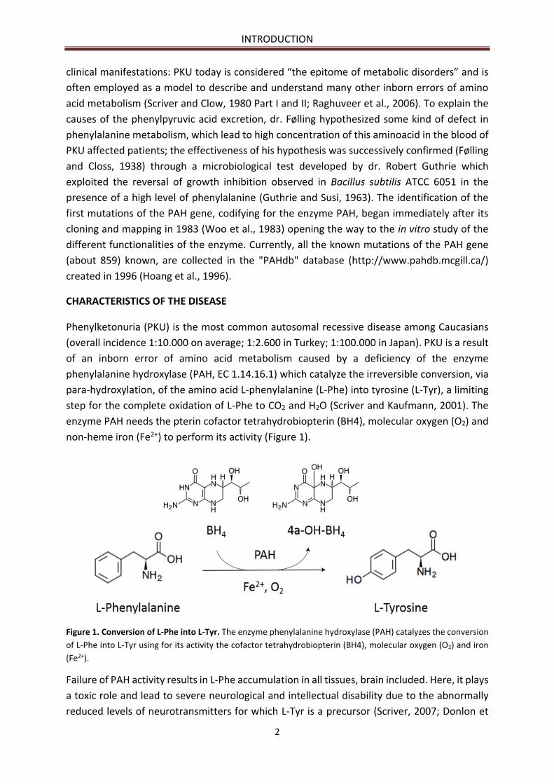
phenylalanine hydroxylase (PAH, EC 1.14.16.1) which catalyze the irreversible conversion, via
para-hydroxylation, of the amino acid L-phenylalanine (L-Phe) into tyrosine (L-Tyr), a limiting
step for the complete oxidation of L-Phe to CO2 and H2O (Scriver and Kaufmann, 2001). The
enzyme PAH needs the pterin cofactor tetrahydrobiopterin (BH4), molecular oxygen (O2) and
non-heme iron (Fe2+) to perform its activity (Figure 1).
Figure 1. Conversion of L-Phe into L-Tyr. The enzyme phenylalanine hydroxylase (PAH) catalyzes the conversion
of L-Phe into L-Tyr using for its activity the cofactor tetrahydrobiopterin (BH4), molecular oxygen (O2) and iron
(Fe2+).
Failure of PAH activity results in L-Phe accumulation in all tissues, brain included. Here, it plays
a toxic role and lead to severe neurological and intellectual disability due to the abnormally
reduced levels of neurotransmitters for which L-Tyr is a precursor (Scriver, 2007; Donlon et
INTRODUCTION
3
al., 2010). Early diagnosis and a quick treatment are able to reduce toxic levels of this
aminoacid, avoiding these serious consequences. Nowadays, many countries include a
neonatal screening such as Guthrie test or more modern system based on tandem mass
spectrometry for the detection of hyperphenylalaninemia (HPA). Moreover, L-Phe itself is an
essential nutrient an it represents a pivotal constituent for protein synthesis. Therefore, PKU
treatment requires the balanced reduction of systemic L-Phe levels without its excessive
depletion in order to guarantee a satisfactory synthesis of L-Tyr. Mutations of PAH gene,
located in chromosome 12 (region 12q22-q24.2, GenBank U49897), is responsible for the
insufficient activity of this cytosolic hepatic enzyme and the establishment of the HPA state.
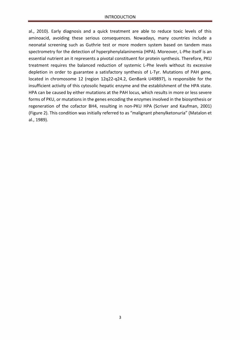
HPA can be caused by either mutations at the PAH locus, which results in more or less severe
forms of PKU, or mutations in the genes encoding the enzymes involved in the biosynthesis or
regeneration of the cofactor BH4, resulting in non-PKU HPA (Scriver and Kaufman, 2001)
(Figure 2). This condition was initially referred to as “malignant phenylketonuria” (Matalon et
al., 1989).
INTRODUCTION
4
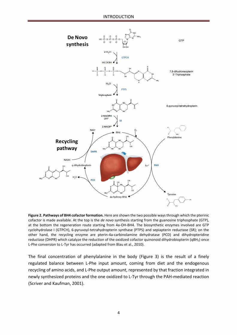
Figure 2. Pathways of BH4 cofactor formation. Here are shown the two possible ways through which the pterinic cofactor is made available. At the top is the de novo synthesis starting from the guanosine triphosphate (GTP), at the bottom the regeneration route starting from 4a-OH-BH4. The biosynthetic enzymes involved are GTP cyclohydrolase I (GTPCH), 6-pyruvoyl-tetrahydropterin synthase (PTPS) and sepiapterin reductase (SR); on the other hand, the recycling enzyme are pterin-4a-carbinolamine dehydratase (PCD) and dihydropteridine reductase (DHPR) which catalyze the reduction of the oxidized cofactor quinonoid dihydrobiopterin (qBH2) once L-Phe conversion to L-Tyr has occurred (adapted from Blau et al., 2010).
The final concentration of phenylalanine in the body (Figure 3) is the result of a finely
regulated balance between L-Phe input amount, coming from diet and the endogenous
recycling of amino acids, and L-Phe output amount, represented by that fraction integrated in
newly synthesized proteins and the one oxidized to L-Tyr through the PAH-mediated reaction
(Scriver and Kaufman, 2001).
De Novo synthesis
Recyclingpathway
INTRODUCTION
5
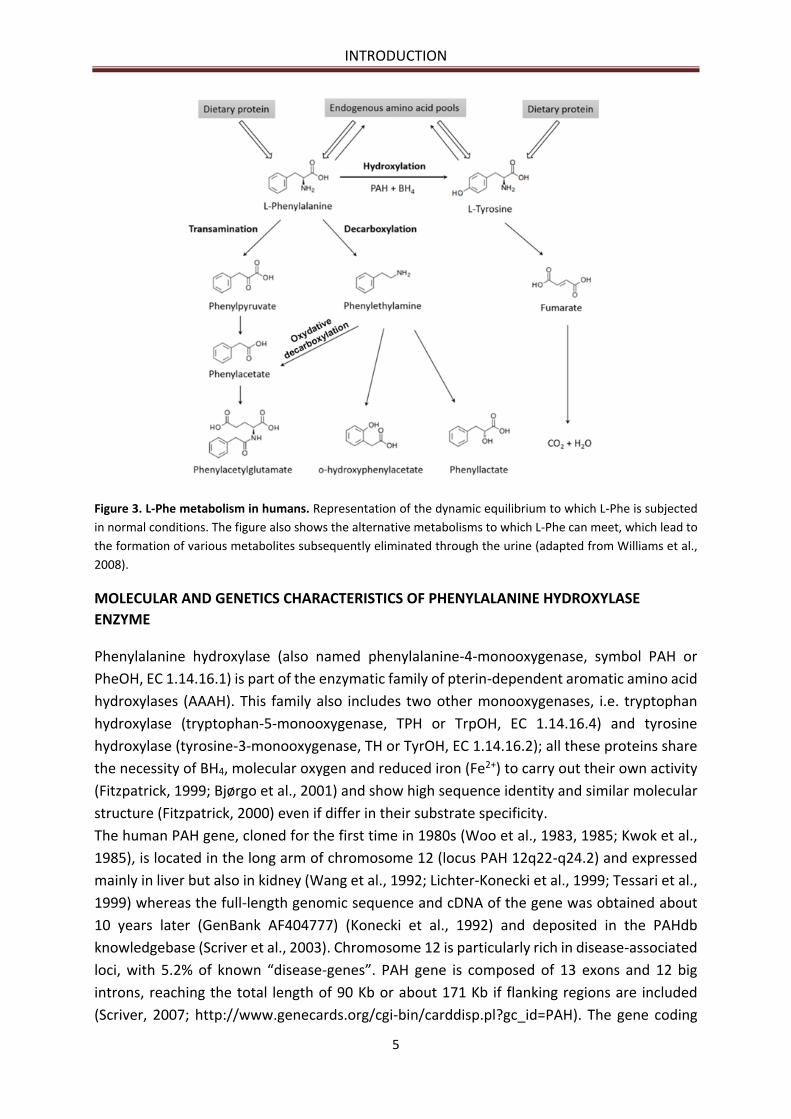
Figure 3. L-Phe metabolism in humans. Representation of the dynamic equilibrium to which L-Phe is subjected
in normal conditions. The figure also shows the alternative metabolisms to which L-Phe can meet, which lead to
the formation of various metabolites subsequently eliminated through the urine (adapted from Williams et al.,
2008).
MOLECULAR AND GENETICS CHARACTERISTICS OF PHENYLALANINE HYDROXYLASE
ENZYME
Phenylalanine hydroxylase (also named phenylalanine-4-monooxygenase, symbol PAH or
PheOH, EC 1.14.16.1) is part of the enzymatic family of pterin-dependent aromatic amino acid
hydroxylases (AAAH). This family also includes two other monooxygenases, i.e. tryptophan
hydroxylase (tryptophan-5-monooxygenase, TPH or TrpOH, EC 1.14.16.4) and tyrosine
hydroxylase (tyrosine-3-monooxygenase, TH or TyrOH, EC 1.14.16.2); all these proteins share
the necessity of BH4, molecular oxygen and reduced iron (Fe2+) to carry out their own activity
(Fitzpatrick, 1999; Bjørgo et al., 2001) and show high sequence identity and similar molecular
structure (Fitzpatrick, 2000) even if differ in their substrate specificity.
The human PAH gene, cloned for the first time in 1980s (Woo et al., 1983, 1985; Kwok et al.,
1985), is located in the long arm of chromosome 12 (locus PAH 12q22-q24.2) and expressed
mainly in liver but also in kidney (Wang et al., 1992; Lichter-Konecki et al., 1999; Tessari et al.,
1999) whereas the full-length genomic sequence and cDNA of the gene was obtained about
10 years later (GenBank AF404777) (Konecki et al., 1992) and deposited in the PAHdb
knowledgebase (Scriver et al., 2003). Chromosome 12 is particularly rich in disease-associated
loci, with 5.2% of known “disease-genes”. PAH gene is composed of 13 exons and 12 big
introns, reaching the total length of 90 Kb or about 171 Kb if flanking regions are included
(Scriver, 2007; http://www.genecards.org/cgi-bin/carddisp.pl?gc_id=PAH). The gene coding
INTRODUCTION
6
sequence (cds, nt 473-1831) is transcribed into a mature mRNA of approximately 2,6 Kb (2680
bp), which is in turn translated into a 452 amino acid monomer
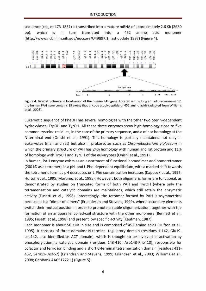
(http://www.ncbi.nlm.nih.gov/nuccore/U49897.1, last update 1997) (Figure 4).
Figure 4. Basic structure and localization of the human PAH gene. Located on the long arm of chromosome 12, the human PAH gene contains 13 exons that encode a polypeptide of 452 amino acids (adapted from Williams et al., 2008).
Eukaryotic sequence of PheOH has several homologies with the other two pterin-dependent
hydroxylases: TrpOH and TyrOH. All these three enzymes show high homology close to five
common cysteine residues, in the core of the primary sequence, and a minor homology at the
N-terminal end (Onishi et al., 1991). This homology is partially maintained not only in
eukaryotes (man and rat) but also in prokaryotes such as Chromobacterium violaceum in
which the primary structure of PAH has 24% homology with human and rat protein and 11%
of homology with TrpOH and TyrOH of the eukaryotes (Onishi et al., 1991).
In human, PAH enzyme exists as an assortment of functional homodimer and homotetramer
(200 kD as a tetramer), in a pH- and L-Phe-dependent equilibrium, with a marked shift towards
the tetrameric form as pH decreases or L-Phe concentration increases (Kappock et al., 1995;
Hufton et al., 1995; Martinez et al., 1995). However, both oligomeric forms are functional, as
demonstrated by studies on truncated forms of both PAH and TyrOH (where only the
tetramerization and catalytic domains are maintained), which still retain the enzymatic
activity (Fusetti et al., 1998). Interestingly, the tetramer formed by PAH is asymmetrical
because it is a “dimer of dimers” (Erlandesen and Stevens, 1999), where secondary elements
switch their mutual position in order to promote a stable oligomerization, together with the
formation of an antiparallel coiled-coil structure with the other monomers (Bennett et al.,
1995; Fusetti et al., 1998) and present low specific activity (Kaufman, 1987).
Each monomer is about 50 KDa in size and is comprised of 452 amino acids (Hufton et al.,
1995). It consists of three domains: N-terminal regulatory domain (residues 1-142, Glu19-
Leu142, also identified as ACT domain), which is thought to be involved in activation by
phosphorylation; a catalytic domain (residues 143-410, Asp143-Phe410), responsible for
cofactor and ferric ion binding and a short C-terminal tetramerization domain (residues 411-
452, Ser411-Lys452) (Erlandsen and Stevens, 1999; Erlandsen et al., 2003; Williams et al.,
2008; GenBank AAC51772.1) (Figure 5).
INTRODUCTION
7
Figure 5. PAH structure. (A) Full-length structure of human phenylalanine hydroxylase monomer obtained by superimposing the catalytic domains of the truncated forms. The red sphere represents iron. (B, C) Two perpendicular views of the full-length PAH model structure. The iron is shown as a gray sphere in all four monomers (adapted from Erlandsen and Stevens, 1999).
PAH working and regulation
PAH activity is tightly regulated by a number of possible mechanisms such as reversible
phosphorylation and substrate activation. The activity of PAH, as previously mentioned,
requires the binding of the cofactor BH4 and molecular oxygen (Figure 1).
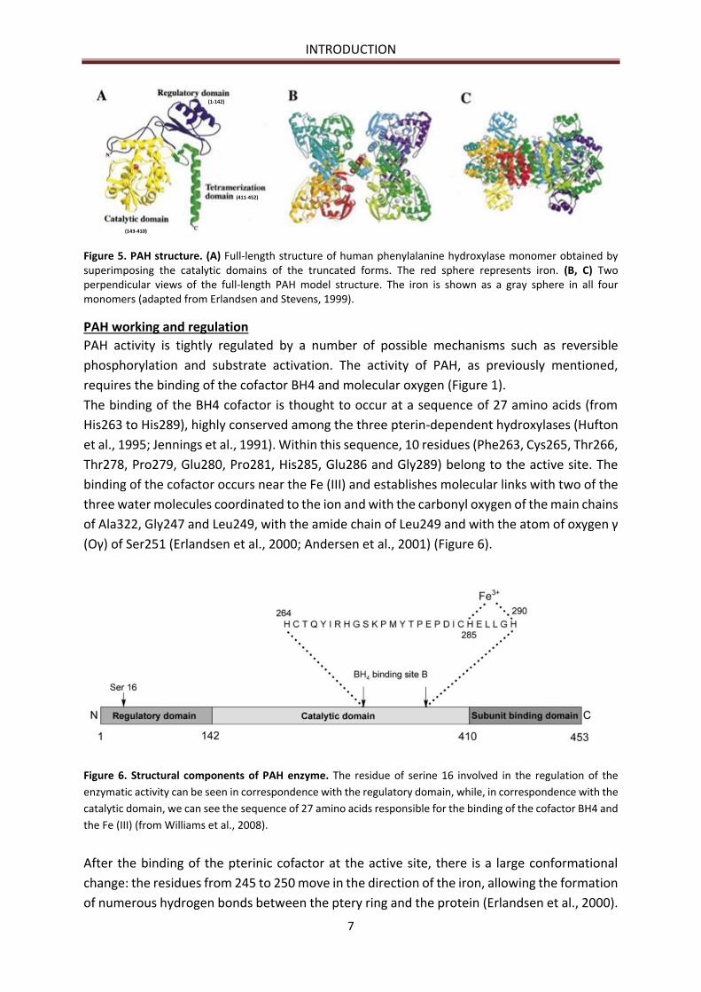
The binding of the BH4 cofactor is thought to occur at a sequence of 27 amino acids (from
His263 to His289), highly conserved among the three pterin-dependent hydroxylases (Hufton
et al., 1995; Jennings et al., 1991). Within this sequence, 10 residues (Phe263, Cys265, Thr266,
Thr278, Pro279, Glu280, Pro281, His285, Glu286 and Gly289) belong to the active site. The
binding of the cofactor occurs near the Fe (III) and establishes molecular links with two of the
three water molecules coordinated to the ion and with the carbonyl oxygen of the main chains
of Ala322, Gly247 and Leu249, with the amide chain of Leu249 and with the atom of oxygen γ
(Oγ) of Ser251 (Erlandsen et al., 2000; Andersen et al., 2001) (Figure 6).
Figure 6. Structural components of PAH enzyme. The residue of serine 16 involved in the regulation of the
enzymatic activity can be seen in correspondence with the regulatory domain, while, in correspondence with the
catalytic domain, we can see the sequence of 27 amino acids responsible for the binding of the cofactor BH4 and
the Fe (III) (from Williams et al., 2008).
After the binding of the pterinic cofactor at the active site, there is a large conformational
change: the residues from 245 to 250 move in the direction of the iron, allowing the formation
of numerous hydrogen bonds between the ptery ring and the protein (Erlandsen et al., 2000).
(411-452)
(1-142)
(143-410)
INTRODUCTION
8
Studies carried out on PAH have shown that a stoichiometric amount of BH4 can be oxidized
in the presence of oxygen by determining the reduction of the enzyme (Marota and Shiman,
1984), i.e. the reduction of Fe3+ to Fe2+, a process necessary for the activation of PAH (Wallick
et al., 1984). This reaction is achieved by transferring an electron from BH4 to Fe3+ and a
second electron to the oxygen molecule (Marota and Shiman, 1984; Wallick et al., 1984; Hill
et al., 1988).
The PAH enzyme is particularly sensitive to its substrates concentrations and its activity is
strictly regulated through different mechanisms that act together to tightly control the
negative effects of an excessive concentration of circulating L-Phe (Heintz et al., 2013).
The first thirty residues of the enzyme (residues 19-33) act as a autoregulatory sequence (ARS)
and contain a residue of serine 16 (Ser16) which has been demonstrated to be the site of
phosphorylation by the cAMP-dependent protein kinase A (cAMP-PKA) (Døskeland et al.,
1996). This sequence is named autoregulatory because it sterically limits the access of the
substrate to the catalytic site of the enzyme (Heintz et al., 2013). When the first 30 N-terminal
residues are removed, PAH shows a higher affinity and a consequent higher rate of L-Phe
conversion (Knappskog et al., 1996).
L-Phe acts as an allosteric activator, promoting the activation of the enzyme, thus increasing
the formation rate of L-Tyr proportionally to the concentration of L-Phe (Døskeland et al.,
1996). This effect is probably due to a conformational modification that the enzyme
undergoes following the binding of L-Phe, a change that also leads to changes in the
spectroscopic properties of PAH (Kappock et al., 1995). The mechanism of activation involves
all the four monomers, inducing modifications in the monomeric structures which promote a
stronger interaction at the dimer interface, whereas the interactions between dimers in a
tetramer are weakened. As a result, the dimer/tetramer equilibrium is shifted towards the
tetrameric form of the enzyme upon binding of L-Phe (Martinez et al., 1995), the volume of
the tetramer increases and a competent catalytic site is exposed (Kappock et al., 1995). L-Phe
binds in a specific allosteric site located in the regulatory domain (Li et al., 2011), different
from the active site of the catalytic domain, and the binding on one site do not automatically
excludes the binding on the other one, although the affinity for the allosteric site is seven-fold
higher (Shiman, 1980; Shiman et al., 1990). In fact, in each monomer the N-terminal tail
stretches over the active site, thus preventing the access of substrate unless L-Phe binds on
the regulatory sequence (Fusetti et al., 1998).
On the other hand, BH4 cofactor acts as an allosteric inhibitor, blocking the conformational
change induced by the substrate (Kaufman, 1993). Indeed, it binds the N-terminal
autoregulatory domain, blocking the access of the substrates to the active site (Teigen and
Martinez, 2003). A possible mechanism behind this allosteric inhibition is the decrease of the
phosphorylation rate of the Ser16 residue by the c-AMP dependent kinase (Døskeland et al.,
1984). The phosphorylation of this residue is facilitated by the conformational change induced
by the L-Phe binding to the ARS site, which in turn facilitates the entry of L-Phe at the active
site, increasing the activation level of the enzyme. In support of this hypothesis, in some
studies it has been observed that the phosphorylated PAH requires less L-Phe to be activated
than the unphosphorylated PAH (Døskeland et al., 1984). Nevertheless, BH4 is necessary for
INTRODUCTION
9
the reduction of the Fe3+ ion to Fe2+, an inevitable prerequisite for enzyme activation (Shiman,
1980).
Shiman and his collaborators (1982) demonstrated that both phosphorylated and
unphosphorylated forms of the enzyme require L-Phe for their activation; hence,
phosphorylation is not equivalent to the allosteric activation, but lowers the energy needed
for its occurrence. This happens by means of two mechanisms: by promoting the transition to
the active state of the protein, and by reducing BH4 affinity for its inhibitory binding site. In
keeping with this evidence it could be explained why in vivo phosphorylated PAH has a higher
affinity for the substrate L-Phe, a higher activation rate and a lower sensitivity to BH4-
mediated inhibition (Kappock and Caradonna, 1996). This important role of L-Phe in the
regulation of PAH activity is reported also in a recent publication (Jaffe, 2017) in which the
authors described the enzyme in terms of a sensitive equilibrium between resting-state PAH
(RS-PAH) and activated PAH (A-PAH) structures. This position depends on L-Phe availability
because when L-Phe levels rise, the PAH structural equilibrium shifts toward A-structures
while at low L-Phe level (<50µM) the enzyme is in RS-form.
PAH GENE MUTATIONS AND DATABASE
Many variations of PAH gene have been described during 25 years of research (Scriver et al.,
2000) with the most commonly variations occurring in exon 3, 6, 7 and 11 (Blau et al., 2014).
The PAH gene is characterized by great allelic heterogeneity, as reported in the open access
PAHvdb database (http://www.biopku.org/home/pah.asp) that harbors 957 variants of this
gene (Blau et al., 2014). To date, 60% of PAH variants are missense mutations, followed by
deletions (13.4%), splice alterations (10.9%), silent or non-sense mutations (7% and 5%
respectively) and small insertion (1%). Large deletions, probably account for 3% (Scriver,
2007). The genotypes and clinical phenotypes are tabulated in the BIOPKU database in which
it can observed that 55% of PKU patients shows a classical phenotype, 27% has a mild
phenotype and the remaining have non-PKU mild HPA. The most common mutations
(c.1222C>T and c.1066-11 G>A) are responsible of abolish PAH activity (DiLella et al., 1987;
Gjetting et al., 2001). Other type of mutations alters PAH activity in a different manner; for
example, alleles c.782G>A and c.1241A>G have respectively about 44% and 57% of the activity
when compared with the wild type enzyme (Zurflüh et al., 2008; Wettstein et al., 2015). Other
variants of the gene are silent mutations with little or no effect on PAH activity (Wettstein et
al., 2015). The alterations which destroy enzyme functionality, named “null” mutations (Zhou
et al., 2012; Mitchell et al., 2011) (such as mutations at splice sites, frameshift as well as
missense mutations), often occur on exons or between introns and exons, interfering with the
correct folding of the protein, the tetramerization process or destroying the catalytic domain,
accelerating its degradation and compromising its catalytic activity (Bai and Song, 2003). The
mutations called "silent" (Zhou et al., 2012; Mitchell et al., 2011), mainly missense mutations,
interfere with the protein folding, with its regulation or with the parameters that regulate and
influence PAH activity: however, with these mutations the enzyme maintain a minimal
residual activity.
INTRODUCTION
10
These last type of variations are most likely to demonstrate increased activity in presence of
BH4. In fact, BH4 seems to be a molecular chaperone for PAH, protecting it against protein
misfolding during its synthesis. This activity is therefore likely to be multifactorial in nature
(Erlandsen et al., 2004; Gersting et al., 2008).
PKU affected patients are generally not homozygous for a single mutation; they are instead
heterozygous (about 75%) for two different allelic alterations. Some patients, who are
compound heterozygous, are phenotypically functional hemizygous, due to a combination of
a severe mutation (such as a null one) with an allele that still allows the production of enzyme,
even if only partially functioning: in those cases, the mutation determines the PKU metabolic
phenotype (Guldberg et al., 1998). This is the principal reason underlying the great phenotypic
diversity associated with the disease, which makes PKU very widespread in spite of its
recessive inheritance pattern (Bercovich et al., 2008; Santos et al., 2010).
Allelic heterogeneity exists also in the gene coding for the enzymes involved in the
biosynthesis or regeneration of the BH4 cofactor (Figure 2). Defects of BH4 synthesis result
from alterations in GTP cyclohydrolase I (GTPCH) or 6-piruvoyl-tetrahydropterine synthase
(PTPS), while alterations of BH4 regeneration result from mutations in the NADH-dependent
dihydrobiopterin reductase enzyme (DHPR) or in the carbinolamine pterina dehydratase
enzyme (PCD) (Mitchell et al., 2011). About 2% of the HPA cases are due to these impairments
whose make very important the careful analysis of the cause responsible for the increase in L-
Phe levels (Blau et al., 2001). This cofactor is part of several and important metabolic
pathways, making the unavailability of BH4 the basis of various pathological changes such as
vascular dysfunction and neurological impairments. Indeed, it influences the synthesis of
catecholamines, serotonin and nitric oxide in the central nervous system (CNS), being used as
a cofactor by TyrOH e TrpOH as well as by all the three forms of nitric oxide synthase (NOS)
and glyceryl ether monooxygenase (Werner et al., 2011).
The population incidence of BH4 deficient forms of PKU is 1 out of 1 million births (Thöny and
Blau, 2006) but even if primary disorders of BH4 metabolism are rare, they must be identified
during a positive newborn screening test in order to start an appropriate and accurate
treatment for the patients. More and comprehensive information about these genes and
enzymes could be obtain on a dedicate website (www.bh4.org).
It is therefore clear that the identification of the correct etiologic agent allows the
development of a specific treatment aimed at limiting the phenotypic effects of the disease:
phenylketonuric patients show a different tolerance with respect to the daily amount of L-Phe
intake, and on these basis, a dietary therapy has been proposed in the 1950s, with first positive
results published in 1953 (Woolf et al., 1951; Bickel, 1953, 1954). In addition, a correlation
between genotype-phenotype does not always exist.
Indeed, PKU has often been defined as a disease born from the discordance between nature
and nurture (Scriver, 2007; Donlon et al., 2010), where the nurture component is the essential
amino acid L-Phe and the nature is represented by the mutation in the PAH gene. The result
of this discordance is HPA, the PKU metabolic phenotype, which leads to the clinical
phenotype of impaired cognitive development and function. The possibility to act externally
on the metabolic manifestation of the disease makes PKU the first genetic disease to have a
INTRODUCTION
11
pharmacological treatment, thus smoothing the negative effects of the gene alterations
(Scriver, 2007).
CLASSIFICATION
Until the late 1980s ”phenylalanine loading test” was applied for the detection of
heterozygote in PKU families (Driscoll et al., 1956) when molecular analysis of PAH gene and
mutations replaced it. This test was developed by Blaskovics (2006) in the mid-1960s and
gained further interest when Guthrie card mass screening allowed to identify not only classic
PKU but also some variants. The test consists of three-day loading of natural protein at 6
months age. Through the loading test was possible to distinguish three types of response.
Type 1 response corresponds to the classic PKU and was characterized by a 72h L-Phe beyond
1200 µmol/L; type 2 response was characterized by a decline of L-Phe levels - despite
continuation of protein loading - from 1200 µmol/L after 2 days to 1200-600 µmol/L after 72h;
in the type three response, L-Phe levels was <600 µmol/L after 72h and corresponds to mild
HPA. About 10% of the patients belong in type 2 response. Despite the success, the Blaskovics
test do not predict the current and future dietary requirements and some patients manifested
signs of intoxication during the test: the test has been thus replaced in practice (Blau et al.,
2011).
Various forms of clinical phenotypes associated with HPA state have been described, so it is
possible to establish different classifications for PKU considering different aspects: the first is
based on the severity of HPA due to the type and position of the PAH mutation, which
determines the rate of enzymatic activity; the second is based on the tolerance to dietary L-
Phe intake and last but not least on the clinical course of the disease and BH4 responsiveness
(Blau et al., 2010). Hence, the classification is primarily made on the basis of the severity of
HPA, considering that the normal L-Phe concentration in the blood of healthy individuals
ranges from 50 to 110 µM (Kure et al., 1999; Blau et al., 2010).
Classification according to blood L-Phe
This classification is primarily made on the basis of the severity of HPA, considering that the
normal L-Phe concentration in the blood of healthy individuals ranges from 50 to 110 µM (Blau
et al., 2010). Known pretreatment L-Phe levels is important but this values are influenced by
some factors such as: the timing of blood L-Phe detection and the diet that the patients have
been received before that time but also from the neonatal catabolism. In addition, the current
practice of blood L-Phe screening in newborns within the third day of life can result in a
negative conclusion (false negative), if L-Phe has no time to reach its maximal concentration
(Blau et al., 2010). However, these parameters have been shown to be used for phenotyping
patients with PKU in about 80% of the treatment centers (Blau et al., 2011).
The phenotyping of patient according to amino acidic pre-treatment levels was introduced in
1980 by Güttler et al. (1980) and defines the following phenotypes:
Classical PKU: pre-treatment L-Phe >1200 µM. This is the most severe form and the
subjects exhibit high risk of suffering from cognitive impairment without treatment
INTRODUCTION
12
Variant PKU: pre-treatment L-Phe between 600 and 1200 µM
Mild HPA or non-PKU HPA: pre-treatment L-Phe between 120 and 600 µM
The class named “variant PKU” was later divided into two subcategories (Guldberg and
Guttler, 1994; Guldberg et al., 1998), resulting in:
Classical PKU: L-Phe >1200 µM (>20 mg/dL)
Moderate PKU: L-Phe between 900 and 1200 µM (15-20 mg/dL)
Mild PKU: L-Phe between 600 and 900 µM (10-15 mg/dL)
Mild HPA: L-Phe above 110 µM but <600 µM
A further classification has been made for the values below 600 µM (Camp et al., 2014). In
particular, we can distinguish, “Mild HPA-gray zone” to describe blood L-Phe levels between
360 and 600 μmol/L (6-10 mg/dL) and “Mild HPA-NT zone” to describe blood L-Phe levels
between 120 and 360 μmol/L (2-6 mg/dL). The difference between the two zones has been
made because the NT zone doesn’t require a treatment while for the gray zone it remain
unclear if a treatment to avoid negative influence on cognitive and executive functioning is
required or not (Hanley, 2011; van Spronsen, 2011).
However, the picture is now more clear thanks to the new Key European guidelines, which
suggest that no intervention is required if the blood L-Phe concentration is less than 360
µmol/L but is recommended when this value is between 360 µmol/L and 600 µmol/L up to
age of 12 years and lifelong treatment is strongly recommended when the concentration is
more than 600 µmol/L (van Spronsen et al., 2017).
Phenylalanine tolerance
Using L-Phe tolerance we can identify three different phenotypes:
Classic PKU: L-Phe tolerance <20 mg/kg body weight/day
Variant PKU: L-Phe tolerance between 20 and 50 mg/kg body weight/day
Mild HPA: L-Phe tolerance >50 mg/kg body weight/day
Subsequently, a subdivision with four phenotypes has been adopted (Guldberg and Guttler,
1994; Guldberg et al., 1998):
Classic PKU: L-Phe tolerance <20 mg/kg/day, corresponding to 250-300 mg L-Phe/day
Moderate PKU: L-Phe tolerance of 20-25 mg/kg/day (350-400 mg/day)
Mild PKU: L-Phe tolerance of 25-50 mg/kg/day (400-600 mg/day)
Mild HPA: patients not requiring dietary restriction
This evaluation is determined with the amount of daily L-Phe intake that a patient can tolerate
without L-Phe reaches the maximum level. L-Phe tolerance is usually determined at the age
of 5 years (Guldberg et al., 1998), but recently has been shown that also at 2 years old it is
possible a reliable determination because the tolerance at 2, 3 and 5 years correlates with
that observed at 10 years age (van Spronsen et al., 2009). On the contrary, L-Phe tolerance
must be reassessed in adulthood in relation to body weight in order to satisfy as much as
INTRODUCTION
13
possible the criterion of 9.1 mg L-Phe/kg ideal body weight/day (MacLeod et al., 2009). This
kind of classification is currently used by 70% of medical centers (Zurfluh et al., 2008).
Clinical course of the disease
Another possibility that allows to discriminate the different phenotypes of PKU, is based on
its clinical course (Blau et al., 2011). In particular, it includes parameters such as the
intellectual outcome, in terms of patient education and IQ, the maximum L-Phe concentration
reached in particular conditions or periods of life (such as non-compliance to the restricted
diet or the occurrence of infectious diseases) and, most importantly, the variations in blood L-
Phe levels and L-Phe/L-Tyr ratio (Luciana et al., 2001; Anastasoaie et al., 2008; Humphrey et
al., 2011). The classification is based on the need for treatment as reported:
PKU: patients who need a strict dietary control of L-Phe levels
Non-PKU HPA: patient who do not need any dietary treatment to keep L-Phe under
control
BH4-responsive PKU: patients who may take advantage from BH4 supplementation.
This type of classification is applied only in 31% of the medical centers (Blau et al., 2011).
An additional classification based on BH4-responsiveness has been proposed by Blau and
Muntau (2002) and consists in BH4-non-responsive HPA and BH4-responsive HPA, the latter
further divided into BH4-responsive PAH deficiency and HPA due to defects in the BH4
pathway. Deficiencies in the BH4 synthesis or recycling enzymes are inherited similarly to the
PAH mutations as autosomic recessive traits, and account for approximately 2% of HPAs
detected in babies by newborn screening (Harding, 2010).
The definition of PKU phenotypes is fundamental for establish the best treatments options, in
counseling, in the outcome prediction and during pregnancy.
The three different systems of classification above reported may help to discriminate the PKU
phenotype but they are not precise parameters (Blau et al., 2011).
SCREENING AND DIAGNOSIS
Hereditary metabolic diseases (HMDs), such as PKU, are rare diseases that if ascertained are
treatable, thus preventing intellectual and general disability (Morrissey et al., 2013). In fact,
an improved and rapid detection and treatment in pediatric practice of HPA has resulted in
increased survival up to adult life.
For this reason, in developed countries all newborns are routinely tested for PKU/HPA soon
after birth according to national screening programs (Dhondt, 2006), since 1960s. Blood
samples are drawn for analysis between the 2nd and the 5th day of life in most centers
(Zaffanello et al., 2003). Today, it is accepted that the best results in terms of sensitivity of
screening tests are obtained in healthy neonate when performed before 24h of life, especially
when L-Phe/L-Tyr ratio is monitored for the diagnosis (Chace et al., 1998; Eastman et al., 2000;
Zaffanello et al., 2003). However, results of early PKU screening should be carefully analyzed
in sick neonate or in neonate under parenteral nutrition or blood transfusion to avoid wrong
INTRODUCTION
14
results (false negatives). Nevertheless, false positives may also origin from an improper
sample preparation or an excessive blood spot thickness, or a combination of two or more of
these factors (Mitchell at al., 2011). In addition, temporarily higher levels of L-Phe might be
due to heterozygosity for PAH deficiency (Hennermann et al., 2004), to maternal PKU or to
other non-PKU disorders but also in prematurely infants which display an immaturity of the
enzymes involved in amino acid metabolism. In all these cases, generally, a second test on
dried blood spot should be performed in order to confirm the first result (Williams et al.,
2008).
Diagnosis mainly consists therefore in the biochemical assessment of blood L-Phe and L-Tyr,
biopterin and neopterin content in blood or urine, and the measurement of specific enzymatic
activities (Blau et al., 2011). The measurement of L-Phe metabolites (phenylpyruvate,
phenylacetate, phenylactate) in urine is not accepted as PKU screening method because their
levels vary considerably between blood and urine and excretion depends upon transaminase
activity which can be low in neonates (Knox, 1970). All the forms of this disease reveal upon
neonatal screening a common pattern of blood L-Phe, which is higher than 120 µM, normal or
reduced L-Tyr concentrations (with a L-Phe/L-Tyr ratio >2) and normal values for the
remaining amino acids (Blau et al., 2010). The analytical methods employed to assess blood L-
Phe are briefly described below.
Guthrie test
The first efficient test for the detection of HPA by newborn screening, was developed in 1960s
by Robert Guthrie (Guthrie and Susi, 1963). The test was based on Bacillus subtilis activity
which requires L-Phe for its grow. This test is very useful for neonatal screening and is
performed by a rapid withdrawal, generally carried out in the hospital or in the doctor's office,
of a small amount of peripheral blood from the heel prick which is then put onto filter paper
cards (Guthrie card). The dried blood spot (DBS) obtained is then submitted to the analysis.
This system has become an accepted facet of newborn care throughout the modern world
(AAP Newborn Screening Task Force) also considering the elevated conservation time of the
cards. Nevertheless, nowadays it is being increasingly replaced by more modern techniques
(e.g. tandem mass spectrometry) characterized by an improved precision, sensitivity,
practicability, and faster time of analysis. However, in recent years several positive aspects
have been emerged on the use of DBS (Demirev, 2013): for this technique a small volumes (µl)
of blood are required and the stability of the compounds (amino acids) remain over a long
period of time (15 years), thus allowing storage and shipping at room temperature (Strnadovà
et al., 2007). For all these reasons, a study carried out by Pecce and colleagues (2013)
demonstrated the effectiveness of the determination of L-Phe and L-Tyr from blood spots
rather than from blood samples normally used to perform analysis in HPLC, indicating this
method as a viable alternative to follow patients with PKU;
Fluorimetric assay:
The fluorimetric assay is a simplified and automated method yielding a lower rate of false
positive results compared to the Guthrie’s test (Blau, 1983; Gerasimova et al., 1989);
INTRODUCTION
15
Reverse-phase liquid chromatography
Analysis by reverse-phase liquid chromatography (Vollmer et al., 1990; Pecce et al., 2013);
Tandem mass-spectrometry (TMS) assay
TMS was recently developed as a fast method to obtain quantitative determination of amino
acids concentrations in small volumes of blood or plasma (Chace et al., 1998). This method
has been also used to simultaneously identify small amounts of amino acids (L-Phe and L-Tyr)
in dried blood spots collected on Guthrie’s cards, providing the L-Phe/L-Tyr ratio and yielding
a low rate of false-positive results (Schulze et al., 1999; Chace et al., 1993, 1998, 2003). In
addition, using TMS it is possible to identify many other inborn errors of metabolism;
Molecular diagnosis
Using molecular diagnosis of PAH locus we can identify mutations and associated polymorphic
haplotypes revealing the number and the nature of the alterations and hence allowing an
evaluation of the potential residual enzymatic activity. For prenatal diagnosis, this system is
really useful since it allows to identify babies with aberrant gene but also to recognize those
genotypes resulting in a milder phenotype and thus presenting a higher probability of BH4-
responsiveness (Blau et al., 2011);
After the identification of a neonates with HPA it is very important to carry out a differential
diagnosis in order to discriminate patients with defect in BH4 synthesis or recycling from those
with a defect on PAH enzyme (Blau, 2011). About 2% of all HPA are due to disorder of BH4
metabolism and their frequency is higher in some countries, such as Turkey or Saudi Arabia,
where the rate of consanguineous marriages tends to maintain the presence of genetic
disorder (Blau et al., 2011).
The discrimination between the two types of disorders should be obtained by the analysis of
urinary neopterin and biopterin, as well as the activity of the enzymes of BH4 metabolism in
blood, with particular attention to DHPR (Scriver and Kaufman, 2001; Blau and Thöny, 2008;
Blau et al., 2011; Mitchell et al., 2011; Blau et al., 2003). In addition, the quantification of
neurotransmitter metabolites (5-hydroxyindoleacetic acid and homovanillic acid), pterins and
folate in cerebrospinal fluid, in association with the execution of the “BH4 loading test”,
provides further information about the disease, thus enabling a correct differentiation among
the various severe forms of PAH or BH4 deficiency (Blau et al., 2003; Blau and Thöny, 2008;
Longo, 2009).
All the determinations requiring blood samples can be performed on a single dried blood spot
by means of tandem mass spectrometry (Blau et al., 2003; Chace et al., 2003).
The BH4 loading test
Today, the BH4 loading test is an important tool not only for the discrimination between HPA
due to PAH deficiency or to BH4 deficiency, but also for the identification of PKU patients
responsive to BH4 administration. An international online survey reported that in the 62% of
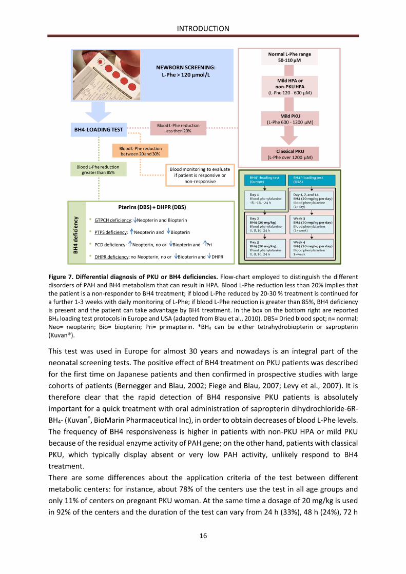
the metabolic centers this test is an integral part of the diagnosis. In Figure 7 is reported the
flow-chart commonly followed to perform the differential diagnosis of PKU or BH4
deficiencies, once HPA has been detected.
INTRODUCTION
16
Figure 7. Differential diagnosis of PKU or BH4 deficiencies. Flow-chart employed to distinguish the different disorders of PAH and BH4 metabolism that can result in HPA. Blood L-Phe reduction less than 20% implies that the patient is a non-responder to BH4 treatment; if blood L-Phe reduced by 20-30 % treatment is continued for a further 1-3 weeks with daily monitoring of L-Phe; if blood L-Phe reduction is greater than 85%, BH4 deficiency is present and the patient can take advantage by BH4 treatment. In the box on the bottom right are reported BH4 loading test protocols in Europe and USA (adapted from Blau et al., 2010). DBS= Dried blood spot; n= normal; Neo= neopterin; Bio= biopterin; Pri= primapterin. *BH4 can be either tetrahydrobiopterin or sapropterin (Kuvan®).
This test was used in Europe for almost 30 years and nowadays is an integral part of the
neonatal screening tests. The positive effect of BH4 treatment on PKU patients was described
for the first time on Japanese patients and then confirmed in prospective studies with large
cohorts of patients (Bernegger and Blau, 2002; Fiege and Blau, 2007; Levy et al., 2007). It is
therefore clear that the rapid detection of BH4 responsive PKU patients is absolutely
important for a quick treatment with oral administration of sapropterin dihydrochloride-6R-
BH4- (Kuvan®, BioMarin Pharmaceutical Inc), in order to obtain decreases of blood L-Phe levels.
The frequency of BH4 responsiveness is higher in patients with non-PKU HPA or mild PKU
because of the residual enzyme activity of PAH gene; on the other hand, patients with classical
PKU, which typically display absent or very low PAH activity, unlikely respond to BH4
treatment.
There are some differences about the application criteria of the test between different
metabolic centers: for instance, about 78% of the centers use the test in all age groups and
only 11% of centers on pregnant PKU woman. At the same time a dosage of 20 mg/kg is used
in 92% of the centers and the duration of the test can vary from 24 h (33%), 48 h (24%), 72 h
Classical PKU(L-Phe over 1200 µM)
Mild PKU(L-Phe 600 - 1200 µM)
Mild HPA or non-PKU HPA
(L-Phe 120 - 600 µM)
Normal L-Phe range50-110 µM
NEWBORN SCREENING:L-Phe > 120 µmol/L
BH4-LOADING TEST
Blood monitoring to evaluate if patient is responsive or
non-responsive
Pterins (DBS) + DHPR (DBS)
• GTPCH deficiency: Neopterin and Biopterin
• PTPS deficiency: Neopterin and Biopterin
• PCD deficiency: Neopterin, no or Biopterin and Pri
• DHPR deficiency: no Neopterin, no or Biopterin and DHPR
BH
4 d
efi
cie
ncy
Blood L-Phe reductiongreater than 85%
Blood L-Phe reduction between 20 and 30%
INTRODUCTION
17
(16%) and, in some centers - especially from US - from 1 to 4 weeks while in Europe shorter
tests were favorite.
The test should be performed early after birth and before the introduction of the low L-Phe
diet, so as blood L-Phe variations upon BH4 treatment are more evident. Blood L-Phe must be
over 400 µM in order to avoid false negative or false positive results (Belanger-Quintana et
al., 2011). Thus, older patient who are already on dietary regimen must increase the protein
intake before and during the testing period, or should undergo a concomitant L-Phe load,
consisting in a single administration of 100 mg L-Phe/kg BW (Blau, 2008; Blau et al., 2010). In
European centers, a reduction of 30% of L-Phe after a twice administration of 20 mg/kg BH4
is considered a positive response while a decrease under the 20% as negative response. For a
reduction in the range between 20% and 30% a daily monitoring of L-Phe for about 3 weeks is
recommended (Belanger-Quintana et al., 2011). Although the test is effective at all ages, its
sensitivity in newborns has been questioned due to liver immaturity and because only 24h
protocols can be employed at this age (Belanger-Quintana et al., 2011). Performing the
analysis as soon as possible allows the early introduction of the restricted diet in non-
responders, favoring breastfeeding or natural protein intake in responder patients but, at the
same time, implies the possibility to miss slow responders (who are mistakenly considered
negative to the test). Moreover, the association between genotype and responsiveness to BH4
is not really true because if on one side it can identify classic PKU, in all the other situations,
above mentioned, it difficulty predict who will respond to the treatment (Blau et al., 2010).
Therefore, it is advised to repeat the test according to longer protocols after 3 months of life,
that is when the liver has reached complete maturity and longer testing protocols can be
applied (Belanger-Quintana et al., 2011). The BIOPKU database (www.biopku.org) reports all
mutations that are correlated with BH4 responsiveness.
PATHOGENIC MECHANISMS OF HPA
Brain development and behavioral outcomes
The main clinical manifestations are due to the disruption of PAH metabolism that causes
accumulation of high levels of L-Phe in the blood, its excessive and toxic concentration in the
brain together with low levels of L-Tyr and its metabolites, which lastly affect different aspects
of brain functioning. The effects of liver PAH mutations on the ability to maintain L-Phe
homeostasis have been well described and the main clinical effects are related to the normal
development of the brain and the physiological development of cognitive functions (Kayaalp
et al., 1997). Untreated HPA is the most common biochemical cause of mental retardation
(intelligence quotient, IQ <30), seizures, microcephaly, epilepsy, motor deficits, severe
intellectual disability and behavioral disturbances, including psychotic, autistic, and aggressive
disorders (Mitchell et al., 2011; Bone et al., 2012). Thus, patients with PKU have lower IQ
scores than normal subjects and they present other deficits in various neuropsychological
functions such as working memory, cognitive and executive functions (Blau et al., 2010; Feillet
et al., 2010). All these conditions are due to both aminergic neurotransmitters depletion, i.e.
dopamine (DA), serotonin (5-HT), and myelin impairment. As extensively reviewed by several
authors (Surtees and Blau, 2000; Bone et al., 2012), many of which use PKU mouse models
INTRODUCTION
18
(Fiori et al., 2017; Pascucci et al., 2002, 2008), the excessive L-Phe exposure is responsible for
the altered development of the brain architecture which include abnormal myelination,
cortical plate width and altered dendritic arborization together with a reduced number of
synaptic spines. In addition, the exposure to high concentration of L-Phe makes the already
formed myelin unstable, thus demyelinated axons undergo a reverse maturation, with
consequent neuronal dysfunction (Cleary et al., 1995). Myelin is a metabolic active membrane
produced by oligodendrocytes and it plays an important role for the fast transmission of action
potentials. White matter pathology characterizes the brain of untreated PKU patients where
neurological deterioration is evident and the impact of metabolic control on impairment of
myelination process is related to specific brain areas. Therefore, in childhood the injury of
visuospatial processing is more evident because occipital regions are the first myelinated area
during development while the frontal regions are myelinated later, so the damage of complex
executive functions is more evident during adolescence and adulthood (Klingberg et al., 1999;
Gogtay et al., 2004; Best and Miller, 2010). In addition, the development of the cerebral cortex
occurs following a precise sequence of events, well defined in time and space, especially as
regards the synapses and dendrites formation in the prefrontal cortex. As reported in a study
conducted on mouse model of PKU (Pascucci et al., 2008), during the critical developmental
period (PND 14-21), different availability of brain amines, with an initial increase of
catecholamines and serotonin which then decrease and return to adult levels, has been
observed. This period represents the most susceptible phase to L-Phe-induced damages, as
extensively demonstrated by studies on animal models (Goldman-Rakic and Brown, 1982;
Thomas et al., 1995; Zhou et al., 1995; Berger-Sweeney and Hohmann, 1997; Chugani et al.,
1999; Puglisi-Allegra et al., 2000; Herlenius and Lagercrantz, 2001, 2004; Cabib et al., 2003;
Pascucci et al., 2008, 2009, 2012; Andolina et al., 2011).
Indeed, before acting as neurotransmitters, biogenic amines represent fundamental signals
for the correct early development of the brain (Lauder, 1993) suggesting that a deficit in the
availability of these amines during the critical periods of development, particularly around the
third week of life in the murine models of PKU, is associated with cognitive dysfunction
(Pascucci et al., 2008). These observations have been confirmed by several studies that
highlight how development of the synapses, the growth of the dendritic tree and its
remodeling (Van Eden and Uylings, 1985; Huttenlocher, 1991; Vitalis and Parnavelas, 2003)
are dramatically affected by the decrease of amines levels in the critical period. In particular,
5-HT was the first neurotransmitter for which the role in brain development, dendritic spines
formation and maintenance and amelioration of synaptic connectivity during postnatal life,
has been demonstrated (Mazer et al., 1997; Whitaker-Azmitia, 2001; Sodhi and Sanders-Bush,
2004).
On the other hand, the decreased levels of neurotransmitters, including dopamine, are related
to cognitive and behavioral disabilities (Diamond, 1996; Puglisi-Allegra et al., 2000; Pascucci
et al., 2002; Joseph and Dyer, 2003). L-Phe belongs to the group of the Large Neutral Amino
Acids (LNAAs), with valine, leucine, isoleucine, threonine, histidine, tryptophan, methionine
and tyrosine. All these amino acids share a common selectively predominant carrier system,
the L-amino acid transporter-1 (LAT-1), to cross the blood-brain barrier (BBB) and enter into
INTRODUCTION
19
the brain (Blau et al., 2010). Binding of LNAA to this transporter is a dynamic and competitive
process (Pardrige, 1998; Boado et al., 1999; Smith, 2000), in fact for each LNAA taken into the
brain another one is excreting (Zielke et al., 2002). Across species LAT-1 appeared to have a
higher affinity for L-Phe than the other LNAAs, and this is particularly marked for the human
species, making it more susceptible to the negative effects of HPA. In fact, an excessive
circulating amount of L-Phe has the ability to saturate the LAT-1 transporter, thanks to its
lowest km value for the carrier respect the other LNAAs, resulting in a L-Phe overload and
decreased amount of the other LNAAs, particularly L-Tyr and L-Trp, in the brain (Surtees and
Blau, 2000; Blau et al., 2010). At the same time, non-L-Phe LNAA export from the brain in
exchange for blood L-Phe is increased (de Groot et al., 2010), carrying a reduction in cerebral
protein synthesis for reduced availability of these non L-Phe aminoacids (Pardridge, 1998; van
Vliet et al., 2015). All this evidence explains why the brain is vulnerable to HPA.
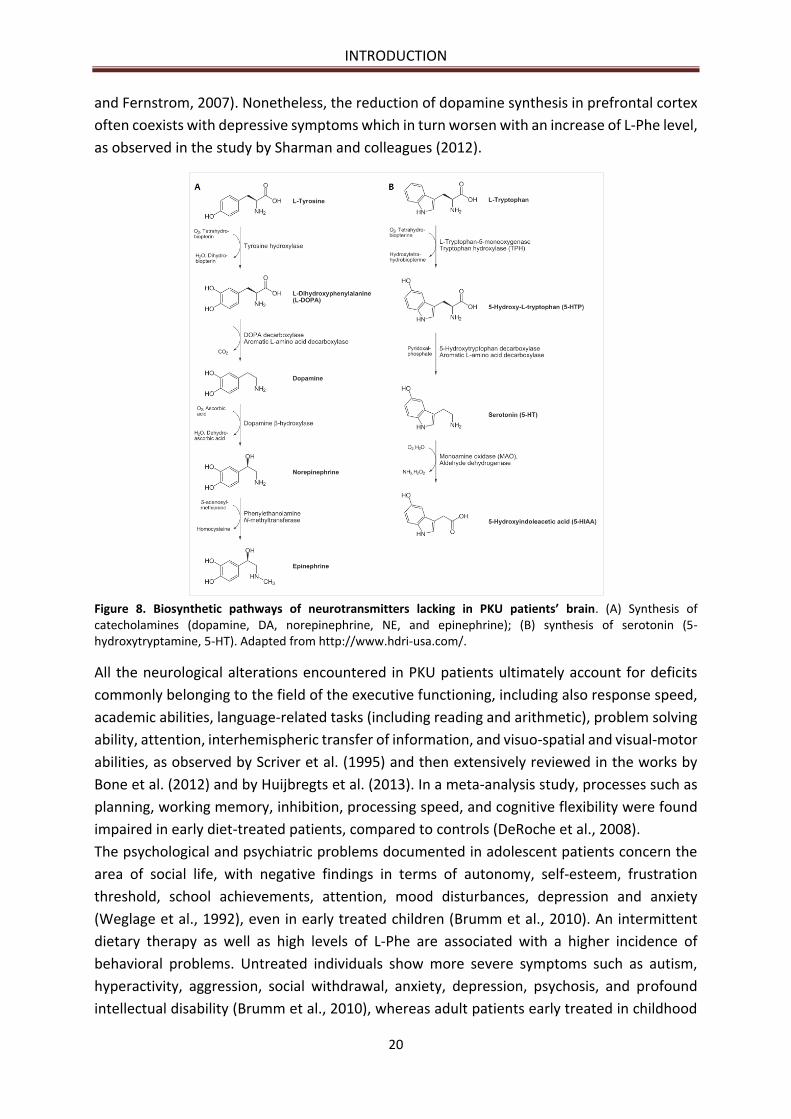
The LNAAs L-Tyr and L-Trp besides their role in protein synthesis, are also precursors for
neurotransmitters, namely dopamine (DA), norepinephrine (NE), epinephrine and serotonin
(5-hydroxytryptamine, 5-HT) respectively (Figure 8) (Surtees and Blau, 2000). Dopamine plays
an important role in motor and cognitive functioning; norepinephrine is involved in learning
and memorization processes, in the arousal of attention, fear and anxiety, and in the
development of the maternal behaviour in females; serotonin is important for neuronal
proliferation, synaptogenesis and morphogenesis (Herlenius and Lagercrantz, 2001, 2004).
Thus, there are two possible mechanisms by which L-Phe alters brain functioning: if on the
one hand the increased L-Phe concentration in brain results in a decreased level of the other
LNAAs including L-Tyr and L-Trp, as described above, on the other hand it acts as a competitive
inhibitor of the other two amino acid hydroxylases, TyrOH and TrpOH (McKean, 1972; Curtius
et al., 1981; Surtees and Blau, 2000; Ogawa and Ichinose, 2006; Pascucci et al., 2008),
generating a lack of their products. In fact, high brain L-Phe was reported to negatively affect
the activity of the other hydroxylases (Surtees and Blau, 2000).
The idea that a deficiency of 5-HT neurotransmitter plays an important role in neurological
disorders due to HPA (Shimada et al., 1993) is supported by the fact that it is also the most
reduced amine in the brain - about 50% - compared with 40% reduction of NE and 30% of DA
(Pascucci et al., 2008). In particular, in a study of Pascucci et al. (2008) it has been
demonstrated that the reduction of 5-HT around 3 postnatal week overlaps with a critical
period for synaptogenesis and dendritic development, thus compromising maturation of
prefrontal cortex (PFC) neurons with the subsequent alteration of the cognitive performance.
Severe lack of whole brain 5-HT during critical post-natal periods and deficits in the level of its
immediate and limiting precursor 5-hydroxytryptophan (5-HTP), is not connected to a
decrease in its initial amino acidic precursor L-Trp; this evidence support the hypothesis of
TrpOH activity inhibition exerted by L-Phe excess, rather than a hampered access of L-Trp
across the BBB (Ogawa and Ichinose, 2006; Pascucci et al., 2009), thus confirming a minor
involvement of L-Trp in the L-Phe induced alterations (Pascucci et al., 2002). On the contrary,
dopamine and its precursor L-3,4-dihydroxyphenylalanine (L-DOPA) are the less affected by
HPA because, when L-Tyr levels are abnormally low and L-Phe is extremely high, the latter can
serve as TyrOH substrate for the production of L-DOPA (Joseph and Dyer, 2003; Fernstrom
INTRODUCTION
20
and Fernstrom, 2007). Nonetheless, the reduction of dopamine synthesis in prefrontal cortex
often coexists with depressive symptoms which in turn worsen with an increase of L-Phe level,
as observed in the study by Sharman and colleagues (2012).
Figure 8. Biosynthetic pathways of neurotransmitters lacking in PKU patients’ brain. (A) Synthesis of catecholamines (dopamine, DA, norepinephrine, NE, and epinephrine); (B) synthesis of serotonin (5-hydroxytryptamine, 5-HT). Adapted from http://www.hdri-usa.com/.
All the neurological alterations encountered in PKU patients ultimately account for deficits
commonly belonging to the field of the executive functioning, including also response speed,
academic abilities, language-related tasks (including reading and arithmetic), problem solving
ability, attention, interhemispheric transfer of information, and visuo-spatial and visual-motor
abilities, as observed by Scriver et al. (1995) and then extensively reviewed in the works by
Bone et al. (2012) and by Huijbregts et al. (2013). In a meta-analysis study, processes such as
planning, working memory, inhibition, processing speed, and cognitive flexibility were found
impaired in early diet-treated patients, compared to controls (DeRoche et al., 2008).
The psychological and psychiatric problems documented in adolescent patients concern the
area of social life, with negative findings in terms of autonomy, self-esteem, frustration
threshold, school achievements, attention, mood disturbances, depression and anxiety
(Weglage et al., 1992), even in early treated children (Brumm et al., 2010). An intermittent
dietary therapy as well as high levels of L-Phe are associated with a higher incidence of
behavioral problems. Untreated individuals show more severe symptoms such as autism,
hyperactivity, aggression, social withdrawal, anxiety, depression, psychosis, and profound
intellectual disability (Brumm et al., 2010), whereas adult patients early treated in childhood
INTRODUCTION
21
display generalized depressed and anxious mood, lack of autonomy, low self-esteem and a
tendency to social isolation; phobias are also typical and the most common one is agoraphobia
(Waisbren and Levy, 1991; Pietz et al., 1997; Brumm et al., 2010).
Adequate control of blood L-Phe concentration is therefore very important for the prevention
of brain deficit. Children with poor metabolic control (L-Phe >400 µM) have reduced executive
functions while children with PKU have behavioral abnormalities, motor dysfunction (Arnold
et al., 1998) and memory impairment (White et al., 2002). In addition, even if several
publications showed a correlation between blood L-Phe fluctuations and intellectual
outcomes, cognition or executive functions, no correlation has been found between this
fluctuation and patients IQ (intelligence quotient) even if it has been identified an influence
on these parameters (Cleary et al., 2013). However, some studies have shown that high blood
L-Phe fluctuation in patients with PKU are associated with lower neurocognitive outcome.
Crucial for the improvement of cognitive function is the metabolic control; indeed, a meta-
analysis of five studies on PKU patients and control showed a significant inverse correlation
between the IQ score and L-Phe levels. This correlation is especially clear during the critical
developmental period (age 0-12 years), even in early treated children: each 100 µM rise in L-
Phe concentration corresponded to a 1.3-3.1 IQ point decline (Burgard, 2000). This result has
been confirmed by a meta-analysis study on children treated since the neonatal age where
the IQ decreases of about 1.9-4.1 point for each 100 µM increase in L-Phe (Blau et al., 2010);
a similar correlation was also found between lifetime L-Phe levels and IQ scores in early-
treated individuals (Waisbren et al., 2007). This condition is due to both dopamine depletion
and myelin impairment.
In addition, HPA includes the reduction of pyruvate kinase activity in the brain (Hörster et al.,
2006), the alteration of glutamatergic neurotransmission (Martynyuk et al., 2005), the
reduction of enzyme 3-hydroxy-3-methyl coenzyme A reductase (HMG-CoA reductase)
(Shefer et al., 2000) as well as the impairment of monoamine oxidase B activity (Ghozlan et
al., 2004).
L-Phe influence on cholesterol biosynthesis and obesity
L-Phe levels play an important role in the inhibition of the rate-limiting enzyme of the
cholesterol biosynthetic pathway in liver and brain, namely 3-hydroxy-3-methylglutaryl-CoA
reductase (HMG-CoA reductase; EC 1.1.1.88), reducing the synthesis of mevalonic acid
(Castillo et al., 1988). The resulting hypocholesterolemia is hypothesized to have a protective
effect against cardiovascular diseases in adults (Williams et al., 2008), but even if PKU children
have lower blood total cholesterol and LDL levels with respect to healthy subjects,
cardiovascular risk has been reported to be the same (Verduci et al., 2016). Elevated levels of
L-Phe have been showed to decrease coenzyme Q10 (ubiquinone-10; CoQ10) concentrations
both in plasma and in lymphocytes. This coenzyme is involved in many functions i.e. acting as
a cofactor in the mitochondrial electron transport chain, preventing LDL oxidation and
representing an antioxidant molecule in mitochondria and lipid membranes (Colomé et al.,
2002).
INTRODUCTION
22
The PKU patients, in particular female, represent those with the highest incidence of obesity
but the correlation is not clear yet (Belanger-Quintana and Martínez-Pardo, 2011; Burrage et
al., 2012; Rocha et al., 2012; Robertson et al., 2013).
MATERNAL PKU
Non-controlled levels of L-Phe during pregnancy are teratogenic for the fetus and can increase
the risk of miscarriage (American Academy of Pediatrics: Committee on Genetics, 2001; Blau
et al., 2010). This condition, called maternal phenylketonuria syndrome or maternal PKU
(MPKU), was firstly described over 60 years ago (Pinto et al., 2017) and is responsible for
intrauterine growth retardation, spontaneous abortion, intrauterine fetal death (IUFD),
congenital heart disease, developmental delay and other important fetal alterations (Levy and
Ghavami, 1996; Rouse et al., 1997; Williams et al., 2008; Prick et al., 2012). Babies delivered
by mothers under MPKU condition show microcephaly, congenital heart disease (CHD),
intellectual or developmental disabilities (IDDs), and facial dysmorphism (FD) together with
low birth weight (defined as small for gestational age, SGA) (Lenke and Levy, 1980; Levy and
Ghavami, 1996; Rouse et al., 1997). Adequate control of L-Phe levels is important not only
during pregnancy but also before conception because the toxic effect of this amino acid is
dangerous in early stages of pregnancy, especially during the first weeks of embryogenesis.
To this purpose, it is essential that affected women follow a strict low L-Phe diet for several
months before conception, in order to stabilize the levels of this amino acid between 100 and
360 μmol/L, thus preventing teratogenic effects on the fetus (Lee et al., 2005); moreover, it is
essential to maintain an optimal blood L-Phe level throughout the all pregnancy.
Despite during pregnancy the phenylalanine tolerance is slightly increased thanks to the
activity of fetal PAH - as has been observed during the second trimester of pregnancy when L-
Phe levels decrease and tolerance of proteins intake increased (Prick et al., 2012) - weekly or
biweekly controls of L-Phe blood levels remain fundamental to avoid fetal impairments
(Australian Society for Inborn Errors of Metabolism). At the same time is important that these
women receive an adequate energy intake, in terms of proteins, fats, carbohydrates and
multivitamin complexes, vitamin B12 and folic acid, in order to guarantee the best conditions
for fetal growth (Koch et al., 2000).
Although some studies have shown that children born by women with untreated
concentrations of L-Phe lewer than 400 μmol/L, may be normal, the "Maternal PKU
Collaborative Study" (MPKUCS) reports that between children born from women with levels
of L-Phe between 120 and 360 μmol/L, 6% showed microcephaly and 4% showed a delay in
post-natal growth. If the concentration of L-Phe exceeds 900 μmol/L, the risk of microcephaly
rises to 85%, post-natal growth delay rises to 51% and intrauterine growth retardation to 26%.
For all these reasons, strictly controlled plasma L-Phe levels should be necessary (Rouse and
Azen, 2004). However, a common problem is the difficulty of sick women in adhering to the
strict diet for poor intellectual and social skills (Koch et al., 2000). For this reason, a pilot
project has been developed to provide a special support and education about the importance
of diet control during pregnancy with the aim of guaranteeing a good health for the newborn
INTRODUCTION
23
child (Waisbren et al., 2000). PKU mothers bearing non-PKU babies (i.e. healthy carriers of a
single mutated allele) are encouraged to breastfeed their children without restriction, since
the single non mutated copy of the PAH gene is sufficient to metabolize the amount of L-Phe
introduced with breast milk (NIH consensus panel, 2001), also because breast milk contains
only 43 mg L-Phe/dl compared to 59-73 mg/dl of infant formulas and 164 mg/dl of cow’s milk
(Berlin et al., 1995).
Following all such recommendations it is possible for PKU mothers to have children with the
same expectancy of cognitive development as non-PKU people (Levy and Ghavami, 1996).
THERAPEUTIC STRATEGIES
Prognosis and outcome depend on time of diagnosis, type of mutations and quickness of
intervention. In fact, the goal of the various therapeutic strategies is to rapidly restoring the
normal levels of L-Phe and L-Tyr in the circulation, in order to eliminate biochemical
abnormalities, improve neurological and psychological performance, and prevent the
syndrome of maternal PKU (Williams et al., 2008). Clinical goals are not only aimed to reduce
L-Phe levels in blood but also to limit its negative effect on other brain aminoacid
concentrations. Nevertheless, there is great discrepancy between the different European
countries and clinical centers in defining the level of L-Phe beyond which it would be
appropriated to intervene (Table 1), especially during the first decade of life, the most
important period of development (van Spronsen et al., 2009; Blau et al., 2010).
This discrepancy further increases beyond the first decade of life, where the gap between
Europe and U.S.A. becomes more pronounced (van Spronsen et al., 2009; Blau et al., 2010).
The most common concentrations identified to this purpose are 360 µM, 400 µM and 600 µM
(Blau et al., 2010) and the L-Phe level considered safe is in the range between 120 and 360
µM, at least until 12 years of age (Koch et al., 1996; NIH consensus, 2001), with the upper
limits rising up to 900 µM after the 12th year of life (NIH consensus, 2001). However, there is
great inconsistency about the target range to be reached in adolescence and adulthood,
resulting in a wide spectrum of disease management and outcomes (Ahring et al., 2009; Blau
et al., 2010) and some studies highlighted the greater importance of considering L-Phe/L-Tyr
ratio rather than the concentration of L-Phe alone, since is considered more involved in the
impairment of executive functions (Sharman et al., 2010).
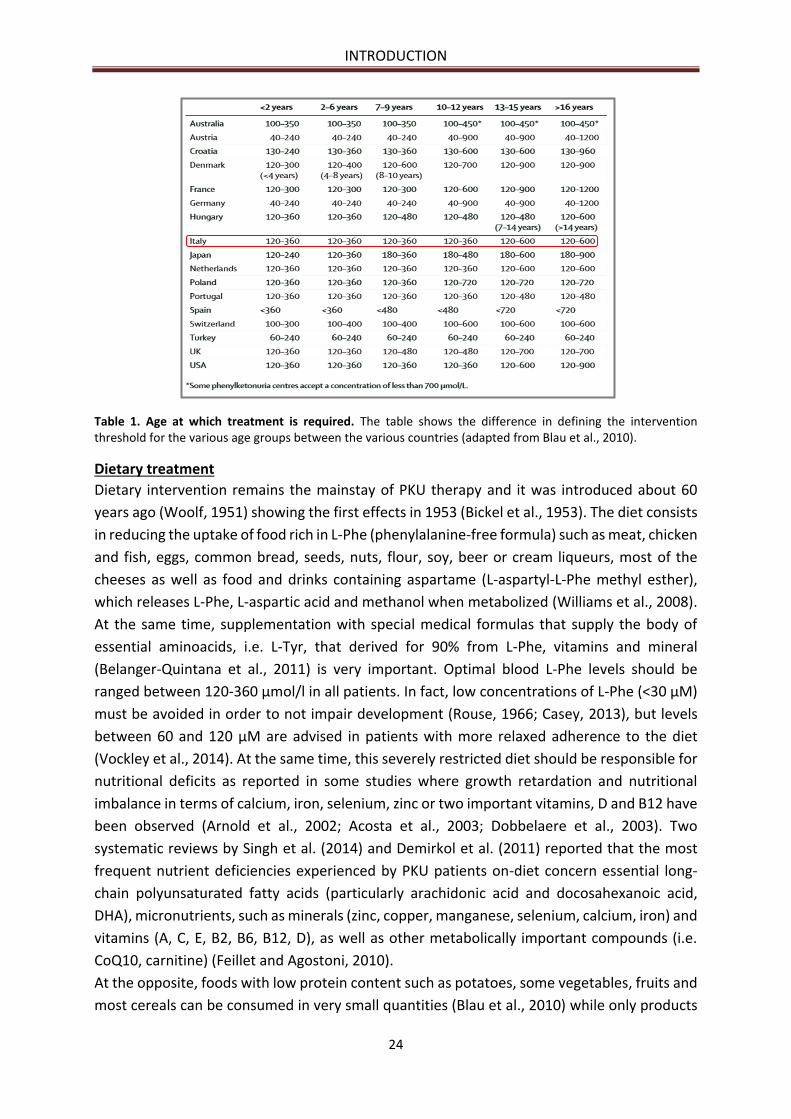
INTRODUCTION
24
Table 1. Age at which treatment is required. The table shows the difference in defining the intervention threshold for the various age groups between the various countries (adapted from Blau et al., 2010).
Dietary treatment
Dietary intervention remains the mainstay of PKU therapy and it was introduced about 60
years ago (Woolf, 1951) showing the first effects in 1953 (Bickel et al., 1953). The diet consists
in reducing the uptake of food rich in L-Phe (phenylalanine-free formula) such as meat, chicken
and fish, eggs, common bread, seeds, nuts, flour, soy, beer or cream liqueurs, most of the
cheeses as well as food and drinks containing aspartame (L-aspartyl-L-Phe methyl esther),
which releases L-Phe, L-aspartic acid and methanol when metabolized (Williams et al., 2008).
At the same time, supplementation with special medical formulas that supply the body of
essential aminoacids, i.e. L-Tyr, that derived for 90% from L-Phe, vitamins and mineral
(Belanger-Quintana et al., 2011) is very important. Optimal blood L-Phe levels should be
ranged between 120-360 µmol/l in all patients. In fact, low concentrations of L-Phe (<30 µM)
must be avoided in order to not impair development (Rouse, 1966; Casey, 2013), but levels
between 60 and 120 µM are advised in patients with more relaxed adherence to the diet
(Vockley et al., 2014). At the same time, this severely restricted diet should be responsible for
nutritional deficits as reported in some studies where growth retardation and nutritional
imbalance in terms of calcium, iron, selenium, zinc or two important vitamins, D and B12 have
been observed (Arnold et al., 2002; Acosta et al., 2003; Dobbelaere et al., 2003). Two
systematic reviews by Singh et al. (2014) and Demirkol et al. (2011) reported that the most
frequent nutrient deficiencies experienced by PKU patients on-diet concern essential long-
chain polyunsaturated fatty acids (particularly arachidonic acid and docosahexanoic acid,
DHA), micronutrients, such as minerals (zinc, copper, manganese, selenium, calcium, iron) and
vitamins (A, C, E, B2, B6, B12, D), as well as other metabolically important compounds (i.e.
CoQ10, carnitine) (Feillet and Agostoni, 2010).
At the opposite, foods with low protein content such as potatoes, some vegetables, fruits and
most cereals can be consumed in very small quantities (Blau et al., 2010) while only products
INTRODUCTION
25
without proteins such as pure sugar, butter, oil and low-protein versions of some foods, such
as bread and pasta, can be eaten without the need to be weighed (Zimmermann et al., 2012).
In order to guarantee the effectiveness of this approach, a rigorous diet control should start
shortly after birth (Blau et al., 2010), better if in the first weeks of age, confirming the
importance of screening programs. In fact, initiation of diet later in life cannot reverse the
brain damage. Moreover, it should be maintained for the entire lifetime in order to avoid any
problem in the adult because recent studies have shown that myelination does not terminate
in adolescence (Romani et al., 2017): this advice denies what done by a large number of clinics
in North America that choose to end the treatment after the first decade of life because they
believed that the process of myelination ended after this period of time (Azen et al., 1991).
Although many people believe that non-executive functions are not negatively affected if L-
Phe levels are maintained less than 1200 μmol/L during adolescence and adulthood without
need to follow a dietary control (Griffiths et al., 2000), this relation is not correct. Some studies
have shown that a less rigorous diet after 12 years, does not negatively influence the IQ score
but is able to compromise other functions i.e. decreasing the cognitive performance and
motor functions in PKU patients compared to healthy subjects (Griffiths et al., 1995). Anyway,
the diet must be carefully monitored, in order to adapt it to individual L-Phe tolerance, age
and growth requirement, illnesses, physical activity and pregnancy in females (Mitchell et al.,
2011; Vockley et al., 2014). Breastfeeding is encouraged in infants in combination with the
medical formula (Blau et al., 2010). Today, the need for a “diet-for-life” is generally accepted,
since if mental disability does not occur when patients have been well controlled during
infancy, many adverse manifestations can develop upon diet relaxation (Smith and Knowles,
2000).
Although dietary treatment has been very effective in prevent severe mental retardation, it
introduces also a large number of problems. First of all, the poor nutritional quality (as shown
above), poor palatability and heavy burden, both economic and social, upon the PKU patients
and their families (Simon et al., 2008). Diet adherence is particularly poor in adolescents and
adults (MacDonald et al., 2010; Ahring et al., 2011) because if during infancy the diet control
is easier because the child’s parents control nutritional intake, during adolescence adherence
to the diet becomes socially demanding due to the impossibility for the children to eat the
food consumed by their peers, inducing them to leave this dietary program. Suboptimal
dietary compliance is also due to psychosocial and emotional factors, commitment required
to parents, lack of reimbursement for food supplements and a low degree of knowledge about
the disease by the families (Crone et al., 2005; Olsson et al., 2007). At the same time, many
adults discontinue the diet during their life or refuse to come back to it after a period of
unrestricted diet. Moreover, strictly treated PKU patients not always eliminate the
neurological defects due to elevated L-Phe level (Belanger-Quintana et al., 2011): thus,
emotional distress, mood disturbances, slow reaction time and impaired social skill continue
to be reported (Pietz et al., 1993; Gassiò et al., 2003; Bik-Multanowski et al., 2008; Dawson et
al., 2011; Jahja et al., 2016).
Current efforts are focused on the development of a dietary treatment which ultimately
results not only in L-Phe control but also in the preservation of patients’ quality of life (Vockley
INTRODUCTION
26
et al., 2014). New dietary treatment includes more palatable foods, large neutral aminoacids
supplementations (LNAAs) (Matalon et al., 2007) and the use of a naturally low-Phe protein,
named glycomacropeptide. In a study of MacDonald et al. (2003) it has been suggested that a
free use of fruit and vegetables containing 51-75mg/100g of L-Phe and poor portions of
potatoes, rice and corn should be safe for children with PKU, making the dietary more
tolerable.
Furthermore, the increased knowledge of the genetic basis of PKU and enzymology has
allowed to develop new intervention approaches aimed to improve the activity of the mutant
enzyme: a clear example is represented by the administration of the BH4 cofactor in
association with the diet (Belanger-Quintana et al., 2011). Orthotropic liver transplantation
(Scriver and Kaufman, 2001), preclinical gene therapy studies and new enzymatic substitution
therapy, are other therapeutic approaches currently under investigation.
Glycomacropeptide
Glycomacropeptide (GMP) is a low-Phe protein of 65 amino acids, representing a minor
fraction of cheese whey, particularly rich in essential amino acids but without Tyr, Phe and Trp
(Laclair et al., 2009) making it useful for the treatment of PKU. This protein comes from the
cheesemaking process, when the bovine k-casein is cut from the chymosin to para-k-casein,
which remains with the curdle, and into GMP, which remains in the cheese whey (Doultani et
al., 2003). The glycomacropeptide can be used in the classical diet when it is extracted in high
purity but if used as a primary source of protein for individuals with PKU, it must be
supplemented with other essential amino acids (Tyr, Trp, His and Leu), vitamins and minerals
(Ney et al., 2009) even if its LNAA content is 3 times higher than other natural proteins (Etzel,
2004). Studies performed on patients that assumed this protein have observed an improved
compliance with the diet, higher metabolic control and better quality of life thanks to the
greater palatability of foods (Lim et al., 2007; Ney et al., 2009).
Large neutral aminoacids (LNAAs) supplementation
LNAAs supplementation has been suggested as valid alternative approach to classical dietary
restriction to avoid disturbances caused by high L-Phe concentrations in early 1948
(Christensen et al., 1948) and was for the first time studied in rats in 1976 (Andersen and
Avins, 1976). This class of amino acids includes tyrosine, tryptophan, threonine, methionine,
valine, isoleucine, leucine and histidine (van Spronsen et al., 2010). All of them are essential
except for tyrosine that, however, becomes essential in PKU patients. Since the first
employment, different combinations of LNAA strategy, including only supplementation with
tyrosine and tryptophan have been investigated. LAT1 transporter has been known having
major affinity for L-Phe instead of LNAAs, so this treatment has the intention to restore the
altered LNAAs transport across the BBB in order to decrease brain L-Phe level (Berry et al.,
1982; Jordan et al., 1985; Pietz et al., 1999), increase the levels of non-Phe LNAAs in the brain
(van Spronsen et al., 2009), increase monoaminergic neurotransmitters concentration (Lou,
1985; Lykkelund et al., 1988; van Vliet et al., 2015, 2016) and, at the same time, reduce blood
L-Phe level (Matalon et al., 2006). This goal is achieved because when LNAAs level increase,
these aminoacids compete with L-Phe for LAT1, reducing the entry of this compound through
INTRODUCTION
27
the BBB and, at the same time, increasing their concentration inside the brain. Besides, has
been hypothesized that LNAAs can reduce L-Phe level competing with its uptake at the gut-
blood barrier and/or increasing protein synthesis and thus L-Phe utilization (Matalon et al.,
2003). This hypothesis has been proven by various studies performed on patients with PKU,
in which a reduction up to 50% of the blood concentration of this amino acid was observed
following the administration of 0.5-1.0 g/Kg/day of LNAA (Matalon et al., 2006, 2007).
Different combinations of LNAAs have been developed and some of these contain arginine
and/or lysine, which don’t belong to LNAAs. In a study of van Vliet et al. (2015) has been
observed that after LNAAs supplementations, in PKU mice brain L-Phe concentrations were
significantly reduced by 26%: this result is perfectly in line with previous study performed on
both PKU patients and mice (van Spronsen et al., 2010; van Vliet et al., 2015) where a
reduction of about 20% and 46%, respectively, were reported. LNAAs supplementation has
been also shown able to increase cerebral serotonin and norepinephrine concentrations, as
also indirectly suggested by the increased blood and urine melatonine concentrations induced
by the treatment, probably by both decreasing brain L-Phe and increasing brain tyrosine and
tryptophan levels (Berry et al., 1982; van Vliet et al., 2015).
LNAAs treatment should represent an alternative strategy to diet control but it is currently
limited to adolescent and adult patients and is not advised during pregnancy, due to the
limited knowledge about the effect on fetal development (Vockley et al., 2014). However, a
recent study (van Vliet et al., 2018) suggests that LNAAs treatment without any L-Phe-
restricted diet in adult PKU mice was able to restore brain monoamine concentration equally
to the severe L-Phe-restricted diet. To this end, this strategy is a promising alternative for all
adult PKU patients with suboptimal compliance to the diet. In order to increase adherence to
LNAAs supplementation, studies to improve taste, tolerability and correct LNAAs doses and
composition would be beneficial (van Vliet et al., 2018).
Tetrahydrobiopterin (BH4) treatment
As reported in the diagnosis section, a little percentage of patients has mutations associated
with a BH4-responsive phenotype. This aspect was remained unexplored until 1999 when Kure
et al (Kure et al., 1999) demonstrated that after a treatment with 20 mg/kg of BH4, in some
patients blood levels of L-Phe was reduced. Since then from 20 to 60% of patients was found
to be BH4 responsive. Generally, in these patients mutations on PAH gene are located in the
entire region but allow to maintain residual activity of the enzyme which can interact with
BH4 as showed in figure 9.
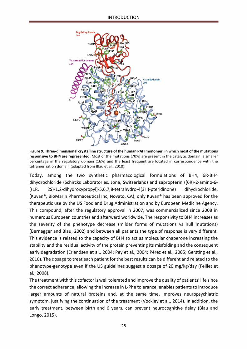
INTRODUCTION
28
Figure 9. Three-dimensional crystalline structure of the human PAH monomer, in which most of the mutations responsive to BH4 are represented. Most of the mutations (70%) are present in the catalytic domain, a smaller percentage in the regulatory domain (16%) and the least frequent are located in correspondence with the tetramerization domain (adapted from Blau et al., 2010).
Today, among the two synthetic pharmacological formulations of BH4, 6R-BH4
dihydrochloride (Schircks Laboratories, Jona, Switzerland) and sapropterin ((6R)-2-amino-6-
[(1R, 2S)-1,2-dihydroxypropyl]-5,6,7,8-tetrahydro-4(3H)-pteridinone) dihydrochloride,
(Kuvan®, BioMarin Pharmaceutical Inc, Novato, CA), only Kuvan® has been approved for the
therapeutic use by the US Food and Drug Administration and by European Medicine Agency.
This compound, after the regulatory approval in 2007, was commercialized since 2008 in
numerous European countries and afterward worldwide. The responsivity to BH4 increases as
the severity of the phenotype decrease (milder forms of mutations vs null mutations)
(Bernegger and Blau, 2002) and between all patients the type of response is very different.
This evidence is related to the capacity of BH4 to act as molecular chaperone increasing the
stability and the residual activity of the protein preventing its misfolding and the consequent
early degradation (Erlandsen et al., 2004; Pey et al., 2004; Pérez et al., 2005; Gersting et al.,
2010). The dosage to treat each patient for the best results can be different and related to the
phenotype-genotype even if the US guidelines suggest a dosage of 20 mg/kg/day (Feillet et
al., 2008).
The treatment with this cofactor is well tolerated and improve the quality of patients’ life since
the correct adherence, allowing the increase in L-Phe tolerance, enables patients to introduce
larger amounts of natural proteins and, at the same time, improves neuropsychiatric
symptom, justifying the continuation of the treatment (Vockley et al., 2014). In addition, the
early treatment, between birth and 6 years, can prevent neurocognitive delay (Blau and
Longo, 2015).
INTRODUCTION
29
Only light adverse events of the treatment have been reported: typically, headache followed
by vomiting, diarrhea or irritation of the respiratory tract. However, since there are no data
about the safety of the treatment on children under 4 years, even if the early administration
has the ability to maintain low blood L-Phe levels, it remains not recommended for this class
of patients.
Despite the advantages above reported and although the treatment with BH4 allows the
possibility of oral administration, it is important to consider the short half-life of sapropterin,
which is about 6.5 hours (3-17 h), its rapid sequestration by the liver or elimination through
urine in the case of oral administration (Harding et al., 2004). In addition, it should be
considered the high costs of the daily therapy, if compared to those of the dietary regimen
(Harding, 2010), and the insufficient L-Phe lowering that sometimes can be observed in
responsive patients.
Gene therapy and liver transplantation
The increasing knowledge in many different scientific fields and the ability of cloning human
and murine PAH cDNA (Woo et al., 1983) have opened the way to gene therapy. Gene therapy
is based on the insertion, by different vectors, of PAH cDNA in organs or tissues, such as the
liver, in order to restore its functionality. Nowadays, gene therapy represents a very promising
approach as alternative therapy to restrictive diet (Kim et al., 2004), also considering that even
just a 10% of PAH activity in mice it is enough to establish a normal metabolism of L-Phe
(Ledley et al., 1985). Various approaches have been tested for this treatment. Among these,
the first was based on in vitro transfer of the PAH gene inside the hepatocytes by its
complementation with a protein recognized by the hepatic receptors allowing its
internalization (Ding et al., 2004). As this system showed some limitations in transduction
efficiency, it was hypothesized to use viral vectors. The first tested vector able to transfer PAH
cDNA to liver was recombinant Adenoviruses. However, although its ability to promote L-Phe
stabilization at the recommended therapeutic range, it was soon abandoned due to the high
immune response developed, which limited the duration of the therapeutic effect and the
possibility of repeated administrations (Eisensmith and Woo, 1996). Recently, the Adeno-
associated recombinant virus (rAAV) has attracted great attention because of its advantages
of being poorly immunogenic, its ability to infect cells that do not divide, its low tissues
specificity and the absence of liver damage (Thomas et al., 2003; Oh et al., 2004). However,
this strategy does not allow long-term effects because rAAV does not integrate its genome
into the infected cells but it remains in episomal form, been lost during the cell regeneration
process. The skeletal muscle, however, thanks to its slower regeneration and its easier
accessibility, represents a very interesting target (Rebuffat et al., 2010). In keeping with
muscle potential role as target, the intramuscular injection of a vector containing the PAH
gene has the ability to protect small children born from sick mice from the teratogenic effects
of HPA (Jung et al., 2008). The transduction of the muscle with only PAH cDNA is not however
sufficient to induce an effective hydroxylation of L-Phe: indeed, in order to obtain the
therapeutic effects, it become necessary the simultaneous expression of the enzymes
involved in the synthesis of BH4 (Ding et al., 2008).
INTRODUCTION
30
The strategy of cell therapy has been proven in several preclinical studies on animal models
of inborn errors of metabolism, such as defects in the urea cycle, in glycogen storage and in
the α1 antitrypsin deficiency. Conversely, the transplantation of hepatocytes with the
functionality PAH/BH4 system, has not been tested in PKU patients yet because the donor
cells have to growth over the autochthonous cells (Harding, 2008). In addition, liver
transplantation makes necessary the administration of immunosuppressant agents and the
high risk of major surgery precludes its routine use.
Enzyme replacement therapy (ERT)
Despite BH4 treatment represents a promising strategy to reduce L-Phe level, patients with a
classical form of PKU can’t benefit of this solution probably for the complete absence of PAH
residual activity and even if the nutritional control or supplementation with LNAAs help to
reduce brain L-Phe levels, enzyme replacement therapy (ERT) represents a valid alternative to
all the different types of intervention today available. ERT consists in restoring a lacking or
non-functioning enzyme with its native counterpart or with an enzyme able to degrade toxic
metabolites or secondary compounds of the reaction catalyzed by the deficient enzyme. ERT
was first proposed by de Duve in 1964 (de Duve, 1964) expressly for lysosomal storage
diseases (LSDs). However, the administration of various active substances by conventional
systems and dosage forms may involve intrinsic difficulties such as a rapid clearance by
premature degradation, inactivation or elimination from the body, resulting in short plasma
half-life and low bioavailability in the desired site of action at the proper concentration. These
problems concern the most therapeutic agents and are commonly due to their susceptibility
to degradation by liver and kidney enzymes, to rapid renal clearance by glomerular filtration,
to their recognition and processing by the reticuloendothelial system and/or to their potential
immunogenicity. In particular, concerning the immune activation, this occurs not only with a
non-human origin of the protein but also against a human enzyme that is absent in the patient
(Schellekens, 2010). The main drawbacks for the enzymes are their suboptimal
physicochemical and pharmacokinetic (PK) properties. Thus, their systemic use by intravenous
administration are limited by their inadequate solubility, physicochemical instability, tissues
toxicity, iper-sensitivity reactions and proteolytic inactivation. These complications generally
lead to the necessity of employing repeated and high dose administration to achieve and
maintain a therapeutic in vivo activity which in turn involves reduced quality and compliance
of patient’s life, inconvenient economic burden and a reduction of the benefit to risk ratio
that, ultimately, results in decreased clinical applications of several drug therapies (Rossi et
al., 2016).
In order to overcome these limitations, several strategies have been adopted over time with
the aim of improving enzyme pharmacokinetics and/or pharmacodynamics properties. The
possible strategies include both amino acid manipulation to mask or remove cleavage sites or
structural interventions on the protein such as conjugation with serum proteins or with
synthetic polymers (e.g. polyethylene glycol, PEG), which are able to reduce immunogenicity
and proteolytic instability, thus extending the therapeutic protein half-life.
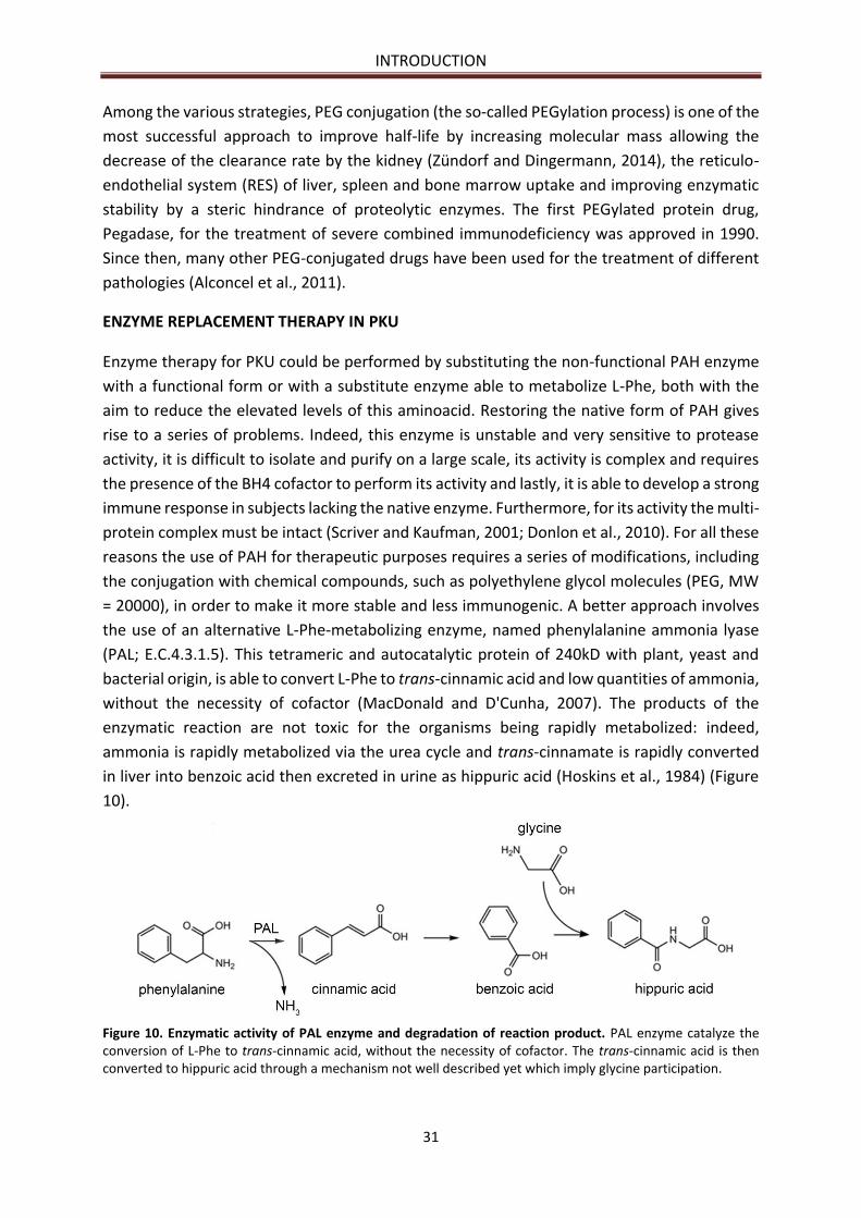
INTRODUCTION
31
Among the various strategies, PEG conjugation (the so-called PEGylation process) is one of the
most successful approach to improve half-life by increasing molecular mass allowing the
decrease of the clearance rate by the kidney (Zündorf and Dingermann, 2014), the reticulo-
endothelial system (RES) of liver, spleen and bone marrow uptake and improving enzymatic
stability by a steric hindrance of proteolytic enzymes. The first PEGylated protein drug,
Pegadase, for the treatment of severe combined immunodeficiency was approved in 1990.
Since then, many other PEG-conjugated drugs have been used for the treatment of different
pathologies (Alconcel et al., 2011).
ENZYME REPLACEMENT THERAPY IN PKU
Enzyme therapy for PKU could be performed by substituting the non-functional PAH enzyme
with a functional form or with a substitute enzyme able to metabolize L-Phe, both with the
aim to reduce the elevated levels of this aminoacid. Restoring the native form of PAH gives
rise to a series of problems. Indeed, this enzyme is unstable and very sensitive to protease
activity, it is difficult to isolate and purify on a large scale, its activity is complex and requires
the presence of the BH4 cofactor to perform its activity and lastly, it is able to develop a strong
immune response in subjects lacking the native enzyme. Furthermore, for its activity the multi-
protein complex must be intact (Scriver and Kaufman, 2001; Donlon et al., 2010). For all these
reasons the use of PAH for therapeutic purposes requires a series of modifications, including
the conjugation with chemical compounds, such as polyethylene glycol molecules (PEG, MW
= 20000), in order to make it more stable and less immunogenic. A better approach involves
the use of an alternative L-Phe-metabolizing enzyme, named phenylalanine ammonia lyase
(PAL; E.C.4.3.1.5). This tetrameric and autocatalytic protein of 240kD with plant, yeast and
bacterial origin, is able to convert L-Phe to trans-cinnamic acid and low quantities of ammonia,
without the necessity of cofactor (MacDonald and D'Cunha, 2007). The products of the
enzymatic reaction are not toxic for the organisms being rapidly metabolized: indeed,
ammonia is rapidly metabolized via the urea cycle and trans-cinnamate is rapidly converted
in liver into benzoic acid then excreted in urine as hippuric acid (Hoskins et al., 1984) (Figure
10).
Figure 10. Enzymatic activity of PAL enzyme and degradation of reaction product. PAL enzyme catalyze the conversion of L-Phe to trans-cinnamic acid, without the necessity of cofactor. The trans-cinnamic acid is then converted to hippuric acid through a mechanism not well described yet which imply glycine participation.
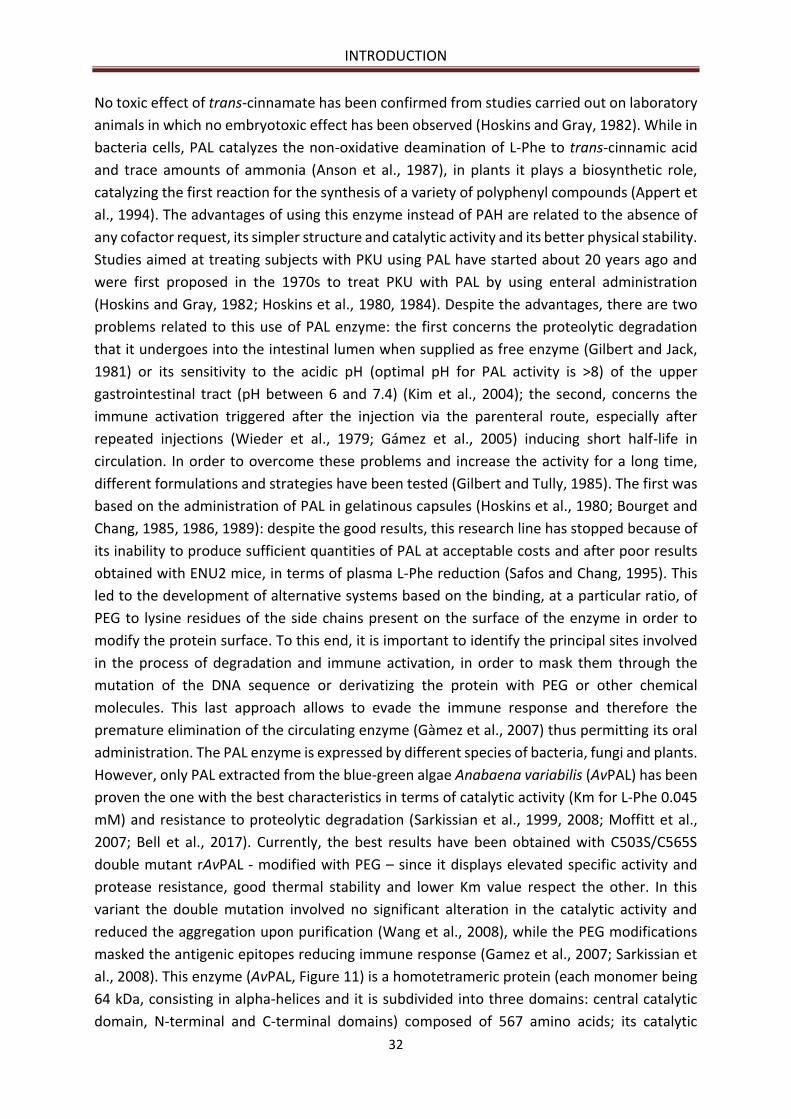
INTRODUCTION
32
No toxic effect of trans-cinnamate has been confirmed from studies carried out on laboratory
animals in which no embryotoxic effect has been observed (Hoskins and Gray, 1982). While in
bacteria cells, PAL catalyzes the non-oxidative deamination of L-Phe to trans-cinnamic acid
and trace amounts of ammonia (Anson et al., 1987), in plants it plays a biosynthetic role,
catalyzing the first reaction for the synthesis of a variety of polyphenyl compounds (Appert et
al., 1994). The advantages of using this enzyme instead of PAH are related to the absence of
any cofactor request, its simpler structure and catalytic activity and its better physical stability.
Studies aimed at treating subjects with PKU using PAL have started about 20 years ago and
were first proposed in the 1970s to treat PKU with PAL by using enteral administration
(Hoskins and Gray, 1982; Hoskins et al., 1980, 1984). Despite the advantages, there are two
problems related to this use of PAL enzyme: the first concerns the proteolytic degradation
that it undergoes into the intestinal lumen when supplied as free enzyme (Gilbert and Jack,
1981) or its sensitivity to the acidic pH (optimal pH for PAL activity is >8) of the upper
gastrointestinal tract (pH between 6 and 7.4) (Kim et al., 2004); the second, concerns the
immune activation triggered after the injection via the parenteral route, especially after
repeated injections (Wieder et al., 1979; Gámez et al., 2005) inducing short half-life in
circulation. In order to overcome these problems and increase the activity for a long time,
different formulations and strategies have been tested (Gilbert and Tully, 1985). The first was
based on the administration of PAL in gelatinous capsules (Hoskins et al., 1980; Bourget and
Chang, 1985, 1986, 1989): despite the good results, this research line has stopped because of
its inability to produce sufficient quantities of PAL at acceptable costs and after poor results
obtained with ENU2 mice, in terms of plasma L-Phe reduction (Safos and Chang, 1995). This
led to the development of alternative systems based on the binding, at a particular ratio, of
PEG to lysine residues of the side chains present on the surface of the enzyme in order to
modify the protein surface. To this end, it is important to identify the principal sites involved
in the process of degradation and immune activation, in order to mask them through the
mutation of the DNA sequence or derivatizing the protein with PEG or other chemical
molecules. This last approach allows to evade the immune response and therefore the
premature elimination of the circulating enzyme (Gàmez et al., 2007) thus permitting its oral
administration. The PAL enzyme is expressed by different species of bacteria, fungi and plants.
However, only PAL extracted from the blue-green algae Anabaena variabilis (AvPAL) has been
proven the one with the best characteristics in terms of catalytic activity (Km for L-Phe 0.045
mM) and resistance to proteolytic degradation (Sarkissian et al., 1999, 2008; Moffitt et al.,
2007; Bell et al., 2017). Currently, the best results have been obtained with C503S/C565S
double mutant rAvPAL - modified with PEG – since it displays elevated specific activity and
protease resistance, good thermal stability and lower Km value respect the other. In this
variant the double mutation involved no significant alteration in the catalytic activity and
reduced the aggregation upon purification (Wang et al., 2008), while the PEG modifications
masked the antigenic epitopes reducing immune response (Gamez et al., 2007; Sarkissian et
al., 2008). This enzyme (AvPAL, Figure 11) is a homotetrameric protein (each monomer being
64 kDa, consisting in alpha-helices and it is subdivided into three domains: central catalytic
domain, N-terminal and C-terminal domains) composed of 567 amino acids; its catalytic
INTRODUCTION
33
activity requires the electrophilic prosthetic group 4-methylideneimidazole-5-one (MIO) and
does not require any external additional cofactor (Moffitt et al., 2007).
Figure 11. A) Side view and B) Top view of the crystal structure of wild-type phenylalanine ammonia lyase from Anabaena variabilis (adapted from Moffitt et al., 2007). The spheres represent the atoms of the four MIO prosthetic groups.
Based on these results, the BioMarin Corp., Novato, introduced the PEGylated form of double-
mutant rAvPAL-PEG enzyme in clinical trials for the ERT of PKU after its applied to the U.S.
Food and Drug Administration (FDA). In the phase I trial (NCT00634660 Government
Identification Code), aimed at evaluating the safety and tolerability of subcutaneous injection
of rAvPAL-PEG in patients with PKU, 35 participants with L-Phe concentration lower than 600
μM, were divided in 7 different cohorts receiving an increasing concentrations of rAvPAL-PEG
formulation. In the high-dose treated cohort, the L-Phe level decreased after a single
inoculation without the development of major adverse events, with the exception of some
injection-site reactions of mild intensity, as recently published by Longo et al. (2014). A phase
II clinical trial (NCT00925054, NCT01560286, NCT00924703, NCT01212744), aimed to
evaluating the efficacy and safety of multiple enzyme injections, has been recently concluded
(Bélanger-Quintana et al., 2011) while, a phase III clinical trial (NCT01819727, NCT01889862)
has been started to assess, through a multicenter study, the safety and tolerability of rAvPAL-
PEG administration and improvements in attention scores compared to pretreatment
baseline. In May 2018, the FDA approved PalynziqTM (pegvaliase-pqpz) in the USA as enzyme
therapy for PKU (ww.fda.gov) and on March 2018 the European Medicines Agency
(www.ema.europa.eu) accepted BioMarine’s submission of a Marketing Authorization
Application (MAA) for pegvaliase. Although PEG system has some important features that
make it suitable for pharmaceutical use such as lack of toxicity and high solubility, this
molecule itself can be immunogenic, especially upon repeated and continuous treatments
(Schellekens et al., 2013; Longo et al., 2014) and it also can modify enzyme activity
compromising the therapeutic effect (Gaberc-Porekar et al., 2008) besides the rather high
costs of the PEGylation process (Hershfield, 1995). Indeed, PALYNZIQ may cause serious side
effects, including, joint pain, hypersensitivity reactions due to formation of antibodies against
A B
INTRODUCTION
34
the product, headache, nausea, dizziness, abdominal pain, throat pain, fatigue, vomiting,
cough and diarrhea as well as anaphylaxis events which require the patients to have auto-
injectable epinephrine available at all times of Palynziq treatment. Therefore, developing a
secure and non-invasive delivery system such as an oral formulation of the therapeutic
enzyme, would be a key milestone (Kang et al., 2010).
In the study by Kang and colleagues (2010), aimed at developing an oral administrable PAL
form, the enzyme AvPAL was engineered by site-direct mutagenesis of residues recognized by
chymotrypsin to make it more resistant to intestinal proteolytic digestion. However, the
different strategies to make the enzyme less degradable by the protease after oral
administration are responsible of reducing its specific activity, making a greater dose of
recombinant PAL and a longer time of contact with its substrate to the gut which is needed to
reduce L-Phe level (Sarkissian and Gamez, 2005). More recently, literature (Isabella et al.,
2018) reported a study in which a synthetic live bacterial strain, SYNB1618, was able to inhibit
increases of serum L-Phe, but nowadays clinical trials to evaluate SYNB1618 safety and efficacy
are underway. At the same time, we must consider that the oral administration of PAL is
limited to patients with mild PKU and the diet control is still recommended to obtain the best
L-Phe control (Bourget and Chang, 1989; Chang et al., 1995): consequently, subcutaneous
administration of the therapeutic enzyme represents the most promising therapeutic
approach
The above mentioned concerns led to an increasing interest in drug delivery systems able to
target the therapeutic agent to the desired site of action with minimal side effects (Pierigè et
al., 2017; De Jong et al., 2008).
Many carriers have been proposed (Allen and Cullis, 2004), including either simple soluble
molecules, such as monoclonal antibodies and biodegradable polymers, or more complex
structures, such as microcapsules and particles, cells, liposomes and erythrocytes (Chiarantini
et al., 1995; Pierigè et al., 2008; Biagiotti et al., 2011; Villa et al., 2015).
Among all possibilities, erythrocytes (red blood cells, RBCs) are the most promising ones
thanks to their unique characteristics and their ability to accumulate L-Phe through a saturable
transport that obeys to Michaelis-Menten kinetics (Pico et al., 1993), which makes them an
ideal carrier system. The first application of RBCs as ERT in PKU system has been performed
by Yew et al. (2013) whom entrapped the prokaryotic PAH, obtained from Chromobacterium
violaceum, a monomeric enzyme of 33 kD more stable and active than mammalian PAH.
Although the system was able to reduce L-Phe level, its efficacy in PKU mice was probably low
due to the lower in vivo vs in vitro enzymatic activity. Better results have been obtained in a
study of Rossi et al. (2014) that demonstrated the ability of RBCs loaded with rAvPAL to act as
circulating bioreactors able to reduce L-Phe levels and confirmed the efficacy of the
treatment, which was able to maintain L-Phe levels between 100-900 µM also after repeated
infusions of 9-10 day-intervals each other. In addition, even though an immune response
against rAvPAL was observed, the ability of the system to metabolize L-Phe has not been
compromised thanks to the protective action carried out by RBCs towards the enzyme (Rossi
et al., 2014). More details about RBCs as delivery system are reported in the section below.
INTRODUCTION
35
DRUG DELIVERY SYSTEMS: RED BLOOD CELLS AS THE BEST CHOICE
RBCs have been identified as the most useful natural carriers for drug delivery since 1960s
(Villa et al., 2016). In the last decades RBCs are being employed for delivery of a variety of
therapeutics, contrast agents and for many other clinical uses. They are considered the ideal
candidates as delivery system because they have unique biological characteristics. Such
qualities include (Beutler et al., 1995; Gutiérrez Millàn et al., 2004; Rossi et al., 2003, 2005;
Hamidi et al., 2007; Pierigè et al., 2008; Muzykantov, 2010; Biagiotti et al., 2011; Leuzzi et al.,
2016; Pierigè et al., 2017):
- Biocompatibility, especially if autologous RBCs are used;
- Biodegradability, without formation of toxic by-products;
- RBCs represent the most abundant cell types in blood (the human body normally
possess 2-3 x 10¹³ RBCs continuously produced at a rate of 2 million per second), they
comprise one quarter of the total number of body cells and are the main component
of blood;
- A long in vivo life-span of 100-120 days, the longest among delivery systems, which has
influence on the encapsulated agent availability;
- An average cellular volume of approximately 90 fL being mostly available for large
amounts of drugs to be encapsulated, since mature RBCs lack nucleus and organelles;
- The biconcave shape, which enables great flexibility and membrane deformability,
making RBCs able to travel across small undamaged capillaries (until 2-3 μm in
diameter) avoiding unwanted extravasation;
- Easiness of ex vivo handling by means of several already existing procedures allowing
the reversible opening of pores on cell membrane (from 10 nm up to 500 nm diameter)
by exploiting RBC ability to behave like an osmometer, shrinking or swelling according
to the salt content of the external medium, thus permitting many non-diffusible large
compounds (such as proteins and peptides) to be loaded inside the cell, maintaining
morphological, biochemical and immunological properties similar to those of native
cells;
- Ability to protect the encapsulated agent from the premature degradation or
inactivation by the host immune system thanks to their biocompatibility (in contrast
with most of the other delivery systems) (Ponder, 2008; Benichou et al., 2009)
promoting a longer persistence of the drug in the body;
- Possibility to modulate the drug pharmacokinetics thus enabling a sustained release of
the therapeutic agent in circulation while providing protection to the organism from
the potential negative effects of peak concentrations of drugs;
- Possibility to use erythrocytes not only as passive carriers but also as active bioreactors
thanks to the presence of the intrinsic pool of enzymes able to process entrapped pro-
drugs and to convert them into active drugs (Hoffman, 1992; Rossi et al., 2001, 2004);
- Possibility to selectively target macrophage cells and the reticulo-endothelial system
(RES) by exploiting the natural mode of macrophage-mediated erythrocyte removal
INTRODUCTION
36
from circulation, thus eliminating drug toxicity to other body districts (Chiarantini et
al., 1995; Serafini et al., 2004; Rossi et al., 2005; Sabatino et al., 2014).
Despite the benefits of RBCs as delivery system, these cells exhibit some drawbacks (Hamidi
et al., 2007): the macrophage-mediated removal of senescent or damaged RBCs might
represent an undesired side-effect, shortening the half-life of the encapsulated drug. In
addition, some molecules may alter RBCs physiology or rapidly leak from them, thus being lost
in the bloodstream. Given their natural origin, RBCs are characterized by an intrinsic variability
that may lead to different rates of success in the loading procedures. Besides, RBCs are viable
cells that need special attention during storage to avoid any alteration of their therapeutic
action once reinfused in vivo. Many strategies have been adopted to overcome this problem,
such as isotonic buffers containing essential nutrients and low temperatures. Furthermore, a
major concern is the potential contamination due to blood origin, the loading equipment used
and the environment where the procedure is carried out. In keeping with this issue, strict
controls are needed to guarantee a correct collection and handling of the erythrocytes, as well
as the safety of the final product.
Despite all, the above mentioned positive features still overcome the negative drawbacks and
RBCs as drug carriers have been studied since the mid-1970s (Updike et al., 1976) and
employed for different purposes, as extensively reviewed by many authors (Hamidi et al.,
2007; Biagiotti et al., 2011; Magnani and Rossi, 2014; Pierigè et al., 2008, 2017; Bhateria et al.,
2014; Zarrin et al., 2014; Rossi et al., 2016). These carriers have been shown to be able to carry
many different agents including antiviral and antineoplastic drugs, therapeutic proteins and
peptides (i.e. vaccines and enzymes), cytokines, oligosaccharides, nucleic acids, anti-
inflammatory drugs (like the glucocorticoid dexamethasone, DEXA) and contrasting agents for
diagnostic purposes (Antonelli et al., 2013; Pierigè et al., 2017). These cells can be also used
as transport systems to obtain a slow release of the pro-drug in the circulation or to selectively
direct a drug in the specific cells responsible and/or capable of erythrophagocytosis. In
addition, their use as circulating bioreactors for ERT makes these cells an extremely versatile
system for the treatment of various pathological conditions.
Several techniques have been developed to produce engineered erythrocytes able to perform
as carrier system (Zolla et al., 1991; Rossi et al., 2003). To this aim, we can identify two
principal groups of techniques: encapsulation within the inner volume of RBCs or drug
attachment on cell surface. The most common procedures for the first purpose include
electroporation (Lizano et al., 2001) or hypotonic hemolysis (Green et al., 1981; Ropars et al.,
1985; Ihler and Tsang, 1987; Ihler and Chi-Wan Tsang, 1987). The second group involve
different covalent or non-covalent cross-linkers able to couple molecules to the RBC surface
(Muzykantov, 2010; Villa, 2016). The most widely used method to load protein into RBCs are
based on hypotonic haemolysis which, in turn, can be divided into hypotonic dilution (Ihler et
al., 1973; DeLoach and Ihler, 1977; Talwar and Jain, 1992), hypotonic dialysis, that is the most
widespread method to load protein into RBCs, (DeLoach and Ihler, 1977; Alvarez et al., 1988;
Gutiérrez Millàn et al., 2004) and hypotonic pre-swelling dilution (Jenner et al., 1981; Tamura
et al., 1988; Ito et al., 1989; Tajerzadeh and Hamidi, 2000). Briefly, all of them consist in
INTRODUCTION
37
causing erythrocytes to swell by putting them into a hypotonic medium that permits the
formation of reversible pores (200-500 Å diameter); after a period of equilibration with a
solution of the molecule to be entrapped, which can cross the pores, RBCs are resealed by
adding a hypertonic solution, which restores the physiologic isotonic environment thus
permitting the pores to close and the membrane to restore its physiological structure.
In order to act as carriers for enzyme, RBC membrane has to be permeable to the target
metabolite, the entrapped enzyme should not alter the physiology of the RBCs and it must not
leak through RBC membrane (Leuzzi et al., 2016). The target of the delivery system is related
to the disease. Indeed, for instance in blood malignancies, the target is the blood depletion of
any metabolite that represents a substrate of the pathological tissue. On the other hand, in a
different group of diseases the enzyme inside RBCs remove toxic endogenous or exogenous
compounds which are a consequence of an inherited disorder of metabolism. The first
attempt was made by Beutler and colleagues (1977), who aimed at treating Gaucher’s disease
by means of RBCs loaded with glucocerebrosidase. Up to now, RBCs loaded with L-
asparaginase have been employed in clinical trials (ClinicalTrials.gov ID NCT01523782) to
remove L-asparagine, a non-essential amino acid necessary for lymphoblastic proliferation in
acute lymphoblastic leukemia (ALL) (Kravtzoff et al., 1990; Kwon et al., 2009; Domenech et al.,
2011; Agrawal et al., 2013); adenosine deaminase (ADA) is another therapeutic enzyme
encapsulated both in its native and PEGylateg form for the treatment of ADA deficiency (Bax
et al., 1996, 2000a-b, 2007). Thymidine phosphorylase has been studied in animal models and
also in patient as enzyme replacement therapy for the rare disease MNGIE (mitochondrial
neurogastrointestinal encephalomyopathy) (Moran et al., 2008; Bax et al., 2013; Levene et al.,
2013).
My PhD EUREKA project, has been involved in analyzing phenylketonuria (PKU), an inherited
metabolic disease that can take advantage from ERT.
For all its possible and promising applications, this carrier system during the years have moved
from academia to industry and is at the moment investigated in several clinical trials by two
major biopharmaceutical companies, ERYTECH pharma (http://erytech.com/) and EryDel
S.p.A (http://www.erydel.com/). The first is the proprietary of an apparatus created in 2006
by Godfrin for industrial applications (Godfrin, 2006), named ERYcaps. The company has used
its technology for the treatment of ALL, currently in Phase 3 clinical trial (NCT01523782), as a
valid alternative to free form of enzyme, with positive results (Godfrin and Bertrand, 2006;
Domenech et al., 2011). At the same time the second biotechnology company EryDel S.p.A
(www.erydel.com) is the proprietary of a non-invasive electromechanical device named Red
Cell loader® (RCL). The RCL is a fully automated apparatus with a dedicated software,
conceived to function with disposable and CE marked kit, designed to reproducibly load
human autologous erythrocytes with different drugs, in safe, sterile and apyrogenic
conditions, as required for the performance of clinical studies so as to yield a final product
suitable for the reinfusion into patients with several pathologic disturbances (Magnani et al.,
1998; Mambrini et al., 2017). Up to now the apparatus has undergone a process of upgrading,
leading to the full-optional machine currently available, which is able to complete the
procedure in about 90 minutes directly in the clinical centers. The device has been included in
INTRODUCTION
38
several clinical studies (ClinicalTrials.gov) both with healthy volunteers (IDs NCT01925859,
NCT02380924) and patients, where it was employed to load DEXA-21-P for the treatment of
inflammatory diseases such as chronic obstructive pulmonary disease (Rossi et al., 2001),
cystic fibrosis (Rossi et al., 2004), Crohn’s disease (Castro et al., 2007) (ID NCT01277289) and
ulcerative colitis (Bossa et al., 2013). The so-called EryDex system has been used in Phase 2
clinical trials for the treatment of ataxia-teleangiectasia (ID NCT01255358; EU Clinical Trial
Register number 2010-022315-19), that it has received the Orphan Drug designation by
European and US Authorities and is now entering a pivotal phase III study
(http://www.erydel.com/en/erydex). Moreover, EryDex system using autologous RBCs allows
a rapid administration of loaded cells to patients avoiding problems of immunological impact
on the host as happens with Erytech technology that employs homologous RBCs. Indeed,
while in autologous approach blood is collected from the patient, in the homologous system
blood is provided by blood banks (Pierigè et al., 2017).
39
AIM OF THE WORK
In a previous 2014 study of Rossi et al., we demonstrated the ability of rAvPAL-RBCs to act as
an efficacious carrier system able to reduce L-Phe levels at safety values. In particular, in the
dose finding study aimed at evaluating the effectiveness of the strategy on a PKU murine
model, two different doses of enzyme (0.5 and 1 IU rAvPAL/mouse) were proven to
dramatically decrease blood L-Phe levels in ENU2 mice, peaking 24 h after treatment with no
difference among doses and slight differences above the range of normal concentrations (50-
110 µM) (Blau et al., 2010), after 8-9 days from the infusion.
In the study of repeated administrations, seven injections of the same dose of rAvPAL-RBCs
suggested by the dose finding study were performed at 9-10 days. Treated mice showed a
reduction in blood L-Phe, whose level remained between healthy control and PKU mice values,
never reaching back their respective pre-treatment level for the entire experimental period
(70 days).
Considering these interesting results, with this new experiment we aim to prove that early
(starting at 15 post natal day) and continuous administration of this carrier system, at 7 day-
intervals between each infusion, normalizing blood and brain L-Phe levels, is able to prevent
the abnormalities found in untreated ENU2 mice: cognitive impairment, brain
neurotransmitter deficit, and neuropathological abnormalities disease. At the same time, we
aim to demonstrate that erythrocytes may act as an effective and safe delivering system for
the enzyme during mouse development as well.
To this purpose we employed the best performing variant of PAL enzyme currently available,
i.e. C503S/C565S double mutant recombinant PAL from the cyanobacterium Anabaena
variabilis (rAvPAL) (Wang et al., 2008), kindly provided by BioMarin Pharmaceutical Inc.
(Novato, CA) in its chemically unmodified (non PEGylated) form.
The trial was designed, as reported below (Figure 12), in order to mimic in a preclinical setting
what happens for early treated PKU patients (Blau et al., 2010):
1. One group of ENU2 mice (n=9) were treated with 8 subsequent intra-venus injections of
rAvPAL-RBCs at 7 day-intervals between infusions. Two other groups (WT mice, n=13
and ENU2 mice n=7) were used as control. At scheduled intervals of time (4 days after II
infusion, 7 days after III, VI and VIII infusion), blood L-Phe and L-Tyr levels were
monitored.
2. At the end of the infusions (PND 68-71), all animals have been evaluated with behavioral
tests and one aliquot of plasma sample has been assessed for anti-rAvPAL IgG response.
3. At the end of behavioral experiments, all animal groups were sacrificed and brains and
blood have been processed for biochemical, morphological and molecular assays.
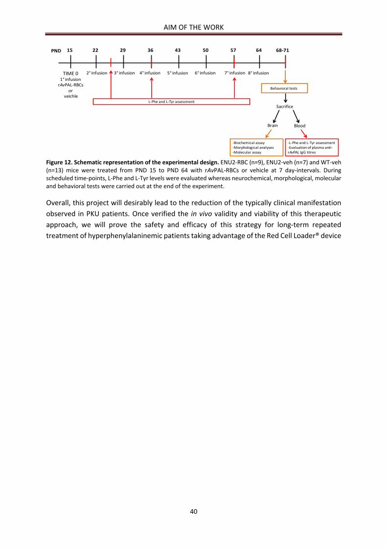
AIM OF THE WORK
40
Figure 12. Schematic representation of the experimental design. ENU2-RBC (n=9), ENU2-veh (n=7) and WT-veh (n=13) mice were treated from PND 15 to PND 64 with rAvPAL-RBCs or vehicle at 7 day-intervals. During scheduled time-points, L-Phe and L-Tyr levels were evaluated whereas neurochemical, morphological, molecular and behavioral tests were carried out at the end of the experiment.
Overall, this project will desirably lead to the reduction of the typically clinical manifestation
observed in PKU patients. Once verified the in vivo validity and viability of this therapeutic
approach, we will prove the safety and efficacy of this strategy for long-term repeated
treatment of hyperphenylalaninemic patients taking advantage of the Red Cell Loader® device
PND 15 22 29 36 43 50 57 64 68-71
TIME 01° infusion
rAvPAL-RBCsor
veichle
2° infusion 3° infusion 4° infusion 5° infusion 6° infusion 7° infusion 8° infusion
L-Phe and L-Tyr assessment
Behavioral testsr
Sacrifice
Brain Blood
-Biochemical assay-Morphological analyses-Molecular assay
-L-Phe and L-Tyr assessment-Evaluation of plasma anti-rAvPAL IgG titres
MATERIALS AND METHODS
41
MATERIALS AND METHODS
ENZYMES
Recombinant AvPAL
Recombinant Phenylalanine Ammonia Lyase from Anabaena variabilis (rAvPAL) was prepared
by BioMarin Pharmaceutical Inc. (Novato, CA) clinical manufacturing group. Briefly, the
protein was cloned in a pIBEX7 plasmid, expressed in the E. coli strain BLR (Novagen) and
purified by anion exchange chromatography followed by hydrophobic interacting
chromatography. It contains two point mutations, Cys503 Ser and Cys565 Ser in order
to prevent aggregation. Purified rAvPAL was concentrated and buffer exchanged by
ultrafiltration/diafiltration to a final concentration of approximately 200 International Units
(IU)/ml. Final concentration was determined by bicinchoninic acid assay (BCA) and activity was
assayed as previously described by Wang et al. (2008). This protein was provided in vials of
200 μl in a Tris-buffered saline solution each containing 2 mM L-Phe. According to the data
provided by the company, the protein had a molecular weight of 61.9 KDa and concentration
equal to 109 mg/ml, equivalent to an enzymatic activity of 203 IU/ml; the specific activity (S.A.)
was 1.86 IU/mg. However, all the parameters have been re-evaluated. Protein concentration
was estimated by Bradford colorimetric assay, as previously reported (Bradford, 1976). This
test is based on the ability of the Coomassie® brilliant Blue G-250 dye to bind arginine,
tryptophan, Tyr, histidine and Phe residues of the protein samples. After the dye bond, the
displacement of the wavelength at which it absorbs, from 465 nm to 595 nm, can be observed.
The increase of absorbance at 595 nm (intensity of the blue color that develops) is
proportional to the amount of protein in the sample. The preparation showed an enzymatic
activity of 104.4 IU/ml and a S.A. of 1.66 IU/mg protein.
ANIMALS
Developing homozygous Pahenu2-/- (ENU2) and Pahenu2+/+ (Wild Type; WT) male mice of the
BTBR background strain, employed in this study, were issued from heterozygous mating
Pahenu2+/-. Genetic characterization was performed starting from little tail tissue. The genetic
ENU2 modification is chemically induced after treatment of BTBR WT mice with N-ethyl-N-
nitrosurea (hence the name). The treatment causes an A>T835C missense mutation at
nucleotide position 835 in exon 7, resulting in a phenylalanine-to-serine amino acid
substitution in position 263 of the protein chain (F263S) (McDonald and Charlton, 1997). Exon
7, the most frequent site for PKU mutations in human (Dworniczak et al., 1992; Scriver et al.,
1996), is a region that encodes the active site of the PAH enzyme (Jennings et al., 1991) and
its mutation affects a Bbs I restriction site creating a Bsm AI recognition site. The homozygous
mice were considered the most appropriate mouse model because the mutation is
responsible for the appearance of a biochemical and neurological phenotype very similar to
human classic PKU disease, with plasma L-Phe levels 10- to 20-fold higher than those of
healthy animals, and reduced PAH enzyme activity (McDonald et al., 1990). At the same time,
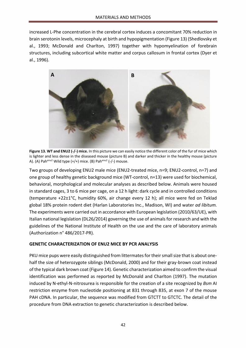
MATERIALS AND METHODS
42
increased L-Phe concentration in the cerebral cortex induces a concomitant 70% reduction in
brain serotonin levels, microcephaly at birth and hypopigmentation (Figure 13) (Shedlovsky et
al., 1993; McDonald and Charlton, 1997) together with hypomyelination of forebrain
structures, including subcortical white matter and corpus callosum in frontal cortex (Dyer et
al., 1996).
Figure 13. WT and ENU2 (-/-) mice. In this picture we can easily notice the different color of the fur of mice which is lighter and less dense in the diseased mouse (picture B) and darker and thicker in the healthy mouse (picture A). (A) Pahenu2 Wild type (+/+) mice. (B) Pahenu2 (-/-) mouse.
Two groups of developing ENU2 male mice (ENU2-treated mice, n=9; ENU2-control, n=7) and
one group of healthy genetic background mice (WT-control, n=13) were used for biochemical,
behavioral, morphological and molecular analyses as described below. Animals were housed
in standard cages, 3 to 6 mice per cage, on a 12 h light: dark cycle and in controlled conditions
(temperature +22±1°C, humidity 60%, air change every 12 h); all mice were fed on Teklad
global 18% protein rodent diet (Harlan Laboratories Inc., Madison, WI) and water ad libitum.
The experiments were carried out in accordance with European legislation (2010/63/UE), with
Italian national legislation (DL26/2014) governing the use of animals for research and with the
guidelines of the National Institute of Health on the use and the care of laboratory animals
(Authorization n° 486/2017-PR).
GENETIC CHARACTERIZATION OF ENU2 MICE BY PCR ANALYSIS
PKU mice pups were easily distinguished from littermates for their small size that is about one-
half the size of heterozygote siblings (McDonald, 2000) and for their gray-brown coat instead
of the typical dark brown coat (Figure 14). Genetic characterization aimed to confirm the visual
identification was performed as reported by McDonald and Charlton (1997). The mutation
induced by N-ethyl-N-nitrosurea is responsible for the creation of a site recognized by Bsm AI
restriction enzyme from nucleotide positioning at 831 through 835, at exon 7 of the mouse
PAH cDNA. In particular, the sequence was modified from GTCTT to GTCTC. The detail of the
procedure from DNA extraction to genetic characterization is described below.
A B
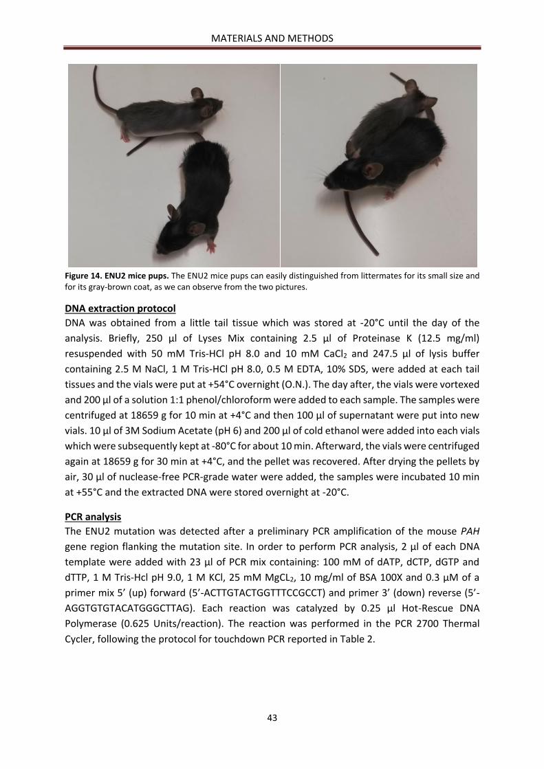
MATERIALS AND METHODS
43
Figure 14. ENU2 mice pups. The ENU2 mice pups can easily distinguished from littermates for its small size and for its gray-brown coat, as we can observe from the two pictures.
DNA extraction protocol
DNA was obtained from a little tail tissue which was stored at -20°C until the day of the
analysis. Briefly, 250 µl of Lyses Mix containing 2.5 µl of Proteinase K (12.5 mg/ml)
resuspended with 50 mM Tris-HCl pH 8.0 and 10 mM CaCl2 and 247.5 µl of lysis buffer
containing 2.5 M NaCl, 1 M Tris-HCl pH 8.0, 0.5 M EDTA, 10% SDS, were added at each tail
tissues and the vials were put at +54°C overnight (O.N.). The day after, the vials were vortexed
and 200 µl of a solution 1:1 phenol/chloroform were added to each sample. The samples were
centrifuged at 18659 g for 10 min at +4°C and then 100 µl of supernatant were put into new
vials. 10 µl of 3M Sodium Acetate (pH 6) and 200 µl of cold ethanol were added into each vials
which were subsequently kept at -80°C for about 10 min. Afterward, the vials were centrifuged
again at 18659 g for 30 min at +4°C, and the pellet was recovered. After drying the pellets by
air, 30 µl of nuclease-free PCR-grade water were added, the samples were incubated 10 min
at +55°C and the extracted DNA were stored overnight at -20°C.
PCR analysis
The ENU2 mutation was detected after a preliminary PCR amplification of the mouse PAH
gene region flanking the mutation site. In order to perform PCR analysis, 2 µl of each DNA
template were added with 23 µl of PCR mix containing: 100 mM of dATP, dCTP, dGTP and
dTTP, 1 M Tris-Hcl pH 9.0, 1 M KCl, 25 mM MgCL2, 10 mg/ml of BSA 100X and 0.3 µM of a
primer mix 5’ (up) forward (5’-ACTTGTACTGGTTTCCGCCT) and primer 3’ (down) reverse (5’-
AGGTGTGTACATGGGCTTAG). Each reaction was catalyzed by 0.25 µl Hot-Rescue DNA
Polymerase (0.625 Units/reaction). The reaction was performed in the PCR 2700 Thermal
Cycler, following the protocol for touchdown PCR reported in Table 2.
MATERIALS AND METHODS
44
Cycle name Temperature
(°C) Time Number of cycles
Activation +95 1 min 1
Denaturation +95 15 sec 2 cycles (for the first
two annealing
temperature)
20 cycles for +58°C
annealing
temperature
10 cycles for +55°C
annealing
temperature
Annealing +64/+61/+58/+55 15 sec
Extension +72 1.30 min
Final Extension and
Ligation +72 10 min 1
Hold +4
Table 2. Thermocycle parameters. The sequence of denaturation, annealing and extension, has been repeated
for 4 time varying annealing temperature from +64°C for the first step to +55°C for the last step, in order to
increased reaction specificity.
At the end of PCR, the 132-bp amplification product was digested by 3 µl of restriction enzyme
Bsm AI (5 U/µl) with the addition of the specific enzyme reaction buffer and by incubation
O.N. at +55°C, allowing a complete PCR product digestion. Bsm AI digestion yielded a
distribution of fragments with a distinct banding pattern for each of three possible genotypes
as observed by gel analysis.
GEL analysis
The restriction fragments were separated by electrophoresis through a 4-15 % polyacrylamide
gel (Mini-Protean TGX Gels) in presence of TBE buffer 1X at 80 V. To each sample, 2 µl of
Bromophenol Blue (1X) were added. At the end of the electrophoresis process, the gel was
stained with ethidium bromide (0.6 nM) and photographed. The gel image was acquired by
Bio-Rad Gel Doc 1000 System. For each of the three possible genotypes, different pattern of
fragments was detected. In particular, WT displayed two fragments of 82 and 50 bp,
heterozygotes generated four fragments of 82, 50, 48 and 34 bp and homozygous mutants
three fragments of 50, 48 and 34 bp (Figure 15).
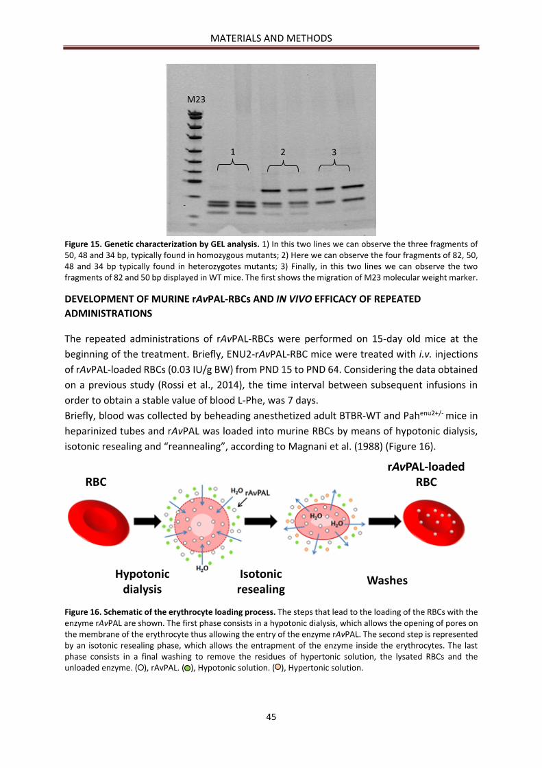
MATERIALS AND METHODS
45
Figure 15. Genetic characterization by GEL analysis. 1) In this two lines we can observe the three fragments of 50, 48 and 34 bp, typically found in homozygous mutants; 2) Here we can observe the four fragments of 82, 50, 48 and 34 bp typically found in heterozygotes mutants; 3) Finally, in this two lines we can observe the two fragments of 82 and 50 bp displayed in WT mice. The first shows the migration of M23 molecular weight marker.
DEVELOPMENT OF MURINE rAvPAL-RBCs AND IN VIVO EFFICACY OF REPEATED
ADMINISTRATIONS
The repeated administrations of rAvPAL-RBCs were performed on 15-day old mice at the
beginning of the treatment. Briefly, ENU2-rAvPAL-RBC mice were treated with i.v. injections
of rAvPAL-loaded RBCs (0.03 IU/g BW) from PND 15 to PND 64. Considering the data obtained
on a previous study (Rossi et al., 2014), the time interval between subsequent infusions in
order to obtain a stable value of blood L-Phe, was 7 days.
Briefly, blood was collected by beheading anesthetized adult BTBR-WT and Pahenu2+/- mice in
heparinized tubes and rAvPAL was loaded into murine RBCs by means of hypotonic dialysis,
isotonic resealing and “reannealing”, according to Magnani et al. (1988) (Figure 16).
Figure 16. Schematic of the erythrocyte loading process. The steps that lead to the loading of the RBCs with the enzyme rAvPAL are shown. The first phase consists in a hypotonic dialysis, which allows the opening of pores on the membrane of the erythrocyte thus allowing the entry of the enzyme rAvPAL. The second step is represented by an isotonic resealing phase, which allows the entrapment of the enzyme inside the erythrocytes. The last phase consists in a final washing to remove the residues of hypertonic solution, the lysated RBCs and the unloaded enzyme. ( ), rAvPAL. ( ), Hypotonic solution. ( ), Hypertonic solution.
RBC
Hypotonicdialysis
Isotonicresealing
Washes
rAvPAL-loadedRBC
1 2 3
M23
MATERIALS AND METHODS
46
Whole blood was centrifuged to remove plasma by 10 min centrifugation at +4°C and 900 g
and then washed twice by 10 min centrifugations at +4°C, at 900 g and 1500 g respectively, in
a physiological saline solution containing: 10 mM 2-[4-(2-hydroxyethyl)piperazin-1-yl]-
ethanesulfonic acid (HEPES, pH 7.4), 154 mM NaCl and 5 mM glucose. The procedure was
carried out with about 46 IU of enzyme (400 µl of protein solution with SA 1.66 IU/mg) added
to RBCs suspended in Hepes solution at 50% haematocrit (Ht). The condition (1 ml final
volume) was dialyzed for 75 min at +4°C in a cellulose tube (14 kDa MWCO, Roth, Karlsruhe,
Germany) placed in a rotating plate vs 50 ml of hypotonic dialysis buffer optimized for murine
RBCs loading, containing 15 mM NaH2PO4, 15 mM NaHCO3 (pH 7.4), 20 mM glucose, 4 mM
MgCl2, 3 mM glutathione and 2 mM ATP. The final osmolarity of the hypotonic solution was
88±7.3 mOsm, measured by Osmometer Fiske Associates, Model 210 (Norwood, MA, USA).
After dialysis, the cells reached about 89±6.2 mOsm (the opening of membrane pores in
murine RBC starts at values ≤150 mOsm). Subsequent resealing and re-annealing steps were
carried out by incubating the pooled dialyzed RBC suspension 5 min at +37°C. Then, PIGPA
solution (100 mM inosine, 20 mM ATP, 10 mM anhydrous glucose, 100 mM sodium pyruvate,
4 mM MgCl2, 190 mM NaCl, 1666 mM KCl and 33 mM NaH2PO4) was added to the RBCs (10%
v/v) to restore isotonicity (300 mOsm) and the suspension was incubated another 25 min at
+37°C under gentle stirring, to allow pore closure. Final washing phase was divided in two
steps. The first washing step was carried out centrifuging 10 min at 360 g and +4°C, to avoid
cell lyses, and the second by 10 min centrifugation at 615 g and +4°C. The amount of
entrapped rAvPAL was quantified by means of a kinetic assay according to Wang et al. (2008)
as briefly summarized below. Hematological parameters were measured by an automatic ABX
Micros® 60 cell counter (Horiba Medical, Irvine, CA) and the percentage of RBC recovery was
calculated between the number of RBCs that underwent the dialysis step and those recovered
at the end of the loading procedure.
Final packed rAvPAL-loaded RBCs were re-suspended in Hepes solution at about 25% Ht and
i.v. infusions of these suspensions, ranging from 50 to 250 µl, were performed based on the
weight gain of developing animals in order to administer 0.03 IU rAvPAL/g BW. For the
experiment, untreated ENU2 mice (ENU2-veh) (n=7) and untreated healthy mice (WT-veh)
(n=13) underwent the same manipulations and received repeated i.v. injections of saline
solution (NaCl 0.9% W/V) following the same schedule as the ENU2 mice treated with loaded
RBCs (ENU2-rAvPAL-RBC mice (n=9)).
Biochemical monitoring of the blood L-Phe and L-Tyr levels, evaluated starting from PND 26
(4 days after II infusion) to PND 70 at the planned time points (7 days after III, VI and VIII
infusion) as showed in Figure 12, were used as an indicator of treatment efficacy.
PHENYLALANINE AMMONIA LYASE ACTIVITY ASSAY
The kinetic assay was performed as reported by Wang et al. (2008), monitoring the formation
of trans-cinnamic acid (tCA) at 290 nm wavelength for 20 min at +30°C (tCA molar extinction
coefficient: 10.238 μmol-1 cm-1 ml-1) (Figure 17).
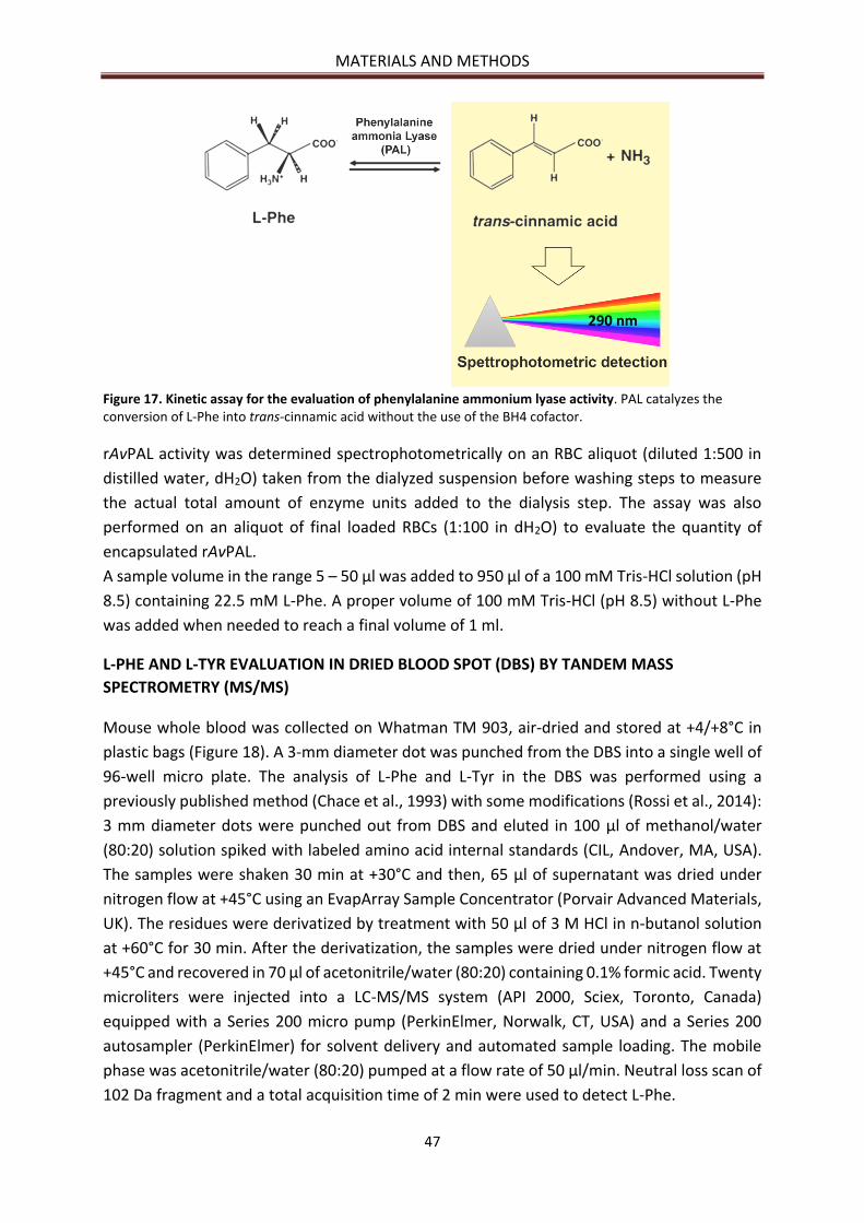
MATERIALS AND METHODS
47
Figure 17. Kinetic assay for the evaluation of phenylalanine ammonium lyase activity. PAL catalyzes the conversion of L-Phe into trans-cinnamic acid without the use of the BH4 cofactor.
rAvPAL activity was determined spectrophotometrically on an RBC aliquot (diluted 1:500 in
distilled water, dH2O) taken from the dialyzed suspension before washing steps to measure
the actual total amount of enzyme units added to the dialysis step. The assay was also
performed on an aliquot of final loaded RBCs (1:100 in dH2O) to evaluate the quantity of
encapsulated rAvPAL.
A sample volume in the range 5 – 50 µl was added to 950 µl of a 100 mM Tris-HCl solution (pH
8.5) containing 22.5 mM L-Phe. A proper volume of 100 mM Tris-HCl (pH 8.5) without L-Phe
was added when needed to reach a final volume of 1 ml.
L-PHE AND L-TYR EVALUATION IN DRIED BLOOD SPOT (DBS) BY TANDEM MASS
SPECTROMETRY (MS/MS)
Mouse whole blood was collected on Whatman TM 903, air-dried and stored at +4/+8°C in
plastic bags (Figure 18). A 3-mm diameter dot was punched from the DBS into a single well of
96-well micro plate. The analysis of L-Phe and L-Tyr in the DBS was performed using a
previously published method (Chace et al., 1993) with some modifications (Rossi et al., 2014):
3 mm diameter dots were punched out from DBS and eluted in 100 μl of methanol/water
(80:20) solution spiked with labeled amino acid internal standards (CIL, Andover, MA, USA).
The samples were shaken 30 min at +30°C and then, 65 μl of supernatant was dried under
nitrogen flow at +45°C using an EvapArray Sample Concentrator (Porvair Advanced Materials,
UK). The residues were derivatized by treatment with 50 μl of 3 M HCl in n-butanol solution
at +60°C for 30 min. After the derivatization, the samples were dried under nitrogen flow at
+45°C and recovered in 70 μl of acetonitrile/water (80:20) containing 0.1% formic acid. Twenty
microliters were injected into a LC-MS/MS system (API 2000, Sciex, Toronto, Canada)
equipped with a Series 200 micro pump (PerkinElmer, Norwalk, CT, USA) and a Series 200
autosampler (PerkinElmer) for solvent delivery and automated sample loading. The mobile
phase was acetonitrile/water (80:20) pumped at a flow rate of 50 μl/min. Neutral loss scan of
102 Da fragment and a total acquisition time of 2 min were used to detect L-Phe.
290 nm
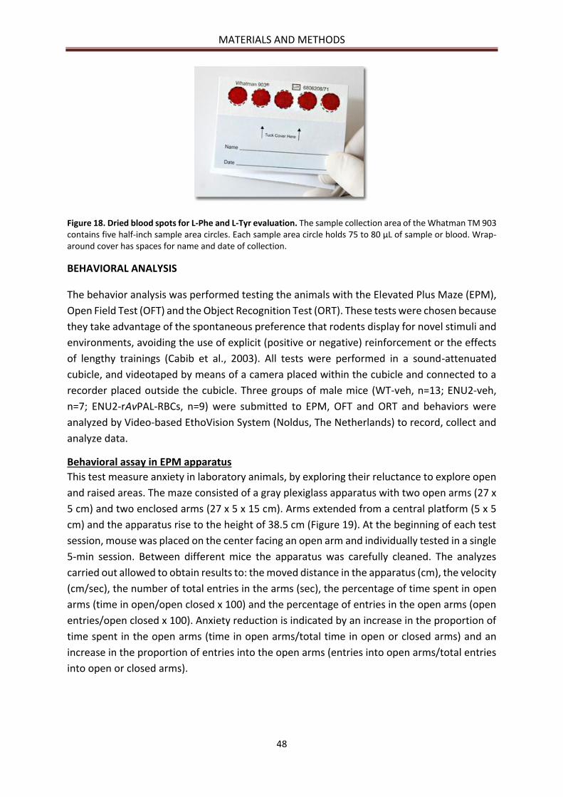
MATERIALS AND METHODS
48
Figure 18. Dried blood spots for L-Phe and L-Tyr evaluation. The sample collection area of the Whatman TM 903 contains five half-inch sample area circles. Each sample area circle holds 75 to 80 µL of sample or blood. Wrap-around cover has spaces for name and date of collection.
BEHAVIORAL ANALYSIS
The behavior analysis was performed testing the animals with the Elevated Plus Maze (EPM),
Open Field Test (OFT) and the Object Recognition Test (ORT). These tests were chosen because
they take advantage of the spontaneous preference that rodents display for novel stimuli and
environments, avoiding the use of explicit (positive or negative) reinforcement or the effects
of lengthy trainings (Cabib et al., 2003). All tests were performed in a sound-attenuated
cubicle, and videotaped by means of a camera placed within the cubicle and connected to a
recorder placed outside the cubicle. Three groups of male mice (WT-veh, n=13; ENU2-veh,
n=7; ENU2-rAvPAL-RBCs, n=9) were submitted to EPM, OFT and ORT and behaviors were
analyzed by Video-based EthoVision System (Noldus, The Netherlands) to record, collect and
analyze data.
Behavioral assay in EPM apparatus
This test measure anxiety in laboratory animals, by exploring their reluctance to explore open
and raised areas. The maze consisted of a gray plexiglass apparatus with two open arms (27 x
5 cm) and two enclosed arms (27 x 5 x 15 cm). Arms extended from a central platform (5 x 5
cm) and the apparatus rise to the height of 38.5 cm (Figure 19). At the beginning of each test
session, mouse was placed on the center facing an open arm and individually tested in a single
5-min session. Between different mice the apparatus was carefully cleaned. The analyzes
carried out allowed to obtain results to: the moved distance in the apparatus (cm), the velocity
(cm/sec), the number of total entries in the arms (sec), the percentage of time spent in open
arms (time in open/open closed x 100) and the percentage of entries in the open arms (open
entries/open closed x 100). Anxiety reduction is indicated by an increase in the proportion of
time spent in the open arms (time in open arms/total time in open or closed arms) and an
increase in the proportion of entries into the open arms (entries into open arms/total entries
into open or closed arms).
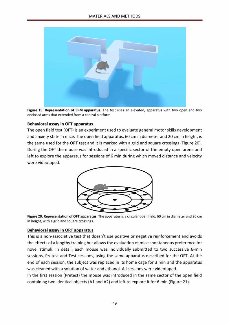
MATERIALS AND METHODS
49
Figure 19. Representation of EPM apparatus. The test uses an elevated, apparatus with two open and two enclosed arms that extended from a central platform.
Behavioral assay in OFT apparatus
The open field test (OFT) is an experiment used to evaluate general motor skills development
and anxiety state in mice. The open field apparatus, 60 cm in diameter and 20 cm in height, is
the same used for the ORT test and it is marked with a grid and square crossings (Figure 20).
During the OFT the mouse was introduced in a specific sector of the empty open arena and
left to explore the apparatus for sessions of 6 min during which moved distance and velocity
were videotaped.
Figure 20. Representation of OFT apparatus. The apparatus is a circular open field, 60 cm in diameter and 20 cm in height, with a grid and square crossings.
Behavioral assay in ORT apparatus
This is a non-associative test that doesn’t use positive or negative reinforcement and avoids
the effects of a lengthy training but allows the evaluation of mice spontaneous preference for
novel stimuli. In detail, each mouse was individually submitted to two successive 6-min
sessions, Pretest and Test sessions, using the same apparatus described for the OFT. At the
end of each session, the subject was replaced in its home cage for 3 min and the apparatus
was cleaned with a solution of water and ethanol. All sessions were videotaped.
In the first session (Pretest) the mouse was introduced in the same sector of the open field
containing two identical objects (A1 and A2) and left to explore it for 6 min (Figure 21).
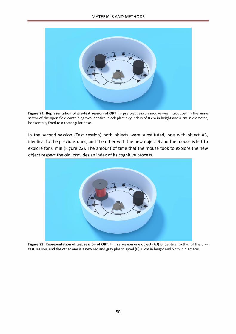
MATERIALS AND METHODS
50
Figure 21. Representation of pre-test session of ORT. In pre-test session mouse was introduced in the same sector of the open field containing two identical black plastic cylinders of 8 cm in height and 4 cm in diameter, horizontally fixed to a rectangular base.
In the second session (Test session) both objects were substituted, one with object A3,
identical to the previous ones, and the other with the new object B and the mouse is left to
explore for 6 min (Figure 22). The amount of time that the mouse took to explore the new
object respect the old, provides an index of its cognitive process.
Figure 22. Representation of test session of ORT. In this session one object (A3) is identical to that of the pre-test session, and the other one is a new red and gray plastic spool (B), 8 cm in height and 5 cm in diameter.
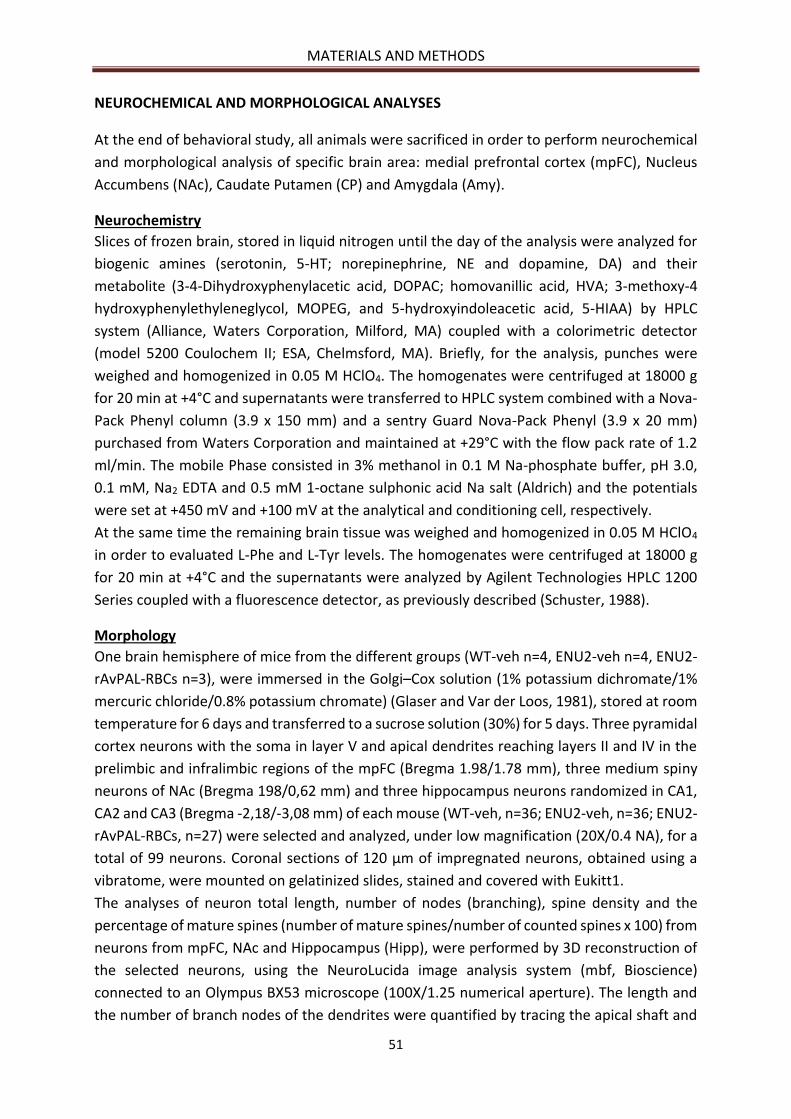
MATERIALS AND METHODS
51
NEUROCHEMICAL AND MORPHOLOGICAL ANALYSES
At the end of behavioral study, all animals were sacrificed in order to perform neurochemical
and morphological analysis of specific brain area: medial prefrontal cortex (mpFC), Nucleus
Accumbens (NAc), Caudate Putamen (CP) and Amygdala (Amy).
Neurochemistry
Slices of frozen brain, stored in liquid nitrogen until the day of the analysis were analyzed for
biogenic amines (serotonin, 5-HT; norepinephrine, NE and dopamine, DA) and their
metabolite (3-4-Dihydroxyphenylacetic acid, DOPAC; homovanillic acid, HVA; 3-methoxy-4
hydroxyphenylethyleneglycol, MOPEG, and 5-hydroxyindoleacetic acid, 5-HIAA) by HPLC
system (Alliance, Waters Corporation, Milford, MA) coupled with a colorimetric detector
(model 5200 Coulochem II; ESA, Chelmsford, MA). Briefly, for the analysis, punches were
weighed and homogenized in 0.05 M HClO4. The homogenates were centrifuged at 18000 g
for 20 min at +4°C and supernatants were transferred to HPLC system combined with a Nova-
Pack Phenyl column (3.9 x 150 mm) and a sentry Guard Nova-Pack Phenyl (3.9 x 20 mm)
purchased from Waters Corporation and maintained at +29°C with the flow pack rate of 1.2
ml/min. The mobile Phase consisted in 3% methanol in 0.1 M Na-phosphate buffer, pH 3.0,
0.1 mM, Na2 EDTA and 0.5 mM 1-octane sulphonic acid Na salt (Aldrich) and the potentials
were set at +450 mV and +100 mV at the analytical and conditioning cell, respectively.
At the same time the remaining brain tissue was weighed and homogenized in 0.05 M HClO4
in order to evaluated L-Phe and L-Tyr levels. The homogenates were centrifuged at 18000 g
for 20 min at +4°C and the supernatants were analyzed by Agilent Technologies HPLC 1200
Series coupled with a fluorescence detector, as previously described (Schuster, 1988).
Morphology
One brain hemisphere of mice from the different groups (WT-veh n=4, ENU2-veh n=4, ENU2-
rAvPAL-RBCs n=3), were immersed in the Golgi–Cox solution (1% potassium dichromate/1%
mercuric chloride/0.8% potassium chromate) (Glaser and Var der Loos, 1981), stored at room
temperature for 6 days and transferred to a sucrose solution (30%) for 5 days. Three pyramidal
cortex neurons with the soma in layer V and apical dendrites reaching layers II and IV in the
prelimbic and infralimbic regions of the mpFC (Bregma 1.98/1.78 mm), three medium spiny
neurons of NAc (Bregma 198/0,62 mm) and three hippocampus neurons randomized in CA1,
CA2 and CA3 (Bregma -2,18/-3,08 mm) of each mouse (WT-veh, n=36; ENU2-veh, n=36; ENU2-
rAvPAL-RBCs, n=27) were selected and analyzed, under low magnification (20X/0.4 NA), for a
total of 99 neurons. Coronal sections of 120 µm of impregnated neurons, obtained using a
vibratome, were mounted on gelatinized slides, stained and covered with Eukitt1.
The analyses of neuron total length, number of nodes (branching), spine density and the
percentage of mature spines (number of mature spines/number of counted spines x 100) from
neurons from mpFC, NAc and Hippocampus (Hipp), were performed by 3D reconstruction of
the selected neurons, using the NeuroLucida image analysis system (mbf, Bioscience)
connected to an Olympus BX53 microscope (100X/1.25 numerical aperture). The length and
the number of branch nodes of the dendrites were quantified by tracing the apical shaft and
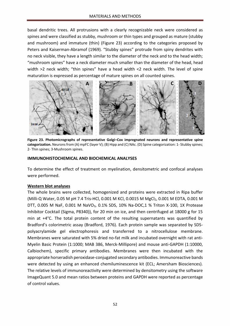
MATERIALS AND METHODS
52
basal dendritic trees. All protrusions with a clearly recognizable neck were considered as
spines and were classified as stubby, mushroom or thin types and grouped as mature (stubby
and mushroom) and immature (thin) (Figure 23) according to the categories proposed by
Peters and Kaiserman-Abramof (1969). “Stubby spines” protrude from spiny dendrites with
no neck visible, they have a length similar to the diameter of the neck and to the head width;
“mushroom spines” have a neck diameter much smaller than the diameter of the head, head
width >2 neck width; “thin spines” have a head width <2 neck width. The level of spine
maturation is expressed as percentage of mature spines on all counted spines.
Figure 23. Photomicrographs of representative Golgi–Cox impregnated neurons and representative spine categorization. Neurons from (A) mpFC (layer V); (B) Hipp and (C) NAc. (D) Spine categorization: 1- Stubby spines; 2- Thin spines; 3-Mushroom spines.
IMMUNOHISTOCHEMICAL AND BIOCHEMICAL ANALYSES
To determine the effect of treatment on myelination, densitometric and confocal analyses
were performed.
Western blot analyses
The whole brains were collected, homogenized and proteins were extracted in Ripa buffer
(Milli-Q Water, 0.05 M pH 7.4 Tris-HCl, 0.001 M KCl, 0.0015 M MgCl2, 0.001 M EDTA, 0.001 M
DTT, 0.005 M NaF, 0.001 M NaVO3, 0.1% SDS, 10% Na-DOC,1 % Triton X-100, 1X Protease
Inhibitor Cocktail (Sigma, P8340)), for 20 min on ice, and then centrifuged at 18000 g for 15
min at +4°C. The total protein content of the resulting supernatants was quantified by
Bradford’s colorimetric assay (Bradford, 1976). Each protein sample was separated by SDS-
polyacrylamide gel electrophoresis and transferred to a nitrocellulose membrane.
Membranes were saturated with 5% dried no-fat milk and incubated overnight with rat anti-
Myelin Basic Protein (1:1000; MAB 386, Merck-Millipore) and mouse anti-GAPDH (1:10000,
Calbiochem), specific primary antibodies. Membranes were then incubated with the
appropriate horseradish peroxidase-conjugated secondary antibodies. Immunoreactive bands
were detected by using an enhanced chemiluminescence kit (ECL; Amersham Biosciences).
The relative levels of immunoreactivity were determined by densitometry using the software
ImageQuant 5.0 and mean ratios between proteins and GAPDH were reported as percentage
of control values.
A B C D
MATERIALS AND METHODS
53
Immunofluorescence analyses
Five mice per group were perfused transcardially with 50 ml of saline solution followed by 50
ml of 4% paraformaldehyde in a 0.1 M phosphate buffer (PB; pH 7.4) under anesthesia. Each
brain was removed immediately, post-fixed in the same fixative for 2 h and, after three washes
in PB, transferred to 30% sucrose in PB solution at +4°C until they sank. Four series of 30 μm-
thick transverse brain sections obtained by means of a freezing microtome were collected in
PB and then incubated O.N. with primary antibody solution, rat anti-MBP (1:1000; MAB 386,
Merck-Millipore), prepared in PB and 0.3% Triton X-100. Each incubation step was followed
by three 5-min rinses in PB. Afterwards, sections were incubated 2 h at RT with a cocktail of
1:200 secondary antibodies (Alexa Fluor 488 conjugated donkey anti-rat and Alexa Fluor 555
conjugated donkey anti-goat). Sections of two brain structures containing many neural fiber
bundles, such as the corpus callosum and the striatum, were examined under a confocal laser
scanning microscope (Zeiss CLSM700, Germany) equipped with two laser lines: argon emitting
at 488 nm, and helium/neon emitting at 543 nm and 633 nm. Confocal settings for image
capture were maintained constant throughout the acquisition of sections from the different
experimental groups of mice in order to avoid staining variability among sections and
experimental groups. Images were exported in TIFF format and analyzed with ImageJ software
(http://rsb. info.nih.gov/ij/; National Institutes of Health). MBP-associated signal was
quantified by manually outlining the areas of interest. Mean signal intensity (F) of the marker
of interest was performed on two squared frames (42 μm per side) on 5 sections sampled to
cover the rostro-caudal extent of the areas of interest (striatum and corpus callosum) entirely
(n=10 samples per mouse). The F/A ratio defines mean fluorescence of individual samples (F)
normalized to total cellular surface (A).
EVALUATION OF PLASMA ANTI-rAvPAL IgG TITER
The immune response against rAvPAL administered through erythrocytes was evaluated by
standard indirect ELISA on the samples collected at the end of behavioral tests (Figure 24).
Blood samples (100 µl) collected in heparin were centrifuged 5 min at 1050 g, plasma was
harvested and frozen until use. ELISA analysis was performed as follows: 2HB flat bottom 96-
well plates (Immulon® microtiter plates, Thermo Scientific, Rochester, NY) were coated
overnight at +4°C with 100 μl of rAvPAL dissolved 1 μg/ml in 50 mM carbonate buffer (15 mM
Na2CO3 and 33.7 mM NaHCO3, pH 9.7). Plates were washed four times with 400 μl/well of PBS
solution (1.37 mM NaCl, 26.8 mM KCl, 32.1 mM Na2HPO4, 14.7 mM KH2PO4) added with 0.05%
(v/v) Tween 20 (TPBS), blocked with 100 μl/well of TPBS plus 2% (w/v) fat free dry milk
(blocking solution) and maintained 1 h at +37°C. Plates were finally washed four times with
TPBS solution. Plasma was thawed, serially diluted in blocking solution in the range of 1:25 –
1:1600 for treated ENU2-rAvPAL-RBC mice, in the range of 1:25 – 1:200 for WT-veh animals,
and dispensed 100 µl/well in duplicate. Plasma antibody binding was obtained incubating the
plates 90 min at +37°C. After four washes with TPBS solution, 100 µl/well of goat anti-mouse
IgG-HRP (Bio-Rad, Hercules, CA), diluted 1:1000 in blocking solution, were added and plates
incubated 60 min at +37°C. After another four washes in TPBS solution, the immune
complexes were revealed adding 100 μl/well of 2,2'-azino-bis (3-ethylbenzothiazoline-6-
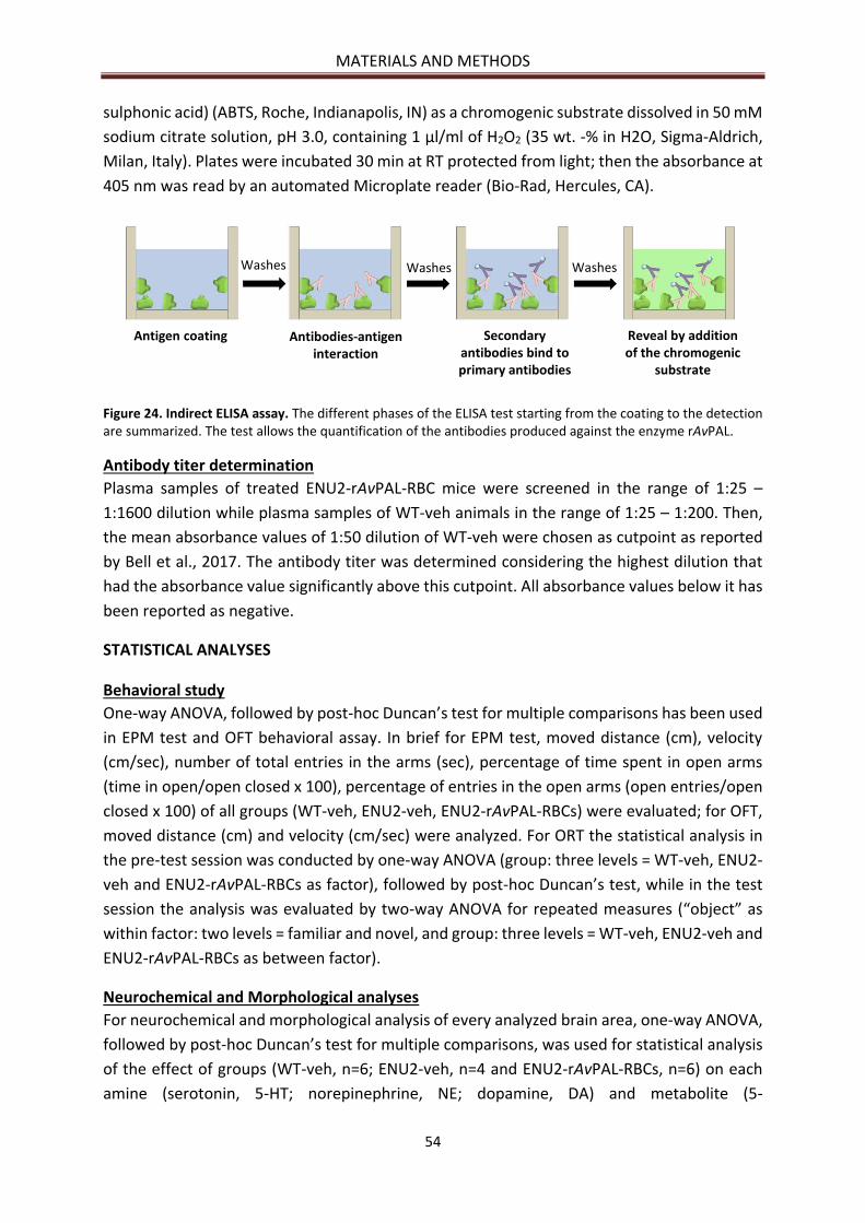
MATERIALS AND METHODS
54
sulphonic acid) (ABTS, Roche, Indianapolis, IN) as a chromogenic substrate dissolved in 50 mM
sodium citrate solution, pH 3.0, containing 1 μl/ml of H2O2 (35 wt. -% in H2O, Sigma-Aldrich,
Milan, Italy). Plates were incubated 30 min at RT protected from light; then the absorbance at
405 nm was read by an automated Microplate reader (Bio-Rad, Hercules, CA).
Figure 24. Indirect ELISA assay. The different phases of the ELISA test starting from the coating to the detection are summarized. The test allows the quantification of the antibodies produced against the enzyme rAvPAL.
Antibody titer determination
Plasma samples of treated ENU2-rAvPAL-RBC mice were screened in the range of 1:25 –
1:1600 dilution while plasma samples of WT-veh animals in the range of 1:25 – 1:200. Then,
the mean absorbance values of 1:50 dilution of WT-veh were chosen as cutpoint as reported
by Bell et al., 2017. The antibody titer was determined considering the highest dilution that
had the absorbance value significantly above this cutpoint. All absorbance values below it has
been reported as negative.
STATISTICAL ANALYSES
Behavioral study
One-way ANOVA, followed by post-hoc Duncan’s test for multiple comparisons has been used
in EPM test and OFT behavioral assay. In brief for EPM test, moved distance (cm), velocity
(cm/sec), number of total entries in the arms (sec), percentage of time spent in open arms
(time in open/open closed x 100), percentage of entries in the open arms (open entries/open
closed x 100) of all groups (WT-veh, ENU2-veh, ENU2-rAvPAL-RBCs) were evaluated; for OFT,
moved distance (cm) and velocity (cm/sec) were analyzed. For ORT the statistical analysis in
the pre-test session was conducted by one-way ANOVA (group: three levels = WT-veh, ENU2-
veh and ENU2-rAvPAL-RBCs as factor), followed by post-hoc Duncan’s test, while in the test
session the analysis was evaluated by two-way ANOVA for repeated measures (“object” as
within factor: two levels = familiar and novel, and group: three levels = WT-veh, ENU2-veh and
ENU2-rAvPAL-RBCs as between factor).
Neurochemical and Morphological analyses
For neurochemical and morphological analysis of every analyzed brain area, one-way ANOVA,
followed by post-hoc Duncan’s test for multiple comparisons, was used for statistical analysis
of the effect of groups (WT-veh, n=6; ENU2-veh, n=4 and ENU2-rAvPAL-RBCs, n=6) on each
amine (serotonin, 5-HT; norepinephrine, NE; dopamine, DA) and metabolite (5-
Washes Washes Washes
Antigen coating Antibodies-antigeninteraction
Secondaryantibodies bind to primary antibodies
Reveal by additionof the chromogenic
substrate

MATERIALS AND METHODS
55
hydroxyindoleacetic acid, 5-HIAA; 3-methoxy-4 hydroxyphenylethyleneglycol, MOPEG; 3-4-
Dihydroxyphenylacetic acid, DOPAC and homovanillic acid, HVA).
Antibody titer determination
The statistical determination of antibody titer was performed applying the T-Test on the
absorbance values of the different plasma diluitions of all experimental groups.
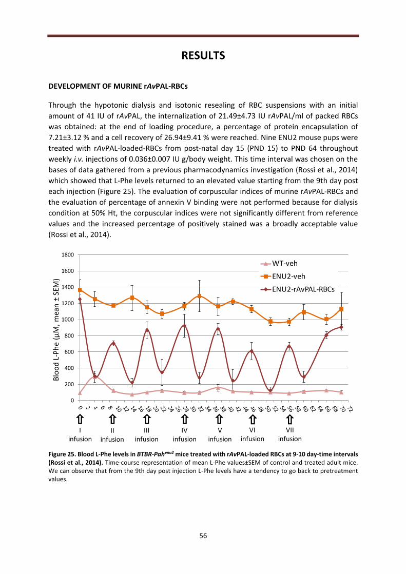
56
RESULTS
DEVELOPMENT OF MURINE rAvPAL-RBCs
Through the hypotonic dialysis and isotonic resealing of RBC suspensions with an initial
amount of 41 IU of rAvPAL, the internalization of 21.49±4.73 IU rAvPAL/ml of packed RBCs
was obtained: at the end of loading procedure, a percentage of protein encapsulation of
7.21±3.12 % and a cell recovery of 26.94±9.41 % were reached. Nine ENU2 mouse pups were
treated with rAvPAL-loaded-RBCs from post-natal day 15 (PND 15) to PND 64 throughout
weekly i.v. injections of 0.036±0.007 IU g/body weight. This time interval was chosen on the
bases of data gathered from a previous pharmacodynamics investigation (Rossi et al., 2014)
which showed that L-Phe levels returned to an elevated value starting from the 9th day post
each injection (Figure 25). The evaluation of corpuscular indices of murine rAvPAL-RBCs and
the evaluation of percentage of annexin V binding were not performed because for dialysis
condition at 50% Ht, the corpuscular indices were not significantly different from reference
values and the increased percentage of positively stained was a broadly acceptable value
(Rossi et al., 2014).
Figure 25. Blood L-Phe levels in BTBR-Pahenu2 mice treated with rAvPAL-loaded RBCs at 9-10 day-time intervals (Rossi et al., 2014). Time-course representation of mean L-Phe values±SEM of control and treated adult mice. We can observe that from the 9th day post injection L-Phe levels have a tendency to go back to pretreatment values.
0
200
400
600
800
1000
1200
1400
1600
1800
Blo
od
L-P
he
(µM
, m
ean
±SE
M)
WT-veh
ENU2-veh
ENU2-rAvPAL-RBCs
I infusion
II infusion
III infusion
IV infusion
V infusion
VIinfusion
VII infusion
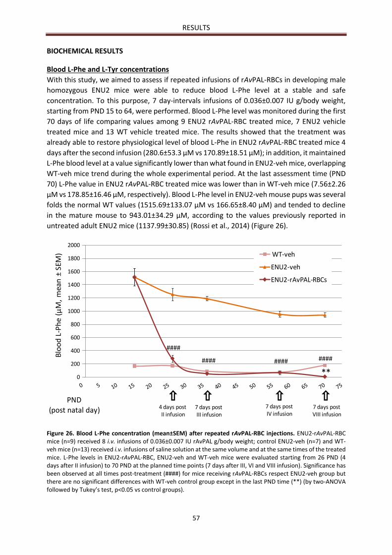
RESULTS
57
BIOCHEMICAL RESULTS
Blood L-Phe and L-Tyr concentrations
With this study, we aimed to assess if repeated infusions of rAvPAL-RBCs in developing male
homozygous ENU2 mice were able to reduce blood L-Phe level at a stable and safe
concentration. To this purpose, 7 day-intervals infusions of 0.036±0.007 IU g/body weight,
starting from PND 15 to 64, were performed. Blood L-Phe level was monitored during the first
70 days of life comparing values among 9 ENU2 rAvPAL-RBC treated mice, 7 ENU2 vehicle
treated mice and 13 WT vehicle treated mice. The results showed that the treatment was
already able to restore physiological level of blood L-Phe in ENU2 rAvPAL-RBC treated mice 4
days after the second infusion (280.6±53.3 µM vs 170.89±18.51 µM); in addition, it maintained
L-Phe blood level at a value significantly lower than what found in ENU2-veh mice, overlapping
WT-veh mice trend during the whole experimental period. At the last assessment time (PND
70) L-Phe value in ENU2 rAvPAL-RBC treated mice was lower than in WT-veh mice (7.56±2.26
µM vs 178.85±16.46 µM, respectively). Blood L-Phe level in ENU2-veh mouse pups was several
folds the normal WT values (1515.69±133.07 µM vs 166.65±8.40 µM) and tended to decline
in the mature mouse to 943.01±34.29 μM, according to the values previously reported in
untreated adult ENU2 mice (1137.99±30.85) (Rossi et al., 2014) (Figure 26).
Figure 26. Blood L-Phe concentration (mean±SEM) after repeated rAvPAL-RBC injections. ENU2-rAvPAL-RBC mice (n=9) received 8 i.v. infusions of 0.036±0.007 IU rAvPAL g/body weight; control ENU2-veh (n=7) and WT-veh mice (n=13) received i.v. infusions of saline solution at the same volume and at the same times of the treated mice. L-Phe levels in ENU2-rAvPAL-RBC, ENU2-veh and WT-veh mice were evaluated starting from 26 PND (4 days after II infusion) to 70 PND at the planned time points (7 days after III, VI and VIII infusion). Significance has been observed at all times post-treatment (####) for mice receiving rAvPAL-RBCs respect ENU2-veh group but there are no significant differences with WT-veh control group except in the last PND time (**) (by two-ANOVA followed by Tukey’s test, p<0.05 vs control groups).
0
200
400
600
800
1000
1200
1400
1600
1800
2000
Blo
od
L-P
he
(µM
, m
ean
±SE
M)
PND (post natal day)
WT-veh
ENU2-veh
ENU2-rAvPAL-RBCs
####
**
#### #### ####
4 days postII infusion
7 days postIII infusion
7 days postIV infusion
7 days postVIII infusion
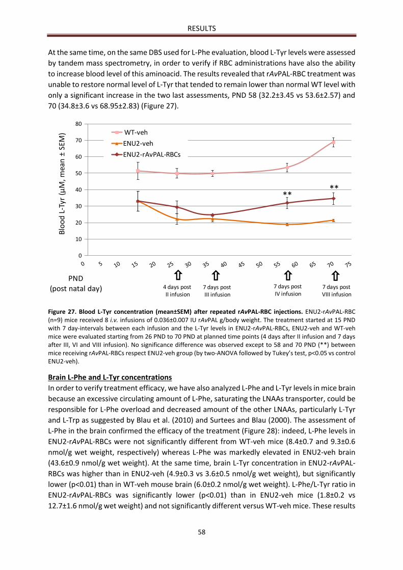
RESULTS
58
At the same time, on the same DBS used for L-Phe evaluation, blood L-Tyr levels were assessed
by tandem mass spectrometry, in order to verify if RBC administrations have also the ability
to increase blood level of this aminoacid. The results revealed that rAvPAL-RBC treatment was
unable to restore normal level of L-Tyr that tended to remain lower than normal WT level with
only a significant increase in the two last assessments, PND 58 (32.2±3.45 vs 53.6±2.57) and
70 (34.8±3.6 vs 68.95±2.83) (Figure 27).
Figure 27. Blood L-Tyr concentration (mean±SEM) after repeated rAvPAL-RBC injections. ENU2-rAvPAL-RBC (n=9) mice received 8 i.v. infusions of 0.036±0.007 IU rAvPAL g/body weight. The treatment started at 15 PND with 7 day-intervals between each infusion and the L-Tyr levels in ENU2-rAvPAL-RBCs, ENU2-veh and WT-veh mice were evaluated starting from 26 PND to 70 PND at planned time points (4 days after II infusion and 7 days after III, VI and VIII infusion). No significance difference was observed except to 58 and 70 PND (**) between mice receiving rAvPAL-RBCs respect ENU2-veh group (by two-ANOVA followed by Tukey’s test, p<0.05 vs control ENU2-veh).
Brain L-Phe and L-Tyr concentrations
In order to verify treatment efficacy, we have also analyzed L-Phe and L-Tyr levels in mice brain
because an excessive circulating amount of L-Phe, saturating the LNAAs transporter, could be
responsible for L-Phe overload and decreased amount of the other LNAAs, particularly L-Tyr
and L-Trp as suggested by Blau et al. (2010) and Surtees and Blau (2000). The assessment of
L-Phe in the brain confirmed the efficacy of the treatment (Figure 28): indeed, L-Phe levels in
ENU2-rAvPAL-RBCs were not significantly different from WT-veh mice (8.4±0.7 and 9.3±0.6
nmol/g wet weight, respectively) whereas L-Phe was markedly elevated in ENU2-veh brain
(43.6±0.9 nmol/g wet weight). At the same time, brain L-Tyr concentration in ENU2-rAvPAL-
RBCs was higher than in ENU2-veh (4.9±0.3 vs 3.6±0.5 nmol/g wet weight), but significantly
lower (p<0.01) than in WT-veh mouse brain (6.0±0.2 nmol/g wet weight). L-Phe/L-Tyr ratio in
ENU2-rAvPAL-RBCs was significantly lower (p<0.01) than in ENU2-veh mice (1.8±0.2 vs
12.7±1.6 nmol/g wet weight) and not significantly different versus WT-veh mice. These results
0
10
20
30
40
50
60
70
80
Blo
od
L-T
yr (
µM
, mea
n ±
SEM
)
PND (post natal day)
WT-veh
ENU2-veh
ENU2-rAvPAL-RBCs
****
4 days postII infusion
7 days postIII infusion
7 days postIV infusion
7 days postVIII infusion
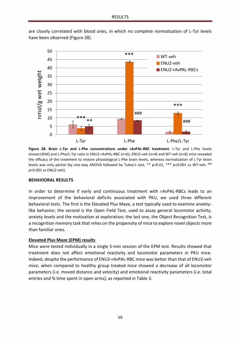
RESULTS
59
are closely correlated with blood ones, in which no complete normalization of L-Tyr levels
have been observed (Figure 28).
Figure 28. Brain L-Tyr and L-Phe concentrations under rAvPAL-RBC treatment. L-Tyr and L-Phe levels
(mean±SEM) and L-Phe/L-Tyr ratio in ENU2-rAvPAL-RBC (n=6), ENU2-veh (n=4) and WT-veh (n=6) mice revealed
the efficacy of the treatment to restore physiological L-Phe brain levels, whereas normalization of L-Tyr brain
levels was only partial (by one-way ANOVA followed by Tukey’s test, ** p<0.01, *** p<0.001 vs WT-veh; ###
p<0.001 vs ENU2-veh).
BEHAVIORAL RESULTS
In order to determine if early and continuous treatment with rAvPAL-RBCs leads to an
improvement of the behavioral deficits associated with PKU, we used three different
behavioral tests. The first is the Elevated Plus Maze, a test typically used to examine anxiety-
like behavior; the second is the Open Field Test, used to assay general locomotor activity,
anxiety levels and the motivation at exploration; the last one, the Object Recognition Test, is
a recognition memory task that relies on the propensity of mice to explore novel objects more
than familiar ones.
Elevated Plus Maze (EPM) results
Mice were tested individually in a single 5-min session of the EPM test. Results showed that
treatment does not affect emotional reactivity and locomotor parameters in PKU mice.
Indeed, despite the performance of ENU2-rAvPAL-RBC mice was better than that of ENU2-veh
mice, when compared to healthy group treated mice showed a decrease of all locomotor
parameters (i.e. moved distance and velocity) and emotional reactivity parameters (i.e. total
entries and % time spent in open arms), as reported in Table 3.
0
5
10
15
20
25
30
35
40
45
50
L-Tyr L-Phe L-Phe/L-Tyr
nm
ol/
g w
et w
eigh
t
WT-veh
ENU2-veh
ENU2-rAvPAL-RBCs
*** **
***
***###
###
RESULTS
60
Moved distance
(cm)
Velocity
(cm/sec)
Total
entries
% time spent in
open arms
WT-veh (n=13) 2112.08±164.19 8.07±1.04 54.76±9.96 18.51±2.82
ENU2-veh (n=7) 1097.57±41.46C 3.70±0.29B 12.21±0.99B 3.06±1.15C
ENU2-rAvPAL-RBCs (n=9)
1456.72±123.06B 5.73±0.94 28.72±6.41A 7.05±1.89B
Table 3. Behavioral responses in the EPM by adult WT-veh, ENU2-veh and ENU2-rAvPAL-RBC mice. The table reported locomotor parameters (moved distance and velocity) and emotional reactivity (total entries and % time in open arms). AP<0.05, BP<0.01 and CP<0.001 compared with WT-veh values. Data are expressed as mean±SEM.
Open Field Test (OFT) results
ENU2 mice receiving rAvPAL-RBC treatment exhibit no significant differences in the moved
distance and velocity into empty Open Field, when compared to ENU2 untreated mice, thus
they maintain motor impairment when compared with WT-veh mice as shown in Table 4.
Moved distance
(cm)
Velocity
(cm/sec) Grooming Rearing
WT-veh (n=13) 2638.59±180.45 9.35±0.56 7.62±3.28 13.21±1.99
ENU2-veh (n=7) 2420.12±294.55 8.02±0.98 22.25±3.72B 0.39±2.26C
ENU2-rAvPAL-RBCs (n=9)
1908.53±268.10A 6.67±0.75A 19.78±3.28A 2.22±1.99C
Table 4. Behavioral responses in the Open Field by adult WT-veh, ENU2-veh and ENU2-rAvPAL-RBC mice. AP<0.05, BP<0.01 and CP<0.001 compared with WT-veh values. Data are expressed as mean±SEM.
Object Recognition Test (ORT) results
ORT is a variant for rodents of the delayed non-matching to sample task that does not require
reinforcement, exploiting spontaneous preference of mice for novelty (Cabib et al., 2003).
During pre-test session, statistical analysis revealed a significant difference among groups for
the time spent exploring the objects. ENU2-veh mice showed a decrease in the time spent
exploring the objects in comparison with the other groups while ENU2-rAvPAL-RBC behaved
similarly to WT-veh mice (Figure 29A). In addition, during the test session, both treated PKU
and healty mice spent more time than ENU2-veh mice exploring a novel object (Figure 29B).
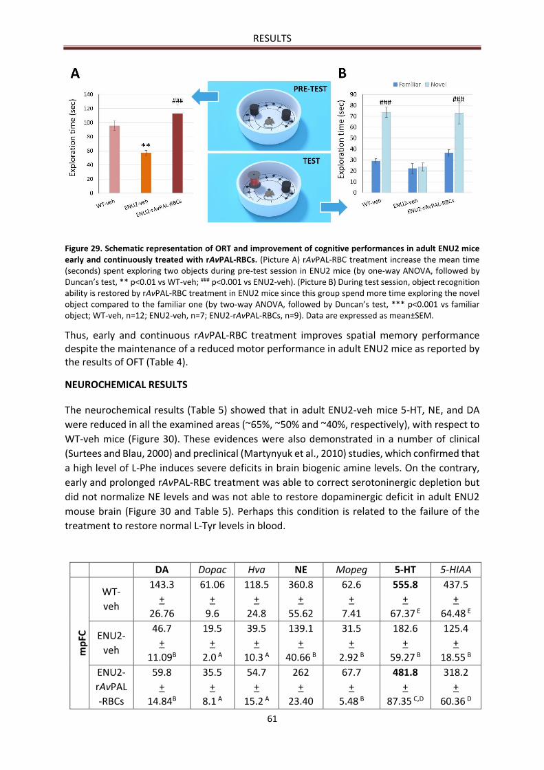
RESULTS
61
Figure 29. Schematic representation of ORT and improvement of cognitive performances in adult ENU2 mice early and continuously treated with rAvPAL-RBCs. (Picture A) rAvPAL-RBC treatment increase the mean time (seconds) spent exploring two objects during pre-test session in ENU2 mice (by one-way ANOVA, followed by Duncan’s test, ** p<0.01 vs WT-veh; ### p<0.001 vs ENU2-veh). (Picture B) During test session, object recognition ability is restored by rAvPAL-RBC treatment in ENU2 mice since this group spend more time exploring the novel object compared to the familiar one (by two-way ANOVA, followed by Duncan’s test, *** p<0.001 vs familiar object; WT-veh, n=12; ENU2-veh, n=7; ENU2-rAvPAL-RBCs, n=9). Data are expressed as mean±SEM.
Thus, early and continuous rAvPAL-RBC treatment improves spatial memory performance despite the maintenance of a reduced motor performance in adult ENU2 mice as reported by the results of OFT (Table 4).
NEUROCHEMICAL RESULTS
The neurochemical results (Table 5) showed that in adult ENU2-veh mice 5-HT, NE, and DA
were reduced in all the examined areas (~65%, ~50% and ~40%, respectively), with respect to
WT-veh mice (Figure 30). These evidences were also demonstrated in a number of clinical
(Surtees and Blau, 2000) and preclinical (Martynyuk et al., 2010) studies, which confirmed that
a high level of L-Phe induces severe deficits in brain biogenic amine levels. On the contrary,
early and prolonged rAvPAL-RBC treatment was able to correct serotoninergic depletion but
did not normalize NE levels and was not able to restore dopaminergic deficit in adult ENU2
mouse brain (Figure 30 and Table 5). Perhaps this condition is related to the failure of the
treatment to restore normal L-Tyr levels in blood.
DA Dopac Hva NE Mopeg 5-HT 5-HIAA
mp
FC
WT-
veh
143.3
+
26.76
61.06
+
9.6
118.5
+
24.8
360.8
+
55.62
62.6
+
7.41
555.8
+
67.37 E
437.5
+
64.48 E
ENU2-
veh
46.7
+
11.09B
19.5
+
2.0 A
39.5
+
10.3 A
139.1
+
40.66 B
31.5
+
2.92 B
182.6
+
59.27 B
125.4
+
18.55 B
ENU2-
rAvPAL
-RBCs
59.8
+
14.84B
35.5
+
8.1 A
54.7
+
15.2 A
262
+
23.40
67.7
+
5.48 B
481.8
+
87.35 C,D
318.2
+
60.36 D
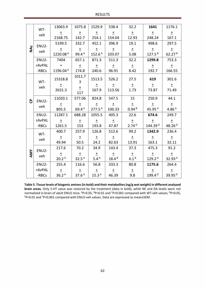
RESULTS
62
NA
c WT-
veh
13063.9
+
2168.75
1075.8
+
142.7
1529.9
+
254.1
538.4
+
154.04
32.2
+
12.93
1641
+
248.24
1176.1
+
167.1
ENU2-
veh
5199.5
+
1220.08 A
332.7
+
99.4 A
452.1
+
152.6 A
396.9
+
103.07
19.1
+
5.08
498.6
+
127.5 B
297.5
+
62.27 B
ENU2-
rAvPAL
-RBCs
7404
+
1196.04 A
657.1
+
174.8
871.3
+
240.6
511.3
+
96.91
32.2
+
8.42
1299.8
+
192.7
753.3
+
166.55
CP
WT-
veh
15318.8
+
2631.3
1011.7
3
+
117
1513.5
+
167.9
526.2
+
113.56
27.5
+
1.73
619
+
73.87
301.6
+
71.49
ENU2-
veh
11020.1
+
805.5
577.06
+
69.4 A
824.8
+
277.5 A
547.5
+
100.33
15
+
0.94 B
250.9
+
45.95 A
44.1
+
4.86 A
ENU2-
rAvPAL
-RBCs
11287.1
+
1261.5
688.28
+
153
1055.5
+
193.8
405.3
+
47.87
22.6
+
2.74 D
674.6
+
144.39 D
249.7
+
48.26 D
AM
Y
WT-
veh
400.7
+
49.94
257.9
+
50.5
126.8
+
24.2
512.6
+
82.63
99.2
+
13.91
1342.9
+
163.1
236.4
+
32.11
ENU2-
veh
217.6
+
20.2 A
70.2
+
32.5 A
34.9
+
5.4 A
143.4
+
18.4 B
37.3
+
4.1 B
475.3
+
129.2 A
91.2
+
32.93 A
ENU2-
rAvPAL
-RBCs
255.4
+
36.2 A
116.6
+
37.6 A
56.8
+
15.3 A
333.3
+
46.39
80.8
+
9.8
1175.6
+
199.4 D
264.4
+
39.95 D
Table 5. Tissue levels of biogenic amines (in bold) and their metabolites (ng/g wet weight) in different analyzed brain areas. Only 5-HT value was restored by the treatment (data in bold), while NE and DA levels were not normalized in brain of adult ENU2 mice. AP<0.05, BP<0.01 and CP<0.001 compared with WT-veh values; DP<0.05,
EP<0.01 and FP<0.001 compared with ENU2-veh values. Data are expressed as mean±SEM.
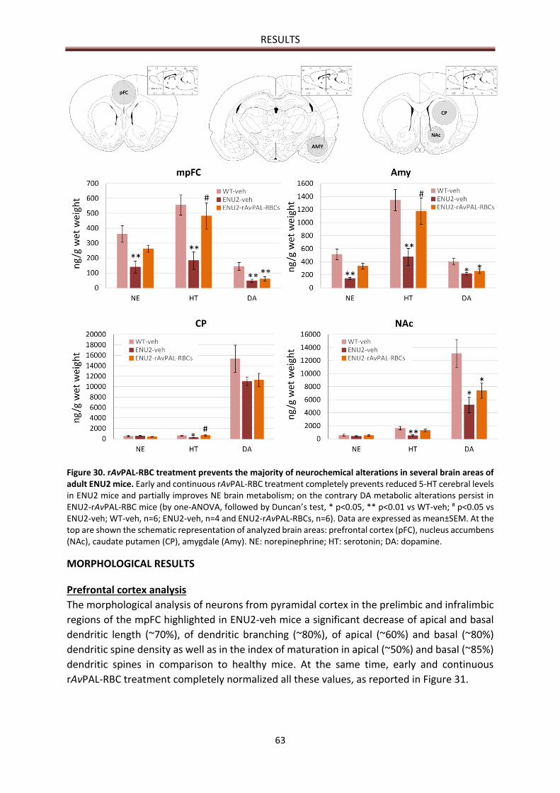
RESULTS
63
Figure 30. rAvPAL-RBC treatment prevents the majority of neurochemical alterations in several brain areas of adult ENU2 mice. Early and continuous rAvPAL-RBC treatment completely prevents reduced 5-HT cerebral levels in ENU2 mice and partially improves NE brain metabolism; on the contrary DA metabolic alterations persist in ENU2-rAvPAL-RBC mice (by one-ANOVA, followed by Duncan’s test, * p<0.05, ** p<0.01 vs WT-veh; # p<0.05 vs ENU2-veh; WT-veh, n=6; ENU2-veh, n=4 and ENU2-rAvPAL-RBCs, n=6). Data are expressed as mean±SEM. At the top are shown the schematic representation of analyzed brain areas: prefrontal cortex (pFC), nucleus accumbens (NAc), caudate putamen (CP), amygdale (Amy). NE: norepinephrine; HT: serotonin; DA: dopamine.
MORPHOLOGICAL RESULTS
Prefrontal cortex analysis
The morphological analysis of neurons from pyramidal cortex in the prelimbic and infralimbic
regions of the mpFC highlighted in ENU2-veh mice a significant decrease of apical and basal
dendritic length (~70%), of dendritic branching (~80%), of apical (~60%) and basal (~80%)
dendritic spine density as well as in the index of maturation in apical (~50%) and basal (~85%)
dendritic spines in comparison to healthy mice. At the same time, early and continuous
rAvPAL-RBC treatment completely normalized all these values, as reported in Figure 31.
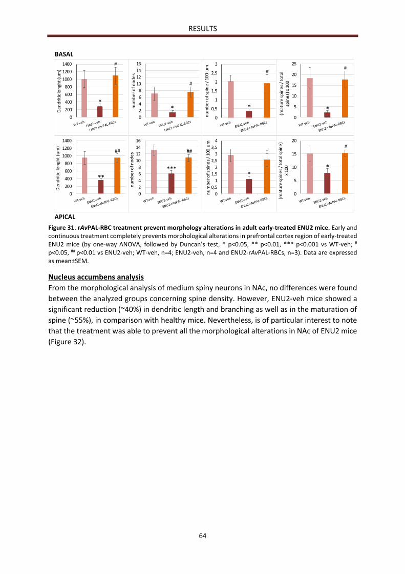
RESULTS
64
Figure 31. rAvPAL-RBC treatment prevent morphology alterations in adult early-treated ENU2 mice. Early and continuous treatment completely prevents morphological alterations in prefrontal cortex region of early-treated ENU2 mice (by one-way ANOVA, followed by Duncan’s test, * p<0.05, ** p<0.01, *** p<0.001 vs WT-veh; #
p<0.05, ## p<0.01 vs ENU2-veh; WT-veh, n=4; ENU2-veh, n=4 and ENU2-rAvPAL-RBCs, n=3). Data are expressed as mean±SEM.
Nucleus accumbens analysis
From the morphological analysis of medium spiny neurons in NAc, no differences were found
between the analyzed groups concerning spine density. However, ENU2-veh mice showed a
significant reduction (~40%) in dendritic length and branching as well as in the maturation of
spine (~55%), in comparison with healthy mice. Nevertheless, is of particular interest to note
that the treatment was able to prevent all the morphological alterations in NAc of ENU2 mice
(Figure 32).
0
0,5
1
1,5
2
2,5
3
nu
mb
er o
f sp
ine
/ 1
00 u
m
0
200
400
600
800
1000
1200
1400
De
nd
riti
cle
ngh
t(u
m)
*
#
*
#
***
##
**
##
BASAL
APICAL
0
200
400
600
800
1000
1200
1400D
en
dri
tic
len
ght(
um
)
*
#
0
2
4
6
8
10
12
14
16
nu
mb
er o
f no
de
s
***
##
0
2
4
6
8
10
12
14
16
nu
mb
er o
f no
de
s
*
#
0
0,5
1
1,5
2
2,5
3
3,5
4
nu
mb
er o
f sp
ine
s /
100
um
*
#
0
5
10
15
20
25
(mat
ure
sp
ine
s /
tota
l sp
ine
s) x
100
0
5
10
15
20
(mat
ure
sp
ine
s /
tota
l sp
ine
) x
100 *
#
*
#
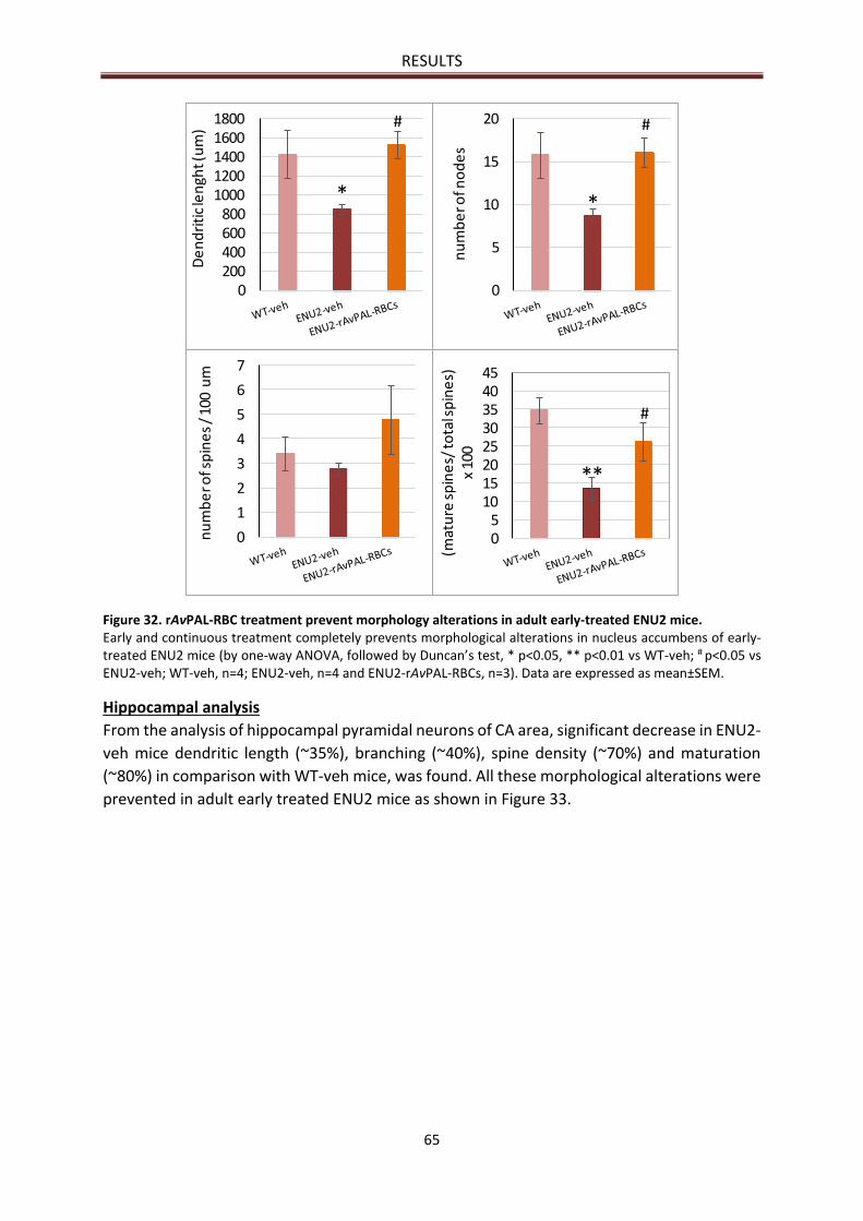
RESULTS
65
Figure 32. rAvPAL-RBC treatment prevent morphology alterations in adult early-treated ENU2 mice. Early and continuous treatment completely prevents morphological alterations in nucleus accumbens of early-treated ENU2 mice (by one-way ANOVA, followed by Duncan’s test, * p<0.05, ** p<0.01 vs WT-veh; # p<0.05 vs ENU2-veh; WT-veh, n=4; ENU2-veh, n=4 and ENU2-rAvPAL-RBCs, n=3). Data are expressed as mean±SEM.
Hippocampal analysis
From the analysis of hippocampal pyramidal neurons of CA area, significant decrease in ENU2-
veh mice dendritic length (~35%), branching (~40%), spine density (~70%) and maturation
(~80%) in comparison with WT-veh mice, was found. All these morphological alterations were
prevented in adult early treated ENU2 mice as shown in Figure 33.
0200400600800
10001200140016001800
De
nd
riti
cle
ngh
t(u
m)
*
#
0
5
10
15
20
nu
mb
er o
f no
de
s
*
#
0
1
2
3
4
5
6
7
nu
mb
er o
f sp
ine
s /
100
um
05
1015202530354045
(mat
ure
sp
ine
s/ to
tal s
pin
es)
x
100
**
#
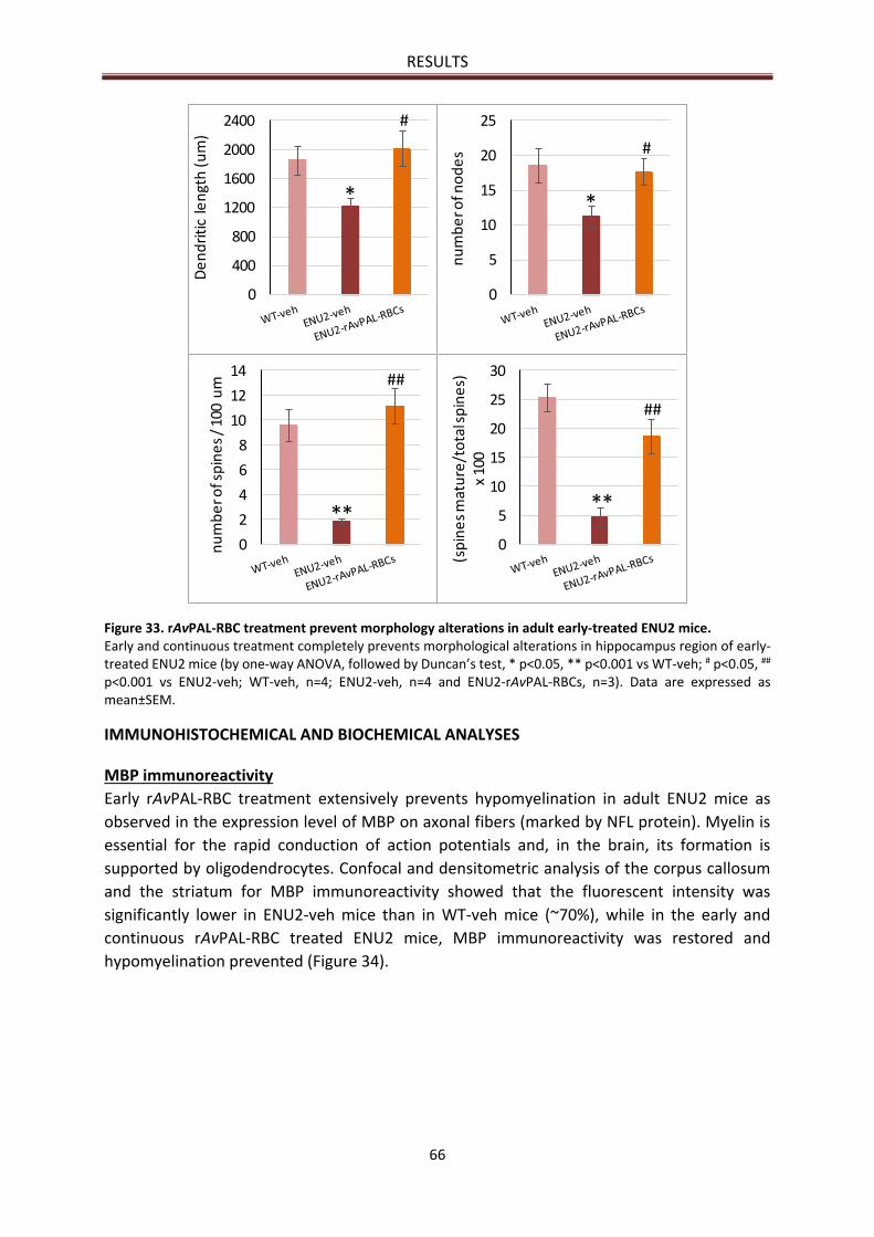
RESULTS
66
Figure 33. rAvPAL-RBC treatment prevent morphology alterations in adult early-treated ENU2 mice. Early and continuous treatment completely prevents morphological alterations in hippocampus region of early-treated ENU2 mice (by one-way ANOVA, followed by Duncan’s test, * p<0.05, ** p<0.001 vs WT-veh; # p<0.05, ##
p<0.001 vs ENU2-veh; WT-veh, n=4; ENU2-veh, n=4 and ENU2-rAvPAL-RBCs, n=3). Data are expressed as mean±SEM.
IMMUNOHISTOCHEMICAL AND BIOCHEMICAL ANALYSES
MBP immunoreactivity
Early rAvPAL-RBC treatment extensively prevents hypomyelination in adult ENU2 mice as
observed in the expression level of MBP on axonal fibers (marked by NFL protein). Myelin is
essential for the rapid conduction of action potentials and, in the brain, its formation is
supported by oligodendrocytes. Confocal and densitometric analysis of the corpus callosum
and the striatum for MBP immunoreactivity showed that the fluorescent intensity was
significantly lower in ENU2-veh mice than in WT-veh mice (~70%), while in the early and
continuous rAvPAL-RBC treated ENU2 mice, MBP immunoreactivity was restored and
hypomyelination prevented (Figure 34).
0
400
800
1200
1600
2000
2400
De
nd
riti
c le
ngt
h (
um
)
0
5
10
15
20
25
nu
mb
er o
f no
de
s
0
2
4
6
8
10
12
14
nu
mb
er o
f sp
ine
s /
100
um
0
5
10
15
20
25
30
(sp
ine
s m
atu
re/t
ota
l sp
ine
s)
x 10
0
**
** **
#
#
##
##
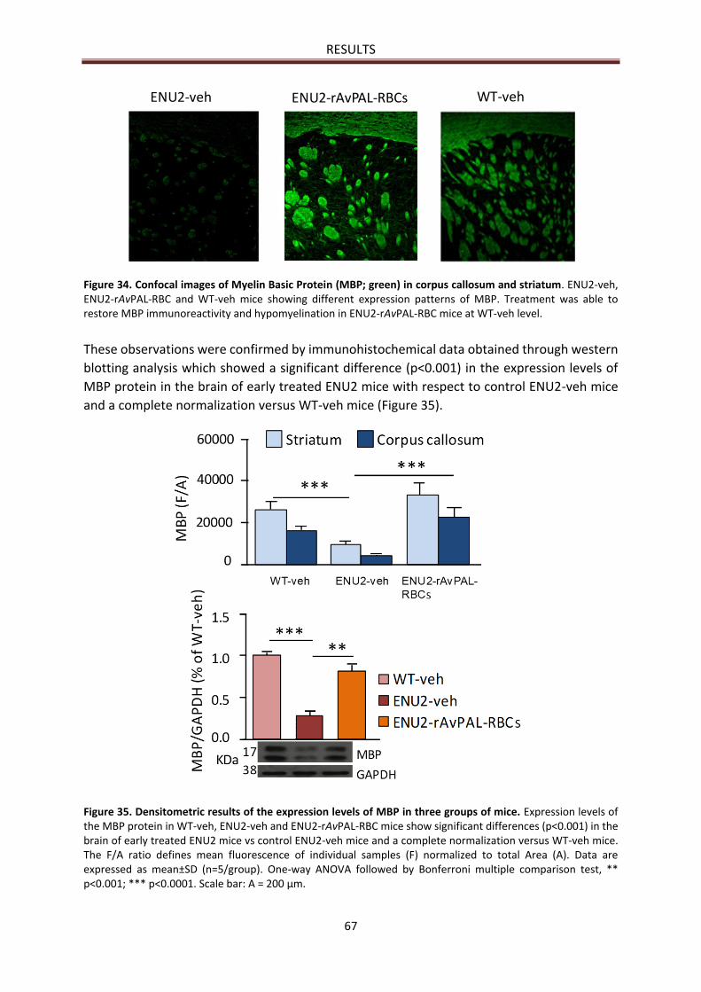
RESULTS
67
Figure 34. Confocal images of Myelin Basic Protein (MBP; green) in corpus callosum and striatum. ENU2-veh, ENU2-rAvPAL-RBC and WT-veh mice showing different expression patterns of MBP. Treatment was able to restore MBP immunoreactivity and hypomyelination in ENU2-rAvPAL-RBC mice at WT-veh level.
These observations were confirmed by immunohistochemical data obtained through western
blotting analysis which showed a significant difference (p<0.001) in the expression levels of
MBP protein in the brain of early treated ENU2 mice with respect to control ENU2-veh mice
and a complete normalization versus WT-veh mice (Figure 35).
Figure 35. Densitometric results of the expression levels of MBP in three groups of mice. Expression levels of the MBP protein in WT-veh, ENU2-veh and ENU2-rAvPAL-RBC mice show significant differences (p<0.001) in the brain of early treated ENU2 mice vs control ENU2-veh mice and a complete normalization versus WT-veh mice. The F/A ratio defines mean fluorescence of individual samples (F) normalized to total Area (A). Data are expressed as mean±SD (n=5/group). One-way ANOVA followed by Bonferroni multiple comparison test, ** p<0.001; *** p<0.0001. Scale bar: A = 200 μm.
sENU2-veh ENU2-rAvPAL-RBCs WT-veh
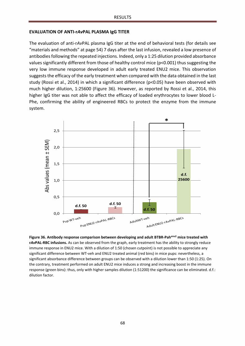
RESULTS
68
EVALUATION OF ANTI-rAvPAL PLASMA IgG TITER
The evaluation of anti-rAvPAL plasma IgG titer at the end of behavioral tests (for details see
“materials and methods” at page 54) 7 days after the last infusion, revealed a low presence of
antibodies following the repeated injections. Indeed, only a 1:25 dilution provided absorbance
values significantly different from those of healthy control mice (p<0.001) thus suggesting the
very low immune response developed in adult early treated ENU2 mice. This observation
suggests the efficacy of the early treatment when compared with the data obtained in the last
study (Rossi et al., 2014) in which a significant difference (p<0.05) have been observed with
much higher dilution, 1:25600 (Figure 36). However, as reported by Rossi et al., 2014, this
higher IgG titer was not able to affect the efficacy of loaded erythrocytes to lower blood L-
Phe, confirming the ability of engineered RBCs to protect the enzyme from the immune
system.
Figure 36. Antibody response comparison between developing and adult BTBR-Pahenu2 mice treated with
rAvPAL-RBC infusions. As can be observed from the graph, early treatment has the ability to strongly reduce
immune response in ENU2 mice. With a dilution of 1:50 (chosen cutpoint) is not possible to appreciate any
significant difference between WT-veh and ENU2 treated animal (red bins) in mice pups: nevertheless, a
significant absorbance difference between groups can be observed with a dilution lower than 1:50 (1:25). On
the contrary, treatment performed on adult ENU2 mice induces a strong and increasing boost in the immune
response (green bins): thus, only with higher samples dilution (1:51200) the significance can be eliminated. d.f.:
dilution factor.
d.f. 50d.f. 50
d.f.
25600
d.f. 500,0
0,5
1,0
1,5
2,0
2,5
Abs
valu
es (m
ean
±SE
M)
*
69
DISCUSSION
Phenylketonuria has been the first inborn error of metabolism, described for the first time in
1934 by Asbjørn Følling, to have been recognized as having a precise chemical etiology, i.e.
hyperphenylalaninemia and the presence of phenylpiruvic acid in the urine (Williams et al.,
2008). This genetic disease is due to a lack of activity of the hepatic enzyme phenylalanine
hydroxylase (PAH, EC 1.14.16.1), who catalyzes conversion of L-Phe to L-Tyr, which can then
be used to synthesize other biologically important molecules (such as neurotransmitters)
preventing, at the same time, toxic accumulation of L-Phe in body fluids. The majority of PAH
mutations, hampering its tetramerization, alters the enzymatic activity (Pey et al., 2003). The
severity of the clinical neurological manifestations characterizing untreated patients, the
widespread diffusion of the disease itself (with its broad phenotypic variety), as well as the
discovery of the etiologic agent have been altogether led to encouraging efforts in the field of
therapy research and development since the first years after PKU discovery in the mid-1930s.
Among the different intervention approaches that have been proposed over the years, a
severe L-Phe-restricted diet still represented today the golden standard of PKU treatments to
lower the blood levels of this aminoacid to the range of 120-360 µmol/L, as recommended by
the American College of Medical Genetics and Genomics (ACMG) guidelines (Vockley et al.,
2014). This approach, aimed at introducing low protein diet with only the amino acid amount
necessary for normal development and functioning of the organism, according to each
patient’s age and specific needs, brings with it numerous negative aspects for patients and
families: poor palatability of foods, high economic cost, possible nutritional deficits that
require adequate dietary supply, social discomfort. All these difficulties are responsible for the
reduced compliance that the patient disclose for this dietary approach, especially in
adolescence and adulthood where from 60 to 80% of the patients partially or totally
abandoned the treatment, with negative neurological consequences. The problems related to
a life-long selective reduction of L-Phe intake have led to the development of alternative
strategies aimed at maintaining physiological blood level of this aminoacid in a way as much
as possible independent from the diet. Among all the different forms of therapy that have
been explored, treatments currently employed in clinical use include the glycomacropeptides
(GMP), a medical food formulas able to overcome some problems related to the classical diet,
large neutral amino acids (LNAA) supplementation and treatment with catalytic cofactor (BH4)
in its pharmacological analog sapropterin dihydrochloride (Kuvan™). BH4 supplementation,
which has been extensively tested in clinical trials, reaching Phase IV in Europe
(ClinicalTrials.gov ID NCT01082328), have been proved to be effective only in patients with
the milder forms of the disease which still maintain a residual PAH activity; thus, for the more
severe forms of PKU, diet and diet-related treatments continue to be the only feasible choice
able to properly reduce blood L-Phe.
Particular interest has emerged for the enzyme replacement therapy with the aim to provide
a long-lasting enzymatic activity able to reduce the accumulation of toxic metabolite without
DISCUSSION
70
adverse events. In particular, this strategy may include both the restoration of the native form
of PAH and the use of an alternative enzyme. This last possibility involves the use of the non-
mammalian enzyme phenylalanine ammonia lyase (PAL, E.C. 4.3.1.24), which is not
structurally and catalytically complex, is physically stable and does not require a cofactor to
carry out its catalytic function (Sarkissian and Gámez, 2005) making this enzyme easier to
employ if compared to PAH. PAL converts L-Phe to trans-cinnamic acid with trace amounts of
ammonia without embryotoxic effects (Hoskins et al., 1984; Liu et al., 2002). Despite positive
results obtained in short-term preclinical studies in which PAL has been administered orally
(Safos and Chang, 1995) or subcutaneously (Sarkissian et al., 1999), some drawbacks were
observed. Among these, the more significant is the host immune reaction against the foreign
enzyme, due to the far phylogenetic distance between PAL’s origin organisms and mammals,
leading to its inactivation especially upon multiple administrations. Several strategies have
been investigated in order to improve enzyme stability and to mask antigenic epitopes to host
immune system. Between these, PAL engineering by site-directed mutagenesis to improve
stability (Wang et al., 2005, 2008) have ultimately led to a double mutant form of recombinant
PAL from the cyanobacterium Anabaena variabilis, which currently represents the best
performing available variant (Sarkissian et al., 2008; Wang et al., 2008). Moreover, PEGylation
process of this modified PAL has been under clinical investigation by the company Biomarin
Pharmaceutical Inc. (Novato, CA) which was engaged in a Phase III multi-center clinical
program, consisting of two studies, PRISM-1 and PRISM-2 (NCT01819727, NCT01889862),
aimed at evaluating the safety and long-term efficacy and tolerability of the rAvPAL-PEG
administration. Very recently, on May 2018, the FDA approved PalynziqTM (composed of
rAvPAL conjugated to PEG) in the USA for the treatment of PKU. Unfortunately, the presence
of PEG in the therapeutic enzyme induces several serious side effect including anaphylaxis
events that require the patients to have auto-injectable epinephrine.
Indeed, although PEG has been used to modify several therapeutic molecules (mostly
enzymes) thanks to its ability to attenuate the immune response against the therapeutic agent
and to improve pharmacodynamic stability (Delgado et al., 1992), the results of previous
Phase I clinical study with rAvPAL-PEG (NCT00634660) had already demonstrated the
production of specific antibodies against the PEG moiety itself, which can result in allergic
manifestations or even in more severe forms of intolerance in already sensitized patients
(Longo, 2014). As reported by many Authors (Shimizu et al., 2012; August et al., 2013; Ishida
and Kiwada, 2013; Schellekens, 2013; Longo et al., 2014; Petersen et al., 2014; Heinriksen et
al., 2015), the production of anti-PEG antibodies could alter the PEG-conjugate biodistribution
and bioavailability by acting on the complex clearance rate (Abu Lila et al., 2013). Because of
these problems, administration through an opportune delivery system could therefore
represent a valid and viable alternative to reduce the antibody response and at the same time
ensure adequate therapeutic efficacy.
There are several useful delivery system but particular interest has been paid to red blood
cells thanks to the best advantages that these cells offer such as biodegradability, non-toxicity
and immunogenicity, the easiness of obtaining them in elevated quantities and the high cell
volume that allows the loading of a relatively large amount of protein (Ihler et al., 1973; Rossi
DISCUSSION
71
et al., 2005; Hamidi et al., 2007; Pierigè et al., 2008; Muzykantov et al., 2010; Biagiotti et al.,
2011; Magnani et al., 2011; Zarrin et al., 2014), making RBCs the ideal carriers for enzymes.
Among the numerous methods proposed for loading proteins into the erythrocytes, hypotonic
dialysis appears the best one, allowing the preservation of biochemical and physiological
characteristics of these cells. Inside RBCs the enzyme is protect from premature inactivation
both by plasma proteases and by neutralizing antibodies, particularly when repeated
administrations are needed (Bax et al., 2000a/b, 2007; Domenech et al., 2011; Agrawal et al.,
2013; Levene et al., 2013). Indeed, a study of Rossi et al. (2014) demonstrated in vivo on adult
ENU2 mice that RBCs might act as effective delivery system for the enzyme rAvPAL protecting
the therapeutic molecule from the immune system and proteases degradation.
It is on these bases that our work recently published (Pascucci et al., 2018) collected more
evidence on the preclinical efficacy of a RBCs-mediated delivery system for the recombinant
enzyme AvPAL confirming the protecting action of these carriers also on developing mice.
The first aim of the present study is to optimize the timetable of rAvPAL-RBCs infusions in
order to improve the efficacy of the treatment in controlling blood L-Phe level. At the same
time, we aim to prove that early (starting at 15 post natal day) and continuous administration
through this carrier system, at 7 day-intervals, normalizing blood and brain L-Phe levels, could
be able to prevent the typically abnormalities found in untreated ENU2 mice: cognitive
impairment, brain neurotransmitter deficit and neuropathological abnormalities.
The trial was conducted using three groups of male ENU2 mice pups, genetic murine model of
PKU fully characterized in the past for both behavioral and biochemical phenotype (Puglisi-
Allegra et al., 2000; Cabib et al., 2003; Pascucci et al., 2008, 2009, 2012, 2013; Andolina et al.,
2011; Rossi et al., 2014; De Jaco et al., 2017): ENU2-rAvPAL-RBC mice (n=9), ENU2-veh mice
(n=7) and WT-veh mice (n=13). This mouse model helps to understand why an elevated level
of L-Phe damages brain functions and could lead to the development of alternative strategies
of therapy. Females mice were not used in the experiment because sexual difference in ENU2
mice has been recently reported regarding the levels of blood and brain L-Phe levels
(females>males), brain dopamine levels (females<males), and cumulative distance to the
visible platform at water maze testing (females>males) denoting a greater cognitive
impairment in females than in males (Winn et al., 2018). In addition, older studies (Broening
et al., 2001; Sarkissian et al., 2008) observed that gender effect on treatment efficacy may be
related to estrogen influence that potentially affects dosing regimen.
We evaluated the efficacy of 8 repeated intra-venus infusions (0.036±0.007 IU g/body weight)
of rAvPAL-RBCs to reduce blood L-Phe level at stable low concentration in male homozygous
ENU2 mice starting from PND 15 to PND 70 at 7 day-intervals. This time interval has been
chosen to optimize the data obtained in the previous study by Rossi et al. (2014) in order to
avoid the end-dose fluctuations of blood L-Phe levels due to the relatively short half-life of
mouse RBCs (6-11 days for loaded murine RBCs, slightly reduced in comparison with native
cells which last for 12-14 days) (Pierigè et al., 2017). Our delivery system results in a more
stable control of blood L-Phe concentration if compared with subcutaneous injections of
rAvPAL-PEG (Bell et al., 2017). Indeed, already at PND 25, blood L-Phe of ENU2-rAvPAL-RBC
mice leveled under 200 μM and remained stable until the end of the trial (PND 70) when L-
DISCUSSION
72
Phe levels were significantly lower than in WT-veh mice. These results were confirmed also
through biochemical analysis of brain L-Phe concentration and L-Phe/L-Tyr ratio which both
levels overlapped those found in WT-veh animals. However, brain L-Tyr level could not be
restored at a physiological value by the therapy, as observed in a study of Joseph and Dyer
(2003) following the supplementation of this aminoacid. A possible reason could lie in the
inability of PAL enzyme to synthesize L-Tyr and/or in the insufficient L-Tyr diet intake.
Preliminary IgG evaluation showed a very slight induction of anti-PAL IgG antibody production
compared with those observed after the administration of the PEGylated PAL (Bell et al., 2017)
or RBCs loaded with not PEGylated PAL in adult animals (Rossi et al., 2014), suggesting the
efficacy of the early treatment in preventing antibody production. However, these results will
be confirmed in future experiments through the evaluation of the immune response not only
at the end of the infusions but also at defined time intervals during the whole experimental
period. Additionally, no evidence of enzyme activity neutralization by increasing IgG titers
could be observed: even though we have analyzed only the last time point, the eight infusions
were all able to act on L-Phe level to the same extent, with no remarkable differences between
the last administrations and the first ones. These results confirm the validity of erythrocytes
as protecting delivery system from immune response as previously demonstrated by Rossi et
al. (2014) and, at same time, they suggest the significance of an early treatment: in fact, during
neonatal period the developing immune system is particularly susceptible to induction of
tolerance (Wu et al., 2011).
To evaluate if early treatment with rAvPAL-RBCs was able to ameliorate performances of adult
ENU2 mice, preventing motor and cognitive impairment typically found in untreated mice
(Cabib et al., 2003; Fiori et al., 2017), some behavioral tests were performed. Generally,
disturbances of spatial learning in adult rats are promoted by late (PND 11-20) but not early
(PND 1-10) pharmacological treatment (Broening et al., 2001). In fact, under early treatment
with rAvPAL-RBCs (PND 15), ENU2 mice developed a normal pattern of exploration of novel vs
familiar objects while elevated levels of L-Phe negatively affected recognition memory and
motor performance in ENU2-veh mice. On the other hand, however, the treatment does not
improve emotional reactivity and locomotors parameters. Indeed, both elevated pluze maze
test and open field test demonstrated that ENU2 mice receiving rAvPAL-RBC treatment exhibit
decrease in all locomotors (distance moved and velocity) and emotional (total entries and %
time in open arms) parameters when compared with healthy mice confirming a maintenance
of the anxiety profile typically found in these animals (Winn et al., 2018). These discrepancies
between the outcome of motor and non-motor functions could be related to the delay in the
beginning of the treatment (PND 15) with respect to the developmental window (PND 11-17)
of motor functions in BTBR mouse (Fiori et al., 2017). At the same time, as mentioned above,
the unimproved reduced levels of brain L-Tyr and DA should be responsible for these results.
All the brain areas analyzed of untreated ENU2 mouse are involved in the declines of biogenic
amines with a gradient such that 5-HT is the most and DA the least affected among the
analyzed amines (Puglisi-Allegra et al., 2000; Pascucci et al., 2008), and mpFC and Amy the
most affected among the cerebral areas (Pascucci et al., 2013). Several studies suggest two
hypotheses that cause brain amine deficiency in PKU. The first is that L-Phe induces the
DISCUSSION
73
inhibition of transport across the blood-brain barrier of the two substrates for dopamine and
serotonin synthesis, tyrosine and tryptophan respectively (Dyer et al., 1996; Pietz et al., 1999)
the second is the interference of L-Phe excess on the enzymes involved in the rate-limiting
steps for dopamine and serotonin synthesis, tryptophan hydroxylase (TPH) and Tyr
hydroxylase (TH) (Kaufman, 1985; Fitzpatrick, 2003).
While early and continuous rAvPAL-RBC treatment was able to restore normal levels of 5-HT
in all analyzed brain area, at value that overlapped with those of healthy control mice, it was
found unable to avoid DA alteration and only partially improved NE brain metabolism. The
cause of the impaired dopaminergic transmission, and related motor functions, remains to be
explicated because, as demonstrated by the present study and by Van Vliet et al. (2016), it
persists under normal blood L-Phe levels. On the contrary, the restoration of normal TH
activity by reduction of blood L-Phe levels have been demonstrated (Pascucci et al., 2012; Van
Vliet et al., 2016). We can also speculate that L-Tyr diet intake, since PAL enzyme is not able
to synthesize this amino acid, was not sufficient to normalize brain L-Tyr levels. Other possible
mechanisms such as a competitive effect of high L-Phe concentration at the level of neuronal
L-Tyr uptake (Fernstrom and Fernstrom, 2007) as well as a downregulation of TH protein in
dopaminergic neurons (Pascucci et al., 2012; Goldfinger et al., 2017) could account for the
impaired synthesis or turnover of DA (Pascucci et al., 2012; Harding et al., 2014).
The ability of rAvPAL-RBC treatment to avoid 5-HT depletion and partially restoration of NE,
prevents the emergence of the characteristic postnatal synaptogenetic alterations found in
untreated ENU2 mice (Andolina et al., 2011). In fact, biogenic amines function as
neurotransmitters but have also an important role in brain development (Herklenius and
Lagercrantz, 2001). In particular, the marked reduction and delay of the peak increase in brain
5-HT availability around week 3 of postnatal life (Pascucci et al., 2008), which occurs during a
critical period for brain maturation (Bennett-Clarke et al., 1994; Cases et al., 1996; Persico et
al., 2001), is responsible for uncorrected synapse formation, dendritic growth and remodeling,
axonal refinement and columnarization (Bhatt et al., 2009). In present study, ENU2-veh mice
present alterations which include: a relevant decrease in apical and basal dendritic length and
branching; a significant reduction in apical and basal dendritic spine density as well as in the
index of spine maturation, and a great decrease in apical and basal dendritic spines due to the
exposure to high levels of L-Phe. On the other end, early and continuous rAvPAL-RBC
treatment completely prevents these morphological alterations in all examined brain areas,
leading to a brain development similar to those of WT-veh mice and confirming a specific
pathogenic role of 5-HT depletion in BTBR mice during this specific developmental window.
This important role of 5-HT as regulator for functions related to brain development,
maintenance of dendritic spines and synaptic connectivity, has already been observed in a
study of Andolina et al. (2011) after supplementation of a precursor of 5-HT, 5-
hydroxytryptophan.
Another important result obtained after rAvPAL-RBC treatment was the preventing
hypomyelination in adult early-treated ENU2 mice that together with gliosis are the
predominant neuropathological findings in brain from untreated mice and patients with PKU
(Malamud, 1966; Dyer et al., 1996; Shefer et al., 2000). High L-Phe level is responsible for
DISCUSSION
74
altered oligodendrocyes ability to form myelin (Dyer et al., 1999) with a relevant reduction of
myelinated axons as observed in hypomyelinated areas of mutant animals (Shefer et al.,
2000). In rodents, proliferation and maturation of oligodendrocytes occur mainly during early
postnatal life (Baumann, 2001; Miller, 2002) and myelination is virtually complete by 30 days
of age (Norton and Poduslo, 1973). An important role is played by MBP that is essential for
the assembly of a mature and functioning myelin membrane (Nave and Werner, 2014) and,
as observed in this study through brain densitometry and western blotting analysis, after 8
repeated infusions of rAvPAL-RBCs its level was restored in brain of treated mice while it
remained severely reduced in untreated PKU mouse. Dysmyelination is responsible for
impaired cognitive and motor functions (Filley, 1998) since these two activities are both
dependent on the integrity of specific brain regions and on the tracts that connect these brain
structures. As demonstrated from previous studies (Cleary et al., 1995), a stricter dietary
control of L-Phe can partially reverse white matter alteration, suggesting that it might be
originated by a derangement of MBP or other axonal proteins (Joseph and Dyer, 2003).
75
CONCLUSION AND FUTURE PERSPECTIVES
We performed the first preclinical longitudinal study exploring and demonstrating that murine
RBCs loaded with recombinant AvPAL are able to act as bioreactors to decrease blood L-Phe
in BTBR-Pahenu2 mice, the most widely used animal model of human PKU and at the same time
we have demonstrated the efficacy of rAvPAL-RBC treatment in preventing the emergence of
clinical phenotype associated with untreated PKU. We have also confirmed the efficacy of RBC
membrane to protect the loaded enzyme from immunological inactivation and their ability to
induce tolerance when the treatment started early.
Considering that the usage of erythrocytes as vehicle has many noticeable advantages and
permits a stable biochemical control, a possible reduction of the frequency of treatment, a
lower cost of the treatment and the avoidance of PEGylation, the results reported in this paper
constitute the basis for the design of a clinical trial to prove safety and efficacy in PKU patients.
Besides, the therapy here proposed might take advantage of Red Cell Loader©, an
electromedical device specifically developed by EryDel S.p.A. for safe and reproducible RBCs
drug loading. This apparatus would be the ideal since it works in sterility and apirogenic
condition, which are a requirement for clinical study (Magnani et al., 1998; Mambrini et al.,
2017).
Our future objective is now to verify if tyrosine supplementation could have the ability to
ameliorate locomotor and emotional parameters and, at the same time, verify the ability of
early treatment to induce immune tolerance through the monitoring of the immune response
during the whole experimental period.
Thanks to the great potential of RBCs for enzymatic replacement therapy, many other
disorders involving enzyme deficiencies, which share with PKU a similar mechanism with
progressive blood accumulation of toxic compounds, could therefore benefit from the
therapeutic approach here described. In addition, the availability of the treatment would
therefore mean for all patients and their families a great improvement of everyday quality of
life if considering that such patients are usually bound to life-long therapy.
76
REFERENCES
AAP Newborn Screening Task Force. Newborn screening: a blueprint for the future. A call for a national agenda on state newborn screening programs. Pediatrics. 2000;106(Suppl):389-422.
Abu Lila AS, Kiwada H, Ishida T. The accelerated blood clearance (ABC) phenomenon: clinical challenge and approaches to manage. J Control Release. 2013;172(1):38-47.
Acosta PB, Yannicelli S, Singh R, Mofidi S, Steiner R, DeVincentis E, Jurecki E, Bernstein L, Gleason S, Chetty M, Rouse B. Nutrient intakes and physical growth of children with phenylketonuria undergoing nutrition therapy. J Am Diet Assoc. 2003;103(9):1167-1173.
Agrawal V, Woo JH, Borthakur G, Kantarjian H, Frankel AE. Red blood cell-encapsulated L-asparaginase: potential therapy of patients with asparagine synthetase deficient acute myeloid leukemia. Protein Pept Lett. 2013;20(4):392-402.
Ahring K, Bélanger-Quintana A, Dokoupil K, Gokmen Ozel H, Lammardo AM, MacDonald A, Motzfeldt K, Nowacka M, Robert M, van Rijn M. Dietary management practices in phenylketonuria across European centres. Clin Nutr. 2009;28(3):231-236.
Ahring K, Bélanger-Quintana A, Dokoupil K, Gokmen-Ozel H, Lammardo AM, MacDonald A, Motzfeldt K, Nowacka M, Robert M, van Rijn M. Blood phenylalanine control in phenylketonuria: a survey of 10 European centres. Eur J Clin Nutr. 2011;65:275-278.
Alconcel SNS, Baas AS, Maynard HD. FDA-approved poly(ethylene glycol)–protein conjugate drugs. Polym Chem. 2011;2:1442-1448.
Allen TM, Cullis PR. Drug delivery systems: entering and mainstream. Science. 2004;303:1818-1822.
Alvarez FJ, Jordán JA, Herráez A, Díez JC, Tejedor MC. Hypotonically loaded rat erythrocytes deliver encapsulated substances into peritoneal macrophages. J Biochem. 1998;123(2):233-239.
American Academy of Pediatrics: Committee on Genetics. Maternal phenylketonuria. Pediatrics. 2001;107:427-428.
Anastasoaie V, Kurzius L, Forbes P, Waisbren S. Stability of blood phenylalanine levels and IQ in children with phenylketonuria. Mol Genet Metab. 2008;95(1-2):17-20.
Andersen AE, Avins L. Lowering brain phenylalanine levels by giving other large neutral amino acids. A new experimental therapeutic approach to phenylketonuria. Arch Neurol. 1976;33:684-686.
Andersen OA, Flatmark T, Hough E. High resolution crystal structures of the catalytic domain of human phenylalanine hydroxylase in its catalytically active Fe(II) form and binary complex with tetrahydrobiopterin. J. Mol. Biol. 2001;314:279-291.
Andolina D, Conversi D, Cabib S, Trabalza A, Ventura R, Puglisi-Allegra S, Pascucci T. 5-Hydroxytryptophan during critical postnatal period improves cognitive performances and promotes dendritic spine maturation in genetic mouse model of phenylketonuria. Int J Neuropsychopharmacol. 2011;14(4):479-489.
Anson JG, Gilbert HJ, Oram JD, Minton NP. Complete nucleotide sequence of the Rhodosporidium toruloides gene coding for phenylalanine ammonia-lyase. Gene. 1987;58:189-199.
Antonelli A, Sfara C, Rahmer J, Gleich B, Borgert J, Magnani M. Red blood cells as carriers in magnetic particle imaging. Biomed Tech (Berl). 2013;58(6):517-525.
REFERENCES
77
Appert C, Logemann E, Hahlbrock K, Schmid J, Amrhein N. Structural and catalytic properties of the four phenylalanine ammonia-lyase isoenzymes from parsley (Petroselinum crispum Nym.). Eur J Biochem. 1994;225:491-499.
Arnold GL, Kramer BM, Kirby RS, Plumeau PB, Blakely EM, Sanger Cregan LS, Davidson PW. Factors affecting cognitive, motor, behavioral and executive functioning in children with phenylketonuria. Acta Paediatr. 1998;87(5):565-570.
Arnold GL, Vladutiu CJ, Kirby RS, Blakely EM, Deluca JM. Protein insufficiency and linear growth restriction in phenylketonuria. J Pediatr. 2002;141(2):243-246.
Asplin JR. Hyperoxaluric calcium nephrolithiasis. Endocrinol Metab Clin North Am. 2002;31(4):927-949.
August KJ, Miller WP, Dalton A, Shinnick S. Comparison of hypersensitivity reactions to PEG-asparaginase in children after intravenous and intramuscular administration. J Pediatr Hematol Oncol. 2013;35(7):e283-6.
Australian Society for Inborn Errors of Metabolism. PKU handbook. Dennison B (ed). Alexandra, Australia: Human Genetics Society of Australian, 2005.
Azen CG, Koch R, Friedman EG, Berlow S, Coldwell J, Krause W, Matalon R, McCabe E, O'Flynn M, Peterson R, et al. Intellectual development in 12-year-old children treated for phenylketonuria. Am J Dis Child. 1991;145:35-39.
Bai JL, Song F. Effect of gene mutation on the function and structure of phenylalanine hydroxylase. Int. J. Pediat. 2003;25:142-144.
Baumann N, Pham-Dinh D. Biology of oligodendrocyte and myelin in the mammalian central nervous system. Physiol Rev. 2001;81(2):871-927.
Bax BE, Bain MD, Fairbanks LD, Simmonds HA, Webster AD, Chalmers RA. Carrier erythrocyte entrapped adenosine deaminase therapy in adenosine deaminase deficiency. Adv Exp Med Biol. 2000;486:47-50. (b)
Bax BE, Bain MD, Fairbanks LD, Webster AD, Chalmers RA. In vitro and in vivo studies with human carrier erythrocytes loaded with polyethyleneglycol-conjugated and native adenosine deaminase. Br J Haematol. 2000;109(3):549-554. (a)
Bax BE, Bain MD, Fairbanks LD, Webster AD, Ind PW, Hershfield MS, Chalmers RA. A 9-yr evaluation of carrier erythrocyte encapsulated adenosine deaminase (ADA) therapy in a patient with adult-type ADA deficiency. Eur J Haematol. 2007;79(4):338-348.
Bax BE, Bain MD, Scarpelli M, Filosto M, Tonin P, Moran N. Clinical and biochemical improvements in a patient with MNGIE following enzyme replacement. Neurology. 2013;81(14):1269-1271.
Bax BE, Fairbanks LD, Bain MD, Simmonds HA, Chalmers RA. The entrapment of polyethylene glycol-bound adenosine deaminase (Pegademase) in human carrier erythrocytes. Biochem Soc Trans. 1996;24(3):442S.
Beck BB, Hoyer-Kuhn H, Göbel H, Habbig S, Hoppe B. Hyperoxaluria and systemic oxalosis: an update on current therapy and future directions. Expert Opin Investig Drugs. 2013;22(1):117-129.
Bélanger-Quintana A, Burlina A, Harding CO, Muntau AC. Up to date knowledge on different treatment strategies for phenylketonuria. Mol Genet Metab. 2011;104 Suppl:S19-25.
Belanger-Quintana A, Martínez-Pardo M. Physical development in patients with phenylketonuria on dietary treatment: a retrospective study. Mol Genet Metab. 2011;104(4):480-484.
REFERENCES
78
Bell SM, Wendt DJ, Zhang Y, Taylor TW, Long S, Tsuruda L, Zhao B, Laipis P, Fitzpatrick PA. Formulation and PEGylation optimization of the therapeutic PEGylated phenylalanine ammonia lyase for the treatment of phenylketonuria. PLoS One. 2017;12(3):e0173269.
Benichou B, Goyal S, Sung C, Norfleet AM, O'Brien F. A retrospective analysis of the potential impact of IgG antibodies to agalsidase beta on efficacy during enzyme replacement therapy for Fabry disease. Mol. Genet. Metab. 2009;96:4-12.
Bennett MJ, Schlunegger MP, Eisenberg D. 3D domain swapping: a mechanism for oligomer assembly. Protein Sci. 1995;4(12):2455-2468.
Bennett-Clarke CA, Leslie MJ, Lane RD, Rhoades RW. Effect of serotonin depletion on vibrissa-related patterns of thalamic afferents in the rat's somatosensory cortex. J Neurosci. 1994;14(12):7594-7607.
Bercovich D, Elimelech A, Zlotogora J, Korem S, Yardeni T, Gal N, Goldstein N, Vilensky B, Segev R, Avraham S, Loewenthal R, Schwartz G, AniksterY. Genotype-phenotype correlations analysis of mutations in the phenylalanine hydroxylase (PAH) gene. J Hum Genet. 2008;53(5):407-418.
Berger-Sweeney J, Hohmann CF. Behavioral consequences of abnormal cortical development: insights into developmental disabilities. Behav Brain Res. 1997;86(2):121-142.
Berlin CM Jr, Levy HL, Hanley WB. Delayed increase in blood phenylalanine concentration in phenylketonuric children initially classified as mild hyperphenylalaninemia. Screening. 1995;4:35-39.
Bernegger C, Blau N. High frequency of tetrahydrobiopterin-responsiveness among hyperphenylalaninemias: a study of 1,919 patients observed from 1988 to 2002. Mol Genet Metab. 2002;77(4):304-313.
Berry HK, Bofinger MK, Hunt MM, Phillips PJ, Guilfoile MB. Reduction of cerebrospinal fluid phenylalanine after oral administration of valine, isoleucine, and leucine. Pediatr Res. 1982;16:751-755.
Best JR, Miller PH. A developmental perspective on executive function. Child Dev. 2010;81(6):1641-1660.
Beutler E, Dale GL, Guinto DE, Kuhl W. Enzyme replacement therapy in Gaucher's disease: preliminary clinical trial of a new enzyme preparation. Proc Natl Acad Sci U S A. 1977;74(10):4620-4623.
Beutler E., Lichtman M.A. In: Coller BS, Kipps TJ (Eds.), Williams Hematology, Fifth Edition, McGraw-Hill, New York, 1995:349-425.
Bhateria M, Rachumallu R, Singh R, Bhatta RS. Erythrocytes-based synthetic delivery systems: transition from conventional to novel engineering strategies. Expert Opin Drug Deliv. 2014;11(8):1219-1236.
Bhatt DH, Zhang S, Gan WB. Dendritic spine dynamics. Annu Rev Physiol. 2009;71:261-282.
Biagiotti S, Paoletti MF, Fraternale A, Rossi L, Magnani M. Drug delivery by red blood cells. IUBMB Life. 2011;63(8):621-631.
Bickel H, Gerrad J, Hickmans EM. Influence of phenylalanine intake on phenylketonuria. Lancet. 1953;265(6790):812-813.
Bickel H. The effects of a phenylalanine-free and phenylalanine-poor diet in phenylpyruvic oligophrenis. Exp Med Surg. 1954;12(1):114-117.
Bik-Multanowski M, Didycz B, Mozrzymas R, Nowacka M, Kaluzny L, Cichy W, Schneiberg B, Amilkiewicz J, Bilar A, Gizewska M, Lange A, Starostecka E, Chrobot A, Wojcicka-Bartlomiejczyk BI, Milanowski A.
REFERENCES
79
Quality of life in noncompliant adults with phenylketonuria after resumption of the diet. J Inherit Metab Dis. 2008;31(Suppl 2):S415-418.
Bjørgo E, de Carvalho RM, Flatmark T. A comparison of kinetic and regulatory properties of the tetrameric and dimeric forms of wild-type and Thr427-->Pro mutant human phenylalanine hydroxylase: contribution of the flexible hinge region Asp425-Gln429 to the tetramerization and cooperative substrate binding. Eur J Biochem. 2001;268(4):997-1005.
Blaskovics ME. Phenylketonuria: loading studies revisited. In: Blau N (Ed.). PKU and BH4: Advances in Phenylketonuria and Tetrahydrobiopterin. SPS Verlagsgesellschaft, Heilbronn. 2006; 104-107.
Blau K. Determination of phenylalanine in filter paper blood spots by a simplified automated fluorimetric method without dialysis. Clin Chim Acta 1983;129:197-200.
Blau N, Bonafé L, Blaskovics ME. "Disorders of phenylalanine and tetrahydrobiopterin metabolism". In: Blau N, Duran M, Blaskovics ME, Gibson KM, eds. Physician's Guide to the Laboratory Diagnosis of Metabolic Diseases. 2nd ed. Heidelberg: Springer-Verlag, 2003: 89-106.
Blau N, Hennermann JB, Langenbeck U, Lichter-Konecki U. Diagnosis, classification, and genetics of phenylketonuria and tetrahydrobiopterin (BH4) deficiencies. Mol Genet Metab. 2011;104 Suppl:S2-9.
Blau N, Longo N. Alternative therapies to address the unmet medical needs of patients with phenylketonuria. Expert Opin Pharmacother. 2015;16(6):791-800.
Blau N, Muntau A. Tetrahydrobiopterin-Responsive Phenylalanine Hydroxylase Deficiency, 34th EMG Workshop Results, Friedrichsdorf, Milupa, 2002.
Blau N, Shen N, Carducci C. Molecular genetics and diagnosis of phenylketonuria: state of the art. Expert Rev Mol Diagn. 2014;14(6):655-671.
Blau N, Thöny B, Cotton RG, Hyland K. Disorders of tetrahydrobiopterin and related biogenic amines. In: Scriver CR, Beaudet AL, Sly WS, Valle D, Childs B, Kinzler K, Vogelstein B (Eds). The Metabolic and Molecular Bases of Inherited Disease. Eighth edition. New York, McGraw-Hill, 2001:1725-1776.
Blau N, Thöny B. Pterins and Related Enzymes. In: Blau N, Duran M, Gibson K, Michael R (Eds). Laboratory Guide to the Methods in Biochemical Genetics. Berlin Heidelberg, Springer, 2008(Chapter 6):665-701.
Blau N, van Spronsen FJ, Levy HL. Phenylketonuria. Lancet. 2010;376:1417-1427.
Blau N. Defining tetrahydrobiopterin (BH4)-responsiveness in PKU. J Inherit Metab Dis. 2008;31(1):2-3.
Boado RJ, Li JY, Nagaya M, Zhang C, Pardridge WM. Selective expression of the large neutral amino acid transporter at the blood-brain barrier. Proc Natl Acad Sci U S A. 1999;96(21):12079-12084.
Bone A, Kuehl AK, Angelino AF. A neuropsychiatric perspective of phenylketonuria I: overview of phenylketonuria and its neuropsychiatric sequelae. Psychosomatics. 2012;53(6):517-523.
Bossa F, Annese V, Valvano MR, Latiano A, Martino G, Rossi L, Magnani M, Palmieri O, Serafini S, Damonte G, De Santo E, Andriulli A. Erythrocytes-mediated delivery of dexamethasone 21-phosphate in steroid-dependent ulcerative colitis: a randomized, double-blind Sham-controlled study. Inflamm Bowel Dis. 2013;19(9):1872-1879.
Bourget L, Chang TM. Effects of oral administration of artificial cells immobilized phenylalanine ammonia-lyase on intestinal amino acids of phenylketonuric rats. Biomater Artif Cells Artif Organs. 1989;17:161-181.
REFERENCES
80
Bourget L, Chang TM. Phenylalanine ammonia-lyase immobilized in microcapsules for the depletion of phenylalanine in plasma in phenylketonuric rat model. Biochim Biophys Acta. 1986;883(3):432-438.
Bourget L, Chang TM. Phenylalanine ammonia-lyase immobilized in semipermeable microcapsules for enzyme replacement in phenylketonuria. FEBS Lett. 1985;180(1):5-8.
Bradford M.M. A rapid and sensitive method for the quantitation of microgram quantities of protein utilizing the principle of protein-dye binding. Anal. Biochem. 1976;72:248-254.
Broening HW, Morford LL, Inman-Wood SL, Fukumura M, Vorhees CV. 3,4-ethylenedioxymethamphetamine (ecstasy)-induced learning and memory impairments depend on the age of exposure during early development. J Neurosci. 2001 1;21(9):3228-3235.
Brumm VL, Bilder D, Waisbren SE. Psychiatric symptoms and disorders in phenylketonuria. Mol Genet Metab. 2010;99(Suppl 1):S59-63.
Burgard P. Development of intelligence in early treated phenylketonuria. Eur J Pediatr. 2000;159(Suppl 2):S74-79.
Burrage LC, McConnell J, Haesler R, O'Riordan MA, Sutton VR, Kerr DS, McCandless SE. High prevalence of overweight and obesity in females with phenylketonuria. Mol Genet Metab. 2012;107(1-2):43-48.
Cabib S, Pascucci T, Ventura R, Romano V, Puglisi-Allegra S. The behavioral profile of severe mental retardation in a genetic mouse model of phenylketonuria. Behav Gen. 2003;33(3):301-310.
Camp KM, Parisi MA, Acosta PB, Berry GT, Bilder DA, Blau N, Bodamer OA, Brosco JP, Brown CS, Burlina AB, Burton BK, Chang CS, Coates PM, Cunningham AC, Dobrowolski SF, Ferguson JH, Franklin TD, Frazier DM, Grange DK, Greene CL, Groft SC, Harding CO, Howell RR, Huntington KL, Hyatt-Knorr HD, Jevaji IP, Levy HL, Lichter-Konecki U, Lindegren ML, Lloyd-Puryear MA, Matalon K, MacDonald A, McPheeters ML, Mitchell JJ, Mofidi S, Moseley KD, Mueller CM, Mulberg AE, Nerurkar LS, Ogata BN, Pariser AR, Prasad S, Pridjian G, Rasmussen SA, Reddy UM, Rohr FJ, Singh RH, Sirrs SM, Stremer SE, Tagle DA, Thompson SM, Urv TK, Utz JR, van Spronsen F, Vockley J, Waisbren SE, Weglicki LS, White DA, Whitley CB, Wilfond BS, Yannicelli S, Young JM. Phenylketonuria Scientific Review Conference: state of the science and future research needs. Mol Genet Metab. 2014;112(2):87-122.
Cases O, Vitalis T, Seif I, De Maeyer E, Sotelo C, Gaspar P. Lack of barrels in the somatosensory cortex of monoamine oxidase A-deficient mice: role of a serotonin excess during the critical period. Neuron. 1996;16(2):297-307.
Casey L. Caring for children with phenylketonuria. Can Fam Physician. 2013;59(8):837-840.
Castillo M, Zafra MF, Garcia-Peregrin E. Inhibition of brain and liver 3-hydroxy-3-methylglutaryl-CoA reductase and mevalonate-5-pyrophosphatedecarboxylase in experimental hyperphenylalaninemia. Neurochem Res. 1988;13:551-555.
Castro M, Rossi L, Papadatou B, Bracci F, Knafelz D, Ambrosini MI, Calce A, Serafini S, Isacchi G, D'Orio F, Mambrini G, Magnani M. Long-term treatment with autologous red blood cells loaded with dexamethasone 21-phosphate in pediatric patients affected by steroid-dependent Crohn disease. J Pediatr Gastroenterol Nutr. 2007;44(4):423-426.
Chace DH, Kalas TA, Naylor EW. Use of tandem mass spectrometry for multianalyte screening of dried blood specimens from newborns. Clin Chem. 2003;49(11):1797-1817.
Chace DH, Millington DS, Terada N, Kahler SG, Roe CR, Hofman LF. Rapid diagnosis of phenylketonuria by quantitative analysis for phenylalanine and tyrosine in neonatal blood spots by tandem mass spectrometry. Clin Chem. 1993;39(1):66-71.
REFERENCES
81
Chace DH, Sherwin JE, Hillman SL, Lorey F, Cunningham GC. Use of phenylalanine-to-tyrosine ratio determined by tandem mass spectrometry to improve newborn screening for phenylketonuria of early discharge specimens collected in the first 24 hours. Clin Chem. 1998;44(12):2405-2409.
Chang TM, Bourget L, Lister C. A new theory of enterorecirculation of amino acids and its use for depleting unwanted amino acids using oral enzyme-artificial cells, as in removing phenylalanine in phenylketonuria. Artif. Cells Blood Substit. Immobil. Biotechnol. 1995;23:1-21.
Chiarantini L, Rossi L, Fraternale A, Magnani M. Modulated red blood cell survival by membrane protein clustering. Mol Cell Biochem. 1995;144(1):53-59.
Christensen HN, Streicher JA, Elbinger RL. Effects of feeding individual amino acids upon the distribution of other amino acids between cells and extracellular fluid. J Biol Chem. 1948;172:515-524.
Chugani DC, Muzik O, Behen M, Rothermel R, Janisse JJ, Lee J, Chugani HT. Developmental changes in brain serotonin synthesis capacity in autistic and nonautistic children. Ann Neurol. 1999;45(3):287-295.
Cleary M, Trefz F, Muntau AC, Feillet F, van Spronsen FJ, Burlina A, Bélanger-Quintana A, Giżewska M, Gasteyger C, Bettiol E, Blau N, MacDonald A. Fluctuations in phenylalanine concentrations in phenylketonuria: a review of possible relationships with outcomes. Mol Genet Metab. 2013;110(4):418-423.
Cleary MA, Walter JH, Wraith JE, White F, Tyler K, Jenkins JP. Magnetic resonance imaging in phenylketonuria: reversal of cerebral white matter change. J Pediatr. 1995;127(2):251-255.
ClinicalTrials.gov Colomé C, Artuch R, Vilaseca MA, Sierra C, Brandi N, Cambra FJ, Lambruschini N, Campistol J. Ubiquinone-10 content in lymphocytes of phenylketonuric patients. Clin Biochem. 2002;35(1):81-84.
Crone MR, van Spronsen FJ, Oudshoorn K, Bekhof J, van Rijn G, Verkerk PH. Behavioural factors related to metabolic control in patients with phenylketonuria. J Inherit Metab Dis. 2005;28:627-637.
Curtius HC, Niederwieser A, Viscontini M, Leimbacher W, Wegmann H, Blehova B, Rey F, Schaub J, Schmidt H. Serotonin and dopamine synthesis in phenylketonuria. Adv Exp Med Biol. 1981;133:277-291.
Dawson C, Murphy E, Maritz C, Chan H, Ellerton C, Carpenter RH, Lachmann RH. Dietary treatment of phenylketonuria: the effect of phenylalanine on reaction time. J Inherit Metab Dis. 2011;34(2):449-454.
de Duve C. From cytases to lysosomes. Fed Proc. 1964;23:1045-1049.
de Groot MJ, Hoeksma M, Blau N, Reijngoud DJ, van Spronsen FJ. Pathogenesis of cognitive dysfunction in phenylketonuria: review of hypotheses. Mol Genet Metab. 2010;99(Suppl 1):S86-89.
De Jaco A, Mango D, De Angelis F, Favaloro FL, Andolina D, Nisticò E, Fiori E, Colamartino M, Pascucci T. Unbalance between Excitation and Inhibition in Phenylketonuria, a Genetic Metabolic Disease Associated with Autism. Int J Mol Sci. 2017;18:941.
De Jong WH, Borm PJ. Drug delivery and nanoparticles: applications and hazards. Int J Nanomedicine. 2008;3(2):133-149.
Delgado C, Francis GE, Fisher D. The uses and properties of PEG-linked proteins. Crit Rev Ther Drug Carrier Syst. 1992;9(3-4):249-304.
Deloach J, Ihler G. A dialysis procedure for loading erythrocytes with enzymes and lipids. Biochim Biophys Acta. 1977;496(1):136-145.
REFERENCES
82
Demirev PA. Dried blood spots: analysis and applications. Anal Chem. 2013;85:779-789.
Demirkol M, Giżewska M, Giovannini M, Walter J. Follow up of phenylketonuria patients. Mol Genet Metab. 2011;104 Suppl:S31-39.
DeRoche K, Welsh M. Twenty-five years of research on neurocognitive outcomes in early-treated phenylketonuria: intelligence and executive function. Dev Neuropsychol. 2008;33(4):474-504.
Dhondt JL. Laboratory diagnosis of phenylketonuria. In: Blau N (Ed). PKU and BH4: Advances in Phenylketonuria and Tetrahydrobiopterin. SPS Verlagsgesellschaft Publications, Heilbronn, 2006:161-179.
Diamond A. Evidence for the importance of dopamine for prefrontal cortex functions early in life. Philos Trans R Soc Lond B Biol Sci. 1996;351(1346):1483-1493.
DiLella AG, Marvit J, Brayton K, Woo SL. An amino-acid substitution involved in phenylketonuria is in linkage disequilibrium with DNA haplotype 2. Nature. 1987;327(6120):333-336.
Ding Z, Harding CO, Rebuffat A, Elzaouk L, Wolff JA, Thöny B. Correction of murine PKU following AAV-mediated intramuscular expression of a complete phenylalanine hydroxylating system. Mol Ther. 2008;16:673-681.
Ding Z, Harding CO, Thöny B. State-of-the-art 2003 on PKU gene therapy. Mol Genet Metab. 2004;81:3-8.
Dobbelaere D, Michaud L, Debrabander A, Vanderbecken S, Gottrand F, Turck D, Farriaux JP. Evaluation of nutritional status and pathophysiology of growth retardation in patients with phenylketonuria. J Inherit Metab Dis. 2003;26(1):1-11.
Domenech C, Thomas X, Chabaud S, Baruchel A, Gueyffier F, Mazingue F, Auvrignon A, Corm S, Dombret H, Chevallier P, Galambrun C, Huguet F, Legrand F, Mechinaud F, Vey N, Philip I, Liens D, Godfrin Y, Rigal D, Bertrand Y. L-asparaginase loaded red blood cells in refractory or relapsing acute lymphoblastic leukaemia in children and adults: results of the GRASPALL 2005-01 randomized trial. Br J Haematol. 2011;153(1):58-65.
Donlon J, Levy HL, Scriver CR. Hyperphenylalanine: Phenylalanine Hydroxilase Deficiency. In: Scriver CR, Beaudet AL, Sly WS, Valle D (Eds). Online metabolic and molecular basis of inherited disease. New York, McGraw-Hill, 2010; Chapter 77.
Døskeland AP, Døskeland SO, Ogreid D, Flatmark T. The effect of ligands of phenylalanine 4-monooxygenase on the cAMP-dependent phosphorylation of the enzyme. J Biol Chem. 1984;259(18):11242-11248.
Døskeland AP, Martinez A, Knappskog PM, Flatmark T. Phosphorylation of recombinant human phenylalanine hydroxylase: effect on catalytic activity, substrate activation and protection against non-specific cleavage of the fusion protein by restriction protease. Biochem J. 1996;313(Pt 2):409-414
Doultani S, Turhan KN, Etzel MR. Whey protein isolate and glycomacropeptide recovery from whey using ion exchange chromatography. J Food Sci. 2003;68:1389-1395.
Driscoll KW, Hsia DY, Knox WE, Troll W. Detection by phenylalanine tolerance tests of heterozygous carriers of phenylketonuria. Nature. 1956;178(4544):1239-1240.
Dworniczak B, Kalaydjieva L, Pankoke S, Aulehla-Scholz C, Allen G, Horst J. Analysis of exon 7 of the human phenylalanine hydroxylase gene: a mutation hot spot? Hum Mutat. 1992;1(2):138-146.
Dyer CA, Kendler A, Philibotte T, Gardiner P, Cruz J, Levy HL. Evidence for central nervous system glial cell plasticity in phenylketonuria. J Neuropathol Exp Neurol. 1996;55(7):795-814.
REFERENCES
83
Dyer CA. Pathophysiology of phenylketonuria. Dev Disabil Res Rev. 1999;5:104-112.
Eastman JW, Sherwin JE, Wong R, Liao CL, Currier RJ, Lorey F, Cunningham G. Use of the phenylalanine:tyrosine ratio to test newborns for phenylketonuria in a large public health screening programme. J Med Screen. 2000;7(3):131-135.
Eisensmith RC, Woo SL. Gene therapy for phenylketonuria. Eur J Pediatr. 1996;155:S16-19.
Erlandsen H, Bjørgo E, Flatmark T, Stevens RC. Crystal structure and site-specific mutagenesis of pterin-bound human phenylalanine hydroxylase. Biochemistry. 2000;39:2208-2217.
Erlandsen H, Patch MG, Gamez A, Straub M, Stevens RC. Structural studies on phenylalanine hydroxylase and implications toward understanding and treating phenylketonuria. Pediatrics. 2003;112(6 Pt 2):1557-1565.
Erlandsen H, Pey AL, Gámez A, Pérez B, Desviat LR, Aguado C, Koch R, Surendran S, Tyring S, Matalon R, Scriver CR, Ugarte M, Martínez A, Stevens RC. Correction of kinetic and stability defects by tetrahydrobiopterin in phenylketonuria patients with certain phenylalanine hydroxylase mutations. Proc Natl Acad Sci U S A. 2004;101(48):16903-16908.
Erlandsen H, Stevens RC. The structural basis of phenylketonuria. Mol Genet Metab. 1999;68(2):103-125.
Etzel MR. Manufacture and use of dairy protein fractions. J Nutr. 2004;134:996S-1002S.
Feillet F, Agostoni C. Nutritional issues in treating phenylketonuria. J Inherit Metab Dis. 2010;33(6):659-664.
Feillet F, Clarke L, Meli C, Lipson M, Morris AA, Harmatz P, Mould DR, Green B, Dorenbaum A, Giovannini M, Foehr E; Sapropterin Research Group. Pharmacokinetics of sapropterin in patients with phenylketonuria. Clin Pharmacokinet. 2008;47(12):817-825.
Feillet F, van Spronsen FJ, MacDonald A, Trefz FK, Demirkol M, Giovannini M, Bélanger-Quintana A, Blau N. Challenges and pitfalls in the management of phenylketonuria. Pediatrics. 2010;126(2):333-341.
Fernstrom JD, Fernstrom MH. Tyrosine, phenylalanine, and catecholamine synthesis and function in the brain. J Nutr. 2007;137(6 Suppl 1):1539S-1547S.
Fiege B, Blau N. Assessment of tetrahydrobiopterin (BH4) responsiveness in phenylketonuria. J Pediatr. 2007;150(6):627-630.
Filley CM. The behavioral neurology of cerebral white matter. Neurology. 1998;50(6):1535-1540.
Fiori E, Oddi D, Ventura R, Colamartino M, Valzania A, D'Amato FR, Bruinenberg V, van der Zee E, Puglisi-Allegra S, Pascucci T. Early-onset behavioral and neurochemical deficits in the genetic mouse model of phenylketonuria. PLoS One. 2017;12(8):e0183430.
Fitzpatrick PF. Mechanism of aromatic amino acid hydroxylation. Biochemistry. 2003;42:14083-14091.
Fitzpatrick PF. Tetrahydropterin-dependent amino acid hydroxylases. Annu Rev Biochem. 1999;68:355-381.
Fitzpatrick PF. The aromatic amino acid hydroxylases. Adv Enzymol Relat Areas Mol Biol. 2000;74:235-294.
Følling A, Closs K. Über das Vorkommen von l-Phenylalanin in Harn und Blut bei Imbecillitas henylpyrouvica. Hoppe-Seyler’s Z Physiol Chem 1938;254:115-116.
REFERENCES
84
Følling A, Mohr OL, Ruud L. Oligophrenia phenylpyrouvica. A recessive syndrome in man. In: Skrifter, Det Norske Vitenskapsakademi i Oslo. I. Mat Naturv Klasse. 1944;13:1-48.
Følling A. Über Ausscheidung von Phenylbrenztraubensäure in den Harn als Stoffwechselanomalie in Verbindung mit Imbezillität. Hoppe-Seyler’s Z Physiol Chem. 1934;227:169-176.
Fusetti F, Erlandsen H, Flatmark T, Stevens RC. Structure of tetrameric human phenylalanine hydroxylase and its implications for phenylketonuria. J Biol Chem. 1998;273(27):16962-16967.
Gaberc-Porekar V, Zore I, Podobnik B, Menart V. Obstacles and pitfalls in the PEGylation of therapeutic proteins. Curr Opin Drug Discov Dev. 2008;11:242-250.
Gámez A, Sarkissian CN, Wang L, Kim W, Straub M, Patch MG, Chen L, Striepeke S, Fitzpatrick P, Lemontt JF, O'Neill C, Scriver CR, Stevens RC. Development of pegylated forms of recombinant Rhodosporidium toruloides phenylalanine ammonia-lyase for the treatment of classical phenylketonuria. Mol Ther. 2005;11(6):986-989.
Gámez A, Wang L, Sarkissian CN, Wendt D, Fitzpatrick P, Lemontt JF, Scriver CR, Stevens RC. Structure-based epitope and PEGylation sites mapping of phenylalanine ammonia-lyase for enzyme substitution treatment of phenylketonuria. Mol Genet Metab. 2007;91:325-334.
Gassió R, Campistol J, Vilaseca MA, Lambruschini N, Cambra FJ, Fusté E. Do adult patients with phenylketonuria improve their quality of life after introduction/resumption of a phenylalanine-restricted diet? Acta Paediatr. 2003;92(12):1474-1478.
Gerasimova NS, Steklova IV, Tuuminen T. Fluorometric method for phenylalanine microplate assay adapted for phenylketonuria screening. Clin Chem. 1989;35:2112-2115.
Gersting SW, Kemter KF, Staudigl M, Messing DD, Danecka MK, Lagler FB, Sommerhoff CP, Roscher AA, Muntau AC. Loss of function in phenylketonuria is caused by impaired molecular motions and conformational instability. Am J Hum Genet. 2008;83(1):5-17.
Gersting SW, Lagler FB, Eichinger A, Kemter KF, Danecka MK, Messing DD, Staudigl M, Domdey KA, Zsifkovits C, Fingerhut R, Glossmann H, Roscher AA, Muntau AC. Pahenu1 is a mouse model for tetrahydrobiopterin-responsive phenylalanine hydroxylase deficiency and promotes analysis of the pharmacological chaperone mechanism in vivo. Hum Mol Genet. 2010;19(10):2039-2049.
Ghozlan A, Varoquaux O, Abadie V. Is monoamine oxydase-B a modifying gene and phenylethylamine a harmful compound in phenylketonuria?. Mol Genet Metab. 2004;83:337-340.
Gilbert HJ, Jack GW. The effect of proteinases on phenylalanine ammonia-lyase from the yeast Rhodotorula glutinis. Biochem J. 1981;199:715-723.
Gilbert HJ, Tully M. Protection of phenylalanine ammonia-lyase from proteolytic attack. Biochem Biophys Res Commun. 1985;131(2):557-563.
Gjetting T, Petersen M, Guldberg P, Güttler F. In vitro expression of 34 naturally occurring mutant variants of phenylalanine hydroxylase: correlation with metabolic phenotypes and susceptibility toward protein aggregation. Mol Genet Metab. 2001;72(2):132-143.
Glaser EM, Van der Loos H. Analysis of thick brain sections by obverse-reverse computer microscopy: application of a new, high clarity Golgi-Nissl stain. J Neurosci Methods. 1981;4(2):117-125.
Godfrin Y, Bertrand Y. L-asparaginase introduced into erythrocytes for the treatment of leukemia (ALL). Oncol News. 2006;1:2-4.
Godfrin Y, inventor; ERYTECH Pharma, assignee. Lysis/resealing process and device for incorporating an active ingredient, in particular asparaginase or inositol hexaphosphate, in erythrocytes. Patent WO 2006016247;2006.
REFERENCES
85
Gogtay N, Giedd JN, Lusk L, Hayashi KM, Greenstein D, Vaituzis AC, Nugent TF 3rd, Herman DH, Clasen LS, Toga AW, Rapoport JL, Thompson PM. Dynamic mapping of human cortical development during childhood through early adulthood. Proc Natl Acad Sci U S A. 2004;101(21):8174-8179.
Goldfinger M, Zeile WL, Corado CR, O'Neill CA, Tsuruda LS, Laipis PJ, Cooper JD. Partial rescue of neuropathology in the murine model of PKU following administration of recombinant phenylalanine ammonia lyase (pegvaliase). Mol Genet Metab. 2017;122(1-2):33-35.
Goldman-Rakic PS, Brown RM. Postnatal development of monoamine content and synthesis in the cerebral cortex of rhesus monkeys. Brain Res. 1982;256(3):339-349.
Green R, Miller J, Crosby W. Enhancement of iron chelation by desferrioxamine entrapped in red blood cell ghosts. Blood. 1981;57(5):866-872.
Griffiths P, Demellweek C, Fay N, Robinson PH, Davidson DC. Wechsler subscale IQ and subtest profile in early treated phenylketonuria. Arch Dis Child. 2000;82:209-215.
Griffiths P, Paterson L, Harvie A. Neuropsychological effects of subsequent exposure to phenylalanine in adolescents and young adults with early-treated phenylketonuria. J Intellect Disabil Res. 1995;39:365-372.
Guldberg P, Güttler F. Mutations in the phenylalanine hydroxylase gene: methods for their characterization. Acta Paediatr Suppl. 1994;407:27-33.
Guldberg P, Rey F, Zschocke J, Romano V, François B, Michiels L, Ullrich K, Hoffmann GF, Burgard P, Schmidt H, Meli C, Riva E, Dianzani I, Ponzone A, Rey J, Güttler F. A European multicenter study of phenylalanine hydroxylase deficiency: classification of 105 mutations and a general system for genotype-based prediction of metabolic phenotype. Am J Hum Genet. 1998;63(1):71-79.
Guthrie R, Susi A. A Simple A simple phenylalanine method for detecting phenylketonuria in large populations of newborn infants. Pediatrics. 1963;32:318-343.
Gutiérrez Millán C, Sayalero Marinero ML, Zarzuelo Castañeda A, Lanao JM. Drug, enzyme and peptide delivery using erythrocytes as carriers. J Control Release. 2004;95(1):27-49.
Güttler F. Hyperphenylalaninemia: diagnosis and classification of the various types of phenylalanine hydroxylase deficiency in childhood. Acta Paediatr Scand Suppl. 1980;280:1-80.
Hamidi M, Zarrin A, Foroozesh M, Mohammadi-Samani S. Applications of carrier erythrocytes in delivery of biopharmaceuticals. J Control Release. 2007;118(2):145-160.
Hanley WB. Non-PKU mild hyperphenylalaninemia (MHP)--the dilemma. Mol Genet Metab. 2011;104(1-2):23-26.
Harding CO, Neff M, Wild K, Jones K, Elzaouk L, Thöny B, Milstien S. The fate of intravenously administered tetrahydrobiopterin and its implications for heterologous gene therapy of phenylketonuria. Mol Genet Metab. 2004;81:52-57.
Harding CO, Winn SR, Gibson KM, Arning E, Bottiglieri T, Grompe M. Pharmacologic inhibition of L-tyrosine degradation ameliorates cerebral dopamine deficiency in murine phenylketonuria (PKU). J Inherit Metab Dis. 2014;37(5):735-743.
Harding CO. New era in treatment for phenylketonuria: Pharmacologic therapy with sapropterin dihydrochloride. Biologics. 2010;4:231-236.
Harding CO. Progress toward cell-directed therapy for phenylketonuria. Clin Genet. 2008;74:97-104.
REFERENCES
86
Heintz C, Cotton RG, Blau N. Tetrahydrobiopterin, its mode of action on phenylalanine hydroxylase, and importance of genotypes for pharmacological therapy of phenylketonuria. Hum Mutat. 2013;34(7):927-936.
Hennermann JB, Loui A, Weber A, Monch E. Hyperphenylalaninemia in a premature infant with heterozygosity for phenylketonuria. J Perinat Med 2004;32:383-385.
Henriksen LT, Harila-Saari A, Ruud E, Abrahamsson J, Pruunsild K, Vaitkeviciene G, Jónsson ÓG, Schmiegelow K, Heyman M, Schrøder H, Albertsen BK; Nordic Society of Paediatric Haematology and Oncology (NOPHO) group. PEG-asparaginase allergy in children with acute lymphoblastic leukemia in the NOPHO ALL2008 protocol. Pediatr Blood Cancer. 2015;62(3):427-33.
Herlenius E, Lagercrantz H. Development of neurotransmitter systems during critical periods. Exp Neurol. 2004;190(Suppl 1):S8-21.
Herlenius E, Lagercrantz H. Neurotransmitters and neuromodulators during early human development. Early Hum Dev. 2001;65(1):21-37.
Hershfield MS. PEG-ADA replacement therapy for adenosine deaminase deficiency: an update after 8.5 years. Clin Immunol Immunopathol. 1995;76:228-232.
Hill MA, Marota JJ, Shiman R. Reaction of rat liver phenylalanine hydroxylase with fatty acid hydroperoxides. Characterization and mechanism. J Biol Chem. 1988;263:5646-5655.
Hoang L, Byck S, Prevost L, Scriver CR. PAH Mutation Analysis Consortium Database: a database for disease-producing and other allelic variation at the human PAH locus. Nucleic Acids Res. 1996;24(1):127-131.
Hoffman JF. On red blood cells, hemolysis and resealed ghosts. Adv Exp Med Biol. 1992;326:1-15.
Hoppe B, Beck BB, Milliner DS. The primary hyperoxalurias. Kidney Int. 2009;75(12):1264-1271.
Hörster F, Schwab MA, Sauer SW, Pietz J, Hoffmann GF, Okun JG, Kölker S, Kins S. Phenylalanine reduces synaptic density in mixed cortical cultures from mice. Pediatr Res. 2006;59:544-548.
Hoskins JA, Gray J. Phenylalanine ammonia lyase in the management of phenylketonuria: the relationship between ingested cinnamate and urinary hippurate in humans. Res Commun Chem Pathol Pharmacol. 1982;35:275-282.
Hoskins JA, Holliday SB, Greenway AM. The metabolism of cinnamic acid by healthy and phenylketonuric adults: a kinetic study. Biomed Mass Spectrom. 1984;11(6):296-300.
Hoskins JA, Jack G, Wade HE, Peiris RJ, Wright EC, Starr DJ, Stern J. Enzymatic control of phenylalanine intake in phenylketonuria. Lancet. 1980;1:392-324.
Hufton SE, Jennings IG, Cotton RG. Structure and function of the aromatic amino acid hydroxylases. Biochem J. 1995;311(Pt 2):353-366.
Huijbregts SC, Gassió R, Campistol J. Executive functioning in context: Relevance for treatment and monitoring of phenylketonuria. Mol Genet Metab. 2013;110 Suppl:S25-30.
Humphrey M, Nation J, Francis I, Boneh A. Effect of tetrahydrobiopterin on Phe/Tyr ratios and variation in Phe levels in tetrahydrobiopterin responsive PKU patients. Mol Genet Metab. 2011; 104(1-2):89-92.
Huttenlocher PR, Bonnier C. Effects of changes in the periphery on development of the corticospinal motor system in the rat. Brain Res Dev Brain Res. 1991;60(2):253-260.
Huttenlocher PR. Dendritic and synaptic pathology in mental retardation. Pediatr Neurol. 1991;7:79-85.
REFERENCES
87
Ihler GM, Glew RH, Schnure FW. Enzyme loading of erythrocytes. Proc Natl Acad Sci U S A. 1973;70(9):2663-2666.
Ihler GM, Tsang HC. Hypotonic hemolysis methods for entrapment of agents in resealed erythrocytes. Methods Enzymol. 1987;149:221-229.
Ihler MG, Chi-Wan Tsang H. In: Green R, Widder KJ (Eds.). Methods in Enzymology, vol. 149, Academic Press, San Diego, 1987:221-229.
Isabella VM, Ha BN, Castillo MJ, Lubkowicz DJ, Rowe SE, Millet YA, Anderson CL, Li N, Fisher AB, West KA, Reeder PJ, Momin MM, Bergeron CG, Guilmain SE, Miller PF, Kurtz CB, Falb D. Development of a synthetic live bacterial therapeutic for the human metabolic disease phenylketonuria. Nat Biotechnol. 2018; 36(9):857-864.
Ishida T, Kiwada H. Anti-polyethyleneglycol antibody response to PEGylated substances. Biol Pharm Bull. 2013;36(6):889-91.
Ito Y, Ogiso T, Iwaki M, Yoneda I, Okuda Y. In vitro stability of insulin-loaded erythrocytes after freezing storage. J Pharmacobiodyn. 1989;12(4):201-207.
Jaffe EK. New protein structures provide an updated understanding of phenylketonuria. Mol Genet Metab. 2017;121(4):289-296.
Jahja R, van Spronsen FJ, de Sonneville LMJ, van der Meere JJ, Bosch AM, Hollak CEM, Rubio-Gozalbo ME, Brouwers MCGJ, Hofstede FC, de Vries MC, Janssen MCH, van der Ploeg AT, Langendonk JG, Huijbregts SCJ. Social-cognitive functioning and social skills in patients with early treated phenylketonuria: a PKU-COBESO study. J Inherit Metab Dis. 2016;39(3):355-362.
Jenner DJ, Lewis DA, Pitt E, Offord RA. The effect of intravenous administration of corticosteroids encapsulated in intact erythrocytes on adyuvant arthritis in the rat. Br J Pharmacol. 1981;73:212-213.
Jennings IG, Kemp BE, Cotton RG. Localization of cofactor binding sites with monoclonal anti-idiotype antibodies: phenylalanine hydroxylase. Proc. Natl. Acad. Sci. U.S.A. 1991;88:5734-5738.
Jervis GA. Phenylpyruvic oligophrenia deficiency of phenylalanine-oxidizing system. Proc Soc Exp Biol Med. 1953;82(3):514-515.
Jervis GA. Studies on phenylpyruvic oligophrenia; the position of the metabolic error. J Biol Chem. 1947;169(3):651-656.
Jordan MK, Brunner RL, Hunt MM, Berry HK. Preliminary support for the oral administration of valine, isoleucine and leucine for phenylketonuria. Dev Med Child Neurol. 1985;27:33-39.
Joseph B, Dyer CA. Relationship between myelin production and dopamine synthesis in the PKU mouse brain. J Neurochem. 2003;86(3):615-626.
Jung SC, Park JW, Oh HJ, Choi JO, Seo KI, Park ES, Park HY. Protective effect of recombinant adeno-associated virus 2/8-mediated gene therapy from the maternal hyperphenylalaninemia in offsprings of a mouse model of phenylketonuria. J Korean Med Sci. 2008;23:877-883.
Kang TS, Wang L, Sarkissian CN, Gámez A, Scriver CR, Stevens RC. Converting an injectable protein therapeutic into an oral form: phenylalanine ammonia lyase for phenylketonuria. Mol Genet Metab. 2010;99:4-9.
Kappock TJ, Caradonna JP. Pterin-Dependent Amino Acid Hydroxylases. Chem Rev. 1996;96(7):2659-2756.
REFERENCES
88
Kappock TJ, Harkins PC, Friedenberg S, Caradonna JP. Spectroscopic and kinetic properties of unphosphorylated rat hepatic phenylalanine hydroxylase expressed in Escherichia coli. Comparison of resting and activated states. J Biol Chem. 1995;270(51):30532-30544.
Kaufman S. Phenylalanine 4-monooxygenase from rat liver. Methods Enzymol. 1987;142:3-17.
Kaufman S. Regulatory properties of phenylalanine, tyrosine and tryptophan hydroxylases. Biochem Soc Trans. 1985;13:433-436.
Kaufman S. The phenylalanine hydroxylating system. Adv Enzymol Relat Areas Mol Biol. 1993;67:77-264.
Kayaalp E., Treacy E., Waters P.J., Byck S., Nowacki P., Scriver C.R. Human phenylalanine hydroxylase mutations and hyperphenylalaninemia phenotypes: a metanalysis of genotype-phenotype correlations. Am J Hum Genet. 1997;61:1309-1317.
Kim W, Erlandsen H, Surendran S, Stevens RC, Gamez A, Michols-Matalon K, Tyring SK, Matalon R. Trends in Enzyme Therapy for Phenylketonuria. Mol Ther. 2004;10:220-224.
Klingberg T, Vaidya CJ, Gabrieli JD, Moseley ME, Hedehus M. Myelination and organization of the frontal white matter in children: a diffusion tensor MRI study. Neuroreport. 1999;10(13):2817-2821.
Knappskog PM, Flatmark T, Aarden JM, Haavik J, Martínez A. Structure/function relationships in human phenylalanine hydroxylase. Effect of terminal deletions on the oligomerization, activation and cooperativity of substrate binding to the enzyme. Eur J Biochem. 1996;242(3):813-821.
Knox WE. Retrospective study pf phenylketonuria:relation of phenylpyruvate excretion to plasma phenylalanine. PKU newsletter. 1970;2.
Koch R, Azen C, Friedman EG, Fishler K, Baumann-Frischling C, Lin T. Care of the adult with phenylketonuria. Eur J Pediatr. 1996;155(Suppl 1):S90-92.
Koch R, Hanley W, Levy H, Matalon R, Rouse B, Trefz F, Guttler F, Azen C, Friedman E, Platt L, de la Cruz F. Maternal phenylketonuria: an international study. Mol Genet Metab. 2000;71:233-239.
Konecki DS, Wang Y, Trefz FK, Lichter-Konecki U, Woo SL. Structural characterization of the 5' regions of the human phenylalanine hydroxylase gene. Biochemistry. 1992;31(35):8363-8368.
Kravtzoff R, Ropars C, Laguerre M, Muh JP, Chassaigne M. Erythrocytes as carriers for L-asparaginase. Methodological and mouse in-vivo studies. J Pharm Pharmacol. 1990;42(7):473-476.
Kure S, Hou DC, Ohura T, Iwamoto H, Suzuki S, Sugiyama N, Sakamoto O, Fujii K, Matsubara Y, Narisawa K. Tetrahydrobiopterin-responsive phenylalanine hydroxylase deficiency. J Pediatr. 1999;135(3):375-378.
Kwok SC, Ledley FD, DiLella AG, Robson KJ, Woo SL. Nucleotide sequence of a full-length complementary DNA clone and amino acid sequence of human phenylalanine hydroxylase. Biochemistry. 1985;24(3):556-561.
Kwon YM, Chung HS, Moon C, Yockman J, Park YJ, Gitlin SD, David AE, Yang VC. L-Asparaginase encapsulated intact erythrocytes for treatment of acute lymphoblastic leukemia (ALL). J Control Release. 2009;139(3):182-189.
Laclair CE, Ney DM, MacLeod EL, Etzel MR. Purification and use of glycomacropeptide for nutritional management of phenylketonuria. J Food Sci. 2009;74:199-206.
Lauder JM. Neurotransmitters as growth regulatory signals: role of receptors and second messengers. Trends Neurosci. 1993;16(6):233-240.
REFERENCES
89
Ledley FD, Grenett HE, DiLella AG, Kwok SC, Woo SL Gene transfer and expression of human phenylalanine hydroxylase. Science. 1985;228:77-79.
Lee PJ, Ridout D, Walter JH, Cockburn F. Maternal phenylketonuria: report from the United Kingdom Registry 1978-97. Arch Dis Child. 2005;90:143-146.
Lenke RR, Levy HL. Maternal phenylketonuria and hyperphenylalaninemia. An international survey of the outcome of untreated and treated pregnancies. N Engl J Med. 1980;303(21):1202-1208.
Leuzzi V, Rossi L, Gabucci C, Nardecchia F, Magnani M. Erythrocyte-mediated delivery of recombinant enzymes. J Inherit Metab Dis. 2016;39(4):519-530.
Levene M, Coleman DG, Kilpatrick HC, Fairbanks LD, Gangadharan B, Gasson C, Bax BE. Preclinical toxicity evaluation of erythrocyte-encapsulated thymidine phosphorylase in BALB/c mice and beagle dogs: an enzyme-replacement therapy for mitochondrial neurogastrointestinal encephalomyopathy. Toxicol Sci. 2013;131(1):311-324.
Levy HL, Ghavami M. Maternal phenylketonuria: a metabolic teratogen. Teratology. 1996;53(3):176-184.
Levy HL, Milanowski A, Chakrapani A, Cleary M, Lee P, Trefz FK, Whitley CB, Feillet F, Feigenbaum AS, Bebchuk JD, Christ-Schmidt H, Dorenbaum A; Sapropterin Research Group. Efficacy of sapropterin dihydrochloride (tetrahydrobiopterin, 6R-BH4) for reduction of phenylalanine concentration in patients with phenylketonuria: a phase III randomised placebo-controlled study. Lancet. 2007;370(9586):504-510.
Li J, Ilangovan U, Daubner SC, Hinck AP, Fitzpatrick PF. Direct evidence for a phenylalanine site in the regulatory domain of phenylalanine hydroxylase. Arch Biochem Biophys. 2011;505(2):250-255.
Lichter-Konecki U, Hipke CM, Konecki DS. Human phenylalanine hydroxylase gene expression in kidney and other nonhepatic tissues. Mol Genet Metab. 1999;67(4):308-316.
Lim K, van Calcar SC, Nelson KL, Gleason ST, Ney DM. Acceptable low-phenylalanine foods and beverages can be made with glycomacropeptide from cheese whey for individuals with PKU. Mol Genet Metab. 2007;92:176-178.
Liu J, Jia X, Zhang J, Xiang H, Hu W, Zhou Y. Study on a novel strategy to treatment of phenylketonuria. Artif Cells Blood Substit Immobil Biotechnol. 2002;30(4):243-257.
Lizano C, Pérez MT, Pinilla M. Mouse erythrocytes as carriers for coencapsulated alcohol and aldehyde dehydrogenase obtained by electroporation in vivo survival rate in circulation, organ distribution and ethanol degradation. Life Sci. 2001;68(17):2001-2016.
Longo N, Harding CO, Burton BK, Grange DK, Vockley J, Wasserstein M, Rice GM, Dorenbaum A, Neuenburg JK, Musson DG, Gu Z, Sile S. Single-dose, subcutaneous recombinant phenylalanine ammonia lyase conjugated with polyethylene glycol in adult patients with phenylketonuria: an open-label, multicentre, phase 1 dose-escalation trial. Lancet. 2014;384(9937):37-44.
Longo N. Disorders of biopterin metabolism. J Inherit Metab Dis. 2009;32(3):333-42. Erratum in: J Inherit Metab Dis. 2009;32(3):457.
Lou H. Large doses of tryptophan and tyrosine as potential therapeutic alternative to dietary phenylalanine restriction in phenylketonuria. Lancet. 1985;2:150-151.
Luciana M, Sullivan J, Nelson CA. Associations between phenylalanine-to-tyrosine ratios and performance on tests of neuropsychological function in adolescents treated early and continuously for phenylketonuria. Child Dev. 2001;72(6):1637-1652.
REFERENCES
90
Lykkelund C, Nielsen JB, Lou HC, Rasmussen V, Gerdes AM, Christensen E, Güttler F. Increased neurotransmitter biosynthesis in phenylketonuria induced by phenylalanine restriction or by supplementation of unrestricted diet with large amounts of tyrosine. Eur J Pediatr. 1988;148:238-245.
MacDonald A, Gokmen-Ozel H, van Rijn M, Burgard P. The reality of dietary compliance in the management of phenylketonuria. J Inherit Metab Dis. 2010;33:665-670.
MacDonald A, Rylance G, Davies P, Asplin D, Hall SK, Booth IW. Free use of fruits and vegetables in phenylketonuria. J Inherit Metab Dis. 2003;26(4):327-338.
MacDonald MJ, D'Cunha GB. A modern view of phenylalanine ammonia lyase. Biochem Cell Biol. 2007 Jun;85(3):273-82. Review. Erratum in: Biochem Cell Biol. 2007;85(6):759.
MacLeod EL, Gleason ST, van Calcar SC, Ney DM. Reassessment of phenylalanine tolerance in adults with phenylketonuria is needed as body mass changes. Mol Genet Metab. 2009;98(4):331-337.
Magnani M, Rossi L, Bianchi M, Fornaini G, Benatti U, Guida L, Zocchi E, De Flora A. Improved metabolic properties of hexokinase-overloaded human erythrocytes. Biochim Biophys Acta. 1988;972(1):1-8.
Magnani M, Rossi L, D'ascenzo M, Panzani I, Bigi L, Zanella A. Erythrocyte engineering for drug delivery and targeting. Biotechnol Appl Biochem. 1998;28 (Pt 1):1-6.
Magnani M, Rossi L. Approaches to erythrocyte-mediated drug delivery. Expert Opin Drug Deliv. 2014;11(5):677-87.
Magnani M, Serafini S, Fraternale A, Antonelli A, Biagiotti S, Pierigè F, Sfara C, Rossi L. Red Blood Cell-Based Delivery of Drugs and Nanomaterials for Therapeutic and Diagnostic Applications. In: Nalwa SH (ed). Encyclopedia of Nanoscience and Nanotechnology. American Scientific Publishers, 2011:309-354.
Malamud N. Neuropathology of phenylketonuria. J Neuropathol Exp Neurol. 1966;25(2):254-268.
Mambrini G, Mandolini M, Rossi L, Pierigè F, Capogrossi G, Salvati P, Serafini S, Benatti L, Magnani M. Ex vivo encapsulation of dexamethasone sodium phosphate into human autologous erythrocytes using fully automated biomedical equipment. Int J Pharm. 2017;517(1-2):175-184.
Marota JJ, Shiman R. Stoichiometric reduction of phenylalanine hydroxylase by its cofactor: a requirement for enzymatic activity. Biochemistry. 1984;23:1303-1311.
Martinez A, Knappskog PM, Olafsdottir S, Døskeland AP, Eiken HG, Svebak RM, Bozzini M, Apold J, Flatmark T. Expression of recombinant human phenylalanine hydroxylase as fusion protein in Escherichia coli circumvents proteolytic degradation by host cell proteases. Isolation and characterization of the wild-type enzyme. Biochem J. 1995;306 (Pt 2):589-597.
Martynyuk AE, Glushakov AV, Sumners C, Laipis PJ, Dennis DM, Seubert CN. Impaired glutamatergic synaptic transmission in the PKU brain. Mol Genet Metab. 2005;86:S34-42.
Matalon R, Michals K, Blau N, Rouse B. Hyperphenylalaninemia due to inherited deficiencies of tetrahydrobiopterin. Adv Pediatr. 1989;36:67-90.
Matalon R, Michals-Matalon K, Bhatia G, Burlina AB, Burlina AP, Braga C, Fiori L, Giovannini M, Grechanina E, Novikov P, Grady J, Tyring SK, Guttler F. Double blind placebo control trial of large neutral amino acids in treatment of PKU: effect on blood phenylalanine. J Inherit Metab Dis. 2007;30(2):153-158.
Matalon R, Michals-Matalon K, Bhatia G, Grechanina E, Novikov P, McDonald JD, Grady J, Tyring SK, Guttler F. Large neutral amino acids in the treatment of phenylketonuria (PKU). J Inherit Metab Dis. 2006;29:732-738.
REFERENCES
91
Matalon R, Surendran S, Matalon KM, Tyring S, Quast M, Jinga W, Ezell E, Szucs S. Future role of large neutral amino acids in transport of phenylalanine into the brain. Pediatrics. 2003;112(6 Pt 2):1570-1574.
Mazer C., Muneyyirci J., Taheny K., Raio N., Borella A., Whitaker-Azmitia P. Serotonin depletion during synaptogenesis leads to decreased synaptic density and learning deficits in the adult rat: a possible model of neurodevelopmental disorders with cognitive deficits. Brain. Res. 1997;760:68-73.
McDonald JD, Bode VC, Dove WF, Shedlovsky A. Pahhph-5: a mouse mutant deficient in phenylalanine hydroxylase. Proc Natl Acad Sci U S A. 1990;87(5):1965-1967.
McDonald JD, Charlton CK. Characterization of mutations at the mouse phenylalanine hydroxylase locus. Genomics. 1997;39(3):402-405.
McDonald JD. Postnatal growth in a mouse genetic model of classical phenylketonuria. Contemp Top Lab Anim Sci. 2000;39(6):54-56.
McKean CM. The effects of high phenylalanine concentrations on serotonin and catecholamine metabolism in the human brain. Brain Res. 1972;47(2):469-476.
Miller RH. Regulation of oligodendrocyte development in the vertebrate CNS. Prog Neurobiol. 2002;67(6):451-467.
Mitchell JJ, Trakadis YJ, Scriver CR. Phenylalanine hydroxylase deficiency. Genet Med. 2011;13(8):697-707.
Moffitt MC, Louie GV, Bowman ME, Pence J, Noel JP, Moore BS. Discovery of two cyanobacterial phenylalanine ammonia lyases: kinetic and structural characterization. Biochemistry. 2007;46(4):1004-1012.
Moran NF, Bain MD, Muqit MM, Bax BE. Carrier erythrocyte entrapped thymidine phosphorylase therapy for MNGIE. Neurology. 2008;71(9):686-688.
Morrissey L, Tiernan CA, Lambert D, O'Reilly E, Treacy EP. Hereditary metabolic diseases (HMDs) in adult practice in Ireland: a preliminary assessment. Ir J Med Sci. 2013;182(4):565-571.
Muzykantov VR. Drug delivery by red blood cells: vascular carriers designed bymother nature. Expert Opin Drug Deliv. 2010;7(4):403-427.
National Institutes of Health Consensus Development Panel. National Institutes of Health Consensus Development Conference Statement: phenylketonuria: screening and management, October 16-18, 2000. Pediatrics. 2001;108(4):972-982.
Nave KA, Werner HB. Myelination of the nervous system: mechanisms and functions. Annu Rev Cell Dev Biol. 2014;30:503-533.
Ney DM, Gleason ST, van Calcar SC, MacLeod EL, Nelson KL, Etzel MR, Rice GM, Wolff JA. Nutritional management of PKU with glycomacropeptide from cheese whey. J Inherit Metab Dis. 2009;32:32-39.
Norton WT, Poduslo SE. Myelination in rat brain: changes in myelin composition during brain maturation. J Neurochem. 1973;21(4):759-773.
Nucleotide database - National Center for Biotechnology Information, U.S. National Library of Medicine Ogawa S, Ichinose H. Effect of metals and phenylalanine on the activity of human tryptophan hydroxylase-2: comparison with that on tyrosine hydroxylase activity. Neurosci Lett. 2006;401(3):261-265.
REFERENCES
92
Oh HJ, Park ES, Kang S, Jo I, Jung SC. Long-term enzymatic and phenotypic correction in the phenylketonuria mouse model by adeno-associated virus vector-mediated gene transfer. Pediatr Res. 2004;56:278-284.
Olsson GM, Montgomery SM, Alm J. Family conditions and dietary control in phenylketonuria. J Inherit Metab Dis. 2007;30:708-715.
Onishi A, Liotta LJ, Benkovic SJ. Cloning and expression of Chromobacterium violaceum phenylalanine hydroxylase in Escherichia coli and comparison of amino acid sequence with mammalian aromatic amino acid hydroxylases. J Biol Chem. 1991;266:18454-18459.
Pardridge WM. Blood-brain barrier carrier-mediated transport and brain metabolism of amino acids. Neurochem Res. 1998;23(5):635-644.
Pascucci T, Andolina D, Mela IL, Conversi D, Latagliata C, Ventura R, Puglisi-Allegra S, Cabib S. 5-Hydroxytryptophan rescues serotonin response to stress in prefrontal cortex of hyperphenylalaninaemic mice. Int J Neuropsychopharmacol. 2009;12(8):1067-1079.
Pascucci T, Andolina D, Ventura R, Puglisi-Allegra S, Cabib S. Reduced availability of brain amines during critical phases of postnatal development in a genetic mouse model of cognitive delay. Brain Res. 2008;1217:232-238.
Pascucci T, Giacovazzo G, Andolina D, Accoto A, Fiori E, Ventura R, Orsini C, Conversi D, Carducci C, Leuzzi V, Puglisi-Allegra S. Behavioral and neurochemical characterization of new mouse model of hyperphenylalaninemia. PLoS One. 2013;8(12):e84697.
Pascucci T, Giacovazzo G, Andolina D, Conversi D, Cruciani F, Cabib S, Puglisi-Allegra S. In vivo catecholaminergic metabolism in the medial prefrontal cortex of ENU2 mice: an investigation of the cortical dopamine deficit in phenylketonuria. J Inherit Metab Dis. 2012;35(6):1001-1009.
Pascucci T, Rossi L, Colamartino M, Gabucci C, Carducci C, Valzania A, Sasso V, Bigini N, Pierigè F, Viscomi MT, Ventura R, Cabib S, Magnani M, Puglisi-Allegra S, Leuzzi V. A new therapy prevents intellectual disability in mouse with phenylketonuria. Mol Genet Metab. 2018;124(1):39-49.ù
Pascucci T, Ventura R, Puglisi-Allegra S, Cabib S. Deficits in brain serotonin synthesis in a genetic mouse model of phenylketonuria. Neuroreport. 2002;13(18):2561-2564.
Pecce R, Scolamiero E, Ingenito L, Parenti G, Ruoppolo M. Optimization of an HPLC method for phenylalanine and tyrosine quantization in dried blood spot. Clin Biochem. 2013;46(18):1892-1895.
Penrose L, Quastel JH. Metabolic studies in phenylketonuria. Biochem J. 1937;31(2):266-274.
Penrose LS. Phenylketonuria--a problem in eugenics. Ann Hum Genet. 1998;62(Pt 3):193-202.
Pérez B, Desviat LR, Gómez-Puertas P, Martínez A, Stevens RC, Ugarte M. Kinetic and stability analysis of PKU mutations identified in BH4-responsive patients. Mol Genet Metab. 2005;86(Suppl 1):S11-16.
Persico AM, Mengual E, Moessner R, Hall FS, Revay RS, Sora I, Arellano J, DeFelipe J, Gimenez-Amaya JM, Conciatori M, Marino R, Baldi A, Cabib S, Pascucci T, Uhl GR, Murphy DL, Lesch KP, Keller F. Barrel pattern formation requires serotonin uptake by thalamocortical afferents, and not vesicular monoamine release. J Neurosci. 2001;21(17):6862-6873.
Peters A, Kaiserman-Abramof IR. The small pyramidal neuron of the rat cerebral cortex. The synapses upon dendritic spines. Z Zellforsch Mikrosk Anat. 1969;100(4):487-506.
Petersen WC Jr, Clark D, Senn SL, Cash WT, Gillespie SE, McCracken CE, Keller FG, Lew G. Comparison of allergic reactions to intravenous and intramuscular pegaspargase in children with acute lymphoblastic leukemia. Pediatr Hematol Oncol. 2014;31(4):311-7.
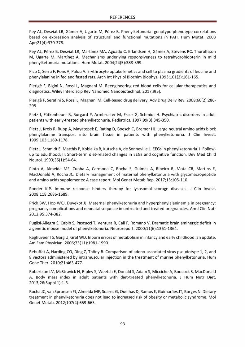
REFERENCES
93
Pey AL, Desviat LR, Gámez A, Ugarte M, Pérez B. Phenylketonuria: genotype-phenotype correlations based on expression analysis of structural and functional mutations in PAH. Hum Mutat. 2003 Apr;21(4):370-378.
Pey AL, Pérez B, Desviat LR, Martínez MA, Aguado C, Erlandsen H, Gámez A, Stevens RC, Thórólfsson M, Ugarte M, Martínez A. Mechanisms underlying responsiveness to tetrahydrobiopterin in mild phenylketonuria mutations. Hum Mutat. 2004;24(5):388-399.
Pico C, Serra F, Pons A, Palou A. Erythrocyte uptake kinetics and cell to plasma gradients of leucine and phenylalanine in fed and fasted rats. Arch Int Physiol Biochim Biophys. 1993;101(2):161-165.
Pierigè F, Bigini N, Rossi L, Magnani M. Reengineering red blood cells for cellular therapeutics and diagnostics. Wiley Interdiscip Rev Nanomed Nanobiotechnol. 2017;9(5).
Pierigè F, Serafini S, Rossi L, Magnani M. Cell-based drug delivery. Adv Drug Deliv Rev. 2008;60(2):286-295.
Pietz J, Fätkenheuer B, Burgard P, Armbruster M, Esser G, Schmidt H. Psychiatric disorders in adult patients with early-treated phenylketonuria. Pediatrics. 1997;99(3):345-350.
Pietz J, Kreis R, Rupp A, Mayatepek E, Rating D, Boesch C, Bremer HJ. Large neutral amino acids block phenylalanine transport into brain tissue in patients with phenylketonuria. J Clin Invest. 1999;103:1169-1178.
Pietz J, Schmidt E, Matthis P, Kobialka B, Kutscha A, de Sonneville L. EEGs in phenylketonuria. I: Follow-up to adulthood; II: Short-term diet-related changes in EEGs and cognitive function. Dev Med Child Neurol. 1993;35(1):54-64.
Pinto A, Almeida MF, Cunha A, Carmona C, Rocha S, Guimas A, Ribeiro R, Mota CR, Martins E, MacDonald A, Rocha JC. Dietary management of maternal phenylketonuria with glycomacropeptide and amino acids supplements: A case report. Mol Genet Metab Rep. 2017;13:105-110.
Ponder K.P. Immune response hinders therapy for lysosomal storage diseases. J Clin Invest. 2008;118:2686-1689.
Prick BW, Hop WCJ, Duvekot JJ. Maternal phenylketonuria and hyperphenylalaninemia in pregnancy: pregnancy complications and neonatal sequelae in untreated and treated pregnancies. Am J Clin Nutr 2012;95:374-382.
Puglisi-Allegra S, Cabib S, Pascucci T, Ventura R, Cali F, Romano V. Dramatic brain aminergic deficit in a genetic mouse model of phenylketonuria. Neuroreport. 2000;11(6):1361-1364.
Raghuveer TS, Garg U, Graf WD. Inborn errors of metabolism in infancy and early childhood: an update. Am Fam Physician. 2006;73(11):1981-1990.
Rebuffat A, Harding CO, Ding Z, Thöny B. Comparison of adeno-associated virus pseudotype 1, 2, and 8 vectors administered by intramuscular injection in the treatment of murine phenylketonuria. Hum Gene Ther. 2010;21:463-477.
Robertson LV, McStravick N, Ripley S, Weetch E, Donald S, Adam S, Micciche A, Boocock S, MacDonald A. Body mass index in adult patients with diet-treated phenylketonuria. J Hum Nutr Diet. 2013;26(Suppl 1):1-6.
Rocha JC, van Spronsen FJ, Almeida MF, Soares G, Quelhas D, Ramos E, Guimarães JT, Borges N. Dietary treatment in phenylketonuria does not lead to increased risk of obesity or metabolic syndrome. Mol Genet Metab. 2012;107(4):659-663.
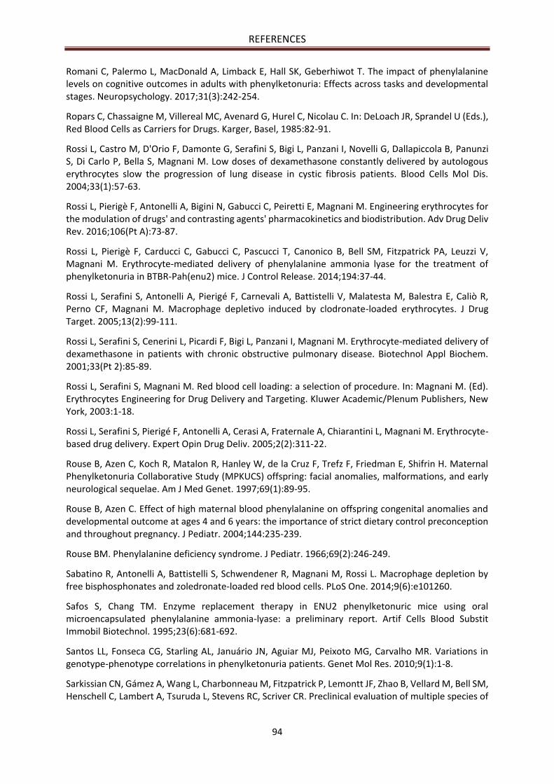
REFERENCES
94
Romani C, Palermo L, MacDonald A, Limback E, Hall SK, Geberhiwot T. The impact of phenylalanine levels on cognitive outcomes in adults with phenylketonuria: Effects across tasks and developmental stages. Neuropsychology. 2017;31(3):242-254.
Ropars C, Chassaigne M, Villereal MC, Avenard G, Hurel C, Nicolau C. In: DeLoach JR, Sprandel U (Eds.), Red Blood Cells as Carriers for Drugs. Karger, Basel, 1985:82-91.
Rossi L, Castro M, D'Orio F, Damonte G, Serafini S, Bigi L, Panzani I, Novelli G, Dallapiccola B, Panunzi S, Di Carlo P, Bella S, Magnani M. Low doses of dexamethasone constantly delivered by autologous erythrocytes slow the progression of lung disease in cystic fibrosis patients. Blood Cells Mol Dis. 2004;33(1):57-63.
Rossi L, Pierigè F, Antonelli A, Bigini N, Gabucci C, Peiretti E, Magnani M. Engineering erythrocytes for the modulation of drugs' and contrasting agents' pharmacokinetics and biodistribution. Adv Drug Deliv Rev. 2016;106(Pt A):73-87.
Rossi L, Pierigè F, Carducci C, Gabucci C, Pascucci T, Canonico B, Bell SM, Fitzpatrick PA, Leuzzi V, Magnani M. Erythrocyte-mediated delivery of phenylalanine ammonia lyase for the treatment of phenylketonuria in BTBR-Pah(enu2) mice. J Control Release. 2014;194:37-44.
Rossi L, Serafini S, Antonelli A, Pierigé F, Carnevali A, Battistelli V, Malatesta M, Balestra E, Caliò R, Perno CF, Magnani M. Macrophage depletivo induced by clodronate-loaded erythrocytes. J Drug Target. 2005;13(2):99-111.
Rossi L, Serafini S, Cenerini L, Picardi F, Bigi L, Panzani I, Magnani M. Erythrocyte-mediated delivery of dexamethasone in patients with chronic obstructive pulmonary disease. Biotechnol Appl Biochem. 2001;33(Pt 2):85-89.
Rossi L, Serafini S, Magnani M. Red blood cell loading: a selection of procedure. In: Magnani M. (Ed). Erythrocytes Engineering for Drug Delivery and Targeting. Kluwer Academic/Plenum Publishers, New York, 2003:1-18.
Rossi L, Serafini S, Pierigé F, Antonelli A, Cerasi A, Fraternale A, Chiarantini L, Magnani M. Erythrocyte-based drug delivery. Expert Opin Drug Deliv. 2005;2(2):311-22.
Rouse B, Azen C, Koch R, Matalon R, Hanley W, de la Cruz F, Trefz F, Friedman E, Shifrin H. Maternal Phenylketonuria Collaborative Study (MPKUCS) offspring: facial anomalies, malformations, and early neurological sequelae. Am J Med Genet. 1997;69(1):89-95.
Rouse B, Azen C. Effect of high maternal blood phenylalanine on offspring congenital anomalies and developmental outcome at ages 4 and 6 years: the importance of strict dietary control preconception and throughout pregnancy. J Pediatr. 2004;144:235-239.
Rouse BM. Phenylalanine deficiency syndrome. J Pediatr. 1966;69(2):246-249.
Sabatino R, Antonelli A, Battistelli S, Schwendener R, Magnani M, Rossi L. Macrophage depletion by free bisphosphonates and zoledronate-loaded red blood cells. PLoS One. 2014;9(6):e101260.
Safos S, Chang TM. Enzyme replacement therapy in ENU2 phenylketonuric mice using oral microencapsulated phenylalanine ammonia-lyase: a preliminary report. Artif Cells Blood Substit Immobil Biotechnol. 1995;23(6):681-692.
Santos LL, Fonseca CG, Starling AL, Januário JN, Aguiar MJ, Peixoto MG, Carvalho MR. Variations in genotype-phenotype correlations in phenylketonuria patients. Genet Mol Res. 2010;9(1):1-8.
Sarkissian CN, Gámez A, Wang L, Charbonneau M, Fitzpatrick P, Lemontt JF, Zhao B, Vellard M, Bell SM, Henschell C, Lambert A, Tsuruda L, Stevens RC, Scriver CR. Preclinical evaluation of multiple species of
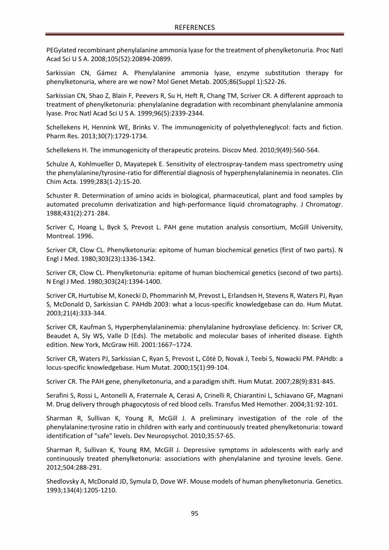
REFERENCES
95
PEGylated recombinant phenylalanine ammonia lyase for the treatment of phenylketonuria. Proc Natl Acad Sci U S A. 2008;105(52):20894-20899.
Sarkissian CN, Gámez A. Phenylalanine ammonia lyase, enzyme substitution therapy for phenylketonuria, where are we now? Mol Genet Metab. 2005;86(Suppl 1):S22-26.
Sarkissian CN, Shao Z, Blain F, Peevers R, Su H, Heft R, Chang TM, Scriver CR. A different approach to treatment of phenylketonuria: phenylalanine degradation with recombinant phenylalanine ammonia lyase. Proc Natl Acad Sci U S A. 1999;96(5):2339-2344.
Schellekens H, Hennink WE, Brinks V. The immunogenicity of polyethyleneglycol: facts and fiction. Pharm Res. 2013;30(7):1729-1734.
Schellekens H. The immunogenicity of therapeutic proteins. Discov Med. 2010;9(49):560-564.
Schulze A, Kohlmueller D, Mayatepek E. Sensitivity of electrospray-tandem mass spectrometry using the phenylalanine/tyrosine-ratio for differential diagnosis of hyperphenylalaninemia in neonates. Clin Chim Acta. 1999;283(1-2):15-20.
Schuster R. Determination of amino acids in biological, pharmaceutical, plant and food samples by automated precolumn derivatization and high-performance liquid chromatography. J Chromatogr. 1988;431(2):271-284.
Scriver C, Hoang L, Byck S, Prevost L. PAH gene mutation analysis consortium, McGill University, Montreal. 1996.
Scriver CR, Clow CL. Phenylketonuria: epitome of human biochemical genetics (first of two parts). N Engl J Med. 1980;303(23):1336-1342.
Scriver CR, Clow CL. Phenylketonuria: epitome of human biochemical genetics (second of two parts). N Engl J Med. 1980;303(24):1394-1400.
Scriver CR, Hurtubise M, Konecki D, Phommarinh M, Prevost L, Erlandsen H, Stevens R, Waters PJ, Ryan S, McDonald D, Sarkissian C. PAHdb 2003: what a locus-specific knowledgebase can do. Hum Mutat. 2003;21(4):333-344.
Scriver CR, Kaufman S, Hyperphenylalaninemia: phenylalanine hydroxylase deficiency. In: Scriver CR, Beaudet A, Sly WS, Valle D (Eds). The metabolic and molecular bases of inherited disease. Eighth edition. New York, McGraw Hill. 2001:1667–1724.
Scriver CR, Waters PJ, Sarkissian C, Ryan S, Prevost L, Côté D, Novak J, Teebi S, Nowacki PM. PAHdb: a locus-specific knowledgebase. Hum Mutat. 2000;15(1):99-104.
Scriver CR. The PAH gene, phenylketonuria, and a paradigm shift. Hum Mutat. 2007;28(9):831-845.
Serafini S, Rossi L, Antonelli A, Fraternale A, Cerasi A, Crinelli R, Chiarantini L, Schiavano GF, Magnani M. Drug delivery through phagocytosis of red blood cells. Transfus Med Hemother. 2004;31:92-101.
Sharman R, Sullivan K, Young R, McGill J. A preliminary investigation of the role of the phenylalanine:tyrosine ratio in children with early and continuously treated phenylketonuria: toward identification of "safe" levels. Dev Neuropsychol. 2010;35:57-65.
Sharman R, Sullivan K, Young RM, McGill J. Depressive symptoms in adolescents with early and continuously treated phenylketonuria: associations with phenylalanine and tyrosine levels. Gene. 2012;504:288-291.
Shedlovsky A, McDonald JD, Symula D, Dove WF. Mouse models of human phenylketonuria. Genetics. 1993;134(4):1205-1210.
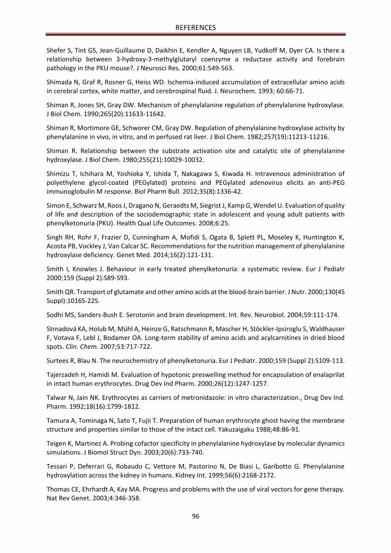
REFERENCES
96
Shefer S, Tint GS, Jean-Guillaume D, Daikhin E, Kendler A, Nguyen LB, Yudkoff M, Dyer CA. Is there a relationship between 3-hydroxy-3-methylglutaryl coenzyme a reductase activity and forebrain pathology in the PKU mouse?. J Neurosci Res. 2000;61:549-563.
Shimada N, Graf R, Rosner G, Heiss WD. Ischemia-induced accumulation of extracellular amino acids in cerebral cortex, white matter, and cerebrospinal fluid. J. Neurochem. 1993; 60:66-71.
Shiman R, Jones SH, Gray DW. Mechanism of phenylalanine regulation of phenylalanine hydroxylase. J Biol Chem. 1990;265(20):11633-11642.
Shiman R, Mortimore GE, Schworer CM, Gray DW. Regulation of phenylalanine hydroxylase activity by phenylalanine in vivo, in vitro, and in perfused rat liver. J Biol Chem. 1982;257(19):11213-11216.
Shiman R. Relationship between the substrate activation site and catalytic site of phenylalanine hydroxylase. J Biol Chem. 1980;255(21):10029-10032.
Shimizu T, Ichihara M, Yoshioka Y, Ishida T, Nakagawa S, Kiwada H. Intravenous administration of polyethylene glycol-coated (PEGylated) proteins and PEGylated adenovirus elicits an anti-PEG immunoglobulin M response. Biol Pharm Bull. 2012;35(8):1336-42.
Simon E, Schwarz M, Roos J, Dragano N, Geraedts M, Siegrist J, Kamp G, Wendel U. Evaluation of quality of life and description of the sociodemographic state in adolescent and young adult patients with phenylketonuria (PKU). Health Qual Life Outcomes. 2008;6:25.
Singh RH, Rohr F, Frazier D, Cunningham A, Mofidi S, Ogata B, Splett PL, Moseley K, Huntington K, Acosta PB, Vockley J, Van Calcar SC. Recommendations for the nutrition management of phenylalanine hydroxylase deficiency. Genet Med. 2014;16(2):121-131.
Smith I, Knowles J. Behaviour in early treated phenylketonuria: a systematic review. Eur J Pediatr 2000;159 (Suppl 2):S89-S93.
Smith QR. Transport of glutamate and other amino acids at the blood-brain barrier. J Nutr. 2000;130(4S Suppl):1016S-22S.
Sodhi MS, Sanders-Bush E. Serotonin and brain development. Int. Rev. Neurobiol. 2004;59:111-174.
Strnadová KA, Holub M, Mühl A, Heinze G, Ratschmann R, Mascher H, Stöckler-Ipsiroglu S, Waldhauser F, Votava F, Lebl J, Bodamer OA. Long-term stability of amino acids and acylcarnitines in dried blood spots. Clin. Chem. 2007;53:717-722.
Surtees R, Blau N. The neurochemistry of phenylketonuria. Eur J Pediatr. 2000;159 (Suppl 2):S109-113.
Tajerzadeh H, Hamidi M. Evaluation of hypotonic preswelling method for encapsulation of enalaprilat in intact human erythrocytes. Drug Dev Ind Pharm. 2000;26(12):1247-1257.
Talwar N, Jain NK. Erythrocytes as carriers of metronidazole: in vitro characterization., Drug Dev Ind. Pharm. 1992;18(16):1799-1812.
Tamura A, Tominaga N, Sato T, Fujii T. Preparation of human erythrocyte ghost having the membrane structure and properties similar to those of the intact cell. Yakuzaigaku 1988;48:86-91.
Teigen K, Martinez A. Probing cofactor specificity in phenylalanine hydroxylase by molecular dynamics simulations. J Biomol Struct Dyn. 2003;20(6):733-740.
Tessari P, Deferrari G, Robaudo C, Vettore M, Pastorino N, De Biasi L, Garibotto G. Phenylalanine hydroxylation across the kidney in humans. Kidney Int. 1999;56(6):2168-2172.
Thomas CE, Ehrhardt A, Kay MA. Progress and problems with the use of viral vectors for gene therapy. Nat Rev Genet. 2003;4:346-358.
REFERENCES
97
Thomas SA, Matsumoto AM, Palmiter RD. Noradrenaline is essential for mouse fetal development. Nature. 1995;374(6523):643-646.
Thöny B, Blau N. Mutations in the BH4-metabolizing genes GTP cyclohydrolase I, 6-pyruvoyl-tetrahydropterin synthase, sepiapterin reductase, carbinolamine-4a-dehydratase, and dihydropteridine reductase. Hum Mutat. 2006;27(9):870-878.
Updike SJ, Wakamiya RT, Lightfoot EN Jr. Asparaginase entrapped in red blood cells: action and survival. Science. 1976;193(4254):681-683.
Van Eden C.G., Uylings H.B. Cytoarchitectonic development of the prefrontal cortex in the rat. J. Comp. Neurol. 1985;241:253-267.
van Spronsen FJ, de Groot MJ, Hoeksma M, Reijngoud DJ, van Rijn M. Large neutral amino acids in the treatment of PKU: from theory to practice. J Inherit Metab Dis. 2010;33:671-676.
van Spronsen FJ, Hoeksma M, Reijngoud DJ. Brain dysfunction in phenylketonuria: is phenylalanine toxicity the only possible cause?. J Inherit Metab Dis. 2009,2:46-51.
van Spronsen FJ, van Rijn M, Dorgelo B, Hoeksma M, Bosch AM, Mulder MF, de Klerk JB, de Koning T, Rubio-Gozalbo ME, de Vries M, Verkerk PH. Phenylalanine tolerance can already reliably be assessed at the age of 2 years in patients with PKU. J Inherit Metab Dis. 2009;32(1):27-31.
van Spronsen FJ, van Wegberg AM, Ahring K, Bélanger-Quintana A, Blau N, Bosch AM, Burlina A, Campistol J, Feillet F, Giżewska M, Huijbregts SC, Kearney S, Leuzzi V, Maillot F, Muntau AC, Trefz FK, van Rijn M, Walter JH, MacDonald A. Key European guidelines for the diagnosis and management of patients with phenylketonuria. Lancet Diabetes Endocrinol. 2017;5(9):743-756.
van Spronsen FJ. Mild hyperphenylalaninemia: to treat or not to treat. J Inherit Metab Dis. 2011;34(3):651-656.
van Vliet D, Bruinenberg VM, Mazzola PN, van Faassen MH, de Blaauw P, Kema IP, Heiner-Fokkema MR, van Anholt RD, van der Zee EA, van Spronsen FJ. Large Neutral Amino Acid Supplementation Exerts Its Effect through Three Synergistic Mechanisms: Proof of Principle in Phenylketonuria Mice. PLoS One. 2015;10(12):e0143833.
van Vliet D, Bruinenberg VM, Mazzola PN, van Faassen MH, de Blaauw P, Pascucci T, Puglisi-Allegra S, Kema IP, Heiner-Fokkema MR, van der Zee EA, van Spronsen FJ. Therapeutic brain modulation with targeted large neutral amino acid supplements in the Pah-enu2 phenylketonuria mouse model. Am J Clin Nutr. 2016;104(5):1292-1300.
van Vliet D, van der Goot E, Bruinenberg VM, van Faassen M, de Blaauw P, Kema IP, Heiner-Fokkema MR, van der Zee EA, van Spronsen FJ. Large neutral amino acid supplementation as an alternative to the phenylalanine-restricted diet in adults with phenylketonuria: evidence from adult Pah-enu2 mice. J Nutr Biochem. 2018;53:20-27.
Verduci E, Banderali G, Moretti F, Lassandro C, Cefalo G, Radaelli G, Salvatici E, Giovannini M. Diet in children with phenylketonuria and risk of cardiovascular disease: A narrative overview. Nutr Metab Cardiovasc Dis. 2016;26(3):171-177.
Villa CH, Pan DC, Zaitsev S, Cines DB, Siegel DL, Muzykantov VR. Delivery of drugs bound to erythrocytes: new avenues for an old intravascular carrier. Ther Deliv. 2015;6(7):795-826.
Villa CH, Seghatchian J, Muzykantov V. Drug delivery by erythrocytes: "Primum non nocere". Transfus Apher Sci. 2016;55(3):275-280.
Vitalis T., Parnavelas J.G. The role of serotonin in early cortical development. Dev. Neurosci. 2003;25:245-256.

REFERENCES
98
Vockley J, Andersson HC, Antshel KM, Braverman NE, Burton BK, Frazier DM, Mitchell J, Smith WE, Thompson BH, Berry SA; American College of Medical Genetics and Genomics Therapeutics Committee. Phenylalanine hydroxylase deficiency: diagnosis and management guideline. Genet Med. 2014;16(2):188-200.
Vollmer DW, Jinks DC, Guthrie R. Isocratic reverse-phase liquid chromatography assay for amino acid metabolic disorders using eluates of dried blood spots. Anal Biochem 1990;189:115-121.
Waisbren SE, Hanley W, Levy HL, Shifrin H, Allred E, Azen C, Chang PN, Cipcic-Schmidt S, de la Cruz F, Hall R, Matalon R, Nanson J, Rouse B, Trefz F, Koch R. Outcome at age 4 years in offspring of women with maternal phenylketonuria: the Maternal PKU Collaborative Study. JAMA. 2000;283:756-762.
Waisbren SE, Levy HL. Agoraphobia in phenylketonuria. J Inherit Metab Dis. 1991;14(5):755-764.
Waisbren SE, Noel K, Fahrbach K, Cella C, Frame D, Dorenbaum A, Levy H. Phenylalanine blood levels and clinical outcomes in phenylketonuria: a systematic literature review and meta-analysis. Mol Genet Metab. 2007;92(1-2):63-70.
Wallick DE, Bloom LM, Gaffney BJ, Benkovic SJ. Reductive activation of phenylalanine hydroxylase and its effect on the redox state of the non-heme iron. Biochemistry. 1984;23:1295-1302.
Wang L, Gamez A, Archer H, Abola EE, Sarkissian CN, Fitzpatrick P, Wendt D, Zhang Y, Vellard M, Bliesath J, Bell SM, Lemontt JF, Scriver CR, Stevens RC. Structural and biochemical characterization of the therapeutic Anabaena variabilis phenylalanine ammonia lyase. J Mol Biol. 2008;380(4):623-635.
Wang L, Gamez A, Sarkissian CN, Straub M, Patch MG, Han GW, Striepeke S, Fitzpatrick P, Scriver CR, Stevens RC. Structure-based chemical modification strategy for enzyme replacement treatment of phenylketonuria. Mol Genet Metab.2005;86(1-2):134-140.
Wang Y, DeMayo JL, Hahn TM, Finegold MJ, Konecki DS, Lichter-Konecki U, Woo SL. Tissue- and development-specific expression of the human phenylalanine hydroxylase/chloramphenicol acetyltransferase fusion gene in transgenic mice. J Biol Chem. 1992;267(21):15105-15110.
Weglage J, Fünders B, Wilken B, Schubert D, Schmidt E, Burgard P, Ullrich K. Psychological and social findings in adolescents with phenylketonuria. Eur J Pediatr. 1992;151(7):522-525.
Werner ER, Blau N, Thöny B. Tetrahydrobiopterin: biochemistry and pathophysiology. Biochem. J. 2011;438:397-414.
Wettstein S, Underhaug J, Perez B, Marsden BD, Yue WW, Martinez A, Blau N. Linking genotypes database with locus-specific database and genotype-phenotype correlation in phenylketonuria. Eur J Hum Genet. 2015;23(3):302-309.
Whitaker-Azmitia P.M. Serotonin and brain development: role in human developmental diseases. Brain Res. Bull. 2001;56:479-485.
White DA, Nortz MJ, Mandernach T, Huntington K, Steiner RD. Age-related working memory impairments in children with prefrontal dysfunction associated with phenylketonuria. J Int Neuropsychol Soc. 2002;8(1):1-11.
Wieder KJ, Palczuk NC, van Es T, Davis FF. Some properties of polyethylene glycol: phenylalanine ammonia-lyase adducts. J Biol Chem. 1979;254:12579-12587.
Williams RA, Mamotte CD, Burnett JR. Phenylketonuria: an inborn error of phenylalanine metabolism. Clin Biochem Rev. 2008;29(1):31-41.
Winn SR, Scherer T, Thöny B, Ying M, Martinez A, Weber S, Raber J, Harding CO. Blood phenylalanine reduction corrects CNS dopamine and serotonin deficiencies and partially improves behavioral performance in adult phenylketonuric mice. Mol Genet Metab. 2018;123(1):6-20.
REFERENCES
99
Woo SL, Güttler F, Ledley FD, Lidsky AS, Kwok SC, DiLella AG, Robson KJ. The human phenylalanine hydroxylase gene. Prog Clin Biol Res. 1985;177:123-135.
Woo SL, Lidsky AS, Güttler F, Chandra T, Robson KJ. Cloned human phenylalanine hydroxylase gene allows prenatal diagnosis and carrier detection of classical phenylketonuria. Nature. 1983;306(5939):151-155.
Woolf LI. Excretion of conjugated phenylacetic acid in phenylketonuria. Biochem J. 1951;49(1):ix-x.
Wu L, Xun L, Yang J, Xu L, Tian Z, Gao S, Zhang Y, Hou P, Shi B. Induction of murine neonatal tolerance against Graves' disease using recombinant adenovirus expressing the TSH receptor A-subunit. Endocrinology. 2011;152(3):1165-1171.
Yew NS, Dufour E, Przybylska M, Putelat J, Crawley C, Foster M, Gentry S, Reczek D, Kloss A, Meyzaud A, Horand F, Cheng SH, Godfrin Y. Erythrocytes encapsulated with phenylalanine hydroxylase exhibit improved pharmacokinetics and lowered plasma phenylalanine levels in normal mice. Mol Genet Metab. 2013;109(4):339-344.
Zaffanello M, Zamboni G, Maffeis C, Tatò L. Neonatal birth parameters of positive newborns at PKU screening as predictors of false-positive and positive results at recall-testing. J Med Screen. 2003;10(4):181-183.
Zarrin A, Foroozesh M, Hamidi M. Carrier erythrocytes: recent advances, present status, current trends and future horizons. Expert Opin Drug Deliv. 2014;11(3):433-47.
Zhou QY, Quaife CJ, Palmiter RD. Targeted disruption of the tyrosine hydroxylase gene reveals that catecholamines are required for mouse fetal development. Nature. 1995;374(6523):640-643.
Zhou YA, Ma YX, Zhang QB, Gao WH, Liu JP, Yang JP, Zhang GX, Zhang XG, Yu L. Mutations of the phenylalanine hydroxylase gene in patients with phenylketonuria in Shanxi, China. Genet Mol Biol. 2012;35:709-713.
Zielke HR, Zielke CL, Baab PJ, Collins RM. Large neutral amino acids auto exchange when infused by microdialysis into the rat brain: implication for maple syrup urine disease and phenylketonuria. Neurochem Int. 2002;40(4):347-354.
Zimmermann M, Jacobs P, Fingerhut R, Torresani T, Thöny B, Blau N, Baumgartner MR, Rohrbach M. Positive effect of a simplified diet on blood phenylalanine control in different phenylketonuria variants, characterized by newborn BH4 loading test and PAH analysis. Mol Genet Metab. 2012;106:264-268.
Zolla L, Lupidi G, Marcheggiani M, Falcioni G, Brunori M. Red blood cells as carriers for delivering of proteins. Ann Ist Super Sanita. 1991;27(1):97-103.
Zündorf I, Dingermann T. PEGylation--a well-proven strategy for the improvement of recombinant drugs. Pharmazie. 2014;69(5):323-326.
Zurflüh MR, Zschocke J, Lindner M, Feillet F, Chery C, Burlina A, Stevens RC, Thöny B, Blau N. Molecular genetics of tetrahydrobiopterin-responsive phenylalanine hydroxylase deficiency. Hum Mutat. 2008;29(1):167-175.

100
INTERNET WEBSITES
History - The World of PKU http://www.pkuworld.org/home/history.asp
PAHdb – Phenylalanine Hydroxylase Locus Knowledgebase http://www.pahdb.mcgill.ca
GeneCards® - Human Gene Database, Weizmann Institute of Science
http://www.genecards.org/cgi-bin/carddisp.pl?gc_id=PAH
Nucleotide database - National Center for Biotechnology Information, U.S. National Library of
Medicine
http://www.ncbi.nlm.nih.gov/nuccore/U49897.1
BIOPKU database, Blau N, Yue W, Perez B
http://www.biopku.org/biopku/
Health Diagnostics and Research Institute, South Amboy, NJ, USA
http://www.hdri-usa.com/
BioMarin Pharmaceutical Inc.
https://www.bmrn.com/about-us/history.php#2008
U S Food and Drug Administration Home Page ww.fda.gov
European Medicines Agency http://www.ema.europa.eu/ema/
ClinicalTrials.gov
https://clinicaltrials.gov/ct2/home
ERYTECH pharma http://erytech.com
EryDel SpA http://www.erydel.com/
ImageJ processing program http://rsb. info.nih.gov/ij/; National Institutes of Health


![ERYTHROCYTES [RBCs]](https://img.pdfslide.net/doc/110x75/56812e48550346895d93dd1e/erythrocytes-rbcs.jpg)


![ERYTHROCYTES [RBCs]](https://img.pdfslide.net/doc/110x75/56813dc0550346895da78963/erythrocytes-rbcs-56ea22b2e2743.jpg)