Embed Size (px)
Citation preview
© 2016. Published by The Company of Biologists Ltd. This is an Open Access article distributed under the terms of the Creative Commons Attribution License
(http://creativecommons.org/licenses/by/3.0), which permits unrestricted use, distribution and reproduction in any medium provided that the original work is properly attributed.
Establishment of a novel retinoblastoma (Rb) nude mouse model by
intravitreal injection of human Rb Y79 cells – Comparison of in vivo analysis
versus histological follow up
Alexander V. Tschulakow1, Ulrich Schraermeyer1, H. Peter Rodemann2, Sylvie
Julien-Schraermeyer1,*
1Division of Experimental Vitreoretinal Surgery, Center for Ophthalmology, Eberhard
Karls University Tuebingen, Germany
2Division of Radiobiology & Molecular Environmental Research, Department of
Radiation Oncology, Eberhard Karls University, Germany.
*Corresponding author: Dr. Sylvie Julien-Schraermeyer, Division of Experimental
Vitreoretinal Surgery, Centre for Ophthalmology, Schleichstrasse 12/1, 72076
Tuebingen, Germany.
Email: [email protected]
Tel.: +49 7071 29 84888, Fax: +49 7071 29 4554
Key words
Retinoblastoma, xenograft, mouse model, SLO, OCT, histology
Summary statement
We present a novel retinoblastoma nude xenograft mouse model which closely
resembles the human disease and allows broad application possibilities.
by guest on September 9, 2018http://bio.biologists.org/Downloaded from
ABSTRACT
Retinoblastoma (Rb) is the most frequent primary intraocular tumour in children and
if let untreated, can cause death. Preclinical animal models that mimic molecular,
genetic, and cellular features of cancers are essential for studying cancer and
searching for promising diagnosis and treatment modalities. There are several
models described for Rb, but none of them fully met our requirements.
The aim of this study was to create a novel xenograft-nude mouse-model with broad
application possibilities, which closely resembles the clinical observations and to
investigate the development and spread of the tumour by using SLO/OCT as well as
histology methods.
We injected human retinoblastoma Y79 cells intravitreally in both eyes of immune-
deficient nude mice. The incidence of retinoblastoma as well as growth velocity were
analysed 3, 6, 9 and 12 weeks after cell injection in vivo by SLO/OCT as well as ex
vivo by electron microscopy (EM) and hematoxylin/eosin (HE) staining. Moreover,
internal organs were histologically screened for potentially occurring metastases .
Already three weeks post-injection, animals developed a retinoblastoma. After five
weeks tumour growth resulted in swelling of the eyes in individual animals and they
showed a similar phenotype to that of untreated Rb patients at advanced stages of
tumour-development. After 12 weeks, 67.5% of all analysed eyes (29 of 42)
contained a retinoblastoma.
At early stages of Rb development, the SLO/OCT analysis correlated with the
histology results. If the tumours were too large, only histological investigations were
feasible.
The ultrastructural characteristics of the xenograft-tumours were very similar to those
described for patient's tumours. In one mouse, brain metastases were observed.
Our retinoblastoma mouse model closely resembles the human disease. SLO/OCT
can be used for the detection of Rb at early stages of development and could be
used for monitoring of the success of future therapies.
by guest on September 9, 2018http://bio.biologists.org/Downloaded from
INTRODUCTION
Retinoblastoma (Rb) is the most common primary intraocular malignancy of infancy
with an incidence of 1/15.000 to 1/20.000 births. It is estimated that Rb affects 7000-
8000 new patients worldwide yearly (Broaddus et al., 2009, Seregard et al., 2004).
Survival rates vary dramatically worldwide. Untreated, mortality is 100%.In 60% of
Rb patients anunilateral Rb tumor is diagnosed at an average age of two years.In
most cases these tumors are not hereditary. In the other 40% of cases Rb is bilateral
and is diagnosed at an average age of one year.. All bilateral and multifocal
unilateral forms belong to a genetic cancer predisposition syndrome and are
hereditary. RB1 gene mutation can be found in all children with a bilateral or familial
form, as well as in 10 to 15% of children with an unilateral form of Rb a (Jehanne et
al., 2014).
Knowledge on tumour genesis was increased enormously with the development of
mouse models for retinoblastoma (Villegas et al., 2013). For example recently, the
chemotherapeutic effect of focal melphalan was investigated in the transgenic
LHBETATAG murine model. This treatment mediates a significant reduction with
respect to the tumour burden, hypoxia and vasculature (Shah et al., 2014).
Ophthalmic imaging like wide-field photography and echography are reliable tools,
not only in the diagnosis but also for detecting regression or progression patterns of
Rb (Villegas et al., 2013, Houston et al., 2011, Shields et al., 2009). Intraocular
calcifications have also been shown to be analysed successfully using
autofluorescence (Ramasubramanian et al., 2011). The role of optical coherence
tomography (OCT) was also investigated in the evaluation of fundus tumours in
children and the use of OCT scans during the management of Rb was approved by
the clinicians (Houston et al., 2013, Shields et al., 2004, Mallipatna et al., 2015).
The aim of this study was to generate an animal model for Rb that closely resembles
human disease for the purpose of developing new therapeutic options or comparing
the efficacy and side-effects of existing treatments. To this aim, retinoblastoma cells
of the human Rb-/- cell line Y79 were intravitreally injected into the eyes of immune-
deficient nude mice to induce tumour growth. Development and spread of the
tumours were characterized by scanning laser ophthalmoscopy (SLO), fluorescein
angiography (FA) and OCT as well as by histology including an analysis at the
ultrastructural level. Moreover, the in vivo and ex vivo follow ups were compared. In
by guest on September 9, 2018http://bio.biologists.org/Downloaded from
addition, ultrastructural analysis of the xenograft Rb tumours was performed in order
to assess the relevance of our Rb mouse model.
MATERIAL AND METHODS
1. Cell Culture
The Y79 retinoblastoma cell line originates from a primary tumour of a 2 1⁄2-year-old
Caucasian female with a maternal history of retinoblastoma in 1971 (Reid et al.,
1974).
The human retinoblastoma Y79 cell line was purchased at ATTC (USA). The cells
were cultured in RPMI-1640 medium (Gibco®, Darmstadt Germany) supplemented
with10% fetal bovine serum and 2 mM L-glutamine. The cells grew as a suspension
culture and were cultured and passaged as recommended by the ATTC. For the
injection, cells from passage 4 were used.
2. Intravitreal injection
24 BALB/c nude mice (female, 3 months old, purchased at Janvier (Laval, France))
were used for the study. The animals were kept in Individually Ventilated Cages
(IVC) in our animal facility.
The mice were handled at all times in accordance with the German Animal Welfare
Act and were under the control of the animal protection agency and under
supervision of veterinarians of the University of Tuebingen. The experiments were
approved by the local authorities (Regierungspräsidium Tuebingen AK 6/12).
Each animal was first anaesthetized with an intraperitoneal injection of a three
component narcosis (0.05 mg fentanyl, 5.00 mg midazolam and 0.5 mg of
medetomidine /1 kg body weight, prepared by the animal protection agency of the
University of Tuebingen).
The pupils were dilated with 1 to 2 drops of Medriaticum drops (Pharmacy of the
University of the University of Tuebingen, Germany) and a drop of topical anesthetic
Novesine (OmniVision, Puchheim, Germany) was applied. Methocel (OmniVision,
by guest on September 9, 2018http://bio.biologists.org/Downloaded from
Puchheim, Germany) eye drops were used to avoid drying of the eyes. Injections
were performed using a surgical microscope. Two microliters of sterile phosphate
buffered saline (Gibco®, Darmstadt Germany) containing 2x10⁴ Y79 human
retinoblastoma cells were injected into the vitreous of each eye through the sclera
using a Hamilton syringe with a 26 gauge cannula. Special care was taken to
prevent lens damage or posterior retinal punctures. After the injection the eyes were
treated with antibiotic eye drops Gentamicin-POS® (Ursapharm, Saarbrücken,
Germany). Finally the mice were subcutaneously injected with an antidote (1.2 mg
naloxon, 0.5 mg flumazenil, 2,5 mg atipamezol/ 1kg body weight, prepared by the
animal protection agency of the University of Tuebingen) which neutralized the
anaesthetic.
The animals were examined 2, 12 and 24 hours after surgery and then daily. Clinical
findings regarding the presence of tumour were recorded.
3. In vivo imaging using SLO/OCT
Three, six, nine and twelve weeks after injection, groups of 5 mice were formed.
Mice which showed tumour-caused phenotypical changes were primarily analysed.
For the analysis a SpectralisTM HRA+OCT (Heidelberg Engineering, Heidelberg,
Germany) SLO/OCT device was used. The whole procedure was performed as
described in (Huber et al., 2009),(Fischer et al., 2009). Briefly, the Spectralis ® was
remodelled to make it usable for the analysis of small rodents by fixing a 78 dpt
double aspheric lens (Volk Optical, Inc., Mentor, OH 44060, U.S.A.) directly to the
outlet of the device, an additional custom-made 100 dpt contact lens directly on the
eyes of the mice. The mice were anaesthetized by a peritoneal injection of a three
component anaesthetic (as described above), the pupils were dilated with 1 to 2
drops of Medriaticum (Pharmacy of the University of the University of Tuebingen,
Germany). Methocel (OmniVision, Puchheim, Germany) eye drops were used to
avoid drying of the eyes and to ensure the adherence of 100 dpt- lenses on the mice
eyes. The mice were put in front of the device on the XYZ-table and positioned for
the analysis. The mice were covered with cloth to avoid hypothermia.
After positioning, the SLO images were taken. In cases when a tumour was
detected, an angiography analysis was also performed. 25 µl of a 2% solution of
by guest on September 9, 2018http://bio.biologists.org/Downloaded from
Fluorescein ® 10% (Alcon Freiburg, Germany) was given subcutaneously into the
mice that made it possible to visualize the retinal and tumour vessels using the FA
(fluorescein angiography) mode of the SLO device. After that the OCT-imaging was
performed. A detailed protocol for anaesthesia and imaging is described elsewhere
(Huber et al., 2009).
4. Histological analysis
Directly after the in vivo analysis the mice were killed by cervical dislocation. One
eye, the brain, lungs, heart, kidney, spleen, and liver were immediately fixed in 4,5%
formalin containing fixation solution (4,5% Roti Histofix, Carl Roth, Karlsruhe,
Germany). The tissues were processed and embedded in paraffin using
conventional automated systems. The blocks were cut to obtain serial 4 µm thick
sections and stained with conventional hematoxylin-eosin (HE). The slides were
examined by the means of a light microscope.
5. Light and electron microscopy (EM)
The other eye of each mouse was fixed in 5% glutaraldehyde for electron
microscopic analysis. After the fixation (min. 3 days) the eyes were screened under a
binocular for areas of interest (aoi), especially tumour-containing areas. The samples
containing these aoi were cut (1mm x 1mm). These specimens were postfixed with
1% OsO4 at room temperature in 0.1 M cacodylate buffer (pH 7.4), en bloc stained
with uranyl acetate and lead citrate, and embedded in Epon after dehydration in a
graded series of acetones. Semi-thin sections (0.2µm) were stained with Toluidine
Blue and examined by light microscopy (Zeiss Axioplan2 imaging, Zeiss, Jena,
Germany). For electron microscopy, the sections were cut ultrathin (0.07 µm) and
analysed with a Zeiss 902 A electron microscope (Zeiss, Jena, Germany).
by guest on September 9, 2018http://bio.biologists.org/Downloaded from
RESULTS
1. Morphological analysis
Starting at week five after the injection, the eyes began to swell in individual animals.
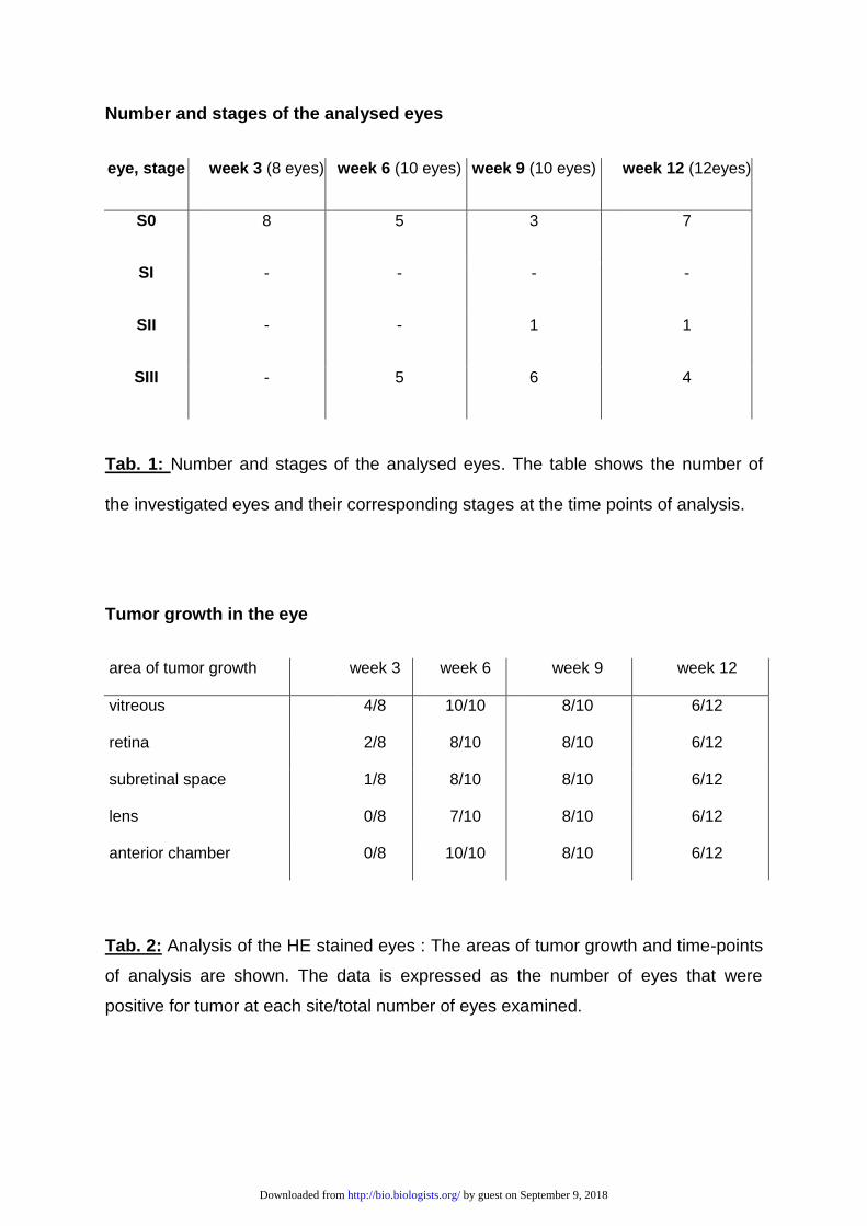
We determined four stages depending on the tumour progression as shown in Fig. 1:
Stage 0 (S0) was considered as the morphology of a normal mouse eye. Stage I (SI)
was reached after the eye was swollen up to 2x of the normal size and showed a
cloudy appearance, stage II (SII) was reached after the eye was swollen up to 3x of
the normal size, stage III (SIII) when the tumour broke through the cornea. Table 1
shows the number of eyes and their corresponding stages at the time points of
analysis.
As shown in Fig. 2, there were some intra individual differences concerning the time
point of the start and the progress of the swelling of the eyes. The earliest cases of
swelling appeared 34 days after injection the latest 70 daysafterwards. In most cases
the swelling started between week five and seven after injection and progressed fast
from stage I (in average 39 days after injection) to stage II (in average 43 days after
injection) and then to stage III (in average 48 days after injection) Fig. 2.
2. In vivo imaging using SLO/OCT
The SLO/OCT analysis could only be performed in stage 0 eyes with tumours at very
early stages or in eyes without a tumour. In eyes with tumours at later stages of
growth (SI-SIII) no analysis was possible, because the tumour covered the fundus.
In all cases in which the SLO/OCT- analysis was possible, the results showed a
good correlation with the results of the histological analysis. Using OCT, not only
could we detect the tumour itself, but could also get information about its growth
characteristics. The tumour shown in Fig. 3, for example, broke through the retina
and began to grow subretinally, which can be clearly seen on the OCT image Fig. 3A
left panel and could later be found on the corresponding H&E stained slide Fig. 3B.
The results of the angiography analysis with fluorescein gave a good picture of the
tumour’s vessel structure Fig. 3C.
by guest on September 9, 2018http://bio.biologists.org/Downloaded from
3. Histological analysis
In the tumour-bearing eyes tumour cells could be observed in the vitreous, in the
retina and in the subretinal space. An overview of the exact number of eyes and
areas of tumour growth up to the time points of analysis is presented in table 2.
For the initial phase of tumour growth (week 3), 8 eyes were analysed. Tumour cells
could be seen in the vitreous and on the retina in 4 of them. In two eyes the tumour
grew through the retina, in one of these eyes even subretinal tumour growth could be
observed.
In all tumour-bearing eyes which were analysed 6 weeks after injection, the tumour
completely replaced the vitreous and grew into the anterior chamber, in seven eyes
the tumour invaded or damaged the lens, in two eyes the tumour did not penetrate
the retina, Fig. 4A) in the other 8 tumour-bearing eyes, a subretinal growth could be
detected (fig. 4B).
In advanced tumours (week 9 and 12) the tumour replaced most of the eye’s
structures like the vitreous, the lens and retina. Here in all 6 tumour-bearing eyes the
sclera was the only part of the eye’s tissue left Fig. 4C.
Histologically the tumours were composed of typical undifferentiated hyperchromatic
cells with scanty cytoplasm having a rosette like growth pattern as described for the
original tumour (Reid et al., 1974). All tumours showed a high mitotic and necrotic
activity.
A tumour was found in sum in 67.5% of the analysed eyes (29 of 42) twelve weeks
after the injection of the Y79 cells.
MetastasesMetastases
We screened tissues near the tumour, like the brain and skull for
metastasesmetastases as well as the kidneys, lung, heart, liver, and spleen for the
appearance of distant metastasesmetastases by analysing HE stained cross
sections of these tissues. Only in one mouse could metastasesmetastases in the
brain be found. The metastasesmetastases were found in the mouse, which after
by guest on September 9, 2018http://bio.biologists.org/Downloaded from
having reached stage III for one eye was kept for the longest period of time (35 days)
before being killed and analysed Fig. 2, *(mouse 16). In this eye the tumour broke
through the sclera in several areas and grew into the brain (not shown).
No distant metastases could be found.
4. Electron microscopy analysis
The ultrastructural analysis of the xenograft–tumours, shown in Fig. 5, showed very
similar characteristics to those described for the original tumour in 1971, like poor
differentiation but still identifiable rosette like growth, large hyperchromatical nuclei
with multiple nucleoli and elaborate convolutions of the nuclei Fig. 5A and numerous
degraded and necrotic cells Fig. 5B (Reid et al., 1974), (Green et al., 1979), (McFall
et al., 1977) (fig 5. a,b). These characteristics are also described as typical for
patient’s Rb tissues (Rodrigues et al., 1986, Allen et al., 1962).
by guest on September 9, 2018http://bio.biologists.org/Downloaded from
DISCUSSION
Preclinical animal models that mimic molecular, genetic, and cellular features of
retinoblastoma are essential for studying this type of cancer.
Currently, two types of retinoblastoma animal models exist: transgenic models and
xenograft models. The transgenic models have been developed from LH-β-Tag
models to conditional gene knock-out models. There are different types of xenograft
models likeorthotopic models and subcutaneous transplantation models. The two
types of Rb models present advantages and disadvantages.
The combination of genetic and xenograft models in retinoblastoma research has
already help to better understand tumour biology and to find more effective diagnosis
and treatments.
Our aim was to create a xenograft mouse model with close resemblance to human
Rb tumours which can be used for broad application possibilities including radio
therapeutic approaches of Rb treatment.
Literature research indicates that in addition to the use of transgenic animals as a
model system for retinoblastoma, another possibility is the use of a xenograft model
which is based on the implantation of human retinoblastoma cells into the eye of
immunodeficient animals. Indeed, retinoblastoma xenograft models are often created
using the cell line Y79. This commercially available human retinoblastoma cell line is
derived from a two-and-a-half-year-old patient, who had a maternal history of
retinoblastoma.
The implantation can be performed in various compartments of the eye, in former
times the anterior chamber was often preferred because it is more accessible for
both the implantation and subsequent observation (Gallie et al., 1977), (Totsuka et
al., 1982). However, in patients the tumour starts its growth in the retina and
penetrates relatively late into the anterior chamber. The latter is physiologically
different to the vitreous body, where the retinoblastoma first encroaches. Thus a
subretinal injection of the retinoblastoma cells is a better reflection of the situation in
humans (del Cerro et al., 1993, Rowe et al., 1992). Unfortunately, this kind of
injection can cause damage to the choroid and retina, which can result in an
unnatural spread of the tumour.
by guest on September 9, 2018http://bio.biologists.org/Downloaded from
Another possibility is the intravitreal injection of tumour cells as described by
(Chevez-Barrios et al., 2000). After the intravitreal implantation of Y79 cells in Rag2
KO mice, tumours formed in the eye and gradually spread over the entire eye, and
later could also be found in the brain. Thus we decided to use an intravitreal injection
as well.
However, the Rag2 KO model developed by Chevez-Barrioz was never used for
radiotherapeutic experiments, but the nude mouse model used by Totsuka was
(Totsuka and Minoda, 1982), so we decided to combine the advantages of both
models to develop another model.
After the injection of Y79 cells, tumour cells proliferated first in the vitreous and then
formed a clearly localised tumour on and through the retina which is not exactly like
retinoblastoma tumours observed in children that originate in the retina. However this
particularity is common with the retinoblastoma mouse model developed by
(Chevez-Barrios et al., 2000) In most of the cases the tumour broke through the
retina and began to grow subretinally. In 2 cases the tumour did not penetrate the
retina at the time point of analysis of 6 weeks (fig. 4 b).
In contrast to the retinoblastoma mouse model developed by Chevez-Barrios et al.
(Chevez-Barrios et al., 2000) in which the authors observe metastases resulting from
migration of tumour cells up the optic nerve, we observed in our model that the
sclera seems to be a strong barrier for the tumour. The tumour needs to grow very
large and have a long time to break through the sclera. During our experiment the
tumour only penetrated the sclera in one mouse eye and formed brain metastases.
The metastases were found in the brain of a mouse, which, after having reached
stage III for the left eye, was kept alive for the longest period of time (35 days) before
being sacrificed and analysed (fig. 2, mouse 16 *). However in the Rag-2 knockout
(KO) mice used by Chevez-Barrios et al. (Chevez-Barrios et al., 2000), the animals
were intravitreally injected with Y79 cells in a similar manner as in our experiment,
but the mice already developed metastases 4 weeks after the injection (Chevez-
Barrios et al., 2000). These results are consistent with that of other groups, who
could show that metastasization metastasesin Rag-2 KO mouse models is stronger
than in nude mice for several human cancer xenografts like sarcoma (Nanni et al.,
2010) breast cancer (Nanni et al., 2012) or adenocarcinoma (Ye et al., 2015) .This
should be considered when choosing a model. Despite the mentioned differences of
by guest on September 9, 2018http://bio.biologists.org/Downloaded from
the metastasization process in Rag2 KO and nude mice, Gallie et al., described a
metastasization of the optic nerve and brain in cyclophosphamide pre-treated nude
mice (Gallie et al., 1977). Unfortunately the authors do not make any statement
about the time point of analysis. In our experiment we had to kill the animals at the
latest 12 weeks after tumour cells injection due to ethical requirements of local
authorities and we consider it very likely that they might develop metastases at a
later time point.
A very important aspect of this work was the use of in vivo approaches like
SLO/OCT for the detection and characterisation of tumours in the mouse eyes and
the comparison of the results with the corresponding results of the histological
analysis, which showed a good correlation as shown in Fig. 3. A similar
funduscopy/OCT based approach was used for the analysis of the tumours in the
eyes of a TAg-RB mouse model by Wenzel et al. with similar results (Wenzel et al.,
2015).
In ophthalmological research, in vivo analysis like SLO/OCT allows multiple analysis
of dynamic biological processes like tumourigenesis, tumour growth and
angiogenesis at certain time points in individual animals and can help to reduce the
number of experimental animals used.
In conclusion, we showed that our Rb mouse model mimics the human disease. The
xenograft tumour samples from our model showed very similar growth
characteristics, cellular appearance and ultrastructural characteristics to that
described for Rb patients tumour tissue samples. This makes our model a promising
tool for the study of retinoblastoma and its potential therapy approaches.
We also show that SLO/OCT can be used for the detection of tumours at early
stages of development and could be used for monitoring the future therapies.
by guest on September 9, 2018http://bio.biologists.org/Downloaded from

Acknowledgments
The authors thank Monika Rittgarn and Sigrid Schultheiss, for their excellent
technical assistance (Division of Experimental Vitreoretinal Surgery, Center for
Ophthalmology, Eberhard Karls University Tuebingen, Germany) and Dr. Tobias
Peters and Norman Rieger (Institute of Ophthalmic Research, Centre for
Ophthalmology, University of Tübingen) for valuable discussions.
We acknowledge support by Deutsche Forschungsgemeinschaft and Open Access
Publishing Fund of University of Tübingen.
Footnotes
Competing interests
The authors declare no competing or financial interests.
Author contributions
S.J designed the experiments. S.J. and .A.T. performed the experiments and
data analysis and wrote the paper. U.S and H.P.R. gave valuable suggestions
and revised the paper. All authors approved the final version of the
manuscript.
Funding
This work was financially supported by the Deutsche Kinderkrebsstiftung
(Project Nr. DKS 2012.08).
by guest on September 9, 2018http://bio.biologists.org/Downloaded from
REFERENCES
ALLEN, R. A., LATTA, H. & STRAATSMA, B. R. 1962. Retinoblastoma. A study of two cases by electron microscopy. Invest Ophthalmol, 1, 728-44.
BOUTRID, H., JOCKOVICH, M. E., MURRAY, T. G., PINA, Y., FEUER, W. J., LAMPIDIS, T. J. & CEBULLA, C. M. 2008. Targeting hypoxia, a novel treatment for advanced retinoblastoma. Invest Ophthalmol Vis Sci, 49, 2799-805.
BROADDUS, E., TOPHAM, A. & SINGH, A. D. 2009. Incidence of retinoblastoma in the USA: 1975-2004. Br J Ophthalmol, 93, 21-3.
CHEVEZ-BARRIOS, P., HURWITZ, M. Y., LOUIE, K., MARCUS, K. T., HOLCOMBE, V. N., SCHAFER, P., AGUILAR-CORDOVA, C. E. & HURWITZ, R. L. 2000. Metastatic and nonmetastatic models of retinoblastoma. Am J Pathol, 157, 1405-12.
DEL CERRO, M., SEIGEL, G. M., LAZAR, E., GROVER, D., DEL CERRO, C., BROOKS, D. H., DILORETO, D., JR. & CHADER, G. 1993. Transplantation of Y79 cells into rat eyes: an in vivo model of human retinoblastomas. Invest Ophthalmol Vis Sci, 34, 3336-46.
FISCHER, M. D., HUBER, G., BECK, S. C., TANIMOTO, N., MUEHLFRIEDEL, R., FAHL, E., GRIMM, C., WENZEL, A., REME, C. E., VAN DE PAVERT, S. A., WIJNHOLDS, J., PACAL, M., BREMNER, R. & SEELIGER, M. W. 2009. Noninvasive, in vivo assessment of mouse retinal structure using optical coherence tomography. PLoS One, 4, e7507.
GALLIE, B. L., ALBERT, D. M., WONG, J. J., BUYUKMIHCI, N. & PULLAFITO, C. A. 1977. Heterotransplantation of retinoblastoma into the athymic "nude" mouse. Invest Ophthalmol Vis Sci, 16, 256-9.
GREEN, A. L., MEEK, E. S., WHITE, D. W., STEVENS, R. H., ACKERMAN, L. D., JUDISCH, G. F. & PATIL, S. R. 1979. Retinoblastoma Y79 cell line: a study of membrane structures. Albrecht Von Graefes Arch Klin Exp Ophthalmol, 211, 279-87.
HOUSTON, S. K., BERROCAL, A. M. & MURRAY, T. G. 2011. The future of diagnostic imaging in retinoblastoma. J AAPOS, 15, 125-6.
HOUSTON, S. K., LAMPIDIS, T. J. & MURRAY, T. G. 2013. Models and discovery strategies for new therapies of retinoblastoma. Expert Opin Drug Discov, 8, 383-94.
HUBER, G., BECK, S. C., GRIMM, C., SAHABOGLU-TEKGOZ, A., PAQUET-DURAND, F., WENZEL, A., HUMPHRIES, P., REDMOND, T. M., SEELIGER, M. W. & FISCHER, M. D. 2009. Spectral domain optical coherence tomography in mouse models of retinal degeneration. Invest Ophthalmol Vis Sci, 50, 5888-95.
JEHANNE, M., BRISSE, H., GAUTHIER-VILLARS, M., LUMBROSO-LE ROUIC, L., FRENEAUX, P. & AERTS, I. 2014. Retinoblastoma: Recent advances. Bull Cancer, 101, 380-387.
MALLIPATNA, A., VINEKAR, A., JAYADEV, C., DABIR, S., SIVAKUMAR, M., KRISHNAN, N., MEHTA, P., BERENDSCHOT, T. & YADAV, N. K. 2015. The use of handheld spectral domain optical coherence tomography in pediatric ophthalmology practice: Our experience of 975 infants and children. Indian J Ophthalmol, 63, 586-93.
MCFALL, R. C., SERY, T. W. & MAKADON, M. 1977. Characterization of a new continuous cell line derived from a human retinoblastoma. Cancer Res, 37, 1003-10.
NANNI, P., NICOLETTI, G., LANDUZZI, L., CROCI, S., MURGO, A., PALLADINI, A., ANTOGNOLI, A., IANZANO, M. L., STIVANI, V., GROSSO, V., MAIRA, S. M., GARCIA-ECHEVERRIA, C., SCOTLANDI, K., DE GIOVANNI, C. & LOLLINI, P. L. 2010. High metastatic efficiency of human sarcoma cells in Rag2/gammac double knockout mice provides a powerful test system for antimetastatic targeted therapy. Eur J Cancer, 46, 659-68.
NANNI, P., NICOLETTI, G., PALLADINI, A., CROCI, S., MURGO, A., IANZANO, M. L., GROSSO, V., STIVANI, V., ANTOGNOLI, A., LAMOLINARA, A., LANDUZZI, L., DI TOMASO, E., IEZZI, M., DE GIOVANNI, C. & LOLLINI, P. L. 2012. Multiorgan metastasis of human HER-2+ breast cancer in Rag2-/-;Il2rg-/- mice and treatment with PI3K inhibitor. PLoS One, 7, e39626.
by guest on September 9, 2018http://bio.biologists.org/Downloaded from

RAMASUBRAMANIAN, A., SHIELDS, C. L., MELLEN, P. L., HAJI, S., HARMON, S. A., VEMUGANTI, G. K. & SHIELDS, J. A. 2011. Autofluorescence of treated retinoblastoma. J AAPOS, 15, 167-72.
REID, T. W., ALBERT, D. M., RABSON, A. S., RUSSELL, P., CRAFT, J., CHU, E. W., TRALKA, T. S. & WILCOX, J. L. 1974. Characteristics of an established cell line of retinoblastoma. J Natl Cancer Inst, 53, 347-60.
RODRIGUES, M. M., WILSON, M. E., WIGGERT, B., KRISHNA, G. & CHADER, G. J. 1986. Retinoblastoma. A clinical, immunohistochemical, and electron microscopic case report. Ophthalmology, 93, 1010-5.
ROWE, S. G., LEE, W. H. & MADREPERLA, S. Subretinal and vitreal growth of human retinoblastoma cells in the mouse eye. Invest Ophthalmol Vis Sci., 1992. 875 (abstr.).
SEREGARD, S., LUNDELL, G., SVEDBERG, H. & KIVELA, T. 2004. Incidence of retinoblastoma from 1958 to 1998 in Northern Europe: advantages of birth cohort analysis. Ophthalmology, 111, 1228-32.
SHAH, N. V., PHAM, D. G., MURRAY, T. G., DECATUR, C., HERNANDEZ, E., SHAH, N. N., CAVALCANTE, M. & HOUSTON, S. K. 2014. Intravitreal and subconjunctival melphalan for retinoblastoma in transgenic mice. J Ophthalmol, 2014, 829879.
SHIELDS, C. L., MASHAYEKHI, A., LUO, C. K., MATERIN, M. A. & SHIELDS, J. A. 2004. Optical coherence tomography in children: analysis of 44 eyes with intraocular tumors and simulating conditions. J Pediatr Ophthalmol Strabismus, 41, 338-44.
SHIELDS, C. L., PALAMAR, M., SHARMA, P., RAMASUBRAMANIAN, A., LEAHEY, A., MEADOWS, A. T. & SHIELDS, J. A. 2009. Retinoblastoma regression patterns following chemoreduction and adjuvant therapy in 557 tumors. Arch Ophthalmol, 127, 282-90.
TOTSUKA, S., AKAZAWA, K. & MINODA, K. 1982. [Transplantation of retinoblastoma into nude mouse. 3. Tumor doubling time of retinoblastoma (author's transl)]. Nihon Ganka Gakkai Zasshi, 86, 418-25.
TOTSUKA, S. & MINODA, K. 1982. Radiation effects on retinoblastoma successively transplanted into nude mouse eyes. Ophthalmologica, 185, 158-67.
VILLEGAS, V. M., HESS, D. J., WILDNER, A., GOLD, A. S. & MURRAY, T. G. 2013. Retinoblastoma. Curr Opin Ophthalmol, 24, 581-8.
WENZEL, A. A., O'HARE, M. N., SHADMAND, M. & CORSON, T. W. 2015. Optical coherence tomography enables imaging of tumor initiation in the TAg-RB mouse model of retinoblastoma. Mol Vis, 21, 515-22.
YE, W., JIANG, Z., LI, G. X., XIAO, Y., LIN, S., LAI, Y., WANG, S., LI, B., JIA, B., LI, Y., HUANG, Z. L., LI, J., FENG, F., LI, S., YAO, H., LIU, Z., CAO, S., XU, L., LI, Y., WU, D., ZENG, L., ZHONG, M., LIU, P., WEN, Z. S., XU, B., YAO, Y., PEI, D. & LI, P. 2015. Quantitative evaluation of the immunodeficiency of a mouse strain by tumor engraftments. J Hematol Oncol, 8, 59.
by guest on September 9, 2018http://bio.biologists.org/Downloaded from
Number and stages of the analysed eyes
eye, stage week 3 (8 eyes) week 6 (10 eyes) week 9 (10 eyes) week 12 (12eyes)
S0 8 5 3 7
SI - - - -
SII - - 1 1
SIII - 5 6 4
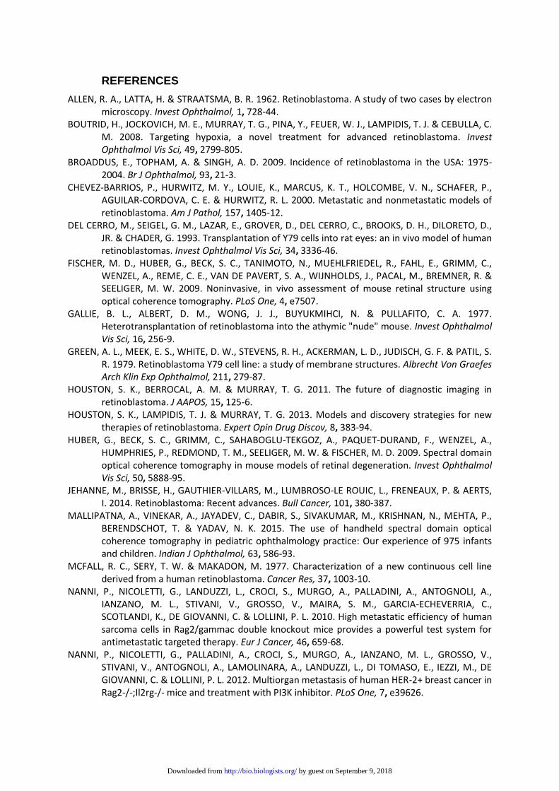
Tab. 1: Number and stages of the analysed eyes. The table shows the number of
the investigated eyes and their corresponding stages at the time points of analysis.
Tumor growth in the eye
area of tumor growth week 3 week 6 week 9 week 12
vitreous 4/8 10/10 8/10 6/12
retina 2/8 8/10 8/10 6/12
subretinal space 1/8 8/10 8/10 6/12
lens 0/8 7/10 8/10 6/12
anterior chamber 0/8 10/10 8/10 6/12
Tab. 2: Analysis of the HE stained eyes : The areas of tumor growth and time-points
of analysis are shown. The data is expressed as the number of eyes that were
positive for tumor at each site/total number of eyes examined.
by guest on September 9, 2018http://bio.biologists.org/Downloaded from
Figures
Results of the morphological analysis
Figure 1: results of the morphological analysis. The pictures a-d show the stages
S0-SIII of the mouse eyes: (A) stage 0: eye of a untreated mouse (S0), (B) stage I:
the eye is swollen up to 2x of the normal size (S I), (C) stage II: the eye is swollen up
to 3x of the normal size, the eye is cloudy (SII), (D) stage III: the tumor breaks
through the cornea (SIII)
by guest on September 9, 2018http://bio.biologists.org/Downloaded from
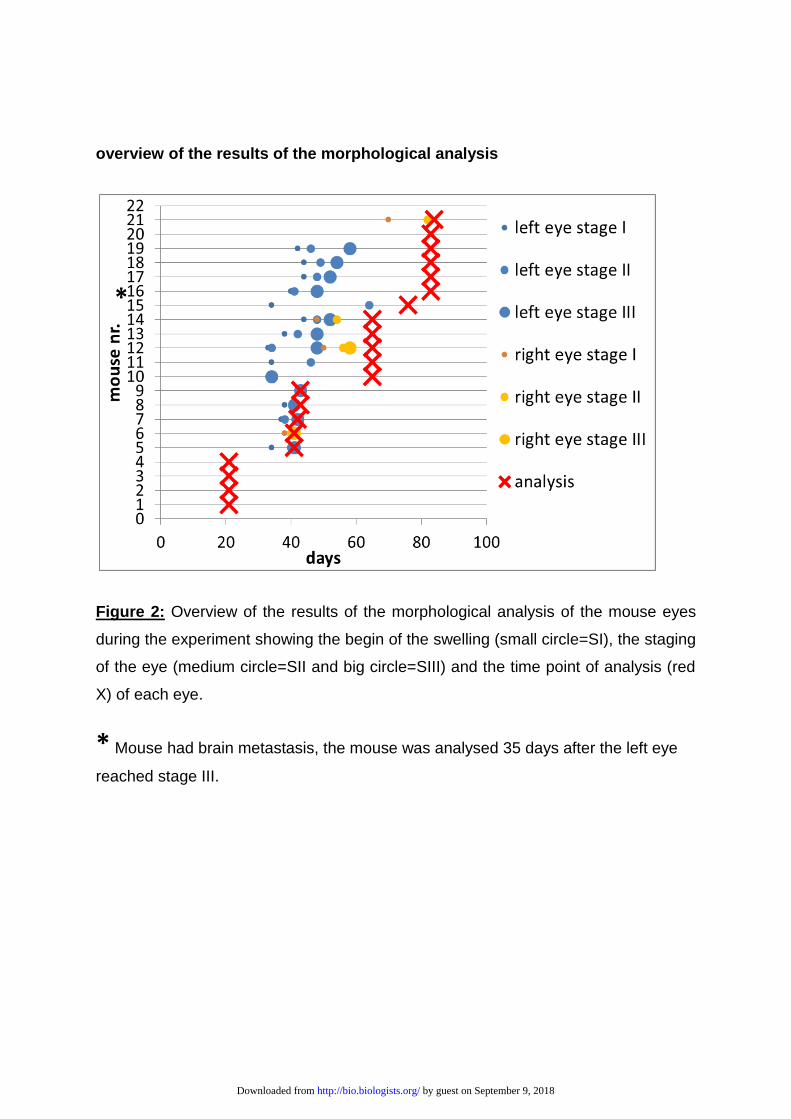
overview of the results of the morphological analysis
Figure 2: Overview of the results of the morphological analysis of the mouse eyes
during the experiment showing the begin of the swelling (small circle=SI), the staging
of the eye (medium circle=SII and big circle=SIII) and the time point of analysis (red
X) of each eye.
* Mouse had brain metastasis, the mouse was analysed 35 days after the left eye
reached stage III.
by guest on September 9, 2018http://bio.biologists.org/Downloaded from
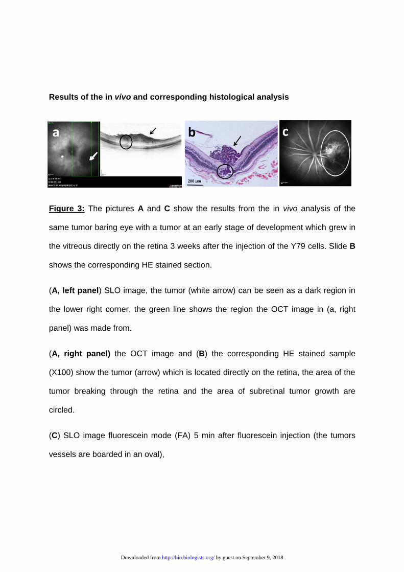
Results of the in vivo and corresponding histological analysis
Figure 3: The pictures A and C show the results from the in vivo analysis of the
same tumor baring eye with a tumor at an early stage of development which grew in
the vitreous directly on the retina 3 weeks after the injection of the Y79 cells. Slide B
shows the corresponding HE stained section.
(A, left panel) SLO image, the tumor (white arrow) can be seen as a dark region in
the lower right corner, the green line shows the region the OCT image in (a, right
panel) was made from.
(A, right panel) the OCT image and (B) the corresponding HE stained sample
(X100) show the tumor (arrow) which is located directly on the retina, the area of the
tumor breaking through the retina and the area of subretinal tumor growth are
circled.
(C) SLO image fluorescein mode (FA) 5 min after fluorescein injection (the tumors
vessels are boarded in an oval),
by guest on September 9, 2018http://bio.biologists.org/Downloaded from
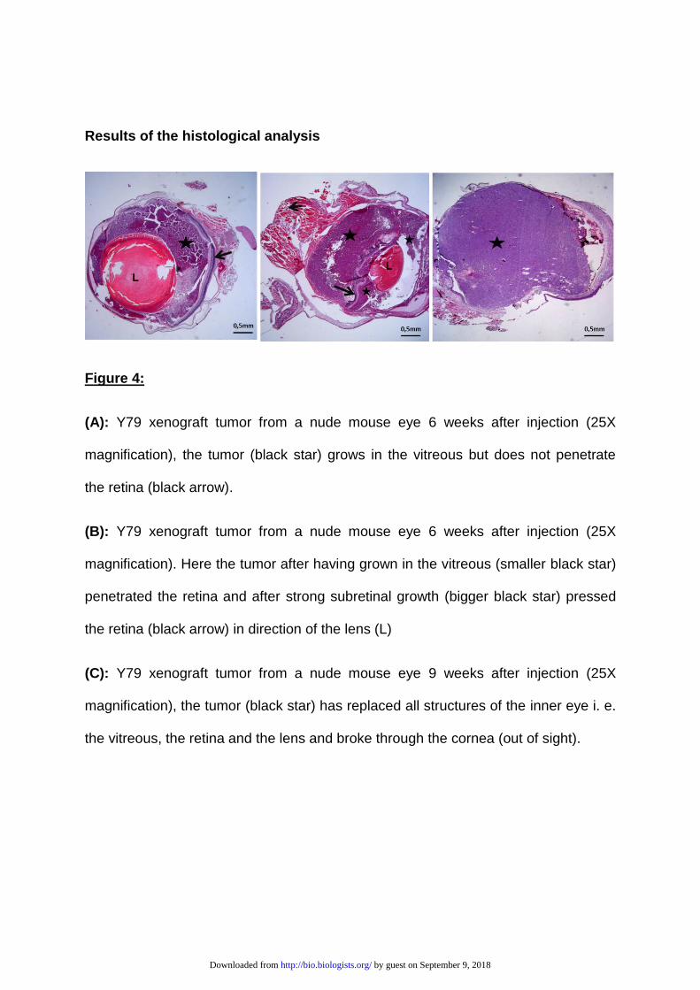
Results of the histological analysis
Figure 4:
(A): Y79 xenograft tumor from a nude mouse eye 6 weeks after injection (25X
magnification), the tumor (black star) grows in the vitreous but does not penetrate
the retina (black arrow).
(B): Y79 xenograft tumor from a nude mouse eye 6 weeks after injection (25X
magnification). Here the tumor after having grown in the vitreous (smaller black star)
penetrated the retina and after strong subretinal growth (bigger black star) pressed
the retina (black arrow) in direction of the lens (L)
(C): Y79 xenograft tumor from a nude mouse eye 9 weeks after injection (25X
magnification), the tumor (black star) has replaced all structures of the inner eye i. e.
the vitreous, the retina and the lens and broke through the cornea (out of sight).
by guest on September 9, 2018http://bio.biologists.org/Downloaded from
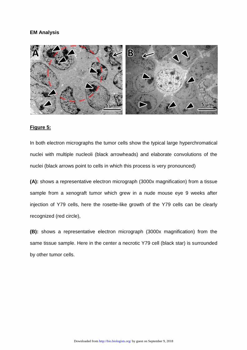
EM Analysis
Figure 5:
In both electron micrographs the tumor cells show the typical large hyperchromatical
nuclei with multiple nucleoli (black arrowheads) and elaborate convolutions of the
nuclei (black arrows point to cells in which this process is very pronounced)
(A): shows a representative electron micrograph (3000x magnification) from a tissue
sample from a xenograft tumor which grew in a nude mouse eye 9 weeks after
injection of Y79 cells, here the rosette-like growth of the Y79 cells can be clearly
recognized (red circle),
(B): shows a representative electron micrograph (3000x magnification) from the
same tissue sample. Here in the center a necrotic Y79 cell (black star) is surrounded
by other tumor cells.
by guest on September 9, 2018http://bio.biologists.org/Downloaded from