Embed Size (px)
Citation preview
Co
CLINICAL GUIDELINE
European Society for Pediatric Gastroenterology,
Hepatology, and Nutrition Guidelines for the Diagnosis of
Coeliac Disease
�S. Husby, yS. Koletzko, zI.R. Korponay-Szabo, §M.L. Mearin, jjA. Phillips, �R. Shamir,#R. Troncone, ��K. Giersiepen, yyD. Branski, zzC. Catassi, §§M. Lelgeman, jjjjM. Maki,
��C. Ribes-Koninckx, ##A. Ventura, and ����K.P. Zimmer, for the ESPGHAN Working Group on
pyright 2012 by
Coeliac is, on behalf of the ESPGHAN Gastroenterology Committee
the years 2004 to 2
recommendations on CD
Received and accepted SFrom the �Hans Chris
University Hospital, tHepatology, Dr. von HUniversity, the zUnivCenter, the §DepartmCenter, the jjUniversand Child Health, thLiver Diseases, SchneFaculty of Medicine,trics and European LDiseases, UniversityResearch, UniversityHadash University Hosita Politecnica delleStatutory Health InsuUniversity of TampeFe University HospitaGarofolo University oPaediatrics and Neona
Address [email protected]
Drs Husby, Koletzko, Korand Giersiepen contriauthors.
Conflict of interest statemCopyright # 2012 by E
Hepatology, and NutGastroenterology, He
DOI: 10.1097/MPG.0b01
136
Disease Diagnos
ABSTRACT
Objective: Diagnostic criteria for coeliac disease (CD) from the European
Society for Paediatric Gastroenterology, Hepatology, and Nutrition (ESP-
GHAN) were published in 1990. Since then, the autoantigen in CD, tissue
transglutaminase, has been identified; the perception of CD has changed
from that of a rather uncommon enteropathy to a common multiorgan
disease strongly dependent on the haplotypes human leukocyte antigen
(HLA)-DQ2 and HLA-DQ8; and CD-specific antibody tests have improved.
Methods: A panel of 17 experts defined CD and developed new diagnostic
criteria based on the Delphi process. Two groups of patients were defined
with different diagnostic approaches to diagnose CD: children with
symptoms suggestive of CD (group 1) and asymptomatic children at
increased risk for CD (group 2). The 2004 National Institutes of Health/
Agency for Healthcare Research and Quality report and a systematic
literature search on antibody tests for CD in paediatric patients covering
ESPGHAN and NASPGHAN. Un
009 was the basis for the evidence-based
-specific antibody testing.
eptember 1, 2011.tian Andersen Children’s Hospital at Odensehe yDivision of Paediatric Gastroenterology andauner Children’s Hospital, Ludwig-Maximilians-
ersity of Debrecen, Medical and Health Scienceent of Paediatrics, Leiden University Medical
ity College London Medical School/Paediatricse �Institute of Gastroenterology, Nutrition andider Children’s Medical Center of Israel, SacklerTel-Aviv University, the #Department of Paedia-aboratory for the Investigation of Food-Induced‘‘Federico II,’’ the ��Centre for Social Policyof Bremen, the yyDepartment of Paediatrics,
spitals, the zzDepartment of Paediatrics, Univer-Marche, the §§Medical Review Board of the
rance Fund, the jjjjPaediatric Research Centre,re and Tampere University Hospital, the ��Lal, the ##Department of Paediatrics, IRCCS Burlof Trieste, and the ����Department for Generaltology, Justus-Liebig University.
and reprint requests to Dr Steffen Husby (e-mail:gionsyddanmark.dk).ponay-Szabo, Mearin, Phillips, Shamir, Troncone,buted equally to the article and are listed as first
ents are listed at the end of the article.uropean Society for Pediatric Gastroenterology,rition and North American Society for Pediatricpatology, and Nutrition3e31821a23d0
Results: In group 1, the diagnosis of CD is based on symptoms, positive
serology, and histology that is consistent with CD. If immunoglobulin A
anti-tissue transglutaminase type 2 antibody titers are high (>10 times the
upper limit of normal), then the option is to diagnose CD without duodenal
biopsies by applying a strict protocol with further laboratory tests. In group
2, the diagnosis of CD is based on positive serology and histology. HLA-
DQ2 and HLA-DQ8 testing is valuable because CD is unlikely if both
haplotypes are negative.
Conclusions: The aim of the new guidelines was to achieve a high
diagnostic accuracy and to reduce the burden for patients and their
families. The performance of these guidelines in clinical practice should
be evaluated prospectively.
(JPGN 2012;54: 136–160)
SYNOPSIS
G uidelines from the European Society for Paediatric Gastro-enterology, Hepatology, and Nutrition (ESPGHAN) for the
diagnosis and treatment of coeliac disease (CD) have not beenrenewed for 20 years. During this time, the perception of CD haschanged from a rather uncommon enteropathy to a common multi-organ disease with a strong genetic predisposition that is associatedmainly with human leukocyte antigen (HLA)-DQ2 and HLA-DQ8.The diagnosis of CD also has changed as a result of the availabilityof CD-specific antibody tests, based mainly on tissue transgluta-minase type 2 (TG2) antibodies.
Within ESPGHAN, a working group was established toformulate new guidelines for the diagnosis of CD based on scien-tific and technical developments using an evidence-based approach.The working group additionally developed a new definition of CD.A detailed evidence report on antibody testing in CD forms the basisof the guidelines and will be published separately. Guidelinestatements and recommendations based on a voting procedure havebeen provided. The goal of this synopsis is to summarise some ofthe evidence statements and recommendations of the guidelines foruse in clinical practice.
Definitions
CD is an immune-mediated systemic disorder elicited bygluten and related prolamines in genetically susceptible individuals
authorized reproduction of this article is prohibited.
and characterised by the presence of a variable combination ofgluten-dependent clinical manifestations, CD-specific antibodies,
JPGN � Volume 54, Number 1, January 2012
Co
HLA-DQ2 or HLA-DQ8 haplotypes, and enteropathy. CD-specificantibodies comprise autoantibodies against TG2, including endo-mysial antibodies (EMA), and antibodies against deamidated formsof gliadin peptides (DGP).
Who Should Be Tested for CD?
CD may present with a large variety of nonspecific signs andsymptoms. It is important to diagnose CD not only in children withobvious gastrointestinal symptoms but also in children with a lessclear clinical picture because the disease may have negative healthconsequences. The availability of serological tests with highaccuracy and other diagnostic tests allows a firm diagnosis tobe made. The interpretation and consequences of the test resultsdiffer between symptomatic and asymptomatic patients in at-risk groups.
Testing for CD should be offered to the following groups:Group 1: Children and adolescents with the otherwise
unexplained symptoms and signs of chronic or intermittent diar-rhoea, failure to thrive, weight loss, stunted growth, delayedpuberty, amenorrhoea, iron-deficiency anaemia, nausea or vomit-ing, chronic abdominal pain, cramping or distension, chronicconstipation, chronic fatigue, recurrent aphthous stomatitis(mouth ulcers), dermatitis herpetiformis–like rash, fracture withinadequate traumas/osteopenia/osteoporosis, and abnormal liverbiochemistry.
Group 2: Asymptomatic children and adolescents with anincreased risk for CD such as type 1 diabetes mellitus (T1DM),Down syndrome, autoimmune thyroid disease, Turner syn-drome, Williams syndrome, selective immunoglobulin A (IgA)deficiency, autoimmune liver disease, and first-degree relativeswith CD.
Diagnostic Tools
CD-specific Antibody TestsCD-specific antibody tests measure anti-TG2 or EMA in
blood. Tests measuring anti-DGP also could be reasonably specific.Laboratories providing CD-specific antibody test results for diag-nostic use should continuously participate in quality control pro-grammes at a national or an international level. Every antibody testused for the diagnosis of childhood CD should be validated againstthe reference standard of EMA or histology in a paediatric popu-lation ranging from infancy to adolescence.
A test is considered as reliable if it shows >95% agreementwith the reference standard. The optimal threshold values forantibody positivity (cutoff value or upper limit of normal[ULN]) of a test should be established. Anti-TG2 and anti-DGPlaboratory test results should be communicated as numeric valuestogether with the specification of the immunoglobulin classmeasured, the manufacturer, the cutoff value defined for thespecific test kit, and (if available) the level of ‘‘high’’ antibodyvalues. It is not sufficient to state only positivity or negativity.Reports on EMA results should contain the specification of theinvestigated immunoglobulin class, cutoff dilution, interpretation(positive or negative), highest dilution still positive, and specifica-tion of the substrate tissue.
For the interpretation of antibody results, total IgA levels inserum, age of the patient, pattern of gluten consumption, and intakeof immunosuppressive drugs should be taken into account. If glutenexposure was short or gluten had been withdrawn for a longerperiod of time (several weeks to years) the negative result is not
JPGN � Volume 54, Number 1, January 2012
pyright 2012 by ESPGHAN and NASPGHAN. Un
reliable. For IgA-competent subjects, the conclusions should bedrawn primarily from the results of IgA class antibody tests. For
www.jpgn.org
subjects with low serum IgA levels (total serum IgA< 0.2 g/L),the conclusions should be drawn from the results of the IgG classCD-specific antibody tests.
HLA Testing for HLA-DQ2 and HLA-DQ8
Typing for HLA-DQ2 and HLA-DQ8 is a useful tool toexclude CD or to make the diagnosis unlikely in the case of anegative test result for both markers. HLA testing should beperformed in patients with an uncertain diagnosis of CD, forexample, in patients with negative CD-specific antibodies and mildinfiltrative changes in proximal small intestinal biopsy specimens.If CD is considered in children in whom there is a strong clinicalsuspicion of CD, high specific CD antibodies are present, and small-bowel biopsies are not going to be performed, then the workinggroup recommends performing HLA-DQ2 and HLA-DQ8 typing toadd strength to the diagnosis. Prospective studies will make clearwhether HLA typing is indeed an efficient and effective diagnostictool in these patients. HLA testing may be offered to asymptomaticindividuals with CD-associated conditions (group 2) to select themfor further CD-specific antibody testing.
Histological Analysis of Duodenal Biopsies
The histological features of the small intestinal enteropathyin CD have a variable severity, may be patchy, and in a smallproportion of patients with CD appear only in the duodenal bulb.The alterations are not specific for CD and may be found inenteropathies other than CD. Biopsies should be taken preferablyduring upper endoscopy from the bulb (at least 1 biopsy) and fromthe second or third portion of duodenum (at least 4 biopsies). Thepathology report should include a description of the orientation, thepresence or not of normal villi or degree of atrophy and cryptelongation, the villus-crypt ratio, the number of intraepitheliallymphocytes (IELs), and grading according to the Marsh-Oberhuber classification.
Diagnostic Approach for a Child or AdolescentWith Symptoms or Signs Suggestive of CD
A test for CD-specific antibodies is the first tool that is usedto identify individuals for further investigation to diagnose or to ruleout CD. Patients who are consuming a gluten-containing diet shouldbe tested for CD-specific antibodies. It is recommended that theinitial test be IgA class anti-TG2 from a blood sample. If total serumIgA is not known, then this also should be measured. In subjectswith either primary or secondary humoral IgA deficiency, at least 1additional test measuring IgG class CD-specific antibodies shouldbe done (IgG anti-TG2, IgG anti-DGP or IgG EMA, or blended kitsfor both IgA and IgG antibodies). In symptomatic patients in whomthe initial testing was performed with a rapid CD antibody detectionkit (point-of-care [POC] tests), the result should be confirmed by alaboratory-based quantitative test. Although published data indicatePOC tests may achieve high accuracy for CD diagnosis, futurestudies must show whether they work equally well when applied inless selected populations and/or when handled by laypeople oruntrained medical staff.
Tests measuring antibodies against DGP may be used asadditional tests in patients who are negative for other CD-specificantibodies but in whom clinical symptoms raise a strong suspicionof CD, especially if they are younger than 2 years. Tests for the
ESPGHAN Guidelines for Diagnosis of Coeliac Disease
authorized reproduction of this article is prohibited.
detection of IgG or IgA antibodies against native gliadin peptides(conventional gliadin antibody test) should not be used for CD
137
Co
diagnosis. Tests for the detection of antibodies of any type (IgG,IgA, secretory IgA) in faecal samples should not be used.
If IgA class CD antibodies are negative in an IgA-competentsymptomatic patient, then it is unlikely that CD is causing thesymptom at the given time point. Further testing for CD is notrecommended unless special medical circumstances (eg, youngerthan 2 years, restricted gluten consumption, severe symptoms,family predisposition or other predisposing disease, immunosup-pressive medication) are present.
In seronegative cases for anti-TG2, EMA, and anti-DGP butwith severe symptoms and a strong clinical suspicion of CD, smallintestinal biopsies and HLA-DQ testing are recommended. Ifhistology shows lesions are compatible with CD but HLA-DQ2/HLA-DQ8 heterodimers are negative, then CD is not likely and anenteropathy caused by a diagnosis other than CD should be con-sidered. In these patients, the diagnosis of CD can be made onlyafter a positive challenge procedure with repeated biopsies.
When duodenal biopsies, taken during routine diagnosticworkup for gastrointestinal symptoms, disclose a histological pat-tern indicative of CD (Marsh 1–3 lesions), antibody determinations(anti-TG2 and, in children younger than 2 years, anti-DGP) andHLA typing should be performed. In the absence of CD-specificantibodies and/or HLA-DQ2 or HLA-DQ8 heterodimers, othercauses of enteropathy (eg, food allergy, autoimmune enteropathy)should be considered.
What Should Be Done When CD-specific AntibodyTests Are Positive?
Children testing positive for CD-specific antibodies shouldbe evaluated by a paediatric gastroenterologist or by a paediatricianwith a similar knowledge of and experience with CD to confirm orexclude CD. A gluten-free diet (GFD) should be introduced onlyafter the completion of the diagnostic process, when a conclusivediagnosis has been made. Health care professionals should beadvised that starting patients on a GFD, when CD has not beenexcluded or confirmed, may be detrimental. A CD-specific anti-body test also should be performed in children and adolescentsbefore the start of a GFD because of suspected or proven allergyto wheat.
The clinical relevance of a positive anti-TG2 or anti-DGPresult should be confirmed by histology, unless certain conditionsare fulfilled that allow the option of omitting the confirmatorybiopsies. If histology shows lesions that are consistent with CD(Marsh 2–3), then the diagnosis of CD is confirmed. If histologyis normal (Marsh 0) or shows only increased IEL counts(>25 lymphocytes per 100 epithelial cells, Marsh 1), then furthertesting should be performed before establishing the diagnosisof CD.
In Which Patients Can the Diagnosis of CD BeMade Without Duodenal Biopsies?
In children and adolescents with signs or symptoms sugges-tive of CD and high anti-TG2 titers with levels >10 times ULN, thelikelihood for villous atrophy (Marsh 3) is high. In this situation, thepaediatric gastroenterologist may discuss with the parents andpatient (as appropriate for age) the option of performing furtherlaboratory testing (EMA, HLA) to make the diagnosis of CDwithout biopsies. Antibody positivity should be verified by EMAfrom a blood sample drawn at an occasion separate from the initialtest to avoid false-positive serology results owing to mislabeling of
Husby et al
pyright 2012 by ESPGHAN and NASPGHAN. Un
blood samples or other technical mistakes. If EMA testing confirmsspecific CD antibody positivity in this second blood sample, then
138
the diagnosis of CD can be made and the child can be started on aGFD. It is advisable to check for HLA types in patients who arediagnosed without having a small intestinal biopsy to reinforce thediagnosis of CD.
Diagnostic Approach for an AsymptomaticChild or Adolescent With CD-associatedConditions
If it is available, HLA testing should be offered as the first-line test. The absence of DQ2 and DQ8 render CD highly unlikelyand no further follow-up with serological tests is needed. If thepatient is DQ8 and/or DQ2 positive, homozygous for only the b-chains of the HLA-DQ2 complex (DQB1�0202), or HLA testing isnot done, then an anti-TG2 IgA test and total IgA determinationshould be performed, but preferably not before the child is 2 yearsold. If antibodies are negative, then repeated testing for CD-specificantibodies is recommended.
Individuals with an increased genetic risk for CD may havefluctuating (or transient) positive serum levels of CD-specificantibodies, particularly anti-TG2 and anti-DGP. Therefore, in thisgroup of individuals (group 2) without clinical signs and symptoms,duodenal biopsies with the demonstration of an enteropathy shouldalways be part of the CD diagnosis. If initial testing was performedwith a rapid CD antibody-detection kit, then a positive test resultalways should be confirmed by a laboratory-based quantitative test.Negative rapid test results in asymptomatic individuals also shouldbe confirmed by a quantitative test whenever the test has beencarried out by laypeople or untrained medical staff and/or reliabilityof the test or circumstances of testing (eg, sufficient gluten intake,concomitant medication, IgA status) are unknown or questionable.
To avoid unnecessary biopsies in individuals with low CD-specific antibody levels (ie,<3 times ULN), it is recommended thatthe more specific test for EMA be performed. If the EMA test ispositive, then the child should be referred for duodenal biopsies. Ifthe EMA test is negative, then repeated serological testing on anormal gluten-containing diet in 3 to 6 monthly intervals is recom-mended.
Follow-up and Challenge Procedures
If the diagnosis of CD is made according to the diagnosticcriteria mentioned above, the family should receive professionaldietary counseling for a GFD. The patients should be followed upregularly for symptomatic improvement and normalisation of CD-specific antibody tests. The time until the antibody titers fall belowthe cutoff for normal depends on the initial level, but in general thisshould be achieved within 12 months after starting the GFD.
In patients fulfilling the diagnostic criteria for CD it isunnecessary to perform small-bowel biopsies on a GFD; however,if there is no clinical response to the GFD in symptomatic patients,after a careful dietary assessment to exclude lack of adherence to aGFD, further investigations are required. These investigations mayinclude further biopsies.
Gluten challenge is not considered necessary except underunusual circumstances. These circumstances include situations inwhich there is doubt about the initial diagnosis. Gluten challengeshould be preceded by HLA typing and assessment of mucosalhistology and always should be performed under medical super-vision, preferably by a paediatric gastroenterologist. Gluten chal-lenge should be discouraged before the child is 5 years old andduring the pubertal growth spurt, unless the child is HLA-DQ2 and
JPGN � Volume 54, Number 1, January 2012
authorized reproduction of this article is prohibited.
HLA-DQ8 negative or has been placed on a GFD without propertesting. The daily gluten intake during gluten challenge should
www.jpgn.org
Co
contain at least the normal amount of gluten intake for children(approximately 15 g/day). IgA anti-TG2 antibody (IgG in low levelsof serum IgA) should be measured during the challenge period. Apatient should be considered to have relapsed (and hence thediagnosis of CD confirmed) if CD-specific antibodies becomepositive and a clinical and/or histological relapse is observed. Inthe absence of positive antibodies or symptoms the challengeshould be considered completed after 2 years; however, additionalbiopsies on a normal diet are recommended because delayed relapsemay occur later in life.
INTRODUCTION AND STRUCTUREESPGHAN guidelines for the diagnosis of CD were last
published in 1990 (1) and at that time represented a significantimprovement in both the diagnosis and management of CD. Since1990, the understanding of the pathological processes of CD hasincreased enormously, leading to a change in the clinical paradigmof CD from a chronic, gluten-dependent enteropathy of childhood toa systemic disease with chronic immune features affecting differentorgan systems. Although CD may occur at any age (2), theseguidelines focus on childhood and adolescence.
The disease etiology is multifactorial with a strong geneticinfluence, as documented in twin studies (3) and in studies showinga strong dependence on HLA-DQ2 and HLA-DQ8 haplotypes (4).A major step forward in the understanding of the pathogenesis ofCD was the demonstration in patients with CD of gluten-reactivesmall-bowel T cells that specifically recognise gliadin peptides inthe context of HLA-DQ2 and HLA-DQ8 (5). Furthermore, thediscovery of TG2 as the major autoantigen in CD led to therecognition of the autoimmune nature of the disease (6). TG2occurs abundantly in the gut and functions to deamidate proteinsand peptides, including gliadin or gliadin fragments, leading toincreased T-cell reactivity in patients with CD (7). This increasedknowledge of CD pathogenesis has led to the further developmentof diagnostic serological tests based on antibody determinationagainst gliadin and TG2-rich endomysium and later TG2.
Tests using DGP as substrate may be of significant value inCD diagnostic testing (8). Antibodies against TG2, EMA, and DGPare hence referred to as CD-specific antibodies, whereas antibodiesagainst native (non-DGP) gliadin are largely nonspecific. Small-bowel biopsies have thus far been considered to be the referencestandard for the diagnosis of CD; however, evidence has beenaccumulating on the diagnostic value of specific CD antibodies, andHLA typing has been used increasingly for diagnostic purposes. Atthe same time, the leading role of histology for the diagnosis of CDhas been questioned for several reasons: histological findings arenot specific for CD, lesions may be patchy and can occur in theduodenal bulb only, and interpretation depends on preparation ofthe tissue and is prone to a high interobserver variability (9). Thediagnosis of CD may then depend not only on the results of small-bowel biopsies but also on information from clinical and family dataand results from specific CD antibody testing and HLA typing.
In 2004, the US National Institutes of Health and the Agencyfor Healthcare Research and Quality (AHRQ) published a com-prehensive evidence-based analysis of the diagnosis and manage-ment of CD (10), which was followed by specific clinical guidelinesfor children by the North American Society for Pediatric Gastro-enterology, Hepatology, and Nutrition (11). In 2008, the UKNational Institute for Health and Clinical Evidence (NICE) pub-lished guidelines for the diagnosis and management of CD ingeneral practice. These guidelines did not challenge the centraland exclusive position of the result of small-bowel biopsies as the
JPGN � Volume 54, Number 1, January 2012
pyright 2012 by ESPGHAN and NASPGHAN. Un
reference standard for the diagnosis of CD. A working group withinESPGHAN was established with the aim of formulating new
www.jpgn.org
evidence-based guidelines for the diagnosis of CD in childrenand adolescents. During the work it became apparent that a newdefinition of CD was necessary, and such a definition is presentedhere. A major goal of the guidelines was to answer the question ofwhether duodenal biopsies with presumed characteristic histologi-cal changes compatible with CD could be omitted in some clinicalcircumstances in the diagnosis of CD. In addition, these guidelinespresent diagnostic algorithms for the clinical diagnosis of childhoodCD.
METHODOLOGIES
Working GroupAn ESPGHAN working group was established in 2007 with
the aim of establishing evidence-based guidelines for the diagnosisof CD in children and adolescents. The members of the group wereESPGHAN members with a scientific and clinical interest in CD,including pathology and laboratory antibody determinations, andwith a broad representation from European countries. A represen-tative of the Association of European Coeliac Societies was amember of the working group. Two epidemiologists also parti-cipated in the working group.
Systematic Searches
The group decided to use an evidence-based approach toselect diagnostic questions, followed by search and evaluation ofthe scientific literature to answer these questions. The guidelineswere based on the available evidence analyses including the AHRQreport from 2004 (10). The search profile of the AHRQ report withregard to specific CD antibodies was used as a template for a newliterature search. At first, a literature search was conducted onarticles from January 2004 to August 2008, supplemented by asecond search from September 2008 to September 2009. Thearticles found were assessed by epidemiologists and evidence-basedmedicine experts from the Centre for Health Technology Assess-ment at the University of Bremen, Germany (www.hta.uni-bre-men.de).
Evidence Report
A key question was whether determination of specific CDantibodies was sufficiently accurate to permit avoidance of small-bowel biopsies to diagnose CD in all of the patients or in selectedpatients. The scientific evidence for this question was specificallysought and antibody analysis is the subject of a full evidence report(11a).
Grades of Evidence
Grading of evidence was sought with levels of evidence(LOE) based on the Grading of Recommendations Assessment,Development, and Evaluation (GRADE) system as a simplifiedversion (12).
Strength of Recommendation
Evidence statements were formulated by the members of theworking group and formed the basis of evidence statements andrecommendations, including grading the evidence. The recommen-dations were based on the degree of evidence and when there was no
ESPGHAN Guidelines for Diagnosis of Coeliac Disease
authorized reproduction of this article is prohibited.
evidence available on the consensus of experts from the workinggroup. The strength of recommendation was chosen to be given with
139
Co
arrows as strong ("") or moderate ("), as explained by Schunemannet al (13).
Voting
To achieve agreement in a range of clinical and diagnosticevidence statements and in recommendations within the areas‘‘who to test,’’ ‘‘specific CD antibodies,’’ ‘‘HLA,’’ and ‘‘small-bowel biopsies,’’ a modified Delphi process that was based onthe work of the GRADE working group. A voting discussionand repeated anonymous voting on the evidence statements andrecommendations was conducted based on an online platformportal (Leitlinienentwicklung, Charite Hospital, Berlin, Germany,www.leitlinienentwicklung.de) to obtain consensus. Four workinggroup members did not participate in the final voting, including themember from the patient organisation and the 2 epidemiologists.
Funding Sources
The production of the guidelines was funded by ESPGHANwith contributions from the coeliac patients’ associations ofGermany, Great Britain, Italy, and Denmark within the Associationof European Coeliac Societies and the national paediatric gastro-enterology societies of Germany and Spain.
DEFINITION AND CLASSIFICATION OF CDThe working group decided to define CD as an immune-
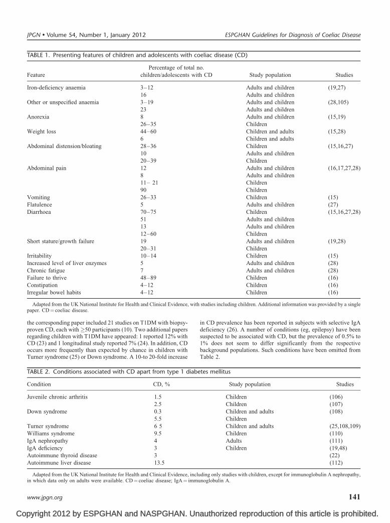
mediated systemic disorder elicited by gluten and related prola-mines in genetically susceptible individuals, characterised by thepresence of a variable combination of gluten-dependent clinicalmanifestations, CD-specific antibodies, HLA-DQ2 and HLA-DQ8haplotypes, and enteropathy. Several classifications of CD havebeen used, most important with distinctions drawn among classical,atypical, asymptomatic, latent, and potential CD. Because atypicalsymptoms may be considerably more common than classic symp-toms, the ESPGHAN working group decided to use the followingnomenclature: gastrointestinal symptoms and signs (eg, chronicdiarrhea) and extraintestinal symptoms and signs (eg, anaemia,neuropathy, decreased bone density, increased risk of fractures).Table 1 provides an extensive list of symptoms and signs of CD inchildren and adolescents.
Silent CD is defined as the presence of positive CD-specificantibodies, HLA, and small-bowel biopsy findings that are compa-tible with CD but without sufficient symptoms and signs to warrantclinical suspicion of CD. Latent CD is defined by the presence ofcompatible HLA but without enteropathy in a patient who has had agluten-dependent enteropathy at some point in his or her life. Thepatient may or may not have symptoms and may or may not haveCD-specific antibodies. Potential CD is defined by the presence ofCD-specific antibodies and compatible HLA but without histologi-cal abnormalities in duodenal biopsies. The patient may or may nothave symptoms and signs and may or may not develop a gluten-dependent enteropathy later.
1. Who to Test
1.1. Evidence BackgroundCD may be difficult to recognise because of the variation in
presentation and intensity of symptoms and signs, and many casesmay actually occur without symptoms. It has been estimated thatonly 1 in 3 to 1 in 7 adult patients with CD are symptomatic (14).The object of this section is to list the symptoms and the concurrent
Husby et al
pyright 2012 by ESPGHAN and NASPGHAN. Un
conditions, which raise sufficient suspicion of CD to warrant furtherinvestigations, so-called CD case finding.
140
CD develops only after the introduction of gluten-containingfoods into a child’s diet. The clinical symptoms of CD may appearin infancy, childhood, adolescence, or adulthood. A GFD in patientswith CD improves or eliminates symptoms and normalises thespecific CD antibodies and histological findings. Therefore, anormal gluten-containing diet with normal quantities of bread,pasta, and other gluten-containing foods should be consumeduntil the end of the diagnostic process. This should be particularlyemphasised to families that consume a low gluten-containing dietbecause of family members diagnosed as having CD. When thediagnosis of CD is suspected in patients who are already receiving aGFD, it is essential that they be placed on a gluten-containing dietbefore initiating the diagnostic process. The length of time of glutenexposure depends on the duration of the GFD. There is no evidencein the scientific literature to suggest the precise amount of glutenthat needs to be ingested to elicit a measurable serological and/orintestinal mucosal response (15). Patients without a conclusivediagnosis of CD, who are already receiving a GFD and do notwant to reintroduce gluten into their diet, must be informed of theconsequences of their decision.
Finally, a GFD is the only lasting treatment for CD. Adher-ence to a GFD in children results in remission of the intestinallesions and promotes better growth and bone mineral density (16). Itis the task of health care professionals to monitor and advise patientsabout adhering to a GFD because compliance with a GFD isvariable and may be as low as 40% (17).
1.2. Evidence Review
Evaluation of the evidence for clinical symptoms of CD wasperformed in the AHRQ report from 2004 for 2 selected signs,anemia and low bone mineral density (10), and included in theNorth American Society for Pediatric Gastroenterology, Hepatol-ogy, and Nutrition guidelines (11). In the 2009 NICE guidelines,data were compiled for a series of symptoms and signs. This sectionis based on these analyses and supplemented with recent literature.
Symptoms and Signs
Gastrointestinal symptoms frequently appear in clinicallydiagnosed childhood CD, including diarrhoea in about 50% ofpatients (15,16,18) and chronic constipation (17). It is unclearwhether chronic abdominal pain is indicative of CD becauserecurrent abdominal pain is so common in childhood. Abdominalpain has been reported as a presenting symptom in 90% of Canadianchildren with CD (18). A shift from gastrointestinal symptoms toextraintestinal symptoms seems to have occurred in children withCD (15,16,19). It is unclear whether this finding reflects a trueclinical variation or an improved recognition of nongastrointestinalforms of CD because of increased awareness of the disease.Researchers have found good evidence that failure to thrive andstunted growth may be caused by CD. The risk of CD in patientswith isolated stunted growth or short stature has been calculated as10% to 40% (20). In some populations, CD is diagnosed inapproximately 15% of children with iron-deficiency anaemia (21).
Associated Conditions
Good evidence exists for the increased prevalence of CD infirst-degree relatives of patients with CD, patients with autoimmunediseases such as T1DM, and autoimmune thyroid disease (22) insome chromosomal aberration disorders and in selective IgA
JPGN � Volume 54, Number 1, January 2012
authorized reproduction of this article is prohibited.
deficiency (Table 2). The prevalence of CD in T1DM has beeninvestigated extensively and is 3% to 12%. The AHRQ report and
www.jpgn.org
Co
TABLE 1. Presenting features of children and adolescents with coeliac disease (CD)
FeaturePercentage of total no.
children/adolescents with CD Study population Studies
Iron-deficiency anaemia 3–12 Adults and children (19,27)16 Adults and children
Other or unspecified anaemia 3–19 Adults and children (28,105)23 Adults and children
Anorexia 8 Adults and children (15,19)26–35 Children
Weight loss 44–60 Children and adults (15,28)6 Children and adults
Abdominal distension/bloating 28–36 Children (15,16,27)10 Adults and children20–39 Children
Abdominal pain 128
Adults and childrenAdults and children
(16,17,27,28)
11– 21 Children90 Children
Vomiting 26–33 Children (15)Flatulence 5 Adults and children (27)Diarrhoea 70–75
51ChildrenAdults and children
(15,16,27,28)
13 Adults and children12–60 Children
Short stature/growth failure 19 Adults and children (19,28)20–31 Children
Irritability 10–14 Children (15)Increased level of liver enzymes 5 Adults and children (28)Chronic fatigue 7 Adults and children (28)Failure to thrive 48–89 Children (16)Constipation 4–12 Children (16)Irregular bowel habits 4–12 Children (16)
with
JPGN � Volume 54, Number 1, January 2012 ESPGHAN Guidelines for Diagnosis of Coeliac Disease
the corresponding paper included 21 studies on T1DM with biopsy-proven CD, each with�50 participants (10). Two additional papersregarding children with T1DM have appeared: 1 reported 12% withCD (23) and 1 longitudinal study reported 7% (24). In addition, CD
Adapted from the UK National Institute for Health and Clinical Evidence,paper. CD¼ coeliac disease.
pyright 2012 by ESPGHAN and NASPGHAN. Un
occurs more frequently than expected by chance in children withTurner syndrome (25) or Down syndrome. A 10-to 20-fold increase
TABLE 2. Conditions associated with CD apart from type 1 diabe
Condition CD, %
Juvenile chronic arthritis 1.52.5
Down syndrome 0.35.5
Turner syndrome 6 5Williams syndrome 9.5IgA nephropathy 4IgA deficiency 3Autoimmune thyroid disease 3Autoimmune liver disease 13.5
Adapted from the UK National Institute for Health and Clinical Evidence, incluin which data only on adults were available. CD¼ coeliac disease; IgA¼ immu
www.jpgn.org
in CD prevalence has been reported in subjects with selective IgAdeficiency (26). A number of conditions (eg, epilepsy) have beensuspected to be associated with CD, but the prevalence of 0.5% to1% does not seem to differ significantly from the respective
studies including children. Additional information was provided by a single
authorized reproduction of this article is prohibited.
background populations. Such conditions have been omitted fromTable 2.
tes mellitus
Study population Studies
Children (106)Children (107)Children and adults (108)ChildrenChildren and adults (25,108,109)Children (110)Adults (111)Children (19,48)
(22)(112)
ding only studies with children, except for immunoglobulin A nephropathy,noglobulin A.
141
Co
1.3. Evidence Statements
Husby et al
1.3.1.
Patients with CD may present with a wide range of symptomsand signs or be asymptomatic. Symptoms in CD are adapted from
the Nguidpy
b.
the
14
ICE guidelines. �Denotes added to the list from the NICEelines, þ denotes a particularly common symptom.
a. G
astrointestinal: Chronic diarrheaþ, chronic constipation,abdominal painþ, nausea vomiting, distended abdomenþ�.Extraintestinal: Failure-to-thriveþ�, stunted growthþ, delayedpuberty, chronic anaemiaþ, decreased bone mineralisation(osteopenia/osteoporosis)þ,dental enamel defects, irritability, chronic fatigue, neuropathy, arthritis/arthralgia,amenorrhea,increased levels of liver enzymesþ.LOE: 2.References (15,16,19,26,27)Total number of votes: 13, Agree: 13, Disagree: 0,
Abstensions: 0
1.3.2.
The following signs or diagnoses may be present when CD isdiagnosed (�data from adults): short stature, amenorrhoea, recurrentaphthous stomatitis (mouth ulcers)�, dental enamel defects, derma-titis herpetiformis, osteopenia/osteoporosis, abnormal liver bio-chemistry.
LOE: 2.References: (16,28)Total number of votes: 13, Agree: 13, Disagree: 0,
Abstensions: 0
1.3.3.
CD has an increased prevalence in children and adolescentswith first-degree relatives with CD (10%–20%), T1DM (3%–12%),Down syndrome (5%–12%), autoimmune thyroid disease (up to 7%),Turner syndrome (2%–5%), Williams syndrome (up to 9%), IgAdeficiency (2%–8%), and autoimmune liver disease (12%–13%).
LOE: 1.References: See Table 1.Total number of votes: 13, Agree 13, Disagree: 0,
Abstentions: 0
1.4. Recommendations
1.4.1.
("") Offer CD testing in children and adolescents with thefollowing otherwise unexplained symptoms and signs: chronicabdominal pain, cramping or distension, chronic or intermittentdiarrhoea, growth failure, iron-deficiency anaemia, nausea orvomiting, chronic constipation not responding to usual treatment,weight loss, chronic fatigue, short stature, delayed puberty, ameor-rhoea, recurrent aphthous stomatitis (mouth ulcers), dermatitisherpetiformis–type rash, repetitive fractures/osteopenia/osteoporo-sis, and unexplained abnormal liver biochemistry.
Total number of votes: 13, Agree: 12, Disagree: 1,Abstentions: 0
1.4.2.
right 2012 by ESPGHAN and NASPGHAN. Un
("") Offer CD testing in children and adolescents withfollowing conditions: T1DM, Down syndrome, autoimmune
2
thyroid disease, Turner syndrome, Williams syndrome, IgAdeficiency, autoimmune liver disease, and first-degree relativeswith CD.
Total number of votes: 13, Agree: 11, Disagree: 2,Abstentions: 0
1.4.3.
("") To avoid false-negative results, infants, children, andadolescents should be tested for CD only when they are consuminga gluten-containing diet. Paediatricians and gastroenterologistsshould always ask before testing whether the patients are consum-ing gluten.
Total number of votes: 13, Agree: 13, Disagree: 0,Abstentions: 0
1.4.4.
("") In infants, CD antibodies should be measured only afterthe introduction of gluten-containing foods as complementary to theinfant’s diet.
Total number of votes: 13, Agree: 13, Disagree: 0,Abstentions: 0
1.4.5.
A GFD should be introduced only after the completion of thediagnostic process, when a diagnosis of CD has been conclusivelymade. Health care professionals should be advised that puttingpatients on a GFD, when CD has not been excluded or confirmed,may be detrimental. GFD is a lifelong treatment, and consuminggluten later can result in significant illness.
Total number of votes: 13, Agree: 13, Disagree: 0, Absten-tions: 0
2. HLA Aspects
2.1. Evidence BackgroundThe principal determinants of genetic susceptibility for CD
are the major histocompatibility class II HLA class II DQA andDQB genes coded by the major histocompatibility region in theshort arm of chromosome 6. More than 95% of patients with CDshare the HLA-DQ2 heterodimer, either in the cis (encoded byHLA-DR3-DQA1�0501-DQB1�0201) or the trans configuration(encoded by HLA-DR11-DQA1�0505 DQB1 0301/DR7-DQA1�0201 DQB1 0202), and most of the remainder have theHLA-DQ8 heterodimer (encoded by DQA1�0301-DQB1�0302).CD is a multigenetic disorder, which means that the expression ofthese HLA-DQ2 or HLA-DQ8 molecules is necessary but notsufficient to cause disease because approximately 30% to 40%of the white population holds the HLA-DQ2 haplotype and only 1%develops CD. Outside the HLA region there are several genomicareas related to CD, controlling immune responses, amongothers the genes encoding for CTLA4, IL2, IL21, CCR3, IL12A,IL-18RAP, RGS1, SH2B3 and TAGAP (29–31). Their contributionto the genetics of CD is relatively small in comparison to that ofHLA-DQ2 and HLA-DQ8. The strong relation between HLAgenetic factors and CD is illustrated by the effect of the HLA-DQ2 gene dose on disease development; HLA-DQ2 homozygousindividuals have an at least 5 times higher risk of disease devel-opment compared with HLA-DQ2 heterozygous individuals (32).
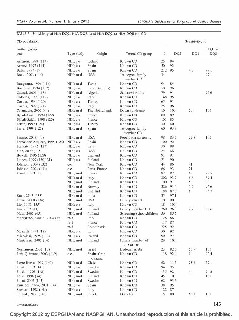
Table 3 presents the sensitivity of HLA-DQ2 and -DQ8 for
JPGN � Volume 54, Number 1, January 2012
authorized reproduction of this article is prohibited.
CD as assessed by the Dutch evidence-based guidelines for CD anddermatitis herpetiformis (33a). Most of the studies included control
www.jpgn.org
Copyright 2012 by ESPGHAN and NASPGHAN. Unauthorized reproduction of this article is prohibited.
TABLE 3. Sensitivity of HLA-DQ2, HLA-DQ8, and HLA-DQ2 or HLA-DQ8 for CD
CD population Sensitivity, %
Author group,year Type study Origin Tested CD group N DQ2 DQ8
DQ2 orDQ8
Arnason, 1994 (113) NIH; c-c Iceland Known CD 25 84Arranz, 1997 (114) NIH; c-c Spain Known CD 50 92Balas, 1997 (39) NIH; c-c Spain Known CD 212 95 4.3 99.1Book, 2003 (115) NIH; m-d USA 1st-degree family
member CD34 97.1
Bouguerra, 1996 (116) NIH; m-d Tunis Known CD 94 84Boy et al, 1994 (117) NIH; c-c Italy (Sardinia) Known CD 50 96Catassi, 2001 (118) NIH; m-d Algeria Saharawi Arabs 79 91 95.6Colonna, 1990 (119) NIH; c-c Italy Known CD 148 95Congia, 1994 (120) NIH; c-c Turkey Known CD 65 91Congia, 1992 (121) NIH; c-c Italy Known CD 25 96Csizmadia, 2000 (44) NIH; m-d The Netherlands Down syndrome 10 100 20 100Djilali-Saiah, 1994 (122) NIH; c-c France Known CD 80 89Djilali-Saiah, 1998 (123) NIH; c-c France Known CD 101 83Erkan, 1999 (124) NIH; c-c Turkey Known CD 30 40Farre, 1999 (125) NIH; m-d Spain 1st-degree family
member CD60 93.3
Fasano, 2003 (40) NIH; m-d USA Population screening 98 83.7 22.5 100Fernandez-Arquero, 1995 (126) NIH; c-c Spain Known CD 100 92Ferrante, 1992 (127) NIH; c-c Italy Known CD 50 88Fine, 2000 (128) NIH; c-c USA Known CD 25 88Howell, 1995 (129) NIH; c-c England Known CD 91 91Iltanen, 1999 (130,131) NIH; c-c Finland Known CD 21 90Johnson, 2004 (132) c-c New York Known CD 44 86 41Johnson, 2004 (132) c-c Paris, France Known CD 66 93 21Karell, 2003 (33) NIH; m-d France Known CD 92 87 6.5 93.5
NIH; m-d Italy Known CD 302 93.7 5.6 89.4NIH; m-d Finland Known CD 100 91 5 96NIH; m-d Norway Known CD 326 91.4 5.2 96.6NIH; m-d England Known CD 188 87.8 8 95.7
Kaur, 2003 (133) NIH; m-d India Known CD 35 97.1Lewis, 2000 (134) NIH; m-d USA Family van CD 101 90Lio, 1998 (135) NIH; c-c Italy Known CD 18 100Liu, 2002 (41) NIH; m-d Finland Family member CD 260 96.9 2.7 99.6Maki, 2003 (45) NIH; m-d Finland Screening schoolchildren 56 85.7Margaritte-Jeannin, 2004 (35) m-d Italy Known CD 128 86
m-d France Known CD 117 87m-d Scandinavia Known CD 225 92
Mazzilli, 1992 (136) NIH; c-c Italy Known CD 50 92Michalski, 1995 (137) NIH; c-c Ireland Known CD 90 97Mustalahti, 2002 (14) NIH; m-d Finland Family member of
CD of DH29 100
Neuhausen, 2002 (138) NIH; m-d Israel Bedouin Arabs 23 82.6 56.5 100Pena-Quintana, 2003 (139) c-c Spain, Gran
CanariaKnown CD 118 92.4 0 92.4
Perez-Bravo 1999 (140) NIH; m-d Chile Known CD 62 11.3 25.8 37.1Ploski, 1993 (141) NIH; c-c Sweden Known CD 94 95Ploski, 1996 (142) NIH; m-d Sweden Known CD 135 92 4.4 96.3Polvi, 1996 (34) NIH; m-d Finland Known CD 45 100 100Popat, 2002 (143) NIH; m-d Sweden Known CD 62 93,6Ruiz del Prado, 2001 (144) NIH; c-c Spain Known CD 38 95Sachetti, 1998 (145) NIH; c-c Italy Known CD 122 87Sumnik, 2000 (146) NIH; m-d Czech Diabetes 15 80 66.7 100
JPGN � Volume 54, Number 1, January 2012 ESPGHAN Guidelines for Diagnosis of Coeliac Disease
www.jpgn.org 143
Co
CD population Sensitivity, %
Author group,year Type study Origin Tested CD group N DQ2 DQ8
DQ2 orDQ8
Tighe, 1992 (147) NIH; c-c Italy Known CD 43 91Tighe, 1993 (148) NIH; c-c Israel Ashkenazi
Jews, known CD34 71
Tumer, 2000 (149) NIH; c-c Turkey Known CD 33 52Tuysuz, 2001 (150) NIH; m-d Turkey Known CD, children 55 84 16.4 90.9Vidales, 2004 (42) m-d Spain Known CD, children 136 94.1 2.1 95.6Zubilaga, 2002 (151) NIH; m-d Spain Known CD 135 92.6 3.7 96SensitivityNo. studies n¼ 55 n¼ 19 n¼ 20Median 91 6.5 96.2p10–p90 82.6–97.0 2.3–50.3 90.2–100p25–p75 86.3–94.0 4.3–22.1 94.6–99.8
IH,eids
TABLE 3. (Continued )
Husby et al JPGN � Volume 54, Number 1, January 2012
groups without results of small-bowel biopsies and were notdesigned primarily to assess the use of HLA typing in the diagnosisof CD. These studies reflect clearly the frequency of HLA-DQ2 andHLA-DQ8 in patients with CD. Table 4 presents the results of thestudies included in the AHRQ report for the diagnosis of CD from2004 (10) and a number of studies published after October 2003. Allof the studies included more than 10 patients with CD. The results ofthe more recent studies did not change the conclusions regarding thesensitivity of HLA-DQ2 and HLA-DQ8 as stated by the AHRQreport. The sensitivity of HLA-DQ2 is high (median 91%; p25–p7586.3%– 94.0%), and if combined with HLA-DQ8 (at least 1 is posi-tive), it is even higher (median 96.2%; p25–p75 94.6%–99.8%),making extremely small the chance of an individual who is negativefor DQ2 and DQ8 to have CD; the small percentage of HLA-DQ2-negative and HLA-DQ8-negative patients is well documented (33–35).
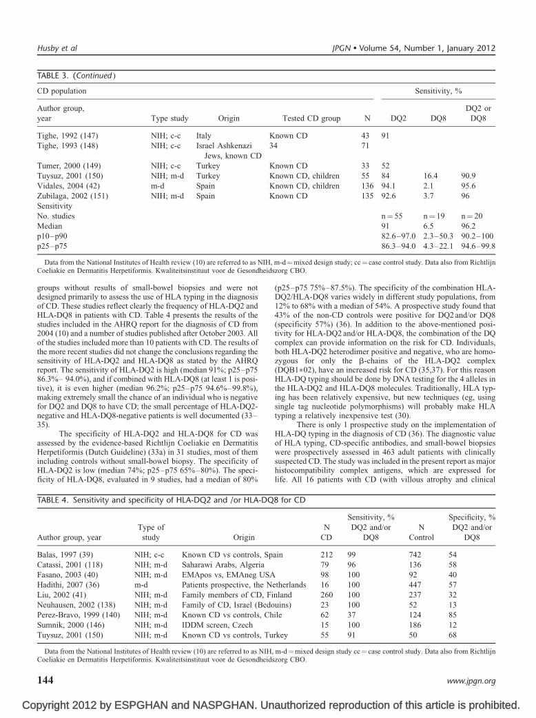
The specificity of HLA-DQ2 and HLA-DQ8 for CD wasassessed by the evidence-based Richtlijn Coeliakie en DermatitisHerpetiformis (Dutch Guideline) (33a) in 31 studies, most of themincluding controls without small-bowel biopsy. The specificity of
Data from the National Institutes of Health review (10) are referred to as NCoeliakie en Dermatitis Herpetiformis. Kwaliteitsinstituut voor de Gesondh
pyright 2012 by ESPGHAN and NASPGHAN. Un
HLA-DQ2 is low (median 74%; p25–p75 65%–80%). The speci-ficity of HLA-DQ8, evaluated in 9 studies, had a median of 80%
TABLE 4. Sensitivity and specificity of HLA-DQ2 and /or HLA-DQ
Author group, yearType of
study Origin
Balas, 1997 (39) NIH; c-c Known CD vs controls, SpCatassi, 2001 (118) NIH; m-d Saharawi Arabs, AlgeriaFasano, 2003 (40) NIH; m-d EMApos vs, EMAneg USAHadithi, 2007 (36) m-d Patients prospective, the NLiu, 2002 (41) NIH; m-d Family members of CD, FiNeuhausen, 2002 (138) NIH; m-d Family of CD, Israel (BedoPerez-Bravo, 1999 (140) NIH; m-d Known CD vs controls, ChSumnik, 2000 (146) NIH; m-d IDDM screen, CzechTuysuz, 2001 (150) NIH; m-d Known CD vs controls, Tu
Data from the National Institutes of Health review (10) are referred to as NIH,Coeliakie en Dermatitis Herpetiformis. Kwaliteitsinstituut voor de Gesondheids
144
(p25–p75 75%–87.5%). The specificity of the combination HLA-DQ2/HLA-DQ8 varies widely in different study populations, from12% to 68% with a median of 54%. A prospective study found that43% of the non-CD controls were positive for DQ2 and/or DQ8(specificity 57%) (36). In addition to the above-mentioned posi-tivity for HLA-DQ2 and/or HLA-DQ8, the combination of the DQcomplex can provide information on the risk for CD. Individuals,both HLA-DQ2 heterodimer positive and negative, who are homo-zygous for only the b-chains of the HLA-DQ2 complex(DQB1�02), have an increased risk for CD (35,37). For this reasonHLA-DQ typing should be done by DNA testing for the 4 alleles inthe HLA-DQ2 and HLA-DQ8 molecules. Traditionally, HLA typ-ing has been relatively expensive, but new techniques (eg, usingsingle tag nucleotide polymorphisms) will probably make HLAtyping a relatively inexpensive test (30).
There is only 1 prospective study on the implementation ofHLA-DQ typing in the diagnosis of CD (36). The diagnostic valueof HLA typing, CD-specific antibodies, and small-bowel biopsieswere prospectively assessed in 463 adult patients with clinicallysuspected CD. The study was included in the present report as major
m-d¼mixed design study; cc¼ case control study. Data also from Richtlijnzorg CBO.
authorized reproduction of this article is prohibited.
histocompatibility complex antigens, which are expressed forlife. All 16 patients with CD (with villous atrophy and clinical
8 for CD
NSensitivity, %
NSpecificity, %
CDDQ2 and/or
DQ8 ControlDQ2 and/or
DQ8
ain 212 99 742 5479 96 136 5898 100 92 40
etherlands 16 100 447 57nland 260 100 237 32uins) 23 100 52 13ile 62 37 124 85
15 100 186 12rkey 55 91 50 68
m-d¼mixed design study cc¼ case control study. Data also from Richtlijnzorg CBO.
www.jpgn.org
Co
response after GFD) were HLA-DQ2 or HLA-DQ8 positive, butthere were no cases of CD among the 255 HLA-DQ2-negative andHLA-DQ8-negative patients. Because the chance of an individualnegative for HLA-DQ2 or HLA-DQ8 having CD is extremelysmall, the main role of HLA-DQ typing in the diagnosis of CDis to exclude the disease or to make it unlikely.
Some evidence exists that HLA-DQ2/HLA-DQ8 typingplays a role in the case-finding strategy in individuals who belongto groups at risk for CD. These individuals include, among others,first-degree relatives of a confirmed case (3) and patients withimmune-mediated as well as nonimmune conditions known to beassociated with CD (Table 2). A negative result for HLA-DQ2/HLA-DQ8 renders CD highly unlikely in these children, and there isno need for subsequent CD antibodies testing of such individuals.
2.2. Evidence Statements
JPGN � Volume 54, Number 1, January 2012
2.2.1.
There is a strong genetic predisposition to CD with the majorrisk attributed to the specific genetic markers known as HLA-DQ2and HLA-DQ8.
LOE: 1.References (10,38)Total number of votes: 13, Agree: 13, Disagree: 0,
Abstentions: 0
2.2.2.
The vast majority of CD patients are HLA-DQ2 (full orincomplete heterodimer) and/or HLA-DQ8 positive.
LOE: 2.References (38,39)Total number of votes: 13, Agree: 13, Disagree: 0,
Abstentions: 0
2.2.3.
Individuals having neither DQ2 nor DQ8 are unlikely to haveCD because the sensitivity of HLA-DQ2 is high (median 91%), andif combined with HLA-DQ8 (at least 1 of them positive), it is evenhigher (96%). The main role of HLA-DQ typing in the diagnosis ofCD is to exclude the disease.
LOE: 2.References (33,39–42)Total number of votes: 13, Agree: 13, Disagree: 0,
Abstentions: 0
2.2.4.
HLA-DQ2 and/or HLA-DQ8 have poor specificity for CD(median 54%), indicating a low positive predictive value for CD.
LOE: 2.References (36,39)Total number of votes: 13, Agree: 13, Disagree: 0,
Abstensions: 0
2.2.5.
HLA-DQ typing should not be done by serology but by DNAtesting for the 4 alleles in the HLA-DQ2 and HLA-DQ8 molecules.New techniques (eg, using tag single nucleotide polymorphisms)will make HLA typing available at a relatively low cost.
pyright 2012 by ESPGHAN and NASPGHAN. Un
LOE: 2.References (35,37,43)
www.jpgn.org
Total number of votes: 13, Agree: 13, Disagree: 0,Abstensions: 0
2.2.6.
HLA-DQ2/HLA-DQ8 typing has a role in the case-findingstrategy in individuals who belong to groups at risk for CD. Anegative result for HLA-DQ2/HLA-DQ8 renders CD highly unli-kely in these children, and hence there is no need for subsequent CDantibodies testing in such individuals.
LOE: 2.References (3,44,45)Total number of votes: 13, Agree: 13, Disagree: 0,
Abstentions: 0
2.3. Recommendations
2.3.1.
("") Offer HLA-DQ2 and HLA-DQ8 typing in patients withuncertain diagnosis of CD, for example, in patients with negative CD-specific antibodies and mild infiltrative changes in small-bowel speci-mens. Negative results render CD highly unlikely in these children.
Total number of votes: 13, Agree: 13, Disagree: 0,Abstentions: 0
2.3.2.
("") In patients with a clinical suspicion of CD, who areHLA-DQ2 negative and HLA-DQ8 negative, offer investigationsfor other causes of the symptoms (ie, different from CD).
Total number of votes: 13, Agree: 13, Disagree: 0,Abstentions: 0
2.3.3.
("") Start the screening for CD in groups at risk by HLA-DQ2 and HLA-DQ8 typing if the test is available. These groupsinclude first-degree relatives of a patient with a confirmed case andpatients with autoimmune and nonautoimmune conditions known tobe associated with CD, such as T1DM, Down syndrome, and Turnersyndrome.
Total number of votes: 13, Agree: 12, Disagree: 0,Abstentions: 1
2.3.4
(") If CD can be diagnosed without performing small-bowelbiopsies in children with strong clinical suspicion of CD and withhigh specific CD antibodies, consider performing HLA-DQ2/HLA-DQ8 typing in these children to add strength to the diagnosis.
Total number of votes: 13, Agree: 12, Disagree: 0,Abstentions: 1
3. Antibodies
3.1. Evidence BackgroundCD is characterised by highly specific autoantibodies
directed against the common CD autoantigen TG2 (10) and byantibodies against DGP (46). EMA are directed against extracellu-lar TG2 (47). Except for DGP antibodies these antibodies aretypically of the IgA class. In IgA-deficient patients with CD, thesame type of antibodies in IgG class can be detected (48).
ESPGHAN Guidelines for Diagnosis of Coeliac Disease
authorized reproduction of this article is prohibited.
Antibodies against TG2 bind in vivo to a patient’s own TG2expressed in the small bowel or in other tissues (eg, liver, muscles,
145
Co
central nervous system) at sites accessible to the antibodies (47,49).Dermatitis herpetiformis is defined by the presence of granular IgAdeposition in the dermal papillae of the skin containing antibodiesagainst tissue transglutaminase type 3 (TG3). The appearance ofCD-specific antibodies in the blood or in tissues may precede thedevelopment of structural abnormalities in the small bowel(50,51).
CD antibodies are not detectable in the blood of all patientswith CD (10,52); however, TG2-specific antibodies may be presentin small-intestine tissue or other tissues of seronegative patients(49,53). Negative antibody results in blood also can be obtained insubjects with dermatitis herpetifomis, after reduction of glutenconsumption or during and after the use of immunosuppressivedrugs (54–56).
3.2. Evidence Review
Antibody Detection
IgA and IgG class anti-TG2 antibodies can be detected inblood samples of patients by various immunoassays (enzyme-linkedimmunosorbent assay, radioimmunoassay, or others) using purifiedor recombinant TG2 antigens or tissue sections/fluids containingTG2. Most often serum is used, but plasma or whole blood also can besuitable sources (57). Immunofluorescent tests such as EMA requiremicroscopic evaluation and may be subject to interobserver varia-bility. Despite these limitations, the specificity of EMA test results is98% to 100% in expert laboratories (10,52), and this test is consideredthe reference standard for CD-specific antibody detection. CD anti-bodies also can be detected by the use of synthetic peptides corre-sponding to deamidated gliadin sequences (46,58).
Antibody Values and Assay Performance
The values for serum anti-TG2 or anti-DGP levels obtainedin a particular test depend on the source (human or animal) of theantigen, quality of the antigen, exposure of the antigen, calibrators,buffers, measuring methods, cutoff values and calculation mode ofthe results, so numerical values obtained with different kits maydiffer substantially. No universally accepted international standardsare available that would allow the expression of antibody amount inabsolute Ig concentrations; however, the majority of commercialkits use a calibration curve with antibody dilutions that providenumerical values that are proportional to antibody concentration inrelative (arbitrary) units.
This is the preferred method for clinical evaluation. Antibodytests that calculate results from the percentage of absorbance valuessupply numerical values that correlate with the logarithmic valuesof antibody concentrations. Despite these differences, many com-mercial anti-TG2 antibody tests have equally high sensitivity andspecificity on the same blood samples (59). Interlaboratory varia-bility also exists (60). In addition, there may be considerable batch-to-batch variability within commercial anti-TG2 assays, whichneeds to be monitored by the use of independent qualitycontrol material.
The performance of a particular antibody test in a clinicalsetting depends on patient characteristics (age, genetic predisposi-tion, IgA deficiency), pretest probability, stage of the disease, andingested amounts of gluten. These factors should be taken intoaccount when interpreting positive and negative antibody resultsand establishing the optimal cutoff limits (55,59,61). This can bedone by receiver operating characteristics curve plotting sensitivity
Husby et al
pyright 2012 by ESPGHAN and NASPGHAN. Un
against 1–specificity. Anti-TG2 antibodies also can be detected insaliva. Sufficient sensitivity and specificity was not achieved with
146
conventional commercially available immunoassays (62,63),although the use of radiobinding assays appeared to be morefavourable (64). There is no reliable method to detect specificCD antibodies from faecal samples (65).
Anti-TG2 antibody detection also can be done from the bloodat the point of contact using rapid test kits (POC test) (57,66,67), butonly as a semiquantitative test for circulating antibodies. Anti-TG2antibodies detection by POC test may achieve a high accuracy forCD diagnosis, and the ESPGHAN evidence report on CD serology(11a) reported a pooled sensitivity of 96.4% and a pooled specificityof 97.7%; however, IgA-antiTG2 or EMA performed better. Pub-lished studies have thus far been based on populations with a highprevalence of CD because 60.3% of all of the patients had biopsy-confirmed CD. Assuming a prevalence of CD in 5% of all sympto-matic children, the positive predictive value would be 68.6% andthe negative predictive value would be 99.8% (11a). The expertiseof the laboratory or of the observers has a great effect on theaccuracy of the results in EMA and rapid tests (67).
Disease Prediction
The positivity for anti-TG2 and/or EMA is associated with ahigh probability for CD in children and adolescents (10,52);however, low levels of anti-TG2 have been described in a numberof conditions unrelated to CD, such as other autoimmune diseases,infections, tumours, myocardial damage, liver disorders, and psor-iasis (68–70). These antibodies are not associated with the EMAreaction, which explains why EMA has higher reliability for thediagnosis of CD. The ESPGHAN evidence report on CD serology(11a) estimates the pooled positive and negative likelihood ratios ofEMA results in the studies performed between 2004 and 2009 as31.8 (95% confidence limit 18.6– 54.0) and 0.067 (95% confidencelimit 0.038–0.12), respectively. Furthermore, EMA results weremore homogeneous than results obtained with other CD antibodytests and had a high diagnostic odds ratio of 553.6. Taken together,these data mean that the presence of CD is likely if the EMA testresult is positive (11a). Remarkably, EMA positivity also is associ-ated with the later development of villous atrophy in the fewreported cases of both adults and children with CD (50,71–73)who initially do not fulfill the histological criteria of CD because ofnormal small-intestinal architecture.
In the ESPGHAN report on CD antibodies, the specificity ofanti-TG2 antibodies measured by enzyme-linked immunosorbentassay was lower than that of EMA testing and varied according tothe test kit used (11a). It was not possible to obtain pooledperformance estimates on sensitivity and specificity resulting fromthe heterogeneity in the evaluated studies, but for 11 of 15 studypopulations the sensitivity reached �90% and for 13 of 15 studypopulations specificity reached �90%. Several studies confirmedthat high concentrations of anti-TG2 antibodies in serum predictvillous atrophy better than low or borderline values (55,74,75).These studies suggested that high anti-TG2 antibody levels can bedefined as those exceeding 10 times ULN in concentration-depen-dent antibody tests based on calibration curves. Testing for anti-TG2 antibodies in serum is the preferred initial approach to find CD.The cutoff for such high values in a number of different commercialtests is examined in Appendix I.
Although tests for anti-DGP antibodies performed favour-ably and much better than antibodies against native gliadin, theirperformance was inferior compared with anti-TG2 or EMA assays(55,11a,76); however, their performance in patients not preselectedby anti-TG2 or EMA testing must be resolved in prospective
JPGN � Volume 54, Number 1, January 2012
authorized reproduction of this article is prohibited.
studies. In addition, their role in the diagnosis in children youngerthan 2 to 3 years required further assessment in large prospective
www.jpgn.org
Co
studies, especially in a head-to-head comparison with anti-TG2 orEMA detection (58,77,78). Conventional or native gliadin antibodytests have, in general, low specificity and sensitivity (10,11a). Someevidence exists, however, that their sensitivity may be higher inchildren younger than 2 years in comparison with EMA and anti-TG2 tests (79). Unfortunately, the specificity is low in this agegroup and makes anti-gliadin antibody tests unhelpful in clinicalpractice. It is thus advisable to obtain a small-intestine biopsysample in young children with severe symptoms suggestive ofCD, even when their serology is negative (73,80). If villous atrophyis found in children who are negative for CD-specific antibodies,then a later gluten challenge procedure always should be performedto confirm CD as a cause of the enteropathy.
IgA deficiency must be taken into consideration in a sub-group of children in the choice of diagnostic tests and the interpret-ation of the results. It is important to exclude IgA deficiency bymeasuring serum total IgA levels. IgA-deficient children can beevaluated on the basis of IgG class tests (26).
3.3. Evidence Statements
JPGN � Volume 54, Number 1, January 2012
3.3.1.
CD is characterised by highly specific autoantibodiesdirected against the common CD autoantigen TG2 (‘‘tissue’’TG), including EMA and by antibodies against DGP.
LOE: 1.References (10,11a)Total number of votes: 13, Agree: 13, Disagree: 0,
Abstentions: 0
3.3.2.
In subjects with normal serum IgA values for age, a positiveIgA class EMA result or a positive IgA class anti-TG2 antibodyresult is considered to be a CD-relevant antibody positivity. In thecase of IgA deficiency, a positive IgG class EMA result, a positiveIgG class anti-TG2 antibody, or a positive IgG class anti-DGPantibody is diagnostically relevant.
LOE: 1.References (10,26,48,11a,78a)Total number of votes: 13, Agree: 13, Disagree: 0,
Abstentions: 0
3.3.3.
It is not required that IgA-competent patients with CD bepositive in both IgA and IgG class CD antibody tests. Isolatedpositivity for IgG class CD antibodies in a person with normalserum IgA levels does not have the same specificity and clinicalrelevance as the positivity of IgA class antibodies.
LOE: 2.References (10,11a)Total number of votes: 13, Agree: 13, Disagree: 0,
Abstentions: 0
3.3.4.
The numeric values obtained with different test kits in anti-TG2 or anti-DGP antibody measurements cannot be directly com-pared because they may differ in their measurement principles,calibrators, and calculation mode of results.
pyright 2012 by ESPGHAN and NASPGHAN. Un
LOE: 2.References (10,59,11a)
www.jpgn.org
Total number of votes: 13, Agree: 13, Disagree: 0,Abstentions: 0
3.3.5.
For blood anti-TG2 antibody tests that use calibration curvesto express antibody concentration, values exceeding 10 times ULNmay be denoted as high antibody positivity. For other tests, valuesconsidered to be high antibody positivity should be established bycomparison with a panel of tests, which are listed in Appendix II.
LOE: 3.References (55,74,75)Total number of votes: 13, Agree: 13, Disagree: 0,
Abstentions: 0
3.3.6.
EMA testing in experienced hands has the highest specificityand positive likelihood ratio for CD among the available serologytools. It is more likely that CD is present if the EMA result ispositive than if another CD antibody result is positive.
LOE: 1.References (11a)Total number of votes: 12, Agree: 12, Disagree: 0,
Abstentions: 1
3.3.7.
The specificity and positive predictive value of serum anti-TG2 antibody measured by immunoassays other than EMA is lowerthan those of positive EMA results. Isolated positivity for anti-TG2,especially in the low positivity range, can occur in conditions thatare unrelated to CD, such as other autoimmune conditions, infec-tions, tumours, or tissue damage.
LOE: 1.References (62,11a–70,81,82)Total number of votes: 12, Agree: 12, Disagree: 0,
Abstentions: 1
3.3.8.
High concentrations of anti-TG2 antibodies in blood (asdefined in statement 2.3.5) predict villous atrophy better thanlow positive or borderline values.
LOE: 2.References (55,74,75)Total number of votes: 13, Agree: 13, Disagree: 0,
Abstentions: 0
3.3.9.
Rapid anti-TG2 antibody detection at the point of contact canperform with high accuracy similar to anti-TG2 antibody detectionby laboratory measurements. The evaluation of rapid tests is lessreliable if done by untrained or laypeople. Quantification as inserum immunoassays is not possible at present.
LOE: 1.References (67,11a)Total number of votes: 12, Agree: 12, Disagree: 0,
Abstentions: 1
3.3.10.
ESPGHAN Guidelines for Diagnosis of Coeliac Disease
authorized reproduction of this article is prohibited.
Anti-TG2 antibody or EMA testing from a blood sample hasa higher accuracy than antibody testing against DGP, unless special
147
Co
patient characteristics are present (IgA deficiency, age younger than2 years).
LOE: 1.References: (11a,76)Total number of votes: 13, Agree: 13, Disagree: 0,
Abstentions: 0
3.3.11.
Anti-TG2 antibodies are detectable in saliva samples frompatients with CD, but the accuracy of available diagnostic tests islower compared with serological tests.
LOE: 3.References (64)Total number of votes: 12, Agree: 12, Disagree: 0,
Abstentions: 1
3.3.12.
Tests for the detection of IgG or IgA antibodies againstnative gliadin (conventional gliadin antibody test) are neithersufficiently sensitive nor sufficiently specific for the detectionof CD.
LOE: 1.References (10,11a)Total number of votes: 13, Agree: 13, Disagree: 0,
Abstentions: 0
3.3.13.
Tests for the detection of CD antibodies of any isotype (IgG,IgA, secretory IgA) in fecal samples are unreliable.
LOE: 3.References (65,11a)Total number of votes: 13, Agree: 13, Disagree: 0,
Abstentions: 0
3.3.14.
The expertise of the laboratory and the selection of the test kitinfluence the accuracy of CD antibody tests.
LOE: 2.References (59,60)Total number of votes: 13, Agree: 13, Disagree: 0,
Abstentions: 0
3.3.15.
Demonstration of in vivo-bound anti-TG2 antibodies on thecell surface in the small bowel or in other tissues supports thediagnosis of CD.
LOE: 2.References (49,50,53,67,73)Total number of votes: 13, Agree: 13, Disagree: 0,
Abstentions: 0
3.4. Recommendations
Husby et al
3.4.1.
("") Every antibody test used for the diagnosis of childhood
pyright 2012 by ESPGHAN and NASPGHAN. Un
CD must be validated in a paediatric population of at least 50 chil-dren with active CD and 100 control children of different ages
148
against the reference of EMA positivity detected under standardconditions in an expert laboratory.
(") Alternatively, a CD test can be validated in childrenagainst reference results of histology or against another anti-TG2antibody test with performance similar to EMA. A test is con-sidered as reliable if it shows >95% agreement with the referencetest.
In both situations, seek statistical advice.Total number of votes: 13, Agree: 12, Disagree: 1,
Abstentions: 0
3.4.2.
("") The optimal threshold values for antibody positivity(ULN) of a test should be established. This is done by receiveroperating characteristics curves plotting sensitivity against speci-ficity at different cutoff levels.
(") In the case of new anti-TG2 antibody measuring tests, it isalso advisable to establish the range of high positivity (in relation toULN).
Total number of votes: 13, Agree: 13, Disagree: 0,Abstentions: 0
3.4.3.
("") Laboratories providing CD antibody test results fordiagnostic use should participate continuously in a quality controlprogramme at a national or a European level.
Total number of votes: 13, Agree: 13, Disagree: 0,Abstentions: 0
3.4.4.
("") Anti-TG2 and anti-DGP laboratory test results shouldbe reported as numeric values together with specification of theIg class measured, the manufacturer, the cutoff value defined forthe specific test kit, and (if available) the level of ‘‘high’’antibody values. It is not sufficient to state only positivity ornegativity. Information on the source of the antigen (natural,recombinant, human, nonhuman) should be provided for in-housemethods.
Total number of votes: 13, Agree: 13, Disagree: 0,Abstentions: 0
3.4.5.
("") Reports on EMA results should contain the specificationof the investigated Ig class, the interpretation of the result (positiveor negative), the cutoff dilution and the specification of the substratetissue. It is also useful to have the information on the highestdilution that is still positive.
Total number of votes: 13, Agree: 13, Disagree: 0,Abstentions: 0
3.4.6.
(") If a rapid or point-of-contact CD antibody test is used by ahealth care professional, the type of the device and class of theinvestigated antibodies and testing for IgA deficiency shouldbe recorded.
JPGN � Volume 54, Number 1, January 2012
authorized reproduction of this article is prohibited.
Total number of votes: 12, Agree: 12, Disagree: 0,Abstentions: 1
www.jpgn.org
Co
3.4.7.
("") A diagnostic test for CD-specific antibody detectionshould be the first tool used to identify patients with symptoms andsigns suggestive of CD for further diagnostic workup (eg, refinedserological testing, HLA typing, small-intestine biopsies) or to ruleout CD. Patients should be tested for CD-specific antibodies whenon a gluten-containing diet.
Total number of votes: 13, Agree: 13, Disagree: 0,Abstentions: 0
3.4.8.
("") For initial testing in symptomatic patients, a quantitativetest detecting IgA class anti-TG2 or EMA from a blood sample isrecommended. If total serum IgA is not known, measurementis recommended.
("") In subjects with either primary or secondary humoralIgA deficiency, at least 1 additional test measuring IgG class CDantibodies (IgG anti-TG2, IgG anti-DGP, or IgG EMA, or blendedkits for both IgA and IgG antibodies) is recommended.
Total number of votes: 13, Agree: 13, Disagree: 0,Abstentions: 0
3.4.9.
(") Rapid CD antibody detection kits meeting the require-ments set forth above for CD antibody
testing can be applied for initial testing.("") Rapid testing is not meant to replace laboratory testing
or to provide a final diagnosis.Total number of votes: 12, Agree: 10, Disagree: 2,
Abstentions: 1
3.4.10.
("") Tests for the detection of IgG or IgA antibodies againstnative gliadin (gliadin antibody or anti- gliadin antibody test)should not be used for detecting CD.
Total number of votes: 13, Agree: 12, Disagree: 13,Abstentions: 0
3.4.11.
(") Tests measuring IgG and/or IgA antibodies againstdeamidated gliadin peptides may be used as additional tests inchildren who are negative for other CD-specific antibodies but inwhom clinical symptoms raise a strong suspicion of CD, especiallyif they are younger than 2 years old.
Total number of votes: 13, Agree: 12, Disagree: 1,Abstentions: 0
3.4.12.
(") The use of tests for the detection of antibodies of any type(IgG, IgA, secretory IgA) in faecal samples are not recommendedfor clinical evaluation.
Total number of votes: 13, Agree: 13, Disagree: 0,Abstentions: 0
3.4.13.
JPGN � Volume 54, Number 1, January 2012
pyright 2012 by ESPGHAN and NASPGHAN. Un
(") Measurements of anti-TG2 or anti-DGP antibodies withthe purpose of demonstrating a decrease in antibody levels after
www.jpgn.org
dietary gluten restriction should be made with the same testingmethod as before treatment.
Total number of votes: 13, Agree: 13, Disagree: 0,Abstentions: 0
3.4.14.
("") For the interpretation of antibody results, serum totalIgA levels, the age of the patient, and the pattern of glutenconsumption should be taken into account.
("") If gluten exposure was short or gluten had been with-drawn for a longer period of time (several weeks to years), thenegative result is not reliable.
Total number of votes: 13, Agree: 13, Disagree: 0,Abstentions: 0
3.4.15.
("") For IgA-competent subjects, the conclusions should bedrawn primarily from the results of the IgA class antibody tests.
("") For IgA-deficient subjects, the conclusions should bedrawn from the results of the IgG class CD antibody tests.
Total number of votes: 13, Agree: 13, Disagree: 0,Abstentions: 0
3.4.16.
("") If IgA class CD antibodies are negative in an IgA-competent symptomatic subject, it is unlikely that CD is causingthe symptom. Further testing for CD is not recommended unlessspecial medical circumstances (child younger than 2 years,restricted gluten consumption, severe symptoms, family predis-position or other predisposing disease, immunosuppressive medi-cations) are present.
Total number of votes: 13, Agree: 13, Disagree: 0,Abstentions: 0
3.4.17.
(") Children found to test positive for CD-specific antibodiesshould be evaluated by a paediatric gastroenterologist to prove or toexclude the presence of CD.
Total number of votes: 13, Agree: 13, Disagree: 0,Abstentions: 0
3.4.18.
("") Skin immunofluorescent study–proven dermatitisherpetiformis also can be regarded as confirmation of glutensensitivity.
Total number of votes: 13, Agree: 13, Disagree: 0,Abstentions: 0
3.4.19.
(") If an IgA-competent subject is negative for all of the IgAclass CD antibodies but has IgG class anti-TG2 or EMA or anti-DGP positivity, a decision on additional testing should be madeafter considering all of the laboratory and clinical parameters,including the clarification of a previous reduction of gluten intake.
ESPGHAN Guidelines for Diagnosis of Coeliac Disease
authorized reproduction of this article is prohibited.
Total number of votes: 13, Agree: 13, Disagree: 0,Abstentions: 0
149
Co
4. Biopsy
4.1. Evidence Review
Histology
A distinct pattern of histological abnormalities has beenobserved in CD (83). The features include partial to total villousatrophy, elongated crypts, decreased villus/crypt ratio, increasedmitotic index in the crypts, increased IEL density, increased IELmitotic index, infiltration of plasma cells, lymphocytes, mast cells,and eosinophils and basophils into the lamina propria. In addition,the absence of an identifiable brush border may be seen as well asabnormalities in the epithelial cells, which become flattened,cuboidal, and pseudostratified. It has become clear that a wholespectrum of histological signs may be present, ranging from anormal villous architecture to severe villous atrophy (83). Accord-ing to the Marsh classification, lesions include infiltrative, hyper-plastic, and atrophic patterns. This classification was modified(84,85). The pathology report should always include a descriptionof the orientation, evaluation of villi (normal or degree of atrophy),crypts, villus/crypt ratio, and number of IELs. IELs in numbers>25/100 epithelial cells suggest an infiltrative lesion (86); however,these changes are not pathognomonic of CD and most of them maybe seen in other entities, such as cow’s milk or soy proteinhypersensitivity, intractable diarrhea of infancy, heavy infestationwith Giardia lamblia, immunodeficiencies, tropical sprue, andbacterial overgrowth. Hence, changes, even the most severe, shouldalways be interpreted in the context of the clinical and serologicalsetting and with consideration of the gluten content of the diet.Finally, there are subjects, often belonging to at-risk groups, withinfiltrative lesions or even completely normal mucosa and yetpositive CD-specific antibodies (72,87,88). Little information isavailable on their natural history and on the need for a GFD inthese subjects.
Low-grade Enteropathy
In the case of mild histological lesions (no villous atrophy,Marsh 1), histology shows low specificity for the diagnosis of CD. Infact, only 10% of subjects presenting infiltrative changes have CD(83,89,90). Positive antibody levels increase the likelihood of CD;however, under these circumstances the sensitivity of serology ismuch less (55,91). Immunohistochemical analysis of biopsies mayimprove specificity: a high count of gd cells (or gd/CD3 ratio) inintestinal mucosae showing Marsh 1 to Marsh 2 changes increases thechances of CD, but requires frozen, nonfixed biopsies. In paraffin-embedded biopsies, counting villous tip IELs also increases thespecificity for CD (92,93). The presence of IgA anti-TG2 depositsin the mucosa seems to be specific for CD and to predict the evolutionto more severe histological patterns (53).
How to Perform a Biopsy
Biopsies can be retrieved by upper endoscopy or by suctioncapsule (94–98). Although duodenal biopsies obtained by suctioncapsule are usually of better quality, upper endoscopy has severaladvantages (eg, shorter procedure time, absence of radiation,multiple biopsies obtained to overcome the possibility of focallesions). Furthermore, endoscopy allows other differential diag-noses to be considered as well as endoscopic patterns suggestive ofCD (eg, absence of folds, scalloped folds, mosaic pattern of themucosa between the folds), although the reliability of these obser-
Husby et al
pyright 2012 by ESPGHAN and NASPGHAN. Un
vations is limited to patients with total or subtotal villous atrophy(85,99).
150
Analysis of multiple biopsies is important. Patchiness of thelesion has been reported (99–102), and in fact, recent work suggeststhat different degrees of severity may be present, even in the samefragment (103). The site where a biopsy is taken remains a matterfor discussion. In a few patients, lesions may be limited to theduodenal bulb (100,101), although this has not been confirmed byothers (103). In conclusion, biopsies should be taken from thesecond/third portion of the duodenum (at least 4 samples), and atleast 1 biopsy should be taken from the duodenal bulb.
When Should a Biopsy Be Taken After Diagnosis?
Patients diagnosed as having CD do not need a histologicalreevaluation on a GFD. The disappearance of symptoms whenpresent and/or normalisation of CD-associated antibodies are suffi-cient to support the diagnosis. If there is no response to GFD, then acareful dietary assessment should be taken to exclude lack ofcompliance and inadvertent exposure to a gluten-containing diet.Further investigations are then required, which could includenew biopsies.
When and How to Perform a Gluten Challenge
Gluten challenge is not necessary in most cases to diagnoseCD, but it may be performed under special circumstances, includingsituations in which doubt exists about the initial diagnosis. Age atdiagnosis of younger than 2 years does not represent a reason forchallenge, unless the diagnosis was made in the absence of positiveCD-specific antibodies (anti-TG2 antibody and EMA) (104). Glu-ten challenge should be discouraged before a child is 5 years old andduring the pubertal growth spurt. Once decided upon, glutenchallenge always should be performed under strict medical super-vision, preferably by a paediatric gastroenterologist. It should bepreceded by HLA testing if not performed previously and by anassessment of duodenal histology. Furthermore, the challengeshould be performed ensuring that a normal amount of gluten inthe diet is ingested. IgA anti-TG2 antibody (IgG anti-TG2 in IgAdeficiency) should be measured during the challenge period. Apatient is considered to have relapsed (and hence the diagnosis ofCD confirmed) if CD antibodies become positive and a clinical and/or histological relapse is observed. In the absence of positiveserology/symptoms, the challenge for practical purposes is con-sidered complete after 2 years, although follow-up should becontinued because relapse may occur at a later time.
4.2. Evidence Statements
JPGN � Volume 54, Number 1, January 2012
4.2.1.
The histological features of the small-intestine enteropathyin CD have a variable severity. The spectrum of histologicalfindings ranges from lymphocytic infiltration of the epitheliumto villous atrophy.
LOE: 1.References (83,84)Total number of votes: 13, Agree: 13, Disagree: 0,
Abstentions: 0
4.2.2.
Patchiness of the lesions may be present.
authorized reproduction of this article is prohibited.
LOE: 1.References (99,101,102)
www.jpgn.org
Co
Total number of votes: 13, Agree: 13, Disagree: 0,Abstentions: 0
4.2.3.
Lesions may be present only at the level of the duodenal bulb.LOE: 2.References (101,102)Total number of votes: 12, Agree: 12, Disagree: 0,
Abstentions: 1
4.2.4.
High IgA anti-TG2 antibody levels are correlated with moresevere histological lesions.
LOE: 1.References (11,55,75)Total number of votes: 13, Agree: 12, Disagree: 0,
Abstentions: 1
4.2.5.
Milder lesions (Marsh 1) are nonspecific because only 10%of subjects presenting this pattern have proven CD.
LOE: 1.References (89,90)Total number of votes: 13, Agree: 13, Disagree: 0,
Abstentions: 0
4.2.6.
In the presence of mild histological lesions a high gd cellcount increases the likelihood for the diagnosis of CD.
LOE: 2.References (89,90)Total number of votes: 13, Agree: 12, Disagree: 0,
Abstentions: 1
4.2.7.
In the presence of mild histological lesions, the presence ofIgA anti-TG2 deposits in the mucosa increases the likelihood for thediagnosis of CD.
LOE: 2.References (73,80)Total number of votes: 13, Agree: 12, Disagree: 0,
Abstentions: 1
4.3. Recommendations
4.3.1.
(") Histological assessment may be omitted in symptomaticpatients (see list in Who to Test) who have high IgA anti-TG2 levels(10 times above ULN), verified by EMA positivity, and are HLA-DQ2 and/or HLA-DQ8 heterodimer positive.
Total number of votes: 13, Agree: 13, Disagree: 0,Abstentions: 0
4.3.2.
JPGN � Volume 54, Number 1, January 2012
pyright 2012 by ESPGHAN and NASPGHAN. Un
(") If all of the criteria in 4.3.1 were fulfilled and thehistological assessment was omitted before the initiation of a
www.jpgn.org
GFD, then follow-up should include significant symptomaticimprovement and normalisation of CD-specific antibody tests.
Total number of votes: 13, Agree: 12, Disagree: 1,Abstentions: 0
4.3.3.
(") If anti-TG2 antibodies are positive only in low concen-trations and EMA testing is negative, then the diagnosis of CD isless likely. A small intestinal biopsy should be performed to clarifywhether CD is present.
Total number of votes: 13, Agree: 13, Disagree: 0,Abstentions: 0
4.3.4.
("") In seronegative patients with strong clinical suspicion ofCD, small-intestine biopsies are recommended.
(") If histology shows lesions compatible with CD, thenHLA-DQ testing should also be performed; however, an entero-pathy other than CD should be considered. In these patients, CDmust be confirmed by a challenge procedure with repeated biopsies.
Total number of votes: 13, Agree: 13, Disagree: 0,Abstentions: 0
4.3.5.
(") In the absence of anti-TG2/EMA, the diagnosis of CD isunlikely. In the case of mild lesions (eg, Marsh 1), additionalsupportive evidence (extended serology, HLA, IgA anti-TG2 intes-tinal deposits, high IEL gd count) should be looked for beforeestablishing the diagnosis of CD.
Total number of votes: 13, Agree: 13, Disagree: 0,Abstentions: 0
4.3.6.
(") When duodenal biopsies, taken during diagnostic workupor by chance, disclose a histological pattern with Marsh 1 to Marsh3 lesions, antibody determinations (anti-TG2 and in childrenyounger than 2 years, anti-DGP) and HLA typing should beperformed. In the absence of positive CD antibodies or compatibleHLA typing other causes of enteropathy (eg, food allergy, auto-immune enteropathy) should be considered.
Total number of votes: 13, Agree: 12, Disagree: 1,Abstentions: 0
4.3.7.
("") It is preferable to take biopsies during upper endoscopy.Total number of votes: 12, Agree: 12, Disagree: 0,
Abstentions: 1
4.3.8.
(") Biopsies should be taken from the bulb (at least 1) andfrom the second or third portion of the duodenum (at least 4).
Total number of votes: 12, Agree: 12, Disagree: 0,Abstentions: 1
4.3.9.
ESPGHAN Guidelines for Diagnosis of Coeliac Disease
authorized reproduction of this article is prohibited.
(") The pathology report should include description of theorientation, evaluation of villi (normal or degree of atrophy), crypts,
151
Co
villus/crypt ratio, and number of IELs. Grading according to theMarsh-Oberhuber classification is recommended.
Total number of votes: 13, Agree: 13, Disagree: 0,Abstentions: 0
4.3.10.
(") Patients on a GFD fulfilling the diagnostic criteria of CDdo not need biopsies.
Total number of votes: 13, Agree: 13, Disagree: 0,Abstentions: 0
4.3.11.
(") If there is no clinical response to a GFD in symptomaticpatients, then after a careful dietary assessment to exclude lack ofcompliance, further investigations are recommended. These inves-tigations may include additional biopsies.
Total number of votes: 13, Agree: 13, Disagree: 0,Abstentions: 0
4.3.12.
(") Gluten challenge is not considered mandatory, exceptunder unusual circumstances. These circumstances include situ-ations in which there is doubt about the initial diagnosis, includingpatients with no CD-specific antibodies before starting a GFD.
Total number of votes: 13, Agree: 13, Disagree: 0,Abstentions: 0
4.3.13.
(") If gluten challenge is indicated, then it should not beperformed before the patient is 5 to 6 years old or during thepubertal growth spurt.
Total number of votes: 13, Agree: 13, Disagree: 0,Abstentions: 0
4.3.14.
("") Gluten challenge should be performed under medicalsupervision, preferably by a paediatric gastroenterologist.
Total number of votes: 13, Agree: 13, Disagree: 0,Abstentions: 0
4.3.15.
(")HLA typing and assessment of duodenal histology shouldbe considered before gluten challenge is instituted.
Total number of votes: 12. Agree: 12, Disagree: 0,Abstentions: 1
4.3.16.
("") The daily dietary intake during gluten challenge shouldcontain a normal amount of gluten (approximately 15 g/day).
Total number of votes: 13, Agree: 13, Disagree: 0,Abstentions: 0
4.3.17.
Husby et al
pyright 2012 by ESPGHAN and NASPGHAN. Un
("") During the challenge period, IgA anti-TG2 antibody(IgG in the case of IgA deficiency) should be measured. A patient
152
should be considered to have relapsed (and hence the diagnosis ofCD confirmed) if CD serology becomes positive and a clinical and/or histological relapse is observed. In the absence of positiveantibodies/symptoms, the challenge should be considered to becompleted after 2 years and biopsies performed. Follow-up shouldbe continued because relapse may occur after >2 years.
Total number of votes: 13, Agree: 11, Disagree: 2,Abstentions: 0
ALGORITHMSTwo algorithms have been developed based on the evidence-
based evidence statements and recommendations. The first algor-ithm (Fig. 1) can be applied to children and adolescents withotherwise unexplained signs and symptoms suggestive of CD. Inthis patient group, the algorithm provides the option to omitduodenal biopsies and histology, but only if certain conditionsare fulfilled. The second algorithm (Fig. 2) should be applied tochildren and adolescents with no signs or symptoms suggestive ofCD, who are investigated because of their increased risk for thedisease (first-degree relatives of CD patients or other chronic,immune-mediated or chromosomal diseases listed in Table 2). Insuch individuals, the clinical workup should look for previouslyundetected disease signs such as iron-deficiency anaemia orelevated liver enzymes, and when these are present, the sympto-matic algorithm applies. It must be emphasised that algorithmsmay not fit 100% of cases and may always allow exceptions;however, the 2 algorithms should fit at least 95% of children andadolescents under consideration. These guidelines did not aim toprepare algorithms for mass screening or for other nonclinicalsituations resulting from accidentally detected CD antibody posi-tivity.
Algorithm 1: Child or Adolescent WithOtherwise Unexplained Symptoms and SignsSuggestive of CD
The initial approach to symptomatic patients is to test foranti-TG2 IgA antibodies and in addition for total IgA in serum toexclude IgA deficiency. As an alternative for total IgA in serum,direct testing for IgG anti-DGP antibodies can be performed. Thedecision to initiate IgA anti-TG2 in this population is based on thehigh sensitivity and specificity of the test, the widespread avail-ability, and low costs compared with EMA IgA antibodies. It is notcost-effective to add further CD-specific tests to the initial diag-nostic workup in symptomatic patients.
If IgA anti-TG2 antibodies are negative and serum total IgAis normal for age (or IgG anti-DGP antibodies are negative), thenCD is unlikely to be the cause of the symptoms; however, certainconditions that are known to give false-negative anti-TG2 resultsmust be considered. These include a diet low in gluten, protein-losing enteropathy, intake of immunosuppressive drugs, andpatients younger than 2 years old. In young children, extendedtests for both IgA and IgG CD-specific antibodies should beperformed after consideration of cow’s-milk protein allergy witha trial of cow’s-milk–free diet. If symptoms are severe, thenduodenal biopsies may be warranted.
If anti-TG2 antibody testing is positive, then patients shouldbe referred to a paediatric gastroenterologist for further diagnosticworkup, which is dependent on serum antibody levels. Patients withpositive anti-TG2 antibody levels lower than 10 times ULN givenby the manufacturer of this particular test should undergo upperendoscopy with multiple biopsies. The paediatric gastroenterologist
JPGN � Volume 54, Number 1, January 2012
authorized reproduction of this article is prohibited.
should discuss with the parents and the patient who is positive foranti-TG2 antibody levels �10 times ULN (as appropriate for age)
www.jpgn.org
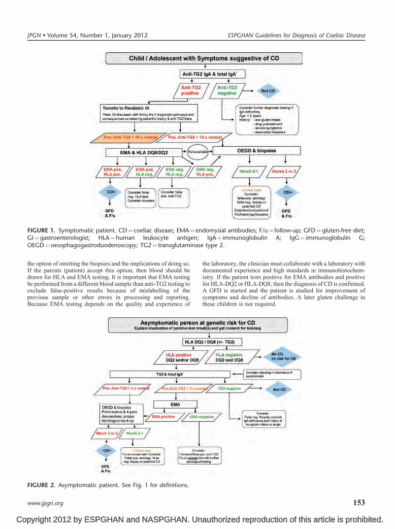
Co
FIGURE 1. Symptomatic patient. CD¼ coeliac disease; EMA¼ endomysial antibodies; F/u¼ follow-up; GFD¼gluten-free diet;GI¼gastroenterologist; HLA¼human leukocyte antigen; IgA¼ immunoglobulin A; IgG¼ immunoglobulin G;
ase
JPGN � Volume 54, Number 1, January 2012 ESPGHAN Guidelines for Diagnosis of Coeliac Disease
the option of omitting the biopsies and the implications of doing so.If the parents (patient) accept this option, then blood should bedrawn for HLA and EMA testing. It is important that EMA testingbe performed from a different blood sample than anti-TG2 testing toexclude false-positive results because of mislabelling of the
OEGD¼oesophagogastroduodenoscopy; TG2¼ transglutamin
pyright 2012 by ESPGHAN and NASPGHAN. Un
previous sample or other errors in processing and reporting.Because EMA testing depends on the quality and experience of
FIGURE 2. Asymptomatic patient. See Fig. 1 for definitions.
www.jpgn.org
the laboratory, the clinician must collaborate with a laboratory withdocumented experience and high standards in immunohistochem-istry. If the patient tests positive for EMA antibodies and positivefor HLA-DQ2 or HLA-DQ8, then the diagnosis of CD is confirmed.A GFD is started and the patient is studied for improvement of
type 2.
authorized reproduction of this article is prohibited.
symptoms and decline of antibodies. A later gluten challenge inthese children is not required.
153
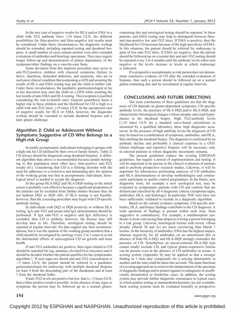
Co
In the rare case of negative results for HLA and/or EMA in achild with TG2 antibody titres �10 times ULN, the differentpossibilities for false-positive and false-negative test results mustbe considered. Under these circumstances, the diagnostic workupshould be extended, including repeated testing and duodenal bio-psies. A small number of cases remain unclear even after extendedevaluation of antibodies and histology specimens. They may requirelonger follow-up and demonstration of gluten dependency of thesymptoms/other findings on a case-by-case basis.
Some deviation from this stepwise procedure may occur inanti-TG2-positive children with classical symptoms (failure tothrive, diarrhoea, distended abdomen, and anaemia), who are insuch poor clinical condition that postponing a GFD and awaiting theresults of HLA and EMA testing may put the child at further risk.Under these circumstances, the paediatric gastroenterologist at hisor her discretion may start the child on a GFD while awaiting thetest results of anti-EMA and HLA testing. This exception is justifiedconsidering the risk-to-benefit ratio: General anaesthesia bears ahigher risk in these children and the likelihood for CD is high in achild with anti-TG2 titers �10 times ULN. In the unexpected caseof negative results for HLA or EMA, however, the diagnosticworkup should be extended to include duodenal biopsies and alater gluten challenge.
Algorithm 2: Child or Adolescent WithoutSymptoms Suggestive of CD Who Belongs to aHigh-risk Group
In totally asymptomatic individuals belonging to groups witha high risk for CD (defined by their own or family history, Table 2)CD always should be diagnosed using duodenal biopsies. A differ-ent algorithm than above is recommended because people belong-ing to this population more often have false-positive anti-TG2results (61). Considering that CD is a lifelong disorder with theneed for adherence to a restrictive and demanding diet, the opinionof the working group was that in asymptomatic individuals, histo-logical proof is needed to accept the diagnosis.
In this group HLA-DQ2 and HLA-DQ8 testing as the initialaction is probably cost-effective because a significant proportion ofthe patients can be excluded from further studies because they donot harbour DQ2 or DQ8 (44). If HLA testing is not feasible,however, then the screening procedure may begin with CD-specificantibody testing.
In individuals with DQ2 or DQ8 positivity or without HLAtesting, IgA anti-TG2 and serum total IgA determination should beperformed. If IgA anti-TG2 is negative and IgA deficiency isexcluded, then CD is unlikely; however, the disease may stilldevelop later in life. Therefore, serological testing should berepeated at regular intervals. No data support any firm recommen-dations, but it was the opinion of the working group members that achild should be investigated by serology every 2 to 3 years to avoidthe detrimental effects of unrecognised CD on growth and bonehealth.
If anti-TG2 antibodies are positive, then signs related to CDshould be searched for (eg, anaemia, elevated liver enzymes) and itshould be decided whether the patient qualifies for the symptomaticalgorithm 1. If such signs are absent and anti-TG2 concentration is>3 times ULN, the patient should be referred to a paediatricgastroenterologist for endoscopy with multiple duodenal biopsies(at least 4 from the descending part of the duodenum and at least1 from the duodenal bulb).
If anti-TG2 levels are positive but low, that is<3 times ULN,
Husby et al
pyright 2012 by ESPGHAN and NASPGHAN. Un
then a false-positive result is possible. In the absence of any signs orsymptoms the person may be followed up on a normal gluten-
154
containing diet and serological testing should be repeated. In thesepatients, anti-EMA testing may help to distinguish between false-and true-positive low anti-TG2 titres. If EMA is positive, then thelikelihood for CD increases because of the high specificity of EMA.In this situation, the patient should be referred for endoscopy inspite of low anti-TG2 titres. If EMA are negative, then the patientshould be followed up on a normal diet and anti-TG2 testing shouldbe repeated every 3 to 6 months until the antibody levels either turnnegative or the levels increase to levels at which endoscopyis indicated.
If a seropositive asymptomatic at-risk person does not demon-strate conclusive evidence of CD after the extended evaluation ofbiopsies, then such a person should be followed up on a normalgluten-containing diet and be reevaluated at regular intervals.
CONCLUSIONS AND FUTURE DIRECTIONSThe main conclusions of these guidelines are that the diag-
nosis of CD depends on gluten-dependent symptoms, CD-specificantibody levels, the presence of HLA-DQ2 and/or HLA-DQ8, andcharacteristic histological changes (villous atrophy and crypt hyper-plasia) in the duodenal biopsy. High TG2-antibody levels(�10 times ULN for a standard curve-based calculation) asmeasured by a qualified laboratory show high diagnostic accu-racies. In the presence of high antibody levels the diagnosis of CDmay be based on a combination of symptoms, antibodies, and HLA,thus omitting the duodenal biopsy. The diagnosis is confirmed by anantibody decline and preferably a clinical response to a GFD.Gluten challenge and repetitive biopsies will be necessary onlyin selected patients in whom diagnostic uncertainty remains.
The present guidelines replace the existing ESPGHANguidelines, but require a period of implementation and testing. Itwill be important to be precise in the clinical evaluation of patientsand to perform prospective research studies. Likewise, it will beimportant for laboratories performing analyses of CD antibodiesand HLA determinations to develop methodologies and continu-ously participate in quality control programmes. In the future, newdiagnostic tools, for example a new serological test, may beevaluated in symptomatic patients with CD and controls that aredefined and classified by all 4 diagnostic criteria (symptoms/signs,antibodies, HLA, and histology). In particular, POC tests have notbeen sufficiently validated to include in a diagnostic algorithm.
Based on the current evidence symptoms, CD-specific anti-bodies, HLA, and biopsy findings contribute to the CD diagnosis. Awide spectrum of findings is present within each item, fromsuggestive to contradictory. For example, a malabsorption syn-drome is more convincing than alopecia or being a person belongingto a risk group. Likewise, histological lesions with severe villousatrophy (Marsh 3b and 3c) are more convincing than Marsh 1lesions. In the hierarchy of antibodies, EMA has the highest impact,whereas negativity for all antibodies on an unrestricted diet orabsence of both HLA-DQ2 and HLA-DQ8 strongly contradict thepresence of CD. Nonetheless, an unconventional HLA-DQ typecannot totally exclude CD, and typical gluten-responsive lesionscan be present even in the absence of CD antibodies in serum. Ascoring system (Appendix II) may be applied so that a strongerfinding in 1 item may compensate for a missing abnormality inanother and the sum could be taken into account. The main functionsof a scoring approach are to assist in the interpretation of the spectrumof diagnostic findings and to protect against overdiagnosis in insuffi-ciently documented or borderline cases. In addition, the scoringsystem may provide further diagnostic reassurance in typical cases
JPGN � Volume 54, Number 1, January 2012
authorized reproduction of this article is prohibited.
in which genetic testing or immunohistochemistry are not available.Such scoring systems must be evaluated formally in prospective
www.jpgn.org
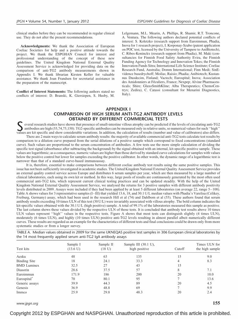
Co
clinical studies before they can be recommended in regular clinicaluse. They do not alter the present recommendations.
Acknowledgments: We thank the Association of EuropeanCoeliac Societies for help and a positive attitude towards theproject. We thank the ESPGHAN Council for interest andprofessional understanding of the concept of these newguidelines. The United Kingdom National External QualityAssessment Service is acknowledged for providing data on thecomparison of anti-TG2 antibody measurements shown inAppendix I. We thank librarian Kirsten Keller for valuableassistance. We thank Joan Frandsen for secretarial assistance inthe preparation of the manuscript.
JPGN � Volume 54, Number 1, January 2012
pyright 2012 by ESPGHAN and NASPGHAN. Un
Conflict of Interest Statements: The following authors stated noconflicts of interest: D. Branski, K. Giersiepen, S. Husby, M.
APPENDCOMPARISON OF HIGH SERUM
OBTAINED BY DIFFERENT
S everal research studies have shown that presence of small-intestineantibodies are high (55,74,75,150). TG2-specific antibodies can be
values are kit specific and show considerable variations. In addition, theThere are 2 main ways to calculate serum antibody results: the maj
comparison to a dilution curve prepared from the serial dilutions of a pcurve). Such values are proportional to the serum concentration of antispecific test signal (absorbance after subtracting the background) by thevalues are logarithmic; as a consequence, numeric values are higher thanbelow the positive control but lower for samples exceeding the positivenarrower than that of a standard curve-based immunoassay.
It is, therefore, essential to make comparisons between differentissue has not been sufficiently investigated in academic studies. The Unitan external quality control service across Europe and distributes 6 seruclinical laboratories, each using its own kit or method. In this way, largecommercial anti-TG2 kits, which represent current clinical testing prKingdom National External Quality Assessment Service, we analysed tlevels distributed in 2009. Assays were included if they had been applieTable A shows values for 3 representative samples (I–III) that yielded 13Freiburg, Germany) assay, which had been used in the research Hill eantibody results exceeding 10 times ULN of this test (30 U/L) were invarkit-specific values obtained with the 30.1 U/L (high positive) sample. AThe last column shows these values divided by the respective ULN of tULN values represent ‘‘high’’ values in the respective tests. Figuremoderately (6 times ULN), and highly (10 times ULN) positive anti-Tcurves. These results are regarded as an example for the characteristics ofsystematic studies or from a longer survey.
TABLE A. Median values obtained in 2009 for the same UKNEQASthe 14 most frequently applied serum anti-TG2 IgA antibody ass
Test kitsSample I(13.6 U)
Sample II(18 U)
Aesku 48 63Binding Site 18 24.1BMD Luminex 32.5 27Diasorin 28.6 37.5Euroimmun 171.9 186Eurospital� 70 80.1Generic assays 39.9 44.3Genesis 36.9 48.8Immco 25.9 29.8
www.jpgn.org
Lelgemann, M.L. Mearin, A. Phillips, R. Shamir, R.T. Troncone,A. Ventura. The following authors declared potential conflicts ofinterest: S. Koletzko (research support from Euroimmun, Phadia,Inova for 1 research project), I. Korponay-Szabo (patent applicationon POC test, licensed by the University of Tampere to AniBiotech),C. Ribes-Koninckx (research support from Phadia), M. Maki (con-sultancies for Finnish Food Safety Authority Evira, the FinnishFunding Agency for Technology and Innovation Tekes; the FinnishInnovation Funds Sitra; International Life Science Institute; CoeliacResearch Fund, Australia; Domm International; Finn Medi; SinE-vidence basedrychoff; Moilas; Raisio; Phadia; Anibiotech; Kustan-nus Duodecim, Finland; Vactech; Eurospital; Inova; Associationdes Amidonniers at Feculiers, France; Nexpep; Alvine Pharmaceu-ticals; Shire; GlaxoSmithKline; Alba Therapeutics; ChemoCen-
ESPGHAN Guidelines for Diagnosis of Coeliac Disease
authorized reproduction of this article is prohibited.
tryx; Zedira), C. Catassi (consultant for Menarini Diagnostics,Italy).
IX IANTI-TG2 ANTIBODY LEVELSCOMMERCIAL TESTS
villous atrophy can be predicted if the levels of circulating anti-TG2measured only in relative units, so numerical values for such ‘‘high’’calculation of results (number and value of calibrators) also differs.ority of available commercial anti-TG2 tests calculate test results byositive sample which correspond to fixed concentrations (standardbodies. A few tests use the more simple calculation of dividing thesignal obtained with an internal, kit-specific positive sample. Thesethose derived by standard curve calculations for samples with valuescalibrator. In other words, the dynamic range of a logarithmic test is
coeliac antibody test results using the same positive samples. Thised Kingdom National External Quality Assessment Service providesm samples per year, which are then measured by a large number of
pools of results are continuously generated by the most often usedactices and can be updated steadily. With the help of the Unitedhe returns for 3 positive samples with different antibody positivityd by at least 5 different laboratories (on average 22, range 5–108)..6, 18, and 30.1 U/L median values with Phadia’s Varelisa (Celikey,t al (74) and Dahlbom et al (55). These authors found that serumiably associated with villous atrophy. The bold column indicates thetotal of 99.1% of the laboratories measured this sample as positive.hose tests. It is concluded that antibody test results above 10 timesA shows that most tests can distinguish slightly (4 times ULN),G2 levels resulting in almost parallel albeit numerically differentdifferent tests and final conclusions could be drawn only from more
positive test samples in 306 European clinical laboratories byays
Sample III (30.1 U),high positive Cutoff
Times ULN forthe high sample
135 15 9.033.3 4 8.343 1557 8 7.1200 20 10.095 7 13.689 20 4.569 7 9.948.3 20 2.4
155
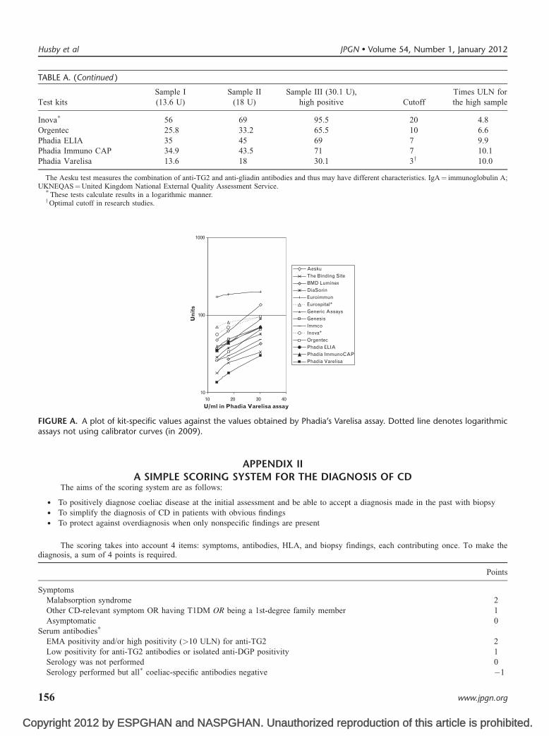
Copyright 2012 by ESPGHAN and NASPGHAN. Unauthorized reproduction of this article is prohibited.
Test kitsSample I(13.6 U)
Sample II(18 U)
Sample III (30.1 U),high positive Cutoff
Times ULN forthe high sample
Inova� 56 69 95.5 20 4.8Orgentec 25.8 33.2 65.5 10 6.6Phadia ELIA 35 45 69 7 9.9Phadia Immuno CAP 34.9 43.5 71 7 10.1Phadia Varelisa 13.6 18 30.1 3y 10.0
The Aesku test measures the combination of anti-TG2 and anti-gliadin antibodies and thus may have different characteristics. IgA¼ immunoglobulin A;UKNEQAS¼United Kingdom National External Quality Assessment Service.�
These tests calculate results in a logarithmic manner.yOptimal cutoff in research studies.
10
100
1000
40302010
U/ml in Phadia Varelisa assay
Un
its
AeskuThe Binding SiteBMD LuminexDiaSorinEuroimmunEurospital*Generic AssaysGenesisImmcoInova*OrgentecPhadia ELIAPhadia ImmunoCAPPhadia Varelisa
FIGURE A. A plot of kit-specific values against the values obtained by Phadia’s Varelisa assay. Dotted line denotes logarithmicassays not using calibrator curves (in 2009).
APPENDIX IIA SIMPLE SCORING SYSTEM FOR THE DIAGNOSIS OF CD
The aims of the scoring system are as follows:
� To positively diagnose coeliac disease at the initial assessment and be able to accept a diagnosis made in the past with biopsy� To simplify the diagnosis of CD in patients with obvious findings� To protect against overdiagnosis when only nonspecific findings are present
The scoring takes into account 4 items: symptoms, antibodies, HLA, and biopsy findings, each contributing once. To make thediagnosis, a sum of 4 points is required.
Points
SymptomsMalabsorption syndrome 2Other CD-relevant symptom OR having T1DM OR being a 1st-degree family member 1Asymptomatic 0
Serum antibodies�
EMA positivity and/or high positivity (>10 ULN) for anti-TG2 2Low positivity for anti-TG2 antibodies or isolated anti-DGP positivity 1Serology was not performed 0Serology performed but all� coeliac-specific antibodies negative �1
TABLE A. (Continued )
Husby et al JPGN � Volume 54, Number 1, January 2012
156 www.jpgn.org
Co
Points
HLAFull HLA-DQ2 (in cis or trans) or HLA-DQ8 heterodimers present 1No HLA performed OR half DQ2 (only HLA-DQB1�0202) present 0HLA neither DQ2 nor DQ8 �1
HistologyMarsh 3b or 3c (subtotal villous atrophy, flat lesion) 2Marsh 2 or 3a (moderately decreased villus height/crypt depth ratio) OR Marsh 0–1 plus intestinal TG2 antibodies 1Marsh 0-1 OR no biopsy performed 0
�Refers in IgA deficiency to IgG class EMA, TG2 and DGP antibodies.
Comments and Explanations for UseBiopsy items were graded by taking into account Villanacci scoring (85) and the clinical utility of the results. We assumed that Marsh 0
or 1 results without any further information could be nonspecific. In contrast, demonstration of antibodies bound to tissue TG2 in the smallbowel adds information to the diagnosis (when available). It is possible to diagnose CD as before even without this possibility. It is notnecessary to have an EMA testing facility, but it is a clear advantage. Some findings that make CD improbable are resulting in negativescoring points. The sum of 4 points may be collected from findings registered at different time points during follow-up if they can be assumedto be gluten dependent. For example, an infant having villous atrophy before the introduction of gluten and normal biopsy at the age of 6 yearswhile normally eating gluten will receive 0 for biopsy.
JPGN � Volume 54, Number 1, January 2012 ESPGHAN Guidelines for Diagnosis of Coeliac Disease
REFERENCES1. Revised criteria for diagnosis of coeliac disease. Report of Working
Group of European Society of Paediatric Gastroenterology and Nutri-tion. Arch Dis Child 1990; 65:909-11.
2. Rewers M. Epidemiology of celiac disease: what are the prevalence,incidence, and progression of celiac disease? Gastroenterology 2005;128(4 Suppl 1):S47–51.
3. Greco L, Romino R, Coto I, et al. The first large population based twinstudy of coeliac disease. Gut 2002;50:624–8.
4. Lundin KE, Sollid LM, Qvigstad E, et al. T lymphocyte recognition ofa celiac disease-associated cis-or trans-encoded HLA-DQ alpha/beta-heterodimer. J Immunol 1990;145:136–9.
5. van de WY, Kooy YM, van Veelen PA, et al. Small intestinal T cells ofceliac disease patients recognize a natural pepsin fragment of gliadin.Proc Natl Acad Sci U S A 1998;95:10050–4.
6. Dieterich W, Ehnis T, Bauer M, et al. Identification of tissue trans-glutaminase as the autoantigen of celiac disease. Nat Med 1997;3:797–801.
7. Sjostrom H, Lundin KE, Molberg O, et al. Identification of a gliadin T-cell epitope in coeliac disease: general importance of gliadin deamida-tion for intestinal T-cell recognition. Scand J Immunol 1998;48:111–5.
8. Mothes T. Deamidated gliadin peptides as targets for celiac disease-specific antibodies. Adv Clin Chem 2007;44:35–63.
9. Corazza GR, Villanacci V, Zambelli C, et al. Comparison of theinterobserver reproducibility with different histologic criteria usedin celiac disease. Clin Gastroenterol Hepatol 2007;5:838–43.
10. Rostom A, Dube C, Cranney A, et al. Celiac disease. Evid Rep TechnolAssess (Summ) 2004;104:1–6.
11. Hill ID, Dirks MH, Liptak GS, et al. Guideline for the diagnosis andtreatment of celiac disease in children: recommendations of the NorthAmerican Society for Pediatric Gastroenterology, Hepatology andNutrition. J Pediatr Gastroenterol Nutr 2005;40:1–19.
11a. Giersiepen K, Lelgemann M, Stuhldreher N, et al. Accuracy ofdiagnostic antibody tests for coeliac disease in children: summaryfrom an evidence report. European Society for Paediatic Gas-troenterology, Hepatology and Nutrition (ESPGHAN) WorkingGroup on Coeliac Disease Diagnosis. J Pediatr Gastroenterol NutrIn press.
12. Ebell MH, Siwek J, Weiss BD, et al. Strength of recommendationtaxonomy (SORT): a patient-centered approach to grading evidence inthe medical literature. J Am Board Fam Pract 2004;17:59–67.
13. Schunemann HJ, Best D, Vist G, et al. Letters, numbers, symbols and
pyright 2012 by ESPGHAN and NASPGHAN. Un
words: how to communicate grades of evidence and recommendations.CMAJ 2003;169:677–80.
www.jpgn.org
14. Mustalahti K, Sulkanen S, Holopainen P, et al. Coeliac disease amonghealthy members of multiple case coeliac disease families. Scand JGastroenterol 2002;37:161–5.
15. Bottaro G, Failla P, Rotolo N, et al. Changes in coeliac diseasebehaviour over the years. Acta Paediatr 1993;82:566–8.
16. Garampazzi A, Rapa A, Mura S, et al. Clinical pattern of celiac diseaseis still changing. J Pediatr Gastroenterol Nutr 2007;45:611–4.
17. Rashid M, Cranney A, Zarkadas M, et al. Celiac disease: evaluation ofthe diagnosis and dietary compliance in Canadian children. Pediatrics2005;116:e754–9.
18. Simmons JH, Klingensmith GJ, McFann K, et al. Impact of celiac auto-immunity on children with type 1 diabetes. J Pediatr 2007;150:461–6.
19. Bottaro G, Cataldo F, Rotolo N, et al. The clinical pattern of sub-clinical/silent celiac disease: an analysis on 1026 consecutive cases.Am J Gastroenterol 1999;94:691–6.
20. van Rijn JC, Grote FK, Oostdijk W, et al. Short stature and theprobability of coeliac disease, in the absence of gastrointestinalsymptoms. Arch Dis Child 2004;89:882–3.
21. Ferrara M, Coppola L, Coppola A, et al. Iron deficiency in childhood andadolescence: retrospective review. Hematology 2006;11: 183–6.
22. Valentino R, Savastano S, Tommaselli AP, et al. Prevalence of coeliacdisease in patients with thyroid autoimmunity. Horm Res 1999;51:124–7.
23. Hansen D, Brock-Jacobsen B, Lund E, et al. Clinical benefit of agluten-free diet in type 1 diabetic children with screening-detectedceliac disease: a population-based screening study with 2 years’follow-up. Diabetes Care 2006;29:2452–6.
24. Salardi S, Volta U, Zucchini S, et al. Prevalence of celiac disease inchildren with type 1 diabetes mellitus increased in the mid-1990 s:an 18-year longitudinal study based on anti-endomysial antibodies.J Pediatr Gastroenterol Nutr 2008;46:612–4.
25. Bonamico M, Pasquino AM, Mariani P, et al. Prevalence and clinicalpicture of celiac disease in Turner syndrome. J Clin Endocrinol Metab2002;87:5495–8.
26. Korponay-Szabo IR, Dahlbom I, Laurila K, et al. Elevation of IgGantibodies against tissue transglutaminase as a diagnostic tool forcoeliac disease in selective IgA deficiency. Gut 2003;52:1567–71.
27. Emami MH, Taheri H, Kohestani S, et al. How frequent is celiacdisease among epileptic patients? J Gastrointestin Liver Dis 2008;17:379–82.
28. Dickey W, McMillan SA, McCrum EE, et al. Association betweenserum levels of total IgA and IgA class endomysial and antigliadin
authorized reproduction of this article is prohibited.
antibodies: implications for coeliac disease screening. Eur J Gastro-enterol Hepatol 1997;9:559–62.
157
Co
29. Monsuur AJ, Wijmenga C. Understanding the molecular basis of celiacdisease: what genetic studies reveal. Ann Med 2006;38:578–91.
30. Monsuur AJ, de Bakker PI, Zhernakova A, et al. Effective detection ofhuman leukocyte antigen risk alleles in celiac disease using tag singlenucleotide polymorphisms. PLoS One 2008;3:e2270.
31. van Heel DA, Franke L, Hunt KA, et al. A genome-wide associationstudy for celiac disease identifies risk variants in the region harboringIL2 and IL21. Nat Genet 2007;39:827–9.
32. Mearin ML, Biemond I, Pena AS, et al. HLA-DR phenotypes inSpanish coeliac children: their contribution to the understanding of thegenetics of the disease. Gut 1983;24:532–7.
33. Karell K, Louka AS, Moodie SJ, et al. HLA types in celiac diseasepatients not carrying the DQA1�05-DQB1�02 (DQ2) heterodimer:results from the European Genetics Cluster on Celiac Disease. HumImmunol 2003;64:469–77.
33a. Richtlijn Coeliakie en Dermatitis Herpetiformis. Kwaliteitsinstituutvoor de Gesondheidszorg CBO. Haarlem, The Netherlands: Neder-landse Vereniging van Maag-Darm. Leverartsen; 2008.
34. Polvi A, Eland C, Koskimies S, et al. HLA DQ and DP in Finnishfamilies with celiac disease. Eur J Immunogenet 1996;23:221–34.
35. Margaritte-Jeannin P, Babron MC, Bourgey M, et al. HLA-DQ relativerisks for coeliac disease in European populations: a study of theEuropean Genetics Cluster on Coeliac Disease. Tissue Antigens2004;63:562–7.
36. Hadithi M, von Blomberg BM, Crusius JB, et al. Accuracy of serologictests and HLA-DQ typing for diagnosing celiac disease. Ann InternMed 2007;147:294–302.
37. van Belzen MJ, Koeleman BP, Crusius JB, et al. Defining the con-tribution of the HLA region to cis DQ2-positive coeliac diseasepatients. Genes Immun 2004;5:215–20.
38. Sollid LM, Markussen G, Ek J, et al. Evidence for a primary associa-tion of celiac disease to a particular HLA-DQ alpha/beta heterodimer.J Exp Med 1989;169:345–50.
39. Balas A, Vicario JL, Zambrano A, et al. Absolute linkage of celiacdisease and dermatitis herpetiformis to HLA-DQ. Tissue Antigens1997;50:52–6.
40. Fasano A, Berti I, Gerarduzzi T, et al. Prevalence of celiac disease in at-risk and not-at-risk groups in the United States: a large multicenterstudy. Arch Intern Med 2003;163:286–92.
41. Liu J, Juo SH, Holopainen P, et al. Genomewide linkage analysis ofceliac disease in Finnish families. Am J Hum Genet 2002;70:51–9.
42. Vidales MC, Zubillaga P, Zubillaga I, et al. Allele and haplotypefrequencies for HLA class II (DQA1 and DQB1) loci in patients withceliac disease from Spain. Hum Immunol 2004;65:352–8.
43. Al-toma A, Goerres MS, Meijer JW, et al. Human leukocyte antigen-DQ2 homozygosity and the development of refractory celiac diseaseand enteropathy-associated T-cell lymphoma. Clin GastroenterolHepatol 2006;4:315–9.
44. Csizmadia CG, Mearin ML, Oren A, et al. Accuracy and cost-effec-tiveness of a new strategy to screen for celiac disease in children withDown syndrome. J Pediatr 2000;137:756–61.
45. Maki M, Mustalahti K, Kokkonen J, et al. Prevalence of celiac diseaseamong children in Finland. N Engl J Med 2003;348:2517–24.
46. Schwertz E, Kahlenberg F, Sack U, et al. Serologic assay based ongliadin-related nonapeptides as a highly sensitive and specific diag-nostic aid in celiac disease. Clin Chem 2004;50:2370–5.
47. Korponay-Szabo IR, Halttunen T, Szalai Z, et al. In vivo targeting ofintestinal and extraintestinal transglutaminase 2 by coeliac autoanti-bodies. Gut 2004;53:641–8.
48. Cataldo F, Lio D, Marino V, et al. IgG(1) antiendomysium and IgGantitissue transglutaminase (anti-tTG) antibodies in coeliac patientswith selective IgA deficiency. Working Groups on Celiac Disease ofSIGEP and Club del Tenue. Gut 2000;47:366–9.
49. Hadjivassiliou M, Maki M, Sanders DS, et al. Autoantibody targetingof brain and intestinal transglutaminase in gluten ataxia. Neurology2006;66:373–7.
50. Kurppa K, Ashorn M, Iltanen S, et al. Celiac disease without villousatrophy in children: a prospective study. J Pediatr 2010;157:373–80.
Husby et al
pyright 2012 by ESPGHAN and NASPGHAN. Un
51. Maki M, Holm K, Koskimies S, et al. Normal small bowel biopsyfollowed by coeliac disease. Arch Dis Child 1990;65:1137–41.
158
52. Lewis NR, Scott BB. Systematic review: the use of serology to excludeor diagnose coeliac disease (a comparison of the endomysial and tissuetransglutaminase antibody tests). Aliment Pharmacol Ther 2006;24:47–54.
53. Salmi TT, Collin P, Korponay-Szabo IR, et al. Endomysial antibody-negative coeliac disease: clinical characteristics and intestinal auto-antibody deposits. Gut 2006;55:1746–53.
54. Bargetzi MJ, Schonenberger A, Tichelli A, et al. Celiac diseasetransmitted by allogeneic non-T cell-depleted bone marrow transplan-tation. Bone Marrow Transplant 1997;20:607–9.
55. Dahlbom I, Korponay-Szabo IR, Kovacs JB, et al. Prediction of clinicaland mucosal severity of coeliac disease and dermatitis herpetiformisby quantification of IgA/IgG serum antibodies to tissue transglutami-nase. J Pediatr Gastroenterol Nutr 2010;50:140–6.
56. Karpati S, Torok E, Kosnai I. IgA class antibody against humanjejunum in sera of children with dermatitis herpetiformis. J InvestDermatol 1986;87:703–6.
57. Raivio T, Kaukinen K, Nemes E, et al. Self transglutaminase-basedrapid coeliac disease antibody detection by a lateral flow method.Aliment Pharmacol Ther 2006;24:147–54.
58. Prause C, Ritter M, Probst C, et al. Antibodies against deamidatedgliadin as new and accurate biomarkers of childhood coeliac disease.J Pediatr Gastroenterol Nutr 2009;49:52–8.
59. Naiyer AJ, Hernandez L, Ciaccio EJ, et al. Comparison of commer-cially available serologic kits for the detection of celiac disease. J ClinGastroenterol 2009;43:225–32.
60. Li M, Yu L, Tiberti C, et al. A report on the International Transglu-taminase Autoantibody Workshop for Celiac Disease. Am J Gastro-enterol 2009;104:154–63.
61. Vecsei A, Arenz T, Heilig G, et al. Influence of age and genetic risk onanti-tissue transglutaminase IgA titers. J Pediatr Gastroenterol Nutr2009;48:544–9.
62. Di Tola M, Barilla F, Trappolini M, et al. Antitissue transglutaminaseantibodies in acute coronary syndrome: an alert signal of myocardialtissue lesion? J Intern Med 2008;263:43–51.
63. Pastore L, Campisi G, Compilato D, et al. Orally based diagnosis ofceliac disease: current perspectives. J Dent Res 2008;87:1100–7.
64. Bonamico M, Ferri M, Nenna R. Tissue transglutaminase autoantibodydetection in human saliva: a powerful method for celiac diseasescreening. J Pediatr 2004;144:632–6.
65. Kappler M, Krauss-Etschmann S, Diehl V, et al. Detection of secretoryIgA antibodies against gliadin and human tissue transglutaminase instool to screen for coeliac disease in children: validation study. BMJ2006;332:213–4.
66. Baviera LC, Aliaga ED, Ortigosa L, et al. Celiac disease screening byimmunochromatographic visual assays: results of a multicenter study.J Pediatr Gastroenterol Nutr 2007;45:546–50.
67. Korponay-Szabo IR, Szabados K, Pusztai J, Uhrin K, Ludmany E,Nemes E, et al. Population screening for coeliac disease in primarycare by district nurses using a rapid antibody test: diagnostic accuracyand feasibility study. BMJ 2007;335:1244–7.
68. Bizzaro N, Tampoia M, Villalta D, Platzgummer S, Liguori M, TozzoliR, et al. Low specificity of anti-tissue transglutaminase antibodiesin patients with primary biliary cirrhosis. J Clin Lab Anal 2006;20:184–9.
69. Ferrara F, Quaglia S, Caputo I, et al. Anti-transglutaminase antibodiesin non-coeliac children suffering from infectious diseases. Clin ExpImmunol 2010;159:217–23.
70. Villalta D, Bizzaro N, Tonutti E, et al. IgG anti-transglutaminaseautoantibodies in systemic lupus erythematosus and Sjogren syn-drome. Clin Chem 2002;48:1133.
71. Collin P, Helin H, Maki M, et al. Follow-up of patients positive inreticulin and gliadin antibody tests with normal small-bowel biopsyfindings. Scand J Gastroenterol 1993;28:595–8.
72. Kurppa K, Collin P, Viljamaa M, et al. Diagnosing mild enteropathyceliac disease: a randomized, controlled clinical study. Gastroenter-ology 2009;136:816–23.
73. Koskinen O, Collin P, Korponay-Szabo I, et al. Gluten-dependentsmall bowel mucosal transglutaminase 2-specific IgA deposits in overt
JPGN � Volume 54, Number 1, January 2012
authorized reproduction of this article is prohibited.
and mild enteropathy coeliac disease. J Pediatr Gastroenterol Nutr2008;47:436–42.
www.jpgn.org
Co
�
74. Hill PG, Holmes GK. Coeliac disease: a biopsy is not always necessaryfor diagnosis. Aliment Pharmacol Ther 2008;27:572–7.
75. Vivas S, Ruiz de Morales JG, Riestra S, et al. Duodenal biopsy may beavoided when high transglutaminase antibody titers are present. WorldJ Gastroenterol 2009;15:4775–80.
76. Lewis NR, Scott BB. Meta-analysis: deamidated gliadin peptideantibody and tissue transglutaminase antibody compared as screen-ing tests for coeliac disease. Aliment Pharmacol Ther 2010;31:73–81.
77. Agardh D. Antibodies against synthetic deamidated gliadin peptidesand tissue transglutaminase for the identification of childhood celiacdisease. Clin Gastroenterol Hepatol 2007;5:1276–81.
78. Liu E, Li M, Emery L, et al. Natural history of antibodies todeamidated gliadin peptides and transglutaminase in early child-hood celiac disease. J Pediatr Gastroenterol Nutr 2007;45:293–300.
78a. National Institute for Health and Clinical Excellence. CG87 type 2diabetes newer agents: NICE guidelines. http://guidance.nice.org.uk/CG87/NICEGuidance/pdf/English. Accessed August 31,2011.
79. Lagerqvist C, Dahlbom I, Hansson T, Jidell E, Juto P, Olcen P, et al.Antigliadin immunoglobulin A best in finding celiac disease inchildren younger than 18 months of age. J Pediatr Gastroenterol Nutr2008;47:428–35.
80. Koskinen O, Collin P, Lindfors K, et al. Usefulness of small-bowelmucosal transglutaminase-2 specific autoantibody deposits in thediagnosis and follow-up of celiac disease. J Clin Gastroenterol2010;44:483–8.
81. Sardy M, Csikos M, Geisen C, et al. Tissue transglutaminase ELISApositivity in autoimmune disease independent of gluten-sensitivedisease. Clin Chim Acta 2007;376:126–35.
82. Damasiewicz-Bodzek A, Wielkoszynski T. Serologic markers of celiacdisease in psoriatic patients. J Eur Acad Dermatol Venereol 2008;22:1055–61.
83. Marsh MN. Grains of truth: evolutionary changes in small intestinalmucosa in response to environmental antigen challenge. Gut 1990;31:111–4.
84. Oberhuber G, Granditsch G, Vogelsang H. The histopathology ofcoeliac disease: time for a standardized report scheme for pathologists.Eur J Gastroenterol Hepatol 1999;11:1185–94.
85. Corazza GR, Villanacci V. Coeliac disease. J Clin Pathol 2005;58:573–4.
86. Dickson BC, Streutker CJ, Chetty R. Coeliac disease: an update forpathologists. J Clin Pathol 2006;59:1008–16.
87. Paparo F, Petrone E, Tosco A, et al. Clinical, HLA, and small bowelimmunohistochemical features of children with positive serum anti-endomysium antibodies and architecturally normal small intestinalmucosa. Am J Gastroenterol 2005;100:2294–8.
88. Simell S, Hoppu S, Hekkala A, et al. Fate of five celiac disease-associated antibodies during normal diet in genetically at-risk childrenobserved from birth in a natural history study. Am J Gastroenterol2007;102:2026–35.
89. Biagi F, Bianchi PI, Campanella J, et al. The prevalence and the causesof minimal intestinal lesions in patients complaining of symptomssuggestive of enteropathy: a follow-up study. J Clin Pathol 2008;61:1116–8.
90. Kakar S, Nehra V, Murray JA, et al. Significance of intraepitheliallymphocytosis in small bowel biopsy samples with normal mucosalarchitecture. Am J Gastroenterol 2003;98:2027–33.
91. Rostami K, Kerckhaert J, Tiemessen R, et al. Sensitivity of antiendo-mysium and antigliadin antibodies in untreated celiac disease:disappointing in clinical practice. Am J Gastroenterol 1999;94:888–94.
92. Jarvinen TT, Kaukinen K, Laurila K, et al. Intraepithelial lymphocytesin celiac disease. Am J Gastroenterol 2003;98:1332–7.
93. Jarvinen TT, Collin P, Rasmussen M, et al. Villous tip intraepitheliallymphocytes as markers of early-stage coeliac disease. Scand JGastroenterol 2004;39:428–33.
94. Achkar E, Carey WD, Petras R, et al. Comparison of suction capsule
JPGN Volume 54, Number 1, January 2012
pyright 2012 by ESPGHAN and NASPGHAN. Un
and endoscopic biopsy of small bowel mucosa. Gastrointest Endosc1986;32:278–81.
www.jpgn.org
95. Barakat MH, Ali SM, Badawi AR, et al. Peroral endoscopic duodenalbiopsy in infants and children. Acta Paediatr Scand 1983;72:563–9.
96. Branski D, Faber J, Freier S, et al. Histologic evaluation of endoscopicversus suction biopsies of small intestinal mucosae in children withand without celiac disease. J Pediatr Gastroenterol Nutr 1998;27:6–11.
97. Granot E, Goodman-Weill M, Pizov G, et al. Histological comparisonof suction capsule and endoscopic small intestinal mucosal biopsies inchildren. J Pediatr Gastroenterol Nutr 1993;16:397–401.
98. Mee AS, Burke M, Vallon AG, et al. Small bowel biopsy for malab-sorption: comparison of the diagnostic adequacy of endoscopic forcepsand capsule biopsy specimens. Br Med J (Clin Res Ed) 1985;291:769–72.
99. Ravelli A, Bolognini S, Gambarotti M, et al. Variability of histologiclesions in relation to biopsy site in gluten-sensitive enteropathy. Am JGastroenterol 2005;100:177–85.
100. Bonamico M, Thanasi E, Mariani P, et al. Duodenal bulb biopsies inceliac disease: a multicenter study. J Pediatr Gastroenterol Nutr2008;47:618–22.
101. Rashid M, MacDonald A. Importance of duodenal bulb biopsies inchildren for diagnosis of celiac disease in clinical practice. BMCGastroenterol 2009;9:78.
102. Weir DC, Glickman JN, Roiff T, et al. Variability of histopathologicalchanges in childhood celiac disease. Am J Gastroenterol 2010;105:207–12.
103. Ravelli A, Villanacci V, Monfredini C, et al. How patchy is patchyvillous atrophy? Distribution pattern of histological lesions in theduodenum of children with celiac disease. Am J Gastroenterol2010;105:2103–10.
104. Korponay-Szabo IR, Kovacs JB, Lorincz M, et al. Prospective sig-nificance of antiendomysium antibody positivity in subsequentlyverified celiac disease. J Pediatr Gastroenterol Nutr 1997;25:56–63.
105. Vilppula A, Collin P, Maki M, et al. Undetected coeliac disease in theelderly: a biopsy-proven population-based study. Dig Liver Dis2008;40:809–13.
106. George DK, Evans RM, Gunn IR. Familial chronic fatigue. PostgradMed J 1997;73:311–3.
107. Lepore L, Martelossi S, Pennesi M, et al. Prevalence of celiac diseasein patients with juvenile chronic arthritis. J Pediatr 1996;129:311–3.
108. Goldacre MJ, Wotton CJ, Seagroatt V, et al. Cancers and immunerelated diseases associated with Down’s syndrome: a record linkagestudy. Arch Dis Child 2004;89:1014–7.
109. Mortensen KH, Cleemann L, Hjerrild BE, et al. Increased prevalenceof autoimmunity in Turner syndrome—influence of age. Clin ExpImmunol 2009;156:205–10.
110. Giannotti A, Tiberio G, Castro M, et al. Coeliac disease in Williamssyndrome. J Med Genet 2001;38:767–8.
111. Collin P, Syrjanen J, Partanen J, et al. Celiac disease and HLA DQ inpatients with IgA nephropathy. Am J Gastroenterol 2002;97:2572–6.
112. Caprai S, Vajro P, Ventura A, et al. Autoimmune liver diseaseassociated with celiac disease in childhood: a multicenter study. ClinGastroenterol Hepatol 2008;6:803–6.
113. Arnason A, Skaftadottir I, Sigmundsson J, et al. The associationbetween coeliac disease, dermatitis herpetiformis and certain HLA-antigens in Icelanders. Eur J Immunogenet 1994;21:457–60.
114. Arranz E, Telleria JJ, Sanz A, et al. HLA-DQA1�0501 and DQB1�02homozygosity and disease susceptibility in Spanish coeliac patients.Exp Clin Immunogenet 1997;14:286–90.
115. Book L, Zone JJ, Neuhausen SL. Prevalence of celiac disease amongrelatives of sib pairs with celiac disease in US families. Am J Gastro-enterol 2003;98:377–81.
116. Bouguerra F, Babron MC, Eliaou JF, et al. Synergistic effect of twoHLA heterodimers in the susceptibility to celiac disease in Tunisia.Genet Epidemiol 1997;14:413–22.
117. Boy MF, La NG, Balestrieri A, et al. Distribution of HLA-DPB1,-DQB1 -DQA1 alleles among Sardinian celiac patients. Dis Markers1995;12:199–204.
118. Catassi C, Doloretta MM, Ratsch IM, et al. The distribution of DQ
ESPGHAN Guidelines for Diagnosis of Coeliac Disease
authorized reproduction of this article is prohibited.
genes in the Saharawi population provides only a partial explanationfor the high celiac disease prevalence. Tissue Antigens 2001;58:402–6.
159
Co
119. Colonna M, Mantovani W, Corazza GR, et al. Reassessment of HLAassociation with celiac disease in special reference to the DP associa-tion. Hum Immunol 1990;29:263–74.
120. Congia M, Cucca F, Frau F, et al. A gene dosage effect of theDQA1�0501/DQB1�0201 allelic combination influences the clinicalheterogeneity of celiac disease. Hum Immunol 1994;40:138–42.
121. Congia M, Frau F, Lampis R, et al. A high frequency of the A30, B18,DR3, DRw52, DQw2 extended haplotype in Sardinian celiac diseasepatients: further evidence that disease susceptibility is conferred byDQ A1�0501, B1�0201. Tissue Antigens 1992;39:78–83.
122. Djilali-Saiah I, Caillat-Zucman S, Schmitz J, et al. Polymorphism ofantigen processing (TAP, LMP) and HLA class II genes in celiacdisease. Hum Immunol 1994;40:8–16.
123. Djilali-Saiah I, Schmitz J, Harfouch-Hammoud E, et al. CTLA-4 genepolymorphism is associated with predisposition to coeliac disease. Gut1998;43:187–9.
124. Erkan T, Kutlu T, Yilmaz E, et al. Human leukocyte antigens in Turkishpediatric celiac patients. Turk J Pediatr 1999;41:181–8.
125. Farre C, Humbert P, Vilar P, et al. Serological markers and HLA-DQ2haplotype among first-degree relatives of celiac patients. CatalonianCoeliac Disease Study Group. Dig Dis Sci 1999;44:2344–9.
126. Fernandez-Arquero M, Figueredo MA, Maluenda C, et al. HLA-linkedgenes acting as additive susceptibility factors in celiac disease. HumImmunol 1995;42:295–300.
127. Ferrante P, Petronzelli F, Mariani P, et al. Oligotyping of Italian celiacpatients with the 11th International Histocompatibility Workshopreagents. Tissue Antigens 1992;39:38–9.
128. Fine KD, Do K, Schulte K, et al. High prevalence of celiac sprue-likeHLA-DQ genes and enteropathy in patients with the microscopiccolitis syndrome. Am J Gastroenterol 2000;95:1974–82.
129. Howell WM, Leung ST, Jones DB, et al. HLA-DRB, -DQA, and -DQBpolymorphism in celiac disease and enteropathy-associated T-celllymphoma. Common features and additional risk factors for malig-nancy. Hum Immunol 1995;43:29–37.
130. Iltanen S, Holm K, Partanen J, et al. Increased density of jejunalgammadeltaþ T cells in patients having normal mucosa—marker ofoperative autoimmune mechanisms? Autoimmunity 1999;29:179–87.
131. Iltanen S, Rantala I, Laippala P, et al. Expression of HSP-65 in jejunalepithelial cells in patients clinically suspected of coeliac disease.Autoimmunity 1999;31:125–32.
132. Johnson TC, Diamond B, Memeo L, et al. Relationship of HLA-DQ8and severity of celiac disease: comparison of New York and Parisiancohorts. Clin Gastroenterol Hepatol 2004;2:888–94.
133. Kaur G, Sarkar N, Bhatnagar S, et al. Pediatric celiac disease in India isassociated with multiple DR3-DQ2 haplotypes. Hum Immunol 2002;63:677–82.
134. Lewis C, Book L, Black J, et al. Celiac disease and human leukocyteantigen genotype: accuracy of diagnosis in self-diagnosed individuals,
Husby et al
pyright 2012 by ESPGHAN and NASPGHAN. Un
31:22–7.
160
135. Lio D, Bonanno CT, D’Anna C, et al. Gluten stimulation induces an invitro expansion of peripheral blood T gamma delta cells from HLA-DQ2-positive subjects of families of patients with celiac disease. ExpClin Immunogenet 1998;15:46–55.
136. Mazzilli MC, Ferrante P, Mariani P, et al. A study of Italian pediatricceliac disease patients confirms that the primary HLA association is tothe DQ(alpha 1�0501, beta 1�0201) heterodimer. Hum Immunol1992;33:133–9.
137. Michalski JP, McCombs CC, Arai T, et al. HLA-DR, DQ genotypes ofceliac disease patients and healthy subjects from the West of Ireland.Tissue Antigens 1996;47:127–33.
138. Neuhausen SL, Weizman Z, Camp NJ, et al. HLA DQA1-DQB1genotypes in Bedouin families with celiac disease. Hum Immunol2002;63:502–7.
139. Pena-Quintana L, Torres-Galvan MJ, Deniz-Naranjo MC, et al. Assess-ment of the DQ heterodimer test in the diagnosis of celiac disease in theCanary Islands (Spain). J Pediatr Gastroenterol Nutr 2003;37:604–8.
140. Perez-Bravo F, Araya M, Mondragon A, et al. Genetic differences inHLA-DQA1� and DQB1� allelic distributions between celiac andcontrol children in Santiago, Chile. Hum Immunol 1999;60:262–7.
141. Ploski R, Ek J, Thorsby E, et al. On the HLA-DQ(alpha 1�0501, beta1�0201)-associated susceptibility in celiac disease: a possible genedosage effect of DQB1�0201. Tissue Antigens 1993;41:173–7.
142. Ploski R, Ascher H, Sollid LM. HLA genotypes and the increasedincidence of coeliac disease in Sweden. Scand J Gastroenterol1996;31:1092–7.
143. Popat S, Hearle N, Wixey J, et al. Analysis of the CTLA4 gene inSwedish coeliac disease patients. Scand J Gastroenterol 2002;37:28–31.
144. Ruiz del Prado MY, Olivares Lopez JL, Lazaro AA, et al. HLA system.Phenotypic and gene frequencies in celiac and healthy subjects fromthe same geographical area. Rev Esp Enferm Dig 2001;93:106–13.
145. Sacchetti L, Calcagno G, Ferrajolo A, et al. Discrimination betweenceliac and other gastrointestinal disorders in childhood by rapid humanlymphocyte antigen typing. Clin Chem 1998;44 (8 Pt 1):1755–7.
146. Sumnik Z, Kolouskova S, Cinek O, et al. HLA-DQA1�05-DQB1�0201 positivity predisposes to coeliac disease in Czech dia-betic children. Acta Paediatr 2000;89:1426–30.
147. Tighe MR, Hall MA, Barbado M, et al. HLA class II alleles associatedwith celiac disease susceptibility in a southern European population.Tissue Antigens 1992;40:90–7.
148. Tighe MR, Hall MA, Ashkenazi A, et al. Celiac disease amongAshkenazi Jews from Israel. A study of the HLA class II allelesand their associations with disease susceptibility. Hum Immunol1993;38:270–6.
149. Tumer L, Altuntas B, Hasanoglu A, et al. Pattern of human leukocyteantigens in Turkish children with celiac disease. Pediatr Int2000;42:678–81.
JPGN � Volume 54, Number 1, January 2012
150. Tuysuz B, Dursun A, Kutlu T, et al. HLA-DQ alleles in patients with
dosage effect, and sibling risk. J Pediatr Gastroenterol Nutr 2000;authorized reproduction of this article is prohibited.
celiac disease in Turkey. Tissue Antigens 2001;57:540–2.
www.jpgn.org