Embed Size (px)
Citation preview
Journal of Food Research; Vol. 2, No. 4; 2013 ISSN 1927-0887 E-ISSN 1927-0895
Published by Canadian Center of Science and Education
37
Evaluation of Antimicrobial Activities of Commercial Herb and Spice Extracts Against Selected Food-Borne Bacteria
Anna M. Witkowska1, Dara K. Hickey1, Mercedes Alonso-Gomez1 & Martin Wilkinson1 1 Department of Life Sciences, University of Limerick, Ireland
Correspondence: Martin Wilkinson, Department of Life Sciences, University of Limerick, Ireland. E-mail: [email protected]
Received: March 28, 2013 Accepted: June 25, 2013 Online Published: June 27, 2013
doi:10.5539/jfr.v2n4p37 URL: http://dx.doi.org/10.5539/jfr.v2n4p37
Abstract
The aim of this study was to evaluate and compare the antimicrobial properties of extracts of thirty types of commercial herbs and spices commonly used in the production of ready meals. Various extracts of spices were evaluated for their antimicrobial activities against Escherichia coli, Listeria innocua, Staphylococcus aureus and Pseudomonas fluorescens using a microdilution broth method. Ethanol and hexane extracts of oregano, clove, sage, rosemary and celery showed relatively strong antimicrobial activities against all bacteria tested. In contrast, water extracts displayed little or no antimicrobial activity. Flow cytometry revealed that cell membrane structures were damaged by spice and herb active extracts, while analysis of intra- and extracellular ATP contents of bacteria indicated that an increase in extracellular ATP was partially due to intracellular leakage. Extract combinations assessed using the checkerboard method did not display synergistic effects, however, some additive effects were observed when oregano was combined with sage or rosemary against L. innocua or S. aureus. This study has demonstrated that some commercial spice extracts have antimicrobial activity against food-borne bacterial species and should be considered as potential antibacterial agents for addition to ready meals.
Keywords: antimicrobial, herbs, spices, extracts, phenolic, permeability, synergy
1. Introduction
Food safety is a fundamental concern for both consumers and food producers alike. Despite the high degree of awareness of food preservation methods there is increasing occurrence of disease outbreaks caused by pathogenic and spoilage microorganisms in foods (Meng & Doyle, 1998). Currently some ready meal manufacturers add synthetic antimicrobial agents such nitrates, nitrites, benzoates to inhibit the growth of food spoilage and food pathogenic microorganisms (Gould, 1996). However, the uncontrolled use of chemical preservatives over the years has led to emergence of microbial resistance to classic antimicrobial agents which has become a major health concern (Kiessling et al., 2002). Consumer awareness and concern that synthetic chemical additives may have some toxic or even carcinogenic effects, has increased the demand for high-quality, minimally processed foods with extended shelf-life, preferably free from or with a reduced level of added chemical antimicrobial agents (Zink, 1997). Recently there has also been an observed trend towards reducing salt levels in ready meals as a result of the proven association between excessive sodium intake, the development of hypertension and increased risk of cardiovascular disease (Desmond, 2006). Therefore, there is growing interest in using natural antimicrobial compounds, including extracts of herbs and spices, as salt replacers or alternatives to synthetic compounds for food preservation (Smid & Gorris, 1999).
Plants, including herbs and spices, contain products of secondary metabolism such as phenolics, phenolic acids, quinones, flavonoids, tannins (Lai & Roy, 2004). Many of these phytochemicals are rich sources of antioxidants and provide defense mechanisms to plants against predation by infectious organisms and insects (Deans & Ritchie, 1987). A number of studies have reported a high correlation between antimicrobial efficacy and the level of phenolic components present in certain herb and spice preparations. Indeed, compounds such as eugenol, carvacrol and carnosic acid present in clove, oregano and rosemary respectively, have been identified as being responsible for antimicrobial activity (Dorman & Deans, 2000; Moreno, Scheyer, Romano, & Vojnov, 2006). In general, crude extracts of herbs and spices are mixtures of several or even dozens of phytochemicals, with the major bioactive compounds constituting up to 85%, while other components are found at trace levels (Burt, 2004;
www.ccsenet.org/jfr Journal of Food Research Vol. 2, No. 4; 2013
38
Lai & Roy, 2004). These bioactive compounds may involve multiple modes of antimicrobial action (Lambert, Skandamis, Coote, & Nychas, 2001) including degradation of the cell wall, disruption of the cytoplasmic membrane, leakage of cellular components, alteration of fatty acid and phospholipid constituents, changes in the synthesis of DNA and RNA and destruction of protein translocation (Shan, Cai, Brooks, & Corke, 2007). Hence it is possible that combining spice and herb extracts could lead to synergistic antimicrobial effects against both food spoilage and pathogenic microorganisms. Information on the occurrence of synergy in herb and spice extracts is slowly emerging, and some combinations of herbal extracts have been reported to display stronger inhibitory effects on food-borne bacteria than when used alone (Lambert et al., 2001). Recently, there has been considerable emphasis on studies involving essential oils of spices as a means of inhibiting the growth of microbes. However, in the manufacture of ready meals there is limited research on the inhibitory effect of different commercial spice extracts including oleoresins and aqueous resins of herbs and spices.
Many studies have concentrated exclusively on antimicrobial effects of one or few herb or spice extracts only. While these data are useful, this information is difficult to compare directly due to a number of factors, such as variability in composition or content of active agents between plants due to origin from various geographical regions, harvesting seasons, growth and drying conditions, or using plant material of different maturity (Cosentino et al., 1999; McGimpsey, Douglas, Van Klink, Beauregard, & Perry, 1994). Other factors which influence the outcome of antimicrobial testing involve differences in experimental design including inoculum size, growth phase, strain susceptibility, culture medium used, etc. Hence there is a need to undertake a comprehensive study of antimicrobial activity of commercial herbs and spice preparations used in ready meal manufacture. Such a study should allow a direct comparison of data arising from the use of standardized microbiological methodologies together with new insights gained from emerging techniques such as flow cytometry (FCM). We have previously reported on microbiological status of commercial herbs and spices used in the manufacture of ready meals (Witkowska, Hickey, Alonso-Gomez, & Wilkinson, 2011) and subsequently we undertook this study in order to evaluate antimicrobial properties of concentrated extracts against selected food-borne bacteria for future potential application in reduced sodium ready meals.
The main objectives of this study were to: (i) undertake an evaluation and comparison of antimicrobial activity of various extracts of a range of commercial herb and spice preparations, (ii) to determine total phenolic content of the most potent herb and spice extracts, and (iii) to determine whether any synergistic effects existed for various combinations of extracts used against selected food spoilage and pathogenic bacteria.
2. Materials and Methods
2.1 Herbs and Spices
Preparations were supplied as dried samples, either rubbed or ground and included: aniseed (Pimpinell aanisum L.), basil (Ocimum basilicum L.), bay (Laurus nobilis L.), black pepper (Piper nigrum L.), cayenne pepper (Capsicum annuum L.), celery (Apium graveolens L.), chili (Capsicum annuum L.), cinnamon (Cinnamomum zeylanicum L.), clove (Syzygium aromaticum L.), coriander (Coriandrum sativum L.), cumin (Cuminum cyminumL.), dill (Anethum graveolens L.), fennel (Foeniculum vulgare Mill.), French onion (Allium cepa L.), garlic (Allium sativum L.), ginger (Zingiber officinale Rosc.), mace (Myristica fragrans Houtt.), marjoram (Origanum majorana L.), mustard (Sinapis alba L.), nutmeg (Myristica fragrans Houtt.), onion (Allium cepa L.), oregano (Origanum vulgare L.), paprika (Capsicum annuum L.), parsley (Petroselinum crispum L.), pimento (Pimentadioica L.), rosemary (Rosmarinus officinalis L.), sage (Salvia officinalis L.), thyme (Thymus vulgaris L.), turmeric (Curcuma longa L.) and white pepper (Piper nigrum L.).
2.2 Preparation of Extracts
Three different types of extracts were prepared for each of the thirty herbs and spices using water, 96% ethanol or hexane as solvents. Extracts were prepared by mixing spice preparation with sterile distilled water, ethanol or hexane respectively, in a 1:10 ratio followed by shaking in the dark for 24 h at room temperature using a universal shaker (Analytica Ltd, Ireland). The mixtures were vacuum filtered through a Whatman No. 4 filter paper, centrifuged at 3800g for 30 min, the supernatants were collected and filtered through 0.2 µm filters (Sarstedt, Germany). The filtrates were evaporated to dryness under vacuum at 70 °C for water extracts, and at 40 °C for ethanol and hexane extracts respectively, using a vacuum evaporator (Genvac Inc., NY, USA). Concentrated extracts were subsequently stored at 4 °C until use.
2.3 Micro-Organisms and Growth Conditions
Bacterial cultures of Listeria innocua (ATCC 33090), and Escherichia coli (ATCC 11303) used in the present studies were obtained from American Type Culture Collection (ATCC), while food isolates of Staphylococcus
www.ccsenet.org/jfr Journal of Food Research Vol. 2, No. 4; 2013
39
aureus, and Pseudomonas fluorescens were kindly provided by the Department Life Sciences culture collection, University of Limerick, Ireland. Stock cultures were maintained at -80 °C in cryovials (Technical Service Consultants Ltd., Lancashire, UK) and subcultured twice onto Tryptone Soya Agar (TSA, Oxoid, Basingstoke, UK) followed by incubation at 37 °C for strains of Listeria innocua, Escherichia coli and Staphylococcus aureus, and at 25 °C for Pseudomonas fluorescens. Working cultures were prepared from subcultures and grown overnight in Tryptone Soya Broth medium (TSB, Oxoid, Basingstoke, UK) under optimal conditions for each microorganism.
2.4 Minimum Inhibitory Concentration (MIC), 50% Growth Reduction (IC50) and Minimum Bactericidal Concentration (MBC)
MIC and IC50 values were determined for extracts displaying antimicrobial properties in screening studies, using a modified microdilution broth method (Sokmen et al., 2004) in 96-well microplates (Sarstedt, Numbrecht, Germany). Briefly, the extracts were first dissolved in DMSO, then diluted in sterile water and tested over a range of concentrations from 1.25 to 40 mg ml-1 against overnight broth cultures of Listeria innocua ATCC 33090, Escherichia coli ATCC 11303, Staphylococcus aureus, and Pseudomonas fluorescens grown to a population 5 x 10⁵ CFU/ml in TSB medium (Oxoid, Basingstoke, UK). DMSO concentration never exceeded 5% (v/v). Microplates were incubated at optimum growth temperature for each bacterial strain and growth was monitored by measuring absorbance at 600 nm every 30 min over 18 h, using a microplate reader (Biotek Instruments Inc, Highland Park, VT, USA). The MIC and IC50 were defined as the lowest concentration of the extract which completely inhibited the growth of a particular microorganism, and the concentration which inhibited 50% of growth, respectively. To determine MBC, a spot on agar test was performed by transferring 5 µl aliquots to TSA plates followed by incubation at appropriate temperatures for 24 h. The MBC was defined as the lowest concentration of the extract, which killed a particular microorganism. Controls were set up with DMSO in amounts corresponding to the highest quantity present in the test solution where appropriate. The experiments were replicated three times on different occasions with duplicate samples analyzed per replicate.
2.5 Kill-Time Study
A kill time study was carried out with the MIC values found previously in the microplate bioassay, using a modification of the viable cells count method of Souza et al. (2006). Double strength TSB broth (5 ml) was inoculated with overnight suspension of bacterial culture to approximately 106 CFU ml-1. Equal volumes of the extracts (5 ml), with concentrations adjusted to obtain final concentrations similar to the MICs previously determined, were added to the tubes and mixed using a vortex for 30 s. All tubes were incubated under optimal conditions for each microorganism. At different time intervals (0, 1, 2, 4, 6, 18 and 24 h) of exposure, 0.1 ml of the suspension was serially diluted up to 10-7 in sterile (0.1% w/v) peptone water (Oxoid, Basingstoke, UK) and 0.1 ml aliquots spread in duplicate on separate TSA plates (Oxoid, Basingstoke, UK) which were incubated for 24 h. The mean numbers of colonies were counted and compared with that of the control where extracts were replaced with sterile distilled water with addition of 5 % DMSO where appropriate. The experiments were replicated three times on different occasions with duplicate samples analyzed per replicate and the results were expressed in log CFU ml-1.
2.6 Total Phenolic Content
Total phenolic content in extracts exhibiting the strongest antimicrobial activity was estimated using the Folin-Ciocalteu colorimetric oxidation/reduction method described by Skerget (2005), with some modifications. Aliquots (0.5 ml) of appropriate dilutions of extracts were oxidized for 4 min with 2.5 ml of Folin-Ciocalteu’s reagent (Sigma-Aldrich, Germany) diluted 10 times with water, and the reaction was neutralized with 2 ml of sodium carbonate (75 g l-1). The samples were incubated for 1h at 25 °C, and the absorbance of the resulting blue colour was measured at 760 nm with a UV-vis mini - 1240 spectrophotometer (Shimadzu Corporation, Kyoto, Japan). For a control sample, 0.5 ml of distilled water was used. A standard curve was generated using various concentrations of gallic acid (0-200mgl-1), which resulted in an R2 value of 0.99, and results were expressed as mg of gallic acid equivalent (GAE) per gram of extract. All values are presented as means of triplicate analyses with duplicate samples analyzed per replicate.
2.7 Determination of Intra- and Extracellular ATP Concentrations
Intra- and extracellular ATP concentrations were measured using methods adapted from Lee et al. (2002) and Caillet et al. (2005). The working cultures of L. innocua, E. coli, S. aureus and P. fluorescens containing approximately 107 CFU ml-1 were centrifuged for 10 min at 1000 g, and the supernatant removed. The cell pellets were washed three times with 0.1 mol l-1 sodium phosphate buffer (pH 7) and a cell suspension was prepared with 9 ml of 0.1 mol l-1 sodium phosphate buffer (pH 7). Subsamples were divided into 1 ml aliquots
www.ccsenet.org/jfr Journal of Food Research Vol. 2, No. 4; 2013
40
and mixed in eppendorf tubes with those ethanol extracts displaying antimicrobial properties with final concentrations adjusted to the MICs previously determined, while untreated controls did not contain extracts. Samples were maintained at room temperature for 30 min (Ultee, Kets, & Smid, 1999), centrifuged for 5 min at 2000g, and incubated on ice to prevent ATP loss. The supernatants containing extracellular ATP were separated from the cell pellets. To determine intracellular ATP levels, cell pellets were washed with 0.85% (w/v) sodium chloride solution and centrifuged for 5 min at 1000 g, and the supernatant discarded. The resulting cell pellets were resuspended in 1 ml of 5x passive lysis buffer (Promega, Madison, WI, USA) and maintained at room temperature for 15 min to disrupt bacterial cells, followed by centrifugation for 1 min at 1200 g. Extra- and intracellular ATP concentrations were determined using an FLAA-1KT ATP assay kit (Sigma, UK) according to manufacturer’s instructions with luminescence measured using a microplate reader set to luminescence mode (Biotek Instruments Inc, Highland Park, VT, USA). To calculate the intra- and extracellular ATP concentrations, a standard ATP curve (55-0.055 ngml-1 ATP) was used to obtain a linear relationship between ATP concentration and the relative light units, which resulted in an R2 value of 0.99. The experiments were replicated at least twice with duplicate samples analyzed per replicate.
2.8 Synergy Studies
Synergy studies were performed using checkerboard method in 96-well microtitre plates (Bonapace, White, Friedrich, & Bosso, 2000), based on the MIC values found in the microplate bioassay. In the serial 2-fold dilution scheme used in checkerboard testing, the MIC concentration was located midway in the dilution ranges used for each extract. Extracts were first dissolved in DMSO and then diluted in sterile water to the highest concentration used. The microplate assay was arranged as follows: extract A was diluted two-fold along the x-axis, while extract B was diluted two-fold along the y-axis. The final volume of extracts in each well was 100 μl comprising of 50 μl of each extract dilution. DMSO concentration never exceeded 5% (v/v). Overnight broth cultures of each strain were adjusted to 106 CFU ml-1 with double-strength TSB, and subsequently 100 µl aliquots were added to the wells. Final volume in each well was 200 µl. To eliminate interference of the tested substance, sterility plates were prepared in parallel with wells containing extract combinations mixed with equal volumes of double strength broth. Growth controls were included and consisted of equal volumes of bacteria in double strength broth and sterile water with addition of 5% (v/v) DMSO where appropriate. The plates were then incubated at optimum temperature for each strain and monitored for growth by measuring change in absorbance at 600 nm over 18 h, using a microplate reader (Biotek Instruments Inc, Highland Park, VT, USA). At the end of the incubation period, the MICs of each extract alone and in the combination (taken as the lowest concentration of both extracts which inhibited growth completely) were recorded. To evaluate the effect of combinations, the fractional inhibitory concentration index (FIC index) was calculated for each of the combinations as follows: FICindex = FICA + FICB, where FICA = (MICAin combination/MICA alone) and FICB = (MICB in combination/MICB alone). The results were interpreted as synergy (FICindex< 0.5), addition (0.5 ≤ FICindex ≤ 1), indifference (1 <FICindex ≤ 4) or antagonism (FICindex> 4) (Gutierrez, Barry-Ryan, & Bourke, 2009). The experiments were replicated at least twice with duplicate samples analysed per replicate.
2.9 Flow Cytometry
Changes in cell membrane permeability of bacteria following treatment with previously selected extracts displaying antimicrobial properties were investigated using flow cytometry (FCM) as described by Cronin and Wilkinson (2008). For this, cells treated for 18h with various concentrations of extracts (40-1.25 mg ml-1) prepared as described above in microdiultion broth method were used. Briefly, bacterial cells were washed twice in sterile filtered (0.2 µm filter, Sarstedt, Germany) PBS (Oxoid, Basingstoke, UK) and resuspended in 200 µl of PBS. Reference populations of permeabilized bacterial cells were generated by heat treatment for 30 min at 80 °C of cultures incubated for 18 h without addition of extracts. Staining of cells was undertaken using a LIVE/DEAD BacLight (Molecular Probes, Leiden, Netherlands) staining reagent, consisting of two nucleic acid stains: green-fluorescent Syto 9, and red-fluorescent propidium iodide (PI). Generally, when used alone, green fluorescing Syto 9 stain, labels all bacterial cells, while red fluorescing PI enters only cells with damaged membranes, causing a reduction in the Syto 9 stain when both dyes are present. Thus bacterial cells with intact membranes stain fluorescent green, whereas bacteria with damaged membranes stain fluorescent red. Aliquots (100 µl) were mixed with 100 µl PI/Syto 9 solution, giving a final concentration of 6 µmol l-1Syto 9 and 30 µmol l-1 PI, and these were incubated for 10 min at room temperature in dark. Samples were run through a Becton Dickinson LSR Flow Cytometer (BD Biosciences, Shannon, Ireland) equipped with 20mW 488 nm Argon-ion laser. The sheath fluid used for all experiments was Facsflow™ (BD Biosciences, Shannon, Ireland). Instrument performance was monitored using Calibrite™ four-colour calibration beads (BD Biosciences, Shannon, Ireland) and Sphero™ Ultra Rainbow six-peak calibration particles (Spherotech Inc., Libertyville, IL, USA). Green
www.ccsenet.org/jfr Journal of Food Research Vol. 2, No. 4; 2013
41
fluorescence (from Syto 9) was captured through a 530/28 nm band-pass filter using the FL1 detector, while red fluorescence (from PI) was captured through a 670 nm long-pass filter using FL3 detector. Ten thousand events per sample were acquired and data analyzed using CellQuest software (BD, Biosciences, Shannon, Ireland). Forward scatter (Cox et al.), side scatters (SS) and fluorescence signals of individual cells passing through the illuminated zone were collected as logarithmic signals. A gate in the dot-plot of FS vs. SS was preset, in order to discriminate bacteria from artifacts. Data were analysed using dot plots, i.e. bivariate displays in which each dot represents one measured event.
2.10 Statistical Analysis
Data were analyzed using Microsoft Excel 2000 9.0.2720 (Microsoft Corporation, Redmond, WA, USA) and GraphPad Prism Version 4.03 statistical software (GraphPad Software, San Diego California USA). Analysis of variance (ANOVA) was carried out, and statistical differences (P < 0.05) between means of pairs were resolved by means of confidence intervals using Tukey’s tests.
3. Results
3.1 Minimum Inhibitory Concentration (MIC), 50% Growth Reduction (IC50) and Minimum Bactericidal Concentration (MBC)
MIC and IC50 values were determined for 90 extracts against L. innocua, E. coli, S. aureus and P. fluorescens using broth dilution assay. Since for many of extracts it was not possible to determine MIC values, due to the absence of, or only weak, antimicrobial activities, IC50 values are presented in Table 1, while MIC and MBC values for ‘active’ extracts displaying stronger antimicrobial properties are presented in Table 2. The results demonstrate that the choice of solvent used to extract herbs and spices had a significant influence on antimicrobial activities. Generally, water extracts of all the herbs and spices displayed little or no antimicrobial activity against all of the bacteria tested. MBC or MIC values could not be determined in the range of concentrations tested for any of the aqueous extracts. However, some weak inhibitory effects were observed for aqueous extracts from a range of herbs and spices including mustard, garlic, onion, nutmeg, mace, pimento, parsley, cinnamon, basil, clove, oregano, sage, rosemary, coriander, marjoram, celery and turmeric (Table 1).
In general, ethanol and hexane extracts of five herbs and spices including oregano, clove, sage, rosemary and celery showed relatively strong antimicrobial activities against four food-borne bacteria. These extracts appeared to exert bacteriostatic or bactericidal effects within the tested range of concentrations (Table 2). Ethanol extracts displayed significantly higher (P < 0.001) antimicrobial activities than their corresponding hexane extracts for all the herbs and spices except celery. Among the ethanol extracts oregano, clove, rosemary and sage displayed complete bactericidal activity towards all the bacterial strains tested over the range 10-40 mg ml-1, while celery extract was bactericidal against L. innocua and S. aureus and bacteriostatic against E. coli and P. fluorescens. Among the hexane extracts only oregano displayed complete bactericidal activity towards all the bacterial strains tested, while for the remaining herbs and spices bactericidal or bacteriostatic effects were noted towards some of the strains tested. Ethanol and hexane extracts of a number of herbs and spices including basil, bay, cinnamon, coriander, cumin, ginger, mace, marjoram, mustard, nutmeg, pimento, thyme and turmeric displayed some weak inhibitory properties as evidenced by a 50% reduction in growth (Table 1). However, in case of spices such as aniseed, black pepper, cayenne pepper, chilli, dill, fennel, French onion, paprika, and white pepper antimicrobial activity was not observed irrespective of solvent used for extraction (P > 0.05).
Although extracts of the five ‘active’ herbs and spices displayed antimicrobial properties towards all microorganisms tested, differences were observed in the response of various bacterial strains. Generally, Gram-positive bacteria were slightly more susceptible to the action of herb and spice extracts than Gram-negative bacteria, as MIC values for most of extracts were one or two double dilutions lower against S. aureus and L. innocua (2.5-20 mg ml-1) than against E. coli and (10-40 mg ml-1), while P. fluorescens proved to be the least sensitive strain to the solvent extracts.
www.ccsenet.org/jfr Journal of Food Research Vol. 2, No. 4; 2013
42
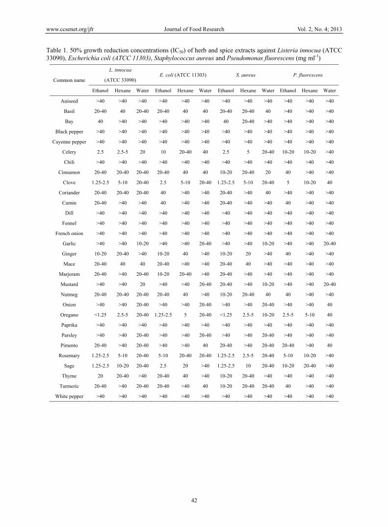
Table 1. 50% growth reduction concentrations (IC50) of herb and spice extracts against Listeria innocua (ATCC 33090), Escherichia coli (ATCC 11303), Staphylococcus aureus and Pseudomonas fluorescens (mg ml-1)
Common name
L. innocua E. coli (ATCC 11303) S. aureus P. fluorescens
(ATCC 33090)
Ethanol Hexane Water Ethanol Hexane Water Ethanol Hexane Water Ethanol Hexane Water
Aniseed >40 >40 >40 >40 >40 >40 >40 >40 >40 >40 >40 >40
Basil 20-40 40 20-40 20-40 40 40 20-40 20-40 40 >40 >40 >40
Bay 40 >40 >40 >40 >40 >40 40 20-40 >40 >40 >40 >40
Black pepper >40 >40 >40 >40 >40 >40 >40 >40 >40 >40 >40 >40
Cayenne pepper >40 >40 >40 >40 >40 >40 >40 >40 >40 >40 >40 >40
Celery 2.5 2.5-5 20 10 20-40 40 2.5 5 20-40 10-20 10-20 >40
Chili >40 >40 >40 >40 >40 >40 >40 >40 >40 >40 >40 >40
Cinnamon 20-40 20-40 20-40 20-40 40 40 10-20 20-40 20 40 >40 >40
Clove 1.25-2.5 5-10 20-40 2.5 5-10 20-40 1.25-2.5 5-10 20-40 5 10-20 40
Coriander 20-40 20-40 20-40 40 >40 >40 20-40 >40 40 >40 >40 >40
Cumin 20-40 >40 >40 40 >40 >40 20-40 >40 >40 40 >40 >40
Dill >40 >40 >40 >40 >40 >40 >40 >40 >40 >40 >40 >40
Fennel >40 >40 >40 >40 >40 >40 >40 >40 >40 >40 >40 >40
French onion >40 >40 >40 >40 >40 >40 >40 >40 >40 >40 >40 >40
Garlic >40 >40 10-20 >40 >40 20-40 >40 >40 10-20 >40 >40 20-40
Ginger 10-20 20-40 >40 10-20 40 >40 10-20 20 >40 40 >40 >40
Mace 20-40 40 40 20-40 >40 >40 20-40 40 >40 >40 >40 >40
Marjoram 20-40 >40 20-40 10-20 20-40 >40 20-40 >40 >40 >40 >40 >40
Mustard >40 >40 20 >40 >40 20-40 20-40 >40 10-20 >40 >40 20-40
Nutmeg 20-40 20-40 20-40 20-40 40 >40 10-20 20-40 40 40 >40 >40
Onion >40 >40 20-40 >40 >40 20-40 >40 >40 20-40 >40 >40 40
Oregano <1.25 2.5-5 20-40 1.25-2.5 5 20-40 <1.25 2.5-5 10-20 2.5-5 5-10 40
Paprika >40 >40 >40 >40 >40 >40 >40 >40 >40 >40 >40 >40
Parsley >40 >40 20-40 >40 >40 20-40 >40 >40 20-40 >40 >40 >40
Pimento 20-40 >40 20-40 >40 >40 40 20-40 >40 20-40 20-40 >40 40
Rosemary 1.25-2.5 5-10 20-40 5-10 20-40 20-40 1.25-2.5 2.5-5 20-40 5-10 10-20 >40
Sage 1.25-2.5 10-20 20-40 2.5 20 >40 1.25-2.5 10 20-40 10-20 20-40 >40
Thyme 20 20-40 >40 20-40 40 >40 10-20 20-40 >40 >40 >40 >40
Turmeric 20-40 >40 20-40 20-40 >40 40 10-20 20-40 20-40 40 >40 >40
White pepper >40 >40 >40 >40 >40 >40 >40 >40 >40 >40 >40 >40
www.ccsenet.org/jfr Journal of Food Research Vol. 2, No. 4; 2013
43
Table 2. Total phenolic content (mg GAE g-1 extract), Minimum inhibitory concentrations (MICs) and Minimum bactericidal concentrations (MBCs) of ‘active’ herb and spice extracts against Listeria innocua (ATCC 33090), Escherichia coli (ATCC 11303), Staphylococcus aureus and Pseudomonas fluorescens (mg ml-1)
Spices and herbs Solvent Total phenolic content L. innocua E. coli S. aureus P. fluorescens
used (Mean ± S.D.) MIC MBC MIC MBC MIC MBC MIC MBC
Oregano ethanol 195.00 ± 4.19 5 5 10 10 2.5 5 10 10
hexane 123.83 ± 5.21 10 10 20 20 10 10 20 40
Sage ethanol 120.20 ± 7.95 5 10 10 20 5 10 20 40
hexane 20.63 ± 4.32 20 >40 40 >40 20 >40 >40 >40
Clove ethanol 185.30 ± 6.80 10 10 10 10 10 10 10 10
hexane 78.87 ± 5.72 20 20 20 40 20 20 40 >40
Rosemary ethanol 145.20 ± 1.71 5 5 20 40 5 5 20 40
hexane 73.03 ± 5.53 10 20 40 >40 10 20 >40 >40
Celery ethanol 35.83 ± 2.25 10 40 20 >40 10 20 20 >40
hexane 26.37 ± 4.50 10 40 40 >40 10 40 40 >40
3.2 Kill-Time Study
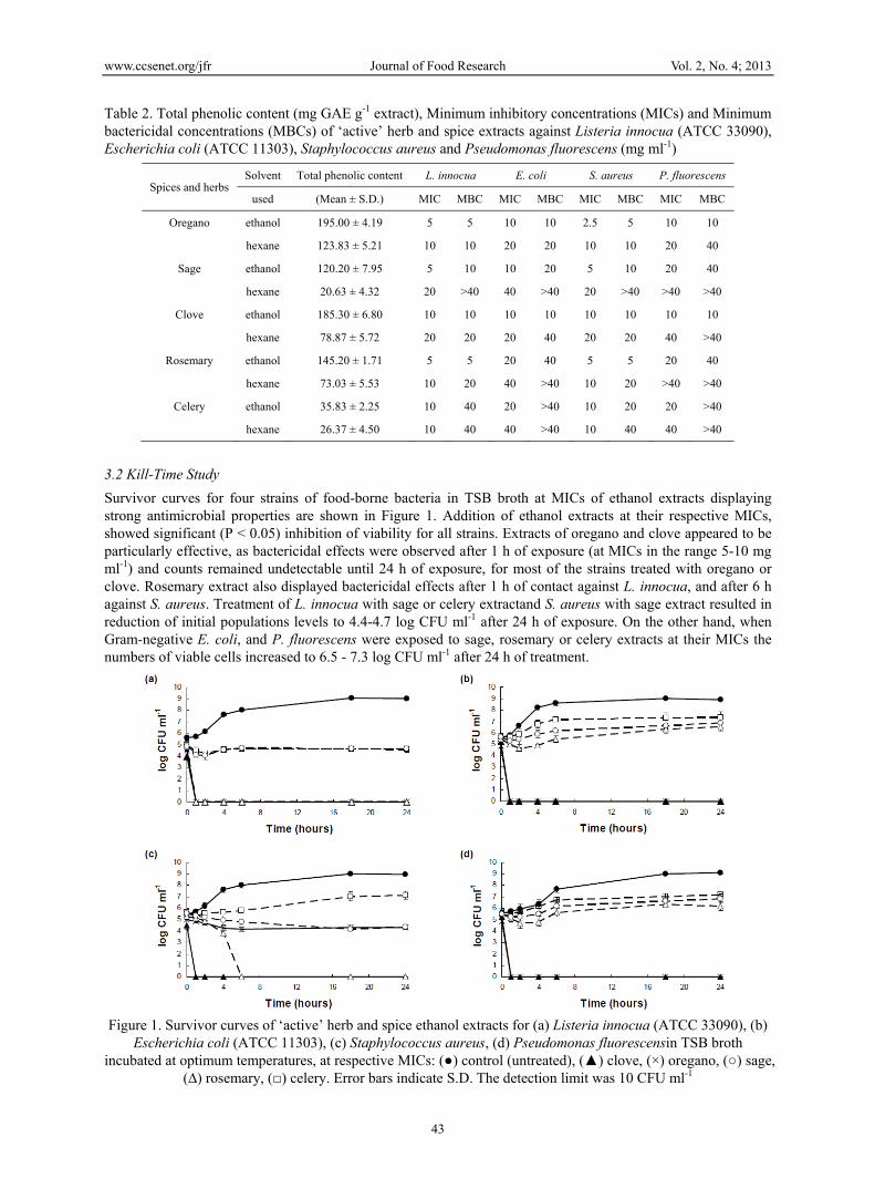
Survivor curves for four strains of food-borne bacteria in TSB broth at MICs of ethanol extracts displaying strong antimicrobial properties are shown in Figure 1. Addition of ethanol extracts at their respective MICs, showed significant (P < 0.05) inhibition of viability for all strains. Extracts of oregano and clove appeared to be particularly effective, as bactericidal effects were observed after 1 h of exposure (at MICs in the range 5-10 mg ml-1) and counts remained undetectable until 24 h of exposure, for most of the strains treated with oregano or clove. Rosemary extract also displayed bactericidal effects after 1 h of contact against L. innocua, and after 6 h against S. aureus. Treatment of L. innocua with sage or celery extractand S. aureus with sage extract resulted in reduction of initial populations levels to 4.4-4.7 log CFU ml-1 after 24 h of exposure. On the other hand, when Gram-negative E. coli, and P. fluorescens were exposed to sage, rosemary or celery extracts at their MICs the numbers of viable cells increased to 6.5 - 7.3 log CFU ml-1 after 24 h of treatment.
Figure 1. Survivor curves of ‘active’ herb and spice ethanol extracts for (a) Listeria innocua (ATCC 33090), (b)
Escherichia coli (ATCC 11303), (c) Staphylococcus aureus, (d) Pseudomonas fluorescensin TSB broth incubated at optimum temperatures, at respective MICs: (●) control (untreated), (▲) clove, (×) oregano, (○) sage,
(∆) rosemary, (□) celery. Error bars indicate S.D. The detection limit was 10 CFU ml-1
www.ccsenet.org/jfr Journal of Food Research Vol. 2, No. 4; 2013
44
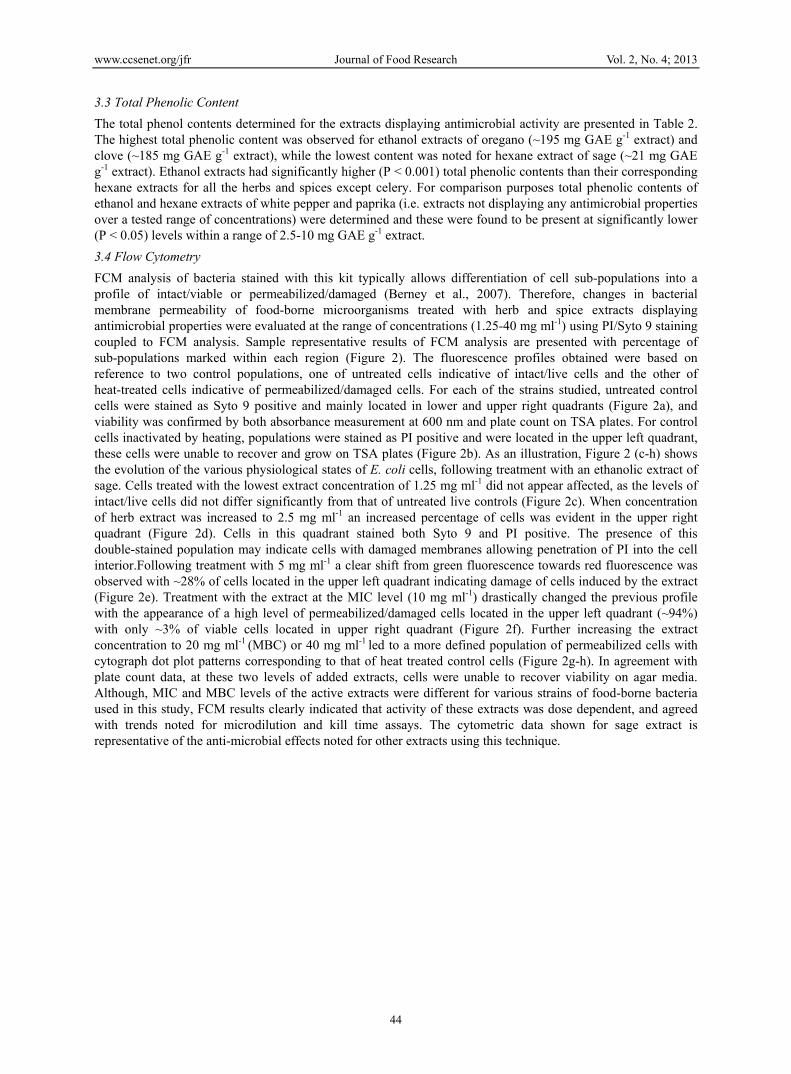
3.3 Total Phenolic Content
The total phenol contents determined for the extracts displaying antimicrobial activity are presented in Table 2. The highest total phenolic content was observed for ethanol extracts of oregano (~195 mg GAE g-1 extract) and clove (~185 mg GAE g-1 extract), while the lowest content was noted for hexane extract of sage (~21 mg GAE g-1 extract). Ethanol extracts had significantly higher (P < 0.001) total phenolic contents than their corresponding hexane extracts for all the herbs and spices except celery. For comparison purposes total phenolic contents of ethanol and hexane extracts of white pepper and paprika (i.e. extracts not displaying any antimicrobial properties over a tested range of concentrations) were determined and these were found to be present at significantly lower (P < 0.05) levels within a range of 2.5-10 mg GAE g-1 extract.
3.4 Flow Cytometry
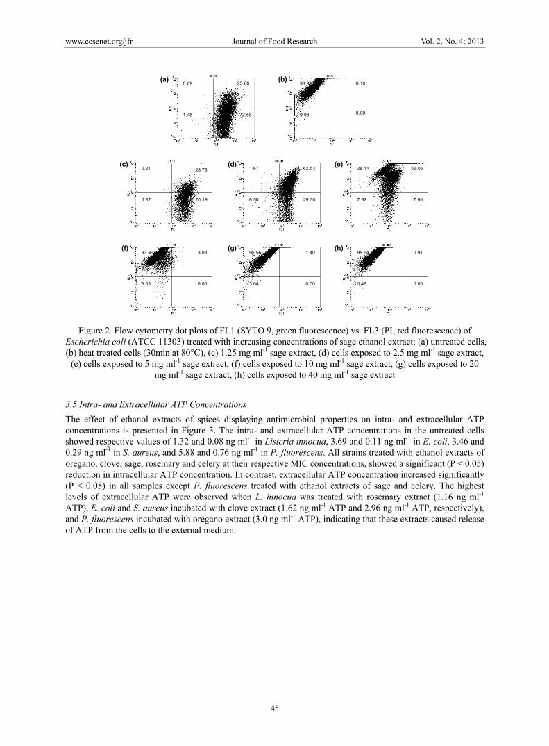
FCM analysis of bacteria stained with this kit typically allows differentiation of cell sub-populations into a profile of intact/viable or permeabilized/damaged (Berney et al., 2007). Therefore, changes in bacterial membrane permeability of food-borne microorganisms treated with herb and spice extracts displaying antimicrobial properties were evaluated at the range of concentrations (1.25-40 mg ml-1) using PI/Syto 9 staining coupled to FCM analysis. Sample representative results of FCM analysis are presented with percentage of sub-populations marked within each region (Figure 2). The fluorescence profiles obtained were based on reference to two control populations, one of untreated cells indicative of intact/live cells and the other of heat-treated cells indicative of permeabilized/damaged cells. For each of the strains studied, untreated control cells were stained as Syto 9 positive and mainly located in lower and upper right quadrants (Figure 2a), and viability was confirmed by both absorbance measurement at 600 nm and plate count on TSA plates. For control cells inactivated by heating, populations were stained as PI positive and were located in the upper left quadrant, these cells were unable to recover and grow on TSA plates (Figure 2b). As an illustration, Figure 2 (c-h) shows the evolution of the various physiological states of E. coli cells, following treatment with an ethanolic extract of sage. Cells treated with the lowest extract concentration of 1.25 mg ml-1 did not appear affected, as the levels of intact/live cells did not differ significantly from that of untreated live controls (Figure 2c). When concentration of herb extract was increased to 2.5 mg ml-1 an increased percentage of cells was evident in the upper right quadrant (Figure 2d). Cells in this quadrant stained both Syto 9 and PI positive. The presence of this double-stained population may indicate cells with damaged membranes allowing penetration of PI into the cell interior.Following treatment with 5 mg ml-1 a clear shift from green fluorescence towards red fluorescence was observed with ~28% of cells located in the upper left quadrant indicating damage of cells induced by the extract (Figure 2e). Treatment with the extract at the MIC level (10 mg ml-1) drastically changed the previous profile with the appearance of a high level of permeabilized/damaged cells located in the upper left quadrant (~94%) with only ~3% of viable cells located in upper right quadrant (Figure 2f). Further increasing the extract concentration to 20 mg ml-1 (MBC) or 40 mg ml-1 led to a more defined population of permeabilized cells with cytograph dot plot patterns corresponding to that of heat treated control cells (Figure 2g-h). In agreement with plate count data, at these two levels of added extracts, cells were unable to recover viability on agar media. Although, MIC and MBC levels of the active extracts were different for various strains of food-borne bacteria used in this study, FCM results clearly indicated that activity of these extracts was dose dependent, and agreed with trends noted for microdilution and kill time assays. The cytometric data shown for sage extract is representative of the anti-microbial effects noted for other extracts using this technique.
www.ccsenet.org/jfr Journal of Food Research Vol. 2, No. 4; 2013
45
Figure 2. Flow cytometry dot plots of FL1 (SYTO 9, green fluorescence) vs. FL3 (PI, red fluorescence) of Escherichia coli (ATCC 11303) treated with increasing concentrations of sage ethanol extract; (a) untreated cells, (b) heat treated cells (30min at 80°C), (c) 1.25 mg ml-1 sage extract, (d) cells exposed to 2.5 mg ml-1 sage extract,
(e) cells exposed to 5 mg ml-1 sage extract, (f) cells exposed to 10 mg ml-1 sage extract, (g) cells exposed to 20 mg ml-1 sage extract, (h) cells exposed to 40 mg ml-1 sage extract
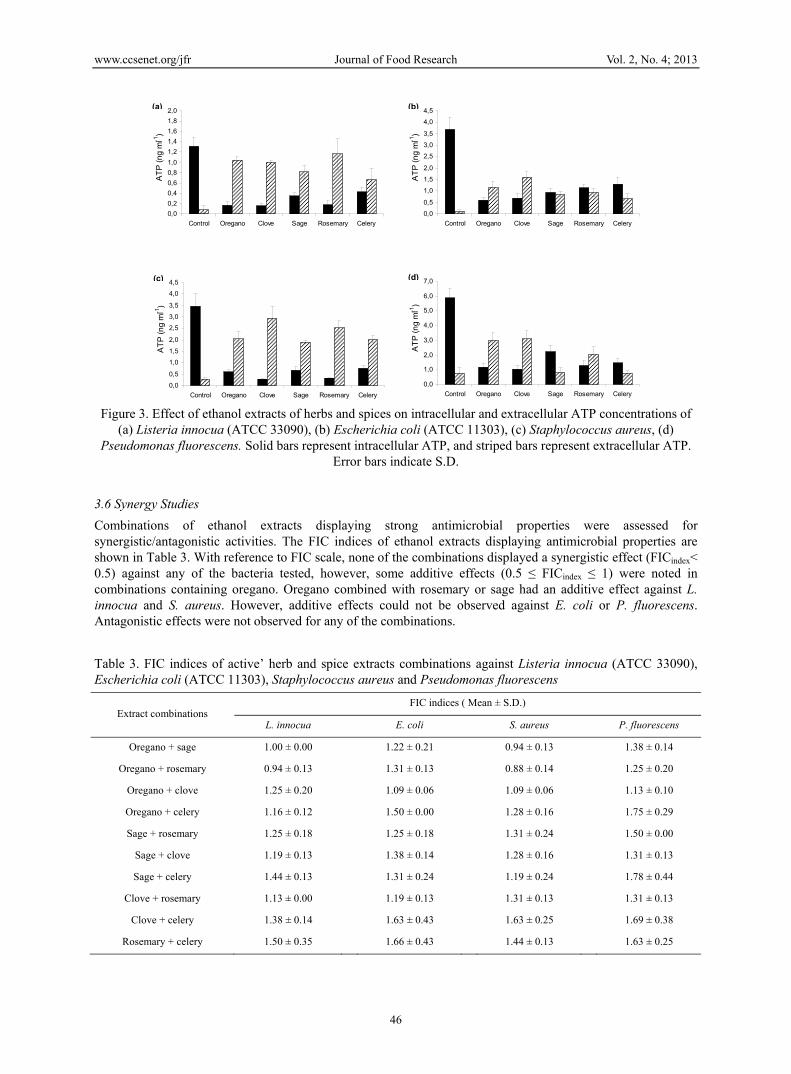
3.5 Intra- and Extracellular ATP Concentrations
The effect of ethanol extracts of spices displaying antimicrobial properties on intra- and extracellular ATP concentrations is presented in Figure 3. The intra- and extracellular ATP concentrations in the untreated cells showed respective values of 1.32 and 0.08 ng ml-1 in Listeria innocua, 3.69 and 0.11 ng ml-1 in E. coli, 3.46 and 0.29 ng ml-1 in S. aureus, and 5.88 and 0.76 ng ml-1 in P. fluorescens. All strains treated with ethanol extracts of oregano, clove, sage, rosemary and celery at their respective MIC concentrations, showed a significant (P < 0.05) reduction in intracellular ATP concentration. In contrast, extracellular ATP concentration increased significantly (P < 0.05) in all samples except P. fluorescens treated with ethanol extracts of sage and celery. The highest levels of extracellular ATP were observed when L. innocua was treated with rosemary extract (1.16 ng ml-1 ATP), E. coli and S. aureus incubated with clove extract (1.62 ng ml-1 ATP and 2.96 ng ml-1 ATP, respectively), and P. fluorescens incubated with oregano extract (3.0 ng ml-1 ATP), indicating that these extracts caused release of ATP from the cells to the external medium.
0.09 25.86
1.48 72.58
(a)98.92 0.10
0.98 0.00
(b)
28.730.21
0.87 70.19
(c) 1.67 62.53
6.50 29.30
(d)28.11 56.06
7.92 7.90
(e)
93.89 3.08
3.03 0.00
(f) 95.56 1.40
3.04 0.00
(g)98.64 0.91
0.46 0.00
(h)
www.ccsenet.org/jfr Journal of Food Research Vol. 2, No. 4; 2013
46
Figure 3. Effect of ethanol extracts of herbs and spices on intracellular and extracellular ATP concentrations of
(a) Listeria innocua (ATCC 33090), (b) Escherichia coli (ATCC 11303), (c) Staphylococcus aureus, (d) Pseudomonas fluorescens. Solid bars represent intracellular ATP, and striped bars represent extracellular ATP.
Error bars indicate S.D.
3.6 Synergy Studies
Combinations of ethanol extracts displaying strong antimicrobial properties were assessed for synergistic/antagonistic activities. The FIC indices of ethanol extracts displaying antimicrobial properties are shown in Table 3. With reference to FIC scale, none of the combinations displayed a synergistic effect (FICindex< 0.5) against any of the bacteria tested, however, some additive effects (0.5 ≤ FICindex ≤ 1) were noted in combinations containing oregano. Oregano combined with rosemary or sage had an additive effect against L. innocua and S. aureus. However, additive effects could not be observed against E. coli or P. fluorescens. Antagonistic effects were not observed for any of the combinations.
Table 3. FIC indices of active’ herb and spice extracts combinations against Listeria innocua (ATCC 33090), Escherichia coli (ATCC 11303), Staphylococcus aureus and Pseudomonas fluorescens
Extract combinations FIC indices ( Mean ± S.D.)
L. innocua E. coli S. aureus P. fluorescens
Oregano + sage 1.00 ± 0.00 1.22 ± 0.21 0.94 ± 0.13 1.38 ± 0.14
Oregano + rosemary 0.94 ± 0.13 1.31 ± 0.13 0.88 ± 0.14 1.25 ± 0.20
Oregano + clove 1.25 ± 0.20 1.09 ± 0.06 1.09 ± 0.06 1.13 ± 0.10
Oregano + celery 1.16 ± 0.12 1.50 ± 0.00 1.28 ± 0.16 1.75 ± 0.29
Sage + rosemary 1.25 ± 0.18 1.25 ± 0.18 1.31 ± 0.24 1.50 ± 0.00
Sage + clove 1.19 ± 0.13 1.38 ± 0.14 1.28 ± 0.16 1.31 ± 0.13
Sage + celery 1.44 ± 0.13 1.31 ± 0.24 1.19 ± 0.24 1.78 ± 0.44
Clove + rosemary 1.13 ± 0.00 1.19 ± 0.13 1.31 ± 0.13 1.31 ± 0.13
Clove + celery 1.38 ± 0.14 1.63 ± 0.43 1.63 ± 0.25 1.69 ± 0.38
Rosemary + celery 1.50 ± 0.35 1.66 ± 0.43 1.44 ± 0.13 1.63 ± 0.25
0,0
0,2
0,4
0,6
0,8
1,0
1,2
1,4
1,6
1,8
2,0
Control Oregano Clove Sage Rosemary Celery
AT
P (
ng
ml-1
)
(a)
0,0
0,5
1,0
1,5
2,0
2,5
3,0
3,5
4,0
4,5
Control Oregano Clove Sage Rosemary Celery
AT
P (
ng
ml-1
)
(b)
0,0
0,5
1,0
1,5
2,0
2,5
3,0
3,5
4,0
4,5
Control Oregano Clove Sage Rosemary Celery
AT
P (
ng
ml-1
)
(c)
0,0
1,0
2,0
3,0
4,0
5,0
6,0
7,0
Control Oregano Clove Sage Rosemary Celery
AT
P (
ng
ml-1
)
(d)
www.ccsenet.org/jfr Journal of Food Research Vol. 2, No. 4; 2013
47
4. Discussion
The type of solvent used to extract herbs and spices appeared to have a major impact on their antimicrobial activity. Generally, extraction of herbs and spices with ethanol and hexane resulted in a product with greater overall antimicrobial activity than extraction with water, as aqueous extracts of all the herbs and spices displayed little or no antimicrobial activity against any of the bacteria tested. This is probably due to the fact that, although the solvents were removed from extracts by evaporation, different chemical compounds were extracted using various solvents, and most of the components with antimicrobial properties are aromatic or saturated organic compounds which are generally more soluble in solvents such as ethanol or methanol (Cowan, 1999). In another study, on antimicrobial activity of Australian herbs, water, ethanol and hexane were also the solvents used for extraction, and it was observed that the aqueous extracts displayed little or no antimicrobial activity (Dupont, Caffin, Bhandari, & Dykes, 2006). Similarly Weerakkody et al. (2010) observed that water extracts of oregano and rosemary had little or no antimicrobial activity compared to ethanol or hexane extracts. On the other hand, Sofia et al. (2007) reported that water extracts of mustard, cinnamon, garlic and clove had good inhibitory activities against E. coli and S. aureus, with MICs in the range of 5-30 mg ml-1. In the present study aqueous extracts over a similar concentration range displayed weak/moderate antimicrobial properties for mustard, cinnamon, garlic and clove, with only 50% growth inhibition noted. However, Sofia et al. (2007), used fresh or dried herbs and spices mixed with distilled water, which was not removed further, while in the present study water was evaporated at 70 °C. Therefore, it is possible that heating may have inactivated some compounds displaying antimicrobial properties, resulting in extracts with lower activities. Indeed, some studies have reported that garlic subjected to autoclaving or heat treatment at 100°C for 20 min, resulted in a complete destruction of inhibitory components (Sato, Terao, & Honma, 1990). In the present study unheated water was used in the extraction of aqueous extracts, and this process is unlikely to have extracted any of the essential oil components (Dupont et al., 2006). Interestingly, in case of garlic, and onion, some antibacterial activity was observed for aqueous but not for ethanol or hexane extracts. Therefore, these results suggest that some water soluble compounds displaying some moderate antimicrobial activities were present in these extracts. More specifically, garlic and onion on crushing were previously found to generate the principal bioactive compound, allicin, through enzymatic conversion of alliin (Ellmore & Feldberg, 1994) and its antimicrobial properties have been well documented.
In this study ethanol and hexane extracts of oregano, clove, sage, rosemary and celery proved to be the most effective against four food-borne bacteria tested. A number of studies have been published on the antimicrobial activities of plant extracts and essential oils against different types of food-borne bacteria (Burt & Reinders, 2003; Hammer, Carson, & Riley, 1999; Moreira, Ponce, del Valle, & Roura, 2005). However, direct comparison of results obtained in various studies is problematic due to a number of factors such as variability in composition of plant extracts as a result of local climatic and environmental conditions, low number of herb and spice samples tested, differences in experimental design including inoculum size, growth phase, strain susceptibility, and culture medium used (Cosentino et al., 1999). Moreover, in many publications the agar disc diffusion method was used to screen antimicrobial potency of plant extracts or essential oils, which should be treated rather as preliminary, qualitative data only (Smith-Palmer, Stewart, & Fyfe, 1998). Nonetheless, a number of previous studies have shown that oregano, clove, rosemary and sage extracts or essential oils displayed the strongest and most consistent inhibitory effects against food-borne bacteria among many herbs and spices tested (Kalemba & Kunicka, 2003; Smith-Palmer et al., 1998; Zaika, 1988). Similar to our results, Zhang et al. (2009) found that ethanol extracts of clove and rosemary displayed significant inhibitory properties against Escherichia coli, Pseudomonas fluorescens and Listeria monocytogenes, when tested at 5-80 mg ml-1. Weerakkody et al. (2010) reported that ethanol or hexane extracts of oregano and rosemary strongly inhibited the growth of S. aureus, Escherichia coli and Listeria monocytogenes, with similar MIC values to those found in the present study. Pandit and Shelef (1994) studied the inhibitory properties of 18 spices and found that clove and rosemary had the highest activity, and that similar to our results ethanol extract of rosemary was listericidal at 5 mg ml-1. Sage extracts and oils were also reported to exhibit significant antibacterial activity against E. coli, S. aureus and Listeria spp. (Sagdic, Karahan, Ozcan, & Ozkan, 2003; Yasar, Sagdic, & Kisioglu, 2005).
On the other hand, celery preparations are not frequently included in antimicrobial screening studies, and limited data concerning bioactivity of this plant exist (Dostbil, 2007). Interestingly, in this study, ethanol and hexane extracts of celery displayed relatively strong antimicrobial properties against all strains tested, with MIC levels comparable to that of celery essential oil reported previously (Hammer et al., 1999). This spice could be particularly useful in extending shelf-life of food products due to its wide range of applications, as celery fruit (seed) extracts are extensively used as flavouring ingredients in various types of products, including soups,
www.ccsenet.org/jfr Journal of Food Research Vol. 2, No. 4; 2013
48
gravies, meat products, frozen dairy desserts, baked goods, candies, puddings, sauces, relishes, snack foods, and beverages (Momin & Nair, 2000).
Many previous studies have demonstrated significant inhibitory activities of thyme and cinnamon (Burt & Reinders, 2003; Oussalah, Caillet, Saucier, & Lacroix, 2006; Smith-Palmer et al., 1998). In contrast, we found that extracts of these seasonings displayed only 50% growth reduction in the tested range of concentrations indicating weak antibacterial activities. As mentioned before, this could probably be due to a number of reasons, the main one being variability in composition resulting from different sources of origin or harvesting seasons (McGimpsey et al., 1994). Indeed, Oussalah et al. (2006) studied the antimicrobial properties of essential oils of several thyme species and found that despite a common botanical origin, the chemical composition and antimicrobial activity varied considerably. Moreover, antimicrobial properties can differ significantly within the same herb or spice species as the proportions of individual components in essential oils of plants are affected by genotype (Arrebola, Navarro, Jimenez, & Ocana, 1994). In agreement with our results, extracts and essential oils of aniseed, black pepper, white pepper, paprika, fennel and dill have been found to be inactive against anumber of food spoilage and pathogenic bacteria (Hammer et al., 1999; Sagdic et al., 2003; Smith-Palmer et al., 1998; Weerakkody et al., 2010).
The microdilution broth method is very useful for determination of the MIC of an antimicrobial compound. However, one of the major problems with the use of turbidimetric analysis to determine MIC values is the minimal range of detection. A bacterial culture with a concentration of 106-107 CFU ml-1 is needed for the spectrophotometer to record a meaningful reading. Thus, in many cases an actively growing culture may be undetected with no increase in absorbance using a spectrophotometer (Kim, Marshall, & Wei, 1995).Therefore, kill-time studies were performed to gain a better insight on bacteriostatic or bactericidal effects of the most potent ethanol extracts. Ethanol extracts of oregano and clove used in the present study appeared to be particularly effective, as bactericidal effects were observed at MICs already after 1 h of exposure, and were maintained during 24 h for both Gram-positive and Gram-negative bacteria. In accordance with our results, Moreira et al. (2005) reported that clove essential oil used at MIC level against E. coli exerted its maximum bactericidal effect after 15 min and its action was maintained during 20 h of incubation. On the other hand, in this study, extracts of sage or celery in most cases exerted bacteriostatic activity against Gram-positive bacteria, while levels of Gram-negative bacteria slightly increased over a 24 h period, indicating that periodic sampling of cultures exposed to extracts should be performed in addition to turbidimetric measurements to determine bacterial survival.
In the present study, data indicates that both Gram-positive and Gram-negative bacteria were affected by the plant extracts tested. However, Gram-positive strains were somewhat more sensitive when treated with herb or spice preparations. This finding is consistent with previous studies on a range of herbs and spices (Farag, Daw, Hewedi, & El-Baroty, 1989; Shan et al., 2007) and may be mainly related to differences in cell wall structure and the outer membrane arrangement between Gram-positive and Gram-negative bacteria. Gram-negative bacteria possess a hydrophilic outer membrane rich in lipopolysaccharide molecules, which serves as a penetration barrier towards macromolecules (Nikaido, 1996). Although this explanation is widely accepted, and true for many essential oils, some researchers have stated that the Gram distinction may have little relation to growth inhibition (Kotzekidou, Giannakidis, & Boulamatsis, 2008) and some herbs and spices are equally effective against both groups of bacteria, one of them being clove (Kim et al., 1995), which was also observed in our study. However, the outer membrane is not completely impermeable as there are porin proteins present in this layer that can create channels large enough to allow restricted passage of molecules with a molecular mass below 600 Da, such as substituted phenolics in herb and spice extracts and essential oils, allowing their slow penetration into the periplasmic space and the cytoplasmic membrane (Abee, Krockel, & Hill, 1995). Thus it is possible that over a longer contact time the active compounds present in oleoresins and essential oils would have the same effect on Gram-negative and Gram-positive bacteria (Deans & Ritchie, 1987).
The total phenolic data are also difficult to compare directly, due to differences in extraction methods, using different solvents or assay methods. Nonetheless, similar levels of total phenols have been reported for some spice extracts in previous studies (Ozkan, Sagdic, Ekici, Ozturk, & Ozcan, 2007). Shan et al. (2007) also observed that significantly higher amounts of total phenols were extracted from rosemary using ethanol than using hexane. There are contradictory reports concerning the relationship between the antimicrobial activity and total phenolic content in herb and spice preparations. Many studies claim that phenolic substances present in herbs and spices may be responsible for antimicrobial activity (Kim et al., 1995). Shan et al. (2007) reported that antibacterial activity of selected dietary spice and medicinal extracts was closely related to the concentration of phenolic compounds. In contrast, Weerakkody et al. (2010) found that this relationship was weak. In the present
www.ccsenet.org/jfr Journal of Food Research Vol. 2, No. 4; 2013
49
study, extracts with relatively high total phenolic content (>100mg GAE g-1 extract) displayed the strongest antimicrobial properties against all bacterial strains tested, while spices such as white pepper or paprika with low total phenolic content did not show any inhibitory properties. Indeed, studies on isolated components of essential oils and extracts confirmed that the highest antibacterial activity was reported for phenols -carvacrol, eugenol, thymol (Dorman & Deans, 2000). Thus, extracts with phenols as their main compounds display the broadest spectrum of activity against microorganisms (Kalemba & Kunicka, 2003). These include oregano containing carvacrol and thymol, and clove with eugenol as the main constituent, followed by rosemary and sage containing rosmarinic acid and other phenolic acids, as well as carnosol, rosmanol and carnosic acids as phenolic diterpenes (Collins & Charles, 1987; Santos-Gomes, Seabra, Andrade, & Fernandes-Ferreira, 2002).
On the other hand, ethanol and hexane extracts of celery with low phenolic contents still possessed relatively strong antimicrobial properties. Similarly, Weerakkody et al. (2010) reported that some herb and spice extracts with low phenolic content, were more inhibitory against some bacterial strains than those containing high levels of phenols. Therefore, this data suggests that although antimicrobial activities of some spices might be attributed to their phenolic compounds, there may be other substances in addition to the phenolics, responsible for the antibacterial properties. Indeed, Misic et al. (2008) studied antimicrobial properties of celery fruit isolates, and reported that preparations characterized by higher concentrations of the main aromatic compounds sedanenolide, sedanolide and 3-n-butylphthalide showed stronger antimicrobial activities against the tested bacteria.
It is generally accepted that aromatic and phenolic compounds exert their antimicrobial activities at the cytoplasmic membrane by changes in its structure and function (Sikkema, Debont, & Poolman, 1995). As plant extracts and essential oils contain dozens of different phytochemicals, it may involve multiple modes of action against bacteria (Lambert et al., 2001). These activities include: interference with the phospholipid bilayer of the cell membrane leading to increase in permeability and leakage of cellular components, impairment of enzymatic mechanisms for energy production and metabolism, disruption of the proton motive force, destruction or inactivation of genetic material (Cox et al., 1998; Kim et al., 1995; Ultee, Bennik, & Moezelaar, 2002), with some mechanisms following on as a consequence of other mechanisms (Shan et al., 2007). In this study, the effects of herb and spice extracts on membrane function were evaluated using differential staining with FCM analysis. Clear differences in cytometric profiles of viable/intact and permeabilized/damaged sub-populations were noted during treatments with ethanolic extracts of sage, oregano, clove, rosemary and celery. Our results indicate that selected herb and spice extracts displayed antimicrobial activity with consequent damage to bacterial cell membranes in Listeria innocua, Staphylococcus aureus, Escherichia coli and Pseudomonas fluorescens. Moreover, FCM analysis demonstrated that the extent of membrane damage was dependent on concentration of spice and herb extractused. To our knowledge, there are few reports showing the effects on cell membrane permeability using FCM of food-borne bacteria subjected to treatment with herb and spice extracts or essential oils. In a study on antimicrobial activity of oregano essential oil against Listeria monocytogenes, FCM analysis coupled with fluorescent staining revealed that membrane disintegration was the primary inactivation mechanism (Paparella et al., 2008). In another study on antibacterial potential of eugenol and carvacrol, the main antimicrobial components of clove and oregano against both Gram-positive and Gram-negative bacteria, it was observed that at bactericidal concentrations the primary mechanism of action of eugenol and carvacrol is disruption of the cytoplasmic membrane, which increases its non-specific permeability (Gill & Holley, 2006). Our results showed that flow cytometry can be a very useful additional technique to traditional methods to evaluate the effects of herb and spice extracts on food spoilage and pathogenic bacteria. Moreover, as FCM is a rapid and sensitive method, it is capable of generating data on the extent of heterogeneity within a population, making it attractive analytical technique for design of effective antimicrobial systems for application in food industry.
Our results showed that upon treatment with herb and spice extracts the concentration of intracellular ATP decreased in all samples against all bacterial strains tested, while the concentration of extracellular ATP increased in all cases except for P. fluorescens treated with celery or sage extracts. This is in agreement with previous studies investigating mechanisms of antimicrobial action of essential oils of herbs and spices (Caillet et al., 2005; Lacroix, Caillet, & Shareck, 2009; Turgis, Han, Caillet, & Lacroix, 2009), and indicates that the increase in extracellular ATP in the presence of herb and spice extracts is at least partially due to intracellular ATP leakage outside the cell, possibly as a consequence of cell envelope disruption by the antimicrobial compounds (Caillet et al., 2005). However, when P. fluorescens was treated with celery or sage extracts, there was no correlation between the intra- and extracellular ATP concentrations, as extracellular ATP concentration remained at the same level as the untreated sample, despite the reduction in intracellular ATP level. This phenomenon was previously observed by Ultee et al. (1999) when Bacillus cereus was treated with carvacrol,
www.ccsenet.org/jfr Journal of Food Research Vol. 2, No. 4; 2013
50
who suggested that depletion of the internal ATP pool resulted from a reduced rate of ATP synthesis or increased ATP hydrolysis, rather than from disruption of the cell membrane. As intracellular ATP may be strongly reduced in presence of a weak ATP efflux it can be concluded that it is partially hydrolysed inside the treated cells (Caillet et al., 2005). However, in contrast to these researchers, Helander et al. (1998) reported that carvacrol and thymol decreased the intracellular ATP pool of E. coli and increased extracellular ATP concentration, indicating disruptive action on the cytoplasmic membrane. P. fluorescens was found to be the most resistant strain to the action of herb and spice extracts, and it is therefore possible that the contact time of 30 min applied in this experiment, may not have been sufficient to cause damage to the bacterial membrane and hence no changes in extracellular ATP pool could be observed. The results of this study are consistent with flow cytometry analysis revealing that the cell membrane structures were impaired by spice and herb active extracts, and indicate that the intracellular ATP was probably leaked through compromised membranes.
Some studies reported that the whole essential oils or extracts have a greater activity than the major components mixed together (Gill, Delaquis, Russo, & Holley, 2002; Lambert et al., 2001) and it was suggested that minor components are critical to antimicrobial activity and may have a synergistic effect or potentiating influence (Burt, 2004). In this study, no synergy according to FIC indices was observed for any of extract combinations, however oregano combined with sage or rosemary exerted some additive effects against L. innocua and S. aureus. As the antimicrobial properties of herb and spice derived preparations depend not only on chemical composition but also on the lipophilic properties and water solubility, combinations of various compounds may have contributed to the observed additive effects (Gutierrez, Barry-Ryan, & Bourke, 2008). Gutierrez et al. (2008) tested efficacy of essential oils in combinations using the same method, and found no synergy but only some additive effects when oregano was combined with rosemary and sage against B. cereus, while oregano in combination with thyme, marjoram or basil showed additive effects against E. coli, P. aeruginosa, or B. cereus. These authors also suggested that, as the plant essential oils or extracts possess similar compositions, their combinations may exhibit an additive rather than a synergistic effect. However, the magnitude of synergistic interactions between the mixtures of extracts and essential oils or their individual components, also reported previously seems to be too low to be of any practical importance (Kalemba & Kunicka, 2003). In contrast, strong synergistic inhibitory effects have been observed between herb and spice extracts or essential oils and some food additives, such as salt, sucrose and low pH (Kivanc & Akgäul, 1988). Therefore, combinations of extracts with other preservation methods might yield stronger synergistic effects.
While there are many studies on the antimicrobial effects of spice extracts, these have been usually conducted for one or a number of herbs and spices and extracted using a single type of solvent. These various studies are difficult to compare directly due to differences in experimental design including method of extract preparation, temperature of solvent removal, inoculum size, growth phase, strain susceptibility, culture medium used, etc. In contrast, the present study is systematic and has provided directly comparable quantitative antimicrobial data for 90 extracts prepared with solvents of various polarity and indicate that crude extracts of selected herbs and spices, possess in vitro activity against food spoilage and pathogenic bacteria. Ethanol extracts of oregano, clove, rosemary, sage and celery displayed bactericidal or bacteriostatic activities with consequent damage to bacterial cell membranes of both Gram-positive and Gram-negative bacteria which was also confirmed by flow cytometry (FCM). In the light of trends towards selection of natural antimicrobial compounds, some of these extracts have the potential to extend the shelf-life or improve the safety of foods including ready meals with reduced salt content. However, antimicrobial activity of spice derived preparations has been reported to diminish within food systems (Kotzekidou et al., 2008). Therefore, further studies on the efficacy of these extracts in a range of ready meal products as well as evaluation of potential interactions of antimicrobial compounds with components of food matrices such as fats, carbohydrates, proteins, pH are warranted. Acknowledgments
This work was funded by the Department of Agriculture, Fisheries and Food under Food Institutional Research Measure (National Development Plan).
References
Abee, T., Krockel, L., & Hill, C. (1995). Bacteriocins: modes of action and potentials in food preservation and control of food poisoning. International Journal of Food Microbiology, 28(2), 169-185. http://dx.doi.org/10.1016/0168-1605(95)00055-0
Arrebola, M. L., Navarro, M. C., Jimenez, J., & Ocana, F. A. (1994). Yield and composition of the essential oil of Thymus serpylloides subsp serpylloides. Phytochemistry, 36(1), 67-72. http://dx.doi.org/10.1016/S0031-9422(00)97014-6
www.ccsenet.org/jfr Journal of Food Research Vol. 2, No. 4; 2013
51
Bonapace, C. R., White, R. L., Friedrich, L. V., & Bosso, J. A. (2000). Evaluation of antibiotic synergy against Acinetobacter baumannii: a comparison with Etest, time-kill, and checkerboard methods. Diagnostic Microbiology and Infectious Disease, 38(1), 43-50. http://dx.doi.org/10.1016/S0732-8893(00)00163-2
Burt, S. (2004). Essential oils: their antibacterial properties and potential applications in foods - a review. International Journal of Food Microbiology, 94(3), 223-253. http://dx.doi.org/10.1016/j.ijfoodmicro.2004.03.022
Burt, S. A., & Reinders, R. D. (2003). Antibacterial activity of selected plant essential oils against Escherichia coli O157 : H7. Letters in Applied Microbiology, 36(3), 162-167. http://dx.doi.org/10.1046/j.1472-765X.2003.01285.x
Caillet, S., Shareck, F., & Lacroix, M. (2005). Effect of gamma radiation and oregano essential oil on murein and ATP concentration of Escherichia coli O157: H7. Journal of Food Protection, 68(12), 2571-2579.
Collins, M. A., & Charles, H. P. (1987). Antimicrobial activity of carnosol and ursolic acid: two anti-oxidant constituents of Rosmarinus officinalis L. Food Microbiology, 4(4), 311-315. http://dx.doi.org/10.1016/S0740-0020(87)80005-9
Cosentino, S., Tuberoso, C. I. G., Pisano, B., Satta, M., Mascia, V., Arzedi, E., & Palmas, F. (1999). In-vitro antimicrobial activity and chemical composition of Sardinian Thymus essential oils. Letters in Applied Microbiology, 29(2), 130-135. http://dx.doi.org/10.1046/j.1472-765X.1999.00605.x
Cowan, M. M. (1999). Plant products as antimicrobial agents. Clin. Microbiol. Rev., 12(4), 564-582.
Cox, S. D., Gustafson, J. E., Mann, C. M., Markham, J. L., Liew, Y. C., Hartland, R. P.,… Wyllie, S. G. (1998). Tea tree oil causes K+ leakage and inhibits respiration in Escherichia coli. Letters in Applied Microbiology, 26(5), 355-358. http://dx.doi.org/10.1046/j.1472-765X.1998.00348.x
Cronin, U. P., & Wilkinson, M. G. (2008). Bacillus cereus endospores exhibit a heterogeneous response to heat treatment and low-temperature storage. Food Microbiology, 25(2), 235-243. http://dx.doi.org/10.1016/j.fm.2007.11.004
Deans, S. G., & Ritchie, G. (1987). Antibacterial properties of plant essential oils. International Journal of Food Microbiology, 5(2), 165-180. http://dx.doi.org/10.1016/0168-1605(87)90034-1
Desmond, E. (2006). Reducing salt: A challenge for the meat industry. Meat Science, 74(1), 188-196. http://dx.doi.org/10.1016/j.meatsci.2006.04.014
Dorman, H. J. D., & Deans, S. G. (2000). Antimicrobial agents from plants: antibacterial activity of plant volatile oils. Journal of Applied Microbiology, 88(2), 308-316. http://dx.doi.org/10.1046/j.1365-2672.2000.00969.x
Dostbil, N. (2007). An investigation of antimicrobial activity of celery (Apium graveolensis L.). Asian Journal of Chemistry, 19(7), 5518-5522.
Dupont, S., Caffin, N., Bhandari, B., & Dykes, G. A. (2006). In vitro antibacterial activity of Australian native herb extracts against food-related bacteria. Food Control, 17(11), 929-932. http://dx.doi.org/10.1016/j.foodcont.2005.06.005
Ellmore, G. S., & Feldberg, R. S. (1994). Allin lyase localization in bundle sheaths of the garlic clove (Allium sativum). American Journal of Botany, 81(1), 89-94. http://dx.doi.org/10.2307/2445567
Farag, R. S., Daw, Z. Y., Hewedi, F. M., & El-Baroty, G. S. A. (1989). Antibacterial activity of some Egyptian spices essential oils. Journal of Food Protection, 52, 665-667.
Gill, A. O., Delaquis, P., Russo, P., & Holley, R. A. (2002). Evaluation of antilisterial action of cilantro oil on vacuum packed ham. International Journal of Food Microbiology, 73(1), 83-92. http://dx.doi.org/10.1016/S0168-1605(01)00712-7
Gill, A. O., & Holley, R. A. (2006). Disruption of Escherichia coli, Listeria monocytogenes and Lactobacillus sakei cellular membranes by plant oil aromatics. International Journal of Food Microbiology, 108(1), 1-9. http://dx.doi.org/10.1016/j.ijfoodmicro.2005.10.009
Gould, G. W. (1996). Methods for preservation and extension of shelf life. International Journal of Food Microbiology, 33(1), 51-64. http://dx.doi.org/10.1016/0168-1605(96)01133-6
www.ccsenet.org/jfr Journal of Food Research Vol. 2, No. 4; 2013
52
Gutierrez, J., Barry-Ryan, C., & Bourke, P. (2008). The antimicrobial efficacy of plant essential oil combinations and interactions with food ingredients. International Journal of Food Microbiology, 124(1), 91-97. http://dx.doi.org/10.1016/j.ijfoodmicro.2008.02.028
Gutierrez, J., Barry-Ryan, C., & Bourke, P. (2009). Antimicrobial activity of plant essential oils using food model media: Efficacy, synergistic potential and interactions with food components. Food Microbiology, 26(2), 142-150. http://dx.doi.org/10.1016/j.fm.2008.10.008
Hammer, K. A., Carson, C. F., & Riley, T. V. (1999). Antimicrobial activity of essential oils and other plant extracts. Journal of Applied Microbiology, 86(6), 985-990. http://dx.doi.org/10.1046/j.1365-2672.1999.00780.x
Helander, I. M., Alakomi, H. -L., Latva-Kala, K., Mattila-Sandholm, T., Pol, I., Smid, E. J., … von Wright, A. (1998). Characterization of the action of selected essential oil components on Gram-negative bacteria. Journal of Agricultural and Food Chemistry, 46(9), 3590-3595. http://dx.doi.org/10.1021/jf980154m
Kalemba, D., & Kunicka, A. (2003). Antibacterial and antifungal properties of essential oils. Current Medicinal Chemistry, 10(10), 813-829. http://dx.doi.org/10.2174/0929867033457719
Kiessling, C. R., Cutting, J. H., Loftis, M., Kiessling, W. M., Datta, A. R., & Sofos, J. N. (2002). Antimicrobial resistance of food-related Salmonella isolates, 1999-2000. Journal of Food Protection, 65(4), 603-608.
Kim, J., Marshall, M. R., & Wei, C. I. (1995). Antibacterial activity of some essential oil components against five foodborne pathogens. Journal of Agricultural and Food Chemistry, 43(11), 2839-2845. http://dx.doi.org/10.1021/jf00059a013
Kivanc, M., & Akgäul, A. (1988). Effect of some essential oil components on the growth of food-borne bacteria and synergism with some food ingredients. Flavour and Fragrance Journal, 3(2), 95-98. http://dx.doi.org/10.1002/ffj.2730030209
Kotzekidou, P., Giannakidis, P., & Boulamatsis, A. (2008). Antimicrobial activity of some plant extracts and essential oils against foodborne pathogens in vitro and on the fate of inoculated pathogens in chocolate. LWT - Food Science and Technology, 41(1), 119-127. http://dx.doi.org/10.1016/j.lwt.2007.01.016
Lacroix, M., Caillet, S., & Shareck, F. (2009). Bacterial radiosensitization by using radiation processing in combination with essential oil: Mechanism of action. Radiation Physics and Chemistry, 78(7-8), 567-570. http://dx.doi.org/10.1016/j.radphyschem.2009.03.012
Lai, P. K., & Roy, J. (Writers). (2004). Antimicrobial and chemopreventive properties of herbs and spices. Current Medicinal Chemistry: Bentham Science Publishers Ltd.
Lambert, R. J. W., Skandamis, P. N., Coote, P. J., & Nychas, G. J. E. (2001). A study of the minimum inhibitory concentration and mode of action of oregano essential oil, thymol and carvacrol. Journal of Applied Microbiology, 91(3), 453-462. http://dx.doi.org/10.1046/j.1365-2672.2001.01428.x
Lee, J. Y., Kim, Y. S., & Shin, D. H. (2002). Antimicrobial synergistic effect of linolenic acid and monoglyceride against Bacillus cereus and Staphylococcus aureus. Journal of Agricultural and Food Chemistry, 50(7), 2193-2199. http://dx.doi.org/10.1021/jf011175a
McGimpsey, J. A., Douglas, M. H., Van Klink, J. W., Beauregard, D. A., & Perry, N. B. (1994). Seasonal variation in essential oil yield and composition from naturalized thymus vulgaris L. in New Zealand. Flavour and Fragrance Journal, 9(6), 347-352. http://dx.doi.org/10.1002/ffj.2730090613
Meng, J., & Doyle, M. P. (1998). Emerging and evolving microbial foodborne pathogens. Bulletin De L Institut Pasteur, 96(3), 151-163. http://dx.doi.org/10.1016/S0020-2452(98)80010-9
Misic, D., Zizovic, I., Stamenic, M., Asanin, R., Ristic, M., Petrovic, S. D., & Skala, D. (2008). Antimicrobial activity of celery fruit isolates and SFE process modeling. Biochemical Engineering Journal, 42(2), 148-152. http://dx.doi.org/10.1016/j.bej.2008.06.008
Momin, R. A., & Nair, M. G. (2000). Mosquitocidal, nematicidal, and antifungal compounds from Apium graveolens L. seeds. Journal of Agricultural and Food Chemistry, 49(1), 142-145. http://dx.doi.org/10.1021/jf001052a
Moreira, M. R., Ponce, A. G., del Valle, C. E., & Roura, S. I. (2005). Inhibitory parameters of essential oils to reduce a foodborne pathogen. LWT-Food Science and Technology, 38(5), 565-570. http://dx.doi.org/10.1016/j.lwt.2004.07.012
www.ccsenet.org/jfr Journal of Food Research Vol. 2, No. 4; 2013
53
Moreno, S., Scheyer, T., Romano, C. S., & Vojnov, A. A. (2006). Antioxidant and antimicrobial activities of rosemary extracts linked to their polyphenol composition. Free Radical Research, 40(2), 223-231. http://dx.doi.org/10.1080/10715760500473834
Nikaido, H. (1996). Outer membrane. In F. C. Neidhardt (Ed.), Escherichia coli and Salmonella: Cellular and Molecular Biology (Vol. 1). Washington, D.C.: American Society for Microbiology Press.
Oussalah, M., Caillet, S., Saucier, L., & Lacroix, M. (2006). Antimicrobial effects of selected plant essential oils on the growth of a Pseudomonas putida strain isolated from meat. Meat Science, 73(2), 236-244. http://dx.doi.org/10.1016/j.meatsci.2005.11.019
Ozkan, G., Sagdic, O., Ekici, L., Ozturk, I., & Ozcan, M. M. (2007). Phenolic compounds of Origanum sipyleum L. extract, and its antioxidant and antibacterial activities. Journal of Food Lipids, 14(2), 157-169. http://dx.doi.org/10.1111/j.1745-4522.2007.00077.x
Pandit, V. A., & Shelef, L. A. (1994). Sensitivity of Listeria monocytogenes to rosemary (Rosmarinus officinalis L.). Food Microbiology, 11(1), 57-63. http://dx.doi.org/10.1006/fmic.1994.1008
Paparella, A., Taccogna, L., Aguzzi, I., Chaves-López, C., Serio, A., Marsilio, F., & Suzzi, G. (2008). Flow cytometric assessment of the antimicrobial activity of essential oils against Listeria monocytogenes. Food Control, 19(12), 1174-1182. http://dx.doi.org/10.1016/j.foodcont.2008.01.002
Sagdic, O., Karahan, A. G., Ozcan, M., & Ozkan, G. (2003). Note: Effect of some spice extracts on bacterial inhibition. Food Science and Technology International, 9(5), 353-358. http://dx.doi.org/10.1177/1082013203038976
Santos-Gomes, P. C., Seabra, R. M., Andrade, P. B., & Fernandes-Ferreira, M. (2002). Phenolic antioxidant compounds produced by in vitro shoots of sage (Salvia officinalis L.). Plant Science, 162(6), 981-987. http://dx.doi.org/10.1016/S0168-9452(02)00052-3
Sato, A., Terao, M., & Honma, Y. (1990). Antibacterial action of garlic extract on food poisoning bacteria. Journal of the Food Hygienic Society of Japan, 31(4), 328-332. http://dx.doi.org/10.3358/shokueishi.31.328
Shan, B., Cai, Y.-Z., Brooks, J. D., & Corke, H. (2007). The in vitro antibacterial activity of dietary spice and medicinal herb extracts. International Journal of Food Microbiology, 117(1), 112-119. http://dx.doi.org/10.1016/j.ijfoodmicro.2007.03.003
Sikkema, J., Debont, J. A. M., & Poolman, B. (1995). Mechanisms of membrane toxicity of hydrocarbons. Microbiological Reviews, 59(2), 201-222.
Skerget, M., Kotnik, P., Hadolin, M., Hras, A. R., Simonic, M., & Knez, Z. (2005). Phenols, proanthocyanidins, flavones and flavonols in some plant materials and their antioxidant activities. Food Chemistry, 89(2), 191-198. http://dx.doi.org/10.1016/j.foodchem.2004.02.025
Smid, E. J., & Gorris, L. G. M. (1999). Natural antimicrobials for food preservation. In M. S. Rahman (Ed.), Handbook of Food Preservation (pp. 285-308). New York: Marcel Dekker.
Smith-Palmer, A., Stewart, J., & Fyfe, L. (1998). Antimicrobial properties of plant essential oils and essences against five important food-borne pathogens. Letters in Applied Microbiology, 26(2), 118-122. http://dx.doi.org/10.1046/j.1472-765X.1998.00303.x
Sofia, P. K., Prasad, R., Vijay, V. K., & Srivastava, A. K. (2007). Evaluation of antibacterial activity of Indian spices against common foodborne pathogens. International Journal of Food Science & Technology, 42, 910-915. http://dx.doi.org/10.1111/j.1365-2621.2006.01308.x
Sokmen, A., Gulluce, M., Akpulat, H. A., Daferera, D., Tepe, B., Polissiou, M., ... Sahin, F. (2004). The in vitro antimicrobial and antioxidant activities of the essential oils and methanol extracts of endemic Thymus spathulifolius. Food Control, 15(8), 627-634. http://dx.doi.org/10.1016/j.foodcont.2003.10.005
Souza, E. L. d., Stamford, T. L. M., & Lima, E. d. O. (2006). Sensitivity of spoiling and pathogen food-related bacteria to Origanum vulgare L. (Lamiaceae) essential oil. Brazilian Journal of Microbiology, 37, 527-532. http://dx.doi.org/10.1590/S1517-83822006000400023
Turgis, M., Han, J., Caillet, S., & Lacroix, M. (2009). Antimicrobial activity of mustard essential oil against Escherichia coli O157:H7 and Salmonella typhi. Food Control, 20(12), 1073-1079. http://dx.doi.org/10.1016/j.foodcont.2009.02.001
www.ccsenet.org/jfr Journal of Food Research Vol. 2, No. 4; 2013
54
Ultee, A., Bennik, M. H. J., & Moezelaar, R. (2002). The phenolic hydroxyl group of carvacrol is essential for action against the food-borne pathogen Bacillus cereus. Applied and Environmental Microbiology, 68(4), 1561-1568. http://dx.doi.org/10.1128/AEM.68.4.1561-1568.2002
Ultee, A., Kets, E. P. W., & Smid, E. J. (1999). Mechanisms of action of carvacrol on the food-borne pathogen Bacillus cereus. Appl. Environ. Microbiol., 65(10), 4606-4610.
Weerakkody, N. S., Caffin, N., Turner, M. S., & Dykes, G. A. (2010). In vitro antimicrobial activity of less-utilized spice and herb extracts against selected food-borne bacteria. Food Control, 21(10), 1408-1414. http://dx.doi.org/10.1016/j.foodcont.2010.04.014
Witkowska, A. M., Hickey, D. K., Alonso-Gomez, M., & Wilkinson, M. G. (2011). The microbiological quality of commercial herb and spice preparations used in the formulation of a chicken supreme ready meal and microbial survival following a simulated industrial heating process. Food Control, 22(3-4), 616-625. http://dx.doi.org/10.1016/j.foodcont.2010.10.014
Yasar, S., Sagdic, O., & Kisioglu, A. N. (2005). In vitro antibacterial effects of single or combined plant extracts (Vol. 3). Helsinki, Finland: WFL publisher.
Zaika, L. L. (1988). Spices and herbs - their antimicrobial activity and its determination. Journal of Food Safety, 9(2), 97-118. http://dx.doi.org/10.1111/j.1745-4565.1988.tb00511.x
Zhang, H. Y., Kong, B. H., Xiong, Y. L. L., & Sun, X. (2009). Antimicrobial activities of spice extracts against pathogenic and spoilage bacteria in modified atmosphere packaged fresh pork and vacuum packaged ham slices stored at 4 degrees C. Meat Science, 81(4), 686-692.
Zink, D. L. (1997). The impact of consumer demands and trends on food processing. Emerging Infectious Diseases, 3(4), 467-469. http://dx.doi.org/10.3201/eid0304.970408
Copyrights
Copyright for this article is retained by the author(s), with first publication rights granted to the journal.
This is an open-access article distributed under the terms and conditions of the Creative Commons Attribution license (http://creativecommons.org/licenses/by/3.0/).