Embed Size (px)
Citation preview
Allergology International Vol 58, No4, 2009 www.jsaweb.jp� 591
Evaluation of Novel Scoring SystemNamed 5-5-5 Exacerbation GradingScale for Allergic ConjunctivitisDiseaseJun Shoji1, Noriko Inada1 and Mitsuru Sawa1
ABSTRACTBackground: The objective of this study is to evaluate the practical usefulness of a scoring system using the5-5-5 exacerbation grading scale for allergic conjunctivitis disease (ACD).Methods: Subjects were 103 patients with ACD including 40 patients with vernal keratoconjunctivitis (VKC),20 patients with atopic keratoconjunctivitis (AKC), and 43 patients with allergic conjunctivitis (AC).
The 5-5-5 exacerbation grading scale consists of the following 3 graded groups of clinical observations: the100-point-grade group (100 points for each observation) includes active giant papillae, gelatinous infiltrates ofthe limbus, exfoliative epithelial keratopathy, shield ulcer and papillary proliferation at lower palpebral conjunc-tiva; the 10-point-grade group (10 points for each observation) includes blepharitis, papillary proliferation withvelvety appearance, Horner-Trantas spots, edema of bulbal conjunctiva, and superficial punctate keratopathy;and the 1-point-grade group (1 point for each observation) includes papillae at upper palpebral conjunctiva, fol-licular lesion at lower palpebral conjunctiva, hyperemia of palpebral conjunctiva, hyperemia of bulbal conjunc-tiva, and lacrimal effusion. The total points in each grade group were determined as the severity score of the 5-5-5 exacerbation grading scale.Results: The median severity scores of the 5-5-5 exacerbation grading scale in VKC, AKC and AC were 243(range: 12-444), 32.5 (11-344), and 13 (2-33), respectively. The severity score of each ACD disease type wassignificantly different (P < 0.001, Kruskal-Wallis test). The severity of each type of ACD was classified as se-vere, moderate, or mild according to the severity score.Conclusions: The 5-5-5 exacerbation grading scale is a useful clinical tool for grading the severity of eachtype of ACD.
KEY WORDSallergic conjunctivitis, atopic keratoconjunctivitis, clinical severity score, vernal keratoconjunctivitis
INTRODUCTIONAllergic conjunctivitis disease (ACD) is a conjunctivalinflammatory disorder caused by an immediate hy-persensitivity response. ACD is divided into severalclinical types such as allergic conjunctivitis (AC),atopic keratoconjunctivitis (AKC) and vernal kerato-conjunctivitis (VKC).1 However, each clinical type ofACD is different in severity and pathognomonic clini-cal manifestations. They are influenced by geneticelements and environmental factors such as season,
climate, and home environment. Therefore, in clinicalpractice with ACD patients, an accurate diagnosis ofthe severity of the disease is essential in selecting themost effective therapy.
Converting clinical observations into clinical scoresis useful when evaluating the clinical severity of ACDobjectively. Results of epidemiological investigationsand measurement of the effects of therapeutic drugsusing clinical scores have been reported.2-7 The effec-tive severity score may make it possible to objectivelydetermine the indication or cancellation stage for
Allergology International. 2009;58:591-597
ORIGINAL ARTICLE
1Department of Ophthalmology, Division of Visual Sciences, NihonUniversity School of Medicine, Tokyo, Japan.Correspondence: Jun Shoji, Department of Ophthalmology, Divi-sion of Visual Sciences, Nihon University School of Medicine, 30−
1 Oyaguchi-kamimachi, Itabashi-ku, Tokyo 173−8610, Japan.Email: [email protected]−u.ac.jpReceived 6 March 2009. Accepted for publication 10 June 2009.�2009 Japanese Society of Allergology
DOI: 10.2332�allergolint.09-OA-0100
Shoji J et al.
592 Allergology International Vol 58, No4, 2009 www.jsaweb.jp�
Table 1 Number and characteristics of the subjects
Sex(Male : Female)
Age (Years)(Mean ± SD)
No. of subjects
: 2221 ± 17.025.743AC : 911 ± 12.424.520AKC : 634 ± 8.316.140VKC
AC, allergic conjunctivitis; AKC, atopic keratoconjunctivitis;
VKC, vernal keratoconjunctivitis.
therapeutic drugs such as antiallergic drugs, im-munosuppressive drugs, or corticosteroids. On theother hand, when we treat patients with ACD, the useof a clinical severity scoring system that reflects sub-tle variations in the severity of ACD is helpful for ourpractice. The scores for a clinical severity scoring sys-tem should be simple and quick to judge, and sever-ity scores should not be determined based on the in-dividual differences of the observers. In addition, bysharing the clinical severity score between the doc-tor, patient and patient’s family, it can increase theirinvolvement in the planning for treatment. It may alsoimprove self-care of the patient.
We used the 5-5-5 exacerbation grading scale toevaluate the clinical severity of patients with ACD.The aim of our study was to evaluate the utility of thisscale.
METHODSSUBJECTSSubjects were 103 patients (103 eyes) with ACD whovisited the Department of Ophthalmology, NihonUniversity Itabashi Hospital, between January 2004and December 2007. For every patients with ACD, in-formed consent for this study was obtained before en-rollment. Patients with ACD were classified into 3groups; 40 patients (40 eyes) with VKC, 20 patients(20 eyes) with AKC, and 43 patients (43 eyes) withAC. Demographic data are shown in Table 1. ACDwas diagnosed based on the following 3 criteria: (1)having slit-lamp microscopic findings of ACD, (2) un-dergone examination for allergen-specific IgE anti-bodies in serum such as house-dust-mite or Japanesecedar pollen by the AlaSTATⓇ (Mitsubishi KagakuIatron, Tokyo, Japan) method, and identified to bepositive for some allergens, (3) patients with non-allergic conjunctivitis, such as infectious conjunctivi-tis, phlyctenular keratoconjunctivitis, superior limbickeratoconjunctivitis, drug toxicity conjunctivitis, andcicatricial conjunctivitis, were excluded from thestudy.
Furthermore, ACD was classified into AC, VKCand AKC according to the characteristics of the fol-lowing clinical observations.8 AC is papillary conjunc-tivitis, which is characterized by itching, burning,redness, chemosis, and lid edema, but without prolif-erative conjunctival lesions such as giant papillae of
the superior tarsal conjunctiva or gelatinous hypertro-phy of the limbus. VKC is a recurrent and chronicconjunctivitis with mucous discharge, characterizedby proliferative conjunctival lesion and keratopathy.AKC is a chronic ocular surface disorder related toatopic dermatitis and characteristics of AKC are re-ported by Hogan9 and Foster.10
THE 5-5-5 EXACERBATION GRADING SCALEThe guidelines for evaluating the 5-5-5 exacerbationgrading scale are demonstrated in Table 2. The criti-cal clinical observations of ACD were classified intothe 100-point-grade group, 10-point-grade group or 1-point-grade group, according to the clinical severityof ACD, and 5 critical findings were identified in eachgrade group. Each positive observation in the 100-point-grade group represented 100 points. Each posi-tive observation in the 10-point-grade group repre-sented 10 points, and each positive observation in the1-point-grade group represented 1 point. When therewere no findings it was scored as 0 points. Finally,the 5-5-5 exacerbation grading scale was determinedby the number of total points scored from clinical ob-servations.
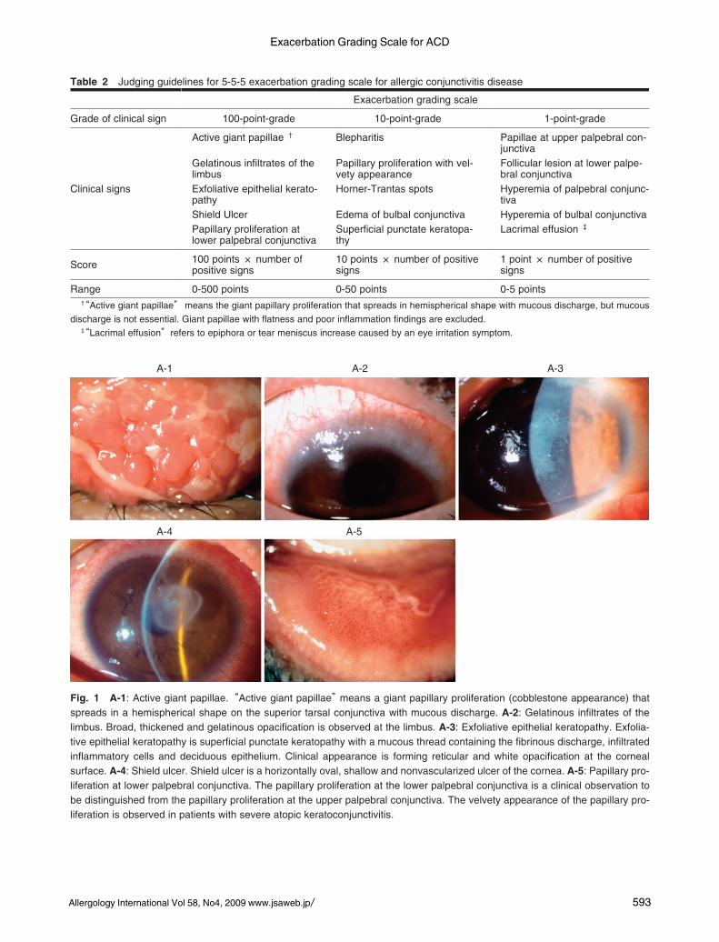
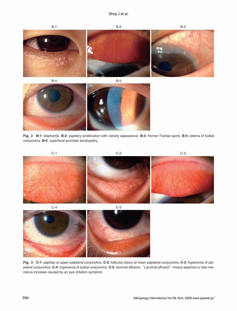
The 100-point-grade group was determined basedon the the presence of the following observations: ac-tive giant papillae (Fig.1 A-1), gelatinous infiltrates ofthe limbus (Fig.1 A-2), exfoliative corneal epitheliopa-thy (Fig.1 A-3), shield ulcer (Fig.1 A-4), and papillaryproliferation at lower tarsal conjunctiva (Fig.1 A-5).The guidelines for evaluation are shown in Figure 1.The 10-point-grade group was determined based onthe presence of the following observations: blephari-tis (Fig.2 B-1), papillary proliferation with velvety ap-pearance (Fig.2 B-2), Horner-Trantas spot (Fig.2 B-3), edema of bulbal conjunctiva (Fig.2 B-4), and su-perficial punctate keratopathy (Fig.2 B-5). The guide-lines for evaluation are shown in Figure 2. The 1-point-grade group was determined based on the pres-ence of the following observations: papillae at uppertarsal conjunctiva (Fig.3 C-1), follicular lesion atlower tarsal conjunctiva (Fig.3 C-2), hyperemia of tar-sal conjunctiva (Fig.3 C-3), hyperemia of bulbal con-junctiva (Fig.3 C-4), and lacrimal effusion (Fig.3 C-5).The guidelines for evaluation are shown in Figure 3.Furthermore, the severity classes of each group weredivided into mild, moderate or severe, according tothe criteria shown in Table 3.
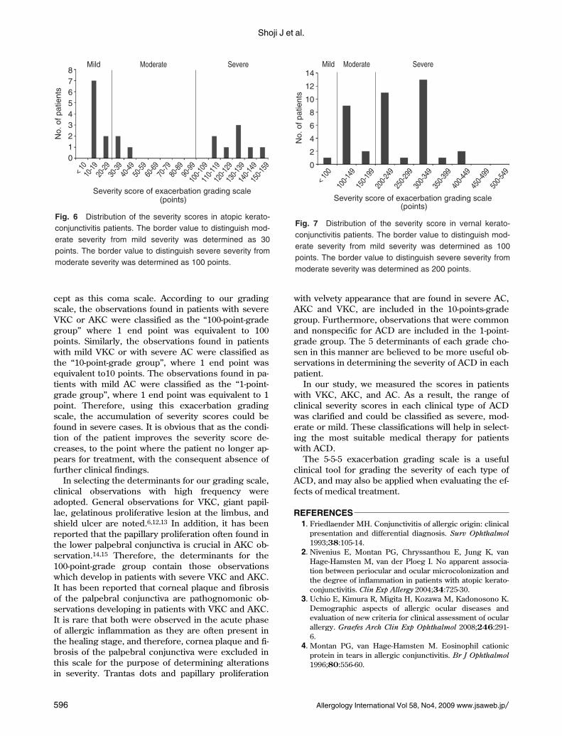
RESULTSResults using the 5-5-5 exacerbation grading scale inpatients with AC, AKC and VKC are shown in Figure4. The median severity scores of the 5-5-5 exacerba-tion grading scale in each group showed 13 in the ACgroup, 32.5 in the AKC group, and 243 in the VKCgroup. A statistically significant difference was foundamong the groups (P < 0.001, Kruskal-Wallis test). Inaddition, the distribution of this scale for the AC,
Exacerbation Grading Scale for ACD
Allergology International Vol 58, No4, 2009 www.jsaweb.jp� 593
Fig. 1 A-1: Active giant papillae.“Active giant papillae” means a giant papillary proliferation (cobblestone appearance) that spreads in a hemispherical shape on the superior tarsal conjunctiva with mucous discharge. A-2: Gelatinous infiltrates of the limbus. Broad, thickened and gelatinous opacification is observed at the limbus. A-3: Exfoliative epithelial keratopathy. Exfoliative epithelial keratopathy is superficial punctate keratopathy with a mucous thread containing the fibrinous discharge, infiltrated inflammatory cells and deciduous epithelium. Clinical appearance is forming reticular and white opacification at the corneal surface. A-4: Shield ulcer. Shield ulcer is a horizontally oval, shallow and nonvascularized ulcer of the cornea. A-5: Papillary proliferation at lower palpebral conjunctiva. The papillary proliferation at the lower palpebral conjunctiva is a clinical observation to be distinguished from the papillary proliferation at the upper palpebral conjunctiva. The velvety appearance of the papillary proliferation is observed in patients with severe atopic keratoconjunctivitis.
A-4 A-5
A-1 A-2 A-3
Table 2 Judging guidelines for 5-5-5 exacerbation grading scale for allergic conjunctivitis disease
Exacerbation grading scale
1-point-grade10-point-grade100-point-gradeGrade of clinical sign
Papillae at upper palpebral con-junctiva
BlepharitisActive giant papillae †
Clinical signs
Follicular lesion at lower palpe-bral conjunctiva
Papillary proliferation with vel-vety appearance
Gelatinous infiltrates of the limbus
Hyperemia of palpebral conjunc-tiva
Horner-Trantas spotsExfoliative epithelial kerato-pathy
Hyperemia of bulbal conjunctivaEdema of bulbal conjunctivaShield UlcerLacrimal effusion ‡ Superficial punctate keratopa-
thyPapillary proliferation at lower palpebral conjunctiva
1 point × number of positive signs
10 points × number of positive signs
100 points × number of positive signsScore
0-5 points0-50 points0-500 pointsRange † “Active giant papillae” means the giant papillary proliferation that spreads in hemispherical shape with mucous discharge, but mucous discharge is not essential. Giant papillae with flatness and poor inflammation findings are excluded. ‡ “Lacrimal effusion”refers to epiphora or tear meniscus increase caused by an eye irritation symptom.
Shoji J et al.
594 Allergology International Vol 58, No4, 2009 www.jsaweb.jp�
Fig. 2 B-1: blepharitis. B-2: papillary proliferation with velvety appearance. B-3: Horner-Trantas spots. B-4: edema of bulbal conjunctiva. B-5: superficial punctate keratopathy.
B-4 B-5
B-1 B-2 B-3
Fig. 3 C-1: papillae at upper palpebral conjunctiva. C-2: follicular lesion at lower palpebral conjunctiva. C-3: hyperemia of palpebral conjunctiva. C-4: hyperemia of bulbal conjunctiva. C-5: lacrimal effusion. “Lacrimal effusion” means epiphora or tear meniscus increase caused by an eye irritation symptom.
C-4 C-5
C-1 C-2 C-3

Exacerbation Grading Scale for ACD
Allergology International Vol 58, No4, 2009 www.jsaweb.jp� 595
Fig. 4 Results of severity score determined by 5-5-5 exac-erbation grading scale in VKC, AKC and AC patients. The severity scores of each type of allergic conjunctivitis disease were significantly different among VKC, AKC and AC patients. (P < 0.001, Kruskal-Wallis test). VKC, vernal kera-toconjunctivitis; AKC, atopic keratoconjunctivitis; AC, aller-gic conjunctivitis.
AC AKC VKCNo. of patients
500
400
300
200
100
0
43 20 40
Median value 13 32.5 243
Range 2-33 11-344 12-444
5-5-
5 sev
erity
obse
rvatio
n sca
le (p
oints)
Fig. 5 Distribution of the severity score in allergic conjunctivitis patients. The border value to distinguish moderate severity from mild severity was determined as 10 points. The border value to distinguish severe severity from moderate severity was determined as 30 points.
No.
of p
atie
nts
Severity score of exacerbation grading scale
20181614121086420
< 5 5-9 10-14 15-19 20-24 25-29 30-34
(points)
SevereModerateMild
Table 3 Criteria of severity classification in allergic conjunctivitis disease
VKCAKCAC
More than two clinical observations classified in 100-point-grade are observed.
At least one of the clinical observa-tion classified in 100-point-grade is observed.
More than three clinical observa-tions classified in 10-point-grade are observed, and those in 100-point-grade are absent.
Severe
Only one clinical observation classi-fied in 100-point-grade is observed.
More than three clinical observa-tions classified in 10-point-grade are observed, and those in 100-point-grade are absent.
Less than two clinical observat-ions classified in 10-point-grade are observed, and those in 100-point-grade are absent.
Moderate
Clinical observation classified in 100-point-grade is absent.
Less than two clinical observations classified in 10-point-grade are ob-served, and those in 100-point-gra-de are absent.
Clinical observation classified in 10- or 100-point-grade is absent.
Mild
AC, allergic conjunctivitis; AKC, atopic keratoconjunctivitis; VKC, vernal keratoconjunctivitis.
AKC and VKC groups are shown as graphs in Figure5, 6, and 7, respectively. The severity classes of eachgroup were divided into mild, moderate or severe, ac-cording to the criteria shown in Table 3. The numberof patients in each severity group is shown in Figure5, 6, and 7.
DISCUSSIONWe set out to determine the practical usefulness of ascoring system using the 5-5-5 exacerbation gradingscale for allergic conjunctivitis disease (ACD). Thisstudy demonstrated that the use of the 5-5-5 exacer-bation grading scale to assess clinical observations ofACD using the clinical severity score is simple andquick, and therefore suitable for follow-up examina-tions. The simplicity of the grading method, judgingrepresentative ACD findings as either present or ab-sent, was reflected in the low inter-observer variabil-
ity. There have been reports on clinical scores of pa-tients with ACD2,3,7 in which the severity of ACD andthe effects of antiallergic drugs were evaluated usingclinical scores. Clinical scores were used to deter-mine a multi-point score for each representative ob-servation of ACD. For each end point, ‘severe’ wasequivalent to 3 points, ‘moderate’ to 2 points and‘mild’ to 1 point. However, the problem with suchclinical scores is that 1 point may represent a differ-ent of severity for each observation category. How-ever, using our method, the 3-graded clinical observa-tions provide the best measure for current severity ofdiseases in patients with ACD. An example of a 3-graded evaluation is the Japanese coma scale, whichis used to evaluate comatose status using a 3-3-9 grad-ing scale.11 A characteristic of this evaluation methodis that the score expresses clinical severity preciselyby giving high points for severe observations and lowpoints for mild observations. The 5-5-5 exacerbationgrading scale used in our study follows the same con-
Shoji J et al.
596 Allergology International Vol 58, No4, 2009 www.jsaweb.jp�
Fig. 6 Distribution of the severity scores in atopic keratoconjunctivitis patients. The border value to distinguish moderate severity from mild severity was determined as 30 points. The border value to distinguish severe severity from moderate severity was determined as 100 points.
SevereModerateMildN
o. o
f pat
ient
s
Severity score of exacerbation grading scale
< 10
10-19
20-29
30-39
40-49
50-59
60-69
70-79
80-89
90-99
100-1
0911
0-119
120-1
2913
0-139
140-1
4915
0-159
(points)
876543210
Fig. 7 Distribution of the severity score in vernal keratoconjunctivitis patients. The border value to distinguish moderate severity from mild severity was determined as 100 points. The border value to distinguish severe severity from moderate severity was determined as 200 points.
No.
of p
atie
nts
SevereModerateMild
< 100
100-1
4915
0-199
200-2
4925
0-299
300-3
4935
0-399
400-4
4945
0-499
500-5
49
Severity score of exacerbation grading scale (points)
14
12
10
8
6
4
2
0
cept as this coma scale. According to our gradingscale, the observations found in patients with severeVKC or AKC were classified as the “100-point-gradegroup” where 1 end point was equivalent to 100points. Similarly, the observations found in patientswith mild VKC or with severe AC were classified asthe “10-point-grade group”, where 1 end point wasequivalent to10 points. The observations found in pa-tients with mild AC were classified as the “1-point-grade group”, where 1 end point was equivalent to 1point. Therefore, using this exacerbation gradingscale, the accumulation of severity scores could befound in severe cases. It is obvious that as the condi-tion of the patient improves the severity score de-creases, to the point where the patient no longer ap-pears for treatment, with the consequent absence offurther clinical findings.
In selecting the determinants for our grading scale,clinical observations with high frequency wereadopted. General observations for VKC, giant papil-lae, gelatinous proliferative lesion at the limbus, andshield ulcer are noted.6,12,13 In addition, it has beenreported that the papillary proliferation often found inthe lower palpebral conjunctiva is crucial in AKC ob-servation.14,15 Therefore, the determinants for the100-point-grade group contain those observationswhich develop in patients with severe VKC and AKC.It has been reported that corneal plaque and fibrosisof the palpebral conjunctiva are pathognomonic ob-servations developing in patients with VKC and AKC.It is rare that both were observed in the acute phaseof allergic inflammation as they are often present inthe healing stage, and therefore, cornea plaque and fi-brosis of the palpebral conjunctiva were excluded inthis scale for the purpose of determining alterationsin severity. Trantas dots and papillary proliferation
with velvety appearance that are found in severe AC,AKC and VKC, are included in the 10-points-gradegroup. Furthermore, observations that were commonand nonspecific for ACD are included in the 1-point-grade group. The 5 determinants of each grade cho-sen in this manner are believed to be more useful ob-servations in determining the severity of ACD in eachpatient.
In our study, we measured the scores in patientswith VKC, AKC, and AC. As a result, the range ofclinical severity scores in each clinical type of ACDwas clarified and could be classified as severe, mod-erate or mild. These classifications will help in select-ing the most suitable medical therapy for patientswith ACD.
The 5-5-5 exacerbation grading scale is a usefulclinical tool for grading the severity of each type ofACD, and may also be applied when evaluating the ef-fects of medical treatment.
REFERENCES1. Friedlaender MH. Conjunctivitis of allergic origin: clinical
presentation and differential diagnosis. Surv Ophthalmol1993;38:105-14.
2. Nivenius E, Montan PG, Chryssanthou E, Jung K, vanHage-Hamsten M, van der Ploeg I. No apparent associa-tion between periocular and ocular microcolonization andthe degree of inflammation in patients with atopic kerato-conjunctivitis. Clin Exp Allergy 2004;34:725-30.
3. Uchio E, Kimura R, Migita H, Kozawa M, Kadonosono K.Demographic aspects of allergic ocular diseases andevaluation of new criteria for clinical assessment of ocularallergy. Graefes Arch Clin Exp Ophthalmol 2008;246:291-6.
4. Montan PG, van Hage-Hamsten M. Eosinophil cationicprotein in tears in allergic conjunctivitis. Br J Ophthalmol1996;80:556-60.
Exacerbation Grading Scale for ACD
Allergology International Vol 58, No4, 2009 www.jsaweb.jp� 597
5. Calonge M, Herreras JM. Clinical grading of atopic kera-toconjunctivitis. Curr Opin Allergy Clin Immunol 2007;7:442-5.
6. Bonini S, Sacchetti M, Mantelli F, Lambiase A. Clinicalgrading of vernal keratoconjunctivitis. Curr Opin AllergyClin Immunol 2007;7:436-41.
7. Leonardi A, Borghesan F, Avarello A, Plebani M, SecchiAG. Effect of lodoxamide and disodium cromoglycate ontear eosinophil cationic protein in vernal keratoconjunc-tivitis. Br J Ophthalmol 1997;81:23-6.
8. Bielory L. Allergic and immunologic disorders of the eye.Part II: Ocular allergy. J Allergy Clin Immunol 2000;106:1019-32.
9. Hogan MJ. Atopic keratoconjunctivitis. Trans Am Oph-thalmol Soc 1952;50:265-81.
10. Foster CS, Calonge M. Atopic keratoconjunctivitis. Oph-
thalmology 1990;97:992-1000.11. Ohta T. Phenomenological aspects of consciousness―its
disturbance in acute and chronic stage. Acta NeurochirSuppl 2005;93:191-3.
12. Bonini S, Bonini S, Lambiase A et al. Vernal Keratocon-junctivitis revisited. A case series of 195 patients withlong-term followup. Ophthalmology 2000;107:1157-63.
13. Leonardi A, Motterle L, Bortolotti M. Allergy and the eye.Clin Exp Immunol 2008;153(Suppl 1):17-21.
14. Hogan MJ. Atopic keratoconjunctivitis. Trans Am Oph-thalmol Soc 1952;50:265-81.
15. Foster CS, Calonge M. Atopic keratoconjunctivitis. Oph-thalmology 1990;97:992-1000.