Embed Size (px)
Citation preview
Evaluation of Pelvic Floor MuscleFunction(PFMF) in Cervical Cancer Patients withQuerleu-Morrow type C HysterectomyA MulticenterStudyShiyan Wang
Peking University People's HospitalHongwu Wen
Peking University First HospitalYunong Gao
Peking University Cancer HospitalQiubo Lv
Beijing HospitalHongyu Li
Third A�liated Hospital of Zhengzhou UniversitySumei Wang
Beijing Chao-Yang HospitalYanlong Wang
Women and Children Health Hospital of XiamenQing Liu
Women and Children Health Hospital of GansuJinsong Han
Peking University Third HospitalHaibo Wang
Peking University Clinical Research InstituteYi Li
Peking University Medical Information CenterNa Yu
Peking University Medical Information CenterQing Wang
Peking University People's HospitalTingting Cao
Peking University People's HospitalSha Wang
Peking University People's Hospital

Huaxin Sun Peking University People's Hospital
Xiuli Sun ( [email protected] )Peking University People's Hospital
Jianliu Wang Peking University People's Hospital
Zhiqi Wang Peking University People's Hospital
Research Article
Keywords: Pelvic �oor muscle strength(PFMS), pelvic �oor muscle function(PFMF),Cervical cancer,QM-C33 hysterectomy,follow-uptime
Posted Date: December 9th, 2020
DOI: https://doi.org/10.21203/rs.3.rs-116903/v1
License: This work is licensed under a Creative Commons Attribution 4.0 International License. Read Full License
1
Evaluation of pelvic floor muscle function(PFMF) in Cervical Cancer patients with1Querleu-Morrow type C hysterectomy:A multicenter study2
Shiyan Wang1,2,Hongwu Wen3, Yunong Gao4, Qiubo Lv5,Hongyu Li6,Sumei Wang7,Yanlong Wang8,Qing3Liu9,Jinsong Han10,Haibo Wang11,Yi Li12,Na Yu12,Qing Wang1,2,Tingting Cao1,2,Sha Wang1,2,Huaxin4Sun1,2,Zhiqi Wang1,2,Xiuli Sun*1,2,Jianliu Wang*1,25
1.Department of OB/GynPeking University People’s Hospital,Beijing,China62.Beijing Key Laboratory of Female Pelvic Floor Disorders,Beijing,China73.Department of Obstetrics and Gynecology, Peking University First Hospital, Beijing, China84.Department of Gynecology, Peking University Cancer Hospital & Institute, Beijing, China95.Department of Obstetrics and Gynecology, Beijing Hospital, Beijing, China106.Department of Obstetrics and Gynecology, Zhengzhou University Third Hospital, Zhengzhou,Henan, China117.Department of Obstetrics and Gynecology, Beijing Chaoyang Hospital, Capital Medical University,Beijing,12China138.Department of Obstetrics and Gynecology, Women and Children Health Hospital of Xiamen, Xiamen,China149.Department of Obstetrics and Gynecology, Women and Children Health Hospital of Gansu,15Lanzhou,Gansu,China1610.Department of Obstetrics and Gynecology, Peking University Third Hospital, Beijing, China1711.Peking University Clinical Research Institute, Beijing, China.1812.Information Center,Medical Department of Peking University19Corresponding author: Jianliu Wang and Xiuli Sun20E-mail: [email protected] and [email protected]: 86 10 8832 435422Fax: 86 10 8832 427023Address: No. 11 Xizhimen South Street, Xicheng Dist., Beijing 100044, China.24
Word count: 328325Author Contributions26Jianliu Wang, Zhiqi Wang, Hongwu Wen,Yunong Gao,Qiubo Lv,Hongyu Li,Sumei27Wang,Yanlong Wang,Qing Liu,Jinsong Hanand Haibo Wang: Project development.28Sha Wang, Shiyan Wang,Qing Wang,Tingting Cao and Huaxin Sun: Data collection.29Na Yu and Yi Li :Data analysis.30Shiyan Wang,Jianliu Wang and Xiuli Sun wrote the main manuscript.31All authors reviewed the manuscript.32Keywords Pelvic floor muscle strength(PFMS), pelvic floor muscle function(PFMF),Cervical cancer, QM-C33hysterectomy, follow-up time34Brief summary The evaluation and analysis of PFMS were studied after Querleu-Morrow type C35hysterectomy and the influencing factors were radiotherapy, chemotherapy and the follow-up.36Acknowledgements37The authors would like to thank Xiaojie Yu and Xiaodan Li for their contribution to the data collection and38Huixin Liu for her contribution in the data analysis.39Clinical Trails NCT number of this study is 02492542.40
4142
43
44
45
2
Abstract46Introduction and Hypothesis To evaluate the pelvic floor muscle function (PFMF) of cervical cancer patients47after type QM-C hysterectomy and to explore the relationship between decreased PFMF and related factors..48Methods This was a multi-centered retrospective cohort study. 181 cervical cancer patients underwent type49QM-C hysterectomy were enrolled from 9 tertiary hospitals. Strength of PFMF were measured by using50neuromuscular apparatus (Phenix U8, French). Risk factors contributed to decreased PFMF was analyzed by51univariate and multivariate ordinal ploytomous logistic regression.52Results Totally 181 patients were investigated in this study.0-3 level of type I muscle fibre strength(MFSI) was5352.6%(95/181),0-3 level of type ⅡA muscle fibre strength(MFSⅡA) was 50%(91/181). Subjective stress54urinary incontinence was 46%(84/181),urinary retention was 27.3%(50/181),dyschezia was5541.5%(75/181),fecal incontinence was 9%(18/181).①MFSI : Multivariate ordinal ploytomous logistic56regression shows that the follow-up time(p<0.05),chemotherapy and radiotherapy (p=0.038) are independent57risk factors of MFSI’s reduction after type QM-C hysterectomy.② MFSⅡA:Multivariate ordinal ploytomous58logistic regression shows that the follow-up time(p<0.05) are independent risk factors of MFSⅡA’s reduction59after type QM-C hysterectomy. The pelvic floor muscle strength(PFMS) increased after 9 months than in 960months after operation, which showed that the PFMS could be recovered after operation.61Conclusions We advocate for more attention and emphasis on the PFMF of Chinese female patients with62cervical cancer postoperation.63Contribution of the Paper The key messages of the article is that PFMF after QM-C hysterectomy have not64been analyzed by current study.The new knowledge added by this study is that 3 months after radical65hysterectomy patients’ should do pelvic floor rehabilitation exercises.66
67Introduction68The morbidity of cervical cancer is still high but advanced technology enables more cervical cancers be69detected at the early stage nowadays. With type QM-C hysterectomy plus pelvic lymphadenectomy, the rate of705-year survival of cervical cancer patients could reach to 95%.[1] However, evidences showed that type QM-C71hysterectomy may seriously decrease the pelvic floor function of the patients, including mainly lower urinary72tract syndrome,[2,3] defecation disorders[4,5] and sexual dysfunction.[6]All of those problems would negatively73affects the life quality of the patients for a long time. Dysfunction of the pelvic floor muscles(PFM)might74result in urinary and faecal incontinence, pelvic organ prolapse (POP), sexual problems, and chronic pain.[7]As75PFM provides support and sphincteric functions to the pelvic organs, investigation to PFM function is crucial76to shot the problems in individuals with pelvic floor disorders (PFD). [8,9]77The PFM was mainly consisted of type I and type II muscle fibers.[10] PFM plays an important role in78maintaining vaginal constriction and the normal position of the organs in pelvis and, thus, further in keeping79the normal function of urethral sphincter and rectal sphincter.[11] Overactive bladder(OAB) and urinary80incontinence(UI) are the two common courses that could have negative impacts on women’s quality of life.81PFM training is the most preferred treatment to increase PFM strength (PFMS) in women with stress urinary82incontinence (SUI).[12-16] The life quality of the patient will be improved if the elements affecting PFMS were83analyzed and treated after type QM-C hysterectomy. However, there were only few studies focusing on the84post QM-C hysterectomy PFMF. Our aim is to identify the elements resulting PFM weakness in patients who85underwent QM-C hysterectomy for cervical cancer and to figure out a way to solve the problems.86The incidence of cervical cancer was high, and it was one of the common malignant tumors in gynecology.87The annual incidence of new cases in China accounted for about 1/3 of the new cases of cervical cancer in the88world[17]. The standard treatments for invasive cervical cancer was surgical treatment and radiotherapy89supplemented by chemotherapy. Annually, about 83.9% of cervical cancer patients in China undergo surgical90treatment with the main procedure to be QM-C hysterectomy and pelvic lymphadenectomy[18]. With the91improvement of cervical cancer screening in recent years, the trend of rejuvenation was obvious and the 5-year92survival rate of patients with early stage of cervical cancers has raised high [19]. Patients with early stage of93
3
cervical cancer could survive or even "cure" after treatment. However, due to the large scope of surgery, the94preoperative removal of para-uterine tissue in QM-C hysterectomy procedures would also damage the pelvic95floor nerves, muscles and fascia, and radiotherapy and chemotherapy would lead to pelvic tissue fibrosis, low96ovarian function and even failure, resulting in postoperative PFD, which seriously affects the life quality of97patients. At present, there was still a lack of large-scale study on the incidence of pelvic floor dysfunction and98related factors in patients with cervical cancer treated with surgery as the main treatment. This study performed99retrospective analyses PFMS measurement and other methods to assess the PFMS of 181 patients with cervical100cancer after QM-C hysterectomy.101
102The PFM plays an important role in supporting the pelvic and abdominal organs and controlling urinary and103fecal continence in addition to their role in the sexual function[20]. The types of PFMF included:MFSI belonged104to the pelvic and abdominal cavity support system, accounting for 70% of the pubic vaginal and puborectalis105muscles, 90% of the pubis caudal muscles, and 68% of the sacral and posterior tibial muscles. The function106was characterized by tonic contraction, long contraction time and long-lasting, and it was not easy to fatigue.107MFSⅡA belonged to the pelvic and abdominal cavity movement system, and their functions were staged108contraction, rapid and short-lived, and were easy to fatigue.[21] Why was PFMS important for female patients?109Decreased PFMS could cause LUTS, defecation disorders, and sexual dysfunction.[22] It would seriously affect110the quality of patients’ life.111
112The fact that cervical cancer patients had the above three PFD diseases might be related to the reduction of113PFMS.114
115According to reports, for women who demonstrated SUI during pregnancy and delivery, postpartum SUI was116mostly linked to weakening of PFM [23]. After being comprehensively and accurately assessed, PFM117rehabilitation was able to enhance PFM strength and muscle tone, which gave rise to improvement of SUI[24-25]118The most popular assessment methods in China for assessing the condition of PFM in postpartum female119patients were the digital palpation and EMG evaluation because of easy access to relevant equipments and low120cost[28]. EMG was mostly utilized to identify muscle activity, measure intramuscular force ratio and record121PFM dysfunction [29]. As a result, we chose EMG as the evaluation method to record the pelvic muscle strength.122Pelvic floor exercise and biofeedback for patients with cervical cancer surgery, and pelvic floor rehabilitation123for patients with PFD could improve the life quality of patients. However, there was no retrospective study for124systematic examination and analysis, and PFMS was not being measured in patients with cervical cancer.125
126127
Materials and Methods1281. Patient recruitment:129The patients will be recruited from the cervical cancer patients who had been underwent QM-C hysterectomy130participating hospitals during January 2012 to March 2015. Patients will be included if they were 1) ≥18 years131of age, 2) underwent QM-C hysterectomy for cervical cancer for 3-24 months, and 3) consent to participation132by signing the informed consent form. Patient will be excluded if they 1) experienced preoperative adjuvant133radiotherapy, 2) had received pelvic floor rehabilitation after surgery, 3) had less than 3cm of main secral134ligament resection and /or less than 3cm of vaginal resection, and 4) could not completed questionnaires or135PFMS evaluation. As the first step of patient enrolment, eligible patients would be examined by two136experienced gynecologic oncologists at the participating hospitals for pelvic floor function, and were staged137according to the patient's medical history and clinical manifestations following the clinical staging criteria of138cervical cancer revised by FIGO in 2009.1391) Patients who met the inclusive criteria would be contacted and invited by the investigators to participate140
the study via explaining the study in detail. An informed consent prier approved by the Institutional141
4
Review Board (IRB) of Peking University People’s Hospital would be signed by the patient and the142doctor when the oatients agreed to participat the study with all her questions about the study fairly143answered and understood. Patients who had died by the study contacting were not included into the144analysis.145
1462. Study Methods147The PFMS and the life quality of the patients were evaluated and the relevant influencing factors were148investigated in a stratification of deferent post-surgery periods as 3-6 months, 7-9 months, 10-12 months,14913-18months, 19-24 months. Eligible patients would went through the procedures as shown in Figure 1 and150described as the follows:1512) Demographic data collection: Demographic data was collected through interviewing the eligible patients152
in a private room, and by an investigator who did not perform treatment on the patient and was blind of153the patient’s history. Data that was collected includes the age, body mass index (BMI), preoperative154comorbidities, number and mode of deliveries, clinical staging, and type of hysterectomy, etc..155
3) Clinical Record Review: Clinical record on the QM-C hysterectomy was checked to confirm the resected156part of the Main sacral ligament was ≥3cm and that of the vagina was ≥ 3 cm. Treatment information157would be also collected for analysis.158
4) Clinic Examination: Detailed physical examination would be conducted by trained investigators and159results recorded, followed by laboratory examination and test as described below.160
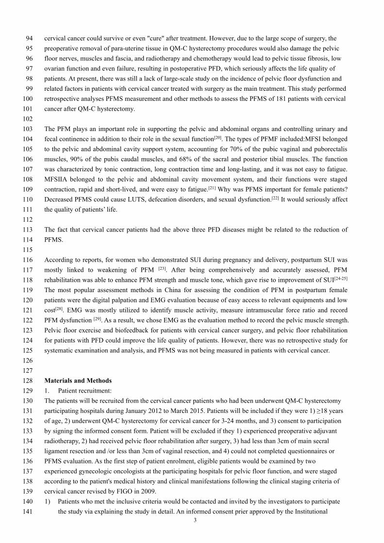
1613. PFMS Examination162Our PFMS examination is followed by relevant guidelines and regulations from a book which is written by163academician Jinghe Lang[21].164Instruments: Phenix USB8 biofeedback system (Electron-IC Concept Lignon Innovation Co., France), the165same equipment as used in Navarro Brazález B’s study [16], was used to test the PFMS of the patients. The166manometer was interfaced with the biofeedback system that was installed in an IBM compatible personal167computer. A vaginal pressure probe of 115 mm in length (Foshan Shanshan Datang Medical Technology Co.,168Ltd. China) inside of an airbag was linked with the manometry. The 41mm-long airbag was used for enlarging169the vagina through inflating it to be oval-sized with air. Manometry and dynamometry were more reliable than170vaginal palpation for the assessment of PFM strength in women with PFD, especially when different raters171were involved[16].172PFM Fibers categorization The PFM fibers were categorized into two types: type I and type II fibers.173Type I fibers, also called sustained contraction muscles, is featured to have long lasting contraction and is not174easy to fatigue. Most of type II fibers commonly distribute in the levator ani muscle. Type II fibers, also called175as rapid contraction muscles, provide quick contraction that lasts very short and are easy to fatigue. They176mostly distribute in the superficial layer of the pelvic floor. PFMS measurement mainly oriented to the strength177of the muscle contraction, the ability of the muscle to resist resistance, the duration, symmetry and fatigue of178the muscle contraction, the ability of the muscle in repeat contractions, and the rapid contraction times.179Research evidence showed that changes of these basic electrophysiological indicators are usually detectable180earlier than pelvic floor dysfunction appears and could be used as an evaluative index for early detection of181pelvic floor dysfunction and the treatment outcomes. Comparing the postoperative PFMS of the two groups of182patients could objectively evaluate the functional status of the pelvic floor.183Measurement of PFMS. The baseline pressure was set to 0cm H2O. Patients were asked to contract their PFM184for measurement of type I muscle fibers strength (MFS-I) by squeezing maximum for 5 times.The total width185of Figure 1 represents 10 seconds in time and the width of the yellow module represents 6 seconds. When the186patient’s MFS-I contraction called the sustained contraction made the red curve reach 40% of the yellow187module height, the patient would be instructed to contract her vagina with maximum power for three times to obtain188the maximum and minimum vaginal manometry values. The peaks of the yellow module in Figure 1 represents the189
5
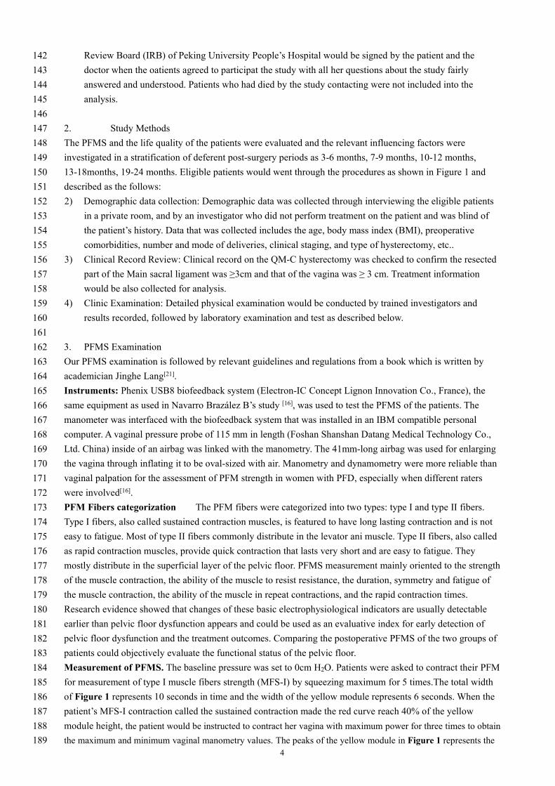
maximum value of the vaginal manometry, while level 0 to level 5 represent the duration of the muscle strength190(MSD) from 0s to 5s, respectively. Figure 2 shows the muscle strength rapid contraction levels when the type191IIA PFM contraction made the red curve reach to 70% - 90% of the yellow module height (referring to Figure1922).193Comparing of the MFS. To identify the factors that impact the MFS of the enrolled patients, they were194categorized into 3 groups according to the therapies they underwent. Patients who underwent surgery only195were referred as Group-S; patients who underwent surgery plus radiotherapy were referred as Group-SR;196patients who underwent surgery and chemotherapy were referred as Group-SC; and those who were treated197with surgery followed by radiotherapy and chemotherapy were referred as Group-SRC. MFS-I and IIA will be198compared among those groups.1994. Statistical Analysis200Statistical analysis was performed using R software programming. Ordinal ploytomous logistic regression was201used to analyze the influencing factors of MFS of type I and IIA after QM-C hysterectomy. The PFMS was202categorized into three groups according to the measured duration levels, with which group A refers to patients203with 0 level MSD, group B refers to those with 1-3 level MSD, while group C refers to those with 4-5 level of204MSD. A p values of < 0.05 was considered statistically significant in comparison of the PFMS in different205periods after QM-C hysterectomy, excluding the confounding factors, and analyzing the influencing factors of206the postoperative PFMS in patients with cervical cancer..207
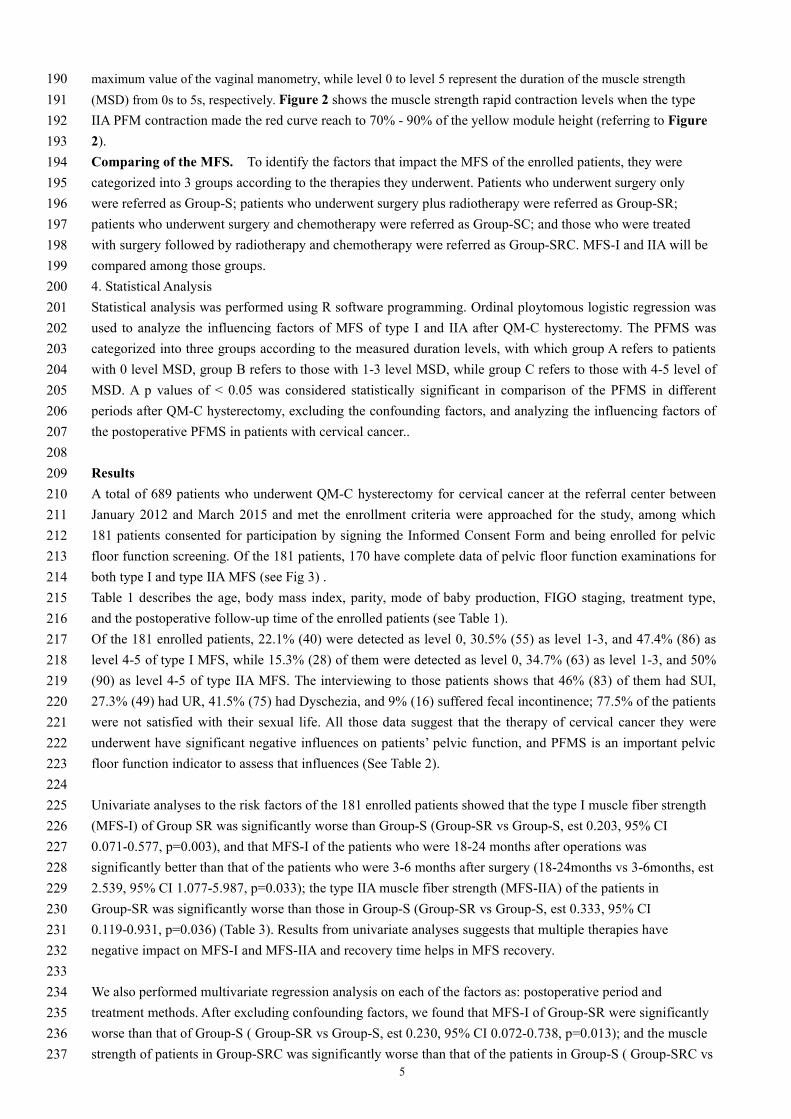
208Results209A total of 689 patients who underwent QM-C hysterectomy for cervical cancer at the referral center between210January 2012 and March 2015 and met the enrollment criteria were approached for the study, among which211181 patients consented for participation by signing the Informed Consent Form and being enrolled for pelvic212floor function screening. Of the 181 patients, 170 have complete data of pelvic floor function examinations for213both type I and type ⅡAMFS (see Fig 3) .214Table 1 describes the age, body mass index, parity, mode of baby production, FIGO staging, treatment type,215and the postoperative follow-up time of the enrolled patients (see Table 1).216Of the 181 enrolled patients, 22.1% (40) were detected as level 0, 30.5% (55) as level 1-3, and 47.4% (86) as217level 4-5 of type I MFS, while 15.3% (28) of them were detected as level 0, 34.7% (63) as level 1-3, and 50%218(90) as level 4-5 of type IIA MFS. The interviewing to those patients shows that 46% (83) of them had SUI,21927.3% (49) had UR, 41.5% (75) had Dyschezia, and 9% (16) suffered fecal incontinence; 77.5% of the patients220were not satisfied with their sexual life. All those data suggest that the therapy of cervical cancer they were221underwent have significant negative influences on patients’ pelvic function, and PFMS is an important pelvic222floor function indicator to assess that influences (See Table 2).223
224Univariate analyses to the risk factors of the 181 enrolled patients showed that the type I muscle fiber strength225(MFS-I) of Group SR was significantly worse than Group-S (Group-SR vs Group-S, est 0.203, 95% CI2260.071-0.577, p=0.003), and that MFS-I of the patients who were 18-24 months after operations was227significantly better than that of the patients who were 3-6 months after surgery (18-24months vs 3-6months, est2282.539, 95% CI 1.077-5.987, p=0.033); the type IIAmuscle fiber strength (MFS-IIA) of the patients in229Group-SR was significantly worse than those in Group-S (Group-SR vs Group-S, est 0.333, 95% CI2300.119-0.931, p=0.036) (Table 3). Results from univariate analyses suggests that multiple therapies have231negative impact on MFS-I and MFS-IIA and recovery time helps in MFS recovery.232
233We also performed multivariate regression analysis on each of the factors as: postoperative period and234treatment methods. After excluding confounding factors, we found that MFS-I of Group-SR were significantly235worse than that of Group-S ( Group-SR vs Group-S, est 0.230, 95% CI 0.072-0.738, p=0.013); and the muscle236strength of patients in Group-SRC was significantly worse than that of the patients in Group-S ( Group-SRC vs237
6
Group-S, est 0.428, 95% CI 0.192-0.954, p=0.038). Muscle strength in Group SC was different with that of238Group-S but not statistically significant ( Group SC vs Group-S, est 0.602, 95% CI 0.388-1.731, P=0.602).239Results from multivariate regressive analyses also suggests the negative impacts of multiple therapies on240MFS-I and MFS-IIA recovery.241
242In comparing the MFS of the patients with different post-treatment periods (PTP), patients with PTP of 18-24243months, 12-18months and 9-12 months are all significantly worse than patients with a PTP of 3-6 months244(18-24 mths vs 3-6m, est 3.126, 95% CI 1.278-7.647, p=0.013; 12-18mths vs 3-6m, est 3.194, 95% CI2451.339-7.617, p=0.009; and 9-12 mths vs 3-6 mths , est 3.816, 95% CI 1.095-13.302, p=0.036, respectively).246However, patients with a PTP of 6-9 months is not significantly worse and those with a PTP of 3-6 (6-9m vs2473-6m, est 1.592, 95% CI 0.641-3.954, p=0.316). This result suggests a time-base trends of MFS that the longer248the PTP is, the worse the MSF is.249
250The results also shows that MFS-ⅡA of the patients in Group-SR was better than that of patients in Group-S251with no statistical significance (Group-SR vs Group-S, est 0.318, 95% CI 0.100-1.009, p=0.052), although the252P value is close to 0.05. It was suggested that radiotherapy might be a risk factor for type II pelvic floor muscle253fibers (SQ-this conclusion is far-fetched and should not be concluded).254
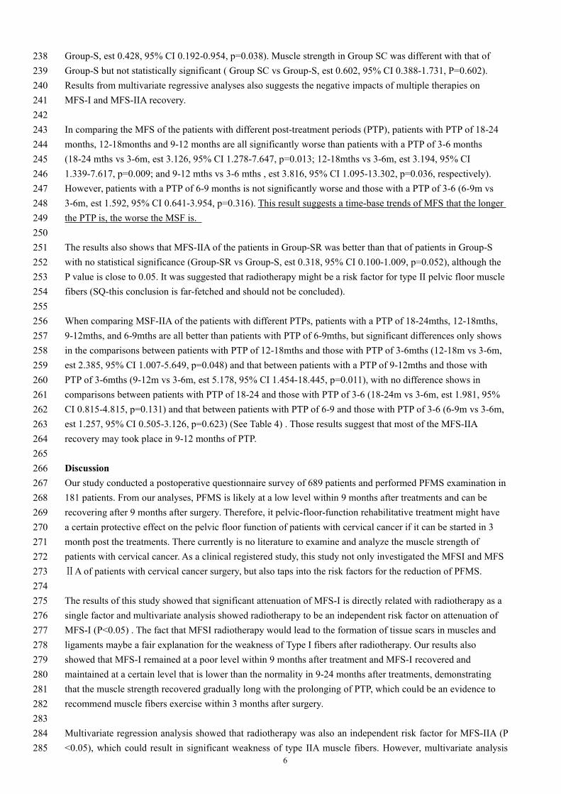
255When comparing MSF-IIA of the patients with different PTPs, patients with a PTP of 18-24mths, 12-18mths,2569-12mths, and 6-9mths are all better than patients with PTP of 6-9mths, but significant differences only shows257in the comparisons between patients with PTP of 12-18mths and those with PTP of 3-6mths (12-18m vs 3-6m,258est 2.385, 95% CI 1.007-5.649, p=0.048) and that between patients with a PTP of 9-12mths and those with259PTP of 3-6mths (9-12m vs 3-6m, est 5.178, 95% CI 1.454-18.445, p=0.011), with no difference shows in260comparisons between patients with PTP of 18-24 and those with PTP of 3-6 (18-24m vs 3-6m, est 1.981, 95%261CI 0.815-4.815, p=0.131) and that between patients with PTP of 6-9 and those with PTP of 3-6 (6-9m vs 3-6m,262est 1.257, 95% CI 0.505-3.126, p=0.623) (See Table 4) . Those results suggest that most of the MFS-IIA263recovery may took place in 9-12 months of PTP.264
265Discussion266Our study conducted a postoperative questionnaire survey of 689 patients and performed PFMS examination in267181 patients. From our analyses, PFMS is likely at a low level within 9 months after treatments and can be268recovering after 9 months after surgery. Therefore, it pelvic-floor-function rehabilitative treatment might have269a certain protective effect on the pelvic floor function of patients with cervical cancer if it can be started in 3270month post the treatments. There currently is no literature to examine and analyze the muscle strength of271patients with cervical cancer. As a clinical registered study, this study not only investigated the MFSI and MFS272ⅡA of patients with cervical cancer surgery, but also taps into the risk factors for the reduction of PFMS.273
274The results of this study showed that significant attenuation of MFS-I is directly related with radiotherapy as a275single factor and multivariate analysis showed radiotherapy to be an independent risk factor on attenuation of276MFS-I (P<0.05) . The fact that MFSI radiotherapy would lead to the formation of tissue scars in muscles and277ligaments maybe a fair explanation for the weakness of Type I fibers after radiotherapy. Our results also278showed that MFS-I remained at a poor level within 9 months after treatment and MFS-I recovered and279maintained at a certain level that is lower than the normality in 9-24 months after treatments, demonstrating280that the muscle strength recovered gradually long with the prolonging of PTP, which could be an evidence to281recommend muscle fibers exercise within 3 months after surgery.282
283Multivariate regression analysis showed that radiotherapy was also an independent risk factor for MFS-IIA (P284<0.05), which could result in significant weakness of type IIA muscle fibers. However, multivariate analysis285
7
showed that MFS-IIA of the patients with PTP of 9-18 months had been significantly improved in comparing286with patients with a PTP of six months, but that of the patients with a PTP of 6-9 months and 18-24 months287had not been significantly bettered, although they are getting little better. This indicates that the improvement288of MSF-IIA can be improved along with the PTP prolonging but the obvious improvement happens during2899-18 month after the treatments (See Fig 4) . Our long-term follow-up on some of the patients showed that,290MFS-IIA could be significantly improved after one and a half years of PTP.291
292As a retrospective analyses, the study has limitation on the enrollment of patients, who are with different293months of PTPs. Perspective study would be designed and conducted if we need to collect data of patients with294a PTP of same time to demonstrate the exact impacts of the risk factors on the MFS. In our study, it was very295difficult to get the specific and objective parameters of PFMS of 181 cervical cancer patients. However, the296results of our study provided valuable evidences to indicate how and when the doctors should have such297patients to exercise pelvic floor muscle after operation.298
299Therefore, we concluded that Radiotherapy is an independent factor to have negative impact on the recovery of300both type I and IIA pelvic floor muscle fibers; Weakness of PFMF can be improved along with the time going301of post treatment period, and most of the recovery taking place during 9-12months of PTP. Pelvic floor muscle302exercise should be prescribed to the patients with 3 months after the treatments.303
304305
Ethical Approval This was a multi-centered and retrospective cohort study, the research protocol was306approved by the Institutional Review Board(IRB) .(IRB number 2015PHB021-04).307Funding sources This work is funded by the National Key Technology R&D Program of China(nos.2019YFC3081005200 and 2019YFC1005203) ,Major scientific and technological project of the Beijing Science and309Technology Committee(D151100001915003) and The National Key Technology R&D Program310(No.2015BAI13B06)311Conflict of interest312None.313
References314[1] Manchana T, Triratanachat S, Sirisabya N, et al. Prevalence and prognostic significance of COX-2315expression in stage IB cervical cancer. Gynecol Oncol,2006,100(3):556–560.316[2] Chun N, Noh GO, Song HJ, Kim SH.Frequency, Intensity and Daily Life Distress of Urinary Dysfunction317in Women with Cervical Cancer after Radical Hysterectomy.J Korean Acad Nurs. 2016 Jun;46(3):400-8. doi:31810.4040/jkan.2016.46.3.400.319[3] Aoun F, Roumeguère T.Lower urinary tract dysfunction following radical hysterectomy.Prog Urol. 2015320Dec;25(17):1184-90. doi: 10.1016/j.purol.2015.08.311. Epub 2015 Sep 8.321[4] Plotti F, Terranova C, Capriglione S,et al.Assessment of Quality of Life and Urinary and Sexual Function322After Radical Hysterectomy in Long-Term Cervical Cancer Survivors.Int J Gynecol323Cancer,2018,May;28(4):818-823.doi:10.1097/IGC.0000000000001239.324[5] Plotti F, Angioli R,Zullo MA, et al. Update on urodynamic bladder dysfunctions after radical325hysterectomy for cervical cancer. Crit Rev OncolHematol, 2011,80(2): 323-329.326[6] Wang X, Chen C, Liu P, et al.The morbidity of sexual dysfunction of 125 Chinese women following327different types of radical hysterectomy for gynaecological malignancies.Arch Gynecol Obstet. 2018328Feb;297(2):459-466. doi: 10.1007/s00404-017-4625-0. Epub 2017 Dec 27.329[7] Bump RC, Norton PA. Epidemiology and natural history of pelvic floor dysfunction.Obstet Gynecol Clin330North Am 1998;25:723–46.331
8
[8] Messelink B, Benson T, Berghmans B, et al. Standardization of terminology of pelvic floor muscle332function and dysfunction: report from the pelvic floor clinical assessment group of the International333Continence Society. Neurourol Urodyn. 2005;380:374-380.334[9] Ashton-Miller J, Delancey JOL. Functional anatomy of the female pelvic floor. Ann N YAcad Sci.3352007;1101:266-296.336[10] Lan Zhu.Basic study of female pelvic floor supporting tissue anatomy and dysfunctional337diseases[J].Chinese Journal of Practical Gynecology and obstetrics,2005(04):18-19.338[11] Sigurdardottir T,Steingrimsdottir T,Geirsson RT,et al.Can postpartum pelvic floor muscle training reduce339urinay and anal incontinence?:An assessor-blinded randomized controlled trial.Am J Obstet Gynecol.2019 Sep34014.doi:10.1016/j.ajog.2019.09.011.PMID:31526791341[12] Angelini K.Pelvic Floor Muscle Training to Manage Overactive Bladder and Urinary Incontinence.Nurs342Womens Health,2017,Feb-Mar;21(1):51-57.doi:10.1016/j.nwh.2016.12.004 PMID:28187840343[13] Thubert T,Bakker E,Fritel X.Pelvic floor muscle traing and pelvic floor disorders in women.Gynecol344Obstet Fertil,2015 May;43(5):389-94.doi:10.1016/j.gyobfe.2015.03.026.PMID:25921509345[14] Nie XF,Ouyang YQ,Wang L,Redding SR.A meta-analysis of pelvic floor muscle training for the346treatment of the treatment of urinary incontinence.Int J Gynaecol Obstet,2017347Sep;138(3):250-255.doi:10.1002/ijgo.12232.PMID:28602038348[15] Dumoulin C, Hay-Smith J. Pelvic floor muscle training versus no treatment,or inactive control treatments,349for urinary incontinence in women.Cochrane Database Syst Rev 2010; CD005654.350[16] Bo K, Talseth T, Holme I. Single blind, randomised controlled trial of pelvic floor exercises, electrical351stimulation, vaginal cones, and no treatment in management of genuine stress incontinence in women. BMJ3521999;318:487–93.353[17] Ware RA and van Nagell JR. Radical hysterectomy with pelvic354lymphadenectomy:indications,technique,and complications. Obstetrics and Gynecology355International,2010,2010:9.356[18] Shuang Li,Ting Hu,Weiguo Lv,et al. Changes in prevalence and clinical characteristics of cervical357cancer in the People’s Republic of China:a study of 10,012 cases from a nationwide working group. The358Oncologist,2013,18(10):1101-1107.359[19] Waggoner SE. Cervical cancer. Lancet,2003, 361(9376):2217–2225.360[20] B∅ K, Hilde G, St?r-Jensen J, Siafarikas F, Tennfjord MK,EnghME.Postpartum pelvic floor muscle361training and pelvic organ prolapse: a randomized trial of primiparous women. Am J Obstet Gynecol.3622015;212(1):38.e1-7.363[21] Lang Jinghe, Huang Xinghua, Wei Lihui, et al. Training materials of Chinese women's pelvic floor364
dysfunction prevention and control project. 2009, First Edition: 11-12365[22] Bump RC, Norton PA. Epidemiology and natural history of pelvic floor dysfunction.Obstet Gynecol Clin366North Am 1998;25:723–46.367[23] Hilde G, Stær Jensen J, Siafarikas F, Engh ME, Brækken IH, Bø K. Impact of childbirth and mode of368delivery on vaginal resting pressure and on PFMS and endurance. Am J Obstet Gynecol. 2013 Jan;369208(1):50.e1370[24] Abrams P, Andersson KE, Birder L, Brubaker L, Cardozo L, Chapple C, et al.; Members of Committees;371Fourth International Consultation on Incontinence. Fourth International Consultation on Incontinence372Recommendations of the International Scientific Committee: evaluation and treatment of urinary incontinence,373pelvic organ prolapse, and fecal incontinence. Neurourol Urodyn. 2010; 29(1):213–40.374[25] Liu YJ, Wu WY, Hsiao SM, Ting SW, Hsu HP,Huang CM. Efficacy of pelvic floor training with surface375electromyography feedback for female stress urinary incontinence. Int J Nurs Pract. 2018 Dec;24(6):e12698.376[26] Celiker Tosun O, Kaya Mutlu E, Ergenoglu AM, Yeniel AO, Tosun G, Malkoc M, et al. Does pelvic floor377muscle training abolish symptoms of urinary incontinence? A randomized controlled trial. Clin Rehabil. 2015378Jun;29(6):525-37.379

9
[27] Woodley SJ, Boyle R, Cody JD, Mørkved S,Hay-Smith EJ. Pelvic floor muscle training for prevention380and treatment of urinary and faecal incontinence in antenatal and postnatal women. Cochrane Database Syst381Rev. 2017 Dec;12:CD007471.382[28] Xinyun Yang,Linling Zhu,Wenjuan Li,et al.Comparisions of Electromyography and Digital Palpation383Measurement of PFMS in Postpartum Women with Stress Urinary Incontinence and Asymptomatic384Parturients:A Cross-Sectional Study.Gynecologic and Obstetric Investigation,2019 on385line.DOI:10.1159/000501825.386[29] Koenig I, Luginbuehl H, Radlinger L. Reliability of pelvic floor muscle electromyography tested on387healthy women and women with pelvic floor muscle dysfunction. Ann Phys Rehabil Med. 2017388Nov;60(6):382–6.389

Figures
Figure 1
Type I muscle strength of grade 5
Figure 2
Type II muscle strength of grade 5
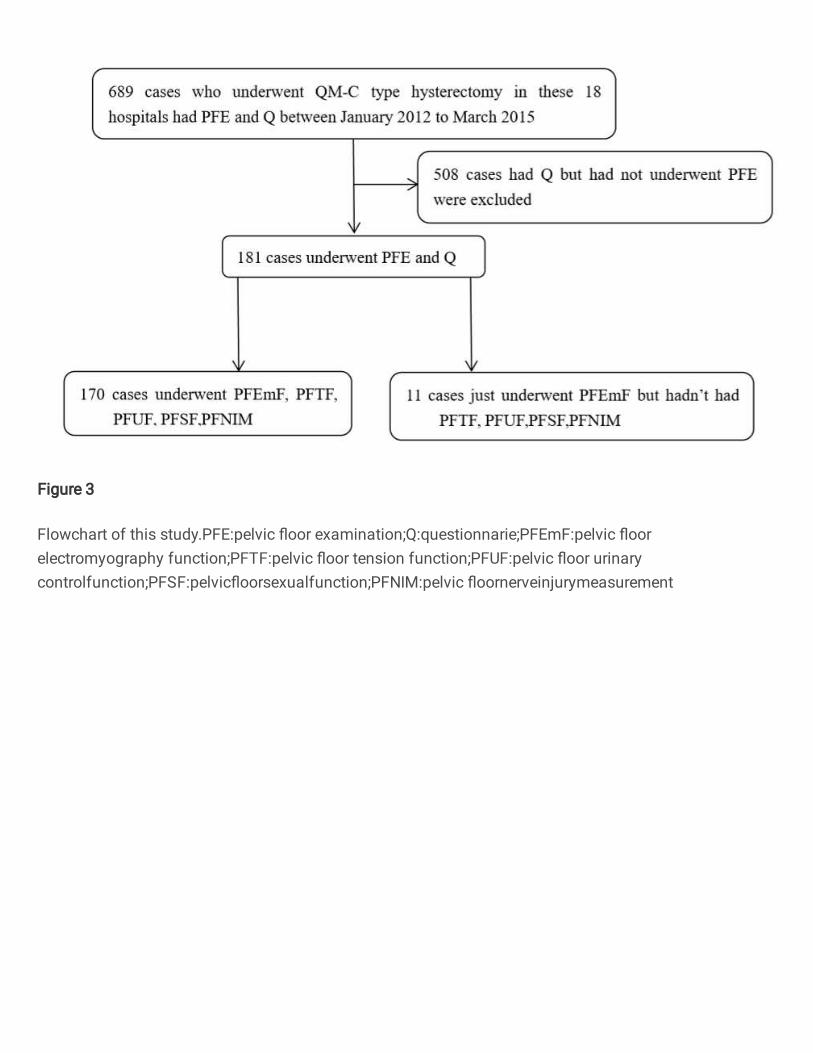
Figure 3
Flowchart of this study.PFE:pelvic �oor examination;Q:questionnarie;PFEmF:pelvic �oorelectromyography function;PFTF:pelvic �oor tension function;PFUF:pelvic �oor urinarycontrolfunction;PFSF:pelvic�oorsexualfunction;PFNIM:pelvic �oornerveinjurymeasurement
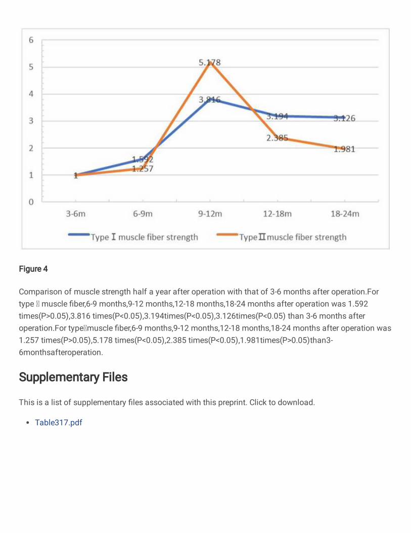
Figure 4
Comparison of muscle strength half a year after operation with that of 3-6 months after operation.Fortype muscle �ber,6-9 months,9-12 months,12-18 months,18-24 months after operation was 1.592times(P>0.05),3.816 times(P<0.05),3.194times(P<0.05),3.126times(P<0.05) than 3-6 months afteroperation.For typemuscle �ber,6-9 months,9-12 months,12-18 months,18-24 months after operation was1.257 times(P>0.05),5.178 times(P<0.05),2.385 times(P<0.05),1.981times(P>0.05)than3-6monthsafteroperation.
Supplementary Files
This is a list of supplementary �les associated with this preprint. Click to download.
Table317.pdf


![Pelvic floor muscle training versus no treatment, or inactive … - 2010.pdf · 2012. 11. 28. · [Intervention Review] Pelvic floor muscle training versus no treatment, or inactive](https://img.pdfslide.net/doc/110x75/5fcc55cc5a78f165476e42b3/pelvic-floor-muscle-training-versus-no-treatment-or-inactive-2010pdf-2012.jpg)