Embed Size (px)
Citation preview
EVOLUTION OF THE VASCULAR SYSTEM IN LINEAGES THAT CONTAIN LIANAS
MARCELO RODRIGO PACE

MARCELO RODRIGO PACE
EVOLUTION OF THE VASCULAR SYSTEM IN
LIANEAGES THAT CONTAIN LIANAS
SÃO PAULO 2015
MARCELO RODRIGO PACE
EVOLUÇÃO DO SISTEMA VASCULAR EM
LINHAGENS QUE CONTÊM LIANAS
Orientação: Prof. Dr. Veronica Angyalossy
SÃO PAULO 2015
Tese apresentada ao Instituto de Biociências da Universidade de São Paulo, para a obtenção do titulo de Doutor em Ciências, na área de Botânica.
COMISSÃO JULGADORA
______________ ______________ Prof. Dr. Prof. Dr.
______________ ______________ Prof. Dr. Prof. Dr.
______________ Prof. Dr. Veronica Angyalossy
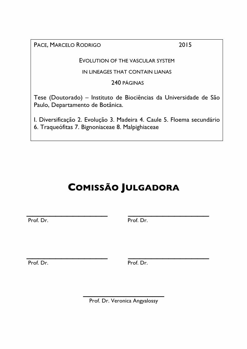
PACE, MARCELO RODRIGO 2015
EVOLUTION OF THE VASCULAR SYSTEM
IN LINEAGES THAT CONTAIN LIANAS
240 PÁGINAS
Tese (Doutorado) – Instituto de Biociências da Universidade de São Paulo, Departamento de Botânica. I. Diversificação 2. Evolução 3. Madeira 4. Caule 5. Floema secundário 6. Traqueófitas 7. Bignoniaceae 8. Malpighiaceae

Nothing in biology makes sense except in the light of evolution.
Theodosius Dobzhansky The American Biology Teacher 35
A Veronica Angyalossy
À mestre que me guiou na construção do conhecimento aqui expresso, pelos14 anos de convivência e por sempre exigir o máximo de mim, acreditar em mim e me ajudar a superar até mesmo minhas próprias expectativas. À amiga, que sempre teve tempo e entendeu uma mão, fosse para sorrir ou chorar juntos,
dedico este trabalho

ABRAÇAR E AGRADECER
Chegar para agradecer e louvar. Louvar o ventre que me gerou
O orixá que me tomou, E a mão da doçura de Oxum que consagrou.
Louvar a água de minha terra O chão que me sustenta, o palco, o massapê,
A beira do abismo, O punhal do susto de cada dia.
Agradecer as nuvens que logo são chuva, Sereniza os sentidos
E ensina a vida a reviver. Agradecer os amigos que fiz
E que mantém a coragem de gostar de mim, apesar de mim... Agradecer a alegria das crianças,
As borboletas que brincam em meus quintais, reais ou não. Agradecer a cada folha, a toda raiz, as pedras majestosas
E as pequeninas como eu, em Aruanda. Agradecer o sol que raia o dia,
A lua que como o menino Deus espraia luz E vira os meus sonhos de pernas pro ar.
Agradecer as marés altas E também aquelas que levam para outros costados todos os males.
Agradecer a tudo que canta no ar, Dentro do mato sobre o mar,
As vozes que soam de cordas tênues e partem cristais. Agradecer os senhores que acolhem e aplaudem esse milagre.
Agradecer, Ter o que agradecer. Louvar e abraçar!
Maria Bethânia
Agradecimentos Quatro anos parecem voar, mas quando busco na memória tudo o que ocorreu nesses tempo, dou-me conta de que não foram poucas coisas. Este doutorado é para mim a realização de um sonho e que foi possível graças a muitos elementos e pessoas, que tentarei fazer jus agradecendo nominalmente aqui, esperando não ter-me esquecido de alguém. Agradeço portanto:
À Universidade de São Paulo, especialmente ao Laboratório de Anatomia Vegetal do Departamento de Botânica do Instituto de Biociências pela oportunidade de me formar aqui com excelente infra-estrutura, cursos e oportunidades e um corpo docente de professores apaixonados pelo que fazem e ensinam que me atraíram para a botânica, para minha própria surpresa. Às agências de fomento FAPESP (2012/01099-8; 2013/10679-0) e CNPq pelo financiamento dessa tese
e tudo o que ela envolveu, incluindo idas ao campo, ida a congressos, contratação de técnicos TT3 para realização de trabalhos práticos e compra de materiais. À minha orientadora Veronica Angyalossy, que sem sombra de dúvidas foi a pessoa mais importante em todo meu processo de formação e que se envolveu plenamente com essa tese, da sua concepção à última linha da conclusão. Veronica, te agradeço por ter sempre acreditado em mim, por ter assinado embaixo nas minhas idéias mais megalomaníacas e sobretudo pelas inúmeras oportunidades que você me brindou ao longo desses quatro anos (ou 14), incluindo-me em palestras, comitês, projetos, como colaborador no seu curso de anatomia da madeira e me estimulando para ir além até de onde achei que pudesse chegar. Obrigado também pela sensibilidade com que sempre me recebeu em momentos pessoalmente mais conturbados. Tenho profunda admiração por você como profissional e como ser humano e me sinto afortunado
por ter tido a oportunidade de te conhecer e de conviver tantos anos com você e sei que só cheguei aqui graças a você.
Aos meus amados pais Adriana De Grandis Pace e Héctor Eduardo Pace agradeço imensamente, tanto pelos pais maravilhosos que são, como por toda ajuda financeira
que me deram, sobretudo nesses últimos 3 anos, pelo apoio incondicional para a realização dessa tese, e sobretudo pelo imenso carinho que recebo de vocês em olhares, palavras e ações quotidianas. À minha amada irmã Gabriella Pace, pela amiga e companheira que é e que sempre fez questão de ser, que sempre está presente na minha vida em todas suas nuances e cuja ajuda foi crucial para a realização desse sonho. Obrigado! À Lisana Rezende, pelas técnicas e apoios que hoje carrego no meu repertório básico de sobrevivência e sem os quais eu não seria a mesma pessoa. Você é especial.
Aos Verônicos André Carvalho Lima, Caian Gerolamo, Carolina Lopes Bastos, Erica Moniz e Mariana Victorio por partilhar da orientação, pelas risadas e pelo excelente convívio diário.
Menção especial à querida amiga, colega e roomie Carolina (Carouo) Lopes Bastos, que se tornou tão especial, uma hiper amiga e que compartilhou comigo um lar pelos últimos 2 anos e inúmeras horas e horas de apoio e terapia mútuos que sem dúvidas influenciaram essa tese.
Realmente muito obrigado!!!!
À Gisele Costa e Tássia dos Santos, técnicas do Laboratório de Anatomia Vegetal, por toda a ajuda e presteza com que sempre receberam todos meus pedidos e pela excelente convivência diária. À grande ajuda técnica que tive e que permitiu que mais de uma centena de espécies e seus indivíduos fossem cortados e processados, envolvendo os mais diversos métodos laboratoriais e plantas incortáveis. Sem esse apoio a realização dessa tese seria inviável. Obrigado Erica Moniz, Mariana Victorio, Rachel Koch, Marli Botânico, André Carvalho Lima e Carolina Lopes Bastos.
Agradeço a todos os amigos e colegas do Laboratório de Anatomia Vegetal dos últimos quatro anos: Aline Siqueira Nunes, André Carvalho Lima, Caian Gerolamo, Carolina Lopes Bastos, Fernanda Cordeiro, Gisele Costa, Giuliano Locosselli, José Hernandes, Keyla Rodrigues, Luiza Teixeira, Mariana Victorio, Natalie Capelli, Rafael Cruz, Raquel Koch, Renata Cassimiro de Lemos, Tássia C. dos Santos, Thalia Gama, Vitor Barão e Yasmin Hirao.
Àqueles amigos do Departamento de Botânica que se tornaram muito especiais, como Anselmo Nogueira, Juliana Lovo, Mariane de Souza-Baena e Suzana Alcantara, que contribuíram sem dúvidas para o meu aprendizado e compleição dessa tese, com discussões, debates, conselhos, risadas e muito companheirismo.
Ao Cassiano Luiz Mecchi pelos quase 11 anos que compartilhamos e por ter sempre apoiado meus sonhos e vibrado com minhas conquistas. Espero que você seja sempre muito feliz, na parte do mundo onde esteja. Conte comigo para o que precisar sempre!
Aos amigos da Finlândia, Helsinki University, Viikki Biocenter 3, Suomen Kielen Kurssi 1 e que apoiaram que eu realizasse esse doutorado e que, apesar dos meus temores e inseguranças, hoje vejo que foi a melhor decisão que poderia ter tomado. Grazie Tahira Anwar e Riccardo Siligato, Kiitos paljon Anne-Maarit Bågman ja Vesa Nousiainen, Hvala vam Milica Maksimovic, Спасибо Katja Shalkovskaya, Obrigado Aime Virkkilä Accorsi. Que bom conhecer vocês.
Aos colaboradores mais recentes, André Marcio Amorim e Suzana Alcantara. Obrigado André por me receber em Ilhéus para sem dúvidas a coleta mais bem sucedida que já participei, com quase 40 espécies de Malpighiaceae coletadas em menos de uma semana e por sempre enviar materiais de excelente qualidade e estar aberto a discussões científicas. À Suzana Alcantara por me receber em Florianópolis para dias de trabalho intenso e eficiente em conjunto, comilanças e até um pouco de praia.
Aos membros da minha banca de qualificação pelos
preciosos comentários e recomendações, Carmen Marcati, José Rubens Pirani e Nanuza Luiza de Menezes.
À Carmen Marcati também pela amizade e companheirismo, hospedagens em Botucatu, risadas.
À querida amiga Neusa Tamaio, que tive o prazer de conviver em campo, congressos e cursos e que sempre foi uma grande incentivadora do meu trabalho e com quem sempre pude contar, além de profunda conhecedora das lianas.
Aos professores do Laboratório de Anatomia Vegetal, Diego Demarco, Gladys Flávia Melo de Pinna, Gregório Ceccantini e Nanuza Luiza de Menezes. Em especial ao Gregório por ter sempre trazido amostras valiosas para mim do campo e incentivado meu trabalho constantemente e à Nanuza pelo imenso carinho que sinto emanar dela sempre que nos encontramos, pelo maravilhoso curso de pós-graduação que nos deu e que nos permitiu entender melhor as plantas como um todo, pela profissional
primorosa e ser humano excepcional! Obrigado.
Ao William Anderson (in memoriam), com o qual pelos primeiros anos da tese troquei e-mails semanais e que me ajudou com seu profundo conhecimento e incomensurável paciência, pois eu não sabia absolutamente nada, a entender as Malpighiáceas, mudanças taxonômicas ocorridas nas últimas décadas, identificação de muitas das
plantas que coletei e pelas trocas de CDs de ópera, outra paixão além das plantas que logo descobrimos compartilhar. Agradeço ainda sua esposa, Christiane Anderson, que gentilmente identificou algumas plantas e também sempre
foi extremamente carinhosa em seus e-mails, mesmo em períodos em que ela estava enfrentando momentos difíceis. À Lúcia G. Lohmann e Richard Olmstead por terem aceitado participar da segunda parte do Capítulo I e publicação do artigo correspondente, pelos comentários e dicas que enriqueceram o manuscrito final.
Aos membros do IAWA Bark Committee, composto majoritariamente de membros sênior ou experientes botânicos já bem estabelecidos, pela acolhida de um aluno dentre eles como igual e pelos agradáveis dias em Brotas (SP), Alan Crivellaro, Alexei Oskolski, Carmen Marcati, Carolina Mittelstaedt, Ekaterina Kotina, Frederic Lens, Leo Junikka, Pieter Baas, Nadezda Nikolaeva, Solange Mazzoni-Viveiros e Teresa Terrazas.
Ao Alex C. Wiedenhoeft, Michael Wiemann e Regis Miller por ter-nos recebido de maneira tão generosa no Forest Products Laboratory em Maddison (Wisconsin), deixando a coleção inteira à nossa disposição para que pudéssemos analisar o material, inclusive noite adentro e nos finais de semana. O Regis foi ainda fundamental para que pudéssemos descobrir a origem de algumas das espécies analisadas.
Ao querido mestre e amigo Antonio Carlos Franco Barbosa, que me ensinou a trabalhar no laboratório ainda no mestrado e cujos ensinamentos temos podido passar adiante e que nos permite obter materiais de qualidade para as análises anatômicas. Agradeço ainda pelo corte de Stereospermum (Bignoniaceae) que fez às pressas e com o primor de sempre.
Ao Pieter Baas, por me receber e me hospedar mais de uma vez em sua casa em Leiden/Leiderdorp (Holanda), pelas revisões e sugestões no primeiro capítulo, pela influência no tema do segundo capítulo e pela doação
de amostras de Malpighiaceae do Velho Mundo, presente na xiloteca de Leiden.
À Michelle Zhjra pela identificação de algumas
madeiras de Coleeae (Bignoniaceae), que ela mesma trouxe de Madagascar e depositou em Madison (WI).
À Maria José Miranda e Rafael Pigozzo por permitirem que fotografasse e analisasse as amostras de Tabebuia, Handroanthus e Sparattosperma (Bignoniaceae) da coleção do Instituto de Pesquisas Tecnológicas (IPT-SP).
Ao Andrew Groover, pelo convite em participar da edição especial do International Journal of Plant Sciences que resultou no Capítulo 1 e pelo incentivo em me dar o grant para o congresso em Ohio, que mudou minha perspectiva para sempre.
Aos colegas e amigos que revisaram cuidadosamente versões anteriores dos manuscritos/capítulos da tese e deram sugestões valiosas que foram incorporadas, André Carvalho Lima, Andrew Groover, Caian Gerolamo, Dewey Litwiller, Giuliano Locosselli, Guillermo Angeles, Pieter Baas e quatro revisores anônimos.
Ao Harri Lorenzi por permitir que por mais de uma vez fôssemos coletar no Instituto Plantarum e que sempre nos recebeu com entusiasmo.
Ao Augusto Francener pela ajuda na identificação
das Malpighiáceas, especialmente Byrsonimas e outras espécies do cerrado.
Ao querido Rafael Felipe de Almeida pelas discussões sobre Mapighiaceae, por coletar materiais valiosos e ajudar na identificação das especies. Obrigado
também pelo incentivo constante e risadas
Agradeço também às amigas de décadas que me acompanharam todos esses anos - escola, graduação, mestrado, doutorado, viagens - e que sempre me apoiaram e apoiam Fernanda Castanho Pereira dos Santos, Juliana Sette Sabbato e Marianne Thamm de Aguiar. Vocês são para lá de especiais na minha vida.
Agradeço aos amigos mais recentes e mais antigos, do dia a dia de São Paulo, pelas viagens, festas, risadas, meriendas, dramas e compreensão pelo último ano e especialmente últimos meses em que tive que dizer mais “nãos” que “sins” pra conseguir terminar a tese e que até disso me ajudaram a rir criando o hashtag #drama_tesis. Obrigado/gracias Abner Mendonça, Andrea Calero, Andrés Mesa, Adriana Ferro, Belén Sala Torres, Danilo Prado, Gustavo Caixeta, Lida Marica Fierro, Marcio Miranda Perez, Muriel Valencia, Pablo Sarmiento, Pilar Marcela Afanador, Rocío Lobo Machín, Viviana Peña.
Por fim, a todos que me ajudaram a coletar as
espécies utilizadas nesse trabalho, diretamente participando em expedições de coleta ou enviando materiais, Alcides Sáenz (Isla Martín García, Argentina), Alexandre Zuntini, André Amorim, Anselmo Nogueira, Arnildo Pott, Arno Fritz, Berta Villagras, Cairo Faleiros, Carolina Lopes Bastos, Carolina Madero (México), Daniel Bazzano (Isla Martín García, Argentina), Daniel Villavoel (Chuquisaca, Bolívia), Erica Moniz, Genise Somner, Geraldo Damasceno Jr., Gonzalo Castillo, Diana Sampaio, Guillermo Angeles (México), Gregório Ceccantini, Ivone Vázques Briones (Tarapoto, Peru), Jefferson Prado, Joan Miró Ortega, Juliana Lovo, Julio Majcher, Kamila Drequeceler, Luiz Carlos Jesus Gomes, Lukas Tadeu Halla Daneu, Marccus Alves, Márdel Lopes, Maria Ana Farinaccio, Milton Groppo Jr., Neusa Tamaio, Pablo Cabanillas (Isla Martín García, Argentina), Renata Udulutsch, Rosani Arruda, Suzana Alcantara, Rafael Felipe de Almeida, Ricardo Zárate (Iquitos, Peru).
Como dito anteriormente, todas essas pessoas contribuíram direta ou indiretamente para que essa tese pudesse existir e portanto têm minha imensa gratidão.
Obrigado!

ABSTRACT
The vascular system of lianas, especially the xylem, has been repeatedly shown to be different, with
lianas having a set of features shared among even distantly related lineages, such as the presence of cambial
variants, wide and long vessels, more abundant axial parenchyma, frequently non-lignified, taller and wider
rays, which are generally heterocellular. In spite of this amount of knowledge, few works have investigated
the impact for the vascular system of the evolution of this habit within lineages whose ancestors are not
lianas, but self-supporting plants. Therefore, in this dissertation we explored wood, phloem and overall stem-
anatomy evolution in lineages that contain lianas and self-supporting plants, using well-supported phylogenies
and detailed anatomical investigations. Within Bignoniaceae (Lamiales), we thoroughly investigated the wood
anatomy, delimiting character states and mapping them onto the last phylogeny for the group, encountering
that eco-physiological and habit transition were the main drivers of modifications in the wood anatomy
in the family. Ring-porous and semi-ring porous woods and helical thickening was found in plants either
growing in higher latitudes or with marked seasonal water regimes, and septate fibres correlated with
scanty axial parenchyma, which are eco-physiological drivers. Evolution of lianas, in turn, drove an increase
in vessel diameter, wide vessels accompanied by very narrow ones, presence of perforated ray cells, scanty
axial parenchyma and cambial variants. Despite the great wood anatomical diversity within the family, major
clades have quite predictive wood anatomy and 9 possible anatomical synapomorphies were raised in this
work to clades previously delimitated exclusively by molecular characters. Within the tracheophytes, we
investigated 26 phylogenetically controlled pairs of lianas and their self-supporting relatives within all major
lineages of tracheophytes (except lycophytes), in order to seek characters evolving in correlation with the
lianescent habit. We found that the sieve elements and sieve pores were always wider in the lianas, and
that the rays were always taller and heterocellular. However, all the main characters of the phloem of the
lianas remained conserved with that of their self-supporting relatives. This evidenced that although a more
efficient photosynthetic conductive system evolved in the phloem of lianas, overall anatomy conserved a high
phylogenetic signal. Within Malpighiaceae, (Malpighiales), lianas are abundant and many cambial variants are
present. However, nothing was known regarding how many types of cambial variants there were in the family
and how they were distributed. We were able to delimit 6 different types of cambial variants that evolved at
least 8 times independently in the family, which ancestrally lacks a cambial variant. Many of these types share
common stages of development and some variants that are anatomically very similar derive from different
ontogenetic trajectories. Within the genera, the variants are conserved, and even between sister groups in the
new and old world, evidencing that cambial variants may be a good indicator of relationships within the family.
Overall, we conclude that lianas greatly impact the evolution of the vascular system in the lineages where
they have evolved, and these modifications normally result in a more efficient water and photosynthates
conduction system and an increased flexibility for climbing.
RESUMO
O sistema vascular das lianas, em especial o xilema, mostrou-se repetidas vezes distinto nas lianas,
com aspectos compartilhados mesmo dentre linhagens distantemente relacionadas, tais como a presença
de variações cambiais, vasos mais largos e longos, parênquima axial mais abundante - frequentemente não-
lignificado - raios mais altos e largos - geralmente heterocelulares. Não obstante todo esse conhecimento,
poucos trabalhos investigaram o impacto da evolução do hábito lianescente no sistema vascular em linhagens
cujos ancestrais não são lianas e sim plantas auto-suportantes. Portanto, nesta tese exploramos o lenho, o floema
e a anatomia caulinar como um todo em linhagens que contêm lianas e plantas auto-suportantes, utilizando
filogenias bem sustentadas e investigações anatômicas detalhadas. Em Bignoniaceae (Lamiales), investigamos
em detalhe a anatomia do lenho, delimitando caracteres e estados de caráter e mapeando-os na filogenia
mais recente do grupo, encontrando que modificações eco-fisiológicas e transições de hábito tiveram grande
impacto na evolução do lenho na família. Anéis porosos e semi-porosos, bem como espessamento espiralado
foram encontrados em plantas crescendo em latitudes mais altas ou em regimes hídricos fortemente sazonais,
ao passo que fibras septadas apareceram correlacionadas com a presença de parênquima axial escasso. A
evolução de lianas, por sua vez, parece ter levado a um aumento no diâmetro dos vasos, contudo dimórficos,
células perfuradas de raio, parênquima axial mais escasso e surgimento de variações cambiais. Apesar da
enorme diversidade dentro de Bignoniaceae, os grandes clados possuem uma anatomia bastante preditiva e
9 possíveis sinapomorfias morfológicas são sugeridas para clados delimitados somente com base em dados
moleculares. Dentro das traqueófitas, investigamos 26 pares filogeneticamente controlados de lianas espécies
auto-suportantes relacionadas pertencentes a todas as principais linhagens de traqueófitas (exceto licófitas),
a fim de buscar caracteres que tenham evoluído em correlação com o hábito lianescente. Encontramos
que os elementos crivados e os poros das placas crivadas têm sempre maior calibre nas lianas, e que os
raios são mais altos e heterocelulares. Contudo, as principais características do floema das lianas se mantêm
conservadas em relação às espécies auto-suportantes relacionadas, evidenciando que as lianas teriam evoluído
um sistema de condução de fotossintetatos mais eficiente, porém preservando um alto sinal filogenético. Em
Malpighiaceae, lianas são abundantes, tal como as variações cambiais. Contudo, pouco se sabe sobre o número
de variações presentes na família ou como elas estariam distribuídas. Aqui delimitamos 6 diferentes tipos de
variação cambial, que teriam evoluído independentemente 8 vezes na família, cujo ancestral é reconstruído
como tendo caule simples. Muitas dessas variações compartilham estágios de desenvolvimento, ao passo que
variações anatomicamente muito similares derivam de trajetórias ontogenéticas distintas. Dentro dos gêneros
as variações se mostraram conservadas e mesmo dentre grupos irmãos do novo e velho mundo, evidenciando
que as variações cambiais seriam bons indicadores de relações na família. De maneira geral, podemos concluir
que lianas impactam significativamente o sistema vascular nas linhagens onde ocorrem e que tais modificações
em geral resultam em um sistema de condução hídrico e de fotossintetatos mais eficiente e também mais
flexível para a escalada.

Contents
General Introduction Lianas and their vascular system 2
References 8
Chapter 1. Diversity and evolution of the secondary xylem in Bignoniaceae Preface 13
Section I Wood anatomy and evolution: a case study in the Bignoniaceae
Resumo & Abstract 15
Introduction 16
Material & Methods 21
Results 28
Discussion 49
References 56
Section II Wood anatomy of major Bignoniaceae clades
Resumo & Abstract 63
Introduction 66
Material & Methods 68
Results 69
Discussion 90
References 97
Apendix I, for sections I and II 100
General conclusions 105
Chapter 1I. The phloem of lianas: a comparative study across the tracheophytes Resumo & Abstract 108
Introduction 109
Material & Methods 114
Results 135
Discussion 155
References 166
Chapter 1II. Diversity of the cambial variants and their distribution in Malpighiaceae Resumo & Abstract 176
Introduction 177
Material & Methods 178
Results 180
Discussion 203
References 210
Appendix I 214
Conclusão Geral 219
Extra file 1. First page of the manuscripts published with results from this work 221
Extra file II. List of works presented in meetings with results from this work 226

GENERAL INTRODUCTION
General Introduction
“Plants become climbers, in order, as it may be presumed, to reach the light, and to expose a large surface of their
leaves to its action and to that of the free air. This is effected by climbers with wonderfully little expenditure of organized
matter, in comparison with trees, which have to support a load of heavy branches by a massive trunk. Hence, no doubt,
it arises that there are so many climbers in all quarters of the world, belonging to so many different orders.”
(Charles Darwin, 1865, pages 107-108 in
The Movements and Habits of Climbing Plants)
The first land plants were likely erect self-supporting plants (Bennici 2008), from which
lianas evolved multiple times since the appearance of tracheophytes (= vascular plants) at around
433 mya (Silvestro et al. 2015). The first fossil records of lianas dates back to the Carboniferous
(Mississippian, ~ 335 mya), with peaks of diversity in the Pennsylvanian (a more recent period of
the Carboniferous) and the Eocene (~50 mya; Burnham 2009, 2015). Lianas are here treated as
any climbing plant that germinates on the soil and that by a range of different mechanisms climb
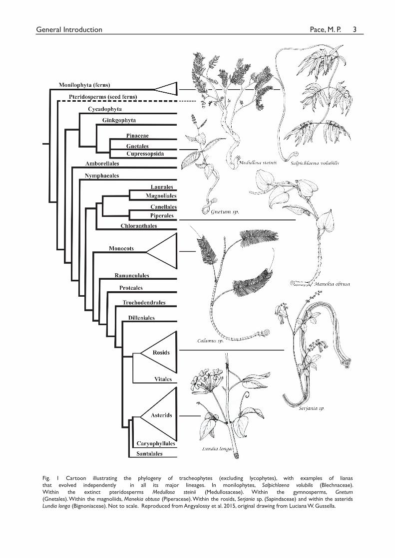
up other plants or vertical supports in search of light (Muller-Dombois & Ellenberg 1974). Lianas
are present in all major tracheophyte lineages (Fig. 1), being present in lycophytes (e.g., Lycopodiella
cernua, Lycopodiaceae, occasionally is a climber; Rowe et al. 2004), ferns (e.g., Lygodium, Schizaeaceae),
extinct progymnosperms (e.g., Medullosa steinii, Medullosaceae; Dunn et al. 2003), gymnosperms (e.g.,
Gnetum, Gnetaceae), and a diverse range of angiosperms, including palms (e.g., the richest genus
of the palm family being lianescent, Calamus; Dransfield et al. 2014), magnoliids (e.g., black pepper,
Piperaceae; Tasmannia cordata, Winteraceae; Feild et al. 2012) and many eudicots (e.g., peas, beans and
wild roses being a few examples).
Architecturally, however, the question is what modifications in the bauplan of a plant have to
occur to make a liana a liana? First of all, climbing can only be achieved either by twining flexible stems
or specialized structures for grasping. Also, lianas ought to have narrower and yet longer stems, since
growing into as massive heavy organisms as trees can impose excess weight and mechanical failure
on their support, while having a higher length is both a side effect of twining and a means of exploring
various tree canopies in search of sunlight. Both these aspects of the liana bauplan seem to have been
conquered multiple times in the transitions from self-supporting to lianas across evolution (at least
133 families contain a few climbers; Gentry 1991), since both morphological modifications adapted
for climbing and taxa with much narrower and longer stems are the rule rather than the exception in
lianescent taxa (Bhambie 1972, Putz 1983, Ewers & Fisher 1991, Rowe et al. 2006), being a textbook
case of convergent evolution (Futuyma 2009).
Pace, M. P.General Introduction 3
Fig. 1 Cartoon illustrating the phylogeny of tracheophytes (excluding lycophytes), with examples of lianas
that evolved independently in all its major lineages. In monilophytes, Salpichlaena volubilis (Blechnaceae).
Within the extinct pteridosperms Medullosa steinii (Medullosaceae). Within the gymnosperms, Gnetum (Gnetales). Within the magnoliids, Manekia obtusa (Piperaceae). Within the rosids, Serjania sp. (Sapindaceae) and within the asterids
Lundia longa (Bignoniaceae). Not to scale. Reproduced from Angyalossy et al. 2015, original drawing from Luciana W. Gussella.
Pace, M. P.General Introduction 4
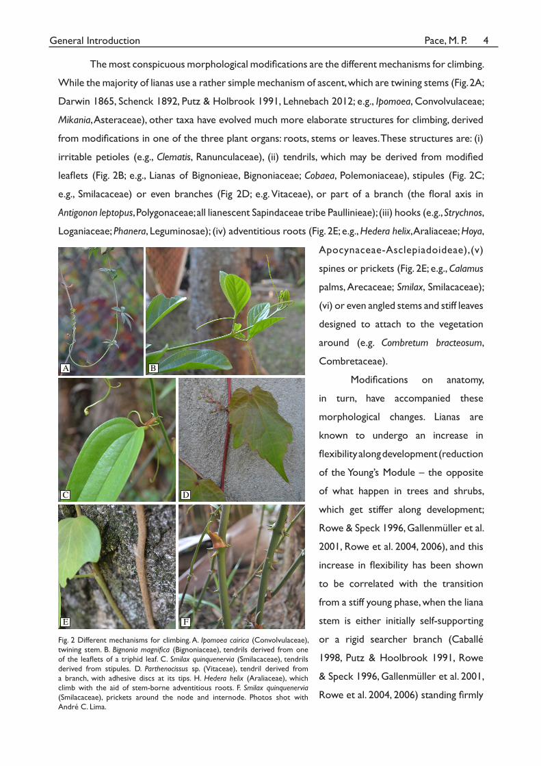
The most conspicuous morphological modifications are the different mechanisms for climbing.
While the majority of lianas use a rather simple mechanism of ascent, which are twining stems (Fig. 2A;
Darwin 1865, Schenck 1892, Putz & Holbrook 1991, Lehnebach 2012; e.g., Ipomoea, Convolvulaceae;
Mikania, Asteraceae), other taxa have evolved much more elaborate structures for climbing, derived
from modifications in one of the three plant organs: roots, stems or leaves. These structures are: (i)
irritable petioles (e.g., Clematis, Ranunculaceae), (ii) tendrils, which may be derived from modified
leaflets (Fig. 2B; e.g., Lianas of Bignonieae, Bignoniaceae; Cobaea, Polemoniaceae), stipules (Fig. 2C;
e.g., Smilacaceae) or even branches (Fig 2D; e.g. Vitaceae), or part of a branch (the floral axis in
Antigonon leptopus, Polygonaceae; all lianescent Sapindaceae tribe Paullinieae); (iii) hooks (e.g., Strychnos,
Loganiaceae; Phanera, Leguminosae); (iv) adventitious roots (Fig. 2E; e.g., Hedera helix, Araliaceae; Hoya,
A B
C D
E FFigure 2. Different mechanism for climbing. A. Ipomoea cairica (Convolvulaceae), twining stem. B. Bignonia magnifi ca (Bignoniaceae), tendrils derived from one of the leafl ets. C. Smilax
quinquenervia (Smilacaceae), tendrils derived from stipules. D. Parthenocissus sp. (Vitaceae),
tendril with adhesive discs, derived from a branch. E. Hedera helix (Araliaceae), climbing
with the aid of stem-borne adventitious roots. F. Smilax quinquenervia (Smilacaceae), prickets
around the node and in the internodes. Photos shot with Andre C. Lima.
Figure 2
Apocynaceae-Asclepiadoideae),(v)
spines or prickets (Fig. 2E; e.g., Calamus
palms, Arecaceae; Smilax, Smilacaceae);
(vi) or even angled stems and stiff leaves
designed to attach to the vegetation
around (e.g. Combretum bracteosum,
Combretaceae).
Modifications on anatomy,
in turn, have accompanied these
morphological changes. Lianas are
known to undergo an increase in
flexibility along development (reduction
of the Young’s Module – the opposite
of what happen in trees and shrubs,
which get stiffer along development;
Rowe & Speck 1996, Gallenmüller et al.
2001, Rowe et al. 2004, 2006), and this
increase in flexibility has been shown
to be correlated with the transition
from a stiff young phase, when the liana
stem is either initially self-supporting
or a rigid searcher branch (Caballé
1998, Putz & Hoolbrook 1991, Rowe
& Speck 1996, Gallenmüller et al. 2001,
Rowe et al. 2004, 2006) standing firmly
Fig. 2 Different mechanisms for climbing. A. Ipomoea cairica (Convolvulaceae),
twining stem. B. Bignonia magnifica (Bignoniaceae), tendrils derived from one
of the leaflets of a triphid leaf. C. Smilax quinquenervia (Smilacaceae), tendrils
derived from stipules. D. Parthenocissus sp. (Vitaceae), tendril derived from
a branch, with adhesive discs at its tips. H. Hedera helix (Araliaceae), which
climb with the aid of stem-borne adventitious roots. F. Smilax quinquenervia (Smilacaceae), prickets around the node and internode. Photos shot with
André C. Lima.

Pace, M. P.General Introduction 5
in the air, circumnutating in search of a support (Darwin 1965, Putz & Holbrook 1991, Isnard & Silk
2009). Whenever the support is found, however, an abrupt change occurs anatomically, resulting
in this decrease in stiffness and increase in flexibility (Putz & Holbrook 1991, Rowe & Speck 1996,
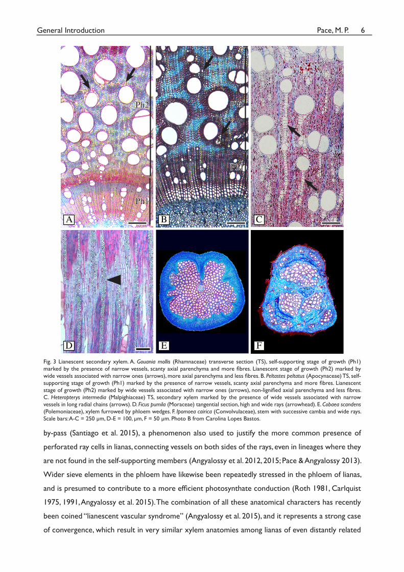
Rowe et al. 2004). Specifically, at the initial self-supporting phase the xylem anatomy is marked by the
presence of narrow vessels, scanty axial parenchyma and the prevalence of fibres (Fig. 3A-B; Rowe et
al. 2004, 2006; Angyalossy et al. 2012, 2015). However, as soon as the plant switches to the lianescent
phase, flexibility increases by the formation of more axial parenchyma (Fig. 3A), which is frequently
non-lignified (Fig. 3B), less fibres, sometimes gelatinous, and wider vessels in the xylem (Fig. 3A-B),
summed to the common appearance of cambial variants at this stage of development (Carquist 1985,
Rowe & Speck 1996, Rowe et al. 2004, Bowling et al. 2009, Crivellaro et al. 2012, Angyalossy et al.
2012, 2015). Cambial variants are alternative forms of secondary growth that generally results in soft
tissues (phloem and/or non-lignified parenchyma) mixed with the rigid secondary xylem, such as the
xylem furrowed by phloem wedges in Bignoniaceae and Polemoniaceae (Fig. 3E) or the successive
cambia in Menispermaceae and Convolvulaceae (Fig. 3F; Schenck 1893, Dobbins 1971, Mennega
1982, Carlquist 2001, Tamaio et al. 2009, Angyalossy et al. 2015). Lianas typically have also higher and
wider rays (Fig. 3D), generally heterocellular mixed or more heterocellular than their close-related
counterparts (Pace & Angyalossy 2013, Angyalossy et al. 2015). The presence of more parenchyma
and less fibres within the xylem is possible only because lianas do not sustain their own bodies,
leaning on their supports (Carlquist 1985, Ewers 1985, Stevens 1987, Gartner 1991, Crivellaro et al.
2012).
Longer and narrower stems, but supporting canopies as large or larger than those of trees
(Putz 1983, Ewers & Fisher 1991), in turn, create a demand for efficient water and photosynthates
transport over long distances, given liana’s high length-low width stem ratio (Putz 1984, Kurzel
et al. 2006). This is achieved by the presence of very wide and long vessels, in fact the widest and
longest known in plants (some up to 500 µm wide and 8 m long; Zimmermann & Jeje 1981, Ewers
1985, Ewers & Fisher 1989, 1991, Isnard & Feild 2015) and with perforation plates that converged
to be simple, even in families where the self-supporting species have scalariform perforation plates
(Ayensu & Stern 1964; Carlquist 1991, Lens et al. 2008). Such features make up an extremely efficient
hydraulic system, shown to transport approximately 3 times more water than that of self-supporting
plants (Isnard & Feild 2015). However, because wide vessels are known to be more vulnerable to
embolism, it is also widespread the presence of narrow vessels associated with the wide vessels
of lianas (Fig. 3A-C; Carlquist 1985, Santiago et al. 2015), a phenomenon named vessel dimorphism
by Carlquist in his work with the carnivore liana Nepenthes (1981). The presence of these narrows
vessels associated with the wide ones act as a guarantee that the water column will not be broken
in the event of embolism in the vulnerable wide vessels, with the narrow vessels acting as a water
Pace, M. P.General Introduction 6
B
D F
A C
E
Fig. 3. Lianescent secondary xylem. A. Gouania mollis (Rhamnaceae) transverse section (TS), self-
supporting stage of growth (Ph1) marked by the presence of narrow vessels, scanty axial paren-
chyma and more fi bres. Lianescent stage of growth (Ph2) marked by wide vessels associated with
narrow ones (arrows), more axial parenchyma and less fi bres. B. Peltastes peltatus (Apocynaceae)
TS, self-supporting stage of growth (Ph1) marked by the presence of narrow vessels, scanty axial
parenchyma and more fi bres. Lianescent stage of growth (Ph2) marked by wide vessels associated
with narrow ones (arrows), non-lignifi ed axial parenchyma and less fi bres. C. Heteropterys inter-
media (Malpighiaceae) TS, secondary xylem marked by the presence of wide vessels associated
with narrow vessels in long radial chains (arrows). D. Ficus pumila (Moraceae) tangential section,
high and wide rays (arrowhead). E. Cobaea scandens (Polemoniaceae), xylem furrowed by phloem
wedges. F. Ipomoea cairica (Convolvulaceae), stem with successive cambia and wide rays. Scale
bars: A-C = 250 um. D-E = 100um, F = 50 um.
Ph1
Ph2
Ph1
Ph2
Fig. 3 Lianescent secondary xylem. A. Gouania mollis (Rhamnaceae) transverse section (TS), self-supporting stage of growth (Ph1)
marked by the presence of narrow vessels, scanty axial parenchyma and more fibres. Lianescent stage of growth (Ph2) marked by
wide vessels associated with narrow ones (arrows), more axial parenchyma and less fibres. B. Peltastes peltatus (Apocynaceae) TS, self-
supporting stage of growth (Ph1) marked by the presence of narrow vessels, scanty axial parenchyma and more fibres. Lianescent
stage of growth (Ph2) marked by wide vessels associated with narrow ones (arrows), non-lignified axial parenchyma and less fibres.
C. Heteropterys intermedia (Malpighiaceae) TS, secondary xylem marked by the presence of wide vessels associated with narrow
vessels in long radial chains (arrows). D. Ficus pumila (Moraceae) tangential section, high and wide rays (arrowhead). E. Cobaea scandens (Polemoniaceae), xylem furrowed by phloem wedges. F. Ipomoea cairica (Convolvulaceae), stem with successive cambia and wide rays.
Scale bars: A-C = 250 µm, D-E = 100, µm, F = 50 µm. Photo B from Carolina Lopes Bastos.
by-pass (Santiago et al. 2015), a phenomenon also used to justify the more common presence of
perforated ray cells in lianas, connecting vessels on both sides of the rays, even in lineages where they
are not found in the self-supporting members (Angyalossy et al. 2012, 2015; Pace & Angyalossy 2013).
Wider sieve elements in the phloem have likewise been repeatedly stressed in the phloem of lianas,
and is presumed to contribute to a more efficient photosynthate conduction (Roth 1981, Carlquist
1975, 1991, Angyalossy et al. 2015). The combination of all these anatomical characters has recently
been coined “lianescent vascular syndrome” (Angyalossy et al. 2015), and it represents a strong case
of convergence, which result in very similar xylem anatomies among lianas of even distantly related
Pace, M. P.General Introduction 7
lineages.
What all the previous works convey is that lianas are special both morphologically and
anatomically. These special features have been noted since the XIX century (Darwin 1865, Van
Thieghem 1884, Avetta 1887, Schenck 1893, Pfeiffer 1926) and stimulated large treatises on the
subject (Schenck 1892, 1893, Obaton 1960, Putz and Mooney 1991, Schnitzer et al. 2015). What at
the present are still meagre are studies that address the evolution of lianas within a phylogenetic
framework, both on a narrow and a wide scale, in order to detect exact how the evolutionary
transition from self-supporting to lianas impact the anatomical diversification of the vascular system,
considering xylem, phloem and the overall stem anatomy. Therefore, the main aim of this PhD
dissertation was to explore xylem, phloem and overall stem architecture evolution within well-
supported phylogenies.
In the first chapter we carried out an in-depth analysis of wood evolution within Bignoniaceae,
a pantropical family with a myriad of different habits, including lianas, shrubs and trees, and occupying
diverse habitats, from humid tropical forests to savannas and temperate forests (Gentry 1980,
Fischer et al. 2004). Our aim was to explore all the wood variable characters in the family to launch
hypotheses on what would be the main drivers of evolution and diversification in this group, how
the diversity in wood structure is distributed in the family and ultimately how wood characters
supported clades in the family, whose relationships have been reconstructed with basis in molecular
sequence data.
In the second chapter, we carried a detailed study of the phloem across all major lineages
of tracheophytes (except lycophytes) in order to investigate a number of taxa of lianas and their
self-supporting relatives to explore whether there is any correlation similar to what described to
the xylem, summarized in the ‘lianescent vascular syndrome’, including confirming or refuting the
hypothesis that lianas would always have wider sieve elements. Also, it included an investigation of
taxa in which two phloem types co-occur: a regular and a variant phloem, resulting from two types
of cambial variants (interxylary phloem and furrowed xylem) and that has been hypothesized as
undergoing a subfunctionalization, with the regular phloem specializing in storage and the variant
phloem specializing in conduction (Pace et al. 2011, Carlquist 2013).
In the third chapter, we explored the stem anatomical diversity in the pantropical family
Malpighiaceae, aiming at delimiting how many different cambial variants are present in the family, how
they are distributed in the phylogeny of the family, their ontogeny, and possible common underlying
features shared between them.
Pace, M. P.General Introduction 8
With these studies we expect to shed light on the main convergent modifications undergone
in the vascular system of plants when they transition from self-supporting to lianas.
LITERATURE CITED
Angyalossy V, Angeles G, Pace MR, Lima AC, Dias-Leme CL, Lohmann LG, Madero-Vega C. 2012. An overview
of the anatomy, development and evolution of the vascular system of lianas. Plant Ecology & Diversity 5: 167-182.
Angyalossy V, Pace MR, Lima AC. 2015. Liana anatomy: a broad perspective on structural evolution of the vascular
system. In: Schnitzer, SA, Bongers F, Burnham R, Putz FE, eds. Ecology of Lianas. Wiley-Blackwell Publishers,
Oxford.
Avetta C. 1887. Contribuzione allo studio delle anomalie di struttura nelle radici delle dicotiledoni. Annuario del
Reale Istituto Botanico di Roma 3: 3-19.
Ayensu ES, Stern WL. 1964. Systematic anatomy and ontogeny of the stem in Passifloraceae. Contributions of the
United States National Herbarium 34: 45-72.
Bennici A. 2008. Origin and early evolution of land plants: problems and considerations. Communicative & Integrative
Biology 1: 212-218.
Bhambie S. 1972. Correlation between form, structure and habit in some lianas. Proceedings of the Indian Academy of
Sciences 75: 246-256.
Bowling AJ, Vaughn KC. 2009. Gelatinous fibers are widespread in coiling tendrils and twining vines. American Journal
of Botany 96: 719-727.
Burnham RJ. 2009. An overview of the fossil record of climbers: bejucos, sogas, trepadoras, lianas, cipós , and vines.
Revista Brasileira de Paleontologia 12: 149-160.
Burnham RJ. 2015. Climbing plants in the fossil record: Paleozoic to present. In: Schnitzer, SA, Bongers F, Burnham R,
Putz FE, eds. Ecology of Lianas. Wiley-Blackwell Publishers, Oxford.
Caballé G. 1998. Le port autoportant des lianes tropicales: une synthèse des stratégies de croissance. Canadian Journal
of Botany 76: 1703-1716.
Carlquist S. 1975. Ecological strategies of xylem evolution. University of California Press, Berkeley.
Carlquist S. 1981. Wood anatomy of Nepenthaceae. Bulletin of the Torrey Botanical Club 108: 324-330.
Carlquist S. 1985. Observations on functional wood histology of vines and lianas: vessel dimorphism, tracheids,
vasicentric tracheids, narrow vessels, and parenchyma. Aliso 11:139–157
Carlquist S. 1991. Anatomy of vine and liana stems: a review and synthesis. Pages 53–73 in FE Putz, HA Mooney, eds.
The biology of vines. Cambridge University Press, Cambridge.
Carlquist S. 2001. Comparative wood anatomy, 2nd edn. Springer, Berlin.
Carlquist S. 2013. Interxylary phloem: diversity and functions. Brittonia 65: 477-495.
Crivellaro A, McCulloh K, Jones FA, Lachenbruch B. 2012. Anatomy and mechanical and hydraulic needs of
Pace, M. P.General Introduction 9
woody climbers contrasted with subshrubs on the island of Cyprus. IAWA Journal 33: 355-373.
Darwin C. 1865. On the movements and habits of climbing plants. Journal of the Linnean Society, London, pp. 118.
Dobbins DR. 1971. Studies on the anomalous cambial activity in Doxantha unguis-cati (Bignoniaceae). II. a case of
differential production of secondary tissues. American Journal of Botany 58: 697-705.
Dransfield J, Uhl NW, Asmussen CB, Baker WJ, Harley M, Lewis C. 2008. Genera Palmarum: the evolution and
classification of palms. Kew Press, London.
Dunn MT, Krings M, Mapes G, Rothwell GW, Mapes RH, Keqin S. 2003. Medullosa steinii sp nov., a seed fern vine
from the Upper Mississippian. Review of Palaeobotany and Palynology 124: 307-324.
Ewers FW. 1985. Xylem structure and water conduction in conifer trees, dicot trees, and lianas. IAWA Bulletin n.s. 6:
309-317.
Ewers FW, Fisher JB. 1989. Variation in vessel length and diameter in stems of six tropical and subtropical lianas.
American Journal of Botany 76: 1452-1459.
Ewers FW, Fisher JB. 1991. Why vines have narrow stems: histological trends in Bauhinia (Fabaceae). Oecologia 88:
233-237.
Feild TS, Chaletet DS, Balun L, Schilling EE, Evans R. 2012. The evolution of angiosperm lianescence without
vessels – climbing mode and wood structure in Tasmannia cordata (Winteraceae). New Phytologist 193: 229-
240.
Fischer E, Theisen I, Lohmann LG. 2004. Bignoniaceae. In: K. Kubitzki and J.W. Kadereit (eds). The Families and
Genera of Vascular Plants VII. Dicotyledons. Lamiales (Except Acanthaceae including Avicenniaceae). Springer-
Verlag, Heidelberg.
Futuyma DJ. 2009. Evolution. 2nd. ed. Sinauer, Sunderland.
Gallenmüller F, Müller U, Rowe N, Speck T. 2001. The growth form of Croton pullei (Euphorbiaceae) - Functional
morphology and biomechanics of a neotropical liana. Plant Biology 3: 50-61.
Gartner BL. 1991. Structural stability and architecture of vines vs. shrubs of poison oak, Toxicodendron diversilobum.
Ecology 72: 2005-2015.
Gentry AH. 1980. Bignoniaceae: Part I (Crescentia and Tourrettieae). Flora Neotropical Monograph 25 (1), The New
York Botanical Garden, New York.
Gentry AH. 1991. The distribution and evolution of climbing plants. In F.E. Putz, and H.A. Mooney (eds), The Biology of
Vines. Cambridge University Press, New York.
Isnard S, Silk WK. 2009. Moving with climbing plants from Charles Darwin’s time into the 21st century. American
Journal of Botany 96: 1205-1221.
Isnard S, Feild TS. 2015. The evolution of angiospem lianescence: a perspective from xylem structure-function. In:
Schnitzer, SA, Bongers F, Burnham R, Putz FE, eds. Ecology of Lianas. Wiley-Blackwell Publishers, Oxford.
Kurzel BP, Schnitzer SA, Carson WP. 2006. Predicting liana crown location from stem diameter in three Panamanian
lowland forests. Biotropica 38: 262-266.
Pace, M. P.General Introduction 10
Lehnebach R. 2012. Evolution et diversité des traits morpho-anatomiques chez les lianes tropicales. Master thesis,
Université Montpellier II, Montpellier.
Lens F, Kårehed J, Baas P, Jansen S, Rabaey D, Huysmans S, Hamann T, Smets E. 2008. The wood anatomy of
polyphyletic Icacinaceae s.l., and their relationship within asterids. Taxon 57: 525-552.
Mennega AW. 1982. Stem structure of the New World Menispermaceae. Journal of the Arnold Arboretum 63: 145-172.
Muller-Dombois D, Ellenberg H. 1974. Aims and methods of vegetation ecology. John Wiley. New York.
Obaton M. 1960. Les lianes ligneuses a structure anormale des forêts denses d’Afrique Occidentale. Annales des
Sciences Naturelles Botanique et Biologie Végétale 12: 1-220.
Pace MR, Angyalossy V. 2013. Wood anatomy and evolution: a case study in the Bignoniaceae. International Journal of
Plant Sciences 174: 1014-1048.
Pace MR, Lohmann LG, Angyalossy V. 2011. Evolution of disparity between the regular and variant phloem in
Bignonieae (Bignoniaceae). American Journal of Botany 98: 602-618.
Pfeiffer H. 1926. Das abnorme Dickenwachstum. In: Lisbauer K. (ed.), Handbuch der Pflanzenanatomie. Gebrüder
Bornstraeger, Berlin.
Putz FE. 1983. Liana biomass and leaf area of a tierra firme forest in the Rio Negro basin, Venezuela. Biotropica 15:
185–189.
Putz FE. 1984. The natural history of lianas on Barro Colorado Island, Panama. Ecology 65: 1713–1724.
Putz FE, Holbrook NM. 1991. Biomechanical studies in vines. In F.E. Putz, and H.A. Mooney (eds), The Biology of Vines.
Cambridge University Press, New York.
Putz FE, Mooney HA. 1991. The biology of vines. Cambridge University Press. Cambridge.
Roth I. 1981. Structural patterns of tropical barks. In: Braun HJ, Carlquist S, Ozenda P, Roth I., eds. Encyclopedia of Plant
Anatomy. Gebrüder Bornstraeger. Berlin.
Rowe NP, Speck T. 1996. Biomechanical characteristics of the ontogeny and growth habit of the tropical liana
Condylocarpon guianense (Apocynaceae). International Journal of Plant Sciences 157: 406-417.
Rowe N, Isnard S, Speck T. 2004. Diversity of mechanical architectures in climbing plants: an evolutionary
perspective. Journal of Plant Growth and Regulation 23: 108-128.
Rowe NP, Isnard S, Gallenmüller F, Speck T. 2006. Diversity of mechanical architectures in climbing plants: an
ecological perspective. In:Herrel A, Rowe NP, Speck T. Biomechanics and Ecology. Dekker.
Santiago LS, Pasquini SC, De Guzman ME. 2015. Physiological implications of the liana growth form. In: Schnitzer,
SA, Bongers F, Burnham R, Putz FE, eds. Ecology of Lianas. Wiley-Blackwell Publishers, Oxford.
Schenck H 1892. Beiträge zur Biologie und Anatomie der Lianen, im Besonderen der in Brasilien einheimischen Arten.
I. Theil. Beiträge zur Biologie der Lianen. In: Schimper AFW. Botanische Mittheilungen aus den Tropen. Gustav
Fisher. Jena.
Schenck H. 1893. Beiträge zur Biologie und Anatomie der Lianen im Besonderen der in Brasilien einheimischen Arten.
II. Theil. Beiträge zur Anatomie der Lianen. In: Schimper AFW. Botanische Mittheilungen aus den Tropen. Gustav
Pace, M. P.General Introduction 11
Fisher. Jena.
Schnitzer SA, Bongers F, Burnham RJ, Putz FE. 2015. Ecology of lianas. Wiley Brackwell, West Sussex.
Silvestro D, Cascales-Minana B, Bacon CD, Antonelli A. 2015. Revisiting the origin and diversification of vascular
plants through a comprehensive Bayesian analysis of the fossil record. New Phytologist 207: 425-436.
Stevens GC. 1987. Lianas as structural parasites: the Bursera simaruba example. Ecology 68: 77-81.
Tamaio N, Vieira RC, Angyalossy V. Origin of successive cambia on stem in three species of Menispermaceae. Revista
Brasileira de Botânica 32: 839-848.
Van Thieghem Ph. 1884. Traité de Botanique. Librairie F. Savy, Paris.
Zimmermann MH, Jeje AA. 1981. Vessel-length distribution in stems of some American woody plants. Canadian
Journal of Botany 59: 1882-1892.

GENERAL CONCLUSION
General conclusions
The evolution of the lianescent habit imprints an enormous overall modification in the
bauplan of a plant. Here we used three different lineages of vascular plants and different approaches
to investigate how the vascular system has evolved in lineages where lianas are present. Bignoniaceae
(Lamiales) within the rosids to investigate the secondary xylem, Malpighiaceae (Malpighiales) within
the asterids to investigate the ontogeny and evolution of the stem, and the tracheophytes as a whole
to investigate the secondary phloem.
In Chapter 1, we analyzed the wood anatomy of 85% of the genera of Bignoniaceae and
encountered that evolution has taken different routes, even opposite, within Bignoniaceae. Classical
linear theories of wood evolution suggest, for instance, that rays evolved from heterocellular to
homocellular, and while this pattern was encountered for some lineages, the opposite pattern was
found too, especially in lineages evolving the lianescent habit. The evolution of lianas greatly impacted
the family, and lianescent lineages such as Bignoniaceae and Tecomeae s.s. are the ones where cambial
variants are encountered, wide vessels, vessel dimorphism, wide heterocellular rays and perforated ray
cells. Occupation of more seasonal climate regimes has also played an important role in the evolution
of Bignoniaceae, and ring porous woods with helical thickening were present in trees and lianas
growing at higher latitudes, in the geographic limits of distribution of the family. However, although
these aspects promoted change in many anatomical features of Bignoniaceae, major clades are still
quite homogeneous anatomically, and at least 9 anatomical synapomorphies could be delimited for
Bignoniaceae, showing the importance of more broad studies of this type for other plant families.
In Chapter 2, we used phylogenetically controlled pairs of lianas and their closest self-
supporting species, sampling monilophytes (ferns), gymnosperms (Ephedraceae), angiosperms
magnoliids, monocots, rosids and asterids. We found that overall, lianas have wider sieve elements,
with wider sieve pores, aspects that contribute to a more efficient conductive system. Similarly to the
xylem, it maintained the taller and heterocellular rays typical of lianas, which has been related to an
increased flexibility for the stems to climb and a better vertical conduction of solutes, respectively.
Self-supporting plants whose most recent common ancestor is known to be a liana were shown to
preserve lianescent features, such as the presence of cambial variants and high and heterocellular
rays, a phenomenon here interpreted as phylogenetic inertia. Plants where two phloem types co-
occur exhibit important dissimilarities in these phloem types, with the variant phloem having wider
sieve elements – more efficient in solute conduction –, indicating an specialization for conduction,
while the regular phloem has tiny sieve tubes, but abundant parenchyma, probably indicating
specialization for storage. Overall, besides from differences in the dimensions of sieve tubes and
ray height and composition, all other phloem features of lianescent species conserve the phloem
220Pace, M. P.General conclusions
anatomy correspondent to the taxa to which they belong. This evidences that while there has been
convergent evolution in lianas towards more efficient photosynthate conduction, the phloem still
carries a high phylogenetic signal.
In Chapter 3, we investigated the stem anatomy of Malpighiaceae, selecting 12 genera in
which we encountered cambial variants to study in detail. Ontogenetic analyses allowed us to delimit
7 different types of cambial variants. When data from ontogeny were mapped onto the phylogeny
of Malpighiaceae we could detect 8 independent evolutions of the cambial variants in the family.
The cambial variants of Malpighiaceae are: (i) Interxylary phloem, present in Dicella (ii) Interxylary
cambia, present in several taxa, being the main cambial variant in Stigmaphyllon and Banisteriospsis
nummifera (iii) Phloem wedges furrowing the xylem, present in Alicia, Callaeum, Diplopterys, Flabellaria,
Heteropterys, Mascagnia, Mezia, Niedenzuella, Peixotoa and Stigmaphyllon. Fissured xylem of 3 different
types, some (iv) without inner xylem partition, as in Diplopterys, some with (v) inner xylem partition,
as in Alicia and Callaeum and some (vi) without complete inner xylem partition, as Flabellaria and
Mezia, and (vii) Asymmetrical stems, apparently exclusively in Heteropterys subsect. Aptychia, in which
some species also develop interxylary cambia. Overall we can conclude that several cambial variants
in Malpighiaceae share common stages of development, while some have almost identical cambial
variants, but deriving from different ontogenetic trajectories. Also, although quite diverse, cambial
variants are generally conserved within the genera and sister genera, even in groups that have
undergone intercontinental disjunctions. Further studies in Malpighiaceae can now explore what is
the impact of these different cambial variants in the process of diversification in the group.
Studies of anatomy within a phylogenetic framework allow to better understand the sequence
of changes anatomy has undergone within evolutionary time and to raise hypothesis on what are the
mechanisms leading to anatomical change over time.