Embed Size (px)
Citation preview
This article is available online at http://www.jlr.org Journal of Lipid Research Volume 51, 2010 2105
Copyright © 2010 by the American Society for Biochemistry and Molecular Biology, Inc.
lular transport of activatable phospholipases and pros-taglandins. J. Lipid Res. 2010. 51: 2105–2120.
Supplementary key words exosome • phosphatidate phosphatase • arachidonic acid • docosahexaenoic acid • prostaglandin
Exosomes are nanovesicles (50–100 nm) released from viable cells, either constitutively or upon activation of cell secretion, but not from lysed or apoptotic cells ( 1 ). They are secreted from an intracellular compartment, the multivesicular bodies (MVB), or late endosomes ( 2 ). The “TfR/tetraspanin/Heat-Shock Protein”-containing exosomes originating from MVB differ from the “CD73 (5 ′ -nucleotidase)/glycophorin/CD45”-containing microves-icles produced by plasma membrane shedding ( 3 ) and from “CD31/Annexin V”-containing apoptotic micropar-ticles ( 4 ).
Exosomes were fi rst characterized as a pathway for elim-ination of obsolete proteins during erythrocyte matura-
Abstract Exosomes are bioactive vesicles released from multivesicular bodies (MVB) by intact cells and participate in intercellular signaling. We investigated the presence of lipid-related proteins and bioactive lipids in RBL-2H3 exo-somes. Besides a phospholipid scramblase and a fatty acid binding protein, the exosomes contained the whole set of phospholipases (A2, C, and D) together with interacting proteins such as aldolase A and Hsp 70. They also contained the phospholipase D (PLD) / phosphatidate phosphatase 1 (PAP1) pathway leading to the formation of diglycerides. RBL-2H3 exosomes also carried members of the three phospholipase A2 classes: the calcium-dependent cPLA 2 -IVA, the calcium-independent iPLA 2 -VIA, and the secreted sPLA 2 -IIA and V. Remarkably, almost all members of the Ras GTPase superfamily were present, and incubation of exosomes with GTP � S triggered activation of phospholi-pase A 2 (PLA 2 )and PLD 2 . A large panel of free fatty acids, including arachidonic acid (AA) and derivatives such as prostaglandin E 2 (PGE 2 ) and 15-deoxy- � 12,14 -prostaglandinJ 2 (15-d PGJ 2 ), were detected. We observed that the exo-somes were internalized by resting and activated RBL cells and that they accumulated in an endosomal compartment. Endosomal concentrations were in the micromolar range for prostaglandins; i.e., concentrations able to trigger pros-taglandin-dependent biological responses. Therefore exo-somes are carriers of GTP-activatable phospholipases and lipid mediators from cell to cell. —Subra, C., D. Grand, K. Laulagnier, A. Stella, G. Lambeau, M. Paillasse, P. De Medina, B. Monsarrat, B. Perret, S. Silvente-Poirot, M. Poirot, and M. Record. Exosomes account for vesicle-mediated transcel-
This work was supported in part by funding from Agence Nationale pour la Recherche contre le SIDA (ANRS) (C.S) and by internal grants from INSERM.
Manuscript received 30 October 2009 and in revised form 27 April 2010.
Published, JLR Papers in Press, April 27, 2010 DOI 10.1194/jlr.M003657
Exosomes account for vesicle-mediated transcellular transport of activatable phospholipases and prostaglandins
Caroline Subra, * ,§ David Grand, ** Karine Laulagnier, †† Alexandre Stella, §§ Gérard Lambeau, *** Michael Paillasse, * ,§ Philippe De Medina, * ,§ Bernard Monsarrat, §§ Bertrand Perret, † Sandrine Silvente-Poirot, * ,§ Marc Poirot, * ,§ and Michel Record 1, * ,§
Metabolism, Oncogenesis and Cell Differentiation Group* and Lipoproteins, Lipid Transport and Dislipidemia Group, † INSERM Research Center 563, Pathophysiology Center of Toulouse Purpan (CPTP), Hôpital Purpan , Toulouse, France ; Paul Sabatier University (UPS) , § Toulouse, France ; Anatomopathology and Cytology Service,** Hôpital Purpan , Toulouse, France ; Biochemistry Department, †† University of Sciences II , Geneva, Switzerland ; Institute of Pharmacology and Structural Biology (IPBS), §§ CNRS , Toulouse, France ; and Institute of Molecular and Cellular Pharmacology,*** CNRS, Sophia-Antipolis University , Valbonne, France
Abbreviations: 15-d PGJ 2 , 15-deoxy- � 12,14 -prostaglandinJ 2; AA, arachidonic acid; BEL, bromo-eno-lactone; DG, diglycerides; DHA, do-cosahexaenoic acid; FABP, fatty acid binding protein; LPC, lysophos-phatidylcholine; MAFP, methyl arachidonyl fl uorophosphonate; Me-indoxam, methyl-indoxam; MVB, multivesicular bodies; PA, phos-phatidic acid; PAP, phosphatidate phosphatase; PC, phosphatidylcho-line; PEt, phosphatidylethanol; PGE 2 , prostaglandin E 2 ; PGF 2 � , pros-taglandin F 2 � ; PLA 2 , phospholipase A 2 ; PLC, phospholipase C; PLD 2 , phospholipase D 2 ; PPAR, peroxysome proliferator activated receptor; Rh-PE, rhodamine-phosphatidylethanolamine.
1 To whom correspondence should be addressed. e-mail: [email protected]
The online version of this article (available at http://www.jlr.org) contains supplementary data in the form of two fi gures.
by guest, on Septem
ber 7, 2018w
ww
.jlr.orgD
ownloaded from
.html http://www.jlr.org/content/suppl/2010/04/27/jlr.M003657.DC1Supplemental Material can be found at:
2106 Journal of Lipid Research Volume 51, 2010
proteins and sPLA 2 antibodies raised against type IIA and V sPLA 2 were produced as described ( 20 ). Polyclonal antibodies against cyclooxygenase (COX)-1 and COX-2 were from Santa Cruz Bio-technology. Rabbit polyclonal anti-PLD antibody (N-PLD4) was from Johnson Pharmaceutical Research Institute (Raritan, NJ) and was kindly supplied by Dr. D. Uhlinger. The HA.11 monoclo-nal mouse anti-HA antibody (clone 16B12) was from Eurogen-tec. Anti-CD63 antibody was from Santa Cruz Biotechnology. The anti-LBPA antibody (6C4) was kindly supplied by Dr. T. Kobayashi, Riken Institute, Tokyo, Japan ( 21 ). Secondary antibod-ies labeled with horseradish peroxidase were from Santa Cruz Biotechnology and PhycoErythrine-labeled antibodies from BD Bioscience. FITC-labeled cholera toxin subunit was from Sigma. Isotype antibodies for fl ow cytometry were from Santa Cruz Bio-technology. Rhodamine-phosphatidylethanolamine (Rh-PE) was from Avanti Polar Lipids (Birmingham, AL). Protease inhibitor cocktail (P8340) was provided by Sigma. Chemical solvents were purchased from Sigma-Aldrich or from Merck for HPLC grade.
Methods Cell lines. RBL-2H3 (also referred to as RBLwt for RBL wild-
type cells) were grown in RPMI 1640 supplemented with 10% (v/v) FCS, 4 mM L-glutamine, 140 units/ml penicillin, and 140 µg/ml streptomycin in a 5% CO 2 humidifi ed atmosphere at 37°C.
Cells overexpressing the human HA-tagged PLD 2 (also re-ferred to as RBLpld2 cells) were obtained by electroporation (250 V, 500 µF) of RBL-2H3 cells with linearized pcDNA3.1 vec-tor containing the HA-tagged cDNA of human PLD 2 . PLD 2 over-expressing cells were selected with G418 (500 µg/ml). Clones grown within one week were recovered in PBS-EDTA, mixed, ex-panded, and stored in liquid nitrogen. The characteristics of the RBLpld2 cell line are reported in the supplemental data.
RBL cell degranulation. Cell secretion was monitored by the amount of 14 C-serotonin released from the MVB compartment. Adherent cells were loaded overnight with 14 C-serotonin, washed, and incubated for 4 h with saturating concentrations of IgE di-rected against dinitrophenol conjugated to serum albumin (DNP-HSA, Sigma). Cell activation was triggered by Fc ε RI cross-linking with DNP-has, and the radioactivity released was mea-sured by scintillation counting.
Exosome preparation. For exosome preparation 1.5 × 10 7 ad-herent cells were harvested with PBS-EDTA and added into 250 ml complete RPMI medium in a spinner bottle for cell culture in suspension. Culture volume was doubled every day in the spinner bottles to maintain a cell density of around 0.25 × 10 6 cells/ml for good cell viability until about 10 9 cells were produced overall.
The cells were spun down, washed with DMEM medium, and concentrated to 10 8 cells in 10 ml of DMEM without FCS to avoid contamination by any microvesicles that might be present in the fetal calf serum. Exosomes were recovered following 20 min cell stimulation by ionomycin (1 µM fi nal) and purifi ed by differen-tial centrifugations as reported previously ( 16 ). Correct exosome preparation required viable cells, which were checked by trypan blue exclusion. Briefl y, viable activated cells were eliminated by centrifugation at 300 g for 5 min. To get rid of possible cell de-bris, the supernatant underwent two consecutive centrifugations at 2000 g for 20 min at 4°C and 10,000 g for 30 min at 4°C. Exo-somes were isolated from the 10,000 g supernatant by ultracen-trifugation at 110,000 g for 70 min at 4°C. The pellet was resuspended in PBS and centrifuged again at 110,000 g for 70 min at 4°C. The fi nal pellet referred to as exosomes was resuspended in PBS for analysis. The quality of the preparations was checked by D 2 O/sucrose discontinuous gradient ( 1 ) and by electron
tion, then as part of an essential process in the immune response, and recently as an enabler of the mechanism that modulates the translational activity of target cells by transferring selected micro RNA between cells ( 5 ). Whether exosomes participate dynamically in lipid metab-olism is not known.
Exosomes appear to be involved in additional intercellu-lar signaling beside soluble agonists. They interact with cell peripheral receptors, such as CD91 ( 6 ), a member of the LDL receptor-related proteins (LRP) receptors, and Tim4 ( 7 ), the phosphatidylserine receptor ( 8 ), a G protein cou-pled receptor (GPCR) member. Other nanovesicles similar to exosomes trigger the Notch signaling pathway ( 9 ).
The exosome biogenesis pathway can be “hijacked” by pathogens like the human immunodefi ciency virus (HIV), by proteins like prions involved in Creutzfeld-Jacob dis-ease ( 10 ), and by the amyloid precursor protein (APP) of Alzheimer’s disease ( 11, 12 ).
Mast cell-derived exosomes trigger functional maturation of dendritic cells (DC) ( 6 ). The DC maturation process has been shown to involve lysophosphatidylcholine and secreted PLA 2 ( 13 ), and prostaglandins ( 14 ). We previously reported that exosomes from RBL-2H3 cells contain a high amount of lysophosphatidylcholine (LPC) ( 15 ) and that phospholi-pase D was involved in exosome release ( 16 ).
We undertook a large analysis of RBL-2H3 exosome content by proteomic high-throughput analysis together with immunodetection and determination of lipolytic activities. We showed that exosomes can behave as “sig-nalosomes” not only by transporting GTP-activatable phospholipases D 2 (PLD 2 ) and phospholipase A 2 (PLA 2 ), but also by carrying the whole set of prostaglandins, in-cluding prostaglandin E2 (PGE 2 ) and the peroxysome proliferator activated receptor � (PPAR � ) agonist 15-deoxy- � 12,14 -prostaglandin J 2 (15d-PGJ 2 ). We observed that exo-somes could traffi c between resting or activated RBL-2H3 cells, thereby modulating RBL-2H3 cell activation by means of the lipid messengers they carry. In addition, exo-somes could constitute a mechanism of entry for 15d-PGJ 2 , as the way this prostaglandin enters cells is as yet unknown ( 17–19 ).
EXPERIMENTAL PROCEDURES
Materials For cell cultures, RPMI 1640, PBS, penicillin, streptomycin,
L-glutamine, and FCS were purchased from Invitrogen. 4,4-Difl u-oro-4-bora-3a,4a-diaza-s-incadene (BODIPY)-PC as phospholipase substrate and BODIPY-ceramide for exosome labeling and up-take detection by immunofl uorescence were obtained from Invit-rogen Molecular Probes and stored in ethanol at � 20°C after dilution. GTP � S was from Sigma. Methyl arachidonyl fl uorophos-phonate (MAFP) was from Calbiochem. Bromoenol lactone (BEL, or HaloEnol Lactone Suicide Substrate) was from Biomol International. Pyrrolidine-1 and Me-indoxam were generous gifts from Prof. M. H. Gelb (University of Washington, Seattle, WA). The cPLA 2 monoclonal antibody (recognizing type IVA) and iPLA 2 polyclonal antibody (recognizing type VIA) were from Santa Cruz Biotechnology. Mouse sPLA 2- IIA and -V recombinant
by guest, on Septem
ber 7, 2018w
ww
.jlr.orgD
ownloaded from
.html http://www.jlr.org/content/suppl/2010/04/27/jlr.M003657.DC1Supplemental Material can be found at:
Exosomes as vectors of phospholipases and prostaglandins 2107
Plasma membrane labeling was fi rst performed on live RBLpld2 cells with fl uorescent cholera toxin added in PBS con-taining 10% BSA at 4°C for 30 min. The cells were washed with PBS, fi xed with 3% PFA for 20 min at 4°C, and permeabilized for 15 min at room temperature with 0.05% saponin in PBS-BSA. HA-PLD 2 location was then detected by incubation with anti-HA antibody diluted at 1/50, followed by incubation with a se-condary antibody (45 min each antibody) at room temperature. Acquisition was performed with a Zeiss LSM 510 confocal microscope.
Measurement of phospholipase activities. Identifi cation of the various phospholipase activities in exosomes was performed by HPLC using a fl uorescent phosphatidylcholine (BODIPY-PC) as substrate. Intact or sonicated (2 × 10 s output 4-5 Micro Tip, Branson Sonifi er) exosomes (50 µg protein) were preincubated 10 min at room temperature in a total volume of 500 µl PBS con-taining 2 mM Ca 2+ /Mg 2+ with 5 µl protease inhibitor cocktail, and as required 50 µM PLA 2 inhibitors (MAFP for cPLA > iPLA 2 ; pyr-rolidine for cPLA 2 ; Me-indoxam for sPLA 2 ; BEL for iPLA 2 ). When calcium was not required, Ca 2+ /Mg 2+ free-PBS was used. Con-centrations of inhibitors and their specifi city have already been documented ( 24, 25 ). When GTP dependency was checked, the nonhydrolysable analog of GTP (GTP � S) was added 10 min before monitoring phospholipase activity. Substrate (1 µl BODIPY-PC, 2.34 µM fi nal) was supplied in ethanol (0.2–2%v/v fi nal). The reaction was performed for 1 h at 37°C. Fluorescent lipids were extracted with 2 × 500 µl 1-n-butanol and were re-solved by HPLC (see below).
Measurement of PA phosphatase activity. Fluorescent phospha-tidic acid (BODIPY-PA) was prepared from BODIPY-PC by in vitro hydrolysis with commercial phospholipase D. For enzymatic PAP activity measurement, 50 µg of exosomes were preincubated for 10 min at room temperature in a total volume of 500 µl of PBS with 2 mM Ca 2+ /Mg 2+ , with or without 100 µM GTP � S, in the presence of 5 µl protease inhibitor cocktail. The reaction was started by addition of 1 µl BODIPY-PA (1 µM fi nal) supplied in ethanol and incubation proceeded at 37°C. Fluorescent lipids were extracted with 2 × 500 µl 1-n-butanol and resolved by HPLC.
HPLC analysis. HPLC separation and quantifi cation of BODIPY-PC-derived products were performed as already reported ( 26 ) on a silica diol column with a solvent fl ow rate of 0.4 ml/min. Fluorescent standards of lysophosphatidylcholine (LPC), phosphatidic acid (PA), diglycerides (DG) and phosphatidyleth-anol (PEt) were prepared by in vitro incubations of BODIPY-PC with appropriate lipolytic enzymes.
Calibration curves for quantifi cation were plotted with BO-DIPY-PC as standard. HPLC peaks across chromatograms were identifi ed by fl uorescent standards injected in the middle of each series of samples to overcome variations in retention times.
Phospholipase immunodetection. For phospholipases A2, cells and exosomes were lysed in Laemmli sample buffer at 95°C for 10 min and sonicated. 10 mM EDTA was added for phospholi-pase D detection. 40 µg of proteins were run on 7.5% SDS-PAGE and transferred onto PVDF membrane. The membranes were saturated with 5% nonfat milk in TBS 0.1% Tween 20 for 1 h at room temperature and blotted at 4°C overnight with mouse or rabbit primary antibodies supplied in blotting buffer. Membranes were then washed and incubated in TBS 0.1% Tween 20 with HRP-labeled anti-mouse IgG or anti-rabbit IgG secondary anti-bodies for 1 h at room temperature.
For sPLA 2 detection, 40 µg of exosome proteins were sepa-rated on a 15% SDS-polyacrylamide gel, compared to 50 ng of
microscopy (performed by D. Lankar, Institut Curie Paris; B. Payré, CMEAB, UPS Toulouse III, France). We also checked the size homogeneity of vesicles obtained using a Zetasizer Nano ZS90 (see below). Protein concentration was determined by the Lowry method ( 22 ) in the presence of 0.1% w/v SDS fi nal.
Size distribution and zeta potential analysis of RBL-2H3-derived exo-somes. The Zetasizer Nano ZS 90 (Malvern Instruments, Orsay, France), allowed the analysis of particles with sizes ranging from 1 nm to 3 µm. Exosomes (50 µg from two pooled preparations) derived from RBLwt or RBLpld 2 cells were diluted in 1 ml PBS, and parameters such as zeta potential (electronegativity) and size distribution were analyzed at 37°C according to the manufac-turer’s instructions (see supplemental Fig. II).
Quantifi cation of exosome vesicles. The correlation between exosome protein content and the number of vesicles was estab-lished by FACS analysis on the basis of the method used to quan-tify the number of circulating microparticles ( 4 ). Exosomes were diluted in PBS-EDTA and the number of vesicles was taken as the number of events in the SSC/FSC quadrant.
Quantifi cation of exosome internalization. Exosomes were la-beled with the fl uorescent lipid probe BODIPY-ceramide so that fl uorescence monitored the amount of vesicles directly ( 16 ). Fluorescent exosomes (25 µg proteins) were incubated with 10 6 adherent cells. At appropriate times, the excess of added exo-somes removed, the cells washed, and cell-associated fl uores-cence monitoring internalized exosomes were extracted with butanol and quantifi ed. The fl uorescence was converted into µg exosome protein using a calibration curve as previously reported ( 16 ).
Confocal microscopy. Internalization of fl uorescent exosomes was monitored under a Zeiss LSM 510 confocal microscope on live cells using LSM 510 software. Cells (3 × 10 4 in RPMI me-dium buffered with 25 mM Hepes) were seeded in LabTek chambers and kept overnight in an incubator. Then medium was removed, and 0.5 ml of the same fresh medium was added. The LabTek chambers were placed into a microscope chamber adaptor warmed to 37°C and with CO 2 fl ow. Exosomes (20 � g), previously made fl uorescent by a 1 h incubation at 37°C with 1.2 � M BODIPY-ceramide ( 23 ) and washed, were added in a small volume (20 � l) into the cell medium and data acquisition started.
The compartment of exosome internalization in target cells was characterized by antibodies directed against late endosome markers. 2 × 10 5 cells were seeded on coverglass in 1 ml RPMI culture medium and incubated for 24 h with 75 µl anti-LBPA antibody (hybridoma supernatant) or 50 µl (10 µg) anti-CD63 antibody. Cells were washed with PBS, then overlaid with 0.5 ml culture medium, and 10 µg fl uorescent (BODIPY-ceramide la-beled) exosomes were added. Incubation proceeded for 4 h at 37°C. Cells were washed with PBS and fi xed with 3.7% PFA for 20 min and washed again. The remaining PFA was quenched with 50 mM NH 4 Cl for 10 min. The cells were washed with PBS, then maintained for 30 min in PBS 3% BSA. Permeabilization was per-formed with 0.05% saponin in PBS 3% BSA for 10 min. The cells were washed and incubated 30 min with appropriate secondary antibodies (anti-mouse PE for LBPA and anti-goat FITC for CD63). Coverslips were mounted with Mowiol, and samples were examined under a LSM 510 confocal microscope.
To label the late endosome compartment with Rhodamine-PE, cells were incubated in suspension at 4°C with 3 µM fi nal of the probe, washed with PBS 3% BSA, and incubated for an addi-tional 3 h at 37°C. Cells were seeded on coverslips and pulsed for 4 h with fl uorescent exosomes. After washing, cells were fi xed with PFA and examined with the LSM 510.
by guest, on Septem
ber 7, 2018w
ww
.jlr.orgD
ownloaded from
.html http://www.jlr.org/content/suppl/2010/04/27/jlr.M003657.DC1Supplemental Material can be found at:

2108 Journal of Lipid Research Volume 51, 2010
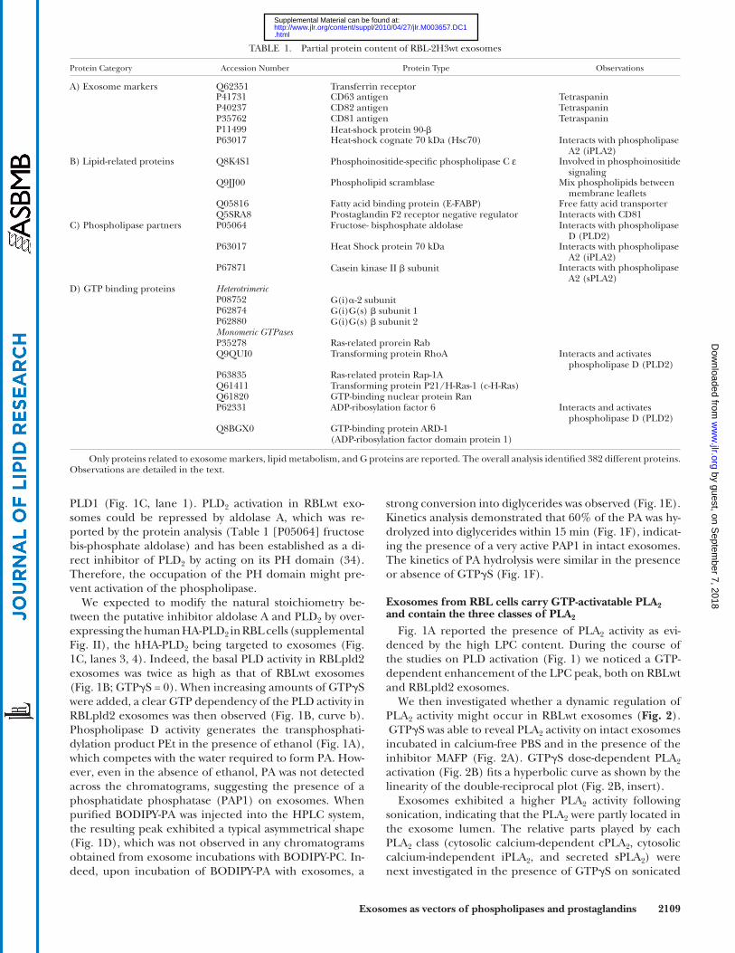
found in the present study are reported in Table 1 . Typical exosome markers, such as the transferring receptor, tet-raspanins (CD63, CD81, CD82), and heat shock proteins, were detected ( Table 1A ), assessing the quality of the preparation. Exosomes were also characterized by their size and their electronegativity (supplemental Fig. II).
Regarding lipid-related proteins, we found a phospho-lipid scramblase, a protein that transports phospholipids between the two membrane leafl ets, in both directions. The presence of this protein was consistent with the lack of membrane phospholipid asymmetry we reported ear-lier in RBL-derived exosomes ( 16 ). Also a member of the fatty acid binding proteins (E-FABP) was detected. FABPs constitute a multigene family of structurally homologous cytosolic proteins that bind and transport polyunsaturated fatty acids, such as arachidonic acid (AA) ( 31 ). Another type of protein was a prostaglandin F 2 receptor negative regulator, also called FPRP ( 32 ). FPRP associates with the PGF 2 � receptor, thereby reducing ligand binding ( 33 ). However, the PGF 2 receptor was not found in exosomes, and the presence of the FPRP protein might be better re-lated to its ability to form a tight complex with the tetra-span molecule CD81 ( 32 ).
Among the phospholipases, only phospholipase C ε hydro-lyzing phosphoinositides was detected ( Table 1B ). Note that proteins known to interact with phospholipases D and A2 were present ( Table 1C ). Fructose bisphosphate aldol-ase interacts directly with phospholipase D isoform PLD 2 and inhibits its activity ( 34 ). The exosomes contained ca-sein kinase II (cK2) that can phosphorylate PLD 2 ( 35 ) and can also interact with sPLA 2 -IIA ( 36 ), precisely one of the sPLA 2 isoforms we detected in the present work. Hsp 70, one of the typical exosome markers ( Table 1A ), has also been shown to interact with iPLA 2 ( 37 ).
Phospholipase C ε has been shown to be regulated by G proteins, either the subunits of heterotrimeric G proteins or monomeric GTPases ( 18 ), both being recovered in the exosomes ( Table 1D ). This prompted us to consider that GTPases could participate in the regulation of exosome lipolytic enzymes. Note that exosomes contained almost all members of the Ras superfamily GTPases [ARF, Rho, Rap, Rab, p21Ras, and Ran ( Table 1D )] except Cdc42 ( 38 ). Possible pathways connecting the Ras superfamily GTPases and phospholipases have been reported. The GTPases RhoA and Arf 6 ( Table 1D ) are direct activators of PLD 2 ( 39, 40 ) from rat or human origin ( 41 ).
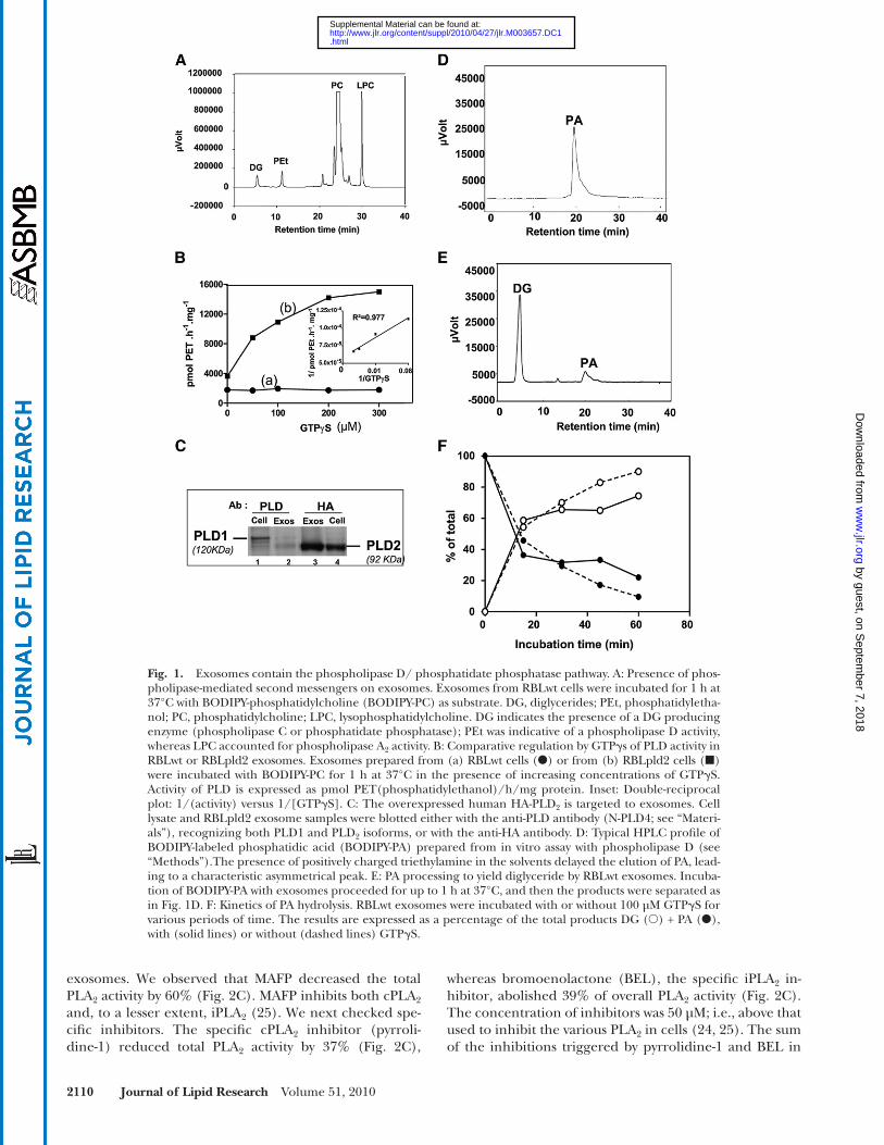
Exosomes contain the PLD/PAP pathway Fig. 1A reports the presence of DG, PEt, and LPC when
RBLwt exosomes were incubated with the fl uorescent and membrane-diffusible phosphatidylcholine. We investi-gated whether an autonomous regulation of the lipolytic enzymes involved in the production of these lipid media-tors could occur in exosomes. Addition of GTP � S up to 300 µM in RBLwt exosomes had no effect on the PLD ac-tivity ( Fig. 1B, curve a). The activity was not increased by exosome sonication. Immunodetection showed the selec-tive sorting of the PLD 2 isoform in exosomes ( Fig. 1C, lane 2) compared with the parental cells, containing mainly
group IIa and V recombinant sPLA 2 proteins as standards, and transferred onto PVDF membrane. Membranes were saturated in NETG buffer (150 mM NaCl, 5 mM EDTA, 50 mM Tris pH 7.4, 0.05% Triton X-100, 0.25% gelatin), washed in PBS 0.05% Tween 20, and incubated in NETG buffer with HRP-labeled anti-rabbit IgG secondary antibodies.
In all cases, the signal was detected by the enhanced chemilu-minescence system from GE Healthcare/Amersham.
Cyclooxygenase detection by fl ow cytometry. Exosomes (5 µg pro-teins) were bound on 4 µm beads (5 µl aldehyde sulfate latex beads; Invitrogen) for 1 h at room temperature under gentle shaking. Unoccupied sites were saturated with vesicle-free FCS for 1 h at room temperature. Beads were spun down, washed with PBS, and resuspended in FACS buffer. Bound exosomes were in-cubated with anti-COX-1 or anti-COX-2 primary antibodies, or control isotype for 1 h at room temperature and spun down, then labeled for 30 min with secondary FITC-labeled antibody. COX expression was analyzed by fl ow cytometry on the FITC channel (FL-1) of a FACSCalibur analyzer (Becton-Dickinson) using set-tings previously reported ( 16 ).
Protein analysis. High-throughput protein analysis was per-formed on 100 µg protein of purifi ed exosome, fi rst separated by one-dimensional SDS-PAGE. The protein gel lane was cut into 16 pieces, which were digested by trypsin. Tryptic peptides were ana-lyzed by nanoLC-MS-MS with a Qstar XL spectrometer (Applied Biosystems). Data were searched against mouse entries in Sprot-Trembl with the Mascot software. Analyses were performed at the IPBS, CNRS, Toulouse, France ( 27 ).
Lipid analysis. Determination of free fatty acids in exosomes was performed at the lipidomics facility of IFR-BMT (Institut Fédératif de Recherche Bio-Medicale de Toulouse), Toulouse, France. RBL-2H3 derived exosomes (100 µg) were incubated in PBS with 2 mM Ca 2+ /Mg 2+ at 37°C for 4 h. The lipids were extracted by the Bligh and Dyer method ( 28 ) in the presence of EGTA in water. Fatty acids were methylated and further analyzed by gas chromatography with an HP5890 instrument ( 29 ).
Prostaglandins were quantifi ed by GC-MS at the lipidomics fa-cility of IMBL/INSA-Lyon, Villeurbanne, France. Lipids from RBL-2H3 derived exosomes (70 µg) were extracted with ethylac-etate, derivatized into pentafl uorobenzyl esters, purifi ed by silica gel TLC using chloroform/ethanol (93:7, v/v), and then modi-fi ed into trimethylsilyl ethers before analysis by GC-MS. Samples were spiked with 10 ng of deuterated prostaglandin standards (Cayman) and GC-MS was carried out with a Hewlett Packard quadrupole mass spectrometer interfaced with a Hewlett Pack-ard gas chromatograph ( 30 ).
Data presentation. HPLC profi les and phospholipase determi-nations representative of at least two independent experiments were plotted. Pooled exosome preparations from two to three experiments were used for protein and lipid analysis, which ful-fi lled the quality control stipulations of the respective facilities (IPBS and IFR-BMT, Toulouse, France; IMBL/INSA-Lyon, Vil-leurbanne, France). Confocal pictures were representative of at least two experiments performed by distinct operators. Errors bars corresponded to SEM from three determinations.
RESULTS
Lipid-related proteins in exosomes To determine which of the diverse lipid-related proteins
were present on the exosomes, we fi rst performed an ex-haustive protein analysis of the vesicles. The proteins
by guest, on Septem
ber 7, 2018w
ww
.jlr.orgD
ownloaded from
.html http://www.jlr.org/content/suppl/2010/04/27/jlr.M003657.DC1Supplemental Material can be found at:
Exosomes as vectors of phospholipases and prostaglandins 2109
strong conversion into diglycerides was observed ( Fig. 1E ). Kinetics analysis demonstrated that 60% of the PA was hy-drolyzed into diglycerides within 15 min ( Fig. 1F ), indicat-ing the presence of a very active PAP1 in intact exosomes. The kinetics of PA hydrolysis were similar in the presence or absence of GTP � S ( Fig. 1F ).
Exosomes from RBL cells carry GTP-activatable PLA 2 and contain the three classes of PLA 2
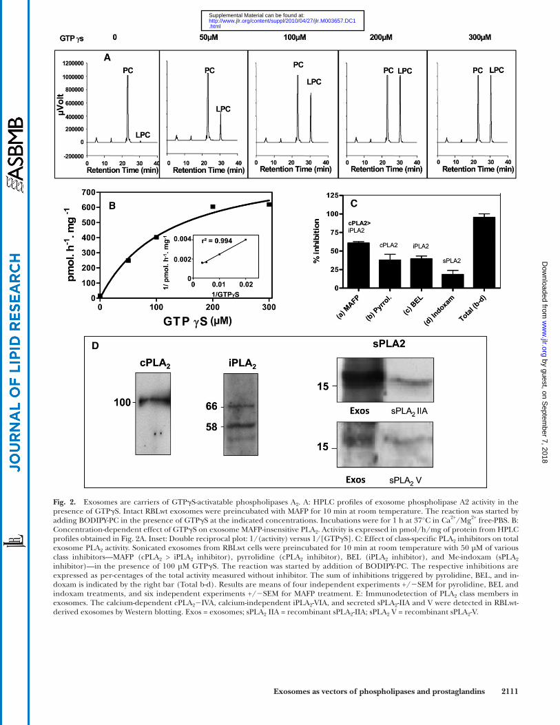
Fig. 1A reported the presence of PLA 2 activity as evi-denced by the high LPC content. During the course of the studies on PLD activation ( Fig. 1 ) we noticed a GTP-dependent enhancement of the LPC peak, both on RBLwt and RBLpld2 exosomes.
We then investigated whether a dynamic regulation of PLA 2 activity might occur in RBLwt exosomes ( Fig. 2 ). GTP � S was able to reveal PLA 2 activity on intact exosomes incubated in calcium-free PBS and in the presence of the inhibitor MAFP ( Fig. 2A ). GTP � S dose-dependent PLA 2 activation ( Fig. 2B ) fi ts a hyperbolic curve as shown by the linearity of the double-reciprocal plot ( Fig. 2B , insert).
Exosomes exhibited a higher PLA 2 activity following sonication, indicating that the PLA 2 were partly located in the exosome lumen. The relative parts played by each PLA 2 class (cytosolic calcium-dependent cPLA 2 , cytosolic calcium-independent iPLA 2 , and secreted sPLA 2 ) were next investigated in the presence of GTP � S on sonicated
PLD1 ( Fig. 1C, lane 1). PLD 2 activation in RBLwt exo-somes could be repressed by aldolase A, which was re-ported by the protein analysis ( Table 1 [P05064] fructose bis-phosphate aldolase) and has been established as a di-rect inhibitor of PLD 2 by acting on its PH domain ( 34 ). Therefore, the occupation of the PH domain might pre-vent activation of the phospholipase.
We expected to modify the natural stoichiometry be-tween the putative inhibitor aldolase A and PLD 2 by over-expressing the human HA-PLD 2 in RBL cells (supplemental Fig. II), the hHA-PLD 2 being targeted to exosomes ( Fig. 1C , lanes 3, 4). Indeed, the basal PLD activity in RBLpld2 exosomes was twice as high as that of RBLwt exosomes ( Fig. 1B ; GTP � S = 0). When increasing amounts of GTP � S were added, a clear GTP dependency of the PLD activity in RBLpld2 exosomes was then observed ( Fig. 1B, curve b). Phospholipase D activity generates the transphosphati-dylation product PEt in the presence of ethanol ( Fig. 1A ), which competes with the water required to form PA. How-ever, even in the absence of ethanol, PA was not detected across the chromatograms, suggesting the presence of a phosphatidate phosphatase (PAP1) on exosomes. When purifi ed BODIPY-PA was injected into the HPLC system, the resulting peak exhibited a typical asymmetrical shape ( Fig. 1D ), which was not observed in any chromatograms obtained from exosome incubations with BODIPY-PC. In-deed, upon incubation of BODIPY-PA with exosomes, a
TABLE 1. Partial protein content of RBL-2H3wt exosomes
Protein Category Accession Number Protein Type Observations
A) Exosome markers Q62351 Transferrin receptorP41731 CD63 antigen TetraspaninP40237 CD82 antigen TetraspaninP35762 CD81 antigen TetraspaninP11499 Heat-shock protein 90- � P63017 Heat-shock cognate 70 kDa (Hsc70) Interacts with phospholipase
A2 (iPLA2)B) Lipid-related proteins Q8K4S1 Phosphoinositide-specifi c phospholipase C ε Involved in phosphoinositide
signalingQ9JJ00 Phospholipid scramblase Mix phospholipids between
membrane leafl etsQ05816 Fatty acid binding protein (E-FABP) Free fatty acid transporterQ5SRA8 Prostaglandin F2 receptor negative regulator Interacts with CD81
C) Phospholipase partners P05064 Fructose- bisphosphate aldolase Interacts with phospholipase D (PLD2)
P63017 Heat Shock protein 70 kDa Interacts with phospholipase A2 (iPLA2)
P67871 Casein kinase II � subunit Interacts with phospholipase A2 (sPLA2)
D) GTP binding proteins Heterotrimeric P08752 G(i) � -2 subunitP62874 G(i)G(s) � subunit 1P62880 G(i)G(s) � subunit 2 Monomeric GTPases P35278 Ras-related prorein RabQ9QUI0 Transforming protein RhoA Interacts and activates
phospholipase D (PLD2)P63835 Ras-related protein Rap-1AQ61411 Transforming protein P21/H-Ras-1 (c-H-Ras)Q61820 GTP-binding nuclear protein RanP62331 ADP-ribosylation factor 6 Interacts and activates
phospholipase D (PLD2)Q8BGX0 GTP-binding protein ARD-1
(ADP-ribosylation factor domain protein 1)
Only proteins related to exosome markers, lipid metabolism, and G proteins are reported. The overall analysis identifi ed 382 different proteins. Observations are detailed in the text. by guest, on S
eptember 7, 2018
ww
w.jlr.org
Dow
nloaded from
.html http://www.jlr.org/content/suppl/2010/04/27/jlr.M003657.DC1Supplemental Material can be found at:
2110 Journal of Lipid Research Volume 51, 2010
whereas bromoenolactone (BEL), the specifi c iPLA 2 in-hibitor, abolished 39% of overall PLA 2 activity ( Fig. 2C ). The concentration of inhibitors was 50 µM; i.e., above that used to inhibit the various PLA 2 in cells ( 24, 25 ). The sum of the inhibitions triggered by pyrrolidine-1 and BEL in
exosomes. We observed that MAFP decreased the total PLA 2 activity by 60% ( Fig. 2C ). MAFP inhibits both cPLA 2 and, to a lesser extent, iPLA 2 ( 25 ). We next checked spe-cifi c inhibitors. The specifi c cPLA 2 inhibitor (pyrroli-dine-1) reduced total PLA 2 activity by 37% ( Fig. 2C ),
Fig. 1. Exosomes contain the phospholipase D/ phosphatidate phosphatase pathway. A: Presence of phos-pholipase-mediated second messengers on exosomes. Exosomes from RBLwt cells were incubated for 1 h at 37°C with BODIPY-phosphatidylcholine (BODIPY-PC) as substrate. DG, diglycerides; PEt, phosphatidyletha-nol; PC, phosphatidylcholine; LPC, lysophosphatidylcholine. DG indicates the presence of a DG producing enzyme (phospholipase C or phosphatidate phosphatase); PEt was indicative of a phospholipase D activity, whereas LPC accounted for phospholipase A 2 activity. B: Comparative regulation by GTP � s of PLD activity in RBLwt or RBLpld2 exosomes. Exosomes prepared from (a) RBLwt cells ( � ) or from (b) RBLpld2 cells (�)were incubated with BODIPY-PC for 1 h at 37°C in the presence of increasing concentrations of GTP � S. Activity of PLD is expressed as pmol PET(phosphatidylethanol)/h/mg protein. Inset: Double-reciprocal plot: 1/(activity) versus 1/[GTP � S]. C: The overexpressed human HA-PLD 2 is targeted to exosomes. Cell lysate and RBLpld2 exosome samples were blotted either with the anti-PLD antibody (N-PLD4; see “Materi-als”), recognizing both PLD1 and PLD 2 isoforms, or with the anti-HA antibody. D: Typical HPLC profi le of BODIPY-labeled phosphatidic acid (BODIPY-PA) prepared from in vitro assay with phospholipase D (see “Methods”).The presence of positively charged triethylamine in the solvents delayed the elution of PA, lead-ing to a characteristic asymmetrical peak. E: PA processing to yield diglyceride by RBLwt exosomes. Incuba-tion of BODIPY-PA with exosomes proceeded for up to 1 h at 37°C, and then the products were separated as in Fig. 1D. F: Kinetics of PA hydrolysis. RBLwt exosomes were incubated with or without 100 µM GTP � S for various periods of time. The results are expressed as a percentage of the total products DG ( � ) + PA ( � ), with (solid lines) or without (dashed lines) GTP � S.
by guest, on Septem
ber 7, 2018w
ww
.jlr.orgD
ownloaded from
.html http://www.jlr.org/content/suppl/2010/04/27/jlr.M003657.DC1Supplemental Material can be found at:
Exosomes as vectors of phospholipases and prostaglandins 2111
Fig. 2. Exosomes are carriers of GTP � S-activatable phospholipases A 2 . A: HPLC profi les of exosome phospholipase A2 activity in the presence of GTP � S. Intact RBLwt exosomes were preincubated with MAFP for 10 min at room temperature. The reaction was started by adding BODIPY-PC in the presence of GTP � S at the indicated concentrations. Incubations were for 1 h at 37°C in Ca 2+ /Mg 2+ free-PBS. B: Concentration-dependent effect of GTP � S on exosome MAFP-insensitive PLA 2 . Activity is expressed in pmol/h/mg of protein from HPLC profi les obtained in Fig. 2A. Inset: Double reciprocal plot: 1/(activity) versus 1/[GTP � S]. C: Effect of class-specifi c PLA 2 inhibitors on total exosome PLA 2 activity. Sonicated exosomes from RBLwt cells were preincubated for 10 min at room temperature with 50 µM of various class inhibitors—MAFP (cPLA 2 > iPLA 2 inhibitor), pyrrolidine (cPLA 2 inhibitor), BEL (iPLA 2 inhibitor), and Me-indoxam (sPLA 2 inhibitor)—in the presence of 100 µM GTP � S. The reaction was started by addition of BODIPY-PC. The respective inhibitions are expressed as per-centages of the total activity measured without inhibitor. The sum of inhibitions triggered by pyrolidine, BEL, and in-doxam is indicated by the right bar (Total b-d). Results are means of four independent experiments +/ � SEM for pyrolidine, BEL and indoxam treatments, and six independent experiments +/ � SEM for MAFP treatment. E: Immunodetection of PLA 2 class members in exosomes. The calcium-dependent cPLA 2 � IVA, calcium-independent iPLA 2 -VIA, and secreted sPLA 2 -IIA and V were detected in RBLwt-derived exosomes by Western blotting. Exos = exosomes; sPLA 2 IIA = recombinant sPLA 2 -IIA; sPLA 2 V = recombinant sPLA 2 -V.
by guest, on Septem
ber 7, 2018w
ww
.jlr.orgD
ownloaded from
.html http://www.jlr.org/content/suppl/2010/04/27/jlr.M003657.DC1Supplemental Material can be found at:
2112 Journal of Lipid Research Volume 51, 2010
Fig. 3. Exosomes carry free fatty acids and arachidonic acid-derived bioactive lipids. A–D: Fatty acid distribution in exosomes. Details for analysis are reported in “Methods.” E: Prostaglandin content of exosomes and parent cells. Prostaglandins were quantifi ed by GC-MS in untreated exosomes (Exos) or GTP � S-treated exosomes (Exos +GTP) and in parent cells (Cells) (RBLwt). GTP � S treatment was per-formed for 1 h at 37°C with 200 µM of the nucleotide. 15d-PGJ 2 = 15-deoxy- � 12,14 -PGJ 2 . PGF 2 � , PGE 2 , PGD 2 = prostaglandins F 2 � , E 2 , and D 2
by guest, on Septem
ber 7, 2018w
ww
.jlr.orgD
ownloaded from
.html http://www.jlr.org/content/suppl/2010/04/27/jlr.M003657.DC1Supplemental Material can be found at:
Exosomes as vectors of phospholipases and prostaglandins 2113
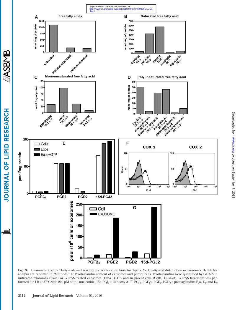
Only one member of the omega-3 series, namely, docosa-hexaenoic acid (DHA), was detected. Interestingly, AA ac-counted for about 30% of the total polyunsaturated fatty acids.
We next investigated whether bioactive lipids derived from AA could be found in exosomes. Quantifi cation of prostaglandins was performed by GC-MS and demon-strated the presence of mainly PGE 2 and 15-deoxy- � 12,14 -PGJ 2 [15d-PGJ 2 ] ( Fig. 3E ). The 15d-PGJ 2 was slightly enriched in the vesicles compared with the parent cells. The respective amounts of the various prostaglandins (PGF 2 � , PGE 2 , PGD 2 , and 15d-PGJ 2 ) in exosomes was not enhanced by incubation with GTP � S ( Fig. 3E ), indicating that the prostaglandins originated either from the basal exosome PLA 2 activities or were loaded in the exosome membrane at the time of their biogenesis in parent cells. Note that COX-1 and COX-2 involved in the early steps of prostaglandin biosynthesis were expressed in exosomes ( Fig. 3F ), indicating that exosomes could be autono-mous biological structures for the biosynthesis of the various prostaglandins. In that respect, the AA concentra-tion in exosome membrane (45 nmoles/mg protein; Fig. 3D ) was in excessive compared with the total membrane pros taglandin concentration (0.31 nmoles/mg protein; Fig. 3E ).
The number of exosome vesicles per unit protein was established and used to calculate the amount of prosta-glandin associated with a defi ned number of vesicles. Compared with the same number of parental cells, exo-somes carried from 12 to 15 times more prostaglandins ( Fig. 3G ). To our knowledge, this is the fi rst report of vesicle-associated release of prostaglandins from cells.
To evaluate the potential of exosomes as vehicles of bio-active lipids, we investigated whether exosomes could traf-fi c between RBL-2H3 cells.
Exosomes are internalized by resting and activated RBL-2H3 cells and concentrate into endosomes
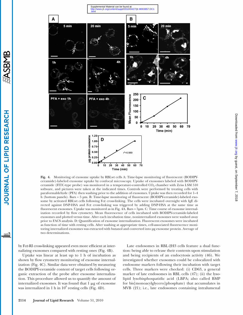
Confocal microscopy performed on living cells showed an accumulation of exosomes on the cell periphery detect-able as soon as 5 min, with subsequent internalization leading to the formation of intracellular aggregates indi-cating storage in an endosomal compartment, as observed after 1 h and 4 h ( Fig. 4A ). Exosome uptake was an active process, as cross-linking of peripheral protein on target cells by paraformaldehyde impaired intracellular exosome accumulation ( Fig. 4A , bottom panels). Only faint, diffuse cell labeling was observed in this case and was attributed to some exchange of the lipidic fl uorescent probe between exosomes and the target cell during the step of exosome interaction with the peripheral cell membrane. We investi-gated whether activated cells, which release exosomes, were also able to internalize them. RBL-2H3 cells activated
exosomes ( Fig. 2C ) led to the reduction of global PLA 2 activity by 76%, indicating that another type of PLA 2 activity was present. Me-indoxam, a specifi c inhibitor of secreted phospholipases ( 42 ) was checked, and it decreased total PLA 2 activity by 19% ( Fig. 2C ). Together, the cumulative effect of the three inhibitors diminished total PLA 2 activity by 95 ± 6.5%. The residual 5% activity might be related to PLA 2 insensitive to the inhibitors, such as some secreted sPLA 2 ( 43 ). Therefore, the three classes of PLA 2 contrib-uted to the global PLA 2 activity detected in the exosomes.
We next assessed the presence of members of the three PLA 2 classes by using specifi c antibodies ( Fig. 2D ). cPLA 2 -IVA was detected as a single band, whereas iPLA 2 -VIA was present as processed forms ( 44 ) The iPLA 2 -VIA can be cleaved at three different sites by caspase 3, which gener-ates various processed forms depending upon the combi-nation of the sites effectively cleaved ( 45 ). Fragmented forms of iPLA 2 , similar to those we reported in Fig. 2D, were observed in erythrocyte-derived exosomes ( 44 ). Pro-teolytic processing has been shown to enhance the iPLA 2 -VIA activity by removing part or complete ankyrin repeats suggested to function as a negative regulator ( 45 ). The form of 66 kDa we observed in Fig. 2D could correspond to the residual protein after caspase 3-mediated cleavage at the DVTD site of the iPLA 2 , leading to the release of the fi rst ankyrin repeat ( 45 ) and making likely that the iPLA 2 -VIA is highly active in exosomes. Among the third class of PLA 2 , namely, secreted sPLA 2 , the presence of sPLA 2 -IIA and sPLA 2 -V groups was observed ( Fig. 2D ). Therefore, RBL exosomes concentrated members of each of the three classes of PLA 2 and are thus a unique cell compartment.
Exosomes as carriers of bioactive lipids A large panel of free fatty acids was recovered from exo-
somes ( Fig. 3A ). Fatty acids could be carried from the pa-rental cells or directly generated within the exosomes by the phospholipase A 2 activities. We previously established that 1 mg of exosome protein contained 230 nmoles phos-pholipid [see Ref. ( 16 )]. Therefore 230 nmoles free fatty acid could be potentially released by the respective PLA 2 activities considering they displayed 100% effi ciency, with an additional amount of free fatty acid originating from the lysophospholipase activity borne by cPLA 2 and iPLA 2 . However, a total amount of 1,420 nmoles free fatty acid/mg exosome protein was measured ( Fig. 3A ), indicating that most of the exosome free fatty acid content was al-ready present at the time of exosome membrane biogen-esis. The chain length of the saturated fatty acids ranged from 14 to 24 carbons ( Fig. 3B ); the major ones were pal-mitic and stearic acids. Monounsaturated fatty acids were es-sentially from the omega-9 series ( Fig. 3C ); oleic acid was the most abundant. Polyunsaturated fatty acids almost exclu-sively contained members of the omega-6 series ( Fig. 3D ).
respectively. F: Exosomes contain cyclooxygenases 1 and 2. Analysis of exosome cyclooxygenase expression by fl ow cytometry, compared to control isotype (gray shaded curves). G: Comparative prostaglandin content of exosomes and parent cells. Prostaglandin content per mg protein plotted in Fig. 3E was converted into cell-equivalents or vesicle (exosome)-equivalents, and normalized to 10 6 cells or 10 6 exosome vesicles, respectively. 1 mg protein corresponded to (71.4 ± 0.54) × 10 5 cells and (5.96 ± 0.13) × 10 5 exosome vesicles.
by guest, on Septem
ber 7, 2018w
ww
.jlr.orgD
ownloaded from
.html http://www.jlr.org/content/suppl/2010/04/27/jlr.M003657.DC1Supplemental Material can be found at:
2114 Journal of Lipid Research Volume 51, 2010
Fig. 4. Monitoring of exosome uptake by RBLwt cells A: Time-lapse monitoring of fl uorescent (BODIPY-ceramide)-labeled exosome uptake by confocal microscopy. Uptake of exosomes labeled with BODIPY-ceramide (FITC-type probe) was monitored in a temperature-controlled CO 2 chamber with Zeiss LSM 510 software, and pictures were taken at the indicated times. Controls were performed by treating cells with paraformaldehyde (PFA) then washing prior to the addition of exosomes. Uptake was then recorded for 1–4 h (bottom panels). Bars = 5 µm. B: Time-lapse monitoring of fl uorescent (BODIPY-ceramide)-labeled exo-some by activated RBLwt cells following Fc ε cross-linking. The cells were incubated overnight with IgE di-rected against DNP-HSA and Fc ε cross-linking was triggered by adding DNP-HSA at the same time as fl uorescent exosomes. Uptake was monitored as in Fig. 4A. Bars = 5µm. C: Time course of exosome internal-ization recorded by fl ow cytometry. Mean fl uorescence of cells incubated with BODIPY-ceramide-labeled exosomes and plotted versus time. After each incubation time, noninternalized exosomes were washed away prior to FACS analysis. D: Quantifi cation of exosome internalization. Fluorescent exosomes were incubated as function of time with resting cells. After washing at appropriate times, cell-associated fl uorescence moni-toring internalized exosomes was extracted with butanol and converted into µg exosome protein. Average of two determinations.
by Fc ε -RI cross-linking appeared even more effi cient at inter-nalizing exosomes compared with resting ones ( Fig. 4B ).
Uptake was linear at least up to 1 h of incubation as shown by fl ow cytometry monitoring of exosome internal-ization ( Fig. 4C ). Similar data were obtained by measuring the BODIPY-ceramide content of target cells following or-ganic extraction of the probe after exosome internaliza-tion. This procedure allowed us to quantify the amount of internalized exosomes. It was found that 1 � g of exosome was internalized in 1 h in 10 6 resting cells ( Fig. 4D ).
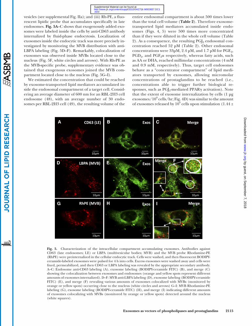
Late endosomes in RBL-2H3 cells feature a dual func-tion: being able to release their contents upon stimulation and being recipients of an endocytosis activity ( 46 ). We investigated whether exosomes could be colocalized with endosome markers following their incubation with target cells. Three markers were checked: (i) CD63, a general marker of late endosomes in RBL cells ( 47 ); (ii) the lyso-lipid lysobisphospatidic acid (LBPA; also called BMP for bis[monoacylglycero]phosphate) that accumulates in MVB ( 21 ); i.e., late endosomes containing intralumenal
by guest, on Septem
ber 7, 2018w
ww
.jlr.orgD
ownloaded from
.html http://www.jlr.org/content/suppl/2010/04/27/jlr.M003657.DC1Supplemental Material can be found at:
Exosomes as vectors of phospholipases and prostaglandins 2115
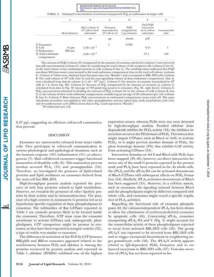
entire endosomal compartment is about 500 times lower than the total cell volume ( Table 2 ). Therefore exosome-transported lipid mediators accumulated inside endo-somes ( Figs. 4, 5 ) were 500 times more concentrated than if they were diluted in the whole cell volume ( Table 2 ). As a consequence, the resulting PGJ 2 endosomal con-centration reached 52 µM ( Table 2 ). Other endosomal concentrations were 33µM, 2.4 µM, and 1.7 µM for PGE 2 , PGD 2 , and PGF 2 � respectively, whereas fatty acids, such as AA or DHA, reached millimolar concentrations (4 mM and 0.9 mM, respectively). Thus, target cell endosomes behave as a “concentrator compartment” of lipid medi-ators transported by exosomes, allowing micromolar concentrations of prostaglandins to be reached (i.e., concentrations able to trigger further biological re-sponses, such as PGJ 2 -mediated PPAR � activation). Note that the extent of exosome internalization by cells (1 µg exosomes/10 6 cells/hr; Fig. 4D ) was similar to the amount of exosomes released by 10 6 cells upon stimulation (1.44 ±
Fig. 5. Characterization of the intracellular compartment accumulating exosomes. Antibodies against CD63 (late endosomes; LE) or LBPA (multivesicular bodies; MVB) and the MVB probe Rhodamine-PE (RhPE) were preinternalized in the cellular endocytic track. Cells were washed, and then fl uorescent BODIPY-ceramide-labeled exosomes were pulsed for 4 h into cells. Excess exosomes were washed away and cells were fi xed, permeabilized, and then CD63 or LBPA labeling was revealed by the appropriate secondary antibody. A–C: Endosome anti-CD63 labeling (A), exosome labeling (BODIPY-ceramide FITC) (B), and merge (C) showing the colocalization between exosomes and endosomes (orange and yellow spots represent different amounts of exosomes internalized). D–F: MVB anti-LBPA labeling (D), exosome labeling (BODIPY-ceramide FITC) (E), and merge (F) revealing various amounts of exosomes colocalized with MVBs (monitored by orange or yellow spots) occurring close to the nucleus (white circles and arrows) G–I: MVB Rhodamine-PE labeling (G), exosome labeling (BODIPY-ceramide FITC) (H), and merge (I) indicating different amounts of exosomes colocalizing with MVBs (monitored by orange or yellow spots) detected around the nucleus (white squares).
vesicles (see supplemental Fig. IIa); and (iii) Rh-PE, a fl uo-rescent lipidic probe that accumulates specifi cally in late endosomes. Fig. 5A –C shows that exogenously added exo-somes were labeled inside the cells by anti-CD63 antibody internalized by fl uid-phase endocytosis. Localization of exosomes inside the endocytic track was more precisely in-vestigated by monitoring the MVB distribution with anti-LBPA labeling (Fig. 5D–F). Remarkably, colocalization of exosomes was observed inside MVBs located close to the nucleus ( Fig. 5F , white circles and arrows). With Rh-PE as the MVB-specifi c probe, supplementary evidence was ob-tained that exogenous exosomes joined the MVB com-partment located close to the nucleus ( Fig. 5G–I ).
We estimated the concentration that could be reached by exosome-transported lipid mediators accumulated in-side the endosomal compartment of a target cell. Consid-ering an average diameter of 600 nm for an RBL-2H3 cell endosome ( 48 ), with an average number of 30 endo-somes per RBL-2H3 cell ( 49 ), the resulting volume of the
by guest, on Septem
ber 7, 2018w
ww
.jlr.orgD
ownloaded from
.html http://www.jlr.org/content/suppl/2010/04/27/jlr.M003657.DC1Supplemental Material can be found at:
2116 Journal of Lipid Research Volume 51, 2010
expression scores, whereas PLDs were not even detected by high-throughput analysis. Purifi ed aldolase dose-dependently inhibits the PLD 2 activity ( 34 ); the inhibitor in-teraction occurs at the PH domain of PLD 2. This interaction might impair GTPases, such as RhoA or Arf6, to activate PLD 2 , or it might prevent another domain of PLD 2 , the phox homology domain (PX) that exhibits GAP activity, from activating GTPases ( 53 ).
Interaction domains on Arf 6 and RhoA with PLD 2 have been mapped ( 39, 40 ); however, no direct interaction be-tween any of the small G proteins reported in the present study and PLA 2 have been reported so far. In whole cells, the cPLA 2 and the sPLA 2 -IIA can be activated downstream of RhoA GTPases with subsequent effects on PGE 2 forma-tion ( 54 ). Similarly, iPLA 2 activation downstream of RhoA has been suggested ( 55 ). However, in a cell-free system, such as exosomes, the signaling network between RhoA and the phospholipases might be different compared with whole cells, and exosomes might reveal a specifi c regula-tion of PLA 2 activities.
Regarding the functional role of exosome phospoli-pases A2, the calcium-independent iPLA 2 has been shown to allow the elimination of erythrocyte-derived exosomes by apoptotic cells ( 44 ). Concerning sPLA 2 , exosomes transporting sPLA 2 IIA and V ( Fig. 3 ) might account for the transcellular activity of these phospholipases reported to occur from activated RBL-2H3 cells ( 56 ). The group sPLA 2 -V was reported to be secreted from RBL-2H3 cells and to trigger eicosanoid biosynthesis in neighboring tar-get granulocytic cells ( 56 ). The sPLA 2 -V activity appears related to IgE-dependent PGD 2 formation and to en-hanced exocytosis in RBL-2H3 cells ( 57 ). Vesicular secre-tion of cPLA 2 has not been reported so far.
0.47 µg), suggesting an effi cient cell-to-cell communica-tion process.
DISCUSSION
Exosomes are nanovesicles released from intact viable cells. They participate in cell-to-cell communication in various physiological and pathological situations, such as the immune response ( 50 ), infl ammation ( 51 ), or athero-genesis ( 7 ). Mast cell-derived exosomes trigger functional maturation of dendritic cells ( 6 ). This maturation process involves secreted PLA 2 ( 13 ) and prostaglandins ( 14 ). Therefore, we investigated the presence of lipid-related proteins and lipid mediators on exosomes derived from the mast cell line RBL-2H3.
High-throughput protein analysis reported the pres-ence of only four proteins related to lipid metabolism. However, we revealed the presence of other lipolytic pro-teins by their activity and by immunodetection. The pres-ence of a high content in monomeric G proteins led us to hypothesize specifi c regulation of these phospholipases in exosomes. The subfamilies of Ras GTPases reported in Table 1 are cytosolic proteins likely to be located inside the exosomes. Therefore, GTP must cross the exosome membrane to activate GTPases and, subsequently, phos-pholipases. GTP transporters might be present in exo-somes, as they have been reported in synaptic vesicles ( 52 ), a type of vesicle very similar to exosomes.
The difference in activation of the PLD by GTP between RBLpld2 and RBLwt exosomes appeared related to the stoichiometry between PLD 2 and aldolase A. Among the proteins recovered by protein analysis and reported in Table 1 , aldolase (P05064) exhibited one of the highest
TABLE 2. Estimated concentration of exosome-transported PGJ 2 in endosomes of target cells
A B C D E F
Mean diameter
Mean volume of 10 6 cells or their
endosomes
Exosomes internalized in 10 6 cells in 1 h
PGJ2 transported by 1 µg exosomes
Final PGJ2 concentrati
on in cells or endosomes
Concentration ratio
ml µg pmoles µM
1) Exosomes 1 0.1932) Cell 15 µm 1.76 × 10 � 3 0.11 13) Endosomes 600 nm4) Total endosome
compartment3.38 × 10 � 6 57.1 519
The amount of PGJ2 (column D) transported by the amount of exosomes plotted in column C was converted into µM concentrations (column E) either by considering the total volume of the recipient cells (column E line 2), or the total volume of late endosomes present in cells (column E line 4). The resulting values indicated that PGJ2 was about 500 times more concentrated in the total endosome compartment than in the total cell volume (column F). Column A: Values were obtained from literature data (see “Results”) and correspond to RBL-2H3 cells. Column B: The total volume of 10 6 cells (line 2) and the corresponding volume of their endosome compartment (line 4) were calculated from data in column A (1 ml = 10 12 µm 3 ). Column C: Net amount of exosome internalized in 10 6 cells in 1 h (from Fig. 4D ). Column D: Amount of PGJ 2 transported by the amount of exosome in column C; calculated from data in Fig. 3E [average of 193 pmol/mg protein in exosomes ( Fig. 3E, right bars)]. Column E: PGJ 2 concentrations obtained by dividing the amount of PGJ 2 (column D) by the volume of cells (column B, line 2) or the volume of their total endosome compartment, considering an average of 30 endosomes per cell (column B, line 4). Column F: Ratio between PGJ 2 concentrations in endosomal compartment versus total cell. The same calculation procedure was applied to the other prostaglandins and two typical fatty acids [arachidonic acid (AA) and docosahexaenoic acid (DHA)] from data in Fig. 3 and reported in “Results.”
a 30 endosomes per cell.
by guest, on Septem
ber 7, 2018w
ww
.jlr.orgD
ownloaded from
.html http://www.jlr.org/content/suppl/2010/04/27/jlr.M003657.DC1Supplemental Material can be found at:
Exosomes as vectors of phospholipases and prostaglandins 2117
zymes, such as COX-1 and COX-2 ( Fig. 3F ), for the early steps of prostaglandin biosynthesis, exosomes could ac-count for transcellular metabolism of prostanoids reported to occur between normal and tumor cells ( 59 ). Arachi-donic acid present in exosomes ( Fig. 3D ) would serve as transcellular biosynthetic precursor. Although eicosanoid transcellular metabolism has been reported to occur at in-
The set of prostaglandins transported by exosomes ( Fig. 3 ) are derived from PGH2, and metabolic conversion of PGH2 has been shown to occur through a transcellular mechanism between two different types of cells, contain-ing either COX-1 and COX-2 or the terminal prostaglan-din synthases ( 58, 59 ). Transporting from cell to cell metabolic precursors, such as PGD 2 ( Fig. 3G ), and en-
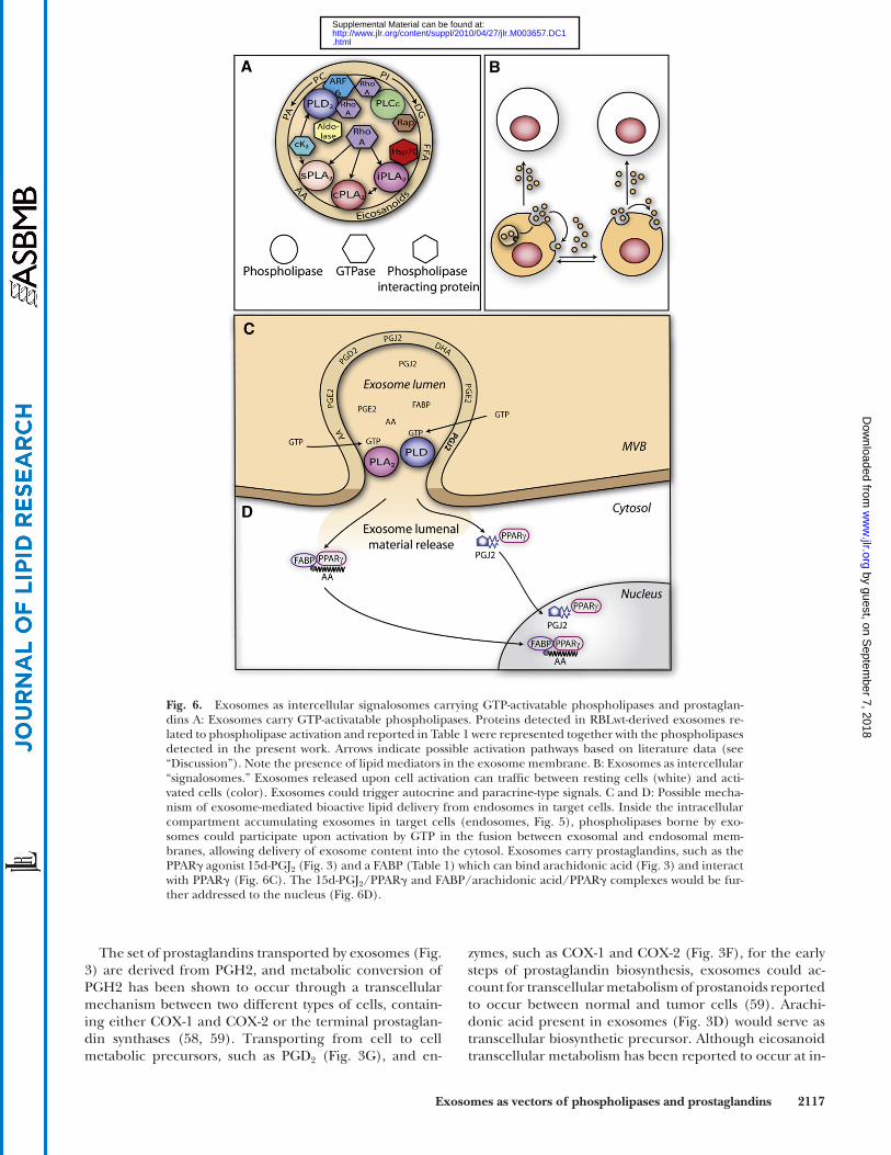
Fig. 6. Exosomes as intercellular signalosomes carrying GTP-activatable phospholipases and prostaglan-dins A: Exosomes carry GTP-activatable phospholipases. Proteins detected in RBLwt-derived exosomes re-lated to phospholipase activation and reported in Table 1 were represented together with the phospholipases detected in the present work. Arrows indicate possible activation pathways based on literature data (see “Discussion”). Note the presence of lipid mediators in the exosome membrane. B: Exosomes as intercellular “signalosomes.” Exosomes released upon cell activation can traffi c between resting cells (white) and acti-vated cells (color). Exosomes could trigger autocrine and paracrine-type signals. C and D: Possible mecha-nism of exosome-mediated bioactive lipid delivery from endosomes in target cells. Inside the intracellular compartment accumulating exosomes in target cells (endosomes, Fig. 5 ), phospholipases borne by exo-somes could participate upon activation by GTP in the fusion between exosomal and endosomal mem-branes, allowing delivery of exosome content into the cytosol. Exosomes carry prostaglandins, such as the PPAR � agonist 15d-PGJ 2 ( Fig. 3 ) and a FABP ( Table 1 ) which can bind arachidonic acid ( Fig. 3 ) and interact with PPAR � (Fig. 6C). The 15d-PGJ 2 /PPAR � and FABP/arachidonic acid/PPAR � complexes would be fur-ther addressed to the nucleus (Fig. 6D).
by guest, on Septem
ber 7, 2018w
ww
.jlr.orgD
ownloaded from
.html http://www.jlr.org/content/suppl/2010/04/27/jlr.M003657.DC1Supplemental Material can be found at:
2118 Journal of Lipid Research Volume 51, 2010
Exosomes could also supply the 15d-PGJ 2 already bound to its receptor, as a recent report indicates the presence of the PPAR � receptor among proteins found in exosomes iso-lated from human serum ( 71 ). Interestingly, the exosome FABP we report in Table1 could bind the AA present in exosomes ( Fig. 3 ) and then interact directly with the PPAR � receptor, the resulting FABP-AA-PPAR � complex being subsequently addressed to the nucleus of target cells to reg-ulate transcription ( Fig. 6C ) ( 72 ). In line with the possible modulation of nuclear receptors by exosome-carried medi-ators, note that PAP1, the diglyceride-generating enzyme we reported in Fig. 1 , has recently been characterized as a transcriptional coactivator of the PPAR � receptor ( 73 ).
Further experiments are required to support the func-tional role of exosomes in RBL-2H3 cells. As a fi rst step, we evaluated whether exosomes could carry suffi cient amounts of the prostaglandin 15d-PGJ 2 to possibly trigger PPAR � activation in target cells. When added to cells, 15d-PGJ 2 has been reported to trigger biological effects in the 10–40 µM range ( 74 ). Exosome accumulation in endo-somes were allowed to reach values > 50 µM ( Table 2 ); i.e., bioactive 15d-PGJ 2 concentrations.
Because of the dynamic regulation of their phospholi-pases by GTP, exosomes appear to behave as “signalo-somes” ( Fig. 6A ). The “signalosomes” would circulate between cells and might regulate their functions whether cells are resting or activated ( Fig. 6B ). Stimulated RBL-2H3 cells feature enhanced endocytosis ( 46 ) and could internalize exosomes they had just released. Preliminary data we obtained indicate that exosomes inhibited Fc ε -mediated degranulation of RBL-2H3 cells. That this effect involves PGE 2 , which is known to inhibit Fc ε RI-mediated exocytosis of mast cells ( 70 ), appears conceivable on the basis of data reported here. Also, by possibly providing 15d-PGJ 2 to PPAR � of target cells, exosomes can repress the transcription of proinfl ammatory mRNAs ( 75 ). Circu-lating simultaneously with allergens that activate cells via Fc ε RI receptors, exosomes appear as a signaling device able to modulate the Fc ε RI-mediated mast cell response by means of phospholipases and lipid mediators that can be activated.
The authors thank Justine Bertrand-Michel (Toulouse) and Michel Guichardant (Lyon) for lipidomics analysis; Bruno Payré for performing the transmission electronic microscopy; P.Winterton for correcting the English manuscript; and Dr. Toshihide Kobayashi (Riken Institute, Tokyo, Japan) for supplying the anti-LBPA antibody.
REFERENCES
1 . Thery , C. , S. Amigorena , G. Raposo , and A. Clayton . 2006 . Isolation and characterization of exosomes from cell culture supernatants and biological fl uids. Curr. Protoc. Cell Biol. Chapter 3: Unit 3.22.
2 . Raposo , G. , H. W. Nijman , W. Stoorvogel , R. Liejendekker , C. V. Harding , C. J. Melief , and H. J. Geuze . 1996 . B lymphocytes secrete antigen-presenting vesicles. J. Exp. Med. 183 : 1161 – 1172 .
3 . Hess , C. , S. Sadallah , A. Hefti , R. Landmann , and J. A. Schifferli . 1999 . Ectosomes released by human neutrophils are specialized functional units. J. Immunol. 163 : 4564 – 4573 .
fl ammation sites between different cell types ( 60 ), one can conceive that RBL-derived exosomes are a mixed popula-tion bearing either the COX-1 and COX-2 or the terminal prostaglandin synthases; therefore, exosome exchange be-tween RBL-2H3 cells would be required to complete the entire prostanoid biosynthesis pathway. In this respect, we showed earlier that RBL-2H3 cells release three distinct subpopulations of exosomes ( 23 ). The present work opens further investigations to understand the mechanisms un-derlying the transcellular metabolism of eicosanoids.
This transcellular metabolism requires exosome traf-fi cking between cells. We have shown that exosomes added to target cells are rapidly internalized ( Fig. 4 ) into the en-docytic track and join the MVB network located close to the nucleus ( Fig. 5 ). It is likely that the GTP-dependent activation of PLD and PLA 2 we observed in exosomes could occur inside the endocytic track of target cells. Many GTPases are present within the endocytosis track, some of them maintained in an active state even in unstimulated cells ( 61 ). GTP-activated phospholipases could participate in exosome fusion with the limiting membrane of the en-dosome, a process called “back-fusion” ( 62, 63 ). This pro-cess allows the lumen content of the exosomes to be released into the cytosol. Back-fusion molecular mecha-nisms require the lipid LBPA, whose biosynthesis involves a cPLA 2 -type activity ( 64, 65 ), as well as a combination of PLA 2 and PLD activities ( 66 ). A previous report describes GTP-dependent cPLA 2 -mediated fusion of secretory gran-ules ( 67 ). Phosphatidic acid resulting from PLD activity is a fusogenic compound in presence of calcium ( 68 ). Di-glycerides generated by the PI-PLCε ( Table 1 ) or the PLD/PA phosphatase pathway ( Fig. 1 ) could participate in exo-some-endosome fusion processes by lowering the surface pressure of the phospholipids ( 69 ). More DG can be ex-pected in RBLpld2 exosomes and could account for the modifi cation of the biophysical parameters (size and elec-tronegativity) shown in supplemental Fig. II. In addition, phospholipid mixing between exosome and endosome membranes triggered by the scramblase we reported in Table 1 would facilitate membrane fusion.
We established in this work that exosomes transport prostaglandins from the parent cells. RBL-2H3 cells fea-ture a mast cell phenotype, and eicosanoids play an essen-tial role in mast cell physiology by regulating their function in host defense and disease ( 70 ). PGE 2 can block Fc ε RI-mediated exocytosis of mast cells ( 70 ). Exosomes, during at least the fi rst 5–20 min ( Fig. 5 ), provide a vehicle for PGE 2 to interact with its respective GPCRs on the periph-ery of target cells. Thereafter, exosome internalization provides the fi rst mechanism described for 15deoxy � 12,14 -PGJ 2 to enter the cells and possibly reach its intracellular targets. Actually, no specifi c peripheral receptors or mech-anisms of entry have been identifi ed to-date for this pros-taglandin ( 17, 19 ). The exosome as a vehicle would allow the plasma membrane to be bypassed and 15d-PGJ 2 to ac-cumulate in the endosomes of target cells, from where the prostaglandin would be released into the cytosol after fu-sion between exosome and endosome membranes. A pos-sible mechanism is summarized in Fig. 6 .
by guest, on Septem
ber 7, 2018w
ww
.jlr.orgD
ownloaded from
.html http://www.jlr.org/content/suppl/2010/04/27/jlr.M003657.DC1Supplemental Material can be found at:
Exosomes as vectors of phospholipases and prostaglandins 2119
2008 . Group IVA phospholipase A2 is necessary for the biogenesis of lipid droplets. J. Biol. Chem. 283 : 27369 – 27382 .
26 . Gayral , S. , P. Deleris , K. Laulagnier , M. Laffargue , J. P. Salles , B. Perret , M. Record , and M. Breton-Douillon . 2006 . Selective activa-tion of nuclear phospholipase D-1 by g protein-coupled receptor agonists in vascular smooth muscle cells. Circ. Res. 99 : 132 – 139 .
27 . Bouyssie , D. , A. Gonzalez de Peredo , E. Mouton , R. Albigot , L. Roussel , N. Ortega , C. Cayrol , O. Burlet-Schiltz , J. P. Girard , and B. Monsarrat . 2007 . Mascot fi le parsing and quantifi cation (MFPaQ), a new software to parse, validate, and quantify proteomics data gen-erated by ICAT and SILAC mass spectrometric analyses: applica-tion to the proteomics study of membrane proteins from primary human endothelial cells. Mol. Cell. Proteomics . 6 : 1621 – 1637 .
28 . Bligh , E. G. , and W. J. Dyer . 1959 . A rapid method of total lipid extraction and purifi cation. Can. J. Biochem. Physiol. 37 : 911 – 917 .
29 . Payre , B. , P. de Medina , N. Boubekeur , L. Mhamdi , J. Bertrand-Michel , F. Terce , I. Fourquaux , D. Goudouneche , M. Record , M. Poirot , et al . 2008 . Microsomal antiestrogen-binding site ligands induce growth control and differentiation of human breast can-cer cells through the modulation of cholesterol metabolism. Mol. Cancer Ther. 7 : 3707 – 3718 .
30 . Soares , A. F. , O. Nosjean , D. Cozzone , D. D’Orazio , M. Becchi , M. Guichardant , G. Ferry , J. A. Boutin , M. Lagarde , and A. Geloen . 2005 . Covalent binding of 15-deoxy-delta12,14-prostaglandin J2 to PPARgamma. Biochem. Biophys. Res. Commun. 337 : 521 – 525 .
31 . Kitanaka , N. , Y. Owada , R. Okuyama , H. Sakagami , M. R. Nourani , S. Aiba , H. Furukawa , M. Watanabe , M. Ono , T. Ohteki , et al . 2006 . Epidermal-type fatty acid binding protein as a negative regulator of IL-12 production in dendritic cells. Biochem. Biophys. Res. Commun. 345 : 459 – 466 .
32 . Stipp , C. S. , D. Orlicky , and M. E. Hemler . 2001 . FPRP, a major, highly stoichiometric, highly specifi c CD81- and CD9-associated protein. J. Biol. Chem. 276 : 4853 – 4862 .
33 . Orlicky , D. J. , R. Berry , and J. M. Sikela . 1996 . Human chromosome 1 localization of the gene for a prostaglandin F2alpha receptor neg-ative regulatory protein. Hum. Genet. 97 : 655 – 658 .
34 . Kim , J. H. , S. Lee , T. G. Lee , M. Hirata , P. G. Suh , and S. H. Ryu . 2002 . Phospholipase D2 directly interacts with aldolase via Its PH domain. Biochemistry . 41 : 3414 – 3421 .
35 . Ganley , I. G. , S. J. Walker , M. Manifava , D. Li , H. A. Brown , and N. T. Ktistakis . 2001 . Interaction of phospholipase D1 with a casein-kinase-2-like serine kinase. Biochem. J. 354 : 369 – 378 .
36 . Shimoyama , Y. , R. Sakamoto , T. Akaboshi , M. Tanaka , and K. Ohtsuki . 2001 . Characterization of secretory type IIA phospholi-pase A2 (sPLA2-IIA) as a glycyrrhizin (GL)-binding protein and the GL-induced inhibition of the CK-II-mediated stimulation of sPLA2-IIA activity in vitro. Biol. Pharm. Bull. 24 : 1004 – 1008 .
37 . Mancuso , D. J. , C. M. Jenkins , and R. W. Gross . 2000 . The genomic organization, complete mRNA sequence, cloning, and expression of a novel human intracellular membrane-associated calcium-independent phospholipase A(2). J. Biol. Chem. 275 : 9937 – 9945 .
38 . Konstantinopoulos , P. A. , M. V. Karamouzis , and A. G. Papavassiliou . 2007 . Post-translational modifi cations and regulation of the RAS superfamily of GTPases as anticancer targets. Nat. Rev. Drug Discov. 6 : 541 – 555 .
39 . Bae , C. D. , D. S. Min , I. N. Fleming , and J. H. Exton . 1998 . Determination of interaction sites on the small G protein RhoA for phospholipase D. J. Biol. Chem. 273 : 11596 – 11604 .
40 . Hiroyama , M. , and J. H. Exton . 2005 . Localization and regulation of phospholipase D2 by ARF6. J. Cell. Biochem. 95 : 149 – 164 .
41 . Le Stunff , H. , L. Dokhac , S. Bourgoin , M. F. Bader , and S. Harbon . 2000 . Phospholipase D in rat myometrium: occurrence of a membrane-bound ARF6 (ADP-ribosylation factor 6)-regulated activ-ity controlled by betagamma subunits of heterotrimeric G-proteins. Biochem. J. 352 : 491 – 499 .
42 . Lambeau , G. , and M. H. Gelb . 2008 . Biochemistry and physiology of mammalian secreted phospholipases A2. Annu. Rev. Biochem. 77 : 495 – 520 .
43 . Singer , A. G. , F. Ghomashchi , C. Le Calvez , J. Bollinger , S. Bezzine , M. Rouault , M. Sadilek , E. Nguyen , M. Lazdunski , G. Lambeau , et al . 2002 . Interfacial kinetic and binding properties of the com-plete set of human and mouse groups I, II, V, X, and XII secreted phospholipases A2. J. Biol. Chem. 277 : 48535 – 48549 .
44 . Blanc , L. , C. Barres , P. Bette-Bobillo , and M. Vidal . 2007 . Reticulocyte-secreted exosomes bind natural IgM antibodies: in-volvement of a ROS-activatable endosomal phospholipase iPLA2. Blood . 110 : 3407 – 3416 .
4 . Werner , N. , S. Wassmann , P. Ahlers , S. Kosiol , and G. Nickenig . 2006 . Circulating CD31+/annexin V+ apoptotic microparticles cor-relate with coronary endothelial function in patients with coronary artery disease. Arterioscler. Thromb. Vasc. Biol. 26 : 112 – 116 .
5 . Valadi , H. , K. Ekstrom , A. Bossios , M. Sjostrand , J. J. Lee , and J. O. Lotvall . 2007 . Exosome-mediated transfer of mRNAs and micro-RNAs is a novel mechanism of genetic exchange between cells. Nat. Cell Biol. 9 : 654 – 659 .
6 . Skokos , D. , H. G. Botros , C. Demeure , J. Morin , R. Peronet , G. Birkenmeier , S. Boudaly , and S. Mecheri . 2003 . Mast cell-derived exosomes induce phenotypic and functional maturation of den-dritic cells and elicit specifi c immune responses in vivo. J. Immunol. 170 : 3037 – 3045 .
7 . Zakharova , L. , M. Svetlova , and A. F. Fomina . 2007 . T cell exosomes induce cholesterol accumulation in human monocytes via phos-phatidylserine receptor. J. Cell. Physiol. 212 : 174 – 181 .
8 . Miyanishi , M. , K. Tada , M. Koike , Y. Uchiyama , T. Kitamura , and S. Nagata . 2007 . Identifi cation of Tim4 as a phosphatidylserine recep-tor. Nature . 450 : 435 – 439 .
9 . Ristorcelli , E. , E. Beraud , S. Mathieu , D. Lombardo , and A. Verine . 2009 . Essential role of Notch signaling in apoptosis of human pan-creatic tumoral cells mediated by exosomal nanoparticles. Int. J. Cancer . 125 : 1016 – 1026 .
10 . Alais , S. , S. Simoes , D. Baas , S. Lehmann , G. Raposo , J. L. Darlix , and P. Leblanc . 2008 . Mouse neuroblastoma cells release prion in-fectivity associated with exosomal vesicles. Biol. Cell . 100 : 603 – 615 .
11 . Schorey , J. S. , and S. Bhatnagar . 2008 . Exosome function: from tu-mor immunology to pathogen biology. Traffi c . 9 : 871 – 881 .
12 . Sharples , R. A. , L. J. Vella , R. M. Nisbet , R. Naylor , K. Perez , K. J. Barnham , C. L. Masters , and A. F. Hill . 2008 . Inhibition of gamma-secretase causes increased secretion of amyloid precursor protein C-terminal fragments in association with exosomes. FASEB J. 22 : 1469 – 1478 .
13 . Perrin-Cocon , L. , S. Agaugue , F. Coutant , A. Masurel , S. Bezzine , G. Lambeau , P. Andre , and V. Lotteau . 2004 . Secretory phospho-lipase A2 induces dendritic cell maturation. Eur. J. Immunol. 34 : 2293 – 2302 .
14 . Thurnher , M. 2007 . Lipids in dendritic cell biology: messengers, effectors, and antigens. J. Leukoc. Biol. 81 : 154 – 160 .
15 . Laulagnier , K. , C. Motta , S. Hamdi , S. Roy , F. Fauvelle , J. F. Pageaux , T. Kobayashi , J. P. Salles , B. Perret , C. Bonnerot , et al . 2004 . Mast cell- and dendritic cell-derived exosomes display a specifi c lipid composition and an unusual membrane organization. Biochem. J. 380 : 161 – 171 .
16 . Laulagnier , K. , D. Grand , A. Dujardin , S. Hamdi , H. Vincent-Schneider , D. Lankar , J. P. Salles , C. Bonnerot , B. Perret , and M. Record . 2004 . PLD2 is enriched on exosomes and its activity is cor-related to the release of exosomes. FEBS Lett. 572 : 11 – 14 .
17 . Scher , J. U. , and M. H. Pillinger . 2005 . 15d-PGJ2: the anti-infl am-matory prostaglandin? Clin. Immunol. 114 : 100 – 109 .
18 . Gandarillas , N. L. , T. D. Bunney , M. B. Josephs , P. Gierschik , and M. Katan . 2009 . In vitro reconstitution of activation of PLCepsilon by Ras and Rho GTPases. Methods Mol. Biol. 462 : 379 – 389 .
19 . Scher , J. U. , and M. H. Pillinger . 2009 . The anti-infl ammatory ef-fects of prostaglandins. J. Investig. Med. 57 : 703 – 708 .
20 . Rouault , M. , C. Le Calvez , E. Boilard , F. Surrel , A. Singer , F. Ghomashchi , S. Bezzine , S. Scarzello , J. Bollinger , M. H. Gelb , et al . 2007 . Recombinant production and properties of binding of the full set of mouse secreted phospholipases A2 to the mouse M-type receptor. Biochemistry . 46 : 1647 – 1662 .
21 . Kobayashi , T. , E. Stang , K. S. Fang , P. de Moerloose , R. G. Parton , and J. Gruenberg . 1998 . A lipid associated with the antiphospho-lipid syndrome regulates endosome structure and function. Nature . 392 : 193 – 197 .
22 . Lowry , O. H. , N. J. Rosebrough , A. L. Farr , and R. J. Randall . 1951 . Protein measurement with the Folin phenol reagent. J. Biol. Chem. 193 : 265 – 275 .
23 . Laulagnier , K. , H. Vincent-Schneider , S. Hamdi , C. Subra , D. Lankar , and M. Record . 2005 . Characterization of exosome sub-populations from RBL-2H3 cells using fl uorescent lipids. Blood Cells Mol. Dis. 35 : 116 – 121 .
24 . Allal , C. , C. Buisson-Brenac , V. Marion , C. Claudel-Renard , T. Faraut , P. Dal Monte , D. Streblow , M. Record , and J. L. Davignon . 2004 . Human cytomegalovirus carries a cell-derived phospholipase A2 required for infectivity. J. Virol. 78 : 7717 – 7726 .
25 . Gubern , A. , J. Casas , M. Barcelo-Torns , D. Barneda , X. de la Rosa , R. Masgrau , F. Picatoste , J. Balsinde , M. A. Balboa , and E. Claro .
by guest, on Septem
ber 7, 2018w
ww
.jlr.orgD
ownloaded from
.html http://www.jlr.org/content/suppl/2010/04/27/jlr.M003657.DC1Supplemental Material can be found at:
2120 Journal of Lipid Research Volume 51, 2010
45 . Lauber , K. , E. Bohn , S. M. Krober , Y. J. Xiao , S. G. Blumenthal , R. K. Lindemann , P. Marini , C. Wiedig , A. Zobywalski , S. Baksh , et al . 2003 . Apoptotic cells induce migration of phagocytes via caspase-3-mediated release of a lipid attraction signal. Cell . 113 : 717 – 730 .
46 . Barbu , A. E. , and I. Pecht . 2005 . Desensitization of mast cells’ secretory response to an immuno-receptor stimulus. Immunol. Lett. 100 : 78 – 87 .
47 . Amano , T. , T. Furuno , N. Hirashima , N. Ohyama , and M. Nakanishi . 2001 . Dynamics of intracellular granules with CD63-GFP in rat ba-sophilic leukemia cells. J Biochem . 129 : 739 – 744 .
48 . Grimberg , E. , Z. Peng , I. Hammel , and R. Sagi-Eisenberg . 2003 . Synaptotagmin III is a critical factor for the formation of the peri-nuclear endocytic recycling compartment and determination of secretory granules size. J. Cell Sci. 116 : 145 – 154 .
49 . Tadokoro , S. , T. Kurimoto , M. Nakanishi , and N. Hirashima . 2007 . Munc18-2 regulates exocytotic membrane fusion positively interact-ing with syntaxin-3 in RBL-2H3 cells. Mol. Immunol. 44 : 3427 – 3433 .
50 . Simons , M. , and G. Raposo . 2009 . Exosomes—vesicular carriers for intercellular communication. Curr. Opin. Cell Biol. 21 : 575 – 581 .
51 . Bhatnagar , S. , K. Shinagawa , F. J. Castellino , and J. S. Schorey . 2007 . Exosomes released from macrophages infected with intracel-lular pathogens stimulate a proinfl ammatory response in vitro and in vivo. Blood . 110 : 3234 – 3244 .
52 . Santos , T. G. , D. O. Souza , and C. I. Tasca . 2006 . GTP uptake into rat brain synaptic vesicles. Brain Res. 1070 : 71 – 76 .
53 . Lee , C. S. , I. S. Kim , J. B. Park , M. N. Lee , H. Y. Lee , P. G. Suh , and S. H. Ryu . 2006 . The phox homology domain of phospholipase D activates dynamin GTPase activity and accelerates EGFR endocyto-sis. Nat. Cell Biol. 8 : 477 – 484 .
54 . Petry , C. , G. Fritz , J. Pfeilschifter , and A. Huwiler . 2004 . Inhibition of Rho modulates cytokine-induced prostaglandin E2 formation in renal mesangial cells. Biochim. Biophys. Acta . 1636 : 108 – 118 .
55 . Maeda , A. , Y. Ozaki , S. Sivakumaran , T. Akiyama , H. Urakubo , A. Usami , M. Sato , K. Kaibuchi , and S. Kuroda . 2006 . Ca2+ -indepen-dent phospholipase A2-dependent sustained Rho-kinase activation exhibits all-or-none response. Genes Cells . 11 : 1071 – 1083 .
56 . Wijewickrama , G. T. , J. H. Kim , Y. J. Kim , A. Abraham , Y. Oh , B. Ananthanarayanan , M. Kwatia , S. J. Ackerman , and W. Cho . 2006 . Systematic evaluation of transcellular activities of secretory phos-pholipases A2. High activity of group V phospholipases A2 to in-duce eicosanoid biosynthesis in neighboring infl ammatory cells. J. Biol. Chem. 281 : 10935 – 10944 .
57 . Sawada , H. , M. Murakami , A. Enomoto , S. Shimbara , and I. Kudo . 1999 . Regulation of type V phospholipase A2 expression and func-tion by proinfl ammatory stimuli. Eur. J. Biochem. 263 : 826 – 835 .
58 . Salvado , M. D. , A. Alfranca , A. Escolano , J. Z. Haeggstrom , and J. M. Redondo . 2009 . COX-2 limits prostanoid production in activated HUVECs and is a source of PGH2 for transcellular metabolism to PGE2 by tumor cells. Arterioscler. Thromb. Vasc. Biol. 29 : 1131 – 1137 .
59 . Folco , G. , and R. C. Murphy . 2006 . Eicosanoid transcellular bio-synthesis: from cell-cell interactions to in vivo tissue responses. Pharmacol. Rev. 58 : 375 – 388 .
60 . Zarini , S. , M. A. Gijon , A. E. Ransome , R. C. Murphy , and A. Sala . 2009 . Transcellular biosynthesis of cysteinyl leukotrienes in vivo during mouse peritoneal infl ammation. Proc. Natl. Acad. Sci. USA . 106 : 8296 – 8301 .
61 . Kondo , H. , R. Shirakawa , T. Higashi , M. Kawato , M. Fukuda , T. Kita , and H. Horiuchi . 2006 . Constitutive GDP/GTP exchange and secretion-dependent GTP hydrolysis activity for Rab27 in platelets. J. Biol. Chem. 281 : 28657 – 28665 .
62 . van der Goot , F. G. , and J. Gruenberg . 2006 . Intra-endosomal mem-brane traffi c. Trends Cell Biol. 16 : 514 – 521 .
63 . Falguieres , T. , P. P. Luyet , and J. Gruenberg . 2009 . Molecular as-semblies and membrane domains in multivesicular endosome dy-namics. Exp. Cell Res. 315 : 1567 – 1573 .
64 . Shinozaki , K. , and M. Waite . 1999 . A novel phosphatidylglycerol-selective phospholipase A2 from macrophages. Biochemistry . 38 : 1669 – 1675 .
65 . Hullin-Matsuda , F. , K. Kawasaki , I. Delton-Vandenbroucke , Y. Xu , M. Nishijima , M. Lagarde , M. Schlame , and T. Kobayashi . 2007 . De novo biosynthesis of the late endosome lipid, bis(monoacylglycero)phosphate. J. Lipid Res. 48 : 1997 – 2008 .
66 . van Blitterswijk , W. J. , and H. Hilkmann . 1993 . Rapid attenuation of receptor-induced diacylglycerol and phosphatidic acid by phos-pholipase D-mediated transphosphatidylation: formation of bis-phosphatidic acid. EMBO J. 12 : 2655 – 2662 .
67 . Sattar , A. A. , and R. Haque . 2007 . Cytosolic PLA2 in zymogen gran-ule fusion and amylase release: inhibition of GTP-induced fusion by arachidonyl trifl uoromethyl ketone points to cPLA2 in G-protein-mediated secretory vesicle fusion. J Biochem . 141 : 77 – 84 .
68 . Blackwood , R. A. , J. E. Smolen , A. Transue , R. J. Hessler , D. M. Harsh , R. C. Brower , and S. French . 1997 . Phospholipase D activity facilitates Ca2+-induced aggregation and fusion of complex lipo-somes. Am. J. Physiol. 272 : C1279 – C1285 .
69 . van Rossum , D. B. , D. Oberdick , Y. Rbaibi , G. Bhardwaj , R. K. Barrow , N. Nikolaidis , S. H. Snyder , K. Kiselyov , and R. L. Patterson . 2008 . TRP_2, a lipid/traffi cking domain that mediates diacylglyc-erol-induced vesicle fusion. J. Biol. Chem. 283 : 34384 – 34392 .
70 . Boyce , J. A. 2007 . Mast cells and eicosanoid mediators: a system of reciprocal paracrine and autocrine regulation. Immunol. Rev. 217 : 168 – 185 .
71 . Looze , C. , D. Yui , L. Leung , M. Ingham , M. Kaler , X. Yao , W. W. Wu , R. F. Shen , M. P. Daniels , and S. J. Levine . 2009 . Proteomic profi ling of human plasma exosomes identifi es PPARgamma as an exosome-associated protein. Biochem. Biophys. Res. Commun. 378 : 433 – 438 .
72 . Tan , N. S. , N. S. Shaw , N. Vinckenbosch , P. Liu , R. Yasmin , B. Desvergne , W. Wahli , and N. Noy . 2002 . Selective cooperation between fatty acid binding proteins and peroxisome proliferator-activated receptors in regulating transcription. Mol. Cell. Biol. 22 : 5114 – 5127 .
73 . Reue , K. , and D. N. Brindley . 2008 . Thematic Review Series: Glycerolipids. Multiple roles for lipins/phosphatidate phosphatase enzymes in lipid metabolism. J. Lipid Res. 49 : 2493 – 2503 .
74 . Cerbone , A. , C. Toaldo , S. Laurora , F. Briatore , S. Pizzimenti , M. U. Dianzani , C. Ferretti , and G. Barrera . 2007 . 4-Hydroxynonenal and PPARgamma ligands affect proliferation, differentiation, and apoptosis in colon cancer cells. Free Radic. Biol. Med. 42 : 1661 – 1670 .
75 . Szanto , A. , and L. Nagy . 2008 . The many faces of PPARgamma: anti-infl ammatory by any means? Immunobiology . 213 : 789 – 803 .
by guest, on Septem
ber 7, 2018w
ww
.jlr.orgD
ownloaded from
.html http://www.jlr.org/content/suppl/2010/04/27/jlr.M003657.DC1Supplemental Material can be found at:


![The Chloroplast-Localized Phospholipases D 4and5 Regulate ...The Chloroplast-Localized Phospholipases D a4and5a Regulate Herbivore-Induced Direct and Indirect Defenses in Rice1[C][W]](https://img.pdfslide.net/doc/110x75/5f0867d77e708231d421d990/the-chloroplast-localized-phospholipases-d-4and5-regulate-the-chloroplast-localized.jpg)