Embed Size (px)
Citation preview
Exploratory study on classification and individualisationof earprints
Lynn Meijermana, Sarah Shollb, Francesca De Contic, Marta Giaconc,Cor van der Lugtd, Andrea Drusinic, Peter Vanezisb, George Maata,*
aBarge’s Anthropologica, Leiden University Medical Centre, P.O. Box 9602, 2300 RC Leiden, The NetherlandsbDepartment of Forensic Medicine and Science, University of Glasgow, Glasgow G12 8QQ, UK
cDepartment of Biology, University of Padova, Via V. Bassi 58B, 35131 Padova, ItalydInstitute for Criminal Investigation and Crime Science (LSOP-ICR), P.O. Box 9016, 7200 GT Zutphen, The Netherlands
Received 16 April 2003; received in revised form 20 October 2003; accepted 27 October 2003
Abstract
The FearID research project is aimed at the individualisation of earprints for the purpose of forensic research. The study
presented here was carried out within the framework of this project. It intends to combine a review of what is known from
literature on the classification and individualisation of earprints with results from a preliminary study of earprints. Possibilities
for, and limitations to, the use of earprints in forensic investigation are addressed. Differences between eliminating a suspect,
placing a suspect at a crime scene, and linking crimes by prints left at different scenes are considered.
# 2003 Elsevier Ireland Ltd. All rights reserved.
Keywords: Earprint; Identification; Individualisation; Classification; Crime scene mark
1. Introduction
When a burglar listens at, for instance, a door or window
before breaking and entering, oils and waxes on the ear leave
a print that can be made visible using techniques similar to
those used when lifting fingerprints. The ‘FearID’ research
project, a collaboration of several European institutes, is
aimed at the individualisation of such an earprint to a person.
The study presented here was compiled within the frame-
work of this project.
An earprint is a two-dimensional reproduction of the parts
of the auricle that touched a surface, like the print of a rubber
stamp. Unlike the regular print surfaces on a stamp, the
elevation and the flexibility of the various morphological
structures of the auricle vary. Some structures will therefore
leave an imprint, while others may not, or do so only partly.
This will depend on the position and elevation of each
morphological structure in relation to the position and
elevation of the other structures. Also, the amount of oil
that is naturally present on the various parts of the auricle
may play a role. Absence of a feature in a print may therefore
be informative of both the condition of the listener and the
morphology of the live ear.
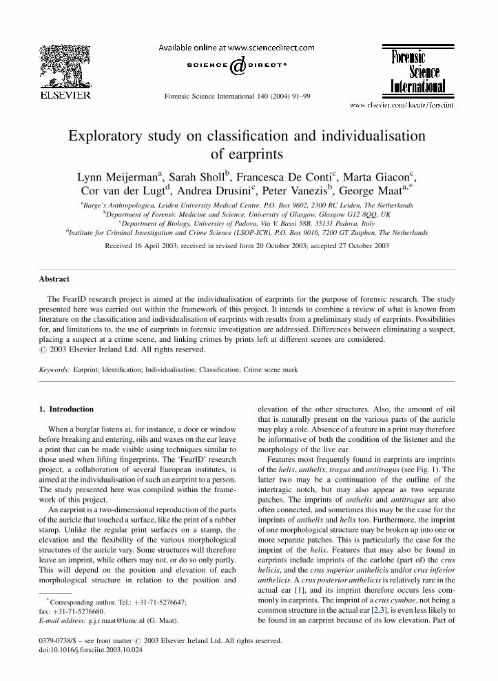
Features most frequently found in earprints are imprints
of the helix, anthelix, tragus and antitragus (see Fig. 1). The
latter two may be a continuation of the outline of the
intertragic notch, but may also appear as two separate
patches. The imprints of anthelix and antitragus are also
often connected, and sometimes this may be the case for the
imprints of anthelix and helix too. Furthermore, the imprint
of one morphological structure may be broken up into one or
more separate patches. This is particularly the case for the
imprint of the helix. Features that may also be found in
earprints include imprints of the earlobe (part of) the crus
helicis, and the crus superior anthelicis and/or crus inferior
anthelicis. A crus posterior anthelicis is relatively rare in the
actual ear [1], and its imprint therefore occurs less com-
monly in earprints. The imprint of a crus cymbae, not being a
common structure in the actual ear [2,3], is even less likely to
be found in an earprint because of its low elevation. Part of
Forensic Science International 140 (2004) 91–99
* Corresponding author. Tel.: þ31-71-5276647;
fax: þ31-71-5276680.
E-mail address: [email protected] (G. Maat).
0379-0738/$ – see front matter # 2003 Elsevier Ireland Ltd. All rights reserved.
doi:10.1016/j.forsciint.2003.10.024
Fig. 1. Examples of earprints, showing inter-individual variation and indicating some characteristic features (features are not necessarily marked
in all prints). Compiled using illustrations from van der Lugt [18]). (1) Crus helicis (anterior section), (2) crus helicis (posterior section), (3) helix,
(4) characteristic notch in inner rim of helix, (5) interruption of helix, (6) Darwinian nodule, (7) Darwinian enlargement, (8) knob on superior part
of helix, (9) anthelix (body of), (10) crus anterior anthelicis, (11) crus superior anthelicis, (12) crus posterior anthelicis, (13) inferior extension of
anthelix, (14) appendix of anthelix, (15) characteristic constriction of anthelix, (16) Tragus, (17) anterior knob of tragus, (18) anterior notch, (19)
antitragus, (20) intertragic notch, (21) posterior auricular furrow, (22) scapha, (23) earlobe, (24) crease in earlobe, (25) pre-auricular area, (26)
creases in pre-auricular area, (27) pre-auricular sinus, (28) skin detail indicating baldness, (29) apex of scapha.
92 L. Meijerman et al. / Forensic Science International 140 (2004) 91–99
the pre-auricular region is, however, often represented in a
print, and seems to provide valuable information due to
characteristic creases of the skin in this area.
Hirschi [4,5] was among the first to recognize the value of
earprints for forensic identification. Ever since, several
studies have been presented, acknowledging the feasibility
of using earprints for this purpose [6–13]. Some basic
questions, however, need to be addressed. How unique is
the human ear, and how stable are its features? Moreover,
how unique is the earprint? Can one ear make prints that vary
substantially, and can two different ears create similar ear-
prints? Knowledge of both variation between several prints
made by a single ear (intra-individual variation) and varia-
tion that occurs between prints that each have been made by
a different ear (inter-individual variation) is of great impor-
tance [14]. Uniqueness of the human ear will be difficult, if
not impossible, to establish. To justify the claim that we can
match an earprint uniquely to an ear, we must establish that
the print resembles other prints from the same ear more than
it resembles prints from another ear. We may attempt to do so
by analysing multiple prints from a large sample of ears,
comparing inter-individual variation with intra-individual
variation over a suitable set of measurable features. An
experimental feature set is suitable only if the inter-indivi-
dual variation is significantly greater than the intra-indivi-
dual variation. For such a comparison, we may use, for
instance, cluster analysis or formal concept analysis
(Ingleby, personal communication). The outcome of this
analysis will be probabilistic. This means we may estimate
the probability of encountering seemingly indistinguishable
prints from different ears. Until now, no such statistical
analysis has been performed. It is, however, part of the work
that the FearID team hopes to accomplish.
An extensive classification of the various features in an
earprint will aid in determining the extent of inter- and intra-
individual variation, and will provide the tools for a statis-
tical analysis. With this study, we aimed to combine a review
of what is known from literature on the subject of classifica-
tion and individualisation of earprints, with results of a
preliminary study of earprints that we have carried out
ourselves.
2. Use of prints in forensic research
Earprints in forensic research can be used for various
purposes. Firstly, a latent earprint found on a scene of crime
may be used to exclude a person as a possible suspect, as
indeed Scaillet [12] excluded one of two possible accom-
plices of two criminals who had confessed to a series of
offences. When utilizing earprints only to dismiss a suspect,
using transparency overlays to establish the degree of simi-
larity will often quickly reveal that a person was not respon-
sible for leaving a latent earprint. Establishing that two
different ears will not make similar earprints is no prere-
quisite when prints are used in this manner. We do, however,
need to be sure that various prints made by one ear do not
vary to the extent that we would not recognise these prints as
being created by a single ear.
Besides eliminating possible subjects from further inves-
tigation, one may also use the latent earprint to increase
evidence against a given suspect, as Scaillet [12] did for the
other of the two possible accomplices mentioned above.
Other examples of this use for earprints are Dubois [6] and
Hirschi [4]. In order to do so, one also has to have a control
print of a possible suspect already at one’s disposal, and the
transparency overlay technique will again quickly reveal the
degree of similarity between the latent earprint and the
control print. Assuming that the latent earprint and the
control print are a (more or less) perfect match, we must
establish that the probability of two similar prints being
made by two different ears is sufficiently close to zero in
order for the latent print to be accepted as evidence.
A third method of using earprints in forensic research may
be applied when there is no suspect available. A latent print
may then be compared to a database containing prints
recovered from crime scenes, each of them linked to a case,
or possibly even a perpetrator through other forms of
evidence or a confession. A database could also contain
control prints taken from large groups of people, or a
combination of both. When using earprints to link cases
this way, it is not sufficient to know that one ear will not
make substantially different prints, and that the chance of
two ears making indistinguishable prints is acceptably small.
We also need to know that, during a certain period of time,
the auricle itself usually does not change to the extent that it
would be impossible to locate a print from the same indi-
vidual at a different age in a database. Depending on how
accurately we need to process the exact dimensions of the
print in order to find an acceptable number of possible
matches, it may be possible that a print in the database is
not found as a possible match anymore after a certain period
of time. This would not mean, however, that another person
risks to be incriminated, since dimensions are merely used
for initial classification and weak linkage, while individua-
lisation will most probably depend on the presence and
position of minutiae, e.g. creases, papules or other details
of the skin, and characteristic notches or angles in the outline
of imprinted features.
3. Variability and stability of the auricle
When addressing the uniqueness of the auricle itself, the
snowflake paradigm—frequently voiced as ‘‘nature never
repeats itself’’—has been applied for forensic purposes [15].
In several publications on the variability of ears and/or
earprints [16–18] and case studies of police investigations
[4,8,19], it was assumed that no two ears are exactly alike.
The assumption was based merely on the absence of two
indistinguishable ears in conducted surveys; the individual-
ity of human ears has never been empirically established
L. Meijerman et al. / Forensic Science International 140 (2004) 91–99 93
[20]. However, as Hoogstrate et al. [21] have stated, avail-
able studies do suggest that the variability between ears is
that large that it might be possible that ears are uniquely
distinct on a limited number of features or characteristics.
Although variability in the morphology of the actual (live)
ears of different people does not automatically lead to a
similar variability in their prints, it may nonetheless be very
informative to study this variation in morphology. It will
assist the interpretation of features in a print, and it will
facilitate the distinction between inter-individual and intra-
individual variation. An indication of the frequency at which
certain morphological structures, such as a Darwinian
nodule or a (pre)auricular sinus, occur in various ears will
further provide information on the value of their imprints for
identification. It is, however, by no means necessary to try to
‘recreate’ a three-dimensional structure from the two-
dimensional print in our minds, in order to be able to
individualise a print, as we can compare prints with prints
and not with the auricle itself.
For a latent earprint to be useful as a means of identifica-
tion, not only the variability between ears (and therefore
earprints) must be sufficient, but the ear (and its prints) must
be relatively stable as well. The auricle, however, does not
remain unchanged throughout life. Some features may be
changed intentionally through piercing; others may change
through disease, or by scarring or other trauma. Provided the
changes are not too extreme, they will not alter the basic
dimensions and characteristics (and therefore diagnostic
features) of the auricle. Once recognised, they may even
make the ears positively more distinguishable from others.
In addition to such relatively sudden changes, the auricle
increases in dimensions due to growth [2,22–24]. Although
this might affect the chance of finding a print in a database
after a long period of time, it is unlikely to make the ear less
‘unique’. Also, natural changes in the auricle (such as
resulting from growth) develop slowly and are small, espe-
cially when considering the time-span we are dealing with in
forensic practice. Here, this is not ‘a lifetime’ but, at most,
the time that passes during which legal prosecution is
possible. Still, a study of the growth of the auricle should
be part of a comprehensive study on ears and earprints, and
the issue will therefore be dealt with in a separate study.
4. Intra-individual variability in prints
Changes in the auricle are not the only possible source for
intra-individual variation in earprints; differences in the way
prints are left, or the material they are left on, may also cause
variation in prints by a single ear. Due to variation in
elevation and flexibility of the various structures of the
auricle, not all features in the prints are created simulta-
neously, nor are they affected in a similar way by changing
pressure. Consequently, the amount of force that is applied to
the surface by the ear during listening may significantly
influence the appearance of the resulting print. Neubert [25]
compared the dimensions of features in a print directly to
dimensions of the auricle. He did so for prints that were
taken at two levels of applied force (‘soft’ and ‘hard’) of both
ears of fifty subjects, and found that length and width of the
auricle usually exceeded the corresponding dimensions of
the unstressed auricle and increased as more force was
applied. He further noted that length increased more (and
more often) than width, that the imprint of the upper part of
the helix differed more from the actual helix than the imprint
of its lower part, and that the minimal width of the imprint of
the anthelix conformed more to the actual minimal width in
the auricle than did the maximal width. The greatest devia-
tion from the unstressed auricle was in the imprint of the
earlobe. The deviation in width was greater than the devia-
tion in length in this feature.
When using prints in forensic research, we need to
familiarize ourselves with the variation between prints made
with various forces, rather than between the morphological
structure and its imprint (although the latter can give some
indication of the former). Saddler [26] studied the influence
of changes in applied force on the various features in a print.
He measured ear length, ear width, anthelix width, and upper
helix width in 92 sets of prints (by 46 left and 46 right ears).
Each set consisted of one print made with ‘soft pressure’ and
one print made with ‘hard pressure’. Width of the imprint of
the anthelix in particular varied with a change in applied
force. In most cases (79% of left-earprints; 70% of right-
earprints), increased force led to an increase in width. In
some prints, however, the width of the imprint of the anthelix
decreased when more force was applied. The total length on
the ear also increased in most prints (in 70% of left-earprints
and 74% of right-earprints). Saddler provided ranges for the
increase in millimetres of ear length (1–6.5 mm) and anthe-
lix width (0.5–5 mm). No specification was, however, given
for the amount of force that was applied, and there was no
indication that the variation in applied force was similar to
variation under natural conditions. Hence, there is no indi-
cation that the variation in dimensions of features in prints he
found during his study would occur in latent (crime scene)
prints by a single ear. Regarding the other dimensions
included in his study, Saddler found that in the majority
of the sets of prints (73% of left-earprints; 60% of right-
earprints), an increase in width of the upper helix could be
observed with increased force. Ear width increased in 47%
of left-earprints and 53% of right-earprints; the remaining
prints showed either no marked difference, or a decrease,
with increased force. Saddler concluded that most dimen-
sions increase with increasing force, but that the imprints of
some structures may change in an unpredictable way.
According to him, this would make searching a database
using metrical characteristics ineffective.
Ingleby et al. [27] tried to avoid the problem of variation
in dimensions due to variation in applied force, by measuring
distances between features in prints while using the intensity
medians lying between the outlines of these features. They
assumed that, as force increases, the edges tend to spread but
94 L. Meijerman et al. / Forensic Science International 140 (2004) 91–99
the intensity medians will remain more or less at the same
position. Dubois [6] came to a similar conclusion when
studying pressure points of an ear that was pressed to a glass
plate.
We carried out a preliminary study of earprints that were
made when subjects were listening naturally at a surface.
Each subject listened a number of times. During each of the
listening efforts, the amount of force applied to the surface
was measured. The resulting prints were dusted using fine
aluminium powder, and preserved on black gel lifters. For
each of thirty different ears, we copied three to five prints
onto transparency sheets to reveal the degree of similarity.
We also compared digitised prints on the computer by
superimposing them onto each other. This preliminary study
has led us to believe that, in addition to the dimensions of the
separate features, the position of these features to each other
may also change when applied force is varied. This may be
the result of flattening of the entire auricle with higher force,
increasing distances between the estimated centre-points or
centre-lines (‘core-lines’) of most features. We believe that
the arrangement of features in a print may further change
with higher force as a result of a different reaction to
increased force by the upper and lower parts of the auricle,
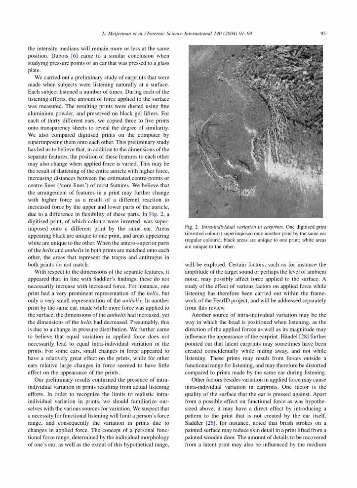
due to a difference in flexibility of these parts. In Fig. 2, a
digitised print, of which colours were inverted, was super-
imposed onto a different print by the same ear. Areas
appearing black are unique to one print, and areas appearing
white are unique to the other. When the antero-superior parts
of the helix and anthelix in both prints are matched onto each
other, the areas that represent the tragus and antitragus in
both prints do not match.
With respect to the dimensions of the separate features, it
appeared that, in line with Saddler’s findings, these do not
necessarily increase with increased force. For instance, one
print had a very prominent representation of the helix, but
only a very small representation of the anthelix. In another
print by the same ear, made while more force was applied to
the surface, the dimensions of the anthelix had increased, yet
the dimensions of the helix had decreased. Presumably, this
is due to a change in pressure distribution. We further came
to believe that equal variation in applied force does not
necessarily lead to equal intra-individual variation in the
prints. For some ears, small changes in force appeared to
have a relatively great effect on the prints, while for other
ears relative large changes in force seemed to have little
effect on the appearance of the prints.
Our preliminary results confirmed the presence of intra-
individual variation in prints resulting from actual listening
efforts. In order to recognize the limits to realistic intra-
individual variation in prints, we should familiarize our-
selves with the various sources for variation. We suspect that
a necessity for functional listening will limit a person’s force
range, and consequently the variation in prints due to
changes in applied force. The concept of a personal func-
tional force range, determined by the individual morphology
of one’s ear, as well as the extent of this hypothetical range,
will be explored. Certain factors, such as for instance the
amplitude of the target sound or perhaps the level of ambient
noise, may possibly affect force applied to the surface. A
study of the effect of various factors on applied force while
listening has therefore been carried out within the frame-
work of the FearID project, and will be addressed separately
from this review.
Another source of intra-individual variation may be the
way in which the head is positioned when listening, as the
direction of the applied forces as well as its magnitude may
influence the appearance of the earprint. Handel [28] further
pointed out that latent earprints may sometimes have been
created coincidentally while hiding away, and not while
listening. These prints may result from forces outside a
functional range for listening, and may therefore be distorted
compared to prints made by the same ear during listening.
Other factors besides variation in applied force may cause
intra-individual variation in earprints. One factor is the
quality of the surface that the ear is pressed against. Apart
from a possible effect on functional force as was hypothe-
sized above, it may have a direct effect by introducing a
pattern to the print that is not created by the ear itself.
Saddler [26], for instance, noted that brush strokes on a
painted surface may reduce skin detail in a print lifted from a
painted wooden door. The amount of details to be recovered
from a latent print may also be influenced by the medium
Fig. 2. Intra-individual variation in earprints. One digitised print
(inverted colours) superimposed onto another print by the same ear
(regular colours); black areas are unique to one print; white areas
are unique to the other.
L. Meijerman et al. / Forensic Science International 140 (2004) 91–99 95
that is used to lift the print. Within the scope of the FearID
project, various media will therefore be tested on their ability
to preserve details.
5. Inter-individual variation: classifying variation
and finding diagnostic features
Various studies have focussed on identifying and classi-
fying variation in prints [18,27,29]. Both metrical and non-
metrical systems, or combinations of the two, were proposed
for the classification of the various phenomena. Ingleby et al.
[27] and Maat [29] proposed methods for a quantitative
classification. Both appointed two fixed landmarks and
connected these two in order to construct a polar axis for
geometrical standardisation. They then calculated distances
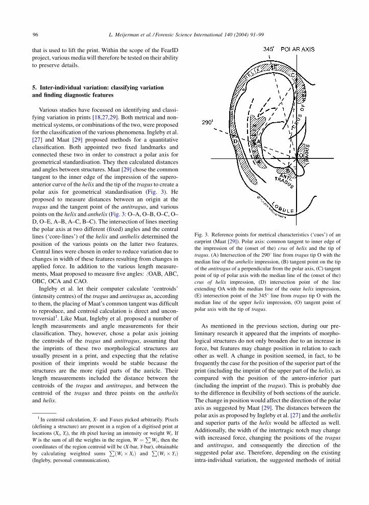
and angles between structures. Maat [29] chose the common
tangent to the inner edge of the impression of the supero-
anterior curve of the helix and the tip of the tragus to create a
polar axis for geometrical standardisation (Fig. 3). He
proposed to measure distances between an origin at the
tragus and the tangent point of the antitragus, and various
points on the helix and anthelix (Fig. 3: O–A, O–B, O–C, O–
D, O–E, A–B, A–C, B–C). The intersection of lines meeting
the polar axis at two different (fixed) angles and the central
lines (‘core-lines’) of the helix and anthelix determined the
position of the various points on the latter two features.
Central lines were chosen in order to reduce variation due to
changes in width of these features resulting from changes in
applied force. In addition to the various length measure-
ments, Maat proposed to measure five angles: ffOAB, ABC,
OBC, OCA and CAO.
Ingleby et al. let their computer calculate ‘centroids’
(intensity centres) of the tragus and antitragus as, according
to them, the placing of Maat’s common tangent was difficult
to reproduce, and centroid calculation is direct and uncon-
troversial1. Like Maat, Ingleby et al. proposed a number of
length measurements and angle measurements for their
classification. They, however, chose a polar axis joining
the centroids of the tragus and antitragus, assuming that
the imprints of these two morphological structures are
usually present in a print, and expecting that the relative
position of their imprints would be stable because the
structures are the more rigid parts of the auricle. Their
length measurements included the distance between the
centroids of the tragus and antitragus, and between the
centroid of the tragus and three points on the anthelix
and helix.
As mentioned in the previous section, during our pre-
liminary research it appeared that the imprints of morpho-
logical structures do not only broaden due to an increase in
force, but features may change position in relation to each
other as well. A change in position seemed, in fact, to be
frequently the case for the position of the superior part of the
print (including the imprint of the upper part of the helix), as
compared with the position of the antero-inferior part
(including the imprint of the tragus). This is probably due
to the difference in flexibility of both sections of the auricle.
The change in position would affect the direction of the polar
axis as suggested by Maat [29]. The distances between the
polar axis as proposed by Ingleby et al. [27] and the anthelix
and superior parts of the helix would be affected as well.
Additionally, the width of the intertragic notch may change
with increased force, changing the positions of the tragus
and antitragus, and consequently the direction of the
suggested polar axe. Therefore, depending on the existing
intra-individual variation, the suggested methods of initial
Fig. 3. Reference points for metrical characteristics (‘cues’) of an
earprint (Maat [29]). Polar axis: common tangent to inner edge of
the impression of the (onset of the) crus of helix and the tip of
tragus. (A) Intersection of the 2908 line from tragus tip O with the
median line of the anthelix impression, (B) tangent point on the tip
of the antitragus of a perpendicular from the polar axis, (C) tangent
point of tip of polar axis with the median line of the (onset of the)
crus of helix impression, (D) intersection point of the line
extending OA with the median line of the outer helix impression,
(E) intersection point of the 3458 line from tragus tip O with the
median line of the upper helix impression, (O) tangent point of
polar axis with the tip of tragus.
1 In centroid calculation, X- and Y-axes picked arbitrarily. Pixels
(defining a structure) are present in a region of a digitised print at
locations (Xi, Yi), the ith pixel having an intensity or weight Wi. If
W is the sum of all the weights in the region, W ¼P
Wi, then the
coordinates of the region centroid will be (X-bar, Y-bar), obtainable
by calculating weighted sumsP
ðWi � XiÞ andP
ðWi � YiÞ(Ingleby, personal communication).
96 L. Meijerman et al. / Forensic Science International 140 (2004) 91–99
classification may introduce unnecessary deviation. In order
to reduce this problem we propose that, when metrical
characteristics (‘cues’) are used for classification, it is better
not to use those related to a polar axis. One can instead
compare the spatial positions of features to each other. One
may for instance measure the shortest distance between
landmarks, or compare the curvature of (parts of) the helix
and anthelix. To reduce the risk of misclassification, one may
choose to only compare neighbouring structures, or neigh-
bouring parts of structures. Intra-individual variation in the
dimensions of, and distances between, features should be
studied in prints made during true efforts of listening.
Examples of non-metrical characteristics, used when
comparing or classifying prints, are the outline of the
imprints of the various morphological structures, and the
appearance, size, and position of minutiae. Two approaches
have been followed in this qualitative approach. One is to
compare the features in a print with the outline of morpho-
logical structures of the auricle when pressed to a glass plate
[19,30]. The other approach is to compare prints with prints
[18,26,29]. Maat [29] suggested recording the presence or
absence, and position, size and form, of a Darwinian nodule,
and the presence or absence of impressions of the tragus,
antitragus and earlobe. He further included recording the
position, size and form of papules and scars, the position and
pattern of creases of the pre-auricular area, anthelix and
helix, and the pattern of hair-related hillocks and dimples of
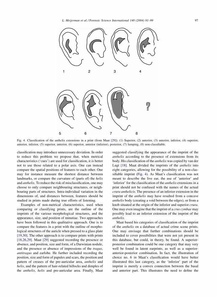
the anthelix, helix and pre-auricular area. Finally, Maat
suggested classifying the appearance of the imprint of the
anthelix according to the presence of extensions from its
body. His classification of the anthelix was copied by van der
Lugt [18]. Maat divided the imprints of the anthelix into
eight categories, allowing for the possibility of a non-clas-
sifiable imprint (Fig. 4). As Maat’s classification was not
meant to describe the live ear, the use of ‘anterior’ and
‘inferior’ for the classification of the anthelix extensions in a
print should not be confused with the names of the actual
crura anthelicis. The presence of an inferior extension in the
imprint of the anthelix may have resulted from a concave
anthelix body (creating a void between the edges), or from a
knob situated at the origin of the inferior and superior crura.
One may even imagine that the imprint of a crus cymbae may
possibly lead to an inferior extension of the imprint of the
anthelix.
Maat based his categories of classification of the imprint
of the anthelix on a database of actual crime scene prints.
One may envisage that further combinations should be
included to cover possibilities that were not yet present in
this database, but could, in theory, be found. A superior-
posterior combination could be one category that may very
well be found in latent earprints, as well as a superior-
anterior-posterior combination. In fact, the illustration of
choice no. 6 in Maat’s classification would have better
illustrated this last category, as the ‘inferior’ part of the
imprint is merely a convex connection between the basal
and anterior part. This illustrates the need to define the
Fig. 4. Classification of the anthelix extensions in a print (from Maat [29]). (1) Superior, (2) anterior, (3) anterior, inferior, (4) superior,
anterior, inferior, (5) superior, anterior, (6) superior, anterior (inferior), posterior, (7) lumping, (8) non-classifiable.
L. Meijerman et al. / Forensic Science International 140 (2004) 91–99 97
boundaries between categories in a classification accurately.
It may further be useful to record which is the strongest of
the extensions, if one is clearly more prominent than the
other(s). The stronger extension will less likely be absent in
prints created with less force than the weaker extension. We
hope to gain a more extensive knowledge on the variability
of each of the various features of a print during the FearID
project. Ideally, we will be able to determine which are the
stable characteristics, and group the less stable ones in such a
manner that, by default, no realistic possibilities are
excluded while classifying prints to search for a matching
print in a database. For any classification scheme of gross
features in a print, achieving inter-subjectivity will be a
challenge. We hope to eventually be able to rely on auto-
mated image-processing techniques for gross feature extrac-
tion through boundary extraction algorithms. This will
decrease the role of the examiner’s perception during the
initial classification.
Not all studies of intra-individual variation in earprints
have used a classification of features, nor may all variation
be easily classified. Iannarelli [16] advised using an overlay
technique for comparing earprints. He further described a
second technique, during which the control print and latent
print are each cut into quarters, and matched together to find
the degree of matching. This technique was adopted by
Kennerley [8] and van der Lugt [18] but might be considered
an unnecessary addition to the overlay technique, only
reducing the possible points of comparison.
As we may expect some variation in the dimensions of the
separate features in different prints of one ear, as well as in
their relative positions, it is likely that the final individua-
lisation of an earprint will greatly depend on physical
minutiae from skin structure. Examples of such minutiae
may be the position, size and/or pattern of creases, papules,
scars, moles, hillocks and dimples, and characteristic
notches along the inner rim of the helix. Comparing different
prints made by the same ear has led us to suspect that
particularly the inner rim of the superior part of the helix
may prove to be very stable, as well as very characteristic.
6. Concluding remarks
Measurements may be hard to reproduce, and classifica-
tions may be subjective. Still, an extensive classification of
features in a print will provide us a tool in order to estimate
the probability of encountering seemingly indistinguishable
prints from different ears. This estimation of false positive
probability will allow us to establish whether or not, and to
what extent, we may use latent earprints in forensic research.
A next phase may then possibly include using earprints for
identification. An (initial) classification of the features in the
latent print will now also facilitate the search for a matching
print in a database. A proper classification of features will
also help in indicating the position of minutiae. It will further
assist interpretation, as imprints of morphological structures
may lump together, or the imprint of a single structure may
appear as separate patches.
Solid knowledge on the variability of earprints left by a
single ear is of great importance during both phases. It will
allow us to record, store and analyse earprints in such a way
that the maximum amount of ‘natural’ variation is taken into
account, without introducing unrealistic variation in prints of
one ear. The latter would likely impede the search for
diagnostic features. Knowing the extent of intra-individual
variability, and recognizing stable features, will aid the
design of a classification system capable of distinguishing
between intra-individual and inter-individual variability.
Acknowledgements
This work was carried out within the framework of the
FearID research project, which was funded by the European
Union. This research project is a collaboration between the
Institute for Criminal Investigation and Crime Science (Zut-
phen, The Netherlands), the National Training Centre for
Scientific Support to Crime Investigation (Durham, UK), the
University of Padova (Italy), the University of Glasgow
(UK), Leiden University Medical Centre (The Netherlands),
The Netherlands Organisation for Applied Scientific
Research (Delft, The Netherlands), the University of Hud-
dersfield (UK), The Netherlands Forensic Institute (Rijs-
wijk, The Netherlands) and NFGD Software Solutions,
Zoetermeer, The Netherlands). The photographs used to
compile Fig. 1 were made by Cor van der Lugt. Jan Lens
drew Figs. 3 and 4. We wish to thank Dr. M. Ingleby for his
helpful remarks and clarification of centroid calculation, as
well as corrections of English text, and Dr. H. de Jong for
critically reading a first draft of the manuscript. We are
further grateful to Ruud van Basten for dusting and lifting
the earprints used during our preliminary research. Two
anonymous referees are thanked for their valuable com-
ments.
References
[1] I. Oepen, Der Identifizierungswert des menschliches Ohres,
Paper Presented at the Institut fur Rechtsmedizin der
Universitat Marburg, 1971.
[2] T. Quelprud, Die Ohrmuschel und ihre Bedeutung fur die
erbbiologische Abstammungsprufung, Der Erbarzt 8 (1935)
121–125.
[3] I. Tillner, Seltene morphologische Merkmale an der mens-
chlichen Ohrmuschel und ihr praktischer Wert fur die
Vaterschaftsbegutachtung, Anthropologische Anzeiger 26
(1963) 294–307.
[4] F. Hirschi, Cambrioleurs internationaux convaincus a l’aide
de preuves peu communes, Revue internationale de police
criminelle 239 (1970) 184–193.
[5] F. Hirschi, Identifizierung von Ohrabdrucken, Kriminalistik
24 (1970) 75–79.
98 L. Meijerman et al. / Forensic Science International 140 (2004) 91–99
[6] N. Dubois, Oren naar hebben, Technisch Informatie Bull. 1
(1988) 7–22.
[7] H.J. Hammer, The identification of earprints secured at the
scene of the crime, Fingerprint World 12 (1986) 49–51.
[8] J. Kennerley, Ear print identification, Paper Presented on the
National Conference on Craniofacial Identification, 4–6
September 1998.
[9] C. van der Lugt, Ear identification—state of the art, Info.
Bull. Shoeprint/Toolmark Examiners 4 (1998) 69–81.
[10] J.W. Osterburg, The Crime laboratory: Case Studies of
Scientific Criminal Investigations, second ed., Clark Board-
man Company Ltd., New York, 1982, pp. 34–35.
[11] G. Pasescu, E. Tanislav, Person identification on the basis of
earprints in the activity of Bucharest Police Department, Info.
Bull. SP/TM Examiners 3 (1997) 10–14.
[12] M. Scaillet, Une identification originale, Revue internationale
de police criminelle 245 (1971) 41–43.
[13] E. Trube-Becker, Identifizierung von Tatern durch Ohrab-
druckspuren, Lecture on the 61st Annual Meeting of the
Deutsche Gesellschaft fur Rechtsmedizin, September 1982,
Wurtzburg, Germany.
[14] C. Champod, I.W. Evett, B. Kuchler, Earmarks as evidence: a
critical review, J. Forensic Sci. 46 (6) (2001) 1275–1284.
[15] J.I. Thornton, The snowflake paradigm, J. Forensic Sci. 31
(1986) 399 (Letter to the Editor).
[16] A.V. Iannarelli, Ear Identification, Forensic Identification
Series, Paramount Publishing Company, Fremont California,
1989, 213 pp.
[17] R. Imhofer, Die Bedeutung der Ohrmuschel fur die
Feststellung der Identitat, Archiv fur Kriminalanthropologie
und Kriminalistik 26 (1906) 150–163.
[18] C. van der Lugt, Earprint Identification, Elsevier Bed-
rijfsinformatie, ’s Gravenhage, 2001, 317 pp.
[19] L. Nitsche, H.J. Hammer, Durch Ohrabdruck Verdachtigen
uberfuhrt, Forum Kriminal, vol. 11, Wiss.-techn. Beilage 5/
75, 1975, pp. 11–15.
[20] A.A. Moenssens, Identification by ear impressions, in: A.A.
Moenssens, J.E. Starrs, C.E. Henderson, F.E. Inbau (Eds.),
Scientific Evidence in Civil and Criminal Cases, Foundation
Press, 1995, pp. 612–614.
[21] A.J. Hoogstrate, H. van den Heuvel, E. Huyben, Ear
identification based on surveillance camera’s images, Sci.
Justice 41 (2001) 167–172.
[22] L. Feenstra, C. van der Lugt, Persoons-identificatie via de
oorschelp, Tijdschr. KNOheelk. 6 (2000) 53–55.
[23] K. Hajnis, Die Veranderungen der Ohrmuscheln beim
Erwachsenen, Z. Morphol. Anthropol. 61 (1969) 42–56.
[24] J.A. Heathcote, Why do old men have big ears? Br. Med. J.
311 (1995) 1668.
[25] F. Neubert, Die Bedeutung der Tateridentifizierung durch
Ohrabdrucke, Med. Diploma Arbeit, Karl-Marx University,
Leipzig, 1985.
[26] K. Saddler, The establishment and evaluation of an ear-print
database at the National Training Centre for Scientific
Support to Crime Investigation, M.Sc. Thesis, University of
Strathclyde, Glasgow, 1996.
[27] M. Ingleby, J. Mason, D. Turner, S. Langdell, Ear-prints: a
neglected forensic resource? University of Huddersfield,
2000, unpublished.
[28] K. Handel, Streiflichter, Kriminalistik 1 (1983) 53–56.
[29] G.J.R Maat, Ear Print Project—Brief report on the pilot
study period September–November 1999, Barge’s Anthro-
pologica, Leiden University Medical Centre, 1999, unpub-
lished.
[30] W. Jung, Uber das Ohr zum Tater—Identifizierung durch
Ohrabdrucke, Kriminalistik 38 (1984) 482–483.
L. Meijerman et al. / Forensic Science International 140 (2004) 91–99 99