Embed Size (px)
Citation preview

EXPRESSION OF BRAF MUTATION IN THYROID NEOPLASMS.
DISSERTATION
SUBMITTED FOR
M.D. IN PATHOLOGY
THE TAMILNADU DR. MGR MEDICAL UNIVERSITY
DEPARTMENT OF PATHOLOGY
PSG INSTITUTE OF MEDICAL SCIENCES & RESEARCH
PEELAMEDU, COIMBATORE – 641004.
TAMILNADU, INDIA
APRIL 2012

CERTIFICATE

CERTIFICATE
This is to certify that the dissertation work entitled “EXPRESSION OF
BRAF MUTATION IN THYROID NEOPLASMS” submitted by Dr. H.Volga
is work done by her during the period of study in the department of Pathology,
PSGIMS & R from June 2009 to April 2012. This work was done under the
guidance of Dr. S.Shanthakumari, Professor, Department of Pathology.
Dr. S.Shanthakumari.M.D Dr. Alamelu Jayaraman.M.D
Professor, Professor & Head of the Department,
Department of Pathology, Department of Pathology,
PSGIMS & R. PSGIMS & R.
Dr. S.Ramalingam.M.D
Principal,
PSGIMS & R,
Coimbatore - 641004.


ACKNOWLEDGEMENT

ACKNOWLEDGEMENT One can count the seeds in a fruit. But none can count the fruits in a seed.
Gratitude is the duty of the fruit which ought to be paid to the seed which the
seed never expects.
My first step and the direction of my journey was introduced to me ,induced
and also enthused by my guide Dr. S. Shanthakumari M.D. Professor ,
Department of Pathology without whose guidance my venture of compiling and
completing my thesis would not have become a reality. I express my heartfelt
gratitude for her untiring efforts in completion of my work.
Gratitude is the memory of the heart. My gratitude goes to faculty of PSG
Center for Molecular Medicine and Therapeutics.
• Dr.S.Ramalingam M.D. Head, PSG CMMT, and also the Principal, for
sparing his precious time from his administrative routine to discuss, analyze
and bring my work to successful completion.
• Dr.Sudha Ramalingam M.D. Assistant Professor, Community Medicine
and PSG CMMT, for offering her valuable guidance in planning, and during
the procedural difficulties and in analysis.

• Dr.Thiagarajan Sairam, Assistant Professor (Research), PSG CMMT,
for guidance in planning, and helping me in designing the primer, and
standardizing the procedures.
• Mrs.Ranjani, Senior Research Fellow, and all members of PSG CMMT
for their technical assistance and execution.
I thank Dr.Alamelu Jayaraman M.D. Professor and Head, Department of
Pathology for allowing me to do this study.
I also thank Dr.Suma B.Pillai, for her valuable comments.
I thank Mrs. Angeline Mary and the other technical staffs, Mrs.Gomathi
and Mrs. Emily, secretary staffs and the attenders of department of pathology.
I also thank my colleagues especially Dr.Ramkumar for his support in my
final work.
I take this opportunity to thank my family members for their support and
kind words of encouragement during my thesis work.

CONTENTS

CONTENTS
PAGE. NO
CERTIFICATE
ETHICAL CLEARANCE CERTIFICATE
ACKNOWLEDGEMENT
1. INTRODUCTION 1
2. AIMS AND OBJECTIVES 4
3. REVIEW OF LITERATURE 5
4. MATERIALS AND METHODS 29
5. RESULTS 36
6. DISCUSSION 49
7. SUMMARY & CONCLUSION 56
8. BIBLIOGRAPHY
9. MASTER CHART

INTRODUCTION

1
INTRODUCTION
The thyroid gland is situated anterior to trachea in lower part of the
neck. It consists of right and left lobe connected across the median plane by
isthmus. The thyroid parenchyma is composed of lobules of thyroid follicles
with intervening thin fibrous tissue septae.Each follicle consists of central
core of colloid surrounded by single layer of follicular epithelial cells. The
parafollicular ‘C’ cells are located at the periphery of the follicles in small
groups or as single cells. [1]
The main function of thyroid gland is production of thyroid hormones
T3 and T4 which regulates the cellular metabolism, oxygen consumption
and protein synthesis. The parafollicular cells secrete calcitonin which in
turn regulates the calcium level in the plasma by a feedback mechanism [1].
Thyroid neoplasms account for 1% of all malignancies and it is the
most common among all endocrine malignancies [2].Environmental and
genetic factors play a major role in thyroid neoplasms due to the
dependence on iodine for hormone production. This organ is also vulnerable
to the genotoxic effects of radioactive iodine and to the non-genotoxic
effects caused by iodine deficiency [3], thereby resulting in neoplastic
lesions.

2
With improved, technically advanced diagnostic screening procedures
like guided FNA procedures, PET scan etc, detection of early neoplastic
lesions are on the rise .When early neoplastic lesions are sampled from
thyroid, morphological assessment to diagnose these, pose a significant
challenge to pathologist and for treatment by the surgeons.
Many a time morphology and immunohistochemistry is adequate for
diagnosing neoplasms of thyroid, [4] but a significant number of thyroid
neoplasms go unnoticed when early lesions are sampled. As genetic
alteration is the earliest change that occurs in oncogenesis and it may be
acquired in the due course of life, it is important to identify these for early
treatment, prognostication and for prevention. Therefore the knowledge of
genetic alteration will help not only in detecting the genesis of thyroid
carcinomas but also might help in treatment and prognostication.
Recent data’s suggest that alterations in RAS-RAF-MAP kinase
signaling pathway is seen in many neoplastic lesions of various organs but
is more frequent in thyroid carcinomas. In papillary thyroid carcinoma
BRAF mutation especially V600E mutation plays a vital role .Studies state
[5, 6] that BRAF mutation is specific for papillary thyroid carcinoma and is
frequently seen in high grade tumors and is associated with poorer
outcomes.
3
We did a Pubmed search for articles on relevant areas. The number of
articles returned for contain keywords in Pubmed search is Nil. (Limits; 10
years, English language, humans).keywords used in MESH “Proto-
oncogene proteins B-raf/genetics” “Thyroid Neoplasms” and “India”.
Thus there is paucity of Indian literature in the area of pathogenesis of
neoplasms of thyroid. The fact that there is a need to establish and conduct
scientific work linking proteins , genes and finding molecular markers for
thyroid neoplasms emphasizes the importance of our work. As 1% of
population world over suffers from thyroid neoplasms, research in this area
would help in treatment, prognostication and possibly prevention of this
major problem.
Therefore we propose to study the prevalence of BRAF V600E
mutation in thyroid neoplasms in PSGIMS&R and its association with
various phenotypic features as an initial step.

AIMS AND OBJECTIVES

4
AIMS AND OBJECTIVES
The aims of this study are
1. To estimate the prevalence of BRAF V600E mutation in thyroid
neoplasms diagnosed at a tertiary care hospital at Coimbatore.
2. To correlate the BRAF V600E mutation with clinicopathological
parameters.

REVIEW OF LITERATURE

5
REVIEW OF LITERATURE
The thyroid gland first appears as a median anlage and two lateral
anlagen. The median anlage develops in the floor of primitive pharynx at the
foramen cecum and grows caudally to become bilobed forming the greater
portion of the thyroid gland and forms follicular epithelial cells. The two
lateral anlagen derived from ultimobranchial bodies’ fuse with median
thyroid anlage and become incorporated into the lateral lobes .Then the
ultimobranchial bodies undergo dissolution phase and forms peripheral
component of cell groups called C cells [1].
Thyroid neoplasms are the most common endocrine neoplasms and
accounts for 1% of all human malignancies [2]. It usually occurs in the young
and in the middle age group. It is more frequent in females [2, 3]. The relative
frequency of papillary carcinoma is high in regions of adequate or high
dietary iodine intake. In regions of iodine deficiency the incidence of
papillary carcinoma is high after iodine supplementation [3]. In endemic
goiter regions the prognosis for thyroid carcinoma is worse, when compared
with regions with an adequate dietary iodine intake [7]. Even in regions with
endemic goiters papillary neoplasms predominate over follicular cancers [3].

6
A report from Health and family Welfare Department, Government of
Tamilnadu [8] from their survey conducted from 1990, states that goiter is
prevalent in all the districts. 18 districts out of 29 have more than 10 %
prevalence. Coimbatore is included among the 18 districts, where the goiter
prevalence is 11.7%.
Studies from India state that papillary thyroid carcinoma is more
common in coastal areas. A report by Professor, N.Dorairajan, on
investigating thyroid cancer [9] states and I quote “In India, thyroid cancer
has a widespread distribution with certain subtypes, notably papillary
cancer, occurring in coastal areas of Tamil Nadu, Andhra Pradesh and
Kerala which are iodine rich. The iodine content of soil modifies
development of these cancers.”He also states “In South India, excess iodine
in diet is related to the higher incidence of papillary cancer compared to
other more malignant subtypes of thyroid cancer.”
The thyroid neoplasms are classified as given below.
CLASSIFICATION OF THYROID TUMORS (WHO 2004) [3] BENIGN
Ø Follicular adenoma

7
MALIGNANT
Ø Papillary carcinoma
Ø Follicular carcinoma
Ø Medullary carcinoma
Ø Poorly differentiated carcinoma (Anaplastic)
Ø Undifferentiated carcinoma (Insular)
Ø Mucinous carcinoma
Ø Muco- epidermoid carcinoma
Ø Squamous cell carcinoma
Ø Others
FOLLICULAR ADENOMA:
Follicular adenoma is a benign tumor enveloped by a thick fibrous
capsule. Morphologically, it is composed of closely packed follicles,
trabeculae or solid sheets of cuboidal cells with pale or darkly stained nuclei
and inconspicuous nucleoli.
Rare variants like atypical follicular adenoma, hyalinizing trabecular
adenoma and Signet- ring cell follicular adenoma [10] are also reported.
Increase in cellularity, mitoses, spontaneous necrosis or infarction but lack
of invasion into the capsule or vasculature are the characteristic features of
atypical follicular adenoma.
8
Hyalinizing trabecular adenoma is characterized by elongated tumor
cells arranged in a wavy trabecular pattern around capillaries. Nuclear
grooves, pseudoinclusions and peri nuclear haloes are prominent.
Predominance of signet ring cells with abundant cytoplasmic vacuoles,
intermixed with groups of follicular cells of normal cytologic features are
the findings reported in Signet- ring cell follicular adenoma.
Hurthle cell adenomas are considered a subtype of follicular
adenoma. They are bright brown in a gross appearance. This neoplasm is
characterized by the presence of Oxyphilic/Hurthle cells arranged in
follicular and or trabecular pattern with partial or complete encapsulation
morphologically; the Hurthle cells are large cells with abundant eosinophilic
granular cytoplasm and a round nucleus. Accumulation of abundant
mitochondria gives a granular appearance to the cytoplasm under the light
microscope. It has a tendency for spontaneous infarction [11].
PAPILLARY CARCINOMA:
The most common of all primary thyroid malignancies is papillary
carcinoma, accounts for about 70- 85% of cases [12]. It can occur in any age
and has a female preponderance. Multifocal disease along with
intrathyroidal extension and metastasizes to regional lymph nodes are the
characteristic features. Extra thyroidal extension can occur and extend

9
beyond the capsule of the thyroid gland to involve structures like larynx,
trachea or esophagus.
Papillary carcinoma classic type is characterized by formation of
complex arborizing papillae with a central fibro vascular core under the
light microscope. The papillae are covered by cells with crowded oval
nuclei. The nuclei show margination of chromatin, overlapping, with
nuclear grooving and intra nuclear cytoplasmic pseudo inclusions. However,
papillary thyroid carcinoma can exhibit a pure follicular pattern or mixed
papillary and follicular pattern. Psammoma bodies are nothing but the
lamellated concretions formed by deposition of calcium is one of the most
important finding in papillary carcinoma [13].
The relationship between Hashimoto’s thyroiditis and papillary
carcinoma is controversial. Reports by Livolsi [14] suggested that the
lymphocytic infiltration of the surrounding thyroid tissue is induced through
autoimmune mechanisms triggered during the development of papillary
carcinoma. Harach et al [15] found that lymphocytic thyroiditis more
commonly associated with papillary thyroid carcinoma than with other types
of thyroid carcinomas. But a study conducted by Dubravka [16] et al showed
no significant association between Hashimoto’s thyroiditis and Papillary
carcinoma.

10
VARIANTS OF PAPILLARY CARCINOMA:
Several morphological variants of papillary carcinoma have been
recognized based on the architecture, growth pattern, cellular morphology
and stromal features. The subtypes are important as some of these subtypes
are more aggressive in their biologic and clinical behaviour irrespective of
their bland appearance. [11]
I. Papillary microcarcinoma:
Papillary micro carcinomas usually measures 1 cm or less in diameter
and often unencapsulated. It is detected incidentally in thyroidectomy
specimens for other indications and is associated with an excellent
prognosis though occasional regional lymph node metastasis is usual [11].
II. Follicular variant:
This neoplasm composed of small to medium sized, irregularly
shaped follicles with abortive papillary formation. Stromal sclerosis and
psammoma bodies can be present. Characteristic nuclear features of
papillary carcinoma are evident. Distant metastasis and vascular invasion
are common. Some of these tumors exhibit encapsulation with an
exceptionally good prognosis (so called Lindsay tumor) [11].

11
III. Tall cell variant:
This is a rare variant and is composed of tumor cells whose heights
are at least three times their widths. Tall cells must be seen in 50% or more
of the tumor areas to make a diagnosis of tall cell variant of papillary
thyroid carcinoma [11] Necrosis, mitotic activity and extra thyroidal
extension are common. These tumors occur in older patients, often males,
with a more aggressive clinical behaviour. This variant is commonly
associated with BRAF mutation reflects its aggressiveness [17].
IV. Oncocytic variant:
This variant of papillary carcinoma comprising of complex branching
papillae with thin fibrovascular stromal core, covered by polygonal
oncocytic cells with abundant granular eosinophilic cytoplasm.[13]The
behaviour is similar to conventional papillary carcinoma.
V. Warthin tumor like variant:
The characteristic feature of this variant is brisk lymphoplasmacytic
infiltrate in the papillary stalks. Papillae were lined by oncocytic cells with
the typical nuclear features of classic papillary carcinoma. It is frequently
associated with Hashimoto thyroiditis [11]. The immuno profile of the
lymphoid cells is no different from chronic lymphocytic thyroiditis.

12
VI. Cribriform variant:
Predominant cribriform pattern, focal papillary architecture, solid and
spindle cell areas interspersed with squamoid morules, characterize this
uncommon variant of papillary carcinoma [12]. The nuclei frequently harbor
eosinophilic, homogenous cytoplasmic inclusions. It typically occurs in
patients with familial adenomatous polyposis or Gardner syndrome. This
tumor is often multifocal and occurs in young women.
VII. Diffuse sclerosing variant:
This rare variant of papillary carcinoma seen more frequently in
children and is associated with a poor prognosis [12]. The typical finding of
this tumor includes diffuse involvement of both the lobes, dense
lymphoplasmacytic infiltrate with extensive lymphatic permeation.
VIII. Clear cell variant:
Tumor cells have extensive clear or vacuolated cytoplasm.
Cytoplasmic clearing is usually due to accumulation of glycogen [10]. The
nuclear features are otherwise typical of papillary carcinoma.
FOLLICULAR CARCINOMA:
It accounts for 5-15% of all thyroid malignancies. The tumor can be
encapsulated or widely invasive. A true capsular and / or a vascular invasion

13
in a follicular neoplasm are mandatory to diagnose follicular carcinoma. It is
more common in an older age group and more common in females. [10, 13]
The criteria to diagnose follicular carcinoma include invasion of the
capsule, invasion through the capsule and invasion into vein in or beyond
the capsule [11].
Tumors with limited focal capsular and / or vascular invasion that are
apparent only on histological examination are termed as ‘Minimally
invasive follicular carcinomas’. ‘Follicular tumors of uncertain malignant
potential’ are designated, if the presence or absence of invasion is not
certain or unequivocal.
Tumors with infiltrative margins and extensive vascular invasion (>4
blood vessel) are the features of widely invasive follicular carcinoma
[10, 11].
Follicular carcinomas lack multifocality and do not invade
lymphatics. But it can metastasize hematogenously to bone and lungs. The
metastatic lesions from follicular carcinoma are histologically similar to the
primary neoplasm in the thyroid. It can also be deceptively bland and mimic
normal thyroid tissue.

14
MEDULLARY CARCINOMA:
Medullary carcinoma is a malignant tumor of C cell origin and is of
great diagnostic importance because of its aggressiveness. It comprises less
than 10% of all malignant thyroid malignancies [11]. Calcitonin secretion is
characteristic. It occurs in the setting of several inherited cancer syndromes
including multiple endocrine neoplasia (MEN) syndromes. Medullary
carcinoma can exhibit trabecular, insular or sheet like growth patterns
traversed by delicate fibro vascular septa. The cells are small with stippled
chromatin. The tumor stroma characteristically contains Amyloid. Tumor
necrosis and mitotic figures are infrequent. Lymphatic invasion and extra
thyroidal involvement by direct extension can be present.
POORLY DIFFERENTIATED CARCINOMA:
Poorly differentiated thyroid carcinoma or insular carcinoma usually
occurs in an older age group and it represents a heterogeneous group of
malignant neoplasms, with varied growth patterns and biological behaviour.
It grows in the form of nests and solid to microfollicular arrangement. The
cells are small, uniform with hyper chromatic or vesicular nucleus and
variable mitotic activity [13].Prominent vascularization, coagulative necrosis,
infiltrative growth pattern and obvious vascular invasion are characteristic.

15
Age more than 45 years; tumor necrosis and mitotic count of more
than 3 per 10 high power fields have been associated with the aggressive
biological behavior.
ANAPLASTIC CARCINOMA:
Anaplastic carcinoma or undifferentiated thyroid carcinoma is a rare
aggressive tumor accounts for about 5 - 10% of all malignant tumors of
thyroid [10].The tumor is usually seen in older patients and in iodine deficient
areas with a rapidly enlarging mass with compression symptoms. Anaplastic
carcinoma exhibits a wide range of morphologic patterns and cell types,
predominant being epithelioid cells, spindle cells and giant cells with focal
squamoid differentiation. The tumor cells exhibit marked anaplasia and also
shows frequent mitoses, extensive coagulative necrosis and marked degree
of invasion into the surrounding soft tissues [3]. Distant metastasis is
frequent. A pre-existing well differentiated thyroid neoplasm, more often
follicular or papillary carcinoma is usually seen in many, if not in most of
the undifferentiated carcinomas.

16
TNM CLASSIFICATION AND STAGING OF THYROID
CARCINOMAS
The staging of the disease explains the spread of cancer and grading
gives the level of differentiation, in combination, these determine clinical
gravity of the disease. The TNM system is endorsed by the International
Union against Cancer (UICC) and the American Joint Commission on
Cancer (AJCC) are commonly used. The staging of thyroid neoplasms is as
given below [3]
T- Primary Tumor
TX: Primary cannot be assessed.
T0: No evidence of primary tumor.
T1: Tumor 2 cm or less in greatest dimension, limited to thyroid.
T2: Tumor more than 2 cm but not more than 4 cm in greatest dimension,
limited to thyroid.
T3: Tumor more than 4 cm in greatest dimension, limited to thyroid or
any tumor with minimal extra thyroidal extension (e.g. extension to
sternothyroid muscle or perithyroid soft tissues) limited to the
thyroid.

17
T4a: Tumor extends beyond the thyroid capsule and invades any of the
following: subcutaneous soft tissues, larynx, trachea, esophagus,
recurrent laryngeal nerve*
T4b: Tumor invades prevertebral fascia, mediastinal vessels, or encases
carotid artery*
T4a* (Anaplastic carcinoma only) Tumor (any size), limited to the
thyroid**
T4b* (Anaplastic carcinoma only) Tumor (any size), extends beyond the
thyroid capsule***.
Notes:
Multifocal tumors of all histological types should be designated (m).
* All Anaplastic/undifferentiated thyroid carcinomas are considered T4.
** Intrathyroidal anaplastic carcinoma – considered surgically resectable.
*** Extra thyroidal anaplastic carcinoma – considered surgically
unresectable.
N-Regional Lymph Nodes
NX: Regional lymph nodes cannot be assessed.
N0: No regional lymph node metastasis.

18
N1: Regional lymph node metastasis.
N1a: Metastasis in Level VI (pretracheal and paratracheal, including
prelaryngeal and Delphian lymph nodes).
N1b: Metastasis in other unilateral, bilateral or contra lateral cervical or
Upper/ superior mediastinal lymph nodes.
M – Distant Metastasis
MX: Distant metastasis cannot be assessed.
M0: No distant metastasis
M1: Distant metastasis
STAGE GROUPING:
For papillary and follicular, medullary and anaplastic/
undifferentiated carcinomas separate stage groupings are recommended.
Unlike most other cancers, thyroid carcinomas are grouped into stages in a
way that considers both the subtype of the neoplasm and the patient’s age [3].

19
Papillary or Follicular under 45 years
Stage I Any T Any N M0
Stage II Any T Any
N
M1
Papillary or Follicular, 45 years and older and Medullary of any age
Stage I T1 N0 M0
Stage II T2 N0 M0
Stage III T3
T1, T2, T3
N0
N1a
M0
M0
Stage IVA T1, T2, T3
T4a
N1b
N0, N1
M0
M0
Stage IVB T4b Any N M0
Stage IVC Any T Any N M1
Anaplastic/ Undifferentiated (all are considered stage IV)
Stage IVA T4a Any N M0
Stage IVB T4b Any N M0
Stage IVC Any T Any N M1

20
GRADING:
Tumor grading in thyroid malignancies is of little significance as
more than 95% of cases are well differentiated using standard grading
criteria [3]. Certain variants such as tall cell variant and diffuse sclerosing
variants of papillary thyroid carcinoma are associated with an aggressive
clinical behaviour. Biological behaviour of follicular carcinoma can be
assigned based on tumor size, local extension and presence of distant
metastasis. Poor survival of medullary carcinoma [13] is determined by the
presence of necrosis, squamous metaplasia and distant metastasis.
MOLECULAR ALTERATIONS IN THYROID MALIGNANCY:
Hundreds of cancer associated genes have been discovered over the
past two decades. The identification of genes and pathways involved will
not only enhance our understanding of the biology of this process, it will
also provide new targets for early diagnosis and facilitate treatment design.
Cancers arise owing to the accumulation of mutations in critical genes
that alter normal programmes of cell proliferation, differentiation and death.
Several molecules that are involved in the pathogenesis of thyroid
cancers are emerging as diagnostic and or prognostic tool for patient
management. Among all thyroid malignancies, papillary carcinomas

21
commonly have one of the following genetic alterations: BRAF point
mutations RET/PTC rearrangements or RAS point mutations [18].
Follicular carcinoma is frequently associated with PAX8/PPARγ
fusion gene and loss of heterozigosity on 3p and 7q loci as well as RAS
mutations [18]. Activating germline point mutations of RET [17] is a feature of
medullary carcinoma are present in 95% of patients with MEN 2.
Poorly differentiated and undifferentiated thyroid cancers have been
associated with inactivating mutations of p53, CTNNB1 mutations, and
BRAF and RAS mutations.RET rearrangement is associated with poorly
differentiated thyroid tumor not in anaplastic carcinoma [19].
BRAF: AN OVERVIEW
BRAF is a Serine - Threonine kinase that belongs to the family of
RAF (20) proteins. It acts upstream of the MEK1/2 kinases in response to
RAS signals.
Structurally RAF protein is divided into two functional domains , the
N - terminal regulatory domain and C- terminal regulatory domain with
three conserved region (CR 1,2 and 3).CR1 and CR2 being present in N
terminal domain and CR3 is situated in C terminal domain [21].
Normally in a cell RAS proteins are attached to the plasma membrane
on the cytoplasmic aspect, the endoplasmic reticulum and the Golgi
22
membrane. They are activated by the growth factor binding to its receptors
.In the inactive state, RAS proteins are bound to GDP. If there is any
stimulation ,exchange of GDP to GTP occurs leading to conformational
change that produces active RAS[2].The activated RAS binds to the RAS
binding domain in CR1 of RAF and recruits RAF to the membrane .This
activates downstream signaling cascade[21]. RAF phosphorylates the
mitogen –activated protein kinase (MAPK).
Extracellular signal-regulated kinases MEK 1 and 2 gets activated
which in turn phosphorylates and activates extracellular signal regulated
kinases ERK 1, and 2. Activated ERK migrates to the nucleus. In the
nucleus, it activates various transcription factors leading to cell cycle
progression which results in cell proliferation and differentiation [22] (Fig 1).
The RAF protein has three isoforms, A-RAF, B-RAF and C-RAF
.Among these BRAF is commonly found in thyroid follicular cells [19]. B-
RAF has a higher basal kinase activity when compared to other isoforms.
Since serine 445 is constitutively phosphorylated in BRAF, a single
mutation at codon 600 results in constitutive activation of BRAF in human
cancers [21, 23].
BRAF protein is expressed in higher levels in hematopoietic cells,
neurons, testicles and is the predominant isoform [19] in thyroid follicular
23
cells and is the most potent activator of the MAPK pathway. Gain-of-
function BRAF mutation provides an alternative route for the aberrant
activation of ERK signaling resulting in constitutive activation of BRAF
kinase that means it is able to phosphorylate MEK as monomers in a RAS
independent manner and chronic stimulation of MAPK pathway resulting in
increased proliferation , decreased survival and differentiation of cells[24]
(Fig 1).

24
Fig 1: Mechanism of BRAF
GF
GF Receptor
GDP GTP
This mechanism is implicated in the tumorigenesis of several human
cancer for example malignant melanoma , thyroid carcinoma ,colorectal
GF-Growth Factor; GDP-Guanosine diphosphate; GTP- Guanosine-triphosphate; MAPK-Mitogen-Activated Protein Kinase
BRAF
Cell cycle progression
Active RAS
Inactive RAS
Plasma membrane
Activation of Transcription Pathway
Increased Cell proliferation
Activation of MAPK Pathway
Mutation
Constitutive activation
Chronic stimulation of MAPK Pathway

25
carcinoma , ovarian carcinoma and carcinomas of biliary tract, ovary,
colon , endometrium ,liver , breast, pancreas and cervix[21,25] .
BRAF EXPRESSION IN THYROID CARCINOMAS:
Many a time diagnosis of thyroid malignancy can be reached by
morphological assessment alone. Immunohistochemical study will be useful
if the tumors exhibit unusual patterns to confirm diagnosis and to establish
the prognosis.
There has been a significant improvement in the knowledge of
molecular alterations over the last two decades in all tumors including
thyroid malignancies. Oncogenic BRAF activation represents the most
prevalent molecular alterations.
BRAF mutations are involved in early thyroid carcinogenesis but it is
not a germline mutation instead it is a somatic genetic alteration [26].
The BRAF mutation occurs early and plays an important role in the
pathogenesis of papillary thyroid carcinoma in which point mutations of the
BRAF gene are the most common mutation to occur in about 40 - 45 % [24].
According to some literature [27] incidence of BRAF mutation in papillary
thyroid carcinoma is 35%-69% of PTC.

26
Most common mutation of BRAF is V600E. Here the missense
thymine(T) to adenine (A) transversion at nucleotide 1799 in exon 15 occur,
resulting in the substitution of a valine by glutamate at residue 600 . The
less common mutation being the K601E mutation found in thyroid
cancer[28].
Many studies have found that BRAF mutation is associated with
poorer clininopathologic outcome [17, 29, 30]. A study conducted by Henderson
et al found that recurrent papillary thyroid carcinoma is significantly
associated with predominant BRAF mutation [31].
Among various subtypes BRAF mutation occurred most commonly in
tall cell variant of PTC followed by conventional PTC and less commonly
in follicular variant of papillary thyroid carcinoma. The Tall cell variant
being the aggressive tumor indicating that BRAF mutation associated with
poorer outcome [26, 32].Cristiana Lupi et al in their study they concluded that
BRAF V600E mutation was found to be associated with follicular variant of
papillary thyroid carcinoma with invasive tumor growth[33].
Other association between mutation and aggressive tumor phenotype
include older age, extrathyroidal tumor invasion, lymph node and distant
metastasis, higher tumor stage stage and poorly different cancer [34, 35].
27
However, in some study BRAF V600E mutation was found to be in patients
with young age group [20].
F.Frasca et al [36] in their study found that presence of BRAF V600E
mutation in PTC s is associated with aggressive tumor behaviour. They also
found that the tumor aggressiveness is independently of tumor size
suggesting that small BRAF positive tumors carry higher risk of progression
and invasiveness than the BRAF negative tumors.
BRAF mutation can be readily tested on thyroid fine needle aspiration
biopsy specimens, with high preoperative predictive probabilities for
clinicopathological outcomes of papillary thyroid carcinoma[37].But it has
limited diagnostic value because [29,38]of the low sensitivity of BRAF
mutation when used in cytologically indeterminate specimens that are
mostly non-PTC and therefore do not harbor BRAF mutation.
However controversies regarding BRAF mutations with poorer
clinico pathologic outcome of papillary thyroid cancers have been reported
in some studies [39, 40].
In thyroid, apart from papillary carcinoma BRAF mutation is also
expressed in anaplastic carcinoma and poorly differentiated carcinoma and
the prevalence is 20-30% and 10-15 % respectively. According to literatures
[19] BRAF mutated poorly differentiated and anaplastic carcinoma will have

28
papillary component. This finding implies that these tumors may progress
from BRAF positive papillary carcinoma.
BRAFV600E mutation is commonly not found in follicular thyroid
cancer and benign thyroid nodules [32]. But BRAF K601E mutation was
detected in follicular adenoma, carcinoma, and follicular variant of PTC.
However Electron et al in their study they observed that BRAF V600E
mutation was expressed in one case of follicular carcinoma [35].
With these as background we proposed to do a study in BRAF V600E
mutation in various thyroid neoplasms at PSGIMS&R a tertiary care
hospital, Coimbatore.

MATERIALS & METHODS

29
MATERIALS AND METHODS
All cases diagnosed as a thyroid neoplasm from Jan 2006 to Sep 2009
in the Department of Pathology, PSG Institute of Medical Sciences and
Research, Coimbatore were considered for this study.
The clinical details of these cases were taken from the medical
records department of PSG IMS&R, after obtaining permission from the
authorities and due IHEC clearance. Age, sex, clinical presentation and
hormone status were obtained by analyzing the case records. The T and N
status of the malignant neoplasms was also noted for staging.
The H&E slides of all the cases were analyzed for the following: the
type of neoplasm, various nuclear features, invasion into the capsule and
vascular spaces, extra thyroidal extension, lymph node metastases, mitoses,
necrosis and presence or absence of Amyloid. Paraffin blocks of those
sections which had high tumor density with less normal thyroid tissue were
included for the study by using H&E stained slides. Paraffin blocks of slides
which showed tumor with large areas of hemorrhage, cystic change and
necrosis were excluded from the study.
Two primers (Forward and Reverse primers) were designed to
amplify a fragment of the exon 15 of BRAF containing site where the

30
V600E mutation occurs.Polymerase Chain Reaction and Restriction
Fragment Length Polymorphism (RFLP) method were carried out for
mutation detection.
The following steps were followed in the methodology. (Fig 2)
STEP 1:
TISSUE PREPARATION FROM FORMALIN FIXED PARAFFIN
EMBEDDED (FFPE) BLOCKS [38]
1) Using a pen, the area of the tissue containing the maximum tumor was
marked on hematoxylin-eosin stained slide.
2) 10-15 μm sections were cut from formalin fixed paraffin embedded
tissues and placed over the plain glass slide.
3) The sections were deparaffinized by immersion in xylene, followed by
hydrated in graded alcohol
4) The H&E stained slide with the marked tumor area was kept over the
unstained section slide.
5) Using the circled area of interest on the unstained tissue section slide as a
guide, a clean scalpel blade was used to scrape the tissue in the area
containing tumor tissue
6) The tissue was placed in a 2 ml of Eppendorf tube.
31
STEP 2:
ISOLATION OF DNA FROM FFPE
PROCEDURE: PHENOL CHLOROFORM METHOD
After placementment of tissue in Eppendorf tube, we dried the tissues
at 50oC, followed by we added 500 µL lysis buffer and 35 µL Proteinase K
(20mg/mL) in each tube .Then the solution was incubated at 60oC for 2-3
hours or until the tissue dissolves. Then we raised the temperature to 95 o C.
Inactivation of Proteinase K was done by incubating the tube for 8
min and then 15 µL RNase (10mg/mL) was added to each tube, followed
by it kept it for incubation at 37 oC for 15 minutes.
Phenol: Chloroform (1ml) was added to each tube and then the
solution was mixed properly. We centrifuged the mixture at 13,000 RPM for
5 minutes, then the supernatant was transferred to fresh tube then we added
equal volume of choloroform and vortexed vigorously, again centrifuged at
13,000RPM for 5 minutes, supernatant was collected in a fresh tube .50 µL
of 30M sodium acetate and 1mL of 100% ethanol added again,
centrifugation was done at the same RPM and time as mentioned above and
then the supernatant was discarded.

32
Finally the pellet was washed in 75% ethanol, followed by
centrifugation at maximum RPM for 5-10 minutes was done. We dried the
pellet and suspension of pellet in 35 µL of Milli Q.
The end product was run on an Agarose gel electrophoresis to check
for the success of DNA isolation process (Fig 3).
STEP 3:
PCR OF ISOLATED DNA USING BRAF PRIMERS
PCR by using BRAF 15F and 15R primers was done to amplify a 224
bp fragment of the exon 15 of BRAF[39] containing the site in which V600E
mutation occurs. PCR reaction were performed in 25 µl of 1.5 mM Mgcl2
with 200µM deoxynucleoside triphosphates ,50-100 ng DNA,0.5 µM of
each primer and 2.5 U Taq polymerase.
Forty cycles with annealing temperature optimized at 59 o C were
used to apply PCR product .PCR amplification was confirmed on 2%
Agarose gel (Fig 4) .The PCR product size was 224 bp.

33
STEP 4:
CLEAN UP OF PCR PRODUCT
PROCEDURE:
We used HIPURA-HIMEDIA clean up kit for this procedure.
1.5 ml of PCR product was taken in a tube and we added 4.5 volume of
SBP binding solution (PCR binding solution), mixed thoroughly by gentle
pipetting. Loading of lysate in Miniprep spin column followed by
centrifugation at 10,000 x g for a minute at room temperature was done. We
discarded the flow through.
Similar procedure was done by using 700µL of diluted wash solution
(HPE) and 500 µL of diluted wash solution (HPE).Finally the empty tube
was centrifuged for 2 minutes at 13,000 x g to dry column matrix, and then
we transferred the column to new 2 ml tube, 30-50 µL of elution buffer was
added. To elute DNA, centrifugation at 13,000 x g for 1 minute was done.
Then we quantified the DNA by using Nanodrop.
STEP 5:
RESTRICTION FRAGMENT LENGTH POLYMORPHISM (RFLP)
RFLP was carried out by digesting the PCR product with TspRI fast
digest (Fermentas). This enzyme digests the wild type alleles and the bands

34
are seen as 120 and 104 bp respectively. We incubated the product at 65⁰C
for 1 1/2 hours and the product was checked in 12 % Polyacrylamide gel
Electrophoresis (PAGE).
Polyacrylamide gel was run in Amersham electrophoresis system at
100 V for about 5 hours and the gel was stained with ethdium bromide.we
viewed the stained gel in a Chemiluminescence gel documentation system
to identify the DNA fragments. The wild type alleles are visualized as two
bands digested by the enzyme. (Fig 5)

35
Fig 2: STEPS IN BRAF MUTATION ANALYSIS IN
VARIOUS THYROID NEOPLASMS
Step 1 Tissue Preparation (FFPE)
Step 2 DNA Extraction (Phenol Chloroform Method)
Step 3 PCR using 15F &15R primers
Step 4 Purification of the PCR product
Step 5 Mutation detection by RFLP Assay
FFPE – FORMALIN FIXED PARAFFIN EMBEDDED TISSUE; PCR-POLYMERASE CHAIN REACTION;
RFLP-RESTRICTION FRAGMENT LENGTH POLYMORPHISM.

RESULTS

36
RESULTS
Department of pathology, PSGIMS&R received 15,739 biopsy
specimens over a period of 3 years and 9 months (January 2006 to
September 2009) of which 2953 were reported as malignant lesions. Of
these 2953 malignancies 64 were thyroid neoplasms giving an overall
incidence of 2.16%. 50 cases were selected from these 64 using the
inclusion and exclusion criteria as mentioned before. The breakup of various
types of thyroid neoplasms is as given in chart I.
CHART I: Types of Thyroid Neoplasms:
Total number of cases: 50
30
17
2 1PAPILLARY CARCINOMA
FOLLICULAR NEOPLASMS
MEDULLARY CARCINOMA
ANAPLASTIC CARCINOMA
The age at presentation of these thyroid neoplasms ranged from 20-70
years as given in the Chart II below, with the mean age of 45 years. There

37
was a female preponderance of thyroid neoplasm reported as shown in Chart
III.
CHART II. Thyroid Neoplasms- Age Distribution
Total number of cases: 50
CHART III: Thyroid Neoplasms- Sex Distribution
Total number of cases 50

38
Among 50 case0s, 42 patients were in euthyroid state, 3 were
hyperthyroid and 3 were in hypothyroid state and for 2 patients, hormone
status was not available as shown in TABLE I.
TABLE I: Thyroid Neoplasms- Hormone Status
Hormone status Euthyroid Hypothyroid Hyperthyroid Not available Total
No of cases 42 3 3 2 50
The gross features of these thyroid neoplasms studied are as given
below.
The size of the tumor ranged from 0.4 to 11.5 cm and Table II shows
the details of the size ranges and the types of thyroid neoplasms .The size of
the tumour is essential for staging the neoplasms.
TABLE II: Thyroid Neoplasms-Size Variation
Size Papillary
carcinoma
Follicular
neoplasm
Medullary
carcinoma
Anaplastic
carcinoma
<2cm 13 1 0 0
2 – 4 cm 13 10 2 0
>4 cm 3 6 0 1
Not assessed 1 0 0 0
Total (50) 30 17 2 1

39
Cystic changes were observed in 6 cases of papillary carcinoma.
Multifocality was noted in 10 cases and 2 patients had lymph node
involvement. Extrathyroidal extension was found in 2 cases.
All 17 cases of follicular neoplasms had a thick fibrous capsule
around. Both the cases of medullary carcinoma had lymph node
involvement. The lone case of anaplastic carcinoma measured 11.5 cm and
involved right lobe and isthmus and also showed extrathyroidal extension.
H&E stained slides were analyzed for the following, microscopic features,
(i) Architectural pattern
(ii) Nuclear atypia
(iii) Vascular invasion
(iv) Capsular invasion
(v) Mitotic activity
(vi) Necrosis
(vii) Presence of Amyloid deposits in case of medullary carcinoma.
Among 30 cases of papillary thyroid carcinoma reported, 23 were
papillary carcinoma classic type (Fig 6), 6 were micropapilary carcinoma
(Fig 7) and the remaining one was encapsulated follicular variant of
papillary carcinoma (Fig. 8) as shown in table III.
40
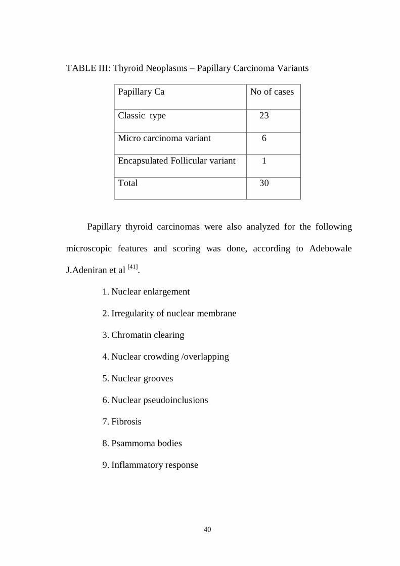
TABLE III: Thyroid Neoplasms – Papillary Carcinoma Variants
Papillary Ca No of cases
Classic type 23
Micro carcinoma variant 6
Encapsulated Follicular variant 1
Total 30
Papillary thyroid carcinomas were also analyzed for the following
microscopic features and scoring was done, according to Adebowale
J.Adeniran et al [41].
1. Nuclear enlargement
2. Irregularity of nuclear membrane
3. Chromatin clearing
4. Nuclear crowding /overlapping
5. Nuclear grooves
6. Nuclear pseudoinclusions
7. Fibrosis
8. Psammoma bodies
9. Inflammatory response

41
The presence of first five features in the sections confined the
specimens into the following scoring categories. If none of the features were
observed the score given is 1, if <10 % of the section studied show above
features the score is 1+, if the section studied revealed 10-50% or >50% of
the above features the scoring was graded as 2+ and 3+ respectively.
The presence of pseudoinclusions were scored as given below .Score
0-No pseudoinclusions /10HPF, Score 1+-1-2 pseudoinclusion /10HPF,
Score 2+ - 3 to 5 pseudoinclusion /10HPF and Score 3+ - >5
pseudoinclusions found in 10 high power fields.
Presence of tumor fibrosis was scored as 0, 1+, 2+, and 3+ for none,
mild, moderate and severe respectively. Presence of inflammatory response
(Chronic inflammatory cell aggregates) and Psammoma bodies were
counted for 10 HPF was scored as 0, 1+ , 2+ and 3+ for 0,1,2 to 5, and > 5
respectively and was correlated with BRAF positive cases as shown in table
Out of 17 cases of follicular neoplasms, 13 cases were folicular
adenoma and 4 were follicular carcinoma. Of the 13 follicular adenomas, 11
were usual type (Fig 9), 1 was signet ring cell type (Fig 10) , and the
remaining one was follicular adenoma with papillary architecture. Among 4
cases of follicular carcinoma, 2 were minimally invasive (Fig 11) , one was
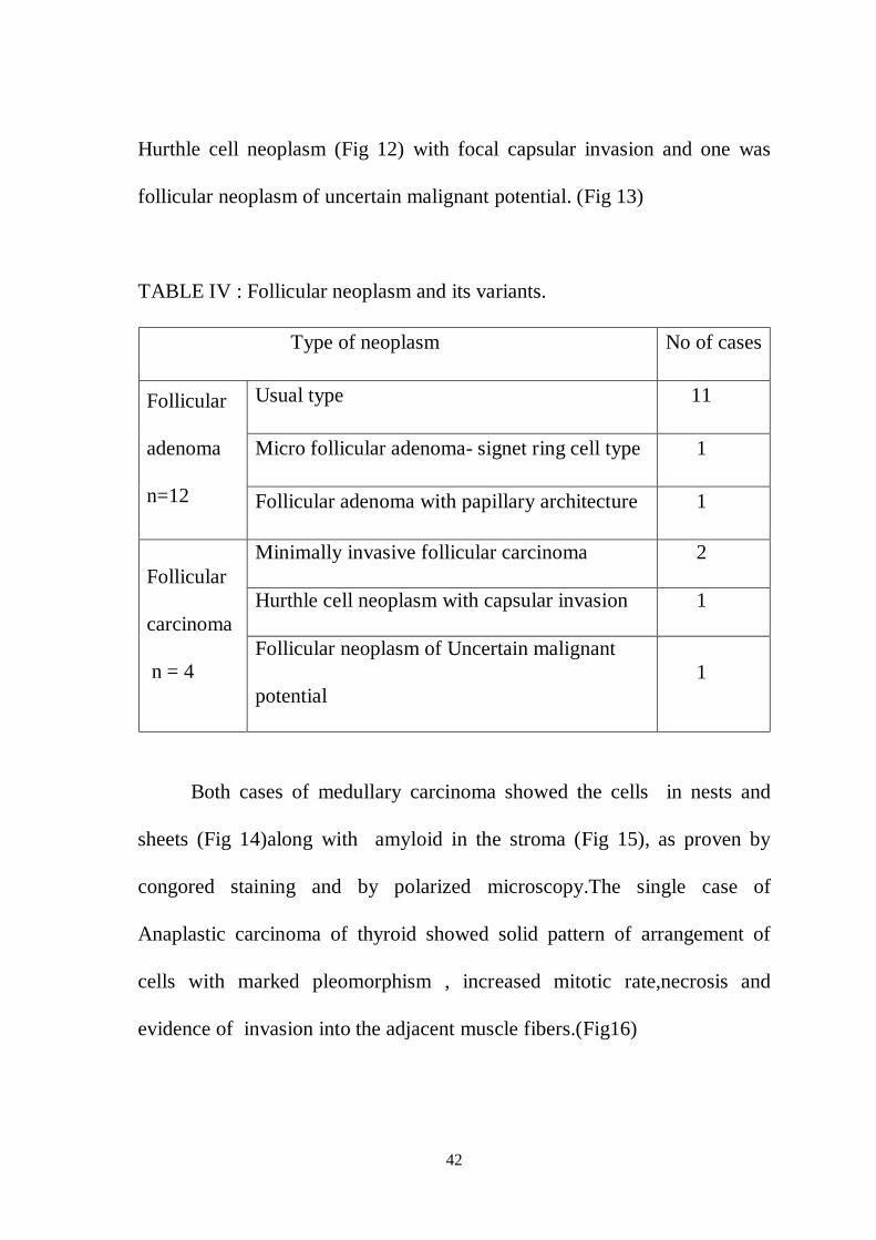
42
Hurthle cell neoplasm (Fig 12) with focal capsular invasion and one was
follicular neoplasm of uncertain malignant potential. (Fig 13)
TABLE IV : Follicular neoplasm and its variants.
Type of neoplasm No of cases
Follicular
adenoma
n=12
Usual type 11
Micro follicular adenoma- signet ring cell type 1
Follicular adenoma with papillary architecture 1
Follicular
carcinoma
n = 4
Minimally invasive follicular carcinoma 2
Hurthle cell neoplasm with capsular invasion 1
Follicular neoplasm of Uncertain malignant
potential 1
Both cases of medullary carcinoma showed the cells in nests and
sheets (Fig 14)along with amyloid in the stroma (Fig 15), as proven by
congored staining and by polarized microscopy.The single case of
Anaplastic carcinoma of thyroid showed solid pattern of arrangement of
cells with marked pleomorphism , increased mitotic rate,necrosis and
evidence of invasion into the adjacent muscle fibers.(Fig16)

43
Staging of these was done according to TNM classification and is as
given in the table V and chart IV.
TABLE V: Thyroid Neoplasms - Stage at Diagnosis
Stage Papillary
carcinoma
Follicular
neoplasms
Medullary
carcinoma
Anaplastic
carcinoma
I 14 17 0 0
II 9 0 0 0
III 7 0 0 0
IVa 0 0 2 0
IVb 0 0 0 1
Total (50) 30 17 2 1
CHART IV: Thyroid Neoplasms - STAGE AT DIAGNOSIS
Total number of cases 50
0
5
10
15
20
STAGE I STAGE II STAGE III STAGE IV a STAGE IV b
NUMBEROF
CASES
STAGE AT PRESENTATION
PAPILLARY CARCINOMA
FOLLICULAR NEOPLASMS
MEDULLARY CARCINOMA
ANAPLASTIC CARCINOMA

44
BRAF MUTATION - RESULTS:
Out of 50 cases studied, DNA could be extracted from 47 cases only.
DNA extraction was not possible for the 3 cases. Of these 47 cases, 14 cases
expressed BRAF mutation. Among these 14, 9 were papillary carcinomas
and 5 were follicular neoplasms. Out of 5 positive follicular neoplasms, 3
were follicular adenoma and the other 2 were follicular carcinoma as shown
in table VI.
TABLE VI: BRAF V600E Positive Cases and Type Of Thyroid Neoplasms
Papillary carcinoma Folicular neoplasm Total
Conventional type Encapsulated
follicular variant
Follicular
Adenoma
Follicular
Carcinoma
14 8 1 3 2
The BRAF V600E mutated cases were analyzed with various
phenotypic features and the results are given in chart V, VI and VII and in
table VII.
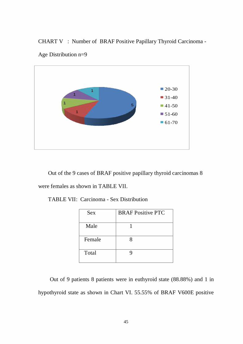
Out of 9 cases of BRAF positive papillary carcinomas, 5 cases were
between the age group of 20-30 yrs and one case each in the 3rd, 4th,5th and
6th decade.
45
CHART V : Number of BRAF Positive Papillary Thyroid Carcinoma -
Age Distribution n=9
Out of the 9 cases of BRAF positive papillary thyroid carcinomas 8
were females as shown in TABLE VII.
TABLE VII: Carcinoma - Sex Distribution
Sex BRAF Positive PTC
Male 1
Female 8
Total 9
Out of 9 patients 8 patients were in euthyroid state (88.88%) and 1 in
hypothyroid state as shown in Chart VI. 55.55% of BRAF V600E positive
5
1
1
11 20-30
31-40
41-50
51-60
61-70

46
cases found to be in stage I( 5/9 cases), stage II was present in 3 cases
(33.33%) and 1 case was found in stage III as given in chart VII.
CHART VI: Number of BRAF Positive Papillary Thyroid Carcinoma-
Hormone Status
CHART VII: Number of BRAF Positive Papillary Thyroid Carcinoma -
Stage at Presentation

47
Scoring for nuclear features and other microscopical features revealed
consistent nuclear features in BRAF positive Papillary Carcinoma s as
shown in Table VIII
TABLE VIII: Scoring for nuclear and other microscopical feature - BRAF
positive papillary carcinoma
S. No NE NI CC NC NG PI
/10HPF Fib
PB
/10HPF
Inflammatory
Cell aggregate
/10HPF
1 1 1 2 3 3 0 1 2 1
2 2 1 3 3 2 0 0 0 0
3 2 1 3 3 2 1 1 0 0
4 2 2 1 3 2 2 1 0 0
5 2 1 3 2 2 1 1 0 0
6 2 1 3 2 1 0 2 2 0
7 2 0 1 3 2 0 0 0 0
8 2 0 1 2 3 0 0 0 0
9 3 3 1 1 2 2 3 3 3
Average
score 2 1.1 2 2.4 2.1 0.6 1 0.7 0.4
NE-Nuclear Enlargement ; NI- Nuclear Irregularity;CC-Chromatin
Clearing;NC-Nuclear Crowding;NG-Nuclear Grooves; PI-Nuclear
Pseudoinclusion;Fib-Fibrosis;PB-Psammoma Bodies.

48
TABLE IX: Distribution of BRAF mutation among different types of
Thyroid Neoplasms
Types of neoplasms BRAF V600E positive (%) Wild Type (%) Total
Papillary Carcinoma 9 (31) 20 (69) 29
Follicular Neoplasm 5 (31.25) 11 (68.75) 16
Medullary Carcinoma 0 1 (100) 1
Anaplastic Carcinoma 0 1 (100) 1
Total 14 (29.8) 33 (70.2) 47
The above table depicts the distribution of BRAF V600E mutation among
different types of thyroid neoplasms.

Fig 3: Agarose Gel Electrophoresis: DNA EXTRACTION
Extraction of DNA from Formalin Fixed Paraffin Embedded tissue .Run on
Agarose gel and was confirmed with a gel doc. Photo which shows the band
confirming the successful DNA isolation.
Fig 4: Agarose Gel Electrophoresis: PCR AMPLIFICATION
Gel electrophoresis image of PCR amplified product, by using 15F and 15R
primers. The product size is 224bp.

Fig 5: Poly Acrylamide Gel Electrophoresis : RESTRICTION
DIGESTION
Control: Well No - 1 (224bp)
Wild type: Well No - 3, 5, 6, 7, 13 (120bp+ 104bp)
Mutant: Well No - 2, 4, 8, 9, 10, 11, 12 (224bp and 224+120+104bp)
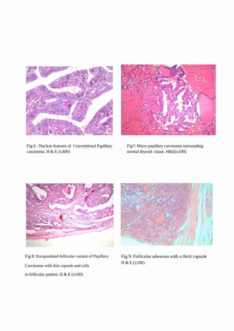
Fig 6 : Nuclear features of Conventional Papillary carcinoma. H & E (x400)
Fig7: Micro papillary carcinoma surrounding normal thyroid tissue. H&E(x100)
Fig 8: Encapsulated follicular variant of Papillary
Carcinoma with thin capsule and cells
in follicular pattern. H & E (x100)
Fig 9: Follicular adenoma with a thick capsule H & E (x100)

Fig 13: Follicular neoplasm uncertain malignant potential (x100)
Fig 10: Signet ring cell type follicular adenoma. H & E (x400)
Fig 11 : Follicular neoplasm with minimal capsular invasion . H & E (x100)
Fig 12: Hurthle cell neoplasm H & E (x400)

Fig 14:Medullary carcinoma H&E (X100) Fig 15:Medullaery carcinoma showing amyloid deposits in the stroma .H & E (x100)
Fig 16.Anaplastic carcinoma H & E (x400)

DISCUSSION

49
DISCUSSION
Malignancies constituted 19.2% among all the biopsies reported at
PSGIMSR during the study period, similar to the incidence reported from
Chennai cancer registry [42]. The thyroid neoplasms constituted 2.16% of all
malignancies (64 over 2953) in this institute and are similar to Chennai
Cancer Registry reports [42] while it is higher than the 1% incidence reported
in the Western countries [12].
Papillary thyroid carcinomas constituted around 64% of thyroid
neoplasms similar to the incidence reported from Chennai cancer registry
(50-90 %).
A report from teaching hospital, Chennai states that the incidence of
papillary thyroid carcinoma is common in coastal areas [43]. Coimbatore is
situated in the western part of Tamilnadu, and is a non-coastal, iodine
deficient area as reported by a population survey conducted in 1991, by the
Tamilnadu health and family welfare. However, the population of
Coimbatore receives dietary iodine supplementation in the form of iodized
salt from 1994[8]. A population survey indicates that high dietary iodine is
associated with a high risk of thyroid neoplasms especially papillary
carcinoma of thyroid [9]. Given the above observations, it is important to

50
plan further studies to estimate average population iodine levels in
Coimbatore.
Further, BRAF V600E mutation is associated with papillary thyroid
carcinomas. Identification of this mutation has changed the algorithm of
treatment approach to papillary thyroid carcinomas. Some of the prevailing
local factors indicated by our biopsies including the increasing incidence,
female preponderance, younger age at presentation and an increase in
incidental finding of papillary carcinoma in thyroidectomy specimens along
with the potential for altering the treatment approach to these patients,
prompted us to do this study.
An extensive literature search on Indian studies for BRAF mutation in
thyroid carcinoma indicated a paucity of literature. For example, a Pubmed
search (from1960-2010) with MESH keywords of “BRAF” and “thyroid
neoplasm” and “India” returned no reports. Thus, we considered it to be
important for us to do this study.
We identified and retrieved paraffin blocks of 50 cases of thyroid
neoplasms from archives of pathology using exclusion and inclusion criteria
as discussed earlier. We could extract DNA from 47 cases only. DNA
extraction was not possible for 3 cases (one each of papillary carcinoma,

51
follicular adenoma and medullary carcinoma) probably due to improper
fixation.
BRAF V600E mutation when present in papillary carcinoma of
thyroid indicates a poorer outcome of the disease [26, 27]. Studies also state
that, though this mutation can be seen in early stages, this is most often
expressed in stage III (30%) when compared to Stage I. The expression of
BRAF mutation in early stages indicates a possible aggressive course and
warrants total thyroidectomy .When present in later stages of the disease,
this is associated with a poorer outcome.
In our study group of 30 papillary carcinomas, DNA was extracted
from 29 cases. 9 cases were positive for BRAF V600E mutation. Hence
31% ( 9/29cases) of papillary carcinoma thyroid expressed BRAF V600E
mutation. Literature search states that BRAF V600E mutation is reported in
40-45% of papillary thyroid carcinomas [22] and we infer from our study that
there is a lesser incidence of V600E mutation expression in papillary thyroid
carcinomas in the subset of population that received treatment in our centre
.But interestingly we found BRAF V600E mutation in 31% of cases of
follicular neoplasms, which is assessed and discussed further down.
Out of 9 cases expressing V600E mutation 5 cases were in the age
group between 20-30 years. Rossella et al [5] states that V600E is expressed

52
in papillary carcinoma occurring in older age group. In our study this is seen
in a younger age group. BRAF V600E is known to induce a more
aggressive phenotype and therefore this may have resulted in clinical
expression in younger age group. As V600E mutation is also associated
with an aggressive course, total thyroidectomy is warranted in cases
expressing V600E in early age group. Therefore surveillance for mutation
especially on FNA samples will prevent more repeat surgeries and help in
better follow-up care of the patient.
5 cases of stage I, 3 cases of stage II and 1 case of stage III expressed
V600E mutation. The analysis of our study shows a significantly higher
expression in stage I and stage II lesions. Therefore we reanalyzed the
reports of papillary thyroid carcinoma. As we are in a tertiary care hospital
with advanced facilities for patient care, radiologist and pathologist were
probably able to detect papillary thyroid carcinoma in early stages, thereby
explaining the higher incidence of V600E mutation in stage I papillary
thyroid carcinomas.
There was no significant correlation with the nuclear features and
BRAF V600E mutation but nuclear crowding, overlapping and grooves
were consistent finding (Table VIII).

53
As previously noted, 5 cases of follicular neoplasm expressed BRAF
V600E mutation with a percentage incidence of 31% .Literature survey
states that V600E mutation is not commonly seen in follicular neoplasms
and a few studies quote no more than 1% incidence of this mutation in
follicular neoplasms [35, 44]. Therefore reanalysis of routine H&E slides were
done and 2 cases had focal papillary architecture with nuclear features
which might have been overlooked in morphological assessment .Excluding
these 2 cases the percentage incidence of V600E mutation in follicular
neoplasm is 18.75%.As bits are given only from the representative areas and
are not sampled extensively on a routine basis, it is likely that papillary
carcinoma especially micro follicular type could have been missed.
Therefore from our study, we infer that a significant number i.e., 31%
of papillary carcinoma (9/29) and 18.75% of follicular neoplasms (3/16)
express V600E mutation. This mutation was expressed more in stage I of
papillary thyroid carcinoma. As BRAF mutation is associated with
aggressive behavior, a close follow up and total thyroidectomy becomes
essential. If mutational studies are done on cytology specimens prior to
surgery, there might be a reduction in number of repeat completion
thyroidectomy procedure as total thyroidectomy is treatment of choice in
BRAF positive cases. It might also be useful to do BRAF mutation studies

54
where there are no firm diagnosis possible by cytology. A larger prospective
study including cytology specimen prior to surgery and surgical resection
specimens of the same patients is essential to prove the utility of V600E
mutation as a routine diagnostic tool. Once developed the primers become
cost effective to the patient. Also as targeted therapies are available,
mutational studies might be the future diagnostic methods of these tumors.
It has been demonstrated that there is an increase in the incidence of
papillary carcinoma where there is high iodine intake [3, 7, 15] either in diet or
in the form of iodine supplementation in a population where there is an
existing V600E mutation. This raises the question over routine high iodine
supplementation for preventing non-neoplastic conditions such as
multinodular goiter. A larger multicentric, prospective study addressing this
might prove to be useful.
As our study showed an increased expression of BRAF V600E
mutation in follicular neoplasms, a larger study of V600E mutation in
follicular neoplasms is vital to look for a change in pattern of expression of
mutational status in Indian population. Algorithm of our approach to this
study is given in Fig 17.

55
Fig 17: Algorithm of the study
Total number of thyroid neoplasms during the study period: 64
Selected cases: 50
30 17 2 1
PTC FN MC AC
• 23 :Classic type
• 6 : Micro ca
• 1: Encapsulated
Follicular variant
• 13: FA
• 4: FC
29 16 1 1
9 -MUTANT
20-WILD
5 -MUTANT
11-WILD 1-WILD 1-WILD
RESULT
PTC- Papillary Thyroid Carcinoma ; FN- Follicular Neoplasm ; FA- Follicular Adenoma;
FC-Follicular Carcinoma; MC-Medullary Carcinoma ; AC-Anaplastic Carcinoma
Exclusion of 14 cases
DNA
EXTRACTION
(47)

SUMMARY &
CONCLUSION

56
SUMMARY AND CONCLUSION
In conclusion, our study on thyroid specimens reported at
PSGIMS&R from January 2006 to September 2009 revealed a 31%
incidence of papillary carcinoma and 18.75% incidence of follicular
neoplasms expressing BRAF V600E mutation. We also infer that BRAF
V600E mutation was seen more in stage I and therefore a close follow up or
completion thyroidectomy becomes essential. The limitation of our study
includes small sample size. Therefore a larger multicentric study
prospective study on both FNA and surgical specimen with long term follow
up is essential to prove the utility of BRAF V600E mutational study as a
routine diagnostic tool for treatment and prognostication.
Probably ,epidemiologic survey of iodine levels and V600E mutation
in patients with multinodular goiter and cases where incidental papillary
thyroid carcinoma reported might go a long way in developing screening
modalities for thyroid neoplasms and prevention of this increasing incidence
of thyroid neoplasms.

BIBLIOGRAPHY

BIBLIOGRAPHY
1. Maria Luisa Carcangiu.Thyroid. In, Stacey E.Mills (ed) ‘Histology
for pathologists’, 3rd edition.Lippincott Williams and Wilkins.2007;
1129-1148.
2. Anirban Maitra, Abul K. Abbas. The Endocrine System. In, Kumar,
Abbas, Fausto (Ed). Robbins and Cotran Pathological basis of disease,
7th edition. Elsevier, W.B. Saunders, 2007; 1155-83.
3. World Health Organization Classification of Tumours. In, Pathology
and Genetics of Tumours of Endocrine Organs. Ed. C Eng, Lyon:
IARC Press, 2004.
4. Carol.C.Cheung,Shereen Ezzat , Jeremy L.Feremy et al.
Immunohistochemical Diagnosis of Papillary Thyroid Carcinoma.Mod
Pathol 2001; 14(4):338-342.
5. Rossella Elisei,Clara Ugolini, David Viola et al.BRAF V600E
Mutation and Outcome of Patients with Papillary Thyroid Carcinoma:
A 15 year Median Follow-Up Study.Journal of Clinical Endocrinolgy
and Metabolism .2008;(93):3943-3949.

6. Nabeel Al-Brahim,Sylvia L.Asa.Thyroid carcinoma.An
overview.Arch Pathol Lab Med.2006;130:1057-1062.
7. Harach HR,Escalanate,Day ES.Thyroid cancer and thyroiditis in
Salta , Argentina:a 40-year study in relation to iodine
prophylaxis.Endocr Pathol 2002;13(3):175-81.
8. Health and family Welfare Department.Government of Tamilnadu.
National Iodine Deficiency Disorders Control Programme Status
Report. http://www.tnhealth.org/dphdbiod.htm.
9. Investigating Thyroid Cancer N.Dorairajan.
http://hindu.com/2001/01/21/stories/1321048d.htm.
10. Zubair W. Baloch, Virginia A. Livolsi. Pathology of thyroid gland.
In, Livolsi (Ed). Endocrine pathology. Churchill Livingstone,
Philadelphia, 2002; 61-102.
11. Chan JKC. Tumours of the thyroid and parathyroid glands. In,
Fletcher CDM (Ed). ‘Diagnostic Histopathology of Tumours’, 3rd
edition, volume 2. Churchill Livingstone, Edinburgh, 1995; 705-64.

12. Rosai J. Thyroid gland. In, Rosai J (Ed). Rosai and Ackerman’s
surgical pathology, 9th edition. St Louis, MO: Mosby, 2004; 515-94.
13. Zubair W. Baloch, Virginia A. Livolsi. Pathology of thyroid and
parathyroid disease. In, Stacey E. Mills (Ed). Sternberg’s Diagnostic
pathology, 4th edition. Lippincott Williams and Wilkins, Philadelphia,
2004; 557-620.
14. Livolsi VA. Papillary neoplasms of the thyroid. Pathologic and
prognostic features. Am. J. Clin. Pathol. 1992; 97:426- 43.
15. Harach HR,Ceballos GA.Thyroid cancer,thyroiditis and dietary
iodine:a review based on the Salta,argentina model.Endocr Pathol.2008
Winter;19(4):209-20.
16. Dubravka Matesa-Anic, Neven Matesa,Nina Pabelic.Coexistence Of
Papillary Carcinoma and Hashimoto’s Thyroiditis.Croat 2009;48:9-12.
17. Rosria ,Alferedo,serio et al.What is New on Thyroid cancer
Biomarkers. Biomarker Insights.2008; 3:237-252.
18. Ronald A.Delellis.Pathology and Genetics of Thyroid
Carcinoma.Journal of Surgical Oncology 2006; 94:662-669.

19. Hussam AL Humadi,Apostolos Zareos,Rafal et al.Genetic Basis and
Gene Therapy Trials for Thyroid Cancer.Cancer Genomics and
Proteomics.2010; 7:31-50
20. Fulvio Basolo, Liborio, Riccardo, Mario, Cristiana, Elisa Sensi et al.
Correlation between the BRAF V600E Mutation and Tumor
Invasiveness in Papillary Thyroid Carcinomas Smaller than 20
Millimeters: Analysis of 1060 Cases. J Clin Endocrinol Metab, Sep
2010, 95(9):4197–4205.
21. Efsevia Vakiani , David B Solit.K-RAS and BRAF :drug targets and
predictive biomarkers.J Pathol.2011;223:219-229.
22. Yorum cohen,Mingzhao,Elizabeth,Zhongmin,Guogern,Barry et
al.BRAF Mutation in Papillary Thyroid Carcinoma.Journal of the
National Cancer Institute.2003;95(8).
23. Mingzhao Xing, Recent Advances in Molecular Biology of Thyroid
Cancer and Their Clinical Implications. Otolaryngol Clin North Am.
2008 Dec; 41(6): 1135–1146.

24. Yuri E.Nikiforov.Molecular Diagnostics of Thyroid tumors.Arch
Pathol Lab Med.2011; 135.
25. Paul,Mathew,Mark Roe,Sharlene ,Dan Niculescu,Valerie et
al.Mechanism of Activation of RAF-ERK Signalling Pathway by
Oncogenic Mutations of B-RAF.Cell .2004;116:855-867.
26. Mingzhao Xing.BRAF Mutation in Papillary Thyroid Carcinoma:
Pathogenic Role,Molecular Bases,and Clinical Implications.Endocrine
reviews .2007;28(7):742-762.
27. Meredith.A.Kato,Thomas J.Fahey.Molecular Markers in Thyroid
Cancer Diagnostics. Surg Clin N Am. 2009; 89:1139-1155.
28. Kepal N.Patel,Bhuvanesh Singh.Genetic Coonsiderations in Thyroid
cancer.Cancer Control.2006;13(20).
29. Yuri E Nikiforov.Molecular analysis of thyroid tumors.Modern
Pathology.2011; 24:S34-S43.
30. Nucera C,Lawler ,Parangi S.BRAF V600E and microenvironment in
thyroid cancer.A functional link to drive cancer progression.Cancer
Res.2011;7:2417-22.
31. Ying C. Henderson, Thomas D, Shellenberger, El-Naggar, Fredrick,
Kathleen et al. High Rate of BRAF and RET/PTC Dual Mutations
Associated with Recurrent Papillary Thyroid Carcinoma. Clinical
Cancer Research, 2009; 15: 485.
32. Tetsuo Kondo,Shereen Ezzat and Sylvia L.Asa.Pathogenetic
mechanisms in thyroid follicular cell neoplasia.Nature
Reviews.2006;Vol6.
33. Christiana ,Riccardo,Clara,Agnese,Piero,Michele et al.Association of
BRAF V600E Mutation with Poor Clinicopathological Outcomes in
500 Consecutive Cases of Papillary Thyroid Carcinoma.The Journal of
Clinical Endocrinology & Metabolism.2007;92(11):4085-4090.
34. Hirataka Akira,Yoshiyasu,Hiroyuki Youhei,Nobuyuki.Clinical
Significance of BRAF (V600E) Mutation and Ki-67 Labelling Index in
Papillary Thyroid Carcinomas.Anticancer Reasearch .2007;27:3645-
3650.
35. Electron kebebew,Julie Weng, Juergen Bauer , Gustavo ranvier ,Orlo
H.Clark,Quan-Yang Duh et al.The prevalence and Prognostic Value of
BRAF Mutation in Thyroid Cancer .Annals of Surgery 2007;3:246.

36. F Frasca , C Nucera ,G Pellegritti ,P Gangemi, M Attard, M
Stella.BRAF (V600E) mutation and the biology of papillary thyroid
cancer.Endocrine –Related Cancer.2008;15:191-205.
37. Giuliana Salvatore, Riccardo, Pinuccia, Alessia caleo, Ilenia,James
fagin et al. Analysis of BRAF Point Mutation and RET/PTC
Rearrangement Refines the Fine-Needle Aspiration Diagnosis of
Papillary Thyroid Carcinoma. The Journal of Clinical Endocrinology &
Metabolism 89(10):5175–5180
38. Leslie R Rowe, Brandon G Bentz, Joel S Bentz . Utility of BRAF
V600E mutation detection in cytologically indeterminate thyroid
nodules.Cytojournal 2006 3:10.
39. Maria Rosaria ,Daniela Posca,Giancarlo,Guido Pettinato,Lucia,
Guido et al.Detection of BRAF mutation in thyroid papillary
carcinomas by mutant allele-specific PCR
amplification(MASA).European Journal of
Endocrinology.2006;154:341-348.

40. Angela, Agustin, manuel, Jonas, Jose Antonio, Jose Cameselle et
al.BRAF mutation associated with other genetic events identifies a
subset of aggressive Papillary Thyroid Carcinoma.Clinical
Endocrinology.2008;68:618-634.
41. Adebowale J.Adeniran ,Zhaowen, Manoj Gandhi, David L,James ,
Thomas et al.Correlation between genetic alterations and microscopic
features,clinical manifestations and prognostic characteristics of thyroid
papillary carcinomas.Am J Surg Pathol.2006;30:216-222.
42. Consolidated Report of Hospital Based Cancer registries 2001-2003.
National Cancer Registry Programme .Indian Council of Medical
Research. New Delhi. April 2007.
43. Pandiarajan, Yuvaraja S. A descriptive study of papillary thyroid
carcinoma in a teaching hospital in Chennai, India. Asian journal of
surgery Oct 2002; 25 (4):300-303.
44. Paula Soares, Vitor Trovisco, Ana Sofia, Jorge, Patricia, Ana Preto.
BRAF mutations and RET/PTC rearrangements are alternative events
in the etiopathogenesis of PTC. Oncogene (2003) 22, 4578–4580.

MASTER CHART

MASTER CHART
S. No HP. No
Age/ Sex
Hormone Status Size Other features Diagnosis Stage BRAF
1. 150/06 30/M Euthyroid 2.6cm - Minimally invasive follicular carcinoma
I Mutant
2. 458/06 48/F Euthyroid 0.4cm - Papillary microcarcinoma I Mutant
3. 747/06 49/F Euthyroid 3.5cm Multifocal, cystic Papillary carcinoma II Wild
4. 888/06 50/F Euthyroid NA Extrathyroidal extension
Papillary carcinoma III Wild
5. 1253/06 51/F Hyperthyroid 0.5cm Multifocal Papillary microcarcinoma I NE
6. 1562/06 32/F Euthyroid 2.3cm - Follicular adenoma I Wild
7. 1801/06 38/M Euthyroid 1.8cm Lymph node involvement Medullary carcinoma IV A Wild
8. 1906/06 48/M NA 4.2cm - Follicular adenoma I NE
9. 2093/06 23/F Euthyroid 5.5cm Multifocal Papillary carcinoma III Wild
10. 2110/06 43/M Euthyroid 2.5cm - Follicular adenoma with papillary architecture I Mutant
11. 2230/06 57/M Euthyroid 3.0cm - FN- UMP I Wild
12. 2232/06 58/M Euthyroid 7.0cm - Microfollicular adenoma- signet ring cell type
I Mutant
13. 2780/06 52/F Euthyroid 0.4-
2.3cm Multifocal Papillary carcinoma II Mutant

14. 2838/06 35/F Euthyroid 9.5cm - Follicular adenoma I Wild
15. 3173/06 34/F Euthyroid 3.0cm - Follicular adenoma I Wild
16. 29/07 51/F Euthyroid 3cm
Cystic Papillary carcinoma II Wild
17. 220/07 70/M Euthyroid 2.0-
4.0cm
Multifocal Papillary carcinoma II Wild
18. 307/07 38/M Euthyroid 0.5cm -
Papillary microcarcinoma I Wild
19. 600/07 67/M Hypothyroid 5.5cm Lymph node involvement
Medullary carcinoma IV A NE
20. 1310/07 43/F Euthyroid 3.3cm -
Follicular adenoma I Wild
21. 1560/07 50/F Euthyroid 0.3-
1.0cm
Multifocal, cystic
Papillary carcinoma I Wild
22. 1572/07 30/F Euthyroid 0.4cm
-
Papillary microcarcinoma I Wild
23. 1651/07 36/F Euthyroid 5.5cm -
Follicular adenoma I Wild
24. 2118/07 45/F Euthyroid 2.5cm -
Follicular adenoma I Wild
25. 2710/07 34/F Euthyroid 3.0cm -
Follicular adenoma I Wild
26. 2874/07 65/F Hypothyroid 1.3cm - Papillary carcinoma I Mutant
27. 3335/07 45/F Euthyroid 6.0cm - Hurthle cell neoplasm I Mutant

28. 3478/07 35/F Hyperthyroid 4.5cm - Follicular adenoma I Wild
29. 3679/07 27/F Euthyroid 3.0cm -
Papillary carcinoma II Wild
30. 3693/07 46/F Euthroid 0.8-
5.0cm
Multifocal
Papillary carcinoma III Wild
31. 648/08 55/F Euthyroid 1.0cm
- Follicular adenoma I Wild
32. 1101/08 30/F Hypothyroid 3.0cm - Minimally invasive follicular carcinoma
I Wild
33. 1376/08 24/F Euthyroid 5.5cm Cystic Papillary carcinoma III Wild
34. 1422/08 53/M Euthyroid 2.0cm - Papillary carcinoma I Wild
35. 1837/08 30/F Euthyroid 0.3cm - Encapsulated follicular
variant of Papillary carcinoma
I Mutant
36. 1865/08 29/M Euthyroid 2.5cm - Papillary carcinoma II Mutant
37. 2062/08 35/F Euthyroid 4.0cm - Follicular adenoma I Mutant
38. 2222/08 27/F Euthyroid 2.4cm - Papillary carcinoma II Mutant
39. 2850/08 25/M Euthyroid 0.5cm - Papillary microcarcinoma
I Wild
40. 3187/08 27/F Hyperthyroid 0.5cm - Papillary microcarcinoma I Wild
41. 4248/08 32/F Euthyroid 1.5cm - Papillary carcinoma I Wild

42. 011/09 45/F Euthyroid 3.5cm - Papillary carcinoma II Wild
43. 229/09 28/F Euthyroid 0.3, 1.4cm
Multifocal, cystic Papillary carcinoma I Mutant
44. 824/09 41/F Euthyroid 0.3-
0.8cm
Multifocal, stromal bone
formation Papillary carcinoma I Wild
45. 915/09 50/M Euthyroid 2.0cm Extrathyroidal extension
Papillary carcinoma III Wild
46. 920/09 59/M Euthyroid 1.0-
3.0cm Multifocal Papillary carcinoma II Wild
47. 1218/09 32/F Euthyroid 1cm Cystic Papillary carcinoma I Mutant
48. 2548/09 20/F Euthyroid 3.5cm
Multifocal, capsular and lymph node involvement
Papillary carcinoma III Mutant
49. 2662/09 40/F Euthyroid 11.5cm Extra thyroidal extension Anaplastic carcinoma IVB Wild
50. 3561/09 45/F NA 3.5cm Lymph node involvement Papillary carcinoma III Wild
FN- UMP – Follicular neoplasm of uncertain malignant potential;
NE- DNA Not Extracted.