Embed Size (px)
Citation preview
B R A I N R E S E A R C H 1 2 5 2 ( 2 0 0 9 ) 1 1 7 – 1 2 9
ava i l ab l e a t www.sc i enced i rec t . com
www.e l sev i e r. com/ l oca te /b ra in res
Research Report
Expression of chondroitin sulfate proteoglycans in barrel fieldof mouse and rat somatosensory cortex
Michiru Nakamuraa, Keiko Nakanoa, Shoko Moritaa, Toshihiro Nakashimaa,Atsuhiko Oohirab, Seiji Miyataa,⁎aDepartment of Applied Biology, Kyoto Institute of Technology, Matsugasaki, Sakyo-ku, Kyoto 606-8585, JapanbDepartment of Perinatology and Neuroglycoscience, Institute for Developmental Research, Aichi Human Service Center, Kasugai,Aichi 480-0392, Japan
A R T I C L E I N F O
⁎ Corresponding author. Fax: +81 75 724 7796.E-mail address: [email protected] (S. MiyAbbreviations: Chase, chondroitinase ABC;
sulfate proteoglycans; CS-6-PG, chondroitinnoglycan; GFAP, glial fibrillar acidic protein; Nday; PNNs, perineuronal nets; PNA, peanut aagglutinin
0006-8993/$ – see front matter © 2008 Elsevidoi:10.1016/j.brainres.2008.11.022
A B S T R A C T
Article history:Accepted 4 November 2008Available online 18 November 2008
Chondroitin sulfate proteoglycans (CSPGs) consist of chondroitin sulfate (CS)glycosaminoglycans (GAGs) and core protein and regulate the migration, axonaloutgrowth, and synaptogenesis in mammalian brains. In the present study, weinvestigated the localization of CSPGs, the effects of sensory deprivation on the densityof perineuronal nets (PNNs), and the effects of chondroitinase ABC (Chase) on theformation of barrel structures in the posterior medial barrel subfield (PMBSF). Indeveloping mouse and rat brains, the immunoreactivity of chondroitin-6-sulfatecontaining proteoglycan (CS-6-PG), phosphacan, and neurocan was stronger at barrelsepta as compared with barrel hollows and surrounding cortex, while the labeling ofWisteria floribunda agglutinin (WFA) was observed at barrel hollows. In adult brains, CS-6-PG-immunoreactive and WFA-labeled PNNs were observed mainly at barrel hollows ofmouse, but they were seen chiefly at barrel septa of rats. Sensory deprivation of facialvibrissae reduced the number of WFA-labeled PNNs at barrel hollows but not at barrelsepta. Intracerebral injection of Chase did not affect the formation of barrel structures inthe PMBSF. These data indicate species-dependent heterogeneity of CSPG expression andactivity-dependent formation of PNNs in the PMBSF, but CS GAGs have no crucial functionin constructing the barrel structures during early postnatal development.
© 2008 Elsevier B.V. All rights reserved.
Keywords:Extracellular matrixGlycosaminoglycanBarrelCritical periodSensory deprivation,Perineuronal netsChondroitin sulfateProteoglycan
1. Introduction
The rodent somatosensory system is an excellent model tostudy neuronal pattern formation and neuronal plasticity(Fox, 1992; Frostig, 2006; for review, see Peterson, 2007). In
ata).ConA, concanavalin A; CO-6-sulfate containing proeuN, neuron-specific nucgglutinin; VVA, Vicia villosa
er B.V. All rights reserved
the primary somatosensory cortex, periphery-related pat-terns of sensory receptors are recapitulated in layer IV as anarray of multineuronal structures called “barrels” (Woolseyand Van der Loos, 1970). In barrels, neurons are arranged incylindrical or oval-shaped rings that are separated from
, cytochrome oxidase; CS, chondroitin sulfate; CSPGs, chondroitinteoglycan; ECM, extracellular matrix molecules; GAG, glycosami-lear antigen; PMBSF, posterior medial barrel subfield; PN, postnatalagglutinin; WFA,Wisteria floribunda agglutinin; WGA, wheat germ
.
118 B R A I N R E S E A R C H 1 2 5 2 ( 2 0 0 9 ) 1 1 7 – 1 2 9
neighbors by hypocellular “septa”. Those cell-dense wallssurround cell-sparse “hollows” containing thalamocorticalafferent terminals (Woolsey and Van der Loos, 1970). Theremoval of sensory information by whisker follicle cauter-ization or denervation induces a disorganization of struc-tural barrel pattern. These barrel structures are consolidatedduring the first few days (critical period) through somato-sensory inputs from whiskers, so that the removal ofsensory information has little effect upon the organizationof barrel structures over postnatal day 5 (PN5) (Van der Loosand Woolsey, 1973; Woolsey and Wann, 1976; Wong-Rileyand Welt, 1980; Schlaggar et al., 1993). The organization ofbarrel structures is primarily determined by a genetic pro-gram such as FGF-8 as well as the general patterning of theneocortex (Fukuichi-Shimogori and Grove, 2001). Moreover,refinement of barrel structures is likely to be guided byactivity-dependent mechanisms, since barrel structures areless clearly defined or absent in mice with genetic knockoutof several genes such as NMDA receptor (Iwasato et al.,2000), phospholipase C β1 (Hannan et al., 2001), adenylcyclase 1 (Welker et al., 1996), and monoamine oxidase A(Cases et al., 1996).
The extracellular matrix (ECM) molecules are involved incell–substrate interactions during morphogenesis of thenervous system in developing brains and plastic modifica-tions of neuronal connectivity in adult brains. Chondroitinsulfate proteoglycans (CSPGs) have been recognized asimportant constituents of the brain's ECM and are con-sidered to participate in neural network formation (forreviews, see Bovolenta and Fernaud-Espinosa, 2000; Galtreyand Fawcett, 2007). CSPGs consist of a core protein andsulfated glycosaminoglycans (GAGs) (Bandtlow and Zimmer-mann, 2000; Yamaguchi, 2000) and are generally known toinhibit neurite outgrowth (Oohira et al., 1991; Katoh-Senbaet al., 1995). Moreover, recent studies have shown that CSPGsare responsible for structural plasticity in adult brains suchas functional recovery after spinal cord injury (Bradburyet al., 2002), axonal sprouting of cerebellar Purkinje neurons(Corvett and Rossi, 2005), and reactivation of ocular dom-inance plasticity in the visual cortex (Pizzorusso et al., 2002,2006).
Perineuronal nets (PNNs) are a specialized form of ECMthat appears at the end of the critical period (Celio et al.,1998). PNNs have been visualized using a variety of methodsincluding the labeling of lectins such as Wisteria floribundaagglutinin (WFA) and Vicia villosa agglutinin (VVA) that havehigh affinities to N-acetylgalactosamine (Härtig et al., 1992;Brückner et al., 1993), the binding of hyaluronic acid bindingprotein to hyaluronic acid (Brückner et al., 1998, 2000), andimmunological staining for CSPGs (Brückner et al., 1998,
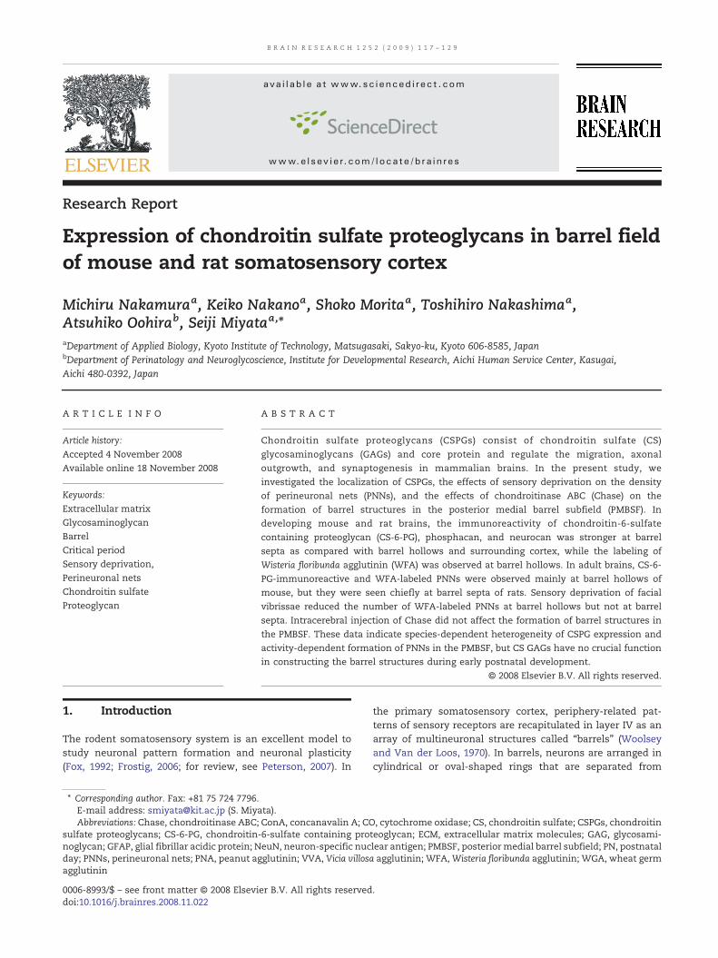
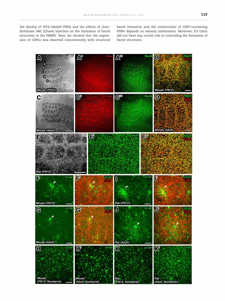
Fig. 1 – Tangential sections showing the histochemical stainingimmunohistochemical staining of NeuN andGFAP in the PMBSF oand adult mice, barrel structures were clearly observed by CO anmagnification view and each barrel hollow was seen clearly by dat highmagnification view. (E–E″) In rats, barrel structureswere clclearly seen by NeuN immunostaining and Nissl staining. (F–K′)(arrowheads) of GFAP-positive astrocytes distributed in the PMBat surrounding cortex (nonbarrel cortex). Scale bars=A and C (50
2000). PNNs are frequently seen around parvalbumin-immu-noreactive GABAergic interneurons in the cerebral cortex(Brückner et al., 1994; Brückner and Grosche, 2001). Duringpostnatal development, PNNs are first detected at the onsetof the period during which the pattern of neuronal activitydetermines the mature synaptic circuitry and neuronalphenotype in the visual cortex (Köppe et al., 1997; Brückneret al., 2000). It is also shown that dark-rearing from birthprolongs the duration of the critical period and attenuates theexpression of several CSPGs (Lander et al., 1997; Pizzorussoet al., 2002). PNNs are well preserved and maintained for 3–10 weeks in slice culture, indicating that the differentiation ofPNNs is part of the developmental program maintained inbrain tissue in vitro (Brückner and Grosche, 2001). It is alsoshown that the formation of PNNs in brain slice culture ispromoted via calcium signaling (Brückner and Grosche, 2001).More recently, we have demonstrated that neurons them-selves are able to construct a PNNs structure in dissociatedcortical culture without glial cells (Miyata et al., 2005, 2007),and later this was confirmed in hippocampal neuron culture(John et al., 2006).
Various ECM molecules are shown to be expressed speci-fically in the somatosensory barrel cortex. Lectin histoche-mistry has revealed that lectin binding is absent from barrelhollows in the early postnatal period using peanut aggluti-nin (PNA), concanavalin A (ConA), and wheat germ agglu-tinin (WGA) (Cooper and Steindler, 1986; Steindler et al.,1995). It has been demonstrated that CSPGs such asneurocan and DSD-1-PG are also absent from barrel hollowsin the early developing brains (Watanabe et al., 1995;Steindler et al., 1995). Tenascin-C, which is able to interactwith CSPGs, is shown to be expressed predominantly atbarrel septa, but tenascin-C-deficient mice show normaldistribution of DSD-1-PG immunoreactivity and PNA labeling(Steindler et al., 1995; Mitrovic et al., 1996; Cybulska-Klosowicz et al., 2004). A recent study has shown that trim-ming of facial vibrissae attenuates the expression of Cat-315-immunoreactive aggrecan without changing the numberof WFA-labeled PNNs (McRae et al., 2007). Moreover, thedeprivation of sensory information from facial vibrissaedecreases the number of VVA-labeled PNNs in the PMBSF(Bahia et al., 2008).
At present, however, the functional significance of CSPGsin pattern formation of barrel structures is not completelyunderstood. In the present study, therefore, we examinedthe spatiotemporal expression of CS GAGs and major CSPGssuch as phosphacan and neurocan in the PMBSF of thesomatosensory cortex using rats and mice to show theheterogeneity of CSPG expression. Furthermore, we investi-gated the effects of sensory deprivation of facial vibrissae on
of CO and red-fluorescence Nissl and thef the somatosenosry cortex usingmice and rats. (A–D) In PN10d Nissl stainings and NeuN immunostaining at lowouble labeling of Nissl staining and NeuN immunostainingearly observed by CO staining, but each barrel hollowwas notThe immunohistochemistry revealed that aggregatesSF of mice and rats in spite of uniform distribution of them0 µm), B, D, and E–K (100 µm).
119B R A I N R E S E A R C H 1 2 5 2 ( 2 0 0 9 ) 1 1 7 – 1 2 9
the density of WFA-labeled PNNs and the effects of chon-droitinase ABC (Chase) injection on the formation of barrelstructures in the PMBSF. Here, we showed that the expres-sion of CSPGs was observed concomitantly with structural
barrel formation and the construction of CSPG-containingPNNs depends on sensory information. Moreover, CS GAGsdid not have any crucial role in controlling the formation ofbarrel structures.
120 B R A I N R E S E A R C H 1 2 5 2 ( 2 0 0 9 ) 1 1 7 – 1 2 9
2. Results
2.1. Comparison of PMBSF structures in mouse and rat
The PMBSF is a subfield of the rodent primary somatosensorycortex that represents 5 rows of large whiskers with exquisiteorder. In the PMBSF of PN10 and adult mice, a typical barrelstructure was clearly visible by staining presynaptic terminalsof thalamic afferent nerves with cytochrome oxidase (CO)
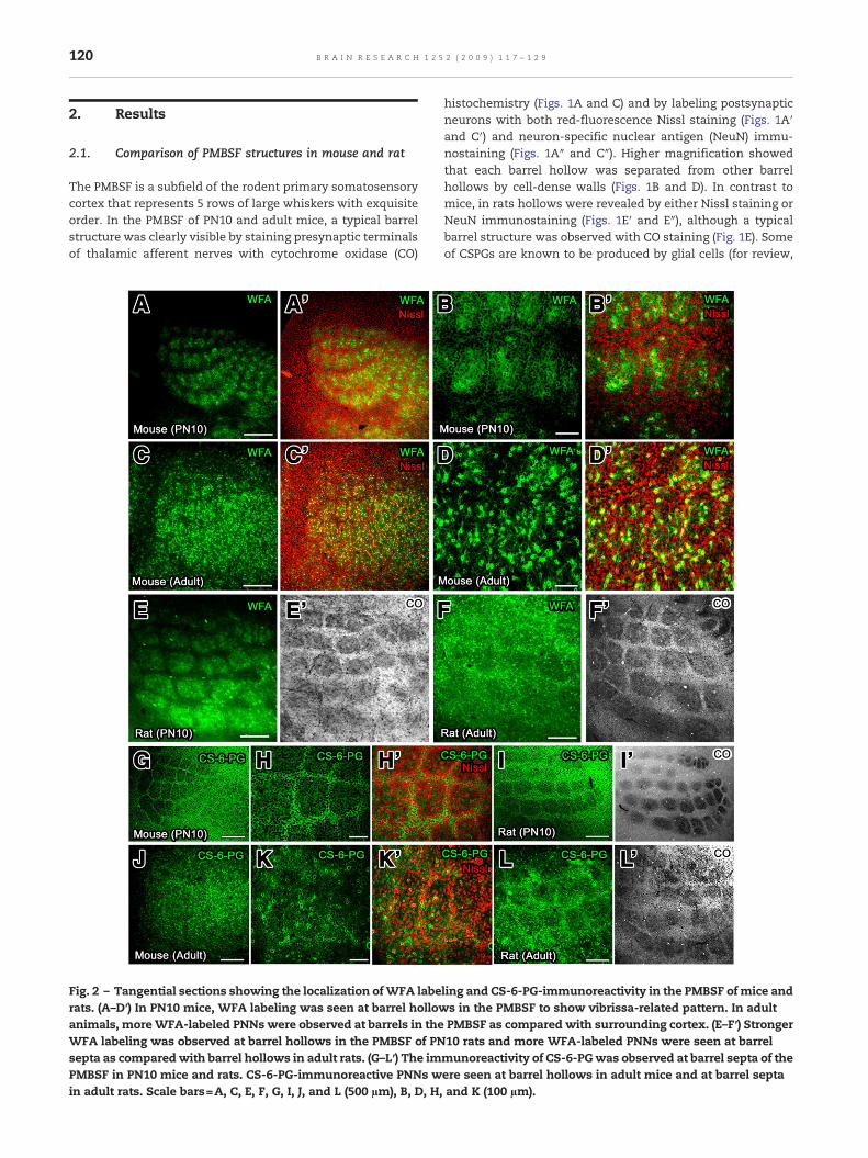
Fig. 2 – Tangential sections showing the localization ofWFA laberats. (A–D′) In PN10 mice, WFA labeling was seen at barrel hollowanimals, moreWFA-labeled PNNs were observed at barrels in theWFA labeling was observed at barrel hollows in the PMBSF of PNsepta as comparedwith barrel hollows in adult rats. (G–L′) The imPMBSF in PN10 mice and rats. CS-6-PG-immunoreactive PNNs win adult rats. Scale bars=A, C, E, F, G, I, J, and L (500 µm), B, D, H,
histochemistry (Figs. 1A and C) and by labeling postsynapticneurons with both red-fluorescence Nissl staining (Figs. 1A′and C′) and neuron-specific nuclear antigen (NeuN) immu-nostaining (Figs. 1A″ and C″). Higher magnification showedthat each barrel hollow was separated from other barrelhollows by cell-dense walls (Figs. 1B and D). In contrast tomice, in rats hollows were revealed by either Nissl staining orNeuN immunostaining (Figs. 1E′ and E″), although a typicalbarrel structure was observed with CO staining (Fig. 1E). Someof CSPGs are known to be produced by glial cells (for review,
ling and CS-6-PG-immunoreactivity in the PMBSF of mice ands in the PMBSF to show vibrissa-related pattern. In adultPMBSF as compared with surrounding cortex. (E–F′) Stronger10 rats and more WFA-labeled PNNs were seen at barrelmunoreactivity of CS-6-PGwas observed at barrel septa of theere seen at barrel hollows in adult mice and at barrel septaand K (100 µm).
121B R A I N R E S E A R C H 1 2 5 2 ( 2 0 0 9 ) 1 1 7 – 1 2 9
see Celio and Blumcke, 1994; Celio et al., 1998), and thereforewe examined the localization of GFAP-positive astrocytes inthe PMBSF of mice and rats (Figs. 1F–K′). While GFAP-positiveastrocytes formed large aggregates in the PMBSF of mice andrats, they were distributed uniformly in the surroundingcortex (nonbarrel regions).
2.2. Spatiotemporal expression of CSPGs in PMBSF
To examine the spatiotemporal expression of CSPGs in thePMBSF, we used WFA lectin and the antibody against chon-droitin-6-sulfate containing proteoglycan (CS-6-PG) (Fig. 2). Itis shown that WFA lectin recognizes N-acetylgalactosaminesof CS GAGs (Härtig et al., 1992; Brückner et al., 1993) andanti-CS-6-PG antibody recognizes 6-sulfated stubs of coreproteins disclosed after Chase treatment (Miyata et al., 2005).Stronger WFA labeling was observed at barrel hollows ascompared with barrel septa and surrounding cortex in PN10mice (Figs. 2A, A′, B, and B′). More WFA-labeled PNNs wereseen at barrel hollows as compared with barrel septa andsurrounding cortex in adult mice (Figs. 2C, C′, D, and D′). InPN10 rats, similar to mice, stronger WFA labeling wasobserved at barrel hollows as compared with barrel septaand surrounding cortex (Figs. 2E and E′), and more WFA-labeled PNNs were seen at barrel septa as compared withbarrel hollows and surrounding cortex (Figs. 2F and F′). Theimmunoreactivity of CS-6-PG was weaker at barrel hollowsas compared with barrel septa and surrounding cortex inPN10 mice and rats (Figs. 2G, H, H′, I, and I′). In adult mice,more CS-6-PG-immunoreactive PNNs were observed at bothbarrel hollows and septa as compared with surroundingcortex (Figs. 2J, K, and K′). In contrast to mice, more CS-6-PG-immunoreactive PNNs were seen at barrel septa of adult ratsas compared with barrel hollows and surrounding cortex(Figs. 2L, and L′). CS-6-PG immunohistochemistry and WFAlabeling histochemistry revealed that spatial expression ofCSPGs in PN5 mice and rats were the same to that seen inPN10 animals.
2.3. Spatiotemporal expression of phosphacan andneurocan in PMBSF
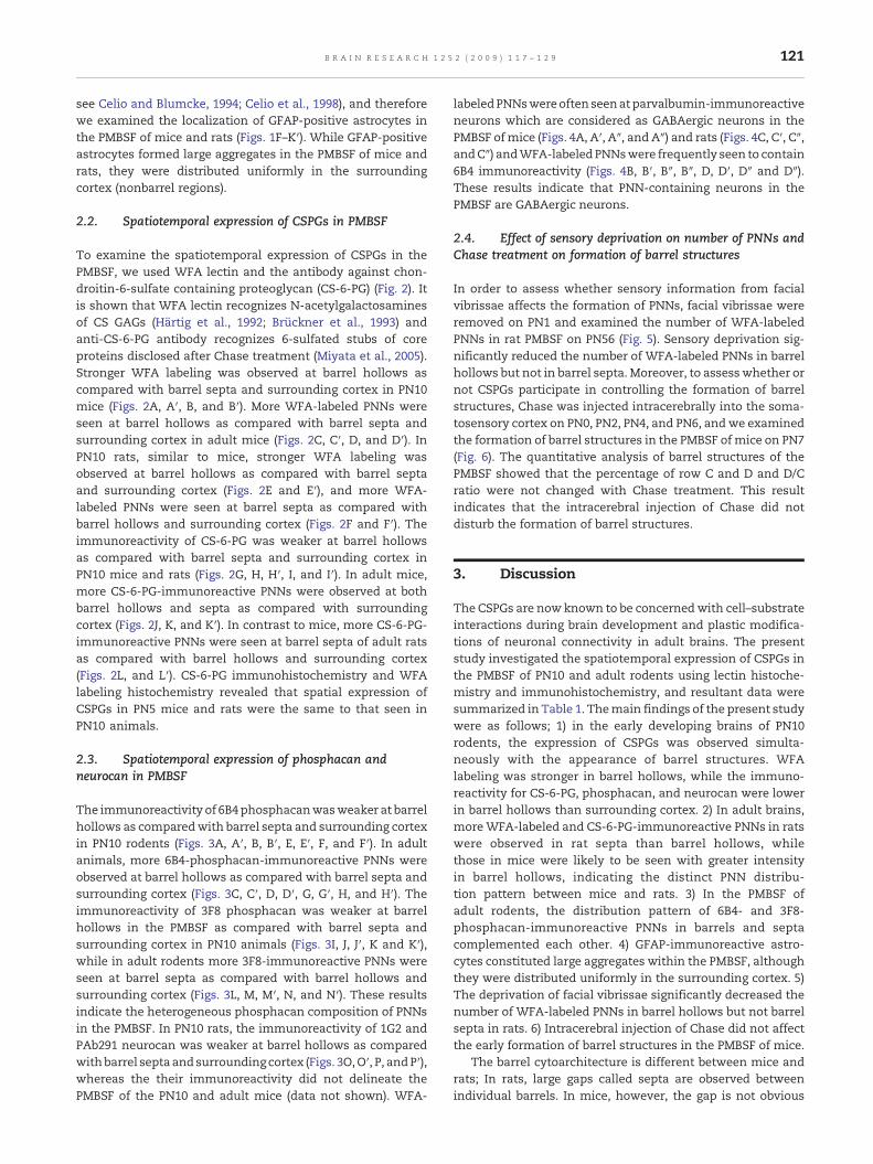
The immunoreactivity of 6B4phosphacanwasweaker at barrelhollows as comparedwith barrel septa and surrounding cortexin PN10 rodents (Figs. 3A, A′, B, B′, E, E′, F, and F′). In adultanimals, more 6B4-phosphacan-immunoreactive PNNs wereobserved at barrel hollows as compared with barrel septa andsurrounding cortex (Figs. 3C, C′, D, D′, G, G′, H, and H′). Theimmunoreactivity of 3F8 phosphacan was weaker at barrelhollows in the PMBSF as compared with barrel septa andsurrounding cortex in PN10 animals (Figs. 3I, J, J′, K and K′),while in adult rodents more 3F8-immunoreactive PNNs wereseen at barrel septa as compared with barrel hollows andsurrounding cortex (Figs. 3L, M, M′, N, and N′). These resultsindicate the heterogeneous phosphacan composition of PNNsin the PMBSF. In PN10 rats, the immunoreactivity of 1G2 andPAb291 neurocan was weaker at barrel hollows as comparedwithbarrel septaandsurrounding cortex (Figs. 3O,O′, P, andP′),whereas the their immunoreactivity did not delineate thePMBSF of the PN10 and adult mice (data not shown). WFA-
labeledPNNswereoftenseenatparvalbumin-immunoreactiveneurons which are considered as GABAergic neurons in thePMBSF ofmice (Figs. 4A, A′, A″, and A″) and rats (Figs. 4C, C′, C″,andC″) andWFA-labeledPNNswere frequently seen to contain6B4 immunoreactivity (Figs. 4B, B′, B″, B″, D, D′, D″ and D″).These results indicate that PNN-containing neurons in thePMBSF are GABAergic neurons.
2.4. Effect of sensory deprivation on number of PNNs andChase treatment on formation of barrel structures
In order to assess whether sensory information from facialvibrissae affects the formation of PNNs, facial vibrissae wereremoved on PN1 and examined the number of WFA-labeledPNNs in rat PMBSF on PN56 (Fig. 5). Sensory deprivation sig-nificantly reduced the number of WFA-labeled PNNs in barrelhollows but not in barrel septa. Moreover, to assesswhether ornot CSPGs participate in controlling the formation of barrelstructures, Chase was injected intracerebrally into the soma-tosensory cortex on PN0, PN2, PN4, and PN6, and we examinedthe formation of barrel structures in the PMBSF of mice on PN7(Fig. 6). The quantitative analysis of barrel structures of thePMBSF showed that the percentage of row C and D and D/Cratio were not changed with Chase treatment. This resultindicates that the intracerebral injection of Chase did notdisturb the formation of barrel structures.
3. Discussion
The CSPGs are now known to be concernedwith cell–substrateinteractions during brain development and plastic modifica-tions of neuronal connectivity in adult brains. The presentstudy investigated the spatiotemporal expression of CSPGs inthe PMBSF of PN10 and adult rodents using lectin histoche-mistry and immunohistochemistry, and resultant data weresummarized in Table 1. Themain findings of the present studywere as follows; 1) in the early developing brains of PN10rodents, the expression of CSPGs was observed simulta-neously with the appearance of barrel structures. WFAlabeling was stronger in barrel hollows, while the immuno-reactivity for CS-6-PG, phosphacan, and neurocan were lowerin barrel hollows than surrounding cortex. 2) In adult brains,moreWFA-labeled and CS-6-PG-immunoreactive PNNs in ratswere observed in rat septa than barrel hollows, whilethose in mice were likely to be seen with greater intensityin barrel hollows, indicating the distinct PNN distribu-tion pattern between mice and rats. 3) In the PMBSF ofadult rodents, the distribution pattern of 6B4- and 3F8-phosphacan-immunoreactive PNNs in barrels and septacomplemented each other. 4) GFAP-immunoreactive astro-cytes constituted large aggregates within the PMBSF, althoughthey were distributed uniformly in the surrounding cortex. 5)The deprivation of facial vibrissae significantly decreased thenumber of WFA-labeled PNNs in barrel hollows but not barrelsepta in rats. 6) Intracerebral injection of Chase did not affectthe early formation of barrel structures in the PMBSF of mice.
The barrel cytoarchitecture is different between mice andrats; In rats, large gaps called septa are observed betweenindividual barrels. In mice, however, the gap is not obvious
122 B R A I N R E S E A R C H 1 2 5 2 ( 2 0 0 9 ) 1 1 7 – 1 2 9
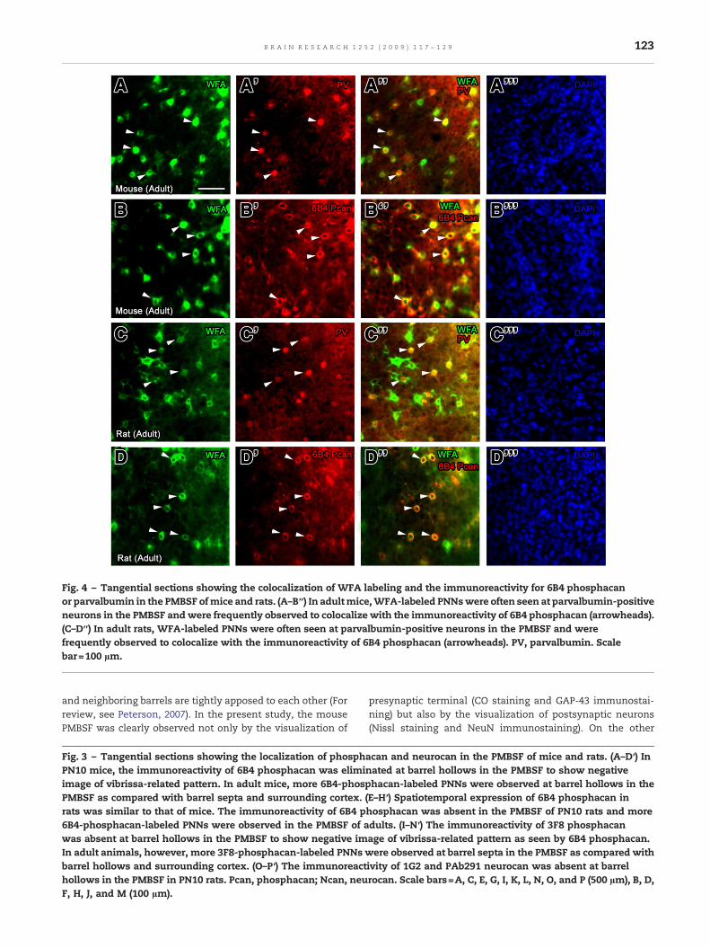
Fig. 4 – Tangential sections showing the colocalization of WFA labeling and the immunoreactivity for 6B4 phosphacanor parvalbumin in the PMBSF ofmice and rats. (A–B″) In adultmice,WFA-labeled PNNswere often seen at parvalbumin-positiveneurons in the PMBSF andwere frequently observed to colocalize with the immunoreactivity of 6B4 phosphacan (arrowheads).(C–D″) In adult rats, WFA-labeled PNNs were often seen at parvalbumin-positive neurons in the PMBSF and werefrequently observed to colocalize with the immunoreactivity of 6B4 phosphacan (arrowheads). PV, parvalbumin. Scalebar=100 µm.
123B R A I N R E S E A R C H 1 2 5 2 ( 2 0 0 9 ) 1 1 7 – 1 2 9
and neighboring barrels are tightly apposed to each other (Forreview, see Peterson, 2007). In the present study, the mousePMBSF was clearly observed not only by the visualization of
Fig. 3 – Tangential sections showing the localization of phosphPN10 mice, the immunoreactivity of 6B4 phosphacan was elimiimage of vibrissa-related pattern. In adult mice, more 6B4-phospPMBSF as compared with barrel septa and surrounding cortex. (rats was similar to that of mice. The immunoreactivity of 6B4 p6B4-phosphacan-labeled PNNs were observed in the PMBSF of awas absent at barrel hollows in the PMBSF to show negative imIn adult animals, however, more 3F8-phosphacan-labeled PNNs wbarrel hollows and surrounding cortex. (O–P′) The immunoreacthollows in the PMBSF in PN10 rats. Pcan, phosphacan; Ncan, neuF, H, J, and M (100 µm).
presynaptic terminal (CO staining and GAP-43 immunostai-ning) but also by the visualization of postsynaptic neurons(Nissl staining and NeuN immunostaining). On the other
acan and neurocan in the PMBSF of mice and rats. (A–D′) Innated at barrel hollows in the PMBSF to show negativehacan-labeled PNNs were observed at barrel hollows in theE–H′) Spatiotemporal expression of 6B4 phosphacan inhosphacan was absent in the PMBSF of PN10 rats and moredults. (I–N′) The immunoreactivity of 3F8 phosphacanage of vibrissa-related pattern as seen by 6B4 phosphacan.ere observed at barrel septa in the PMBSF as compared with
ivity of 1G2 and PAb291 neurocan was absent at barrelrocan. Scale bars=A, C, E, G, I, K, L, N, O, and P (500 µm), B, D,
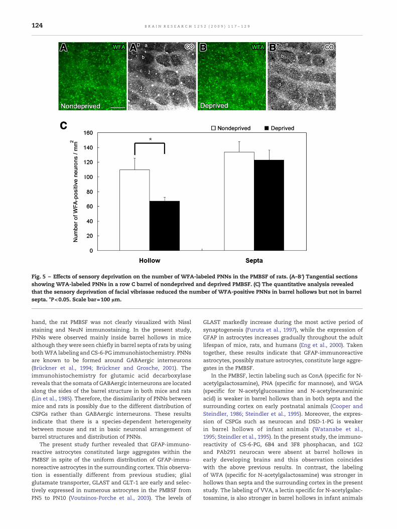
Fig. 5 – Effects of sensory deprivation on the number of WFA-labeled PNNs in the PMBSF of rats. (A–B′) Tangential sectionsshowing WFA-labeled PNNs in a row C barrel of nondeprived and deprived PMBSF. (C) The quantitative analysis revealedthat the sensory deprivation of facial vibrissae reduced the number of WFA-positive PNNs in barrel hollows but not in barrelsepta. *P<0.05. Scale bar=100 µm.
124 B R A I N R E S E A R C H 1 2 5 2 ( 2 0 0 9 ) 1 1 7 – 1 2 9
hand, the rat PMBSF was not clearly visualized with Nisslstaining and NeuN immunostaining. In the present study,PNNs were observed mainly inside barrel hollows in micealthough theywere seen chiefly in barrel septa of rats by usingbothWFA labeling and CS-6-PG immunohistochemistry. PNNsare known to be formed around GABAergic interneurons(Brückner et al., 1994; Brückner and Grosche, 2001). Theimmunohistochemistry for glutamic acid decarboxylasereveals that the somata of GABAergic interneurons are locatedalong the sides of the barrel structure in both mice and rats(Lin et al., 1985). Therefore, the dissimilarity of PNNs betweenmice and rats is possibly due to the different distribution ofCSPGs rather than GABAergic interneurons. These resultsindicate that there is a species-dependent heterogeneitybetween mouse and rat in basic neuronal arrangement ofbarrel structures and distribution of PNNs.
The present study further revealed that GFAP-immuno-reactive astrocytes constituted large aggregates within thePMBSF in spite of the uniform distribution of GFAP-immu-noreactive astrocytes in the surrounding cortex. This observa-tion is essentially different from previous studies; glialglutamate transporter, GLAST and GLT-1 are early and selec-tively expressed in numerous astrocytes in the PMBSF fromPN5 to PN10 (Voutsinos-Porche et al., 2003). The levels of
GLAST markedly increase during the most active period ofsynaptogenesis (Furuta et al., 1997), while the expression ofGFAP in astrocytes increases gradually throughout the adultlifespan of mice, rats, and humans (Eng et al., 2000). Takentogether, these results indicate that GFAP-immunoreactiveastrocytes, possibly mature astrocytes, constitute large aggre-gates in the PMBSF.
In the PMBSF, lectin labeling such as ConA (specific for N-acetylgalactosamine), PNA (specific for mannose), and WGA(specific for N-acetylglucosamine and N-acetylneuraminicacid) is weaker in barrel hollows than in both septa and thesurrounding cortex on early postnatal animals (Cooper andSteindler, 1986; Steindler et al., 1995). Moreover, the expres-sion of CSPGs such as neurocan and DSD-1-PG is weakerin barrel hollows of infant animals (Watanabe et al.,1995; Steindler et al., 1995). In the present study, the immuno-reactivity of CS-6-PG, 6B4 and 3F8 phosphacan, and 1G2and PAb291 neurocan were absent at barrel hollows inearly developing brains and this observation coincideswith the above previous results. In contrast, the labelingof WFA (specific for N-acetylgalactosamine) was stronger inhollows than septa and the surrounding cortex in the presentstudy. The labeling of VVA, a lectin specific for N-acetylgalac-tosamine, is also stronger in barrel hollows in infant animals
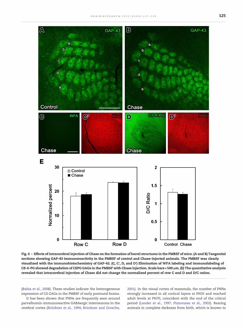
Fig. 6 – Effects of intracerebral injection of Chase on the formation of barrel structures in the PMBSF ofmice. (A and B) Tangentialsections showing GAP-43 immunoreactivity in the PMBSF of control and Chase-injected animals. The PMBSF was clearlyvisualized with the immunohistochemistry of GAP-43. (C, C′, D, and D′) Elimination of WFA labeling and immunolabeling ofCS-6-PG showeddegradation of CSPGGAGs in the PMBSFwith Chase injection. Scale bars=500 µm. (E) The quantitative analysisrevealed that intracerebral injection of Chase did not change the normalized percent of row C and D and D/C ration.
125B R A I N R E S E A R C H 1 2 5 2 ( 2 0 0 9 ) 1 1 7 – 1 2 9
(Bahia et al., 2008). These studies indicate the heterogeneousexpression of CS GAGs in the PMBSF of early postnatal brains.
It has been shown that PNNs are frequently seen aroundparvalbumin-immunoreactive GABAergic interneurons in thecerebral cortex (Brückner et al., 1994; Brückner and Grosche,
2001). In the visual cortex of mammals, the number of PNNsstrongly increased in all cortical layers at PN35 and reachedadult levels at PN70, coincident with the end of the criticalperiod (Lander et al., 1997; Pizzorusso et al., 2002). Rearinganimals in complete darkness from birth, which is known to
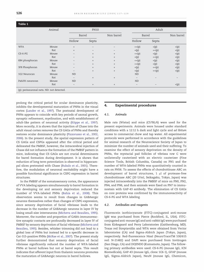
Table 1
Animal PN10 Adult
Barrel Non barrel Barrel Non barrel
Hollow Septa Hollow Septa
WFA Mouse + − − ++(p) +(p) +(p)Rat + − − +(p) ++(p) +(p)
CS-6-PG Mouse − + + ++(p) +(p) +(p)Rat − + + +(p) ++(p) +(p)
6B4 phosphocan Mouse − + + ++(p) +(p) +(p)Rat − + + ++(p) +(p) +(p)
3F8 Phosphocan Mouse − + + +(p) ++(p) +(p)Rat − + + +(p) ++(p) +(p)
1G2 Neurocan Mouse ND NDRat − + + − − −
Pab291 neurocan Mouse ND NDRat − + + − − +(p)
(p): perineuronal nets. ND: not detected.
126 B R A I N R E S E A R C H 1 2 5 2 ( 2 0 0 9 ) 1 1 7 – 1 2 9
prolong the critical period for ocular dominance plasticity,inhibits the developmental maturation of PNNs in the visualcortex (Lander et al., 1997). The postnatal development ofPNNs appears to coincide with key periods of axonal growth,synaptic refinement, myelination, and with establishment ofadult-like pattern of neuronal activity (Köppe et al., 1997).More recently, it is shown that the injection of Chase into theadult visual cortex removes the CS GAGs of PNNs and therebyrestores ocular dominance plasticity (Pizzorusso et al., 2002,2006). In the present study, the spatial expression pattern ofCS GAGs and CSPGs appeared after the critical period anddelineated the PMBSF, however, the intracerebral injection ofChase did not influence the formation of the PMBSF pattern inmice, indicating that CS GAGs are not crucial determinantsfor barrel formation during development. It is shown thatreduction of long term potentiation is observed in hippocam-pal slices pretreated with Chase (Bukalo et al., 2001). There-fore, the modulation of neuronal excitability might have apossible functional significance in CSPG expression in barrelstructures.
In the PMBSF of the somatosensory cortex, the appearanceof VVA labeling appears simultaneously to barrel formation inthe developing rat and sensory deprivation reduced thenumber of VVA-labeled PNNs (Bahia et al., 2008). Thisobservation seems to result from changes in GABAergicneurons themselves rather than changes of CSPG expression,since sensory deprivation of facial vibrissae leads to thedecrease in the number of GABAergic neurons in layer IV bylosing small-size interneurons (Micheva and Beaulieu, 1995).Moreover, the number and proportion of GABA-immunoreac-tive synaptic contacts are profoundly decreased in layer IV ofPMBSF by sensory deprivation of facial vibrissae (Micheva andBeaulieu, 1995). Besides, whisker trimming did not lead to aglobal loss of PNNs but instead led to a specific decrease inCat-315-positive PNNs (McRae et al., 2007). The present studyfurther demonstrated that sensory deprivation of facialvibrissae significantly reduced the number of WFA-labeledPNNs at barrel hollows but not at barrel septa. This resultindicates that afferent input from thalamic neurons promotesthe maturation of GABAergic neurons in barrel hollows.
4. Experimental procedures
4.1. Animals
Male rats (Wistar) and mice (C57BL/6) were used for thepresent experiments. Animals were housed under standardconditions with a 12:12 h dark and light cycle and ad libitumaccess to commercial chow and tap water. All experimentalprotocols were performed in accordance with the guidelinesfor animal research of the Neuroscience Society of Japan tominimize the number of animals used and their suffering. Toexamine the effect of sensory deprivation on the density ofPNNs, the mystacial pad follicles of vibrissa row C wereunilaterally cauterized with an electric cauterizer (FineScience Tools, British Columbia, Canada) on PN1 and thenumber of WFA-labeled PNNs was quantitatively counted inrats on PN56. To assess the effects of chondroitinase ABC ondevelopment of barrel structures, 1 µl of protease-freechondroitinase ABC (20 U/ml, Seikagaku, Tokyo, Japan) wasinjected intracerebrally into the PMBSF of mice on PN0, PN2,PN4, and PN6, and then animals were fixed on PN7 to immu-nostain with GAP-43 antibody. The elimination of CS GAGson core proteins was confirmed by the immunoreactivity ofCS-6-PG and WFA labeling.
4.2. Antibodies and reagents
Fluorescein isothiocyanate (FITC)-conjugated anti-mouseIgM was purchased from Pierce (Rockford, IL, USA). FITC-conjugated anti-mouse IgG andanti-rabbit IgGwere purchasedfrom Kirkegaard and Perry Laboratories (Gaithersburg, MD).Texas red Streptavidin and WFA were obtained from Vectorlaboratories (CA) and Sigma-Aldrich Japan (Tokyo, Japan),respectively. Red-fluorescence Nissl (NeuroTraceTM 530/615red: N-21482) and DAPI were purchased from Invitrogen(San Diego, CA) and DOJINDO (Kumamoto, Japan). The follow-ing primary antibodies were used: CS-6-PG (mouse IgG, ICNBiomedicals), GAP-43 (mouse IgG, clone 1C6-1), GFAP (mouseIgG, Sigma-Aldrich Japan), NeuN (mouse IgG, Chemicon,
127B R A I N R E S E A R C H 1 2 5 2 ( 2 0 0 9 ) 1 1 7 – 1 2 9
Temecula, CA), neurocan (mouse IgG, clone 1G2; Watanabeet al., 1995), neurocan (rabbit IgG, serum PAb291, Matsui et al.,1994), parvalbumin (mouse IgG, Sigma-Aldrich Japan), phos-phacan (mouse IgG, clone 3F8; Developmental Studies Hybri-doma Bank, Iowa, IA), and phosphacan (mouse IgM, clone 6B4,Maeda et al., 1995).
4.3. Tissue fixation and sectioning
Under deep anesthesia with pentobarbital (80 mg/kg), rats andmice were perfused with heparinized phosphate-bufferedsaline (PBS; pH 7.4) followed by 4% paraformaldehyde in0.1 M phosphate buffer (PB, pH 7.4). Brains were removed fromthe skull and sectioned midsagittally into two halves. Theywere then postfixed overnight and flattened as describedpreviously (Jhaveri et al., 1991). After several washes with PBS,tangential sections of flattened hemispheres were cut into50 µm-thick sections on a vibratome (DTK-1000 microslicer,DSK, Kyoto, Japan).
4.4. Histochemistry
CO histochemistry was performed as described by Silver-man and Tootel (1987). Briefly, PFA-fixed sections wererinsed with 10% sucrose in 0.1 M PB and then incubated for10 min in 10% sucrose/0.05 M Tris buffer (pH 7.6) containing275 mg/l of cobalt chloride and washed in 0.1 M PB, andincubated for 4–6 h at 40 °C in 0.1 M PB containing 0.5 g/l of3,38-diaminobenzidine tetrahydrochloride (Sigma-AldrichJapan), 50 g/l of sucrose, 75 mg/l of cytochrome C (type III;Sigma-Aldrich Japan), and 20 mg/l of catalase.
For immunostaining of various CSPGs, GFAP, and NeuN,standard immunofluorescence techniques were performedon the free-floating cerebral sections as described in ourprevious paper (Miyata et al., 2001). Briefly, the sectionswere pretreated with 25 mM glycine PBS for 20 min andincubated with 5% normal goat serum (NGS) in PBScontaining 0.3% Triton X-100 (PBST) overnight. Then sec-tions were incubated with the following primary antibodiesin the PBST containing 5% NGS for 3 days at 4 °C: 6B4 anti-phosphacan (dilution 1:40), 3F8 anti-phosphacan (dilution1:5), 1G2 anti-neurocan (dilution 1:10), PAb291 anti-neurocan(dilution 1:100), anti-NeuN (dilution 1:300), and anti-GFAP(dilution 1:100). The sections were then incubated withFITC-conjugated anti-mouse IgG, anti-mouse IgM, or anti-rabbit IgG (10 µg/ml) in PBST for 2 h in PBST containing red-fluorescence Nissl (dilution 1:200).
For immunostaining of CS-6-PG, the sections were treatedwith chondroitinase ABC to remove CS GAG chains before theimmunofluorescence procedure, since anti-6′CSPG antibodiesrecognize CS-6-sulfated stub of core proteins disclosed afterchondroitinase ABC treatment. Shortly, the sections wereincubated with 0.1 unit/ml of chondroitinase ABC (Seikagaku)in 0.1 M Tris–HCl buffer (pH 8.0) containing 30 mM sodiumacetate, 1 mM PMSF, 5 mM NEM, 10 µg/ml of pepstatin, and10 µg/ml of aprotinin for 3 h at 37 °C. After several washingswith PBS, the sections were incubated overnight with 5% NGSin PBST and then with 6′CSPG (dilution 1:200) in the PBSTcontaining 5% NGS for 3 days at 4 °C. The sections were thenincubated with FITC-conjugated anti-mouse IgG (10 µg/ml)
for 2 h. Red-fluorescence Nissl was added at the same timeas the secondary antibodies.
For labeling of WFA, the sections were pretreated with25 mM glycine PBS for 20 min and incubated with biotinylatedWFA (Sigma Aldrich Japan, 10 µg/ml) in PBST for 2 d at 4 °C.The sections were then incubated with FITC-conjugatedstreptavidin (10 µg/ml) for 2 h. Red-fluorescence Nissl wereadded at the same time as the secondary antibodies. For triplelabeling, the sections were incubated with the followingprimary antibodies in PBST containing 5% NGS for 3 days at4 °C: anti-phosphacan (clone 6B4; dilution 1:40), and anti-parvalbumin (dilution 1:250). The sections were then incu-bated with biotinylated anti-mouse IgG or IgM (7.5 µg/ml) inthe PBST for 2 h followed by Texas Red Streptavidin (20 µg/ml)in the PBST for 2 h. They were incubated with biotinylatedWFA (10 µg/ml) in the PBST for 2 days at 4 °C and then treatedwith FITC-conjugated streptavidin (10 µg/ml) and DAPI(dilution 1:1,000) in PBST for 2 h in PBST.
The sections were mounted on glass slides and sealed withVectashield (Vector Lab). Observation was made using aLSM510 laser-scanning confocal microscope (Carl Zeiss, Ger-many). The sequential scanning of recording configuration forFITC was used to avoid bleed-through of FITC. Images(1024×1024) were saved as TIF files with a Zeiss LSM510Image-Browser software and organized with Photoshop 7.0.Omission of the primary antibody did not produce any visibleimmunostaining on these preparations.
4.5. Quantification
Measurements of the entire large barrels area of mousePMBSF and surface areas devoted to each row (C to D) weremade using the software Image J. For each hemisphere, areasC and D were normalized for PMBSF area. The D/C ratio wasused as a plasticity index (Schlaggar et al., 1993; Rebsamet al., 2005). The number of WFA-labeled PNNs was countedin barrel hollows and septa as described by others (Bahiaet al., 2008). All values were expressed as means±SE andstatistical analysis was performed by ANOVA followed byFisher's test with α=0.05 considered as significant.
Acknowledgments
This work was supported in part by a Scientific ResearchGrant from the Japan Society for the Promotion of Science(No. 19500293) and the Salt Science Research Foundation(No. 0838 to S.M.). The 3F8 antibody, developed by Drs. R. K.Margolis and R. U. Margolis, was obtained from the Deve-lopmental Studies Hybridoma Bank developed under theauspices of the NICHD and maintained by the University ofIowa, Department of Biological Sciences, Iowa City, IA52242.
R E F E R E N C E S
Bahia, C.P., Houzel, J.-C., Picanco-Diniz, C.W., Pereira, A., 2008.Spatiotemporal distribution of proteoglycans in the developing
128 B R A I N R E S E A R C H 1 2 5 2 ( 2 0 0 9 ) 1 1 7 – 1 2 9
rat's barrel field and the effects of early deafferentation.J. Comp. Neurol. 510, 145–157.
Bandtlow, C.E., Zimmermann, D.R., 2000. Proteoglycans in thedeveloping brain: new conceptual insights for old proteins.Physiol. Rev. 80, 1267–1290.
Bradbury, E.J., Moon, L.D., Popat, R.J., King, V.R., Bennett, G.S., Patel,P.N., Fawcett, J.W., McMahon, S.B., 2002. Chondroitinase ABCpromotes functional recovery after spinal cord injury. Nature416, 636–640.
Bovolenta, P., Fernaud-Espinosa, I., 2000. Nervous systemproteoglycans as modulators of neurite outgrowth. Prog.Neurobiol. 61, 113–132.
Brückner, G., Brauer, K., Härtig, W., Wolff, J.R., Rickmann, M.J.,Derouiche, A., Delpech, B., Girard, N., Oertel, W.H.,Reichenbach, A., 1993. Perineuronal nets provide a polyanionic,glia-associated form of microenvironment around certainneurons in many parts of the brain. Glia 8, 183–200.
Brückner, G., Seeger, G., Brauer, K., Härtig, W., Kacza, J., Bigl, V.,1994. Cortical areas are revealed by distribution patterns ofproteoglycan components and parvalbumin in the Mongoliangerbil and rat. Brain Res. 658, 67–86.
Brückner, G., Bringmann, A., Härtig, W., Köppe, G., Delpech, B.,Brauer, K., 1998. Acute and long-lasting changes inextracellular-matrix chondroitin-sulfate proteoglycansinduced by injection of chondroitinase ABC in the adult ratbrain. Exp. Brain Res. 121, 300–310.
Brückner, G., Grosche, J., Schmidt, S., Härtig, W., Margolis, R.U.,Delpech, B., Seidenbecher, C.I., Czaniera, R., Schachner, M.,2000. Postnatal development of perineuronal nets in wild-typemice and in a mutant deficient in tenascin-R. J. Comp. Neurol.428, 616–629.
Brückner, G., Grosche, J., 2001. Perineuronal nets show intrinsicpatterns of extracellular matrix differentiation in organotypicslice culture. Exp. Brain Res. 137, 83–93.
Bukalo, O., Schachner, M., Dityatev, A., 2001. Modification ofextracellular matrix by enzymatic removal of chondroitinsulfate and by lack of tenascin-R differentially affects severalforms of synaptic plasticity in the hippocampus. Neuroscience104, 359–369.
Cases, O., Vitalis, T., Seif, I., De Maeyer, E., Sotelo, C., Gaspar, P.,1996. Lack of barrels in the somatosensory cortex ofmonoamine oxidase A-deficient mice, role of aserotonin excess during the critical period. Neuron 16,297–307.
Celio, M.R., Blümcke, I., 1994. Perineuronal nets-a specializedform of extracellular matrix in the adult nervous system. BrainRes. Rev. 19, 128–145.
Celio, M.R., Spreafico, R., De Bias, S., Vitellaro-Zuccarello, L., 1998.Perineuronal nets, past and present. Trends Neurosci. 21,510–515.
Cooper, N.G.F., Steindler, D.A., 1986. Lectins demarcate the barrelsubfield in the somatosensory cortex of the early postnatalmouse. J. Comp. Neurol. 249, 157–169.
Corvett, L., Rossi, F., 2005. Degradation of chondroitinsulfate proteoglycans induces sprouting of intact Purkinjeaxons in the cerebellum of the adult rat. J. Neurosci. 25,7150–7158.
Cybulska-Klosowicz, A., Zakrzewska, R., Pyza, E., Kossut, M.,Schachner, M., 2004. Reduced plasticity of cortical whiskerrepresentation in adult tenascin-C-deficient mice aftervibrissectomy. Eur. J. Neurosci. 20, 1538–1544.
Eng, L.F., Chirnikar, R.S., Lee, Y.L., 2000. Glial fibrillary acidicprotein: GFAP-thirty-one years (1969–2000). Neurochem. Res.25, 1439–1451.
Fox, K., 1992. A critical period for experience-dependent synapticplasticity in rat barrel cortex. J. Neurosci. 12, 826–1836.
Frostig, R.D., 2006. Functional organization and plasticity in theadult rat barrel cortex, move out-of-the-box. Curr. Opin.Neurobiol. 16, 445–450.
Fukuichi-Shimogori, T., Grove, E.A., 2001. Neocortex patterningby the secreted signaling molecule FGF8. Science 294,1071–1074.
Furuta, A., Rothstein, J.D., Martin, L.J., 1997. Glutamate transporterprotein subtypes are expressed differentially during rat CNSdevelopment. J. Neurosci. 17, 8363–8375.
Galtrey, C.M., Fawcett, J.W., 2007. The role of chondroitin sulfateproteoglycans in regeneration and plasticity in the centralnervous system. Brain Res. Rev. 85, 2813–2823.
Hannan, A.J., Blakemore, C., Katsnelson, A., Vitalis, T., Huber,K.M., Bear, M., Roder, J., Kim, D., Shin, H.S., Kind, P.C.,2001. PLC-beta1, activated via mGluRs mediatesactivity-dependent differentiation in cerebral cortex.Nat. Neurosci. 4, 282–288.
Härtig, W., Brauer, K., Brückner, G., 1992. Wisteria floribundaagglutininlabeled nets surround parvalbumin-containingneurons. NeuroReport 3, 869–872.
Iwasato, T., Datwani, A., Wolf, A.M., Nishiyama, H., Taguchi, Y.,Tonegawa, S., Knopfel, T., Erzurumlu, R.S., Itohara, S., 2000.Cortex-restricted disruption of NMDAR1 impairs neuronalpatterns in the barrel cortex. Nature 406, 726–731.
Jhaveri, S., Erzurumlu, R.S., Crossin, K., 1991. Barrel constructionin rodent neocortex, role of thalamic afferents versusextracellular matrix molecules. Proc. Natl. Acad. Sci. U. S. A.88, 4489–4493.
John, N., Krügel, H., Frischknecht, R., Smalla, K.-H., Schultz, C.,Kreutz, M.R., Gundelfinger, E.D., Seidenbecher, C.I., 2006.Brevican-containing perineuronal nets of extracellular matrixin dissociated hippocampal primary cultures. Mol. Cell.Neurosci. 31, 774–784.
Katoh-Senba, R., Matsuda, M., Kato, K., Oohira, A., 1995.Chondroitin sulphate proteoglycans in the rat brain,candidates for axon barriers of sensory neurons and thepossible modification by laminin of their actions. Eur. J.Neurosci. 7, 613–621.
Köppe, G., Brückner, G., Brauer, K., Härtig, W., Bigl, V., 1997.Developmental patterns of proteoglycan-containingextracellular matrix in perineuronal nets and neuropile of thepostnatal rat brain. Cell Tissue Res. 288, 33–41.
Lander, C., Kind, P., Maleski, M., Höckfield, S., 1997. A familyof activity-dependent neuronal cell-surface chondroitinsulfateproteoglycans in cat visual cortex. J. Neurosci. 17,1928–1939.
Lin, C.-S., Lu, S.M., Schmechel, D.E., 1985. Glutamic aciddecarboxylase immunoreactivity in layer IV of barrel cortex ofrat and mouse. J. Neurosci. 5, 1934–1939.
Maeda, N., Hamanaka, H., Oohita, A., Noda, M., 1995.Purification, characterization and developmentalexpression of a brain-specific chondroitin sulfateproteoglycan, 6B4 proteoglycan/phosphacan. Neuroscience67, 23–35.
Matsui, F., Watanabe, E., Oohira, A., 1994. Immunologicalidentification of two proteoglycan fragments derived fromneurocan, a brain-specific chondroitin sulfate proteoglycan.Neurochem. Int. 25, 425–431.
McRae, P.A., Rocco, M.M., Kelly, G., Brumberg, J.C., Matthews, R.T.,2007. Sensory deprivation alters aggrecan and perineuronalnet expression in the mouse barrel cortex. J. Neurosci. 27,5405–5413.
Micheva, K.D., Beaulieu, C., 1995. An anatomical substrate forexperience-dependent plasticity of the rat barrel field cortex.Proc. Natl. Acad. Sci. U. S. A. 92, 11834–11838.
Mitrovic, N., Mohajeri, H., Schachner, M., 1996. Effects ofNMDA receptor blockage in the developing ratsomatosensory cortex on the expression of the glia-derivedextracellular matrix glycoprotein tenascin-C. Eur. J. Neurosci.8, 1793–1802.
Miyata, S., Takamatsu, S., Maekawa, S., Matsumoto, K.,Watanabe, K., Kiyohara, T., Hatton, G.I., 2001. Plasticity of
129B R A I N R E S E A R C H 1 2 5 2 ( 2 0 0 9 ) 1 1 7 – 1 2 9
neurohypophysial terminals with increased hormonal releaseduring dehydration: ultrastructural and biochemical analyses.J. Comp. Neurol. 343, 413–427.
Miyata, S., Nishimura, Y., Hayashi, N., Oohira, A., 2005.Construction of perineuronal net-like structure by corticalneurons in culture. Neuroscience 136 , 95–104.
Miyata, S., Nishimura, Y., Nakashima, T., 2007. Perineuronal netsprotect against amyloid β-protein neurotoxicity in culturedcortical neurons. Brain Res. 1150, 200–206.
Oohira, A., Matsui, F., Katoh-Senba, R., 1991. Inhibitory effectsof brain chondroitin sulfate proteoglycans on neuriteoutgrowth from PC12D cells. J. Neurosci. 11, 822–827.
Peterson, C.C.H., 2007. The functional organization of the barrelcortex. Neuron 56, 339–355.
Pizzorusso, T., Medini, P., Berardi, N., Chierzi, S., Fawcett, J.W.,Maffei, L., 2002. Reactivation of ocular dominanceplasticity in the adult visual cortex. Science 898, 1248–1251.
Pizzorusso, T., Medini, P., Landi, S., Baldini, A., Berardi, N.,Maffei, L., 2006. Structural and functional recovery fromearly monocular deprivation in adult rats. Proc. Natl. Acad.Sci. U. S. A. 103, 8517–8522.
Rebsam, A., Seif, I., Gaspar, P., 2005. Dissociating barreldevelopment and lesion-induced plasticity in the mousesomatosensory cortex. J. Neurosci. 19, 706–710.
Schlaggar, B.L., Fox, K., O, Leary, D.D.M., 1993. Postsynaptic controlof plasticity in developing somatosensory cortex. Nature 364,623–626.
Silverman, M.S., Tootel, R.B., 1987. Modified technique forcytochrome oxidase histochemistry, increased stainingintensity and compatibility with 2-deoxyglucoseautoradiography. J. Neurosci. Methods 19, 1–10.
Steindler, D.A., Settles, D., Erickson, H.P., Laywell, E.D., Yoshiki, A.,Faissner, A., Kusakabe, M., 1995. Tenascin knockout mice,
barrels, boundary molecules, and glial scars. J. Neurosci. 15,1971–1983.
Van der Loos, H., Woolsey, T.A., 1973. Somatosensory cortex,structural alterations following early injury to sense organs.Science 179, 395–398.
Voutsinos-Porche, B., Knott, G., Tanaka, K., Quairiaux, C., Welker,E., Bonento, G., 2003. Glial glutamate transporters andmaturation of the mouse somatosensory cortex. Cereb. Cortex13, 1110–1121.
Watanabe, E., Aono, S., Matsui, F., Yamada, Y., Naruse, I., Oohira,A., 1995. Distribution of a brain-specific proteoglycan,neurocan, and the corresponding mRNA during the formationof barrels in the rat somatosensory cortex. Eur. J. Neuorsci. 7,547–554.
Welker, E., Armstrong-James, M., Bronchti, G., Ourednik, W.,Gheorghita-Baechler, F., Dubois, R., Guernsey, D.L., Van derLoos, H., Neumann, P.E., 1996. Altered sensory processing inthe somatosensory cortex of the mouse mutant barrelless.Science 271, 1864–1867.
Wong-Riley, M.T., Welt, C., 1980. Histochemical changes incytochrome oxidase of cortical barrels after vibrissal removalin neonatal and adult mice. Proc. Natl. Acad. Sci. U. S. A. 77,2333–2337.
Woolsey, T.A., Van der Loos, H., 1970. The structuralorganization of layer IV in the somatosensory region (SI) ofmouse cerebral cortex. The description of a cortical fieldcomposed of discrete cytoarchitectonic units. Brain Res. 17,205–242.
Woolsey, T.A., Wann, J., 1976. Area changes in mouse corticalbarrels following vibrissal damage at different postnatal ages.J. Comp. Neurol. 170, 53–66.
Yamaguchi, Y., 2000. Lecticans, organizers of the brainextracellular matrix. Cell. Mol. Life Sci. 57, 276–289.