Embed Size (px)
Citation preview
EXTRA ORAL RADIOGAPHS
Extra oral radiographic examination
Extra oral radiographs include allviews made of orofacial region withfilm positioned extra orally. Thedentists often used these views toexamine area not fully covered withintra oral films or to visualize skull
and facial structures.
Skull Projections
Radiographic examination of the skull requires patience, attention to detail, and practice to achieve satisfactory results.
1-POSTEROANTERIOR PROJECTION* The straight posteroanterior (PA) projection is so
named because the x-ray beam passes in a posterior-toanterior direction through the skull.*This projection is used to examine the skull for
disease,trauma,ordevelopmentalabnormalities.* It also provides a good record for detecting
progressive changes in the mediolateral dimensions ofthe skull, including asymmetric growth.
*In addition, the PA projection offers goodvisualization of facial structures, including the frontaland ethmoid sinuses, nasal fossae, and orbits.
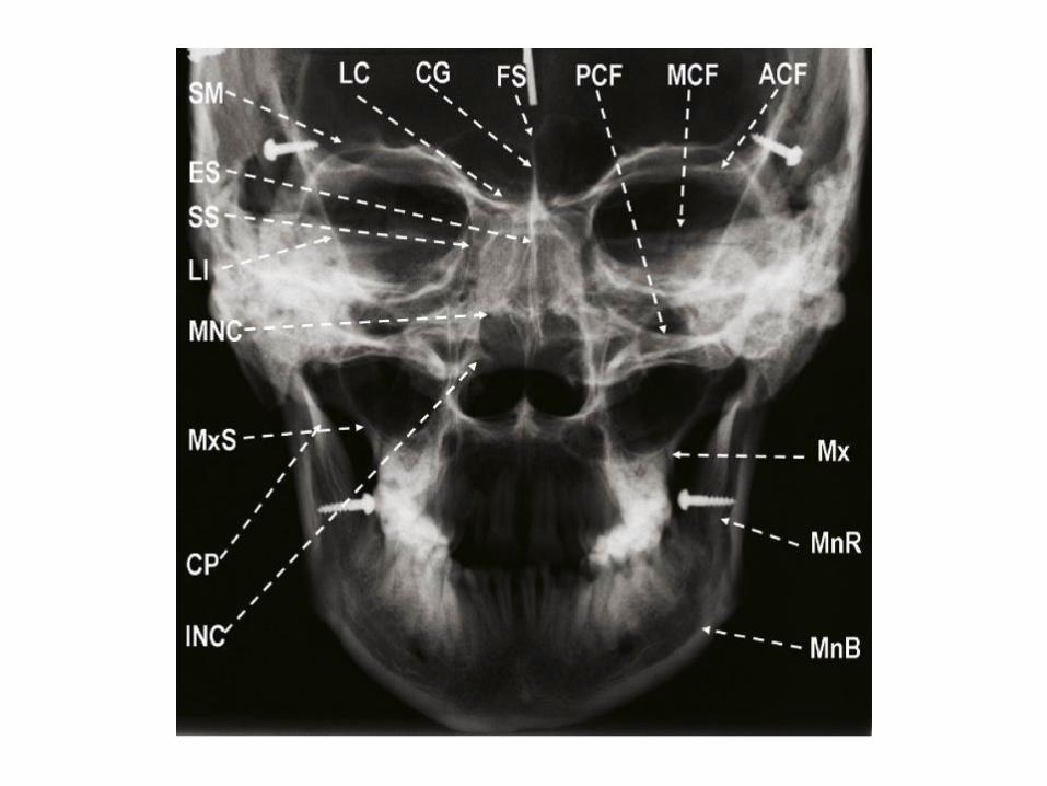
Representative image obtained from the posteroanteriorskull projection (Frankfort horizontal parallel). LC, laminacribrosa; CG, crista galli; FS, frontal sinus; PCF, posterior cranial
fossa;MCF, middle cranial fossa (petrous temporal); ACF, anterior
cranialfossa; Mx, maxilla; MnR, mandibular ramus; MnB, mandibular
body,INC, inferior nasal conchae; CP, coronoid process of mandible;
MxS,maxillary sinus; MNC, middle nasal conchae; LI, linea innominata
(innominateline); SS, sphenoid sinus; ES, ethmoid sinus; SM, supraorbital
margin.
3-WATERS'PROJECTION (Inclined PA,)*The Waters' projection (also called the
occipitomental projection) is a variation of the PA view.* It is particularly useful for evaluating the
maxillary sinuses.* In addition, it demonstrates the frontal and
ethmoid sinuses, the orbit, the zygomaticofrontal suture, and the nasal cavity.*It also demonstrates the position of the coronoid process of the mandible between the maxilla and the zygomatic arch.
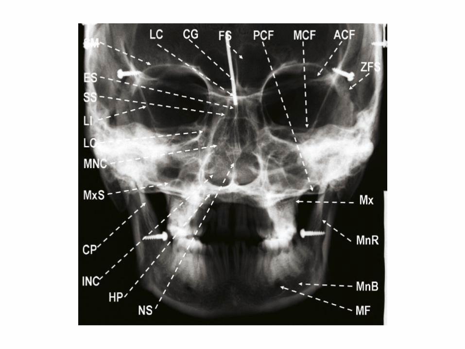
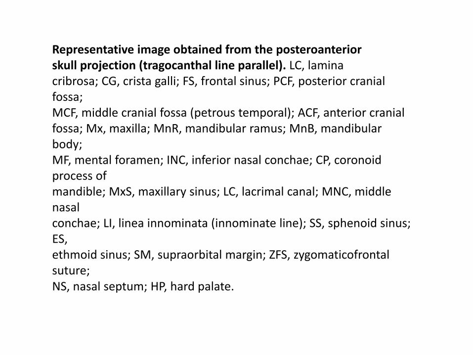
Representative image obtained from the posteroanteriorskull projection (tragocanthal line parallel). LC, laminacribrosa; CG, crista galli; FS, frontal sinus; PCF, posterior cranial fossa;MCF, middle cranial fossa (petrous temporal); ACF, anterior cranialfossa; Mx, maxilla; MnR, mandibular ramus; MnB, mandibular body;MF, mental foramen; INC, inferior nasal conchae; CP, coronoid process ofmandible; MxS, maxillary sinus; LC, lacrimal canal; MNC, middle nasalconchae; LI, linea innominata (innominate line); SS, sphenoid sinus; ES,ethmoid sinus; SM, supraorbital margin; ZFS, zygomaticofrontalsuture;NS, nasal septum; HP, hard palate.
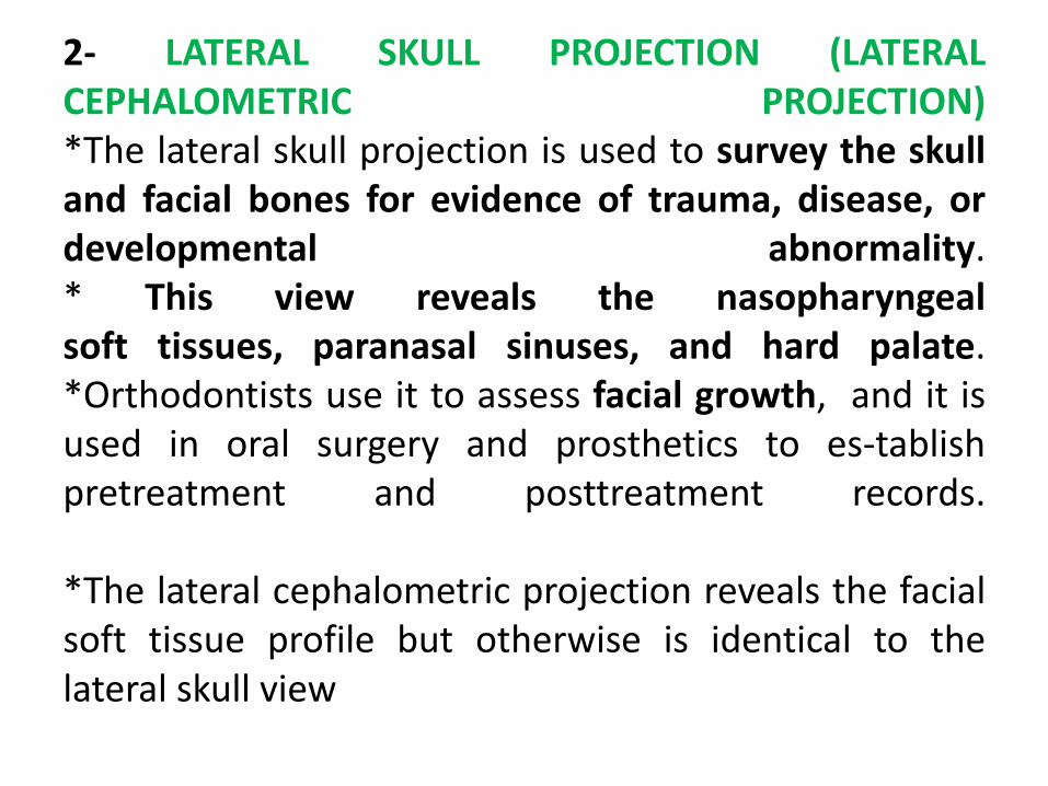
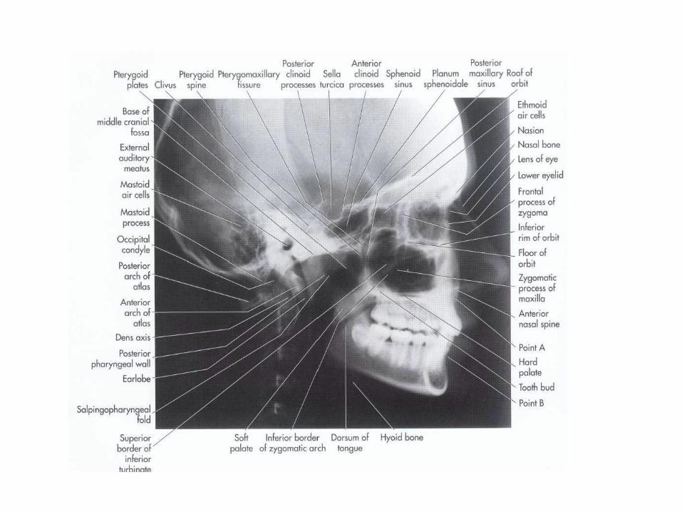
2- LATERAL SKULL PROJECTION (LATERALCEPHALOMETRIC PROJECTION)*The lateral skull projection is used to survey the skulland facial bones for evidence of trauma, disease, ordevelopmental abnormality.* This view reveals the nasopharyngealsoft tissues, paranasal sinuses, and hard palate.*Orthodontists use it to assess facial growth, and it isused in oral surgery and prosthetics to es-tablishpretreatment and posttreatment records.
*The lateral cephalometric projection reveals the facialsoft tissue profile but otherwise is identical to thelateral skull view
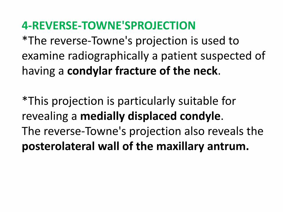
4-REVERSE-TOWNE'SPROJECTION*The reverse-Towne's projection is used to examine radiographically a patient suspected of having a condylar fracture of the neck.
*This projection is particularly suitable for revealing a medially displaced condyle.The reverse-Towne's projection also reveals the posterolateral wall of the maxillary antrum.
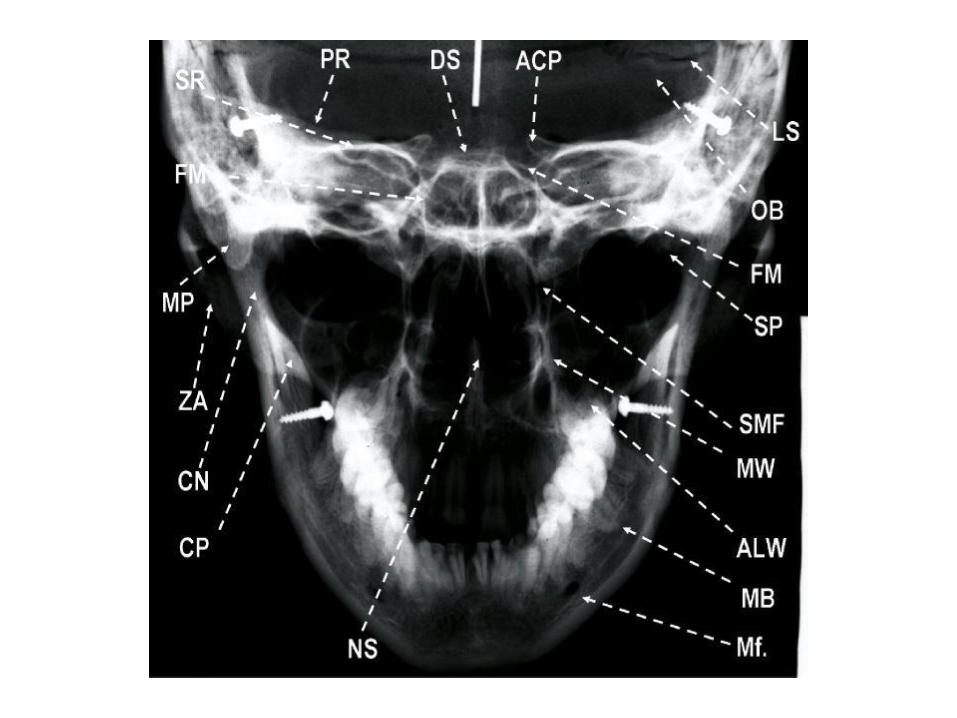
Representative image obtained from the reverseTowne projection. CN, condylar neck; CP, superimposedcoronoid processand ascending ramus of mandible; ZA, zygomatic arch; PR,
petrousridge of the temporal bone; DS, dorsum sella; LS, lambdoidsuture; ACP,anterior clinoid process; FM, foramen magnum; OB, occipitalbone; SP,styloid process; ALW, anterolateral wall, maxillary antrum; MW,
medialwall, maxillary antrum; Mf, mental foramen; NS, nasal septum;
SMF,sphenomaxillary fissure; SR, supraorbital ridge of the orbit; MP,
mastoidprocess; MB, mandibular body; SS, sphenoid sinus.
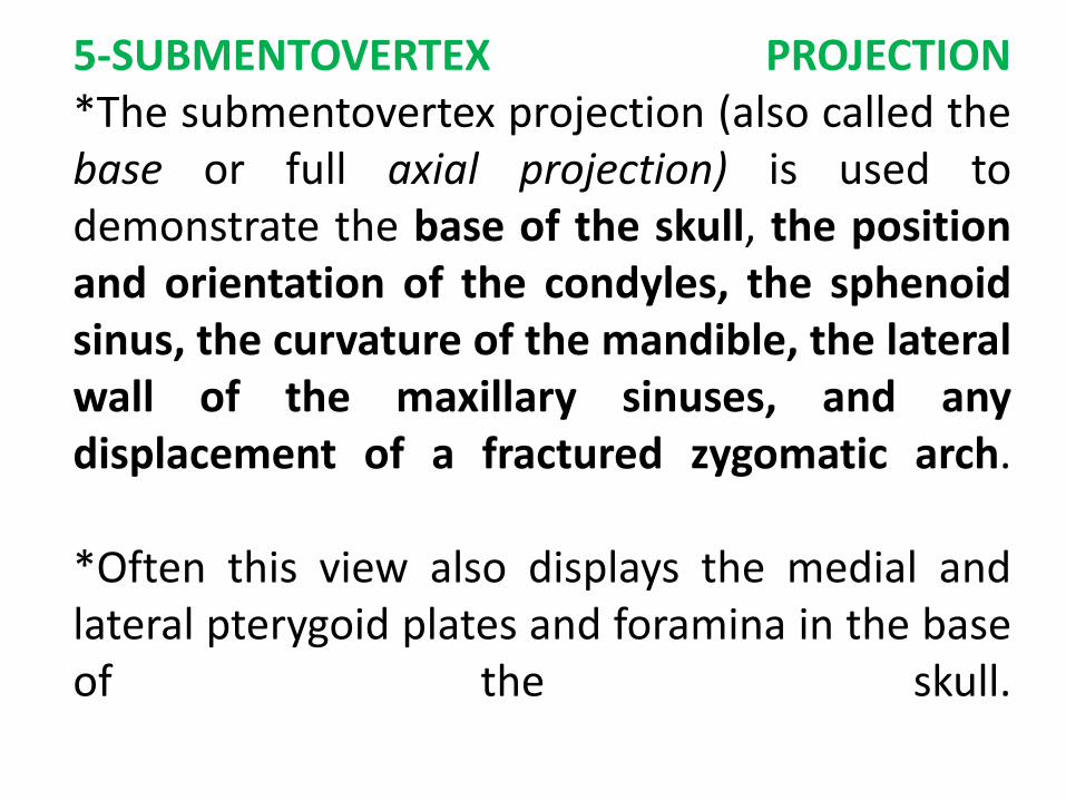
5-SUBMENTOVERTEX PROJECTION*The submentovertex projection (also called thebase or full axial projection) is used todemonstrate the base of the skull, the positionand orientation of the condyles, the sphenoidsinus, the curvature of the mandible, the lateralwall of the maxillary sinuses, and anydisplacement of a fractured zygomatic arch.
*Often this view also displays the medial andlateral pterygoid plates and foramina in the baseof the skull.
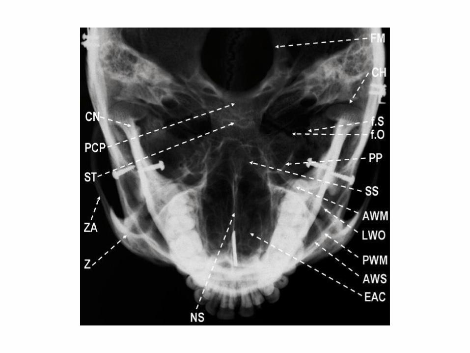
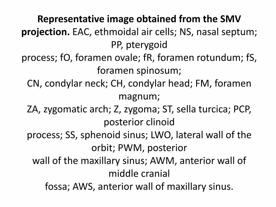
Representative image obtained from the SMVprojection. EAC, ethmoidal air cells; NS, nasal septum;
PP, pterygoidprocess; fO, foramen ovale; fR, foramen rotundum; fS,
foramen spinosum;CN, condylar neck; CH, condylar head; FM, foramen
magnum;ZA, zygomatic arch; Z, zygoma; ST, sella turcica; PCP,
posterior clinoidprocess; SS, sphenoid sinus; LWO, lateral wall of the
orbit; PWM, posteriorwall of the maxillary sinus; AWM, anterior wall of
middle cranialfossa; AWS, anterior wall of maxillary sinus.
Mandibular Oblique Lateral ProjectionTwo oblique lateral projections commonly areused to examine the mandible, one for the bodyand one for the ramus.
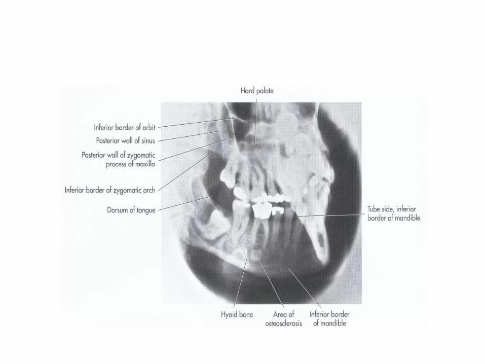
MANDIBULAR BODY PROJECTIONThe mandibular body projection demonstratesthe premolar-molar region and the inferiorborder of the mandible. It provides muchbroader coverage than is possible withperiapical projections
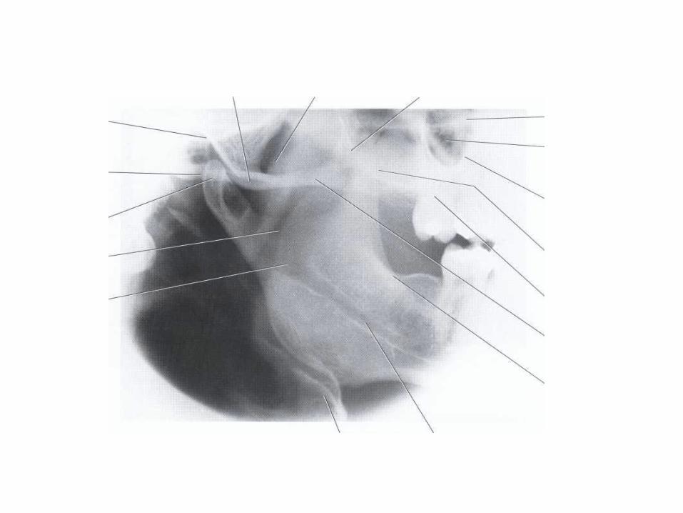
MANDIBULAR RAMUS PROJECTIONThe mandibular ramus projection gives a view ofthe ramus from the angle of the mandible tothe condyl. It often is very useful for examiningthe third molar regions of the maxilla and
mandible.
TemproMandibulat-joint-projectionsTMJ tomography help in visualization of the condyle, articulator eminence and glenoid fossa. It can be also used to determine the joint space and to evaluate the extent of movement of condyle when the mouth is open.
1-Transcranial-view:It used in visualization the superior surface of the condyle and the articulator eminence, the joint space also seen
2-Transorbital view:It demonstrate the entire latero-medial articulating surface of the body, the condyle and articulator eminence and the condylar neck.
3-Transpharyngeal view:
It also called infra cranial view, it demonstrate the angular process from the mid mandibular ramus of the condyle . This tech. helps in diagnosis of facture of the condyle of and condylar neck and in detection alteration in condyle morphology.


![2 extra-oral radiography[1]](https://img.pdfslide.net/doc/110x75/587f1a9e1a28ab350c8b5b19/2-extra-oral-radiography1.jpg)