Embed Size (px)
Citation preview

Disclaimer
This movie is an educational resource only and should not be used to manage your health. The information in this presentation has been intended to help consumers understand the structure and function of anatomical components of the eye.
Multimedia Health Education
EYE ANATOMY
MULTIMEDIA HEALTH EDUCATION MANUAL
TABLE OF CONTENTS
SECTION CONTENT
2 . Eye Anatomy a. External Anatomy
b. Internal Anatomy
1 . Introduction
3 . Mechanics of Vision
Multimedia Health EducationEYE ANATOMY
c. Lateral View
Unit 1: Introduction Multimedia Health Education
EYE ANATOMY
Introduction
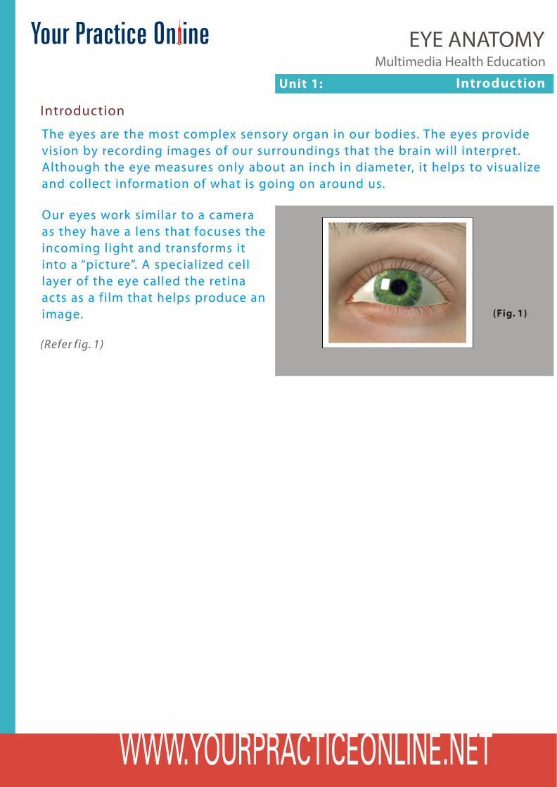
The eyes are the most complex sensory organ in our bodies. The eyes provide vision by recording images of our surroundings that the brain will interpret. Although the eye measures only about an inch in diameter, it helps to visualize and collect information of what is going on around us.
Our eyes work similar to a camera as they have a lens that focuses the incoming light and transforms it into a “picture”. A specialized cell layer of the eye called the retina acts as a film that helps produce an image.
(Refer fig. 1)
(Fig. 1)
Unit 2: Eye Anatomy Multimedia Health Education
EYE ANATOMY
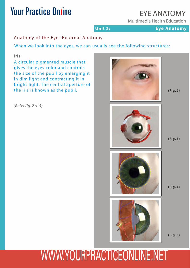
Anatomy of the Eye- External Anatomy
When we look into the eyes, we can usually see the following structures:
Iris:A circular pigmented muscle that gives the eyes color and controls the size of the pupil by enlarging it in dim light and contracting it in bright light. The central aperture of the iris is known as the pupil. (Fig. 2)
(Fig. 3)
(Fig. 4)
(Fig. 5)
(Refer fig. 2 to 5)
Unit 2: Eye Anatomy Multimedia Health Education
EYE ANATOMY
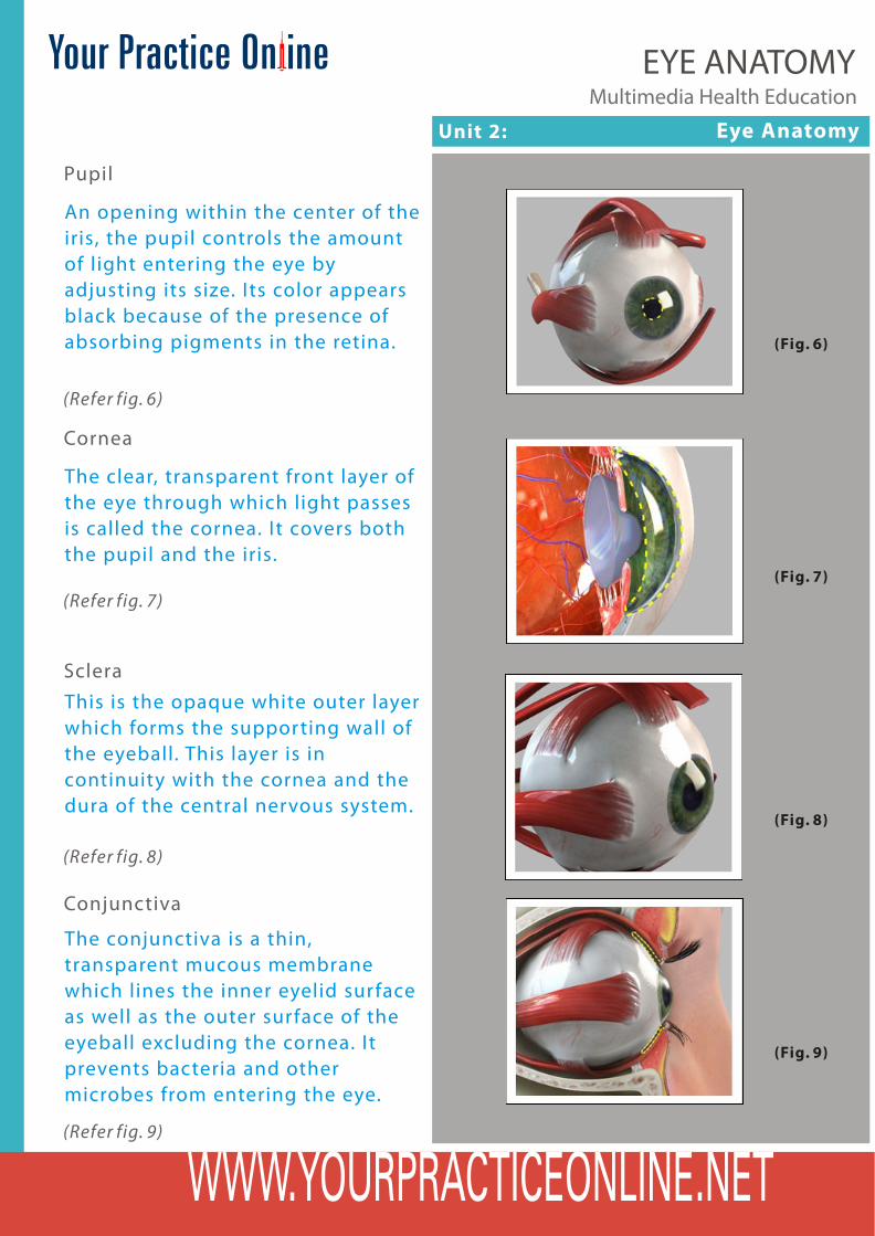
Pupil
An opening within the center of the iris, the pupil controls the amount of light entering the eye by adjusting its size. Its color appears black because of the presence of absorbing pigments in the retina. (Fig. 6)
(Refer fig. 6)
Cornea
The clear, transparent front layer of the eye through which light passes is called the cornea. It covers both the pupil and the iris.
(Fig. 7)
(Refer fig. 7)
Sclera
This is the opaque white outer layer which forms the supporting wall of the eyeball. This layer is in continuity with the cornea and the dura of the central nervous system.
(Refer fig. 8)
(Fig. 8)
(Fig. 9)
Conjunctiva
The conjunctiva is a thin, transparent mucous membrane which lines the inner eyelid surface as well as the outer surface of the eyeball excluding the cornea. It prevents bacteria and other microbes from entering the eye.
(Refer fig. 9)
Unit 2: Eye Anatomy Multimedia Health Education
EYE ANATOMY
Anatomy of the Eye-Internal Anatomy
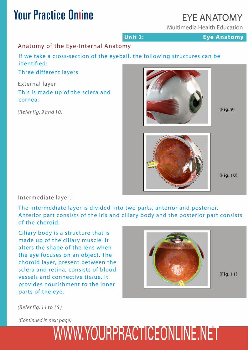
If we take a cross-section of the eyeball, the following structures can be identified:Three different layers
External layerThis is made up of the sclera and cornea.
(Fig. 9)
(Fig. 10)
(Refer fig. 9 and 10)
Intermediate layer:
The intermediate layer is divided into two parts, anterior and posterior. Anterior part consists of the iris and ciliary body and the posterior part consists of the choroid.
Ciliary body is a structure that is made up of the ciliary muscle. It alters the shape of the lens when the eye focuses on an object. The choroid layer, present between the sclera and retina, consists of blood vessels and connective tissue. It provides nourishment to the inner parts of the eye.
(Fig. 11)
(Continued in next page)
(Refer fig. 11 to 15 )

Unit 2: Eye Anatomy Multimedia Health Education
EYE ANATOMY
(Fig. 12)
(Fig. 13)
(Fig. 14)
(Fig. 15)
(Refer fig. 11 to 15 )
Anatomy of the Eye-Internal Anatomy
Unit 2: Eye Anatomy Multimedia Health Education
EYE ANATOMY
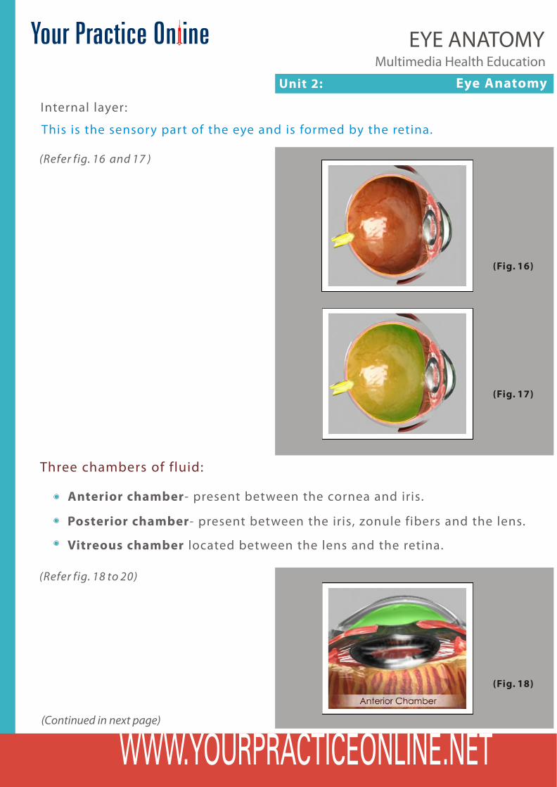
Internal layer:
This is the sensory part of the eye and is formed by the retina.
(Fig. 16)
(Fig. 17)
Anterior chamber- present between the cornea and iris.
Three chambers of fluid:
Posterior chamber- present between the iris, zonule fibers and the lens.
Vitreous chamber located between the lens and the retina.
(Fig. 18)
(Refer fig. 16 and 17 )
(Continued in next page)
(Refer fig. 18 to 20)
Unit 2: Eye Anatomy Multimedia Health Education
EYE ANATOMY
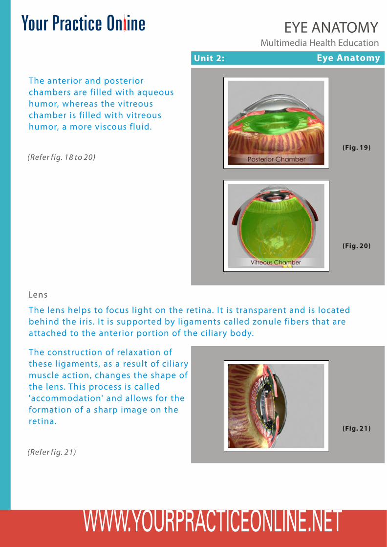
The anterior and posterior chambers are filled with aqueous humor, whereas the vitreous chamber is filled with vitreous humor, a more viscous fluid.
(Fig. 19)
(Fig. 20)
Lens
The lens helps to focus light on the retina. It is transparent and is located behind the iris. It is supported by ligaments called zonule fibers that are attached to the anterior portion of the ciliary body.
The construction of relaxation of these ligaments, as a result of ciliary muscle action, changes the shape of the lens. This process is called 'accommodation' and allows for the formation of a sharp image on the retina.
(Refer fig. 18 to 20)
(Fig. 21)
(Refer fig. 21)
Unit 2: Eye Anatomy Multimedia Health Education
EYE ANATOMY
Anatomy of the Eye-Lateral View
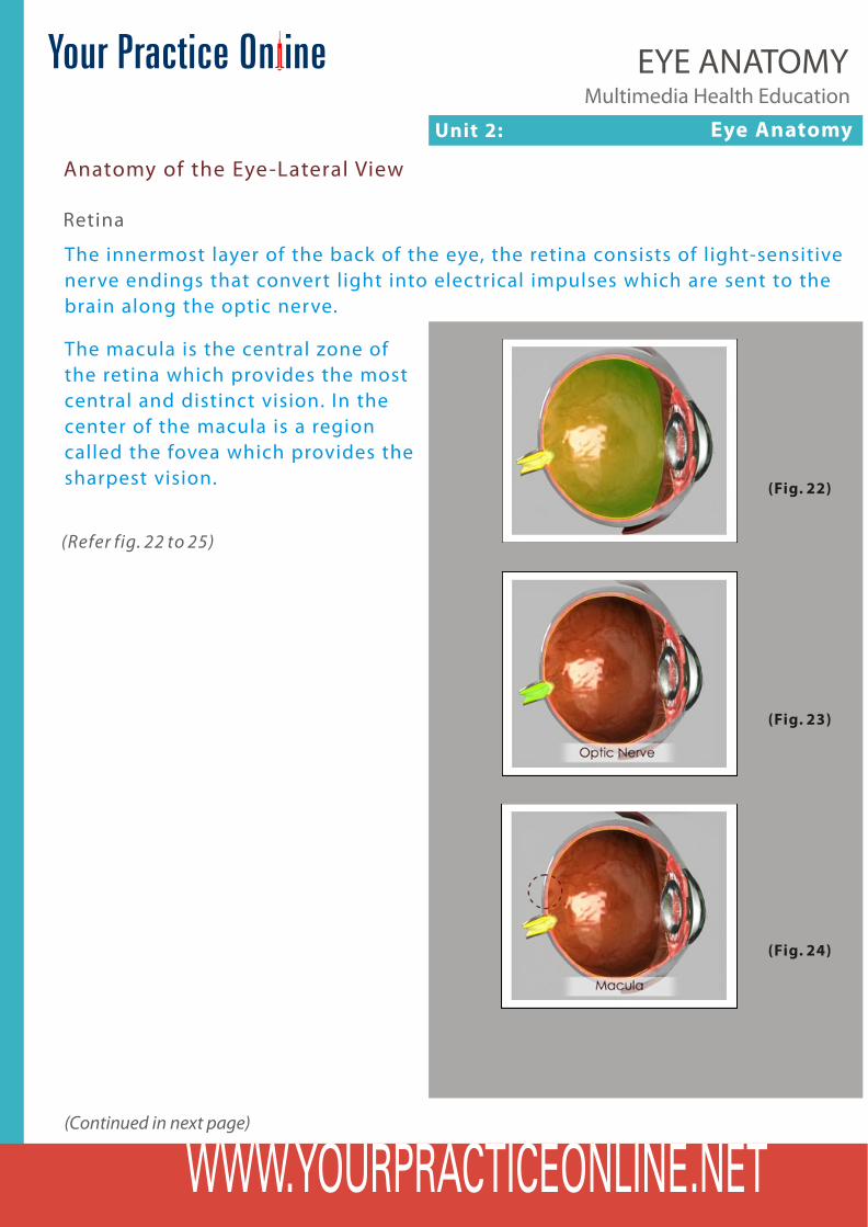
Retina
The innermost layer of the back of the eye, the retina consists of light-sensitive nerve endings that convert light into electrical impulses which are sent to the brain along the optic nerve.
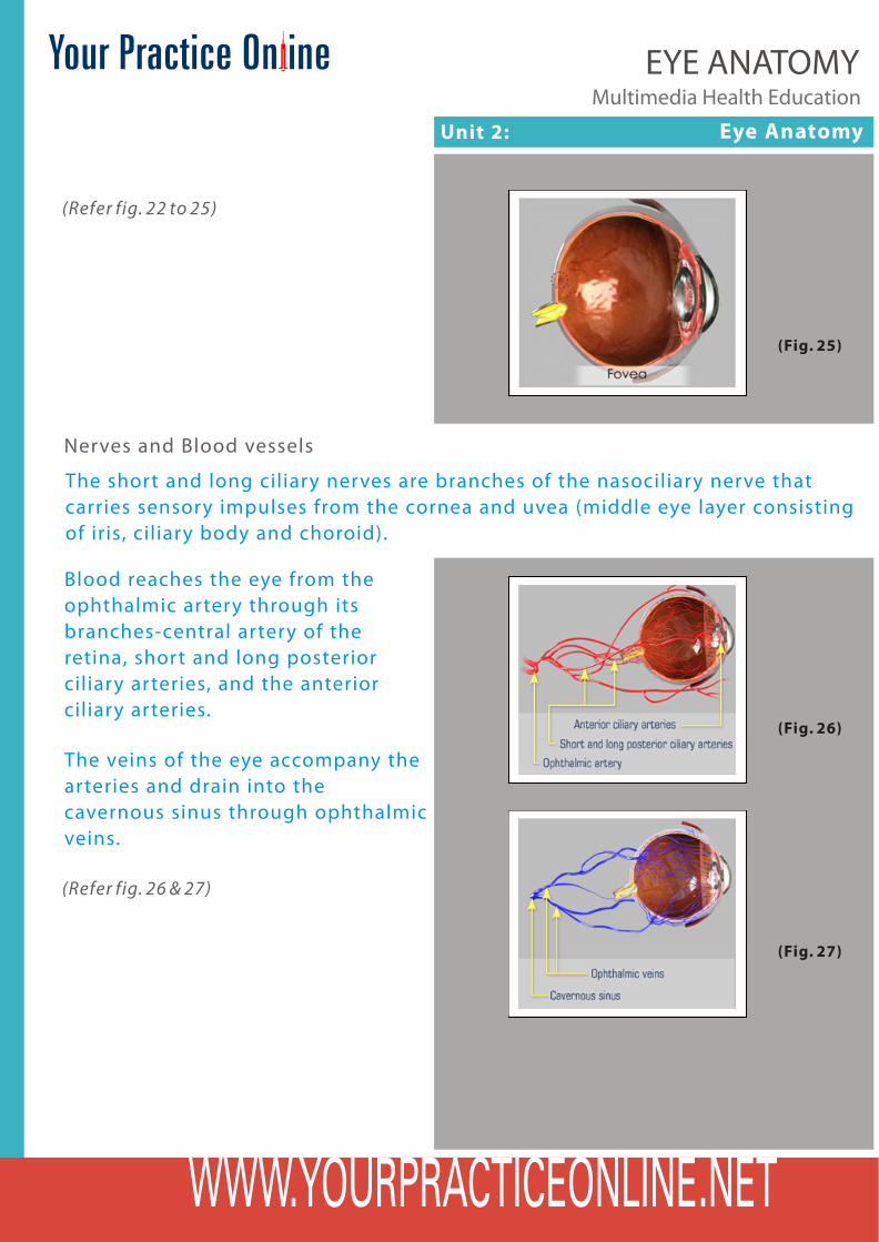
The macula is the central zone of the retina which provides the most central and distinct vision. In the center of the macula is a region called the fovea which provides the sharpest vision. (Fig. 22)
(Fig. 23)
(Fig. 24)
(Refer fig. 22 to 25)
(Continued in next page)
Unit 2: Eye Anatomy Multimedia Health Education
EYE ANATOMY
(Fig. 25)
(Refer fig. 22 to 25)
Nerves and Blood vessels
The short and long ciliary nerves are branches of the nasociliary nerve that carries sensory impulses from the cornea and uvea (middle eye layer consisting of iris, ciliary body and choroid).
Blood reaches the eye from the ophthalmic artery through its branches-central artery of the retina, short and long posterior ciliary arteries, and the anterior ciliary arteries.
(Fig. 26)
(Fig. 27)
The veins of the eye accompany the arteries and drain into the cavernous sinus through ophthalmic veins.
(Refer fig. 26 & 27)
Unit 2: Eye Anatomy Multimedia Health Education
EYE ANATOMY
Optic Nerve
The optic nerve is made up of several million nerve fibers. It carries visual impulses from the retina to the brain.
(Fig. 28)
Optic disc or optic nerve head.
Optic disc is a circular area at the back of the eye where the optic nerve joins the retina. This area does not respond to light stimulation because of the absence of photoreceptors and is known as "blind spot”
(Refer fig. 28)
(Fig. 29)
(Refer fig. 29)
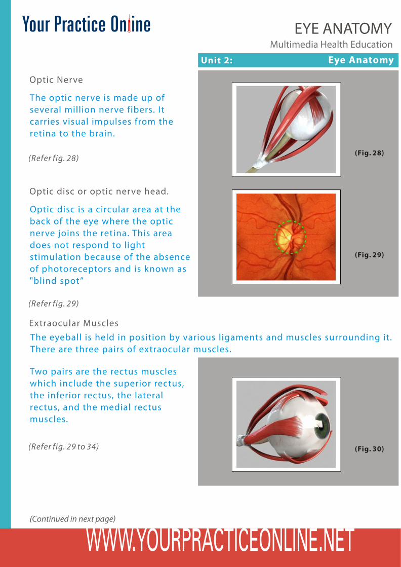
Extraocular MusclesThe eyeball is held in position by various ligaments and muscles surrounding it. There are three pairs of extraocular muscles.
(Fig. 30)(Refer fig. 29 to 34)
(Continued in next page)
Two pairs are the rectus muscles which include the superior rectus, the inferior rectus, the lateral rectus, and the medial rectus muscles.

Unit 2: Eye Anatomy Multimedia Health Education
EYE ANATOMY
They run straight along the eyeball and insert into the bony orbit of the skull. The other pair of muscles in oblique muscles which include the superior and inferior oblique muscles. These muscles help in rotation of the eyeball and also allow the image to be focused on the fovea of the central retina.
(Fig. 31)
(Fig. 32)
(Fig. 33)
(Fig. 34)
(Refer fig. 29 to 34)
Unit 3: Mechanics of Vision
Multimedia Health EducationEYE ANATOMY
Mechanics of Vision
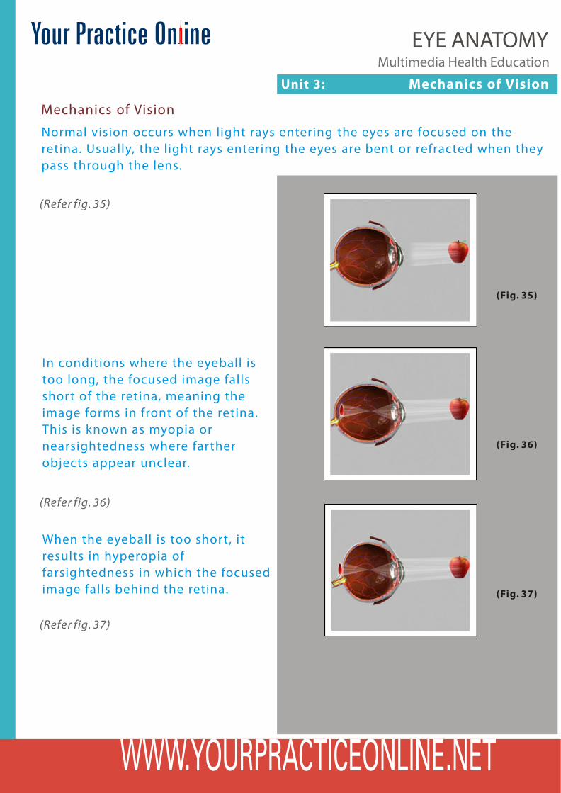
Normal vision occurs when light rays entering the eyes are focused on the retina. Usually, the light rays entering the eyes are bent or refracted when they pass through the lens.
(Fig. 35)
(Refer fig. 35)
In conditions where the eyeball is too long, the focused image falls short of the retina, meaning the image forms in front of the retina. This is known as myopia or nearsightedness where farther objects appear unclear.
(Fig. 36)
(Fig. 37)
When the eyeball is too short, it results in hyperopia of farsightedness in which the focused image falls behind the retina.
(Refer fig. 36)
(Refer fig. 37)
Unit 3: Mechanics of Vision
Multimedia Health EducationEYE ANATOMY
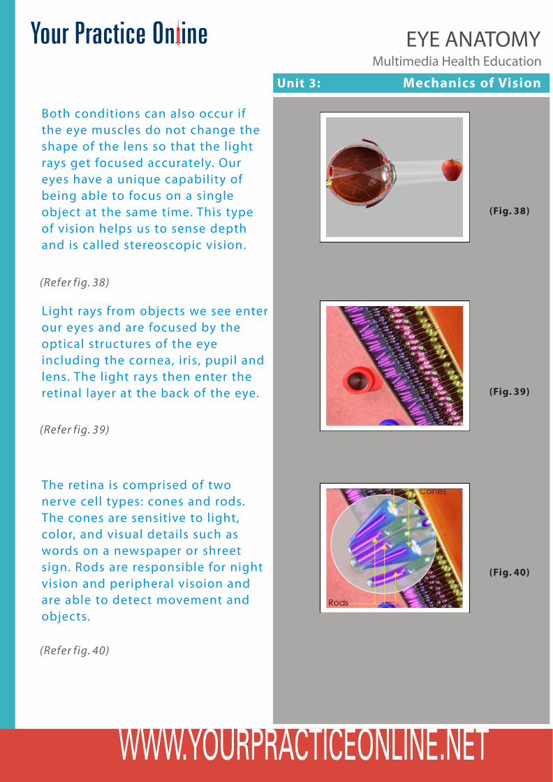
Both conditions can also occur if the eye muscles do not change the shape of the lens so that the light rays get focused accurately. Our eyes have a unique capability of being able to focus on a single object at the same time. This type of vision helps us to sense depth and is called stereoscopic vision.
Light rays from objects we see enter our eyes and are focused by the optical structures of the eye including the cornea, iris, pupil and lens. The light rays then enter the retinal layer at the back of the eye.
The retina is comprised of two nerve cell types: cones and rods. The cones are sensitive to light, color, and visual details such as words on a newspaper or shreet sign. Rods are responsible for night vision and peripheral visoion and are able to detect movement and objects.
(Fig. 38)
(Refer fig. 38)
(Fig. 39)
(Refer fig. 39)
(Fig. 40)
(Refer fig. 40)

Unit 3: Disclaimer
Although every effort is made to educate you on Normal Anatomy of the Eye, there will be specific information that will not be discussed. Talk to your Ophthalmologist or health care provider about any questions you may have.
Disclaimer
Multimedia Health EducationEYE ANATOMY

YOUR SURGERY DATE
Physician's Name :
Physician's Signature:
Date :
Patient’s Name :
Patient’s Signature:
Date :
READ YOUR BOOK AND MATERIAL
VIEW YOUR VIDEO/CD/DVD/ WEBSITE
PRE - HABILITATION
ARRANGE FOR BLOOD
MEDICAL CHECK UP
ADVANCE MEDICAL DIRECTIVE
PRE - ADMISSION TESTING
FAMILY SUPPORT REVIEW
Multimedia Health EducationEYE ANATOMY


![태양광을 이용한 저전력의 LED 표지병 설계 · 자전거용 도로표지병을 [Fig. 6]과 같이 설계하였다. [Fig. 6] Cycletrack Cat's eye stud 2.2 제어회로설계](https://img.pdfslide.net/doc/110x75/60060dccc9e40b653c4eea44/foee-oe-e-led-oee-e-e-eeoeoee.jpg)