Embed Size (px)
Citation preview

Eye and Ear
1. Eye 1) The wall of eyeball
① Fibrous tunic: DCT ---cornea: ---sclera: DCT ---corneal limbus(corneoscleral limbus)Cornea:/anterior 1/6 of fibrous tunic, transparent, bulges
slightly anteriorly/connect with sclera
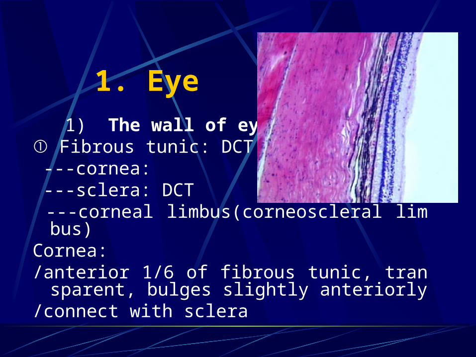
/five layers: corneal epithelium:
i. stratified, squamous non-keratinising epithelium
ii. 5-6 layers of regular arranged cells iii. basal cells have remarkable regeneratin
g ability iv. rich in nerve terminal
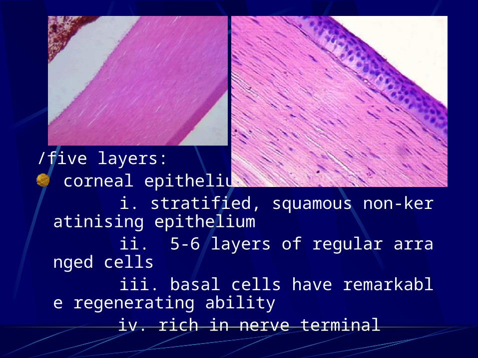
anterior limiting lamina: i. a clear uniform membrane, 10-16u
m thick ii. contain collagenous fibrils and ma
trix iii. cannot regenerate
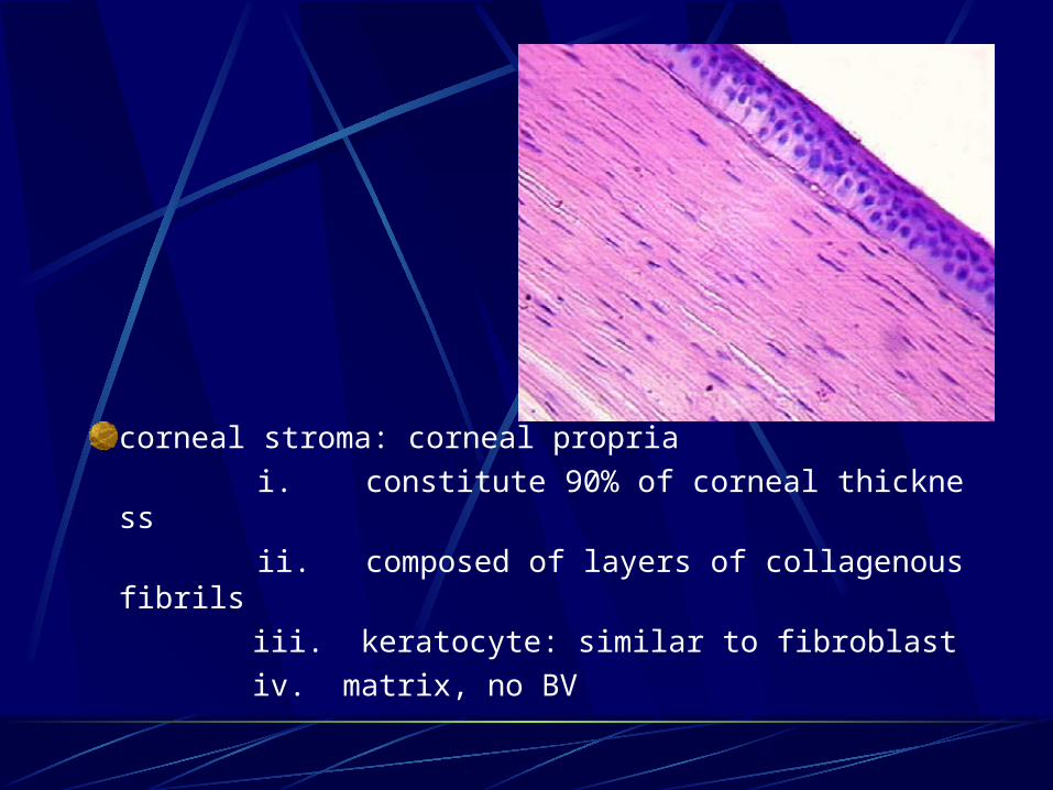
corneal stroma: corneal propria
i. constitute 90% of corneal thickness
ii. composed of layers of collagenous fibrils
iii. keratocyte: similar to fibroblast
iv. matrix, no BV
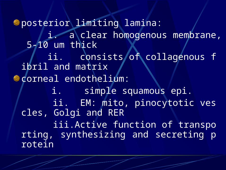
posterior limiting lamina: i. a clear homogenous membrane, 5-10
um thick ii. consists of collagenous fibril and matri
xcorneal endothelium:
i. simple squamous epi. ii. EM: mito, pinocytotic vescles, Golgi a
nd RER iii.Active function of transporting, synthes
izing and secreting protein
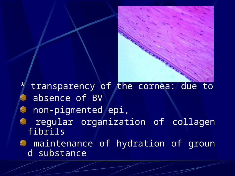
* transparency of the cornea: due to absence of BV non-pigmented epi, regular organization of collagen fibrils maintenance of hydration of ground substance
② Vascular tunic(uvea): LCT with BV and melanocytes
---iris
---ciliary body
---choroid
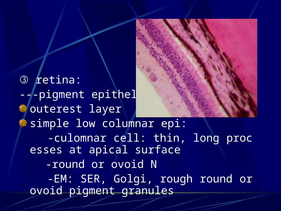
③ retina: ---pigment epithelium:
outerest layersimple low columnar epi:
-culomnar cell: thin, long processes at apical surface
-round or ovoid N -EM: SER, Golgi, rough round or ovoid pigme
nt granules

-function:
i. protect visual cell
ii. involve in replace of membranous disc
iii. store vitamin A and involve in the synthesis of rhodopsin
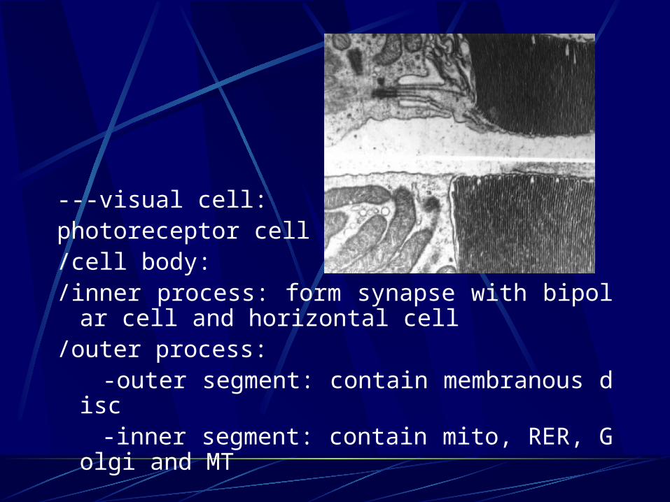
---visual cell: photoreceptor cell/cell body:/inner process: form synapse with bipolar cell a
nd horizontal cell/outer process: -outer segment: contain membranous disc -inner segment: contain mito, RER, Golgi and
MT
/rod cell:-110,000,000-120,000,000-deep-stained N-outer process: cylindrical-outer segment: membranous disc-invagination
of cell membrane but separated with cell membrane(exfoliated and ingested by pigment cell) -rhodopsin(visual purpke)= 11-cisretinal(retinene) + opsin
-inner process: spherule(end in a terminal expansion)
-feel dim light

/cone cell: -6,500,000-7,000,000-large N, paler-stained-outer process: conical-outer segment: membranous disc, not separate
d, no exfoliation of disks-iodopsin(photopsin)= 11-cisretinal + opsin(diffe
rent)-inner process: pedicle-feel blight light(red-558nm, green-531nm, blue-
419nm)
---bipolar cell: /large N/contain RER,mito and Golgi/dendrite: synapse with photoreceptor and horiz
ontal neuron/axon: form synapse with dendrite of ganglion c
ell/classification: -rod bipolar cell -midget bipolar cell -flat bipolar cell
---ganglion cell:
/multipolar neuron:
/dendrite: synapse with bipolar, amacrine cell and interplexiform cell
/axon: make up optic nerve
/classification: midget ganglion cell and diffuse ganglion cell
---interneurons: /located in layer of bipolar cell/horizontal cell, amacrine cell, interplexiform cell---radial neuroglia cell: Muller cell/neuroglial cell/thin and long cell, with ovoid, deep-stained N/processes: end at outer limiting membrane and
inner limiting membrane/function: supporting, protecting, nourishing and
insulating function
Under LM: retina can be divided into ten layers i. layer of pigment epithelium: pigment epithelial
cell ii. layer of rods and cones iii. outer limiting membrane: outer processes of
Muller cell iv. outer nuclear layer: N of visual cells v. outer plexiform layer: inner process of visual c
ell, dendrites of bipolar cell and processes of horizontal cell
vi. inner nuclear layer: cell body of bipolar cell, horizontal cell, amacrine cell and interplexiform cell and Muller cell
vii.inner plexiform layer: axon of bipolar cell, dendrites of ganglion cell, processes of amacrine cell and interplexiform cell
viii. layer of ganglion cells: cell body of ganglion cell
ix. layer of optic fibers: axons of ganglion cell
x.inner limition membrane: formed by connection each other of inner processes of Muller cells
* macula lutea:/definition: a small area of retina at posterior po
lar of retina, contains a yellow pigment and is non-vascularised, so called yellow spot
/3mm in D/central fovea: shallow depression, 1.5mm in D/thinnest retina: 0.1mm/contain only cone cell, no rod cell/one visual cell connects with one bipolar cell, a
nd one bipolar cell forms synapse with one ganglion cell
/have most clear vision
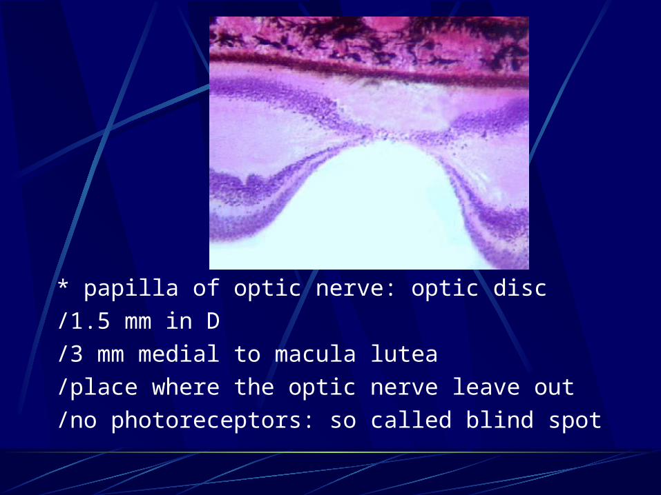
* papilla of optic nerve: optic disc
/1.5 mm in D
/3 mm medial to macula lutea
/place where the optic nerve leave out
/no photoreceptors: so called blind spot
2. Ear
---the external ear---the external ear
---the middle ear---the middle ear
---the inner ear---the inner ear

1) inner ear: labyrinth
---osseous labyrinth: a system of canals and cavities in compact bone
the vestibule
semicircular canal
cochlea
---membranous labyrinth: usually lined by simple squamous epi. except:
membrane semicircular canal: crista ampullaries
saccule and utricle: macula utriculi and macula sacculi
cochlear duct: spiral organ
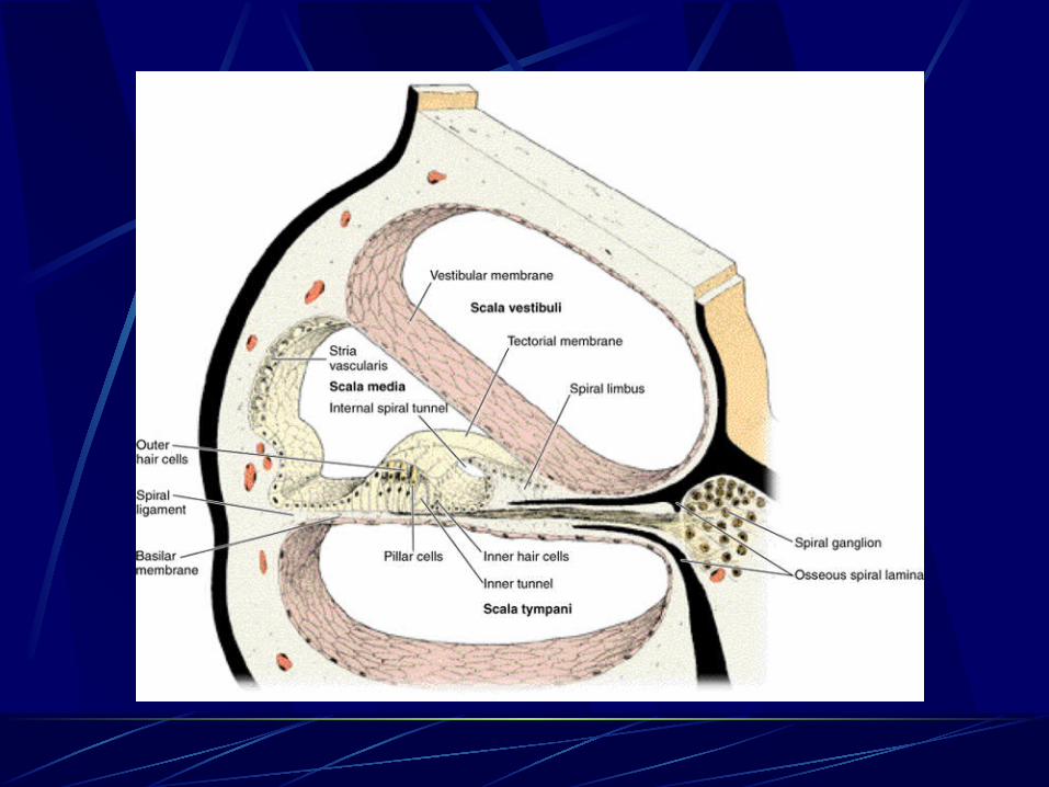
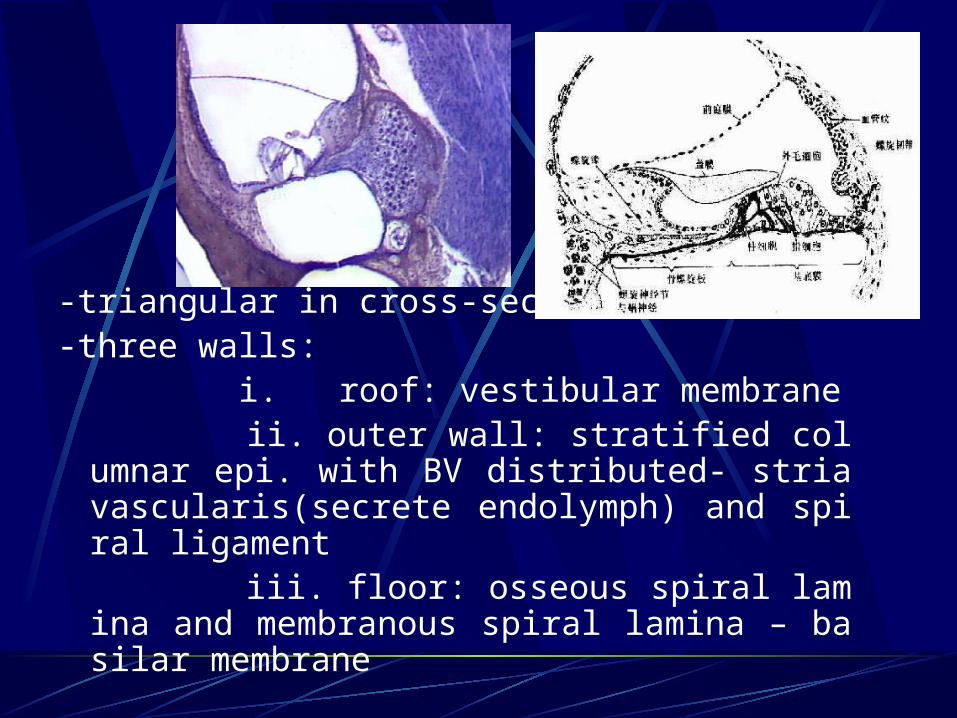
-triangular in cross-section-three walls: i. roof: vestibular membrane ii. outer wall: stratified columnar epi. with
BV distributed- stria vascularis(secrete endolymph) and spiral ligament
iii. floor: osseous spiral lamina and membranous spiral lamina – basilar membrane
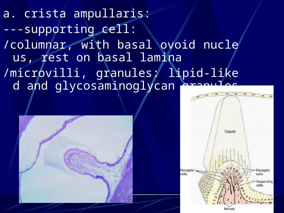
a. crista ampullaris:---supporting cell: /columnar, with basal ovoid nucleus, rest
on basal lamina/microvilli, granules: lipid-liked and glycos
aminoglycan granules
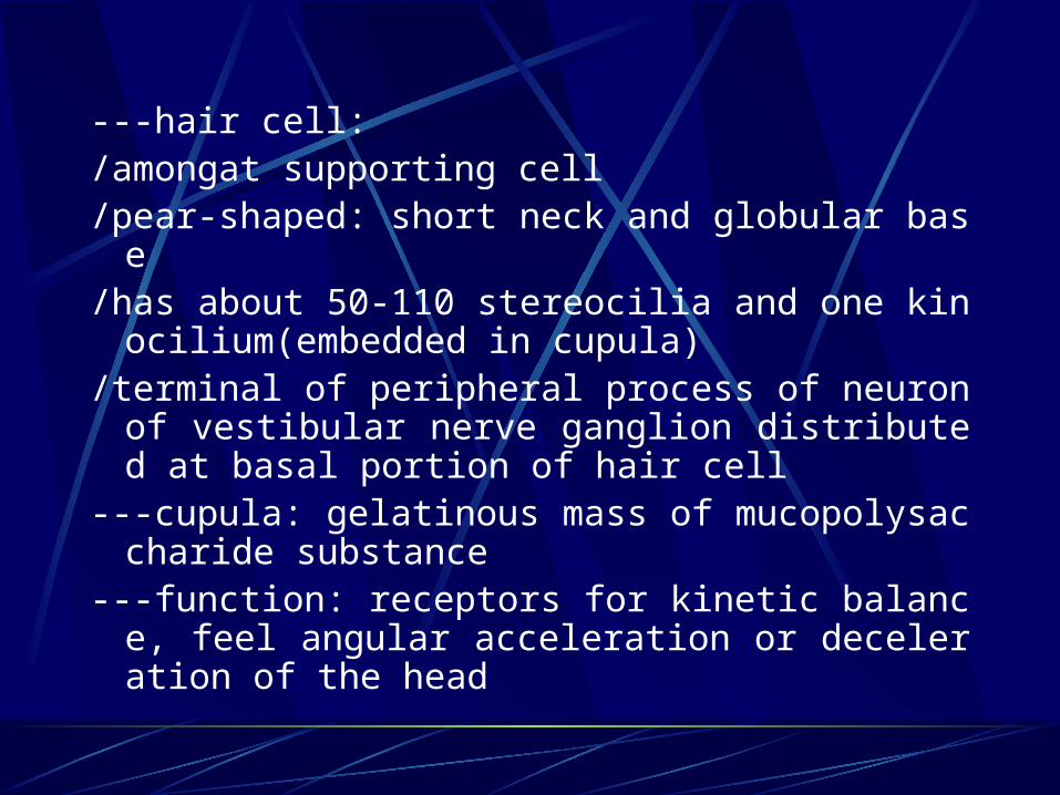
---hair cell: /amongat supporting cell/pear-shaped: short neck and globular base/has about 50-110 stereocilia and one kinociliu
m(embedded in cupula)/terminal of peripheral process of neuron of vest
ibular nerve ganglion distributed at basal portion of hair cell
---cupula: gelatinous mass of mucopolysaccharide substance
---function: receptors for kinetic balance, feel angular acceleration or deceleration of the head
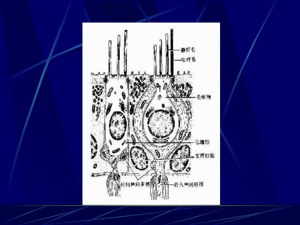
b. macula utriculi and macula sacculi: macula acustica---supporting cell---hair cell: 30-60 stereocilia
and one kinocilium
---otolithic membrane: gelatinous mucopolysaccharide substance containing small crystalline bodies of calcium carbonate---function: receptors of static balance, feel linear acceleration or deceleration and change in position of the head
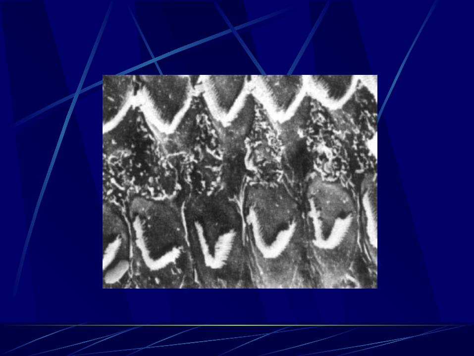
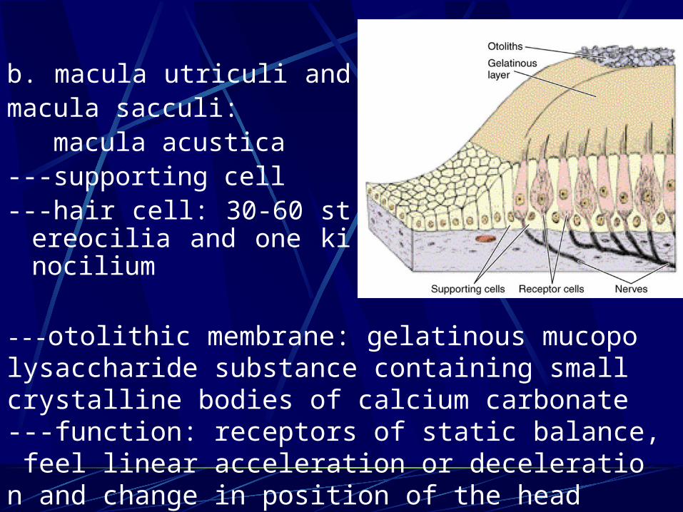
c. spiral organ: Corti organ---supporting cell:
pillar cell:-two rows: inner and outer pollar cell: tall, colum
nar in shape, -inner tunnel
phalahgeal cell:-inner phalangeal cell: one row, is situated
next to inner pillar cell-outer phalangeal cell: 3-5 rows, lateral to
the outer pillar cells-tall columnar cells rest on basilar membr
ane-phalangeal process: enclosed the low pa
rt of hair cell
---hair cell: -inner hair cell: a row of pear-shaped cell, supp
orted by inner phalangeal cell-outer hair cell: 3-5 rows, supported by outer ph
alangeal cell-“V” or “W” shaped-arranged stereocilia on free
surface---peripheral processes of neuron of spiral gangl
ion distribute at basal portion of hair cell---tectorial membrane---auditory string: 2000, located in basilar memb
rane, collagen-liked thin filament---function: receptor of sound