Embed Size (px)
Citation preview
ORIGINAL RESEARCH ARTICLE Open Access
Face and content validity of a virtual-realitysimulator for myringotomy with tubeplacementCaiwen Huang1, Horace Cheng2, Yves Bureau4,5, Sumit K. Agrawal1,2,6*† and Hanif M. Ladak1,2,3,5*†
Abstract
Background: Myringotomy with tube insertion can be challenging for junior Otolaryngology residents as it is oneof the first microscopic procedures they encounter. The Western myringotomy simulator was developed to allowtrainees to practice microscope positioning, myringotomy, and tube placement. This virtual-reality simulator is viewedin stereoscopic 3D, and a haptic device is used to manipulate the digital ear model and surgical tools.
Objective: To assess the face and content validity of the Western myringotomy simulator.
Methods: The myringotomy simulator was integrated with new modules to allow speculum placement, manipulationof an operative microscope, and insertion of the ventilation tube through a deformable tympanic membrane.A questionnaire was developed in consultation with instructing surgeons. Fourteen face validity questionsfocused on the anatomy of the ear, simulation of the operative microscope, appearance and movement ofthe surgical instruments, deformation and cutting of the eardrum, and myringotomy tube insertion. Six contentvalidity questions focused on training potential on surgical tasks such as speculum placement, microscopepositioning, tool navigation, ear anatomy, myringotomy creation and tube insertion. A total of 12 participantsfrom the Department of Otolaryngology—Head and Neck Surgery were recruited for the study. Prior tocompleting the questionnaire, participants were oriented to the simulator and given unlimited time to practice untilthey were comfortable with all of its aspects.
Results: Responses to 12 of the 14 questions on face validity were predominantly positive. One issue of concern waswith contact modeling related to tube insertion into the eardrum, and the second was with the movement of the bladeand forceps. The former could be resolved by using a higher resolution digital model for the eardrum to improve contactlocalization. The latter could be resolved by using a higher fidelity haptic device. With regard to content validity, 64 % ofthe responses were positive, 21 % were neutral, and 15 % were negative.
Conclusions: The Western myringotomy simulator appears to have sufficient face and content validity. Furtherdevelopment with automated metrics and skills transference testing is planned.
Keywords: Myringotomy, Education, Simulator, Virtual reality, Face validity
* Correspondence: [email protected]; [email protected]†Equal contributors1Department of Electrical and Computer Engineering, Western University,London, ON, CanadaFull list of author information is available at the end of the article
© 2015 Huang et al. Open Access This article is distributed under the terms of the Creative Commons Attribution 4.0International License (http://creativecommons.org/licenses/by/4.0/), which permits unrestricted use, distribution, andreproduction in any medium, provided you give appropriate credit to the original author(s) and the source, provide a link tothe Creative Commons license, and indicate if changes were made. The Creative Commons Public Domain Dedication waiver(http://creativecommons.org/publicdomain/zero/1.0/) applies to the data made available in this article, unless otherwise stated.
Huang et al. Journal of Otolaryngology - Headand Neck Surgery (2015) 44:40 DOI 10.1186/s40463-015-0094-2
IntroductionMyringotomy with tube insertion is one of the mostcommon procedures in Otolaryngology—Head & NeckSurgery, and is encountered by residents throughouttheir training. Despite the fact that it is a ubiquitous pro-cedure, the instruction of junior trainees, who often havelittle experience in microscopic procedures, is oftenchallenging. Montague et al. [1] have analyzed surgicalerrors through video analysis of actual procedures andnote that the 4 most frequently occurring errors in orderfrom most to least occurring include (1) failure to per-form a unidirectional myringotomy, (2) making multipleattempts to place the tube, (3) making multiple attemptsto complete the myringotomy, and (4) setting the micro-scope magnification too high. More serious intraopera-tive complications can also occur including externalauditory canal lacerations, medial displacement of tubesinto the middle ear, and vascular injuries [2–4]. Al-though surgical residents can eventually perform stand-ard cases well, they often struggle with narrow canals,retracted tympanic membranes, T-tubes, and proceduresperformed under local anaesthestic. The goal of simula-tion is to decrease the learning curve prior to enteringthe operating, minimize complications in patients, andprovide the ability to practice difficult cases.Several physical models have been described in the lit-
erature to provide practice without potential harm to pa-tients [5–9]. Generally, these consist of a tube to mimicthe ear canal with a synthetic membrane attached to oneend to represent the eardrum. These models do not ap-pear to have gained general acceptance in residency pro-grams, presumably because they are not able torepresent anatomical variability easily and the mechan-ical properties of the materials used do not mimic thatof the actual tissues.Compared with physical models, simulators based on
virtual-reality (VR) technologies have the ability to simu-late difficult anatomy, model various pathologies, pro-vide automated feedback, and even allow trainees topractice on patient-specific models generated from CT/MRI scans. VR-based simulators have been applied inOtolaryngology, especially for endoscopic sinus surgery[10–14] and for temporal bone drilling [15–18].In VR simulators, the trainee interacts with realistic
3D digital models of anatomical structures and viewsthem using 3D displays. Simulated tissues can be operatedupon using digital representations of actual surgical toolsthat can be moved in the workspace using devices such asa haptic arm. The sensation of contact force between adigital surgical tool and simulated tissue can be computedand applied to the trainee’s hand via the haptic arm.The Auditory Biophysics Laboratory at Western
University has developed and reported on several as-pects of VR-based myringotomy simulation. A blade
navigation software system [19, 20] and a system forreal-time deformation and cutting of the tympanic mem-brane [21] were implemented on different software plat-forms as separate training modules. These versions ofthe simulator were not integrated and they did not in-clude speculum placement, operating microscope con-trols for positioning/zooming, or tube insertion throughthe myringotomy.As recently reported [22], the Western myringotomy
simulator has integrated the previous modules into a com-mon software platform. Moreover, new software moduleshave been added to allow the user to adjust their surgicalview through positioning and tilting of the virtualspeculum and operative microscope, and to allow inser-tion of a ventilation tube into the myringotomy created ina deformable tympanic membrane. The goal is to furtherexpand this simulator in the future to allow trainees toraise tympanomeatal flaps andto eventually perform tym-panoplasty/ossiculoplasty on patient-specific anatomy.In order for training simulators to be accepted into a
residency curriculum, a variety of validation studies needto be conducted starting with face validity and culminat-ing in the demonstration that skills acquired in the VRenvironment transfer to the OR (operating room) envir-onment. Face validity refers to the degree to which asimulation appears like the real situation [23] and con-tent validity measures whether the simulator would beappropriate or useful in training [24, 25]. Although facevalidity has previously been established for individualsoftware modules [19–21], validation testing has notbeen performed on the current integrated system, whichsimulates the entire procedure from microscope posi-tioning to ventilation tube insertion [22].The objective of this paper is to determine the face
and content validity of the new integrated Western myr-ingotomy simulator.
MethodsSimulatorAn overview of the major features of the simulator isgiven here; in-depth technical details on the system canbe found in a previous publication [22]. The simulatorconsists of 3 major components: the simulation software,a display system, and a haptic arm as shown in Fig. 1.The simulation software was developed in the AuditoryBiophysics Laboratory at Western University [19–22].The simulator runs on a Z420 Hewitt-Packard personalcomputer, equipped with an Intel(R) Xeon E5-1620 pro-cessor (Intel Corp., Sanata Clara, CA) and a NVIDIAQuadro 4000 graphics card (NVIDIA Corp., Santa Clara,CA). The system is capable of real-time rendering of the3D digital models of the ear, surgical tools, and tympanicmembrane as shown in Fig. 2a. The simulator can im-port various ear canal and tympanic membrane models,
Huang et al. Journal of Otolaryngology - Head and Neck Surgery (2015) 44:40 Page 2 of 8
however for the purposes of this study, a normalpediatric ear canal and tympanic membrane was used.The system also incorporates multi-point collision detec-tion to monitor for all interactions between the virtualtools and virtual ear and performs real-time deformationand tissue cutting as required. The software displays themodels and all interactions on a silver screen mirror thatis part of the DevinSense Display 300 system (DevinSenseDisplay Solutions, Sundbyberg, Sweden). When the screenis viewed using active 3D glasses (Nvidia Corp., SantaClara, CA) provided with the DevinSense system, the 3Ddigital scene consisting of the virtual ear and tools appearsto exist in the space below the silver screen mirror. Thedisplay in this region is correctly co-located with the hap-tic arm (Omni haptic arm, Geomagic, Inc., Morrisville,NC) so movements of the haptic arm appear to occur inthe same space as the 3D scene. Using the haptic arm, theuser can move the virtual surgical tools. Currently, a singlehaptic arm is used to control the various instruments,however a second haptic arm could be added to simultan-eously manipulate multiple instruments (e.g. speculumand myringotomy blade).The haptic arm can be used to position and rotate the
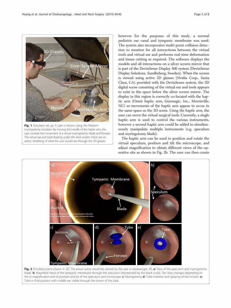
virtual speculum, position and tilt the microscope, andadjust magnification to obtain different views of the op-erative site as shown in Fig. 2b. The user can then create
Fig. 2 Simulator scene shown in 2D. The actual scene would be viewed by the user in stereoscopic 3D. a) View of the speculum and myringotomyblade. b) Magnified views of the tympanic membrane through the speculum (represented by the black circle). The view changes depending onthe (i) magnification and (ii) position and tilt of the speculum and microscope. c) Myringotomy d) Tube insertion and splaying of the incision. e)Tube in final position with middle ear visible through the lumen of the tube
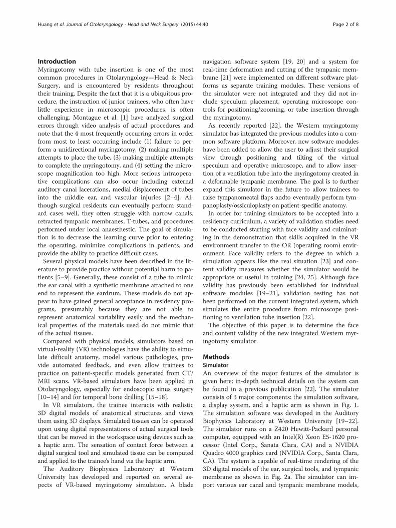
Fig. 1 Simulator set up. A user is shown using the Westernmyringotomy simulator. By moving the handle of the haptic arm, theuser controls the movement of a virtual myringotomy blade and forceps.The virtual ear and tools floating under the silver screen mirror are anartistic rendering of what the user would see through the 3D glasses
Huang et al. Journal of Otolaryngology - Head and Neck Surgery (2015) 44:40 Page 3 of 8
a myringotomy as shown in Fig. 2c using a virtual myr-ingotomy blade; the position and orientation of the bladeare controlled by moving the handle of the haptic arm.A tube may be inserted using virtual forceps, which isalso controlled by the user using the haptic device[Fig. 2d]. The opening and closing of the forceps can betoggled using a button on the haptic arm. During tubeinsertion, the eardrum deforms and the incision splaysas the tube enters the myringotomy. The tube may alsobe repositioned with various instruments until it is in itsfinal position [Fig. 2e].
ParticipantsResearch ethics board approval was obtained fromWestern University (#105239) and participants werecontacted via telephone or electronic mail. All partici-pants were recruited from the Department of Otolaryn-gology - Head & Neck Surgery, Western University. Atotal of 12 subjects agreed to participate, which includedseven junior Otolaryngology residents (postgraduateyears 1 to 3) and five senior Otolaryngologists who rou-tinely performed ventilation tube insertions in theirpractice. These groups were chosen to reflect the targetgroup of the simulator (junior residents) as well as ex-perts in the field (Otolaryngologists). The participantsdid not have any previous exposure to myringotomysimulation.
ProtocolAll participants were initially given an orientation ses-sion which consisted of: 1) an information sheet outlin-ing the software features of the simulator, 2) ademonstration video of how to perform a myringotomyand tube insertion using the simulator controls, and 3) alive demonstration of the simulator and haptic arm. Thesame graduate student and surgical resident performedthe orientation session for each participant, and a stan-dardized script was used to ensure consistency. The par-ticipants were specifically asked to perform the taskslisted in Table 1 so that they could comment on all thevarious aspects of the simulator. Finally, the participantswere given an unlimited period of time to use the
simulator until they felt comfortable completing the faceand content validity questionnaires.
QuestionnairePreviously, we had tested individual software modulesfocusing on blade navigation [19], haptics [20] and tym-panic membrane deformation and cutting [21]. Sincethis new simulator [22] refined each of these compo-nents, including the graphical representations of the earand virtual tools, and included new features such asmicroscope handling, speculum positioning and tube in-sertion, the Myringotomy Surgery Simulation Scale(MS3) used in previous publications [20, 21] was modi-fied to include these features. The questionnaire was di-vided into three sections (A, B, and C) with a total of 20questions. Section A included 14 questions focusing onface validity as listed in Table 2. The appearance andrealism of the surgical instruments; anatomy of the aur-icle, ear canal and eardrum; movement of surgical in-struments; deformation and cutting of the eardrum; tubeinsertion and 3D microscopic view of the scene wereassessed.Section B included six questions focusing on content
validity as listed in Table 3. These questions were usedto determine training potential on specific surgical tasks.In Sections A and B, study participants were asked to
answer each question using a 7-point Likert scale, anequal appearing interval measurement. The scale hadvalues of “1”—Strongly Disagree, 2—“Mostly Disagree”,3—“Disagree”, 4—“Neither Agree/Disagree”, 5—“Agree”,6—“Mostly Agree” and 7—“Strongly Agree”.In Section C, a free-form comment area was provided
for each participant to provide feedback to elaborate on
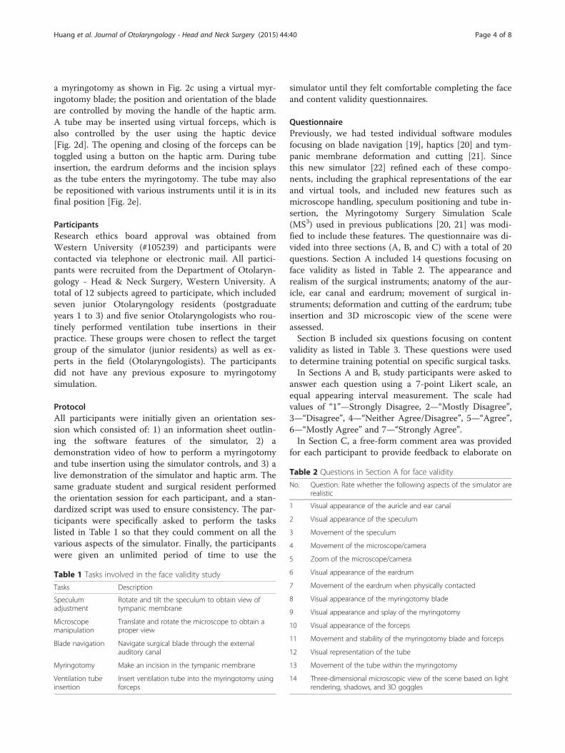
Table 1 Tasks involved in the face validity study
Tasks Description
Speculumadjustment
Rotate and tilt the speculum to obtain view oftympanic membrane
Microscopemanipulation
Translate and rotate the microscope to obtain aproper view
Blade navigation Navigate surgical blade through the externalauditory canal
Myringotomy Make an incision in the tympanic membrane
Ventilation tubeinsertion
Insert ventilation tube into the myringotomy usingforceps
Table 2 Questions in Section A for face validity
No. Question: Rate whether the following aspects of the simulator arerealistic
1 Visual appearance of the auricle and ear canal
2 Visual appearance of the speculum
3 Movement of the speculum
4 Movement of the microscope/camera
5 Zoom of the microscope/camera
6 Visual appearance of the eardrum
7 Movement of the eardrum when physically contacted
8 Visual appearance of the myringotomy blade
9 Visual appearance and splay of the myringotomy
10 Visual appearance of the forceps
11 Movement and stability of the myringotomy blade and forceps
12 Visual representation of the tube
13 Movement of the tube within the myringotomy
14 Three-dimensional microscopic view of the scene based on lightrendering, shadows, and 3D goggles
Huang et al. Journal of Otolaryngology - Head and Neck Surgery (2015) 44:40 Page 4 of 8
previous questions and to address issues not covered inSections A and B.
Statistical analysisThe responses were initially divided by group (juniorresident or practising Otolaryngologist), and the median,quartiles, minimum, and maximum response valueswere computed for each question. The sample size wasmaximized to include all eligible participants at a singleacademic institution. For each question, the Mann–Whitney U-test was used to test the significance of thedifferences in responses between the two groups. A fre-quency distribution histogram was plotted to investigatethe number of favourable responses (score ≥ 5), neutralresponses (score = 4), and negative responses (score ≤ 3)to each question. All data were computed and analysedusing the SPSS statistical software (SPSS Inc, Chicago,IL). The significance was set at p ˂ .05 and the Holm-Bonferroni method was used to correct for multiplecomparisons.
ResultsDemographicsThe first group was comprised of seven junior Otolaryn-gology residents in postgraduate years 1 to 3. They were
all familiar with the operating microscope and the proced-ure, however they were in the active phase of learningwith each resident having performed fewer than 20 myrin-gotomy and tube insertions in training. The second grouphad five fellowship trained Otolaryngologists who rou-tinely performed myringotomy and tube insertions in theirpractice. Each member of this group had performed atleast 200 procedures since completing their fellowship.
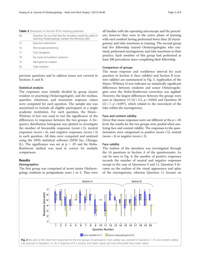
Comparison of groupsThe mean response and confidence interval for eachquestion in Section A (face validity) and Section B (con-tent validity) are summarized in Fig. 3. Application of theMann–Whitney U-test indicates no statistically significantdifferences between residents and senior Otolaryngolo-gists once the Holm-Bonferroni correction was applied.However, the largest differences between the groups wereseen in Question 13 (U = 5.5, p = 0.043) and Question 20(U = 7, p = 0.097), which related to the movement of thetube within the myringotomy.
Face and content validityGiven that mean responses were not different at the p = .05level, the results for the two groups were pooled when ana-lyzing face and content validity. The responses to the ques-tionnaires were categorized as positive (score ≥ 5), neutral(score = 4) or negative (score ≤ 3).
Face validityThe realism of the simulator was investigated throughthe 14 questions in Section A of the questionnaire. Ascan be seen in Fig. 4, the number of positive responsesexceeds the number of neutral and negative responsesexcept in the case of Questions 9 and 11. Question 9 fo-cuses on the realism of the visual appearance and splayof the myringotomy, whereas Question 11 focuses on
Table 3 Questions in Section B for training potential
No Question: Do you feel that the simulator would be useful inteaching Otolaryngology trainees the following skills
15 Speculum placement
16 Microscope positioning
17 Tool navigation
18 Ear canal and eardrum anatomy
19 Myringotomy creation
20 Tube insertion
Fig. 3 Box plot of the Likert item responses for the two groups of participants. Face validity was assessed in Questions 1–14, and content validitywas assessed in Questions 15–20. A response of 4 is neutral, and higher values are more favourable than lower values
Huang et al. Journal of Otolaryngology - Head and Neck Surgery (2015) 44:40 Page 5 of 8
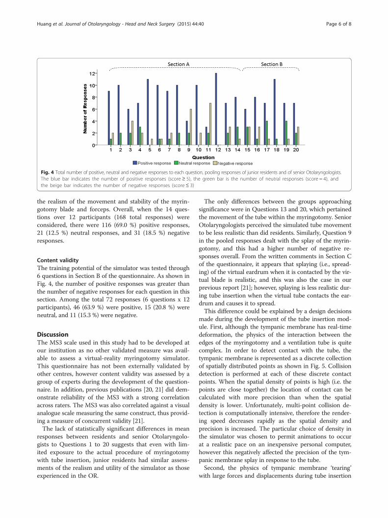
the realism of the movement and stability of the myrin-gotomy blade and forceps. Overall, when the 14 ques-tions over 12 participants (168 total responses) wereconsidered, there were 116 (69.0 %) positive responses,21 (12.5 %) neutral responses, and 31 (18.5 %) negativeresponses.
Content validityThe training potential of the simulator was tested through6 questions in Section B of the questionnaire. As shown inFig. 4, the number of positive responses was greater thanthe number of negative responses for each question in thissection. Among the total 72 responses (6 questions x 12participants), 46 (63.9 %) were positive, 15 (20.8 %) wereneutral, and 11 (15.3 %) were negative.
DiscussionThe MS3 scale used in this study had to be developed atour institution as no other validated measure was avail-able to assess a virtual-reality myringotomy simulator.This questionnaire has not been externally validated byother centres, however content validity was assessed by agroup of experts during the development of the question-naire. In addition, previous publications [20, 21] did dem-onstrate reliability of the MS3 with a strong correlationacross raters. The MS3 was also correlated against a visualanalogue scale measuring the same construct, thus provid-ing a measure of concurrent validity [21].The lack of statistically significant differences in mean
responses between residents and senior Otolaryngolo-gists to Questions 1 to 20 suggests that even with lim-ited exposure to the actual procedure of myringotomywith tube insertion, junior residents had similar assess-ments of the realism and utility of the simulator as thoseexperienced in the OR.
The only differences between the groups approachingsignificance were in Questions 13 and 20, which pertainedthe movement of the tube within the myringotomy. SeniorOtolaryngologists perceived the simulated tube movementto be less realistic than did residents. Similarly, Question 9in the pooled responses dealt with the splay of the myrin-gotomy, and this had a higher number of negative re-sponses overall. From the written comments in Section Cof the questionnaire, it appears that splaying (i.e., spread-ing) of the virtual eardrum when it is contacted by the vir-tual blade is realistic, and this was also the case in ourprevious report [21]; however, splaying is less realistic dur-ing tube insertion when the virtual tube contacts the ear-drum and causes it to spread.This difference could be explained by a design decisions
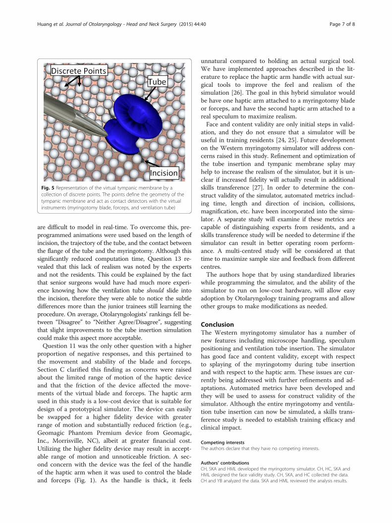
made during the development of the tube insertion mod-ule. First, although the tympanic membrane has real-timedeformation, the physics of the interaction between theedges of the myringotomy and a ventilation tube is quitecomplex. In order to detect contact with the tube, thetympanic membrane is represented as a discrete collectionof spatially distributed points as shown in Fig. 5. Collisiondetection is performed at each of these discrete contactpoints. When the spatial density of points is high (i.e. thepoints are close together) the location of contact can becalculated with more precision than when the spatialdensity is lower. Unfortunately, multi-point collision de-tection is computationally intensive, therefore the render-ing speed decreases rapidly as the spatial density andprecision is increased. The particular choice of density inthe simulator was chosen to permit animations to occurat a realistic pace on an inexpensive personal computer,however this negatively affected the precision of the tym-panic membrane splay in response to the tube.Second, the physics of tympanic membrane ‘tearing’
with large forces and displacements during tube insertion
Fig. 4 Total number of positive, neutral and negative responses to each question, pooling responses of junior residents and of senior Otolaryngologists.The blue bar indicates the number of positive responses (score ≥ 5), the green bar is the number of neutral responses (score = 4), andthe beige bar indicates the number of negative responses (score ≤ 3)
Huang et al. Journal of Otolaryngology - Head and Neck Surgery (2015) 44:40 Page 6 of 8
are difficult to model in real-time. To overcome this, pre-programmed animations were used based on the length ofincision, the trajectory of the tube, and the contact betweenthe flange of the tube and the myringotomy. Although thissignificantly reduced computation time, Question 13 re-vealed that this lack of realism was noted by the expertsand not the residents. This could be explained by the factthat senior surgeons would have had much more experi-ence knowing how the ventilation tube should slide intothe incision, therefore they were able to notice the subtledifferences more than the junior trainees still learning theprocedure. On average, Otolaryngologists’ rankings fell be-tween “Disagree” to “Neither Agree/Disagree”, suggestingthat slight improvements to the tube insertion simulationcould make this aspect more acceptable.Question 11 was the only other question with a higher
proportion of negative responses, and this pertained tothe movement and stability of the blade and forceps.Section C clarified this finding as concerns were raisedabout the limited range of motion of the haptic deviceand that the friction of the device affected the move-ments of the virtual blade and forceps. The haptic armused in this study is a low-cost device that is suitable fordesign of a prototypical simulator. The device can easilybe swapped for a higher fidelity device with greaterrange of motion and substantially reduced friction (e.g.,Geomagic Phantom Premium device from Geomagic,Inc., Morrisville, NC), albeit at greater financial cost.Utilizing the higher fidelity device may result in accept-able range of motion and unnoticeable friction. A sec-ond concern with the device was the feel of the handleof the haptic arm when it was used to control the bladeand forceps (Fig. 1). As the handle is thick, it feels
unnatural compared to holding an actual surgical tool.We have implemented approaches described in the lit-erature to replace the haptic arm handle with actual sur-gical tools to improve the feel and realism of thesimulation [26]. The goal in this hybrid simulator wouldbe have one haptic arm attached to a myringotomy bladeor forceps, and have the second haptic arm attached to areal speculum to maximize realism.Face and content validity are only initial steps in valid-
ation, and they do not ensure that a simulator will beuseful in training residents [24, 25]. Future developmenton the Western myringotomy simulator will address con-cerns raised in this study. Refinement and optimization ofthe tube insertion and tympanic membrane splay mayhelp to increase the realism of the simulator, but it is un-clear if increased fidelity will actually result in additionalskills transference [27]. In order to determine the con-struct validity of the simulator, automated metrics includ-ing time, length and direction of incision, collisions,magnification, etc. have been incorporated into the simu-lator. A separate study will examine if these metrics arecapable of distinguishing experts from residents, and askills transference study will be needed to determine if thesimulator can result in better operating room perform-ance. A multi-centred study will be considered at thattime to maximize sample size and feedback from differentcentres.The authors hope that by using standardized libraries
while programming the simulator, and the ability of thesimulator to run on low-cost hardware, will allow easyadoption by Otolaryngology training programs and allowother groups to make modifications as needed.
ConclusionThe Western myringotomy simulator has a number ofnew features including microscope handling, speculumpositioning and ventilation tube insertion. The simulatorhas good face and content validity, except with respectto splaying of the myringotomy during tube insertionand with respect to the haptic arm. These issues are cur-rently being addressed with further refinements and ad-aptations. Automated metrics have been developed andthey will be used to assess for construct validity of thesimulator. Although the entire myringotomy and ventila-tion tube insertion can now be simulated, a skills trans-ference study is needed to establish training efficacy andclinical impact.
Competing interestsThe authors declare that they have no competing interests.
Authors’ contributionsCH, SKA and HML developed the myringotomy simulator. CH, HC, SKA andHML designed the face validity study. CH, SKA, and HC collected the data.CH and YB analyzed the data. SKA and HML reviewed the analysis results.
Fig. 5 Representation of the virtual tympanic membrane by acollection of discrete points. The points define the geometry of thetympanic membrane and act as contact detectors with the virtualinstruments (myringotomy blade, forceps, and ventilation tube)
Huang et al. Journal of Otolaryngology - Head and Neck Surgery (2015) 44:40 Page 7 of 8
CH, YB, SKA and HML wrote the manuscript. SKA and HML were primarysupervisors for CH and HC. All authors read and approved the final manuscript.
Authors’ informationHML and SKA were co-senior authors on this study.
AcknowledgementsThe authors would like to thank the Natural Sciences and EngineeringResearch Council of Canada (NSERC), Medtronic of Canada Ltd., and theOntario Research Fund (ORF) for financial support of this project.
Author details1Department of Electrical and Computer Engineering, Western University,London, ON, Canada. 2Department of Otolaryngology – Head and NeckSurgery, Schulich School of Medicine and Dentistry, Western University,London, ON, Canada. 3Biomedical Engineering Graduate Program, WesternUniversity, London, ON, Canada. 4Lawson Health Research Institute, London,ON, Canada. 5Department of Medical Biophysics, Western University, London,ON, Canada. 6London Health Sciences Centre, Room B1-333, UniversityHospital, 339 Windermere Rd., London N6A 5A5ON, Canada.
Received: 18 May 2015 Accepted: 12 October 2015
References1. Montague M, Lee MSW, Hussain SSM. Human error identification: an analysis of
myringotomy and ventilation tube insertion. Arch Otolaryngol Head NeckSurg. 2004;130:1153–7.
2. Brodish BN, Woolley AL. Major vascular injuries in children undergoingmyringotomy for tube placement. Am J Otolaryngol. 1999;20:46–50.
3. Kumar M, Khan AM, Davis S. Medial displacement of grommets: anunwanted sequel of grommet insertion. J Laryngol Otol. 2000;114:448–9.
4. Groblewski JC, Harley EH. Medial migration of tympanostomy tubes: anoverlooked complication. Int J Pediatr Otorhinolaryngol. 2006;70:1707–14.
5. Walker T, Duvvi S, Kumar BN. The wigan grommet trainer. Clin Otolaryngol.2006;31:349–50.
6. Duijvestein M, Borgstein J. The bradford grommet trainer. Clin Otolaryngol.2006;31:163.
7. Leong A, Kundu S, Martinez-Devesa P, Aldren C. Artificial ear: a training toolfor grommet insertion and manual dexterity. ORL. 2006;68:115–7.
8. Hong P, Webb AN, Corsten G, Balderston J, Haworth R, Ritchie K, et al. Ananatomically sound surgical simulation model for myringotomy andtympanostomy tube insertion. Int J Pediatr Otorhinolaryngol. 2008;78:522–9.
9. Volsky PG, Hughley BB, Peirce SM, Kesser BW. Construct validity of asimulator for myringotomy with ventilation tube insertion. Otolaryngology -Head and Neck Surg. 2009;141:603–8.
10. Weghorst S, Airola C, Oppenheimer P, Edmond CV, Patience T, Heskamp D,et al. Validation of the madigan ESS simulator. Stud Health TechnolInformat. 1998;50:399–405.
11. Anil SM, Kato Y, Hayakawa M, Yoshida K, Nagahisha S, Kanno T. Virtual3-dimensional preoperative planning with the Dextroscope for excisionof a 4th ventricular ependymoma. Minim Invasive Neurosurg.2007;50:65–70.
12. Audette M, Delingette H, Fuchs A, Astley O, Chinzei K. A topologicallyfaithful, tissue-guided, spatially varying meshing strategy for computingpatient-specific head models for endoscopic pituitary surgery simulation.Stud Health Technol Inform. 2006;119:22–7.
13. Tolsdorff B, Pommert A, Höhne KH, Petersik A, Pflesser B, Tiede U, et al.Virtual reality: a new paranasal sinus surgery simulator. Laryngoscope.2010;120:420–6.
14. Varshney R, Frenkiel S, Nguyen LHP, Young M, Del Maestro R, Zeitouni A,et al. The McGill simulator for endoscopic sinus surgery (MSESS): a validationstudy. J Otolaryngol Head Neck Surg. 2014;43:40.
15. Wiet GJ, Stredney D, Kerwin T, Hittle B, Fernandez SA, Abdel-Rasoul M, et al.Virtual temporal bone dissection system: development and testing.Laryngoscope. 2012;122 Suppl 1:S1–S12.
16. Morris D, Sewell C, Barbagli F, Salisbury K, Blevins NH, Girod S. Visuohapticsimulation of bone surgery for training and evaluation. IEEE Comput GraphAppl. 2006;26:48–57.
17. Sewell C, Morris D, Blevins NH, Dutta S, Agrawal S, Barbagli F, et al. Providingmetrics and performance feedback in a surgical simulator. Comput Aided Surg.2008;13:63–81.
18. Arora A, Khemani S, Tolley N, Singh A, Budge J, Varela DA, et al. Face andcontent validation of a virtual reality temporal bone simulator. OtolaryngolHead Neck Surg. 2012;146:497–503.
19. Wheeler B, Doyle PC, Chandarana S, Agrawal S, Husein M, Ladak HM.Interactive computer-based simulator for training in blade navigation andtargeting in myringotomy. Comput Meth Programs Biomed. 2010;98:130–9.
20. Sowerby LJ, Rehal G, Husein M, Doyle PC, Agrawal S, Ladak HM. Developmentand face validity testing of a three-dimensional myringotomy simulator withhaptic feedback. J Otolaryngol Head Neck Surg. 2010;39:122–9.
21. Ho AK, Alsaffar H, Doyle PC, Ladak HM, Agrawal SK. Virtual realitymyringotomy simulation with real-time deformation: development andvalidity testing. Laryngoscope. 2012;122:1844–51.
22. Huang C, Agrawal SK, Ladak HM. Virtual-reality simulator for training inmyringotomy with tube placement. BC: Vancouver; 2014. Proceedingsof the 37th Canadian medical and biological engineering conference:20–23 May 2014.
23. Carter FJ, Schijven MP, Aggarwal R, Grantcharov T, Francis NK, Hanna GB,et al. Consensus guidelines for validation of virtual reality surgicalsimulators. Surg Endosc. 2005;19:1523–32.
24. Gallagher AG, Ritter EM, Satava RM. Fundamental principles of validation, andreliability rigorous science for the assessment of surgical education andtraining. Surg Endosc. 2003;17:1525–9.
25. Schout BM, Hendrikx AJ, Scheele F, Bemelmans BL, Scherpbier AJ. Validationand implementation of surgical simulators: a critical review of present, past,and future. Surg Endosc. 2010;24:536–46.
26. Coles TR, John NW, Sofia G, Gould DA, Caldwell DG. Modification ofcommercial force feedback hardware for needle insertion simulation. StudHealth Technol Inform. 2011;163:135–7. MMVR18 – Medicine Meets VirtualReality 2011 Poster: 8–12 February 2011; Newport Beach, CA.
27. Hamstra SJ, Brydges R, Hatala R, Zendejas B, Cook DA. Reconsidering fidelityin simulation-based training. Acad Med. 2014;89:387–92.
Submit your next manuscript to BioMed Centraland take full advantage of:
• Convenient online submission
• Thorough peer review
• No space constraints or color figure charges
• Immediate publication on acceptance
• Inclusion in PubMed, CAS, Scopus and Google Scholar
• Research which is freely available for redistribution
Submit your manuscript at www.biomedcentral.com/submit
Huang et al. Journal of Otolaryngology - Head and Neck Surgery (2015) 44:40 Page 8 of 8