Embed Size (px)
Citation preview
Logo
False-Positive Somatostatin Receptor Scintigraphy: Really?
Dr. Augusto Llamas-Olier, Dr. Maria Cristina Martinez, Dr. Emperatriz Angarita, Dr. Amelia De Los ReyesNuclear medicine department.
Instituto Nacional de Cancerologia. Bogota, Colombia.
Logo
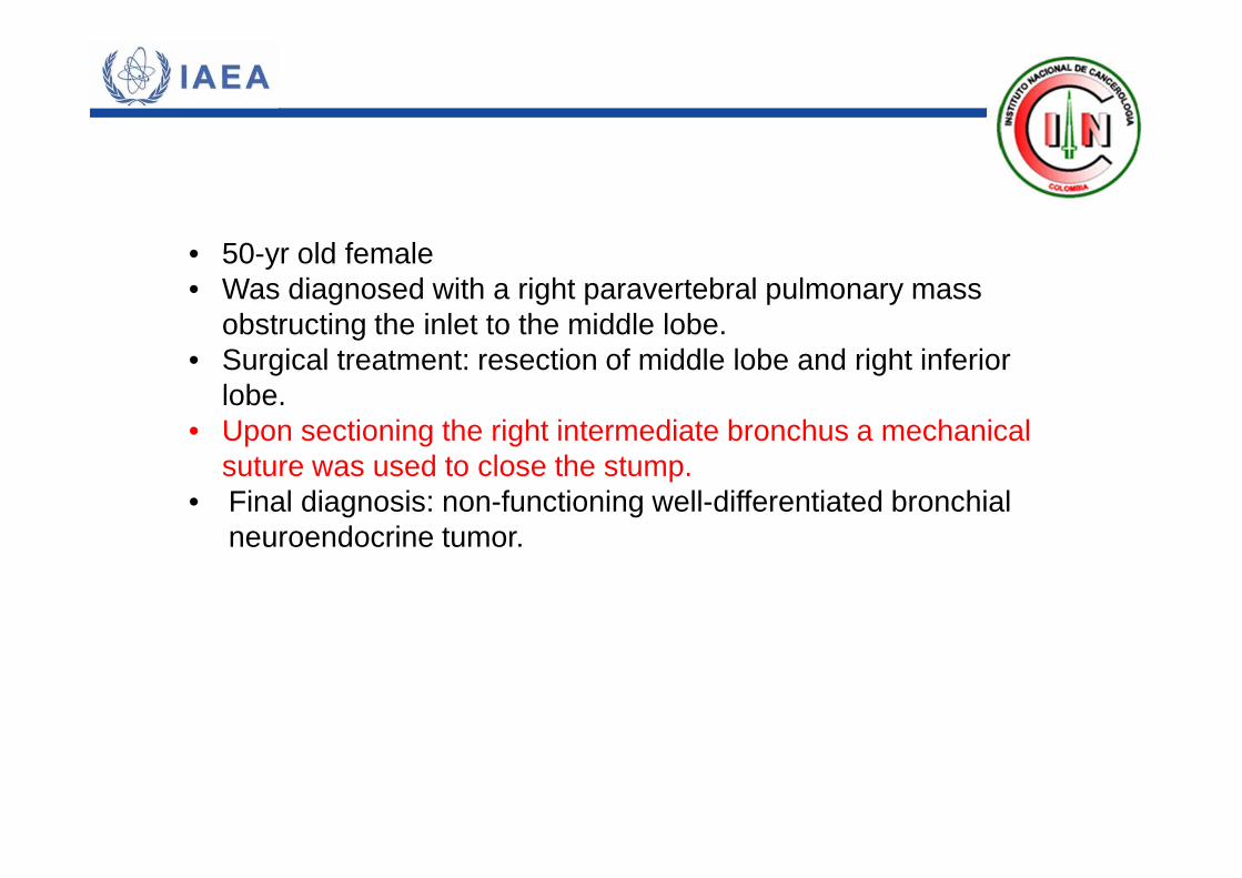
• 50-yr old female• Was diagnosed with a right paravertebral pulmonary mass
obstructing the inlet to the middle lobe.• Surgical treatment: resection of middle lobe and right inferior
lobe.• Upon sectioning the right intermediate bronchus a mechanical
suture was used to close the stump.• Final diagnosis: non-functioning well-differentiated bronchial
neuroendocrine tumor.
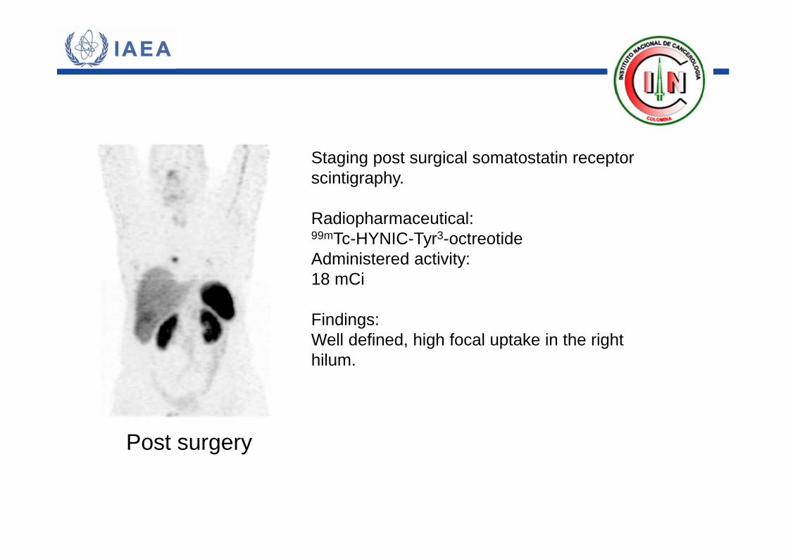
Post surgery
Staging post surgical somatostatin receptor scintigraphy.
Radiopharmaceutical: 99mTc-HYNIC-Tyr3-octreotideAdministered activity:18 mCi
Findings:Well defined, high focal uptake in the right hilum.
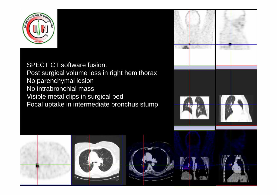
SPECT CT software fusion.Post surgical volume loss in right hemithoraxNo parenchymal lesionNo intrabronchial massVisible metal clips in surgical bedFocal uptake in intermediate bronchus stump
• Fiberoptic bronchoscopy: otherwise unremarkable right bronchial stump.
• Bronchoalveolar lavage: no infectious agents, no tumor cells. Moderately reactive inflammatory process in respiratory epithelium (polymorphonuclear cells: 90%).
• Bronchial stump biopsy: negative for tumor involvement.• Serum chromogranin A: 16 ng/ml.
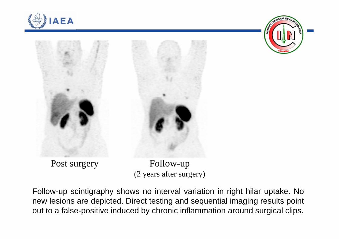
Post surgery Follow-up(2 years after surgery)
Follow-up scintigraphy shows no interval variation in right hilar uptake. Nonew lesions are depicted. Direct testing and sequential imaging results pointout to a false-positive induced by chronic inflammation around surgical clips.

Teaching PointsTo adequately determine that a localization result is a false-positive or true-positive requires either direct examination (bysurgery, cytology or biopsy), other imaging studies to clarify thelesion or extensive follow-up and careful comparison to theclinical context to determine the true nature of the lesion.
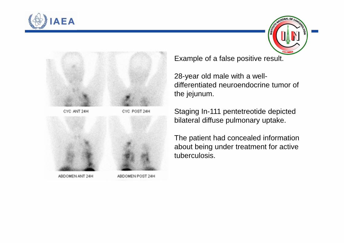
Example of a false positive result.
28-year old male with a well-differentiated neuroendocrine tumor of the jejunum.
Staging In-111 pentetreotide depicted bilateral diffuse pulmonary uptake.
The patient had concealed information about being under treatment for active tuberculosis.

Teaching PointsGranulomatosis disease (sarcoidosis, tuberculosis, Wegener's)as well as both benign and malignant breast diseases arereported to possess high-affinity somatostatin receptors thatbind octreotide and to be imaged on somatostatin receptorscintigraphy.
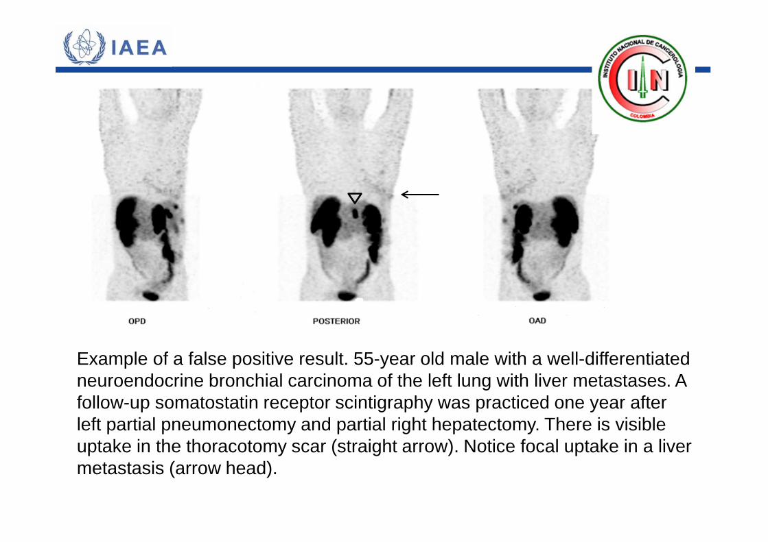
Example of a false positive result. 55-year old male with a well-differentiated neuroendocrine bronchial carcinoma of the left lung with liver metastases. A follow-up somatostatin receptor scintigraphy was practiced one year after left partial pneumonectomy and partial right hepatectomy. There is visible uptake in the thoracotomy scar (straight arrow). Notice focal uptake in a liver metastasis (arrow head).
Teaching PointsOperative sites have been reported to result in false-positivelocalization on somatostatin receptor scintigraphy, perhapsbecause of activated lymphocytes or inflammatory cells that areknown to possess somatostatin receptors and, in somepatients, remain positive on SRS for several months aftersurgery.
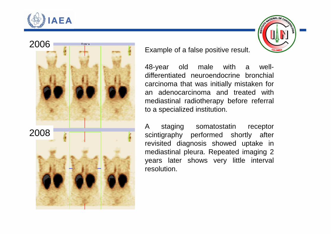
Example of a false positive result.
48-year old male with a well-differentiated neuroendocrine bronchialcarcinoma that was initially mistaken foran adenocarcinoma and treated withmediastinal radiotherapy before referralto a specialized institution.
A staging somatostatin receptorscintigraphy performed shortly afterrevisited diagnosis showed uptake inmediastinal pleura. Repeated imaging 2years later shows very little intervalresolution.
2006
2008
Teaching PointsRadiation pneumonitis can occur 1-8 months after treatment.From 8 months onwards, lung fibrosis may appear.
Indium-111-pentetreotide scans are strongly or moderatelypositive in symptomatic patients examined 2-5 months afterradiotherapy.
The uptake mechanism of In-111-pentetreotide in areas ofradiation pneumonitis remains to be elucidated. Radiationpneumonitis has been explained by early vascular alterationsfollowed by epithelial changes and ablation of type II alveolarcells which will eventually also result in early surfactant releaseinto the alveoli Enhanced production and release of cytokinesfrom alveolar macrophages has been postulated as acomponent in the mechanism of radiation injury.
DiscussionTrue false-positive results are neither false (because they representsomatostatin receptor-positive lesions) nor positive (because they are notrelated to the pathology under scrutiny).
Despite its increased use, there has been only one systematic study of theoccurrence of false-positive somatostatin receptor analogue localization(Gibril et al).
Although several large studies report that somatostatin receptorscintigraphy has excellent specificity it is nevertheless important that thesepoints be systematically examined.
Discussion (II)
Newer radiolabeled somatostatin analogs which can be used in PETimaging, and which have a higher affinity for the somatostatin receptor,especially receptor subtype-2, have been developed.
The most likely candidates to become the new standard forsomatostatin receptor imaging are 68Ga-DOTA0-Tyr3-octreotate and68Ga-DOTA0-Tyr3-octreotide.

Teaching Points (II)
The most common causes for ‘false positive’ results are: radiationpneumonitis, accessory spleen, surgical scar tissue, nodulargoitre, ventral hernia, bacterial pneumonia, respiratory infections,common cold (nasal uptake), cerebrovascular accident,concomitant granulomatous disease, diffuse breast uptake andconcomitant second primary tumour.
False positive readings can be induced by the tracer’s physiologicpathways or contamination: focal collection of stools, gallbladderuptake, adrenal uptake and urine contamination.
References
• Kwekkeboom DJ, Kam BL, Van Essen M, Teunissen JJM,Van Eijck CHJ, Valkema R, et al. Somatostatin receptor-based imaging and therapy of gastroenteropancreaticneuroendocrine tumors. Endoc Rel Cancer 2010;17:R53–R73.
• Gibril F, Reynolds JC, Chen CC, Yu F, Goebel SU, SerranoJ, et al. Specificity of Somatostatin Receptor Scintigraphy: Aprospective study and effects of false-positive localizationson management in patients with gastrinomas. J NucI Med1999;40:539-553.
• Valdes-Olmos R, Van Zandwijk N, Boersma LJ, HoefnagelCA, Baas P, Baars JB, et al. Radiation pneumonitis imagedwith indium-111-pentetreotide. J Nucl Med 1996;37:584-585.