Embed Size (px)
Citation preview
J. clin. Path. (1963), 16, 65
Familial histiocytic reticulosis(familial haemophagocytic reticulosis)
VALERIE J. MARRIAN AND N. G. SANERKIN
From the Departments of Child Health and Pathology, University of Bristol
SYNOPSIS The clinical and pathological findings are recorded in two siblings who died in earlyinfancy from familial histiocytic ('haemophagocytic') reticulosis. The nature of this condition isobscure but probably represents a primary histiocytosis. The only other family on record is thatdescribed by Farquhar and Claireaux (1952) and by Farquhar, Macgregor, and Richmond (1958).
This unusual disorder was first described by Farquharand Claireaux (1952), who recorded a family withthree siblings the first two of whom died in earlyinfancy, the third sibling recovering from what ap-peared to be a mild form of the disease. Subsequently,Farquhar et al. (1958) reported a fourth sibling in thesame family who died from an identical condition.In all three fatal cases, considered by these authorsto represent the homozygous state in a familial dis-order, the infants had developed anorexia, vomiting,irritability, and pallor during the first three monthsof life and were found to have hepato-splenomegalywith progressive anaemia, granulocytopenia, andthrombocytopenia. In all cases, the peripheral bloodshowed numbers of smudged cells and a small pro-,portion of atypical mononuclear cells. The survivingsibling had developed severe anorexia at the age of10 weeks. Smudged cells and atypical mononuclearswere found in her blood films until the age of 18months, but she never had any hepato-splenomegalyand had since remained quite healthy, apparentlyrepresenting the heterozygous state of this disorder.
Necropsy in the three fatal cases was reported tohave shown a remarkable proliferation of histiocytesthroughout the reticulo-endothelial system, withprominent phagocytosis of blood cells, especially ofthe red cells. The authors of the original paper con-cluded that the condition was not Letterer-Siwe's'non-lipid reticulo-endotheliosis'. Their suggestionthat it might be identical with histiocytic medullaryreticulosis was refuted by Marshall (1956).The present paper describes a second family in
which the two offspring died in early infancy from acondition identical with that described by Farquharet al. (1952, 1958). Although the first case was cor-rectly diagnosed as a malignant reticulosis, the exactReceived for publication 7 August 1962.
nature of the disease was not firmly established untilafter the death of the second sibling, when a searchof the literature was made with special reference tohistiocytic medullary reticulosis and Letterer-Siwedisease. Because of this, the detailed haematologicalinvestigations performed by Farquhar et al. (1958)could not be carried out on these infants, in particularthe red-cell survival times and the detection of auto-antibodies. Unfortunately, the parents themselves onreligious grounds declined to undergo any tests fordetermination of their own red-cell survival times.The father, aged 22 at the birth of the first child, had
been diagnosed as a case of pulmonary tuberculosis twoand a half years previously. He was treated with I.N.A.H.and P.A.S. and is now apparently healthy. The mother,aged 20, is an epileptic controlled by treatment withphenobarbitone and for a period with diamox and para-dione. They are not blood relatives. On neither side of thefamily has there been similar illness or death in infancy.The parents consented to simple tests on their blood,
the results of which are given below:Father Hb 99 %, R.B.C. 4,600,000/c.mm., P.C.V. 42%.
W.B.C. 10,800/c.mm. (polymorphs 5,000/c.mm.). DirectCoombs test negative.Mother Hb 82 %, R.B.C. 4,200,000/c.mm., P.C.V. 3500,
W.B.C. 9,200/c.mm. (polymorphs 6,200/c.mm.). DirectCoombs test negative.CASE 1 The first-born, a male infant, was deliveredspontaneously at term after a normal pregnancy (birthweight 7 lb. 11 oz.). He was given B.C.G. at the age of6 days. He appeared quite healthy until the eighth weekwhen he was admitted to hospital with one day's historyof irritability, photophobia, anorexia, projectile vomiting,and grunting respiration. He was pale, shocked, andmiserable (temperature 102-7°F., pulse 160/min., respira-tion 48/min.). He had mild coryza but no evidence of anyother infection. There was no cutaneous rash, and nolymphadenopathy or hepato-splenomegaly was notedinitially. Lumbar puncture showed normal cerebrospinal
65
on 15 July 2018 by guest. Protected by copyright.
http://jcp.bmj.com
/J C
lin Pathol: first published as 10.1136/jcp.16.1.65 on 1 January 1963. D
ownloaded from
Valeirie J. Ma) l iani and N. G. Sanel,kin
fluid. Haemoglobin was 58 oo; a blood film was ortho-chromic with some anisocytosis; platelets appeared to bepresent in normal numbers; reticulocytes were less than1 oo; W.B.C. 4,400/c.mm. Plasma was not icteric. Theblood group was 0 Rh (D) positive. The direct Coombstest was negative, as was the Paul Bunnell test. A tibialmarrow puncture showed normoblastic erythropoiesis,with a myeloid-erythroid ratio of 4:3. A non-catheterspecimen of urine contained albumin +, scanty whitecells, and culture yielded Bact. coli and enterococci.Staph. aureus, resistant to penicillin only, was isolatedfrom the nose. He was treated empirically with tetra-cycline, 62 5 mg. six-hourly, and transfused with 250 ml.of whole blood after which the haemoglobin rose to 94'0.An intravenous pyelogram, performed on the fourth dayafter admission, showed no significant abnormalities, buta soft tissue mass was noted in the left upper abdomen.By the tenth day the liver was enlarged two finger-breadths and a large mobile swelling was felt in the lefthypochondrium which was presumed to be the spleen.On the 17th day the haemoglobin had fallen to 54 o andthe red cells showed hypochromia and anisocytosis;W.B.C. 6,200/c.mm. (polymorphs 750/c.mm.) and 160smudged cells per 100 white cells. A purpuric rash wasnoted about this time. An exploratory laparotomy per-formed on the 20th day showed considerable ascites anda very large soft spleen which was removed. His post-operative condition was poor. He collapsed a few hourslater and died the same day.The excised spleen weighed 157 g. and was large and
soft, with a smooth capsular surface free from adhesions.The cut surface was homogeneous, soft and pink, withindistinct Malpighian bodies. Histological examinationshowed changes identical with those found in the spleenof the second case, and will be described later.Post-mortem examination The body was that of a
very pale male infant weighing 5,365 g. There was nojaundice, oedema, or external haemorrhage. The abdomenwas protuberant, with a sutured surgical wound on theleft side. The abdominal cavity contained about 10 ml. ofblood-stained fluid with a few clots. The pleural andpericardial cavities contained small amounts of clearyellow fluid.The significant naked-eye abnormalities in the internal
organs were as follows: A haematoma of about 10 ml.was found in the splenic bed. The liver was enlarged andweighed 379 g.; it was pale brown in colour, firm andfibrous in texture, but smooth in outline. The lymphnodes of the neck and abdomen were slightly enlarged,soft, discrete, and pink. The bone marrow in the sternumand femur appeared normal. The other organs werenormal though pale. A Meckel's diverticulum was present.The histological findings were similar to those of Case 2
and will be described later.
CASE 2 The second child, a girl, was born one yearlater after an uneventful pregnancy and delivery (birthweight 8 lb. 6 oz.). B.C.G. was given at the age of 7 days.At the age of 4 months she became unusually quiet andsleepy. She developed a febrile illness associated withvomiting and nasal 'snuffles' but no other respiratorysymptoms. One week later her eyes became puffy, and
within a few days the swelling had involved the entire faceand neck. She was admitted to hospital after two weeks'illness looking very ill (temperature 104-4 F., pulse184/min., respiration 62 min.). She was extremely palewith a bluish tinge of the lips. Her face was grosslyoedematous and there was a sero-sanguinous nasal dis-charge. The abdomen was very distended with demon-strable ascites. The spleen was palpable threefingerbreadths and the liver two fingerbreadths belowthe costal margin. Haemoglobin was 39°/, W.B.C.2,700/c.mm. (polymorphs 720/c.mm., reticulocytes 2 2 O)platelets 6,000/c.mm. The blood film showed mildgeneralized hypochromia with normocytosis but occa-sional macrocytes, scanty platelets, and granulocytopeniawith an increase in juvenile forms. There were manysmudged cells and some atypical mononuclear cells withheavily stained distorted nuclei. The blood group wasA Rh (D) positive. Coombs test was not performed. Totalserum proteins were 3 g.A100 ml. with low albumin andglobulin. The serum cholesterol level was 260 mg./l00 ml.,and the total serum lipids 2 34 g./100 ml. The urineshowed a trace of albumin, normal sediment, and nocytomegalic inclusion bodies were found. A nasal swabyielded coagulase-negative Staph. aureus resistant topenicillin only. She was treated empirically with penicillinand streptomycin and transfused with 190 ml. of wholeblood. She deteriorated progressively with falling haemo-globin, increasing splenomegaly and abdominal disten-sion. She developed ecchymoses in the eyelids and bledfrom the ears. A fine purpuric rash appeared over theabdomen and thighs. On the fourth day after admissionthe polymorphonuclear count had fallen to less than50/c.mm. and platelets were not detectable. A tibialmarrow puncture showed moderate cellularity with veryactive erythropoiesis, predominantly normoblastic butwith minor megaloblastic changes. The granulocyticseries was depressed, showing mainly promyelocytes, re-duced numbers of myelocytes, very few juvenile poly-morphs and extremely few segmented cells. Megakaryo-cytes were scanty with abnormal nuclei and practically noplatelets. Reticulum cells were slightly increased innumbers and many showed pale vacuolated cytoplasm.No malignant cells were seen and there was no evidenceof leukaemia. She died on the eighth day after admissionat the age of 5 months.Post-mortem examination The body was that of a
female infant weighing 6,500 g., with marked pallor,ecchymoses over the eyelids and groins, and a generalizedpurpuric rash maximal over the trunk and thighs. Therewas no obvious jaundice. Generalized pitting oedema waspresent. The abdomen was prominently distended. Smallcollections of straw-coloured fluid were found in allthe serous cavities, with subserous petechial haemorrh-ages.The significant naked-eye abnormalities in the internal
organs were as follows. The spleen was grossly enlarged,weighing 195 g. and extending down to the left iliac fossa.It was firm, with a smooth capsular surface without anypericapsular adhesions and a uniform purplish-red cutsurface with indistinct Malpighian bodies. The liver wasmoderately enlarged and weighed 440 g., with sharpmargins and smooth capsular surface; the cut surface
66
on 15 July 2018 by guest. Protected by copyright.
http://jcp.bmj.com
/J C
lin Pathol: first published as 10.1136/jcp.16.1.65 on 1 January 1963. D
ownloaded from
Familial histiocytic reticulosis (familial haemophagocytic reticulosis)
showed no abrnormality apart from obvious pallor. Thelymph nodes were not enlarged to any remarkable degree;they were discrete, soft and rather oedematous, with apinkish cut surface. The bone marrow was uniformlydeep red throughout. An incidental finding was thepresence of bilateral bydroureter and hydronephrosis dueto congenital uretero-vesical stenosis, without evidence ofany pyelonephritis.
HISTOLOGY
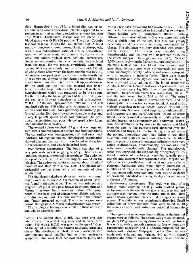
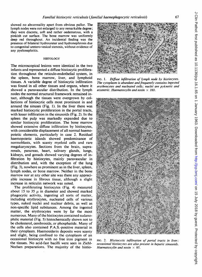
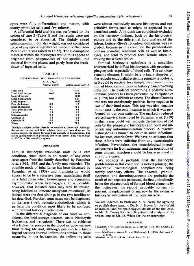
The microscopical lesions were identical in the twoinfants and represented a diffuse histiocytic prolifera-tion throughout the reticulo-endothelial system, inthe spleen, bone marrow, liver, and lymphoidtissues. A variable degree of histiocytic infiltrationwas found in all other tissues and organs, where itshowed a paravascular distribution. In the lymphnodes the normal structural framework remained in-tact, although the tissues were overgrown by col-lections of histiocytic cells most prominent in andaround the sinuses (Fig. 1). In the liver there wasmarked histiocytic proliferation in the portal tracts,with lesser infiltration in the sinusoids (Fig. 2). In thespleen the pulp was markedly expanded due tosimilar histiocytic proliferation. The bone marrowshowed extensive diffuse infiltration by histiocytes,with considerable displacement of all normal haemo-poietic elements, particularly in case 2. Residualhaemopoietic islands showed predominance ofnormoblasts, with scanty myeloid cells and raremegakaryocytes. Sections from the brain, supra-renals, pancreas, heart, salivary glands, lungs,kidneys, and gonads showed varying degrees of in-filtration by histiocytes, mainly paravascular indistribution and, with the exception of the lung(Fig. 3), nowhere as prominent as in the liver, spleen,lymph nodes, or bone marrow. Neither in the bonemarrow nor at any other site was there any appreci-able increase in fibrous tissue, although a slightincrease in reticulin network was noted.The proliferating histiocytes (Fig. 4) measured
about 15 to 35 tt in diameter and showed markedphagocytic activity, ingesting all sorts of matter,including erythrocytes, nucleated cells of varioustypes, naked nuclei and nuclear debris, as well asnon-specific lipid substances. Among the ingestedmatter, the erythrocytes were by far the mostnumerous. Many of the histiocytes contained sudano-philic material (Fig. 5) histochemically shown not tobe cholesterol, cerebroside, or phosphatide. Many ofthe cells also contained P.A.S.-positive material intheir cytoplasm. Haemosiderin deposits were scantyand slight, being confined to the cytoplasm of anoccasional histiocyte with no free iron pigment inthe tissues. No acid-fast bacilli were seen in Ziehl-Neelsen preparations. The majority of the histio-
FIG. 1. Diffuse infiltration of lymph node by histiocytes.The cytoplasm is abundant andfrequently contains ingestederythrocytes and nucleated cells; nuclei are pyknotic andeccentric. Haematoxylin and eosin x 160.
FIG. 2. Histiocytic infiltration of portal tracts in liver;occasional histiocvtes are also present in hepatic sinusoids.Haematoxylin and eosin x 95.
67
on 15 July 2018 by guest. Protected by copyright.
http://jcp.bmj.com
/J C
lin Pathol: first published as 10.1136/jcp.16.1.65 on 1 January 1963. D
ownloaded from

Valerie J. Marrian and N. G. Sanerkin
FIG. 3. Peribronchial and perivascular infiltration by histiocytes in lung. The infiltrate is typicallyinterstitial, not bronchopneumonic. Haematoxylin and eosin x 95.
\,: A\
cS.s.X*N,\'....v r 4S
I ; *8 $ - &~
-:
FIG. 4. High-power view of histiocytes in the medulla ofalymph node. Arrow points at a histiocyte containing severalerythrocytes. Haematoxylin and eosin x 600. .4
+.c,.; .5 ° .C .
FIG. 5. Cuff of sudanophilic histiocytes around a cerebralarteriole. Frozen section of brain stained Sudan HIl. x 150.
68
.4..
N: -
on 15 July 2018 by guest. Protected by copyright.
http://jcp.bmj.com
/J C
lin Pathol: first published as 10.1136/jcp.16.1.65 on 1 January 1963. D
ownloaded from
Familial histiocytic reticulosis (familial haemophagocytic reticulosis)
cytes were fully differentiated and mature, withscanty primitive cells and few mitoses.A differential lipid analysis was performed on the
spleen of case 2 (Table I) and the results were notconsidered to be abnormal. The slightly raisedsphingomyelin, 1-3% dry weight, was not consideredto be of any special significance, since in a Niemann-Pick spleen it was raised to 13 2 %. The sudanophilicmaterial within the histiocytes would thus appear tooriginate from phagocytosis of non-specific lipidmaterial from the plasma and partly from the break-down of ingested cells.
TABLE IDIFFERENTIAL LIPID ANALYSIS OF THE SPLEEN
% Dry BasisNormal Spleen Spleen from Case 2
Total lipidTotal lipid hexoseNeutral hexoseLipid hexosamineLipid sulphurFree cholesterolEster cholesterolTotal phosphatideLecithinSphingomyelinCephalin ACephalin BWater
7-30-41
0-09
0-80-13-11-80-31-40-6
78-2
8.40-310-08Nil0.021-0
0-13-41-61-30-40-1
77-8
The lipid analysis on the spleen is not considered abnormal. Althoughthe neutral hexose and lipid sulphur have not been done on thenormal spleen, the results for case 2 are unlikely to be abnormal. Theraised sphingomyelin is probably due to the formol. This was as highas 13-2 in a Niemann-Pick spleen.
DISCUSSION
Familial histiocytic reticulosis must be a rare
condition, since there is no record of any othercases apart from the family described by Farquharet al. (1952, 1958) and the family now recorded. Thepossible mode of inheritance has been discussed byFarquhar et al. (1958) and transmission wouldappear to be by a recessive gene, manifesting itselfin a fatal form when homozygous and remainingasymptomatic when heterozygous. It is possible,however, that isolated cases may well be missed,being labelled as 'obscure malignant reticuloses', as
indeed were the first siblings in the two families so
far described. Further, some cases may be diagnosedas Letterer-Siwe's reticulo-endotheliosis which isperhaps the condition most likely to be confusedwith familial histiocytic reticulosis.
In the differential diagnosis of our cases we con-
sidered the lipid-storage diseases, acute histiocyticleukaemia, and 'reticulosis'. There was no evidenceof a leukaemic process in the blood or bone marrow
films during life and, although post-mortem histo-logical sections showed infiltrations similar to thoseoccurring in the leukaemias, the infiltrating cells
were almost exclusively mature histiocytes and notprimitive forms such as might be expected in anacute leukaemia. A lipidosis was confidently excludedon the necropsy findings, both by the histologicalpicture and by the differential lipid analysis of thespleen. Letterer-Siwe's reticulo-endotheliosis was ex-cluded, because in this condition the proliferationscontain primitive reticulum cells as well as histio-cytes, and tend to produce focal lesions often in-volving the skeletal tissues.
Familial histiocytic reticulosis is a conditioncharacterized by diffuse histiocytosis with prominentphagocytosis, especially of blood cells. The aetiologyremains obscure. It might be a primary disorder ofthe reticulo-endothelial system, a primary reticulosis,or it could be reactive, for example, to auto-immuniza-tion of blood cells or to some histiocytosis-provokinginfection. The evidence concerning a possible auto-immune process has been presented by Farquhar etal. (1958) but is difficult to assess. The direct Coombstest was not consistently positive, being negative intwo of their fatal cases. This test was also negativein our case 1, the only instance in which it was per-formed on our own patients. The markedly reducedred-cell survival time noted by Farquhar et al. (1958)in their cases could well indicate destruction of redcells by the phagocytic histiocytes and need not im-plicate any auto-immunization process. A reactivehistiocytosis is known to occur in some infections,for instance, enteric fever. We have no evidence thatthese infants were suffering from any ascertainableinfection. Nevertheless, the bacteriological investi-gations were far from adequate, and the possibility ofsome unusual infection should be borne in mind inany future cases.We consider it probable that the histiocytic
proliferation in this condition is indeed primary, theobservable haematological complications beingmerely secondary effects. The anaemia, granulo-cytopenia, and thrombocytopenia are probably theresult of two separate processes, the first undoubtedlybeing the phagocytosis of formed blood elements bythe histiocytes; the second, probably no less im-portant, is replacement of marrow by the extensivehistiocytic infiltration of the bone marrow.
We are indebted to Professor A. V. Neale for agreeingto publish these cases, to Dr. N. J. Brown for the morbidanatomical and histopathological details of the first case,to Mr. A. Tingey for the differential lipid analysis of thespleen, and to Mr. D. White for the photography.
REFERENCES
Farquhar, J. W., and Claireaux, A. E. (1952). Arch. Dis. Childh., 27,519.MacGregor, Agnes R., and Richmond, J. (1958). Brit. med. J.,2, 1561.
Marshall, A. H. E. (1956). J. Path. Bact., 71, 61.
69
on 15 July 2018 by guest. Protected by copyright.
http://jcp.bmj.com
/J C
lin Pathol: first published as 10.1136/jcp.16.1.65 on 1 January 1963. D
ownloaded from


![Title [原著]An Autopsy Case of Histiocytic Medullary ...okinawa-repo.lib.u-ryukyu.ac.jp/bitstream/20.500.12001/...The findings of this case were those of histiocytic medullary reticulosis,](https://img.pdfslide.net/doc/110x75/610486836382170066209593/title-ean-autopsy-case-of-histiocytic-medullary-okinawa-repolibu-.jpg)