Embed Size (px)
Citation preview
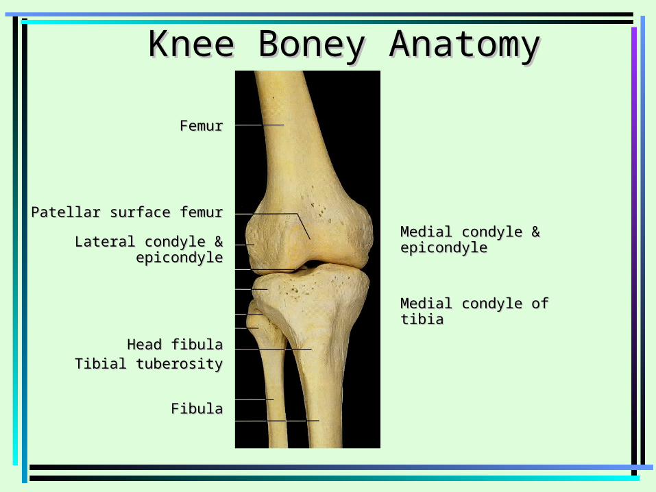
FemurFemur
Patellar surface femurPatellar surface femur
Lateral condyle & epicondyleLateral condyle & epicondyle
Head fibulaHead fibulaTibial tuberosityTibial tuberosity
FibulaFibula
Knee Boney AnatomyKnee Boney Anatomy
Medial condyle & epicondyleMedial condyle & epicondyle
Medial condyle of tibiaMedial condyle of tibia
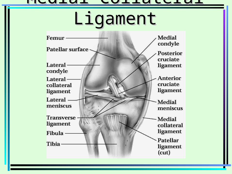
Medial Collateral LigamentMedial Collateral Ligament
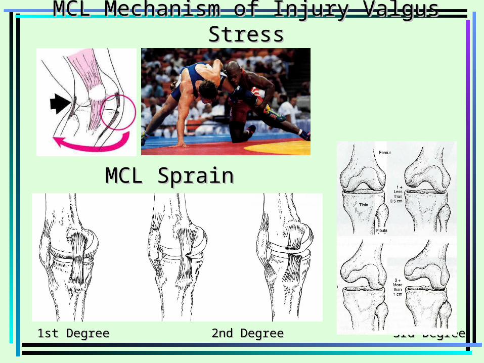
1st Degree 2nd Degree 3rd Degree1st Degree 2nd Degree 3rd Degree
MCL SprainMCL Sprain
MCL Mechanism of Injury Valgus StressMCL Mechanism of Injury Valgus Stress
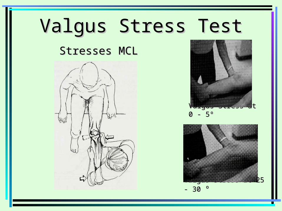
Valgus Stress TestValgus Stress TestStresses MCLStresses MCL
Valgus Stress at 0 - 5ºValgus Stress at 0 - 5º
Valgus Stress at 25 - 30 Valgus Stress at 25 - 30 ºº
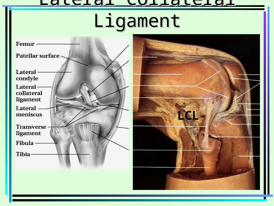
Lateral Collateral LigamentLateral Collateral Ligament
LCL-
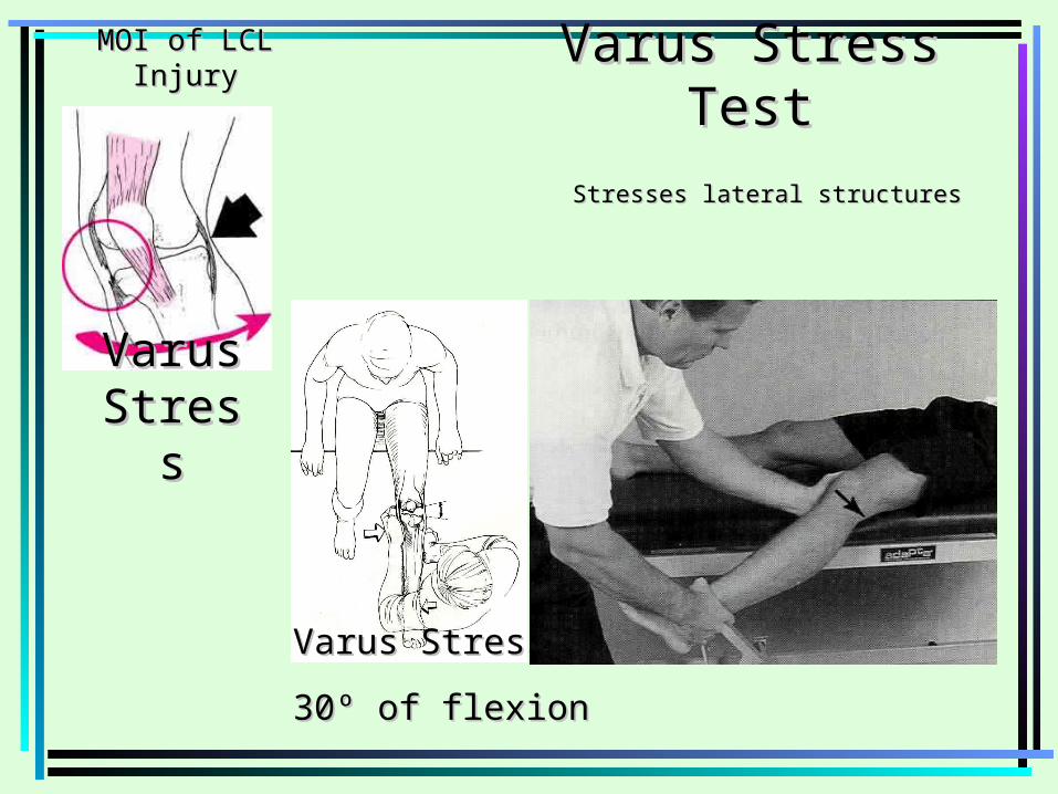
Varus Stress TestVarus Stress Test
Stresses lateral structuresStresses lateral structures
Varus Stress at 0 Varus Stress at 0 ºº and 25 º to 30º of flexion and 25 º to 30º of flexion
Varus Varus StressStress
MOI of LCL InjuryMOI of LCL Injury
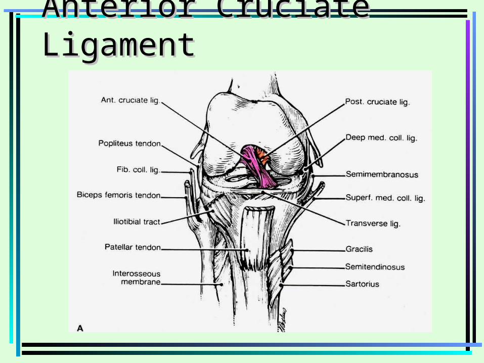
Anterior Cruciate LigamentAnterior Cruciate Ligament
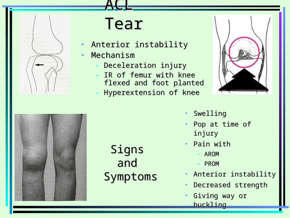
ACL TearACL Tear• Anterior instabilityAnterior instability• MechanismMechanism
– Deceleration injuryDeceleration injury– IR of femur with knee flexed and IR of femur with knee flexed and
foot plantedfoot planted– Hyperextension of kneeHyperextension of knee
Signs Signs and and
SymptomsSymptoms
• SwellingSwelling
• Pop at time of injuryPop at time of injury
• Pain with Pain with – AROMAROM
– PROMPROM
• Anterior instabilityAnterior instability
• Decreased strengthDecreased strength
• Giving way or bucklingGiving way or buckling
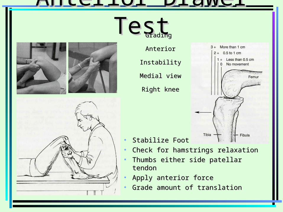
Anterior Drawer TestAnterior Drawer Test
• Stabilize FootStabilize Foot• Check for hamstrings relaxationCheck for hamstrings relaxation• Thumbs either side patellar tendonThumbs either side patellar tendon• Apply anterior forceApply anterior force• Grade amount of translationGrade amount of translation
Grading Grading
Anterior InstabilityAnterior Instability
Medial viewMedial view
Right kneeRight knee
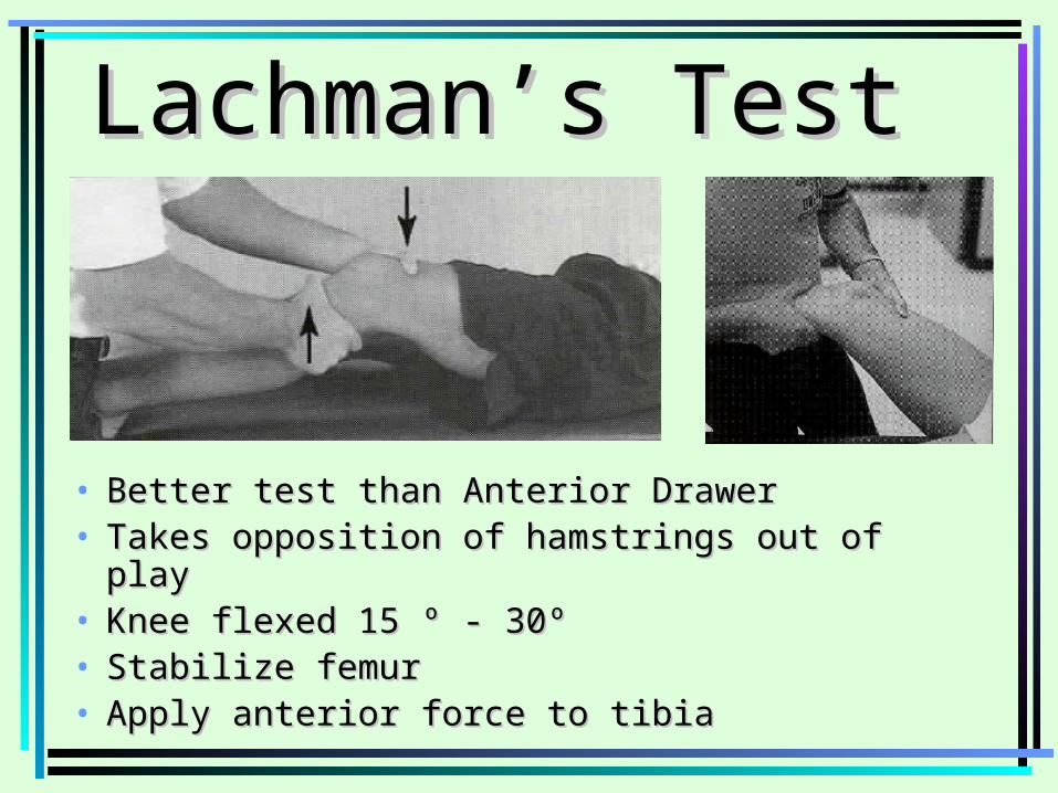
Lachman’s Test Lachman’s Test
• Better test than Anterior DrawerBetter test than Anterior Drawer• Takes opposition of hamstrings out of playTakes opposition of hamstrings out of play• Knee flexed 15 º - 30ºKnee flexed 15 º - 30º• Stabilize femurStabilize femur• Apply anterior force to tibiaApply anterior force to tibia
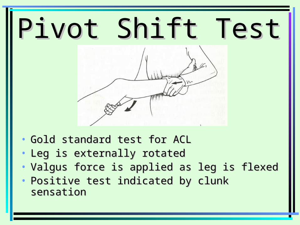
Pivot Shift Test Pivot Shift Test
• Gold standard test for ACLGold standard test for ACL• Leg is externally rotatedLeg is externally rotated• Valgus force is applied as leg is flexedValgus force is applied as leg is flexed• Positive test indicated by clunk sensationPositive test indicated by clunk sensation
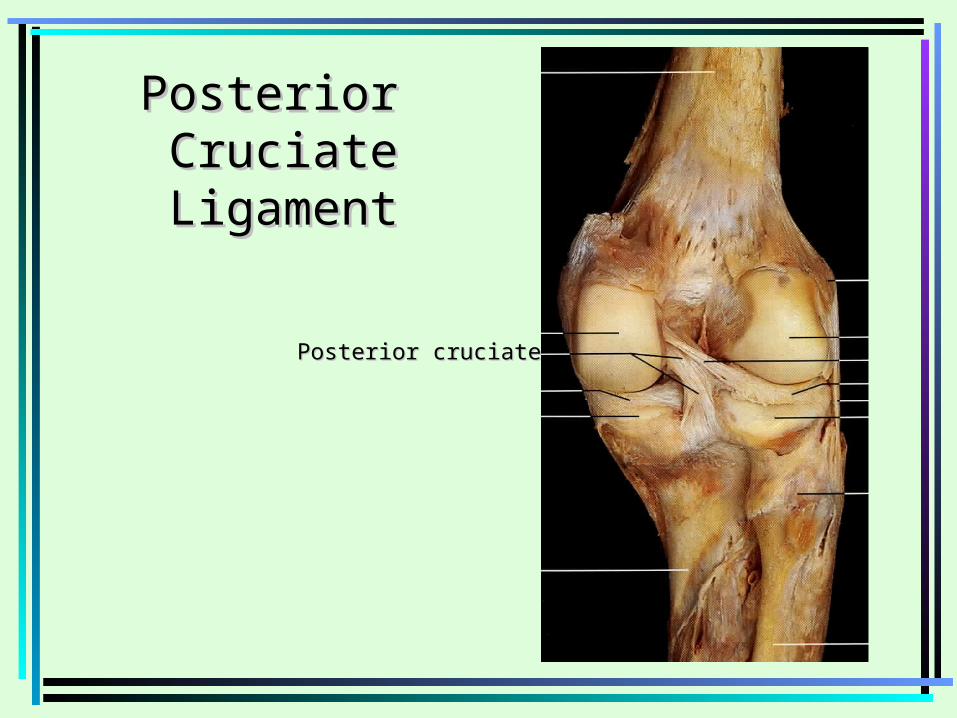
Posterior Posterior CruciateCruciateLigamentLigament
Posterior cruciatePosterior cruciate

Posterior Sag TestPosterior Sag Test• Posterior Cruciate vs Anterior CruciatePosterior Cruciate vs Anterior Cruciate• Athlete supineAthlete supine• Both knees flexed 90’Both knees flexed 90’• Observe laterallyObserve laterally
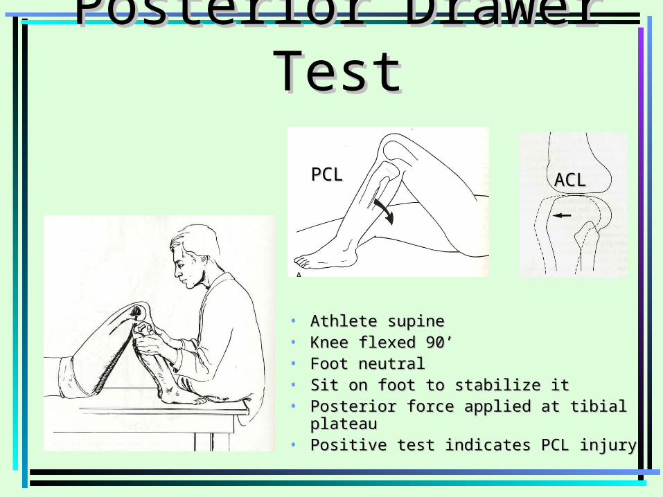
Posterior Drawer TestPosterior Drawer Test
• Athlete supineAthlete supine• Knee flexed 90’Knee flexed 90’• Foot neutralFoot neutral• Sit on foot to stabilize itSit on foot to stabilize it• Posterior force applied at tibial plateauPosterior force applied at tibial plateau• Positive test indicates PCL injuryPositive test indicates PCL injury
PCLPCL ACLACL
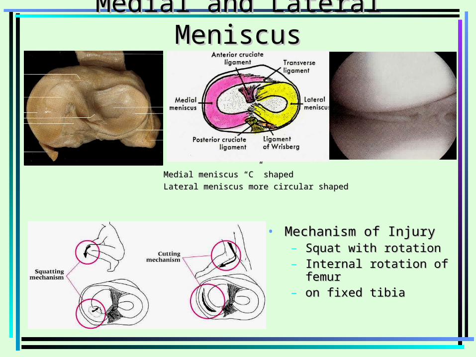
Medial and Lateral MeniscusMedial and Lateral Meniscus
Medial meniscus “C” shapedMedial meniscus “C” shaped
Lateral meniscus more circular shapedLateral meniscus more circular shaped
• Mechanism of InjuryMechanism of Injury– Squat with rotationSquat with rotation– Internal rotation of femur Internal rotation of femur – on fixed tibiaon fixed tibia
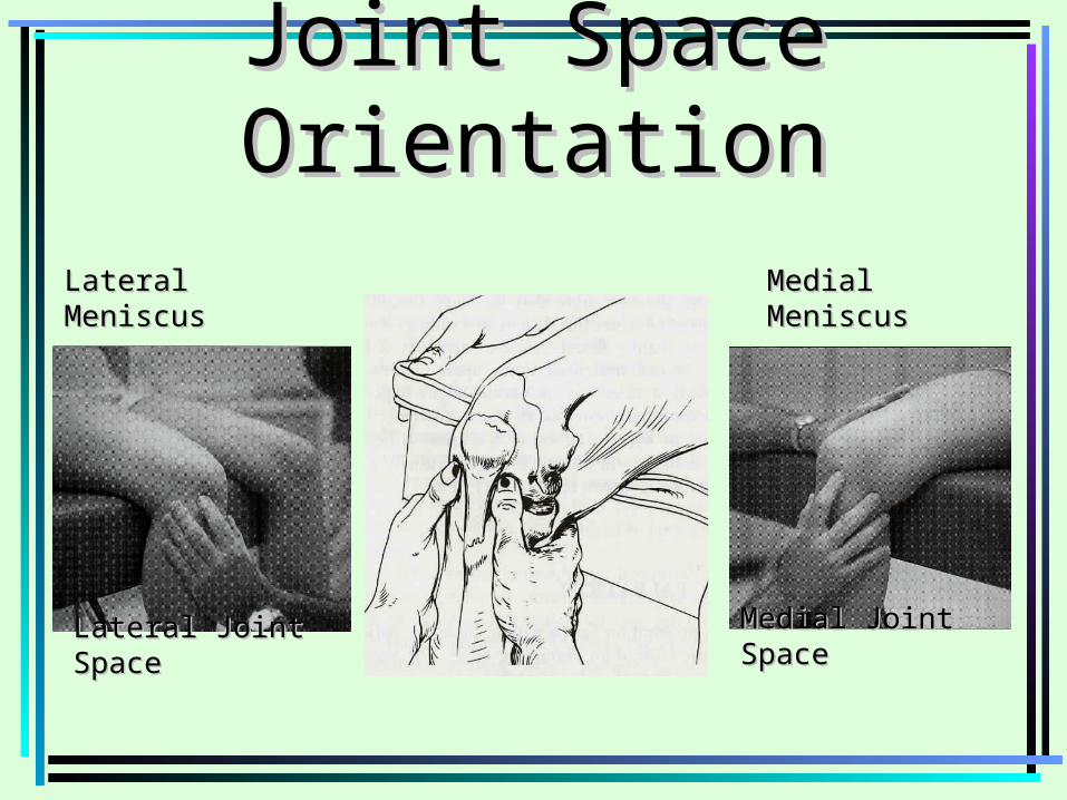
Joint Space OrientationJoint Space Orientation
Medial Joint SpaceMedial Joint Space
Lateral MeniscusLateral Meniscus
Lateral Joint SpaceLateral Joint Space
Medial MeniscusMedial Meniscus
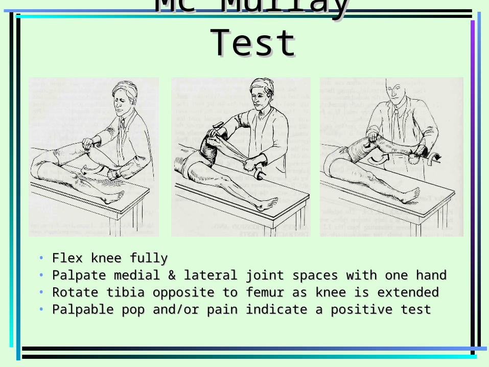
Mc Murray TestMc Murray Test
• Flex knee fullyFlex knee fully• Palpate medial & lateral joint spaces with one handPalpate medial & lateral joint spaces with one hand• Rotate tibia opposite to femur as knee is extendedRotate tibia opposite to femur as knee is extended• Palpable pop and/or pain indicate a positive testPalpable pop and/or pain indicate a positive test
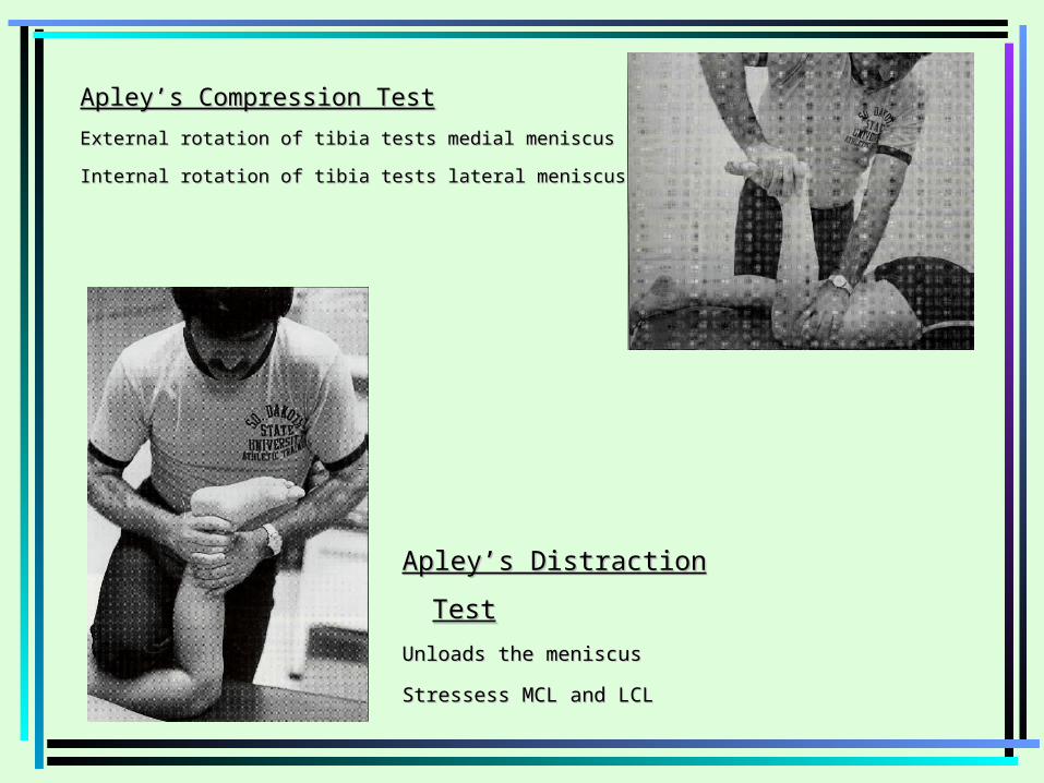
Apley’s Compression TestApley’s Compression Test
External rotation of tibia tests medial meniscus External rotation of tibia tests medial meniscus
Internal rotation of tibia tests lateral meniscusInternal rotation of tibia tests lateral meniscus
Apley’s Distraction TestApley’s Distraction Test
Unloads the meniscus Unloads the meniscus
Stressess MCL and LCLStressess MCL and LCL