Embed Size (px)
Citation preview
Final Progress Report for Research Projects Funded by
Health Research Grants
Instructions: Please complete all of the items as instructed. Do not delete instructions. Do not
leave any items blank; responses must be provided for all items. If your response to an item is
“None”, please specify “None” as your response. “Not applicable” is not an acceptable response
for any of the items. There is no limit to the length of your response to any question. Responses
should be single-spaced, no smaller than 12-point type. The report must be completed using
MS Word. Submitted reports must be Word documents; they should not be converted to pdf
format.
1. Grantee Institution: The Pennsylvania State University
2. Reporting Period (start and end date of grant award period): 1/1/2010 - 12/31/2013
3. Grant Contact Person (First Name, M.I., Last Name, Degrees): John Anthony, MPA
4. Grant Contact Person’s Telephone Number: 814 935 1081
5. Grant SAP Number: 4100050904
6. Project Number and Title of Research Project: 42. Molecular Analysis of Breast Cancer
CTC Heterogeneity & Drug Sensitivity in Preclinical Models & Patients
7. Start and End Date of Research Project: 7/20/2012 - 12/31/2013
8. Name of Principal Investigator for the Research Project: Wafik S. El-Deiry, MD
9. Research Project Expenses.
9(A) Please provide the total amount of health research grant funds spent on this project for
the entire duration of the grant, including indirect costs and any interest earned that was
spent:
$ 351,457
9(B) Provide the last names (include first initial if multiple individuals with the same last
name are listed) of all persons who worked on this research project and were supported with
health research funds. Include position titles (Principal Investigator, Graduate Assistant,
Post-doctoral Fellow, etc.), percent of effort on project and total health research funds
expended for the position. For multiple year projects, if percent of effort varied from year to
year, report in the % of Effort column the effort by year 1, 2, 3, etc. of the project (x% Yr 1;
z% Yr 2-3).
2
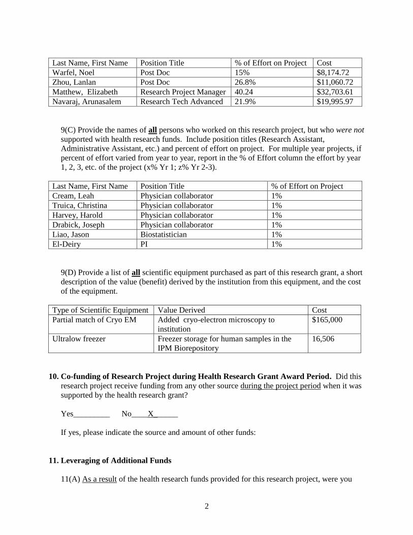
Last Name, First Name Position Title % of Effort on Project Cost
Warfel, Noel Post Doc 15% $8,174.72
Zhou, Lanlan Post Doc 26.8% $11,060.72
Matthew, Elizabeth Research Project Manager 40.24 $32,703.61
Navaraj, Arunasalem Research Tech Advanced 21.9% $19,995.97
9(C) Provide the names of all persons who worked on this research project, but who were not
supported with health research funds. Include position titles (Research Assistant,
Administrative Assistant, etc.) and percent of effort on project. For multiple year projects, if
percent of effort varied from year to year, report in the % of Effort column the effort by year
1, 2, 3, etc. of the project (x% Yr 1; z% Yr 2-3).
Last Name, First Name Position Title % of Effort on Project
Cream, Leah Physician collaborator 1%
Truica, Christina Physician collaborator 1%
Harvey, Harold Physician collaborator 1%
Drabick, Joseph Physician collaborator 1%
Liao, Jason Biostatistician 1%
El-Deiry PI 1%
9(D) Provide a list of all scientific equipment purchased as part of this research grant, a short
description of the value (benefit) derived by the institution from this equipment, and the cost
of the equipment.
Type of Scientific Equipment Value Derived Cost
Partial match of Cryo EM Added cryo-electron microscopy to
institution
$165,000
Ultralow freezer Freezer storage for human samples in the
IPM Biorepository
16,506
10. Co-funding of Research Project during Health Research Grant Award Period. Did this
research project receive funding from any other source during the project period when it was
supported by the health research grant?
Yes_________ No____X______
If yes, please indicate the source and amount of other funds:
11. Leveraging of Additional Funds
11(A) As a result of the health research funds provided for this research project, were you
3
able to apply for and/or obtain funding from other sources to continue or expand the
research?
Yes_________ No____X______
If yes, please list the applications submitted (column A), the funding agency (National
Institutes of Health—NIH, or other source in column B), the month and year when the
application was submitted (column C), and the amount of funds requested (column D). If
you have received a notice that the grant will be funded, please indicate the amount of funds
to be awarded (column E). If the grant was not funded, insert “not funded” in column E.
Do not include funding from your own institution or from CURE (tobacco settlement funds).
Do not include grants submitted prior to the start date of the grant as shown in Question 2. If
you list grants submitted within 1-6 months of the start date of this grant, add a statement
below the table indicating how the data/results from this project were used to secure that
grant.
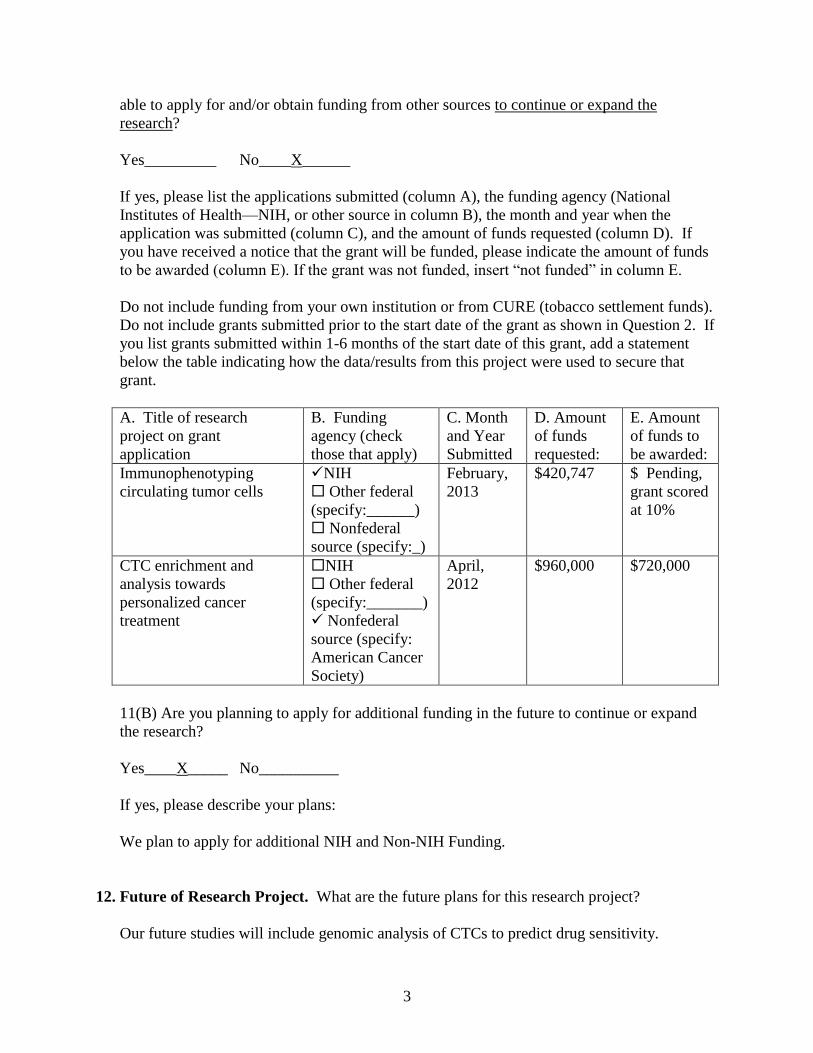
A. Title of research
project on grant
application
B. Funding
agency (check
those that apply)
C. Month
and Year
Submitted
D. Amount
of funds
requested:
E. Amount
of funds to
be awarded:
Immunophenotyping
circulating tumor cells
NIH
Other federal
(specify:______)
Nonfederal
source (specify:_)
February,
2013
$420,747 $ Pending,
grant scored
at 10%
CTC enrichment and
analysis towards
personalized cancer
treatment
NIH
Other federal
(specify:_______)
Nonfederal
source (specify:
American Cancer
Society)
April,
2012
$960,000 $720,000
11(B) Are you planning to apply for additional funding in the future to continue or expand
the research?
Yes____X_____ No__________
If yes, please describe your plans:
We plan to apply for additional NIH and Non-NIH Funding.
12. Future of Research Project. What are the future plans for this research project?
Our future studies will include genomic analysis of CTCs to predict drug sensitivity.
4
13. New Investigator Training and Development. Did students participate in project
supported internships or graduate or post-graduate training for at least one semester or one
summer?
Yes___X______ No__________
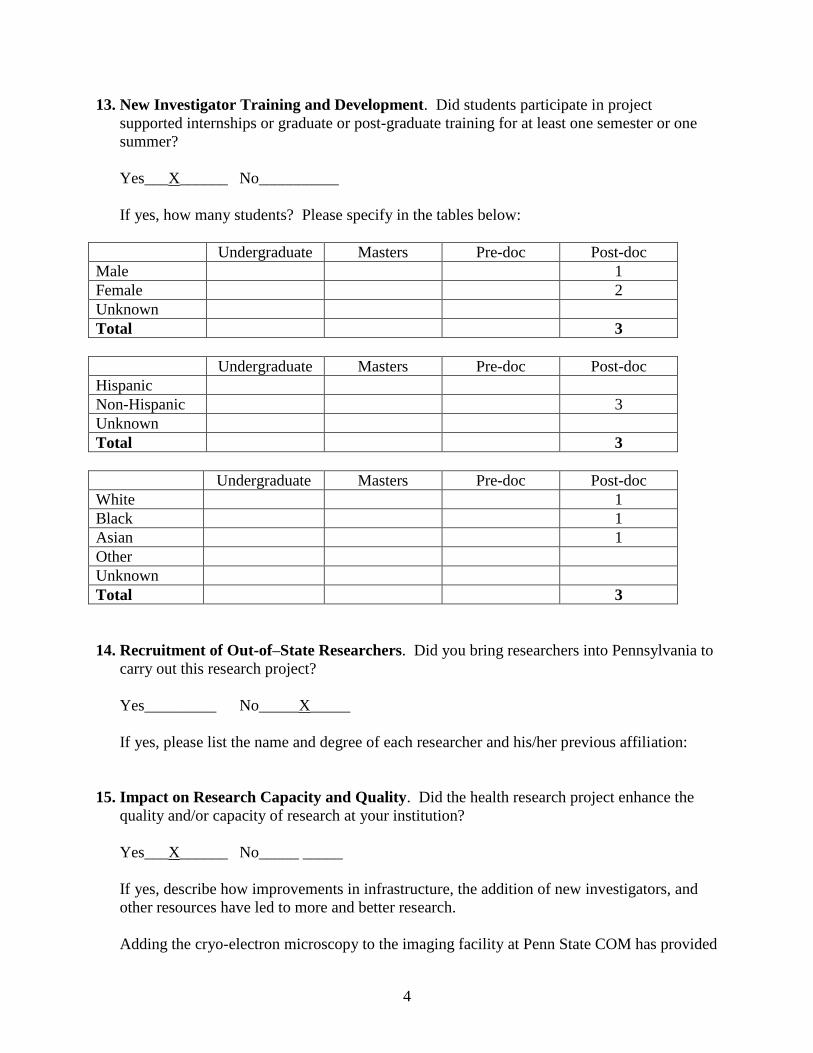
If yes, how many students? Please specify in the tables below:
Undergraduate Masters Pre-doc Post-doc
Male 1
Female 2
Unknown
Total 3
Undergraduate Masters Pre-doc Post-doc
Hispanic
Non-Hispanic 3
Unknown
Total 3
Undergraduate Masters Pre-doc Post-doc
White 1
Black 1
Asian 1
Other
Unknown
Total 3
14. Recruitment of Out-of–State Researchers. Did you bring researchers into Pennsylvania to
carry out this research project?
Yes_________ No_____X_____
If yes, please list the name and degree of each researcher and his/her previous affiliation:
15. Impact on Research Capacity and Quality. Did the health research project enhance the
quality and/or capacity of research at your institution?
Yes___X______ No_____ _____
If yes, describe how improvements in infrastructure, the addition of new investigators, and
other resources have led to more and better research.
Adding the cryo-electron microscopy to the imaging facility at Penn State COM has provided

5
multiple benefits to Penn State researchers. The cryo transmission electron microscope
(cryoEM) - a JEOL model JEM-2100 that is a 200 kV Transmission Electron Microscope
(TEM), with a Lanthanum Boride (LaB6) emitter, a full Cryo package, CCD camera and a
stage-tilting capability for tomography. This particular cryo-TEM is one of the easiest to
operate and maintain, making it the choice of many cryo TEM facilities as a first instrument
to acquire. It functions better than comparable instruments to fulfill the basic needs of a
general research environment and is often retained as a training instrument when facilities
expand because it has a reputation for versatility in a multi-user environment and is relatively
inexpensive to repair and maintain. The cryoEM collects images at the sub-cellular and
molecular level, as well as collecting data that can be processed into 3-D maps.
A wide variety of structural and functional studies have been made possible in our
centralized location. The instrumentation placed in the optimized space offers the powerful
tools of direct visualization that can guide and complement other research approaches
focusing on a variety of health issues including viral infections (including HIV and herpes),
cancer, diabetes and its complications (primarily diabetic retinopathy, muscle protein
synthesis, kidney and brain complications), heart disease, and degenerative diseases of the
central and peripheral nervous system (including Parkinson’s disease, Alzheimer’s disease
and restless leg syndrome). Acquiring CryoEM for the imaging core is meeting existing and
rapidly growing needs for an equipped biomedical research imaging facility dedicated to
experimental approaches in the life sciences. The new microscope at Penn State has allowed
the necessary training of students and researchers in cryoEM data processing, since the
biggest advantage offered to students and researchers by the new equipment is realized by the
cryoEM data processing that produces the 3-D map.
16. Collaboration, business and community involvement.
16(A) Did the health research funds lead to collaboration with research partners outside of
your institution (e.g., entire university, entire hospital system)?
Yes_________ No____X______
If yes, please describe the collaborations:
16(B) Did the research project result in commercial development of any research products?
Yes_________ No___X_______
If yes, please describe commercial development activities that resulted from the research
project:
16(C) Did the research lead to new involvement with the community?
Yes_________ No____X______

6
If yes, please describe involvement with community groups that resulted from the
research project:
17. Progress in Achieving Research Goals, Objectives and Aims. List the project goals, objectives and specific aims (as contained in the grant agreement).
Summarize the progress made in achieving these goals, objectives and aims for the period
that the project was funded (i.e., from project start date through end date). Indicate whether
or not each goal/objective/aim was achieved; if something was not achieved, note the reasons
why. Describe the methods used. If changes were made to the research
goals/objectives/aims, methods, design or timeline since the original grant application was
submitted, please describe the changes. Provide detailed results of the project. Include
evidence of the data that was generated and analyzed, and provide tables, graphs, and figures
of the data. List published abstracts, poster presentations and scientific meeting presentations
at the end of the summary of progress; peer-reviewed publications should be listed under
item 20.
This response should be a DETAILED report of the methods and findings. It is not sufficient
to state that the work was completed. Insufficient information may result in an unfavorable
performance review, which may jeopardize future funding. If research findings are pending
publication you must still include enough detail for the expert peer reviewers to evaluate the
progress during the course of the project.
Health research grants funded under the Tobacco Settlement Act will be evaluated via a
performance review by an expert panel of researchers and clinicians who will assess project
work using this Final Progress Report, all project Annual Reports and the project’s strategic
plan. After the final performance review of each project is complete, approximately 12-16
months after the end of the grant, this Final Progress Report, as well as the Final Performance
Review Report containing the comments of the expert review panel, and the grantee’s written
response to the Final Performance Review Report, will be posted on the CURE Web site.
There is no limit to the length of your response. Responses must be single-spaced below,
no smaller than 12-point type. If you cut and paste text from a publication, be sure
symbols print properly, e.g., the Greek symbol for alpha () and beta (ß) should not
print as boxes () and include the appropriate citation(s). DO NOT DELETE THESE
INSTRUCTIONS.
7
The objective of the project is to isolate circulating tumor cells (CTCs) from a mouse model of
breast cancer as well as from patients with metastatic breast cancer and perform two types of
analysis on the recovered cells including (1) single cell analysis of gene mutations, gene
expression, protein marker expression, and (2) analysis of drug sensitivity on the isolated
growing CTCs. The heterogeneity of the molecular analysis will be unraveled among the initially
isolated CTCs and this will be compared with the growing cells as well as the primary tumor
from either the mouse model or the human patients. We have developed a mouse model of breast
cancer using H-Ras transformed human mammary epithelial cells and BRCA1 knockdown. The
tumors that result are rapidly growing, and show features of the epithelial-to-mesenchymal
transition, invasiveness and angiogenesis. For analysis of drug sensitivity we will evaluate effect
of dose of all FDA approved drugs in a high-throughput 384 well plate format. Our preliminary
data suggests that as few as 20 recovered tumor cells can grow to over a million cells in 2-3
weeks and that as few as 25 cells in a well can reliably provide information regarding tumor cell
drug sensitivity. Our preliminary data shows clearly that many patients with advanced colorectal
or breast cancer can have dozens and sometimes hundreds or thousands of CTCs in a single tube
of blood. The objectives will be accomplished through:
Specific Aim #1: Molecular analysis of heterogeneity of isolated CTCs from a mouse model or
from patients with advanced breast cancer, and
Specific Aim #2: Analysis of drug sensitivity (IC50) against hundreds of FDA-approved drugs
and drug combinations on recovered CTCs from a mouse model or from patients with advanced
breast cancer.
Specific Aim #1: Molecular analysis of heterogeneity of isolated CTCs from a mouse model or
from patients with advanced breast cancer.
Sub Aim 1A: To establish xenograft mouse models for sampling and testing the presence of
Circulating Tumor Cells (CTCs) of breast cancer using FDA-approved Veridex CellSearch
System.
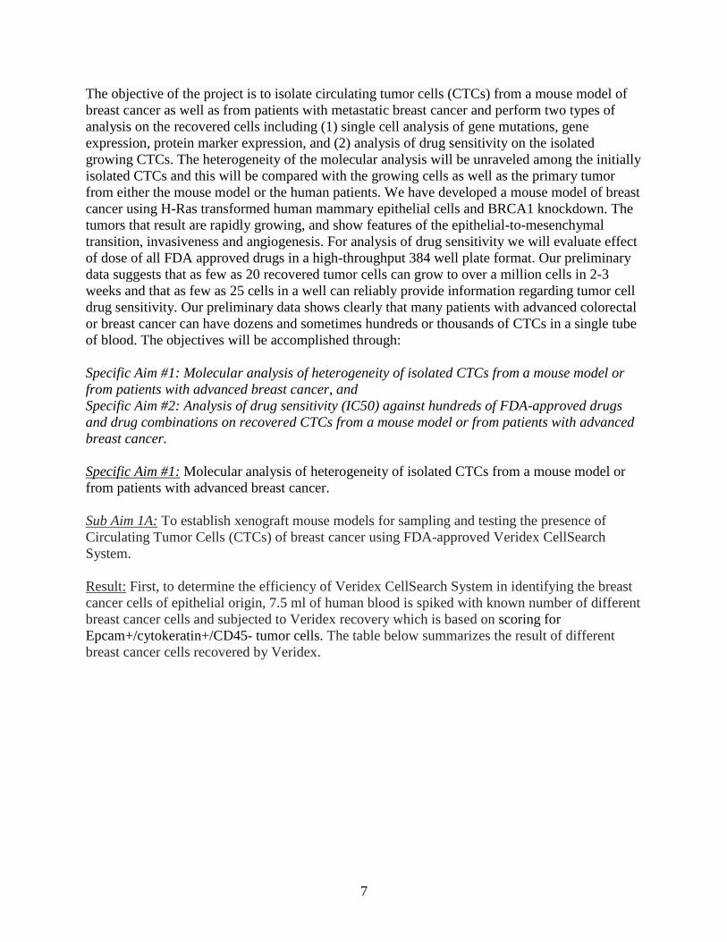
Result: First, to determine the efficiency of Veridex CellSearch System in identifying the breast
cancer cells of epithelial origin, 7.5 ml of human blood is spiked with known number of different
breast cancer cells and subjected to Veridex recovery which is based on scoring for
Epcam+/cytokeratin+/CD45- tumor cells. The table below summarizes the result of different
breast cancer cells recovered by Veridex.
8
Circulating Tumor Cells are metastatic which share many properties of cancer stem cells.
Recent studies indicate that breast cancer is initiated by breast cancer stem cells (BCSCs) which
express CD44+/high CD24-/low surface markers. Mammospheres are non-adherent spherical cell
clusters grown in selective culture conditions. Our experimental results using mammosphere
cultures of different breast cancer cells such as MDA-MB-231, show drastic enrichment of
breast cancer stem-like cells with the phenotype of CD44+/high CD24-/low in flow cytometry
analysis. These mammosphere-derived cells are implanted into mammary fat pads of nude mice
and sampling blood (200 µl) from such tumor bearing animals is done regularly at two week
intervals to look for the presence of CTCs, by scoring for Epcam+/cytokeratin+/CD45- tumor
cells using Veridex CellSearch System. As these metastatic breast cancer cell lines stably over-
express ZS-Green protein, isolated ZS-Green positive CTCs are further subjected to breast
cancer specific - multiplexed marker analysis.
9
A) B)
C)
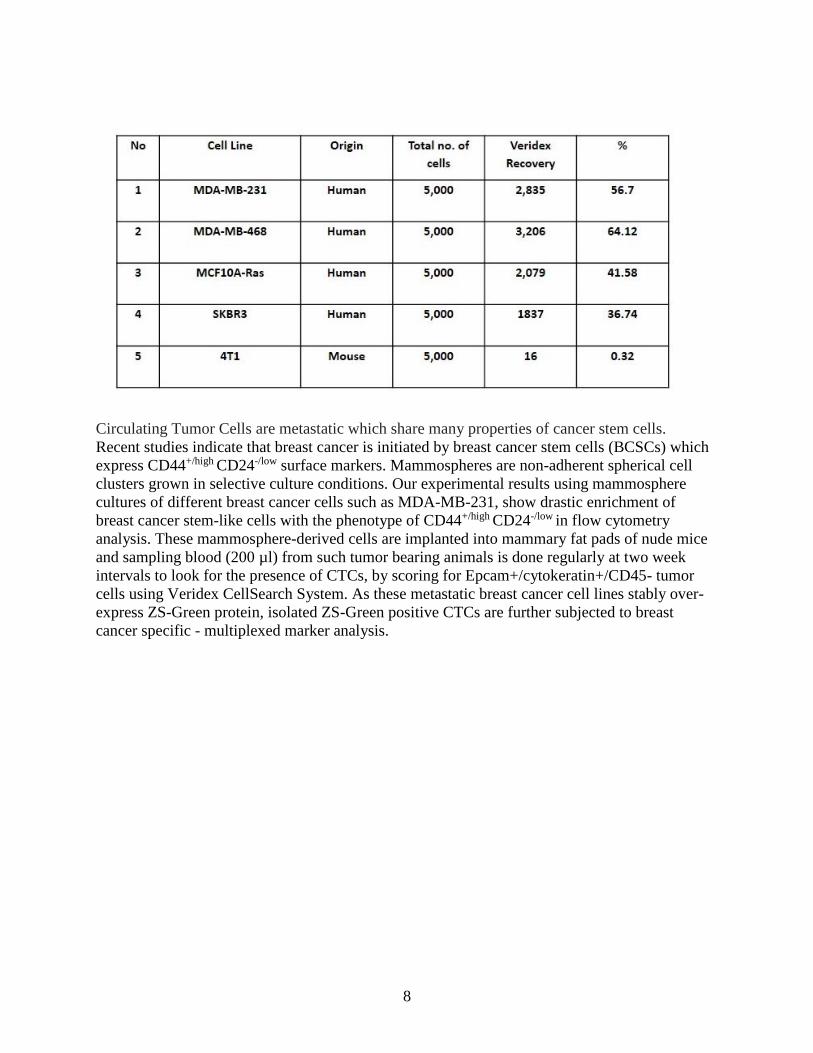
Figure 1. Enrichment of stem-like cancer cells in mammosphere. A) Growth of mammosphere
cultures of MDA-MB-231 cells both in the presence and absence of different growth factors. B)
Flow cytometry to show mammosphere cultures are enriched for stem-like breast cancer cells
which have CD44-high, CD24-low population. C) In vivo bioluminescence imaging of
mammosphere cultures of MDA-MB-231-Luciferase cells, implanted into mammary fat pads of
nude mice.
At every two weeks regular intervals, blood is collected from tumor bearing animals till the
endpoint of experiment when the tumor size reaches 2 cm in diameter. Mouse blood (200 µl) is
immediately mixed with 7.5 ml of normal, human blood in CellSearch tube and subjected to
Veridex analysis for the presence of CTCs. Such analysis often results in unexpected, clotting of
blood in Veridex CellSearch tubes (which are coated with anti-coagulants) and aggregation of
blood cells in those samples.
To overcome this problem arising due to clotting and to develop simpler ways to detect CTCs in
mouse blood, a mouse metastatic breast cancer cell line 4T1 is stably over-expressed with both
luciferase and ZSGreen through retro viral transductions. These 4T1-Luc-ZSGreen cells are also
stained with CellVue Maroon dye, which has excitation maximum at 647 nm and emission
maximum at 667 nm. 4T1-Luc-ZSGreen-CellVue maroon cells are implanted into mammary fat
pads and tumor size is periodically measured using Xenogen IVIS bioluminescence imaging
system. Through tail vein, animal blood is collected (200 µl) periodically and examined for the

10
presence of cells with green fluorescent protein and CellVue stain, under fluorescence
microscopic field. When tumor size is around 2 cm in diameter, animals are sacrificed, internal
organs are imaged, nearly 1 ml of blood is collected through intra cardiac puncture, spotted on a
glass slide by cytospin and examined under fluorescence microscopy for the presence of
ZSGreen and cellvue maroon positive circulating tumor cells.
11
A)
B) C)
D)
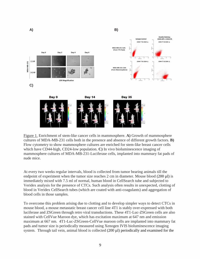
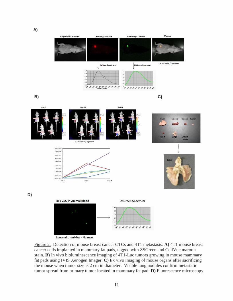
Figure 2. Detection of mouse breast cancer CTCs and 4T1 metastasis. A) 4T1 mouse breast
cancer cells implanted in mammary fat pads, tagged with ZSGreen and CellVue maroon
stain. B) In vivo bioluminescence imaging of 4T1-Luc tumors growing in mouse mammary
fat pads using IVIS Xenogen Imager. C) Ex vivo imaging of mouse organs after sacrificing
the mouse when tumor size is 2 cm in diameter. Visible lung nodules confirm metastatic
tumor spread from primary tumor located in mammary fat pad. D) Fluorescence microscopy
12
analysis of mouse blood spotted on a glass slide showing ZSGreen positive, metastatic
circulating tumor cells.
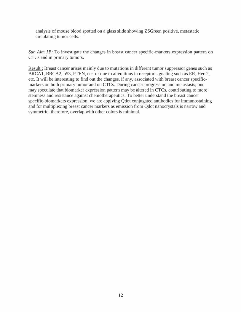
Sub Aim 1B: To investigate the changes in breast cancer specific-markers expression pattern on
CTCs and in primary tumors.
Result : Breast cancer arises mainly due to mutations in different tumor suppressor genes such as
BRCA1, BRCA2, p53, PTEN, etc. or due to alterations in receptor signaling such as ER, Her-2,
etc. It will be interesting to find out the changes, if any, associated with breast cancer specific-
markers on both primary tumor and on CTCs. During cancer progression and metastasis, one
may speculate that biomarker expression pattern may be altered in CTCs, contributing to more
stemness and resistance against chemotherapeutics. To better understand the breast cancer
specific-biomarkers expression, we are applying Qdot conjugated antibodies for immunostaining
and for multiplexing breast cancer markers as emission from Qdot nanocrystals is narrow and
symmetric; therefore, overlap with other colors is minimal.
13
A)
B)
C)
Figure 3. Q-Dot immunostaining of different breast cancer specific-markers. A) MDA-MB-
231 cells are grown on chambered slides and stained with CD44-Qdot525nm antibody.
B) MDA-MB-468 cells are similarly stained with EGFR-Qdot625nm antibody. C) Skbr-3
cells grown on chambered slides are stained with Her2-Qdot565nm antibody.
At present, as shown above, different breast cancer cells, cultured on slides are stained
successfully with Qdot conjugated antibodies specific for breast cancer biomarkers. Efforts
are underway to stain these markers on primary tumor tissue specimens and on isolated CTCs
from clinical samples.
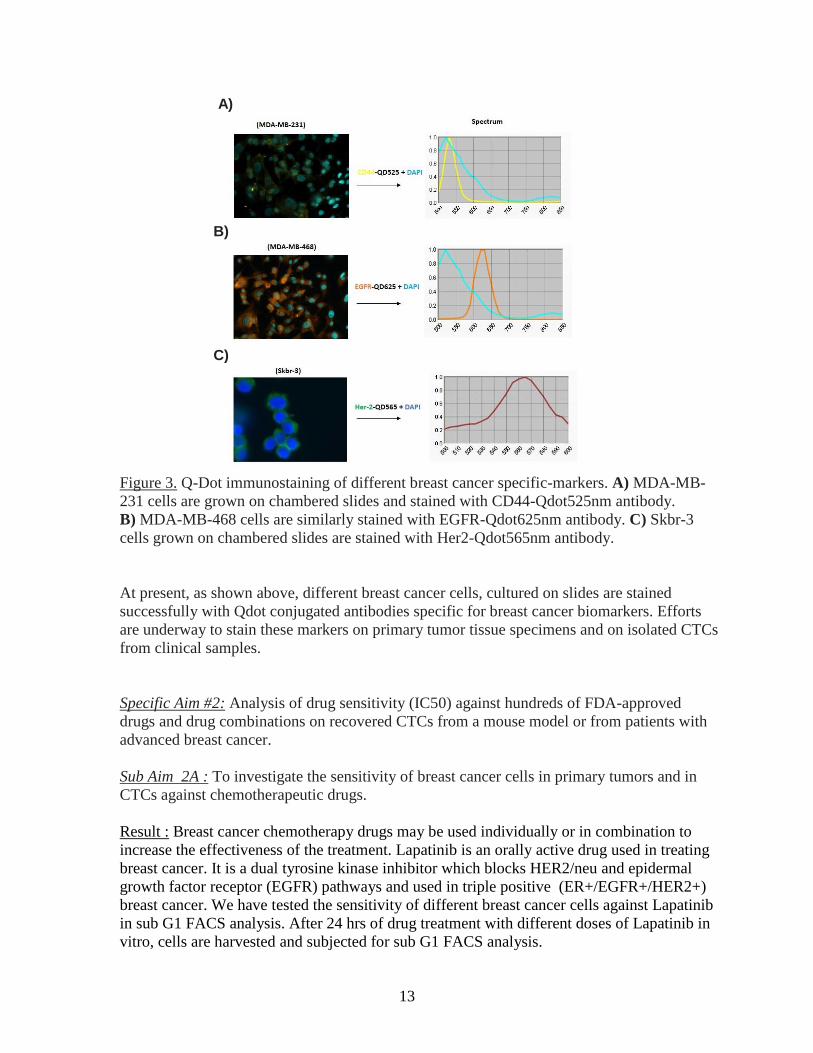
Specific Aim #2: Analysis of drug sensitivity (IC50) against hundreds of FDA-approved
drugs and drug combinations on recovered CTCs from a mouse model or from patients with
advanced breast cancer.
Sub Aim 2A : To investigate the sensitivity of breast cancer cells in primary tumors and in
CTCs against chemotherapeutic drugs.
Result : Breast cancer chemotherapy drugs may be used individually or in combination to
increase the effectiveness of the treatment. Lapatinib is an orally active drug used in treating
breast cancer. It is a dual tyrosine kinase inhibitor which blocks HER2/neu and epidermal
growth factor receptor (EGFR) pathways and used in triple positive (ER+/EGFR+/HER2+)
breast cancer. We have tested the sensitivity of different breast cancer cells against Lapatinib
in sub G1 FACS analysis. After 24 hrs of drug treatment with different doses of Lapatinib in
vitro, cells are harvested and subjected for sub G1 FACS analysis.
14
Figure 4. Varying sensitivity of breast cancer cells in vitro against Lapatinib drug, in sub G1
FACS analysis.
We have continued to make progress in isolating circulating tumor cells (CTCs) from breast
cancer patients and multiplexing with multiple markers. In a metastatic breast cancer (estrogen
receptor negative) patient with metastasis to the bone we have identified breast cancer cells both
by size-based enrichment followed by multiple marker analysis as well as by Veridex Cell
Search.
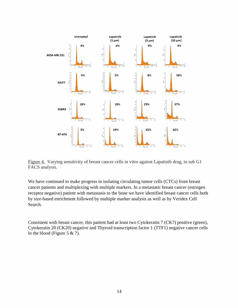
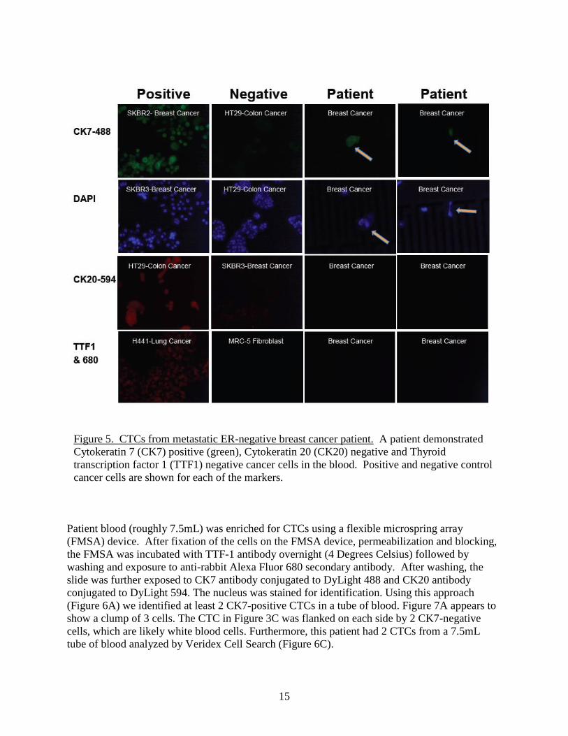
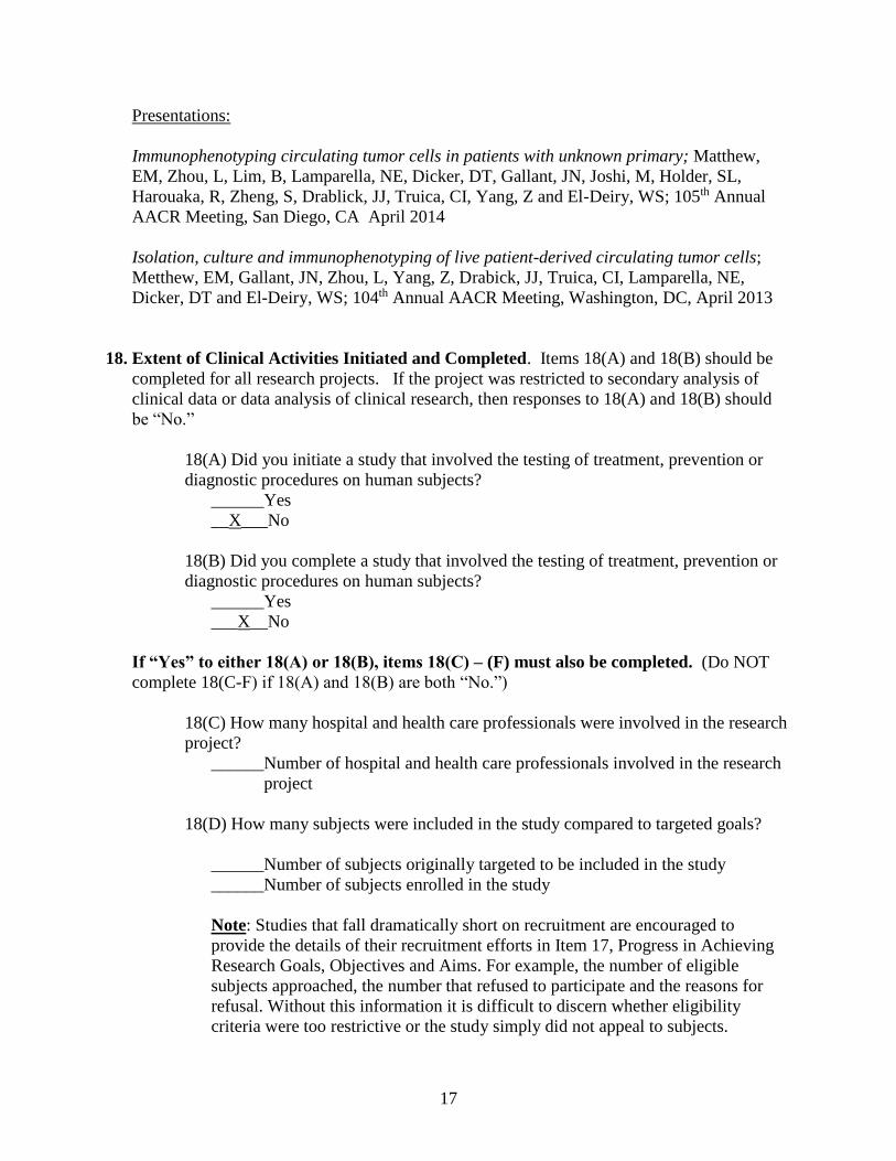
Consistent with breast cancer, this patient had at least two Cytokeratin 7 (CK7) positive (green),
Cytokeratin 20 (CK20) negative and Thyroid transcription factor 1 (TTF1) negative cancer cells
in the blood (Figure 5 & 7).
15
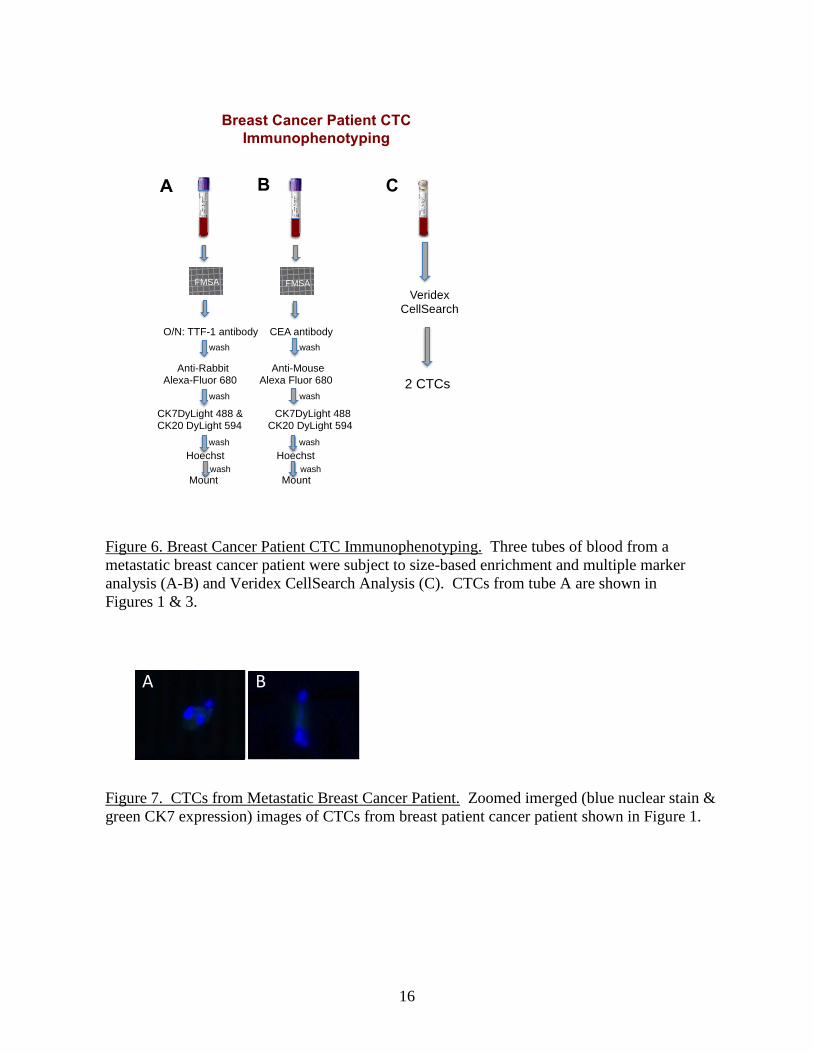
Patient blood (roughly 7.5mL) was enriched for CTCs using a flexible microspring array
(FMSA) device. After fixation of the cells on the FMSA device, permeabilization and blocking,
the FMSA was incubated with TTF-1 antibody overnight (4 Degrees Celsius) followed by
washing and exposure to anti-rabbit Alexa Fluor 680 secondary antibody. After washing, the
slide was further exposed to CK7 antibody conjugated to DyLight 488 and CK20 antibody
conjugated to DyLight 594. The nucleus was stained for identification. Using this approach
(Figure 6A) we identified at least 2 CK7-positive CTCs in a tube of blood. Figure 7A appears to
show a clump of 3 cells. The CTC in Figure 3C was flanked on each side by 2 CK7-negative
cells, which are likely white blood cells. Furthermore, this patient had 2 CTCs from a 7.5mL
tube of blood analyzed by Veridex Cell Search (Figure 6C).
Figure 5. CTCs from metastatic ER-negative breast cancer patient. A patient demonstrated
Cytokeratin 7 (CK7) positive (green), Cytokeratin 20 (CK20) negative and Thyroid
transcription factor 1 (TTF1) negative cancer cells in the blood. Positive and negative control
cancer cells are shown for each of the markers.
16
Breast Cancer Patient CTC
Immunophenotyping
Veridex
CellSearch
2 CTCs
FMSA FMSA
O/N: TTF-1 antibody CEA antibody
Anti-Rabbit Anti-Mouse Alexa-Fluor 680 Alexa Fluor 680
wash wash
wash wash
CK7DyLight 488 & CK7DyLight 488 CK20 DyLight 594 CK20 DyLight 594
Hoechst Hoechst
Mount Mount
wash wash
wash wash
A B C
Figure 6. Breast Cancer Patient CTC Immunophenotyping. Three tubes of blood from a
metastatic breast cancer patient were subject to size-based enrichment and multiple marker
analysis (A-B) and Veridex CellSearch Analysis (C). CTCs from tube A are shown in
Figures 1 & 3.
AB
Figure 7. CTCs from Metastatic Breast Cancer Patient. Zoomed imerged (blue nuclear stain &
green CK7 expression) images of CTCs from breast patient cancer patient shown in Figure 1.
17
Presentations:
Immunophenotyping circulating tumor cells in patients with unknown primary; Matthew,
EM, Zhou, L, Lim, B, Lamparella, NE, Dicker, DT, Gallant, JN, Joshi, M, Holder, SL,
Harouaka, R, Zheng, S, Drablick, JJ, Truica, CI, Yang, Z and El-Deiry, WS; 105th Annual
AACR Meeting, San Diego, CA April 2014
Isolation, culture and immunophenotyping of live patient-derived circulating tumor cells;
Metthew, EM, Gallant, JN, Zhou, L, Yang, Z, Drabick, JJ, Truica, CI, Lamparella, NE,
Dicker, DT and El-Deiry, WS; 104th Annual AACR Meeting, Washington, DC, April 2013
18. Extent of Clinical Activities Initiated and Completed. Items 18(A) and 18(B) should be
completed for all research projects. If the project was restricted to secondary analysis of
clinical data or data analysis of clinical research, then responses to 18(A) and 18(B) should
be “No.”
18(A) Did you initiate a study that involved the testing of treatment, prevention or
diagnostic procedures on human subjects?
______Yes
__X___No
18(B) Did you complete a study that involved the testing of treatment, prevention or
diagnostic procedures on human subjects?
______Yes
___X__No
If “Yes” to either 18(A) or 18(B), items 18(C) – (F) must also be completed. (Do NOT
complete 18(C-F) if 18(A) and 18(B) are both “No.”)
18(C) How many hospital and health care professionals were involved in the research
project?
______Number of hospital and health care professionals involved in the research
project
18(D) How many subjects were included in the study compared to targeted goals?
______Number of subjects originally targeted to be included in the study
______Number of subjects enrolled in the study
Note: Studies that fall dramatically short on recruitment are encouraged to
provide the details of their recruitment efforts in Item 17, Progress in Achieving
Research Goals, Objectives and Aims. For example, the number of eligible
subjects approached, the number that refused to participate and the reasons for
refusal. Without this information it is difficult to discern whether eligibility
criteria were too restrictive or the study simply did not appeal to subjects.
18
18(E) How many subjects were enrolled in the study by gender, ethnicity and race?
Gender:
______Males
______Females
______Unknown
Ethnicity:
______Latinos or Hispanics
______Not Latinos or Hispanics
______Unknown
Race:
______American Indian or Alaska Native
______Asian
______Blacks or African American
______Native Hawaiian or Other Pacific Islander
______White
______Other, specify:
______Unknown
18(F) Where was the research study conducted? (List the county where the research
study was conducted. If the treatment, prevention and diagnostic tests were offered in
more than one county, list all of the counties where the research study was
conducted.)
19. Human Embryonic Stem Cell Research. Item 19(A) should be completed for all research
projects. If the research project involved human embryonic stem cells, items 19(B) and
19(C) must also be completed.
19(A) Did this project involve, in any capacity, human embryonic stem cells?
______Yes
__X__ No
19(B) Were these stem cell lines NIH-approved lines that were derived outside of
Pennsylvania?
_____Yes
____ No
19(C) Please describe how this project involved human embryonic stem cells:
20. Articles Submitted to Peer-Reviewed Publications.
20(A) Identify all publications that resulted from the research performed during the funding
period and that have been submitted to peer-reviewed publications. Do not list journal
19
abstracts or presentations at professional meetings; abstract and meeting presentations should
be listed at the end of item 17. Include only those publications that acknowledge the
Pennsylvania Department of Health as a funding source (as required in the grant
agreement). List the title of the journal article, the authors, the name of the peer-reviewed
publication, the month and year when it was submitted, and the status of publication
(submitted for publication, accepted for publication or published.). Submit an electronic
copy of each publication or paper submitted for publication, listed in the table, in a PDF
version 5.0.5 (or greater) format, 1,200 dpi. Filenames for each publication should include
the number of the research project, the last name of the PI, and an abbreviated title of the
publication. For example, if you submit two publications for Smith (PI for Project 01), one
publication for Zhang (PI for Project 03), and one publication for Bates (PI for Project 04),
the filenames would be:
Project 01 – Smith – Three cases of isolated
Project 01 – Smith – Investigation of NEB1 deletions
Project 03 – Zhang – Molecular profiling of aromatase
Project 04 – Bates – Neonatal intensive care
If the publication is not available electronically, provide 5 paper copies of the publication.
Note: The grant agreement requires that recipients acknowledge the Pennsylvania
Department of Health funding in all publications. Please ensure that all publications listed
acknowledge the Department of Health funding. If a publication does not acknowledge the
funding from the Commonwealth, do not list the publication.
Title of Journal
Article:
Authors: Name of Peer-
reviewed
Publication:
Month and
Year
Submitted:
Publication
Status (check
appropriate box
below):
Submitted
Accepted
Published
20(B) Based on this project, are you planning to submit articles to peer-reviewed publications
in the future?
Yes____x_____ No__________
If yes, please describe your plans:
Predicting therapy response in live tumor cells isolated with the flexible micro spring array
device. Gallant JN, Matthew EM, Cheng H, Harouaka R, Lamparella NE, Kunkel M, Yang
Z, Harvey HA, Cream LV, Kumar SM, Robertson GP, Zheng S, Drabick JJ, Truica CI, El-
Deiry WS. Cell Cycle 12: 2132-43, 2013.
20
21. Changes in Outcome, Impact and Effectiveness Attributable to the Research Project.
Describe the outcome, impact, and effectiveness of the research project by summarizing its
impact on the incidence of disease, death from disease, stage of disease at time of diagnosis,
or other relevant measures of outcome, impact or effectiveness of the research project. If
there were no changes, insert “None”; do not use “Not applicable.” Responses must be
single-spaced below, and no smaller than 12-point type. DO NOT DELETE THESE
INSTRUCTIONS. There is no limit to the length of your response.
Circulating tumor cells (CTCs) in patients with metastatic breast cancer are shed into the
blood circulation and provide valuable information about a patient’s prognosis as well as
their response to therapy. We believe that a molecular analysis (gene mutations and proteins)
of CTCs and a study of our ability to kill living recovered tumor cells from a patient’s blood
sample (drug sensitivity) will provide important information to the treating physician to
guide therapy decisions. Because cancer cells even within a given patient are heterogeneous
and may require combination therapy to target all the different kinds of malignant cells, we
believe that a ‘liquid biopsy’ of the circulating cells in patients with advanced aggressive
breast cancer will allow us to personalize therapy by testing treatments on the patient’s own
tumor cells before treatments are given to patients.
22. Major Discoveries, New Drugs, and New Approaches for Prevention Diagnosis and
Treatment. Describe major discoveries, new drugs, and new approaches for prevention,
diagnosis and treatment that are attributable to the completed research project. If there were
no major discoveries, drugs or approaches, insert “None”; do not use “Not applicable.”
Responses must be single-spaced below, and no smaller than 12-point type. DO NOT
DELETE THESE INSTRUCTIONS. There is no limit to the length of your response.
None.
23. Inventions, Patents and Commercial Development Opportunities.
23(A) Were any inventions, which may be patentable or otherwise protectable under Title 35
of the United States Code, conceived or first actually reduced to practice in the performance
of work under this health research grant? Yes No X
If “Yes” to 23(A), complete items a – g below for each invention. (Do NOT complete items
a - g if 23(A) is “No.”)
a. Title of Invention:
b. Name of Inventor(s):
c. Technical Description of Invention (describe nature, purpose, operation and physical,
chemical, biological or electrical characteristics of the invention):

21
d. Was a patent filed for the invention conceived or first actually reduced to practice in
the performance of work under this health research grant?
Yes No
If yes, indicate date patent was filed:
e. Was a patent issued for the invention conceived or first actually reduced to practice in
the performance of work under this health research grant?
Yes No
If yes, indicate number of patent, title and date issued:
Patent number:
Title of patent:
Date issued:
f. Were any licenses granted for the patent obtained as a result of work performed under
this health research grant? Yes No
If yes, how many licenses were granted?
g. Were any commercial development activities taken to develop the invention into a
commercial product or service for manufacture or sale? Yes No
If yes, describe the commercial development activities:
23(B) Based on the results of this project, are you planning to file for any licenses or patents,
or undertake any commercial development opportunities in the future?
Yes_________ No___X_______
If yes, please describe your plans:
24. Key Investigator Qualifications. Briefly describe the education, research interests and
experience and professional commitments of the Principal Investigator and all other key
investigators. In place of narrative you may insert the NIH biosketch form here; however,
please limit each biosketch to 1-2 pages.
22
BIOGRAPHICAL SKETCH Provide the following information for the Senior/key personnel and other significant contributors in the order listed on Form Page 2.
Follow this format for each person. DO NOT EXCEED FOUR PAGES.
NAME
El-Deiry, Wafik S. POSITION TITLE
Chief, Division of Hematology/Oncology, Penn State Associate Director, Translational Research, PSHCI eRA COMMONS USER NAME (credential, e.g., agency login)
WEL-DEIRY
EDUCATION/TRAINING (Begin with baccalaureate or other initial professional education, such as nursing, include postdoctoral training and residency training if applicable.)
INSTITUTION AND LOCATION DEGREE
(if applicable) MM/YY FIELD OF STUDY
University of Miami College of Arts & Sciences B.S. 06/81 Chemistry/Math University of Miami School of Medicine M.D. 06/87 Medicine University of Miami School of Medicine (So Lab) Ph.D. 06/87 Biochemistry The Johns Hopkins University (Vogelstein Lab) post-doc 06/94 Mol. Genetics of Cancer
A. Personal Statement Dr. El-Deiry’s research is focused on unraveling cell death pathways involved in tumor suppression and mechanisms of cancer therapeutic responses. Dr. El-Deiry discovered the p21(WAF1) cell cycle inhibitor as a p53 target gene and mediator of p53-dependent tumor suppression. This is the most highly cited original research paper published in Cell. His group discovered TRAIL death receptor DR5 and elucidated regulation of TRAIL signaling and DR5 by p53. He is developing strategies to reverse therapy resistance by targeting restoration of the p53 pathway in tumors with mutated or dysfunctional p53 with small molecules. Dr. El-Deiry has significant expertise in cancer biology, in vivo imaging, drug screening, and targeting of cell cycle, cell death, cancer stem cells and signal transduction pathways for cancer therapy. He has mentored over 60 post-doctoral fellows and PhD students and has played an active role in the career development of clinical fellows and faculty for 20 years. He serves as Associate Director for Translational Research at his cancer center and PI of several translational clinical protocols. Dr. El-Deiry along with his laboratory research group and clinical colleagues is well-positioned to pursue translational research to develop new cancer therapeutics.
B. Positions and Honors Positions and Employment 07/87-06/88 Osler Intern in Medicine, The Johns Hopkins Hospital, Baltimore, MD 07/88-06/90 Junior and Senior Assistant Resident, Osler Medical Service, Johns Hopkins Hosp.,
Baltimore 07/90-06/94 Senior Clinical Fellow in Medical Oncology, The Johns Hopkins Oncology
Center, Baltimore 07/94-06/99 Assistant Professor of Medicine, Department of Medicine, Univ. of Penn. School
of Medicine 01/95-08/04 Assistant Investigator, Howard Hughes Medical Institute 06/95-06/99 Assistant Professor of Genetics, University of Pennsylvania School of Medicine 12/95-02/10 Active Medical Staff, Hospital of the University of Pennsylvania 06/96-02/10 Attending Staff (Solid Tumor Oncology), Hospital of the University of
Pennsylvania 02/99-06/99 Assistant Professor of Pharmacology, University of Pennsylvania School of
Medicine 07/99-06/05 Associate Professor of Medicine (Tenured), Genetics, and Pharmacology, Univ.
of Pennsylvania 12/04-02/10 Co-Director, Radiation Biology & Imaging Program, Abramson Comprehensive
Cancer Center 07/05-02/10 Professor of Medicine (Hem/Onc), Genetics, and Pharmacology, University of
Pennsylvania 02/07-02/10 Associate Director for Physician-Scientist Training (Hematology/Oncology)
23
08/13-02/14 Interim Director, Penn State Hershey Cancer Institute 01/09- American Cancer Society Research Professor 03/10- Rose Dunlap Professor of Medicine and Chief, Hem/Onc, Penn State Hershey
Medical Center 03/10- Associate Director for Translational Research, Penn State Hershey Cancer
institute 02/11- Program Leader, Experimental Therapeutics Program, Penn State Hershey
Cancer Institute Honors and Other Professional Activities 1998-04 NIH Pathology B Study Section and Tumor Progression & Metastasis Study
Section Member 1999- Member, American Society for Clinical Investigation (ASCI) 2001- Editor-in-Chief, Cancer Biology and Therapy 2005-07 NIH Molecular Oncogenesis Study Section (MONC) Member 2005 Institute for Scientific Information Highly Cited Researcher (Molecular Biology &
Genetics) 2005-10 Member, Institutional Review Board, University of Pennsylvania 2007-10 Associate Editor, Journal of Clinical Investigation (ASCI) 2008 Member, Association of American Physicians 2008- Reviewer, State of Maryland Stem Cell Research Program (yearly study section
meeting) 2010 Chairperson, AACR Special Conference on Cell Death Mechanisms and Cancer
Therapy 2010 Organizer, 15th International p53 Workshop, Philadelphia 2011- NIH ZRG1 BMCT-C(09) Study Section (co-Chair 6/12, 10/12, 2/13, 6/13, 12/13,
2/14) 2012 Elected Fellow, American College of Physicians 2013 SPORE (2/13; 11/13; 2/14), CCSG study sections (2/13 Northwestern, 5/13
MSKCC, 5/14 Duke) 2013-15 Principal Investigator, American Cancer Society-IRG Grant, Penn State University 2013-14 President, Interurban Clinical Club (historic physician-scientist honor society) 2014 Organizer, 4th Intl Conference on Tumor Progression and Therapeutic Resistance,
Boston, MA 2014 Elected Member, Johns Hopkins University Society of Scholars
Active Clinical Trials (El-Deiry as PI at Penn State) 1. A Randomized, Multi-Center, Blinded, Placebo-Controlled Study of Mapatumumab
([HGS1012], A Fully-Human Monoclonal Antibody to Trail-R1) in Combination with Sorafenib as First-Line Therapy in Subjects with Advanced Hepatocellular Carcinoma.
2. Prevalence of Stem Cell and Prognostic Markers in Circulating Tumor Cells of Patients with Metastatic Colorectal Cancer Undergoing Chemotherapy.
3. A Phase Ib, Open-Label, Dose-Escalation Study of the Safety and Pharmacokinetics of Multiple Doses of Dulanermin Administered Intravenously in Combination with Camptosar/Erbitux Chemotherapy or the Folfiri Regimen with or without Bevacizumab in Subjects with Previously Treated Metastatic Colorectal Cancer. (Closed to accrual).
4. Variable Patterns of Plasma 5-Fluorouracil Levels During Dose Optimization of Infusional 5-FU in Colorectal Cancer Patients.
5. Plk2-TSC interactions and tumor progression in colorectal cancer. (IRB Protocol No. 36137EM). Investigator-initiated.
6. Development of multi-probe panels for characterization of circulating tumor cells (IRB Protocol No. 35227NHR). Investigator-initiated.
7. Sensitivity of TRAIL and TRAIL-based Therapies in Colon Cancer Tissue and Stroma. (IRB Protocol No. 35650EP). Investigator-initiated.
8. A Randomized Double-Blind, Placebo-Controlled Study of the Efficacy and Safety of Monotherapy MORAb-004 Plus Best Supportive Care in Subjects with Chemorefractory Colorectal Cancer (MORAb-004-202-CRC; PSHCI 12-005). (IRB Protocol No. 40816). Sponsor: Morphotek.
24
9. STEAM (Sequencing Triplet with Avastin and Maintenance): FOLFOXIRI/Bevacizumab Regimens (Concurrent and Sequential) vs. FOLFOX/Bevacizumab in First-Line Metastatic Colorectal Cancer (ML28442) (PSHCI 12-120). (IRB Protocol No. 42555). Sponsor: Genentech.
10. Detecting Circulating Tumor Cells using the Veridex CellSearch in Patients Being Evaluated for Carcinoma of Unknown Primary & Utilizing Post-Veridex Single Cell Multiplex Assay in an Attempt to Identify the Primary Site of Origin (PSHCI 12-002). (IRB Protocol No. 39706). Investigator-initiated.
11.Quinacrine-Capecitabine Combinatorial Therapy for Advanced Stage Colorectal Adenocarcinoma: A Phase I/II Clinical Trial (PSHCI 11-099). (IRB Protocol No. 40877). Investigator-initiated. Investigator IND 114242 granted from FDA (El-Deiry; Approval Date 5/17/12 from FDA for Study to proceed).
Licensure: Pennsylvania Medical License MD 054226L exp 12/31/14; DEA: BE4121860 exp 8/31/15 C. Selected Peer-reviewed Publications (Selected from 227 original manuscripts & 118 reviews) 1. El-Deiry, W.S., Kern, S.E., Pietenpol, J.A., Kinzler, K.W., and Vogelstein, B. Definition of a
consensus binding site for p53. Nature Genetics 1:45, 1992. 2. Kastan, M.B., Zhan, Q., El-Deiry, W.S., Carrier, F., Jacks, T., Walsh, W.V., Plunkett, B.S.,
Vogelstein, B., and Fornace, Jr., A. J. A mammalian cell cycle checkpoint pathway utilizing p53 and GADD45 is defective in Ataxia Telangiectasia. Cell 71:587, 1992.
3. El-Deiry WS, Tokino T, Velculescu VE, Levy DB, Parsons R, Trent JM, Lin D, Mercer WE, Kinzler KW, and Vogelstein B. WAF1, a potential mediator of p53 tumor suppression. Cell, 75:817-825, 1993.
4. Zeng, Y.-X., Somasundaram, K., and El-Deiry, W.S. AP-2 inhibits cancer cell growth and activates p21WAF1/CIP1 expression. Nature Genetics, 15:78-82, 1997.
5. Somasundaram, K., Zhang, H., Zeng, Y.-X., Houvras, H., Peng, Y., Zhang, H., Wu, G.S., Licht, J.D., Weber, B.L., and El-Deiry, W.S. Arrest of the cell cycle by the tumour suppressor BRCA1 requires the CDK-inhibitor p21WAF1/CIP1. Nature, 389:187-190, 1997.
6. Wu GS, Burns TF, McDonald III ER, Jiang W, Meng R, Krantz ID, Kao G, Gan D-D, Zhou J-Y, Muschel R, Hamilton SR, Spinner NB, Markowitz S, Wu G, and El-Deiry, WS. KILLER/DR5, a DNA damage-inducible p53-regulated cell death receptor gene. Nature Genetics, 17:141-143, 1997.
7. Sax JK, Fei P, Murphy ME, Bernhard EJ, Korsmeyer SJ, and El-Deiry, WS. BID transcriptional regulation by p53 contributes to chemosensitivity. Nature Cell Biology, 4:842-849, 2002. PMID: 12402042
8. Ricci MS, Kim S-H, Ogi K, Plastaras JP, Ling J, Wang W, Jin Z, Liu YY, Dicker DT, Chiao PJ, Flaherty, KT, Smith, CD, and El-Deiry WS. Reduction of TRAIL-induced Mcl-1 and CIAP2 expression by c-Myc or Bay 43-9006 (Sorafenib) sensitizes resistant human cancer cells to TRAIL-induced death. Cancer Cell, 12:66-80, 2007.
9. Finnberg, N., Klein-Szanto, A.J.P., and El-Deiry, W.S. TRAIL-R knockout promotes susceptibility to chronic inflammation and tumorigenesis. J. Clinical Investigation, 118:111-123, 2008.
10. Dolloff, N.G., Mayes, P.A., Hart, L.S., Dicker, D.T., Humphreys, R. and El-Deiry, W.S. Off-target lapatinib activity sensitizes colon cancer cells through TRAIL death receptor up-regulation. Science Translational Medicine, 3:92-102, 2011.
11. Mayes, P.A., Dolloff, N.G., Daniel, C.J., Liu, J.J., Hart, L.S., Kuribayashi, K., Allen, J.E., Jee, D.I., Dorsey, J.F., Liu, Y.Y., Dicker, D.T., Brown, J.M., Furth, E.E., Klein, P.S., Sears, R.C., and El-Deiry, W.S. Overcoming hypoxia-induced apoptotic resistance through combinatorial inhibition of GSK- Cancer Research, 71:5265-5275, 2011.
12. Allen, J.E., Krigsfeld, G., Mayes, P.A., Patel, L., Dicker, D.T., Patel, A.S., Dolloff, N.G., Messaris, E., Scata, K.A., Wang, W., Zhou, J-Y., Wu, G.S. and El-Deiry, W.S. Dual inactivation of Akt and ERK by TIC10 signals Foxo3a nuclear translocation, TRAIL gene induction and potent anti-tumor effects. Science Translational Medicine, 5:50-62, 2013. Covered by Nature News.
25
13. Warfel, N.A., Dolloff, N.G., Dicker, D.T., Malysz, J., and El-Deiry, W.S. CDK1 stabilizes HIF- Cell Cycle, 12:3689-3701, 2013.
14. Kline, C.L., Schiccitano, A., Zhu, J., Beachler, C., Sheikh, H., Harvey, H., Mackley, H., McKenna, K., Staveley-O’Carroll, K., Poritz, L., Mesaris, E., Stewart, D., Sivik, J., and El-Deiry, W.S. Personalized dosing via pharmacokinetic monitoring of 5-Fluorouracil (5-FU) may reduce toxicity in early or late stage colorectal cancer patients treated with infusional 5-FU-based chemotherapy regimens. Clinical Colorectal Cancer, in press, 2014.
15. Hong, B., Prabhu, V.V., Zhang, S., van den Heuvel, A.P.J., Dicker, D.T., Kopelovich, L., and El-Deiry, W.S. Prodigiosin rescues deficient p53 signaling and anti-tumor effects via up-regulating p73 and disrupting its interaction with mutant p53. Cancer Research, in press, 2014.
Books:
1. El-Deiry, W.S., Ed. Tumor Suppressor Genes: Pathways and Isolation Strategies. In Methods in Molecular Biology, Humana Press, Volume 222 (pp. 1-504), March 2003.
2. El-Deiry, W.S., Ed. Tumor Suppressor Genes: Regulation, Function, and Medicinal Applications. In Methods in Molecular Biology, Humana Press, Volume 223 (pp. 1-657), April 2003.
3. El-Deiry, W.S., Ed. Death Receptors in Cancer Therapy. Cancer Drug Discovery and Development Series. Humana Press, pp. 1-374, 2004.
4. El-Deiry, W.S. Ed. Tumor Progression and Therapeutic Resistance. Annals of the New York Academy of Sciences. Volume 1059 (pp. 1-198), November, 2005.
5. El-Deiry, W.S. Ed. Impact of Genetic Targets on Cancer Therapy. Advances in Experimental Medicine and Biology, Volume 779 (pp. 1-446), Springer, January, 2013. D. Research Support
Ongoing Research RP-09-095-01 (El-Deiry) 1/1/09 – 12/31/14 American Cancer Society Proposal Title: American Cancer Society Research Professorship: Molecularly targeted therapy for colon and liver cancer. Major goals of the project: The Professorship provides discretionary funding to support Dr. El-Deiry’s research program focused on rationally designed anti-cancer therapy based on understanding mechanisms of therapy resistance in the laboratory. Translational efforts will develop and test novel combinations in preclinical animal models and eventually clinical trials.
ACS 124171-IRG-13-042-01-IRG (PI: El-Deiry) 1/1/2013 – 12/31/2015 American Cancer Society Proposal Title: American Cancer Society Institutional Research Grant. Major Goals of the project: The ACS IRG award will provide vital resources that will have a direct and rapid impact on the growth of our cancer research base and will support the career development of junior investigators at a time of historic lows in the availability of extramural support. The IRG pilot awards will be given to investigators within their first 6 years of independent research.
NIH R01 CA173453 (PI: El-Deiry) 07/01/2013 – 06/30/2018 TIC10 anti-tumor effect through regulation of Foxo3a and TRAIL. Major Goals of the Project: (1) Identify TIC10 induced effects on Foxo3a expression, phosphorylation and subcellular localization. (2) Elucidate the role of Akt and ERK kinases in the mechanism of action of TIC10. (3) Determine the differential regulation of TRAIL gene transcription by FOXO family members.
26
NIH R01 CA176289 (PI: El-Deiry) 12/01/2013 – 11/30/2018 Targeting the oncogenic mutant p53 signaling in colorectal cancer therapy. Major Goals of the Project: (1) Evaluate the efficacy and safety of Prodigiosin and other p53-restoring compounds as lead compounds to target mutant p53 in colorectal cancer. (2) Test the ability of p53-restoring compounds to target cancer stem cells in colorectal cancer harboring mutant p53.
NIH R01 CA135273-01A1 (PI: El-Deiry) 05/18/09-02/28/15 (NCE) NIH/National Cancer Institute Proposal Title: Modulation of sensitivity & imaging therapeutic response of hypoxic cancer cells. Major goals of the project: (1) To investigate mechanisms of resistance to TRAIL therapy under hypoxia with specific focus on c-Myc, Mcl-1, HIF and NFkB signaling pathways. (2) To investigate mechanisms of sensitization to TRAIL under hypoxia by novel small molecules, SLMs, isolated from high throughput chemical library screening. (3) To investigate the impact of the tumor microenvironment on TRAIL plus SLM sensitivity through non-invasive in vivo imaging to tumor hypoxia and anti-tumor effects.
NIH R21 CA141395 (PI: El-Deiry) 7/1/11-6/30/14 (NCE) NIH/NCI Project Title: Novel myeloma stem cell markers for therapy Major Goals of the Project: (1) Identify novel MM cancer stem cell specific antigens and generate antibodies using phage display technology. (2) Validate antibody specificity for clonogenic MM precursor cells using in vitro clonogenicity assays as well as in vivo tumorigenicity models in mice.
Completed Research – Last 3 years (2009 – 2012) NIH U54 CA105008 (overall PI: El-Deiry) 9/30/03-8/31/09 Network for Translational Research in Optical Imaging. Project 1 PI: Imaging Cancer Therapeutic Response;
Major goals included: (1) Imaging apoptosis as a marker of therapeutic response in tumor
xenografts. (2)
Imaging gene induction responses during cancer therapeutic responses in vivo, and (3) Using
molecular
imaging to identify and test novel anti-cancer agents capable of reversing therapeutic
resistance.
NIH U54 CA105008 (PI: El-Deiry) 9/30/03-8/31/09 Network for Translational Research in Optical Imaging Administrative Core (Core Director: El-Deiry), Bioluminescence Core (Core Director: El-Deiry)
NIH R01 CA123258 (El-Deiry) 7/1/06-6/30/12 NIH/National Cancer Institute Proposal Title: Hypoxia signaling to checkpoint activation and apoptosis Major goals: (1) To investigate hypoxia-induced apoptotic checkpoint. (2) To examine requirements in tumor suppression of p53 targets and other pathways modulated by hypoxia in vivo.
NIH N01-CN43302-WA-17 (PI: El-Deiry) 07/01/09-10/29/12 NIH/NCI (sub contract from Cornell) Project Title: Targeting the TSGp53 signaling pathway in cancer prevention. Major goals of the project: (1) To identify hits from a high-throughput 140,000 NCI chemical library screen, with the ability to restore functional

27
transcriptional activity and induce tumor cell death or inhibit cell growth. (2) To prioritize hits with regard to genotoxicity, therapeutic window in culture, and target validation and scale up synthesis for in vivo chemoprevention studies in an animal model.
NIH N01-CN43302-WA-27 (PI: El-Deiry) 9/24/10-10/29/12 NIH/NCI (sub contract from Cornell) Project Title: PreClinical In Vitro/In Vivo Screening Assays for Cancer Preventive Agent Development. Major Goals of the project: (1) To identify hits from an additional 50K commercially available (Chembridge) chemical library, by screening for compounds with the ability to restore functional transcriptional activity and induce tumor cell death. (2) To prioritize hits with regard to genotoxicity, therapeutic window in culture, and target validation and scale up synthesis for in vivo chemoprevention studies in an animal model.
28
BIOGRAPHICAL SKETCH
Provide the following information for the Senior/key personnel and other significant contributors. Follow this format for each person. DO NOT EXCEED FOUR PAGES.
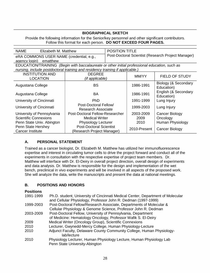
NAME Elizabeth M. Matthew POSITION TITLE
Post-Doctoral Scientist (Research Project Manager) eRA COMMONS USER NAME (credential, e.g., agency login) ematthew EDUCATION/TRAINING (Begin with baccalaureate or other initial professional education, such as nursing, include postdoctoral training and residency training if applicable.)
INSTITUTION AND LOCATION
DEGREE (if applicable)
MM/YY FIELD OF STUDY
Augustana College BS 1986-1991 Biology (& Secondary Education)
Augustana College BA 1986-1991 English (& Secondary Education)
University of Cincinnati PhD 1991-1999 Lung Injury
University of Cincinnati Post-Doctoral Fellow/ Research Associate
1999-2003 Lung Injury
University of Pennsylvania Post-Doctoral Fellow-Researcher 2003-2009 Cancer Biology Scientific Connexions Medical Writer 2009 Oncology Penn State Univ. Abington Physiology Lecturer 2010 Human Physiology Penn State Hershey Cancer Institute
Post-Doctoral Scientist (Research Project Manager)
2010-Present Cancer Biology
A. PERSONAL STATEMENT
Trained as a cancer biologist, Dr. Elizabeth M. Matthew has utilized her immunofluorescence expertise and interest in circulating tumor cells to drive the project forward and conduct all of the experiments in consultation with the respective expertise of project team members. Dr. Matthew will interface with Dr. El-Deiry in overall project direction, overall design of experiments and data analysis. Dr. Matthew is responsible for the design and implementation of the wet bench, preclinical in vivo experiments and will be involved in all aspects of the proposed work. She will analyze the data, write the manuscripts and present the data at national meetings.
B. POSITIONS AND HONORS
Positions 1991-1999 Ph.D. student, University of Cincinnati Medical Center, Department of Molecular
and Cellular Physiology, Professor John R. Dedman (1997-1999) 1999-2003 Post-Doctoral Fellow/Research Associate, Departments of Molecular &
Cellular Physiology & Genome Science, Professor John R. Dedman 2003-2009 Post-Doctoral Fellow, University of Pennsylvania, Department of Medicine: Hematology Oncology, Professor Wafik S. El-Deiry 2009 Medical Writer (Oncology Group), Scientific Connexions 2010 Lecturer, Gwynedd-Mercy College, Human Physiology-Lecture 2010 Adjunct Faculty, Delaware County Community College, Human Physiology-
lab/lecture 2010 Physiology Lecturer, Human Physiology Lecture, Human Physiology Lab Penn State University-Abington
29
2010-Present Post-Doctoral Scientist (Research Project Manager), Penn State University Cancer Institute, Professor Wafik S. El-Deiry Other Experience and Professional Memberships 1987-1990 Laboratory teaching assistant, Human Physiology, Biology Principles I 1991 Student teacher, High school Biology, High school English 1994-1995 Treasurer/Secretary, Physiology Graduate Student Association 2001-2002 Molecular Methods in Physiology Course Lecture: “Analysis of Gene Array Data” 2002-2008 Mentored undergraduate, medical and Ph.D. students on research projects 2004-Present Associate Member, American Cancer Society 2009-2010 Reviewer, Cell Death and Differentiation, Peer-reviewed manuscript Awards and Fellowships 1987-1988 Froiland Scholarship in Biology, Augustana College 1991-1998 Research Assistant in Physiology, University of Cincinnati 1999-2000 NIH Post-Doctoral Training Grant, Department of Molecular & Cellular Physiology 2003-2005 NIH Post-Doctoral Training Grant, Department of Urology 2014 GTC Bio Tumor Progression & Therapeutic Resistance, Boston, MA. Poster
presentation award: Immunophenotyping Circulating Tumor Cells in Patients with Unknown Primary.
C. SELECTED PUBLICATIONS
Cyclosporin A protects lung function from hyperoxic damage. E. Matthew, R. Pun, M. Simonich, H. Iwamoto and J. Dedman. Am. J.Physiol. 276 (Lung Cell. Mol. Physiol. 20) : L786-L795, 1999
A murine model of smoke inhalation. E. Matthew, G. Warden and J. Dedman. Am. J.Physiol. 280(Lung Cell. Mol. Physiol.) : L716-723, 2001
The protection of lungs from hyperoxic injury : Gene expression analysis of Cyclosporin A therapy. E. Matthew, L. Kutcher and J. Dedman Physiol. Genomics 14 : 129-138, 2003
CARPs are ubiquitin ligases that promote MDM2-independent p53 and phospho-p53ser20 degradation. Yang W, Rozan LM, McDonald ER 3rd, Navaraj A, Liu JJ, Matthew EM, Wang W, Dicker DT, El-Deiry WS. J Biol Chem. 282(5): 3273-81, 2007. Replication stress, defective S-phase checkpoint and increased cell death in Plk2 deficient human cancer cells. Matthew EM,, Yen TJ, Dicker D, Dorsey JF, Yang W, Navaraj A and El-Deiry WS. Cell Cycle 6(20):2571-2578 2007 Reduced cell death, invasive and angiogenic features conferred by BRCA1-deficiency in mammary epithelial cells transformed with H-Ras. Navaraj A, Finnberg N, Dicker DT, Yang W, Matthew EM, El-Deiry WS. Cancer Biol Ther: 8(24):2417-44, 2009 The p53 target Plk2 interacts with TSC proteins impacting mTOR signaling, tumor growth, and chemosensitivity under hypoxic conditions. Matthew EM, Hart LS, Astrinidis A, Navaraj A, Dolloff NG, Dicker DT, Henske EP, El-Deiry WS. Cell Cycle: 8 (24):1-8, 2009 Predicting therapy response in live tumor cells isolated with the flexible micro spring array device. Gallant JN*, Matthew EM*, Cheng H, Harouaka R, Lamparella NE, Kunkel M, Yang Z, Harvey HA,

30
Cream LV, Kumar SM, Robertson GP, Zheng S, Drabick JJ, Truica CI, El-Deiry WS. Cell Cycle 12: 2132-43, 2013. *these authors have contributed equally to this work.
D. RESEARCH SUPPORT
Ongoing Research Support None