Embed Size (px)
Citation preview
SERIES ‘‘RESPIRATORY MONITORING: REVISITING CLASSICALPHYSIOLOGICAL PRINCIPLES WITH NEW TOOLS’’Edited by M.I. Polkey, R. Farre and A.T. Dinh-XuanNumber 3 in this Series
Flow limitation and dynamic hyperinflation:
key concepts in modern respiratory
physiologyP.M.A. Calverley* and N.G. Koulouris#
ABSTRACT: Fashions in ideas, like clothes, come and go. From approximately 1950–1980,
physiological research was seen as the key discipline in understanding lung disease and was at
the cutting edge of pulmonary science. Subsequently, its importance has been down played amid
a widely accepted but unfounded assumption that we now have a perfect working understanding
of the physiological behaviour of the respiratory system in health and disease. Although it seems
improbable that completely new disciplines within respiratory physiology will emerge with
fundamentally different ways of describing the mechanical or gas exchanging function of the lung,
advances in computing and new observations in disease have highlighted previously
unsuspected physiological abnormalities that have changed the way we view lung disease and
the interface between disordered lung mechanics, symptomatology and disability.
This is especially true for the two related physiological concepts of expiratory flow limitation
and dynamic hyperinflation, which are now being taken from the physiological laboratory to the
bedside with dramatic effect. Each arises from well-established theoretical and practical
observations first made 40 yrs ago and now adapted to a range of settings, particularly in the
field of obstructive lung disease. This review focuses on how these conditions are defined and
assessed and what evidence there is that they might be important in lung disease.
KEYWORDS: Asthma, chronic obstructive pulmonary disease, exercise, flow limitation, hyper-
inflation, physiological concepts
TIDAL EXPIRATORY FLOW LIMITATION ANDITS MEASUREMENTConventional techniquesThe term expiratory flow limitation (EFL) is usedto indicate that maximal expiratory flow isachieved during tidal breathing and it is char-acteristic of intrathoracic airflow obstruction. Itshould be noted that some experts use the termchronic airflow limitation as a synonym forchronic obstructive pulmonary disease (COPD),to indicate the reduction in maximum expiratoryflow that occurs in this disease (and indeed inother pulmonary diseases); however, this term
does not imply that EFL actually occurs duringtidal breathing [1–3].
The presence of EFL during tidal breathingpromotes dynamic pulmonary hyperinflation(DH) and intrinsic positive end-expiratory pres-sure (PEEPi), with concomitant increase of workof breathing, functional impairment of inspira-tory muscle function and adverse effects onhaemodynamics [4]. This, together withflow-limiting dynamic airway compressionduring tidal breathing, may contribute to dys-pnoea [5].
AFFILIATIONS
*Clinical Science Centre, University
Hospital Aintree, Liverpool, UK.#Respiratory Function Laboratory,
Dept of Respiratory Medicine,
University of Athens Medical School,
‘‘Sotiria’’ Hospital, Athens, Greece.
CORRESPONDENCE
N.G. Koulouris
Respiratory Function Laboratory
Dept of Respiratory Medicine
University of Athens Medical School
‘‘Sotiria’’ Hospital
Athens
Greece
Fax: 30 2107223420
E-mail: [email protected]
Received:
September 30 2004
Accepted:
October 12 2004
European Respiratory Journal
Print ISSN 0903-1936
Online ISSN 1399-3003
Previous articles in this series: No. 1: Man WD-C, Moxham J, Polkey MI. Magnetic stimulation for the measurement of respiratory
and skeletal muscle function. Eur Respir J 2004; 24: 846–860. No. 2: Farre R, Montserrat JM, Navajas D. Noninvasive monitoring of
respiratory mechanics during sleep. Eur Respir J 2004; 24: 1052–1060.
186 VOLUME 25 NUMBER 1 EUROPEAN RESPIRATORY JOURNAL
Eur Respir J 2005; 25: 186–199
DOI: 10.1183/09031936.04.00113204
Copyright�ERS Journals Ltd 2005
According to a recently proposed attractive and provocativehypothesis [6], the transition from peripheral airway disease toovert COPD in smokers who are destined to develop COPD ischaracterised by three sequential stages, in which EFL plays acentral role: Stage I, the closing volume eventually exceeds thefunctional residual capacity; Stage II, EFL first develops; andStage III, DH progressively increases, leading to dyspnoea andexercise limitation. The presence of airway closure (Stage I)and EFL (Stage II) in the tidal volume range may promoteperipheral airway injury and accelerate the abnormalities oflung function [7–9]. This enhances inflammation due to smokeper se, leading to severe functional and structural abnormalitieswithin the lung. This vicious cycle cannot be reversed, possiblyapart from in Stage I.
Despite the severe consequences of EFL, the prevalence andclinical significance of this phenomenon have not beenadequately studied in COPD, asthma and patients with otherpulmonary and nonpulmonary disease.
By definition, finding of EFL requires the demonstration of anincrease in transpulmonary pressure with no increase inexpiratory flow. Therefore, direct assessment of EFL requiresdetermination of iso-volume relationships between flow andtranspulmonary pressure, an approach that is the goldstandard. However, this method is technically complex, timeconsuming and invasive, because it requires the passage of anoesophageal balloon [10, 11].
Until recently, the conventional method used to detect EFLduring tidal breathing was one proposed by HYATT [3] in 1961.It consists of correctly superimposing a flow–volume loop of atidal breath within a maximum flow–volume curve. Thisanalysis and the ‘‘concept of EFL’’ have been the kernel forunderstanding respiratory dynamics. EFL is not present whenthe patient breathes below the maximal expiratory flow–volume (MEFV) curve. According to this technique, normalsubjects do not reach EFL, even at maximum exercise [1, 2]. Incontrast, EFL is present when a patient seeks to breathe tidallyalong or higher than the MEFV curve. It has long beensuggested that patients with severe COPD may exhibit EFLeven at rest, as reflected by the fact that they breathe tidallyalong or above their MEFV curve [1–6]. However, theconventional method to detect ELF, based on the comparisonMEFV and tidal expiratory flow–volume curves, has severalmethodological deficiencies. These include the following. 1)Thoracic gas compression artefacts. To minimise such errors,volume should be measured with a body plethysmograph,instead of using, as is common practice, a pneumotachographor a spirometer [12]. The corollary of this is that, in practice,EFL may be assessed only in seated subjects at rest. 2) Incorrectalignment of tidal expiratory flow–volume and MEFV curves.Such alignment is usually made considering the total lungcapacity (TLC) as a fixed reference point. This assumption maynot always be valid [13, 14]. 3) Effect of previous volume andtime history. Since the previous volume and time history of aspontaneous tidal breath is necessarily different from that of aforced vital capacity (FVC) manoeuvre, it is axiomatic thatcomparison of tidal expiratory flow–volume with MEFVcurves is problematic. In fact, there is not a single MEFVcurve, but rather a family of different curves, which dependon the time-course of the inspiration preceding the FVC
manoeuvre [15–17]. Therefore, comparison of tidal expiratoryflow–volume and MEFV curves is incorrect. 4) Respiratorymechanics and time constant inequalities are different duringthe tidal and maximal expiratory efforts, also makingcomparisons of the two flow–volume curves problematic [18–20]. 5) Exercise may result in bronchodilation or bronchocon-striction and other changes of lung mechanics, which may alsoaffect correct comparisons of the two flow–volume curves [21].6) Patient cooperation. Another important limitation of theconventional method is that it requires patient cooperation.This is not always feasible [13, 14].
From the above considerations, it appears that the detection ofEFL based on comparison of tidal expiratory flow–volumewith MEFV curves is not valid, even when a body-box is used.In fact, this has been clearly demonstrated in several studies[22–25]. As a result, the use of the conventional method is nolonger recommended.
The negative expiratory pressure techniqueRecently, in order to overcome the technical and conceptualdifficulties discussed above, the negative expiratory pressure(NEP) method has been introduced [22–25] (fig. 1). The NEPtechnique has been applied and validated in mechanicallyventilated intensive care unit (ICU) patients by concomitantdetermination of iso-volume flow–pressure relationships [23,26] (fig. 2). This method does not require FVC manoeuvres,collaboration on the part of the patient or use of a bodyplethysmograph, and can be used during spontaneouslybreathing subjects in any body position [27], during exercise[24, 28, 29] and in an ICU setting [7, 8, 23, 30–32]. With thismethod, the volume and time history of the control and testexpiration are the same.
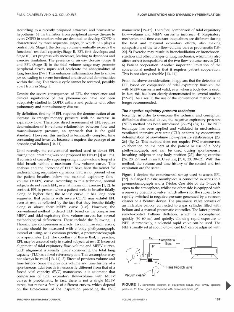
Figure 1 depicts the experimental set-up used to assess EFL[22]. A flanged plastic mouthpiece is connected in series to apneumotachograph and a T-tube. One side of the T-tube isopen to the atmosphere, whilst the other side is equipped witha one-way pneumatic valve, which allows for the subject to berapidly switched to negative pressure generated by a vacuumcleaner or a Venturi device. The pneumatic valve consists ofan inflatable balloon connected to a gas cylinder filled withhelium and a manual pneumatic controller. The latter permitsremote-control balloon deflation, which is accomplishedquickly (30–60 ms) and quietly, allowing rapid exposure toNEP. Alternatively, a solenoid rapid valve can be used. TheNEP (usually set at about -3 to -5 cmH2O) can be adjusted with
������������
���� ������������
����������������
���
��
FIGURE 1. Schematic diagram of equipment setup. Pao: airway opening
pressure; V9: flow. Figure reproduced with permission from [22].
P.M.A. CALVERLEY AND N.G. KOULOURIS FLOW LIMITATION AND DYNAMIC HYPERINFLATION
cEUROPEAN RESPIRATORY JOURNAL VOLUME 25 NUMBER 1 187
a potentiometer on the vacuum cleaner or by controlling theVentouri device. Flow is measured with the heated pneumo-tachograph and pressure at the airway opening is simulta-neously measured through a side port on the mouthpiece.Volume is obtained by digital integration of the flow signal[22–25].
While performing the test, the subjects should be watchedclosely for leaks at the mouthpiece. By monitoring the volumerecord over time on the chart recorder, the absence of leaks andelectrical drift can be ensured by the fact that, after the NEPtests, the end-expiratory lung volume (EELV) returns to thepre-NEP level. Only those tests in which there is no leak arevalid [33].
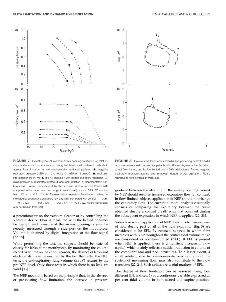
The NEP method is based on the principle that, in the absenceof pre-existing flow limitation, the increase in pressure
gradient between the alveoli and the airway opening causedby NEP should result in increased expiratory flow. By contrast,in flow-limited subjects, application of NEP should not changethe expiratory flow. The current authors’ analysis essentiallyconsists of comparing the expiratory flow–volume curveobtained during a control breath with that obtained duringthe subsequent expiration in which NEP is applied [22, 23].
Subjects in whom application of NEP does not elicit an increaseof flow during part or all of the tidal expiration (fig. 3) areconsidered to be EFL. By contrast, subjects in whom flowincreases with NEP throughout the control tidal volume rangeare considered as nonflow-limited (NFL). If EFL is presentwhen NEP is applied, there is a transient increase of flow(spike), which mainly reflects a sudden reduction in volume ofthe compliant oral and neck structures. To a lesser extent, asmall artefact, due to common-mode rejection ratio of thesystem of measuring flow, may also contribute to the flowtransients [22–24]. Such spikes are useful markers of EFL.
The degree of flow limitation can be assessed using fourdifferent EFL indices: 1) as a continuous variable expressed asper cent tidal volume in both seated and supine positions
�
�
�
���
���
���
���
���
���
���
� �����!����"
�#$�%�
�
�
�
�
�
�
�
�
�
��
�
�
�
�
�
�
����
�&
� �����!����"
�#$�%�
'&
�
���
���
��(
���
��)
�)��)�%)%��%�)��������*
�
�
�
�
�
�
�
�
�
�
�
�
�
�
�
�
�
�
�
�
��
�
�
�
�
�����
FIGURE 2. Expiratory iso-volume flow–airway opening pressure (Pao) relation-
ships under control conditions and during test breaths with different methods to
assess flow limitation in two mechanically ventilated patients. $: negative
expiratory pressure (NEP) of -10 cmH2O; #: NEP of -5 cmH2O; &: expiration
into atmosphere (ATM); m and e: expiration with added expiratory resistance; n:
static pressure of respiratory system during lung deflation. a) Representative non-
flow-limited patient, as indicated by the increase in flow with NEP and ATM
compared with control. –––: no change in volume (DV); – - - –: 0.2 L DV; – – – –:
0.4 L DV; ??????: 0.6 L DV. b) Representative expiratory flow-limited patient, as
indicated by unchanged expiratory flow and ATM compared with control. ––: 0 DV;
– –: 0.1 L DV; – - - –: 0.2 L DV; - - - -: 0.3 L DV; ??????: 0.4 L DV. Figure reproduced
with permission from [23].
�
�
�
%�
%�
+��"
�#$�%�
�&
�
�
�
%�
%�
+��"
�#$�%�
'&
%� � ��������#
FIGURE 3. Flow–volume loops of test breaths and preceding control breaths
of two representative bronchiectatic patients with different degrees of flow limitation:
a) not flow limited; and b) flow limited over ,50% tidal volume. Arrows: negative
expiratory pressure applied and removed; dotted arrow: expiration. Figure
reproduced with permission from [54].
FLOW LIMITATION AND DYNAMIC HYPERINFLATION P.M.A. CALVERLEY AND N.G. KOULOURIS
188 VOLUME 25 NUMBER 1 EUROPEAN RESPIRATORY JOURNAL
(fig. 3) [22]; 3) as a discrete variable in the form of a twoclassification, i.e. NFL in the seated position, EFL in the seatedposition 3) as a discrete variable in the form of three categoriesof classification, i.e. NFL both seated and supine, EFL supinebut not seated, EFL both seated and supine [22]; and 4) as adiscrete variable in the form of the five-categories classification(five-point EFL score) [25].
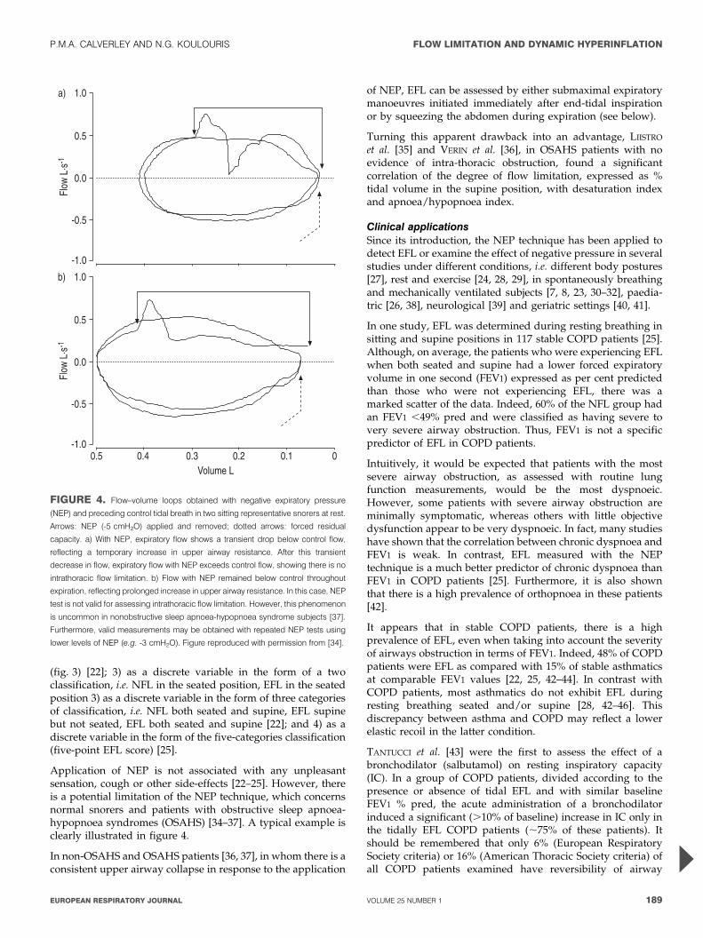
Application of NEP is not associated with any unpleasantsensation, cough or other side-effects [22–25]. However, thereis a potential limitation of the NEP technique, which concernsnormal snorers and patients with obstructive sleep apnoea-hypopnoea syndromes (OSAHS) [34–37]. A typical example isclearly illustrated in figure 4.
In non-OSAHS and OSAHS patients [36, 37], in whom there is aconsistent upper airway collapse in response to the application
of NEP, EFL can be assessed by either submaximal expiratorymanoeuvres initiated immediately after end-tidal inspirationor by squeezing the abdomen during expiration (see below).
Turning this apparent drawback into an advantage, LIISTRO
et al. [35] and VERIN et al. [36], in OSAHS patients with noevidence of intra-thoracic obstruction, found a significantcorrelation of the degree of flow limitation, expressed as %tidal volume in the supine position, with desaturation indexand apnoea/hypopnoea index.
Clinical applicationsSince its introduction, the NEP technique has been applied todetect EFL or examine the effect of negative pressure in severalstudies under different conditions, i.e. different body postures[27], rest and exercise [24, 28, 29], in spontaneously breathingand mechanically ventilated subjects [7, 8, 23, 30–32], paedia-tric [26, 38], neurological [39] and geriatric settings [40, 41].
In one study, EFL was determined during resting breathing insitting and supine positions in 117 stable COPD patients [25].Although, on average, the patients who were experiencing EFLwhen both seated and supine had a lower forced expiratoryvolume in one second (FEV1) expressed as per cent predictedthan those who were not experiencing EFL, there was amarked scatter of the data. Indeed, 60% of the NFL group hadan FEV1 ,49% pred and were classified as having severe tovery severe airway obstruction. Thus, FEV1 is not a specificpredictor of EFL in COPD patients.
Intuitively, it would be expected that patients with the mostsevere airway obstruction, as assessed with routine lungfunction measurements, would be the most dyspnoeic.However, some patients with severe airway obstruction areminimally symptomatic, whereas others with little objectivedysfunction appear to be very dyspnoeic. In fact, many studieshave shown that the correlation between chronic dyspnoea andFEV1 is weak. In contrast, EFL measured with the NEPtechnique is a much better predictor of chronic dyspnoea thanFEV1 in COPD patients [25]. Furthermore, it is also shownthat there is a high prevalence of orthopnoea in these patients[42].
It appears that in stable COPD patients, there is a highprevalence of EFL, even when taking into account the severityof airways obstruction in terms of FEV1. Indeed, 48% of COPDpatients were EFL as compared with 15% of stable asthmaticsat comparable FEV1 values [22, 25, 42–44]. In contrast withCOPD patients, most asthmatics do not exhibit EFL duringresting breathing seated and/or supine [28, 42–46]. Thisdiscrepancy between asthma and COPD may reflect a lowerelastic recoil in the latter condition.
TANTUCCI et al. [43] were the first to assess the effect of abronchodilator (salbutamol) on resting inspiratory capacity(IC). In a group of COPD patients, divided according to thepresence or absence of tidal EFL and with similar baselineFEV1 % pred, the acute administration of a bronchodilatorinduced a significant (.10% of baseline) increase in IC only inthe tidally EFL COPD patients (,75% of these patients). Itshould be remembered that only 6% (European RespiratorySociety criteria) or 16% (American Thoracic Society criteria) ofall COPD patients examined have reversibility of airway
���
��)
���
%��)
%���
+��"
�#$�%�
�&
���
��)
���
%��)
%���
+��"
�#$�%�
'&
��) ��� ��( ��� ��� ��������#
FIGURE 4. Flow–volume loops obtained with negative expiratory pressure
(NEP) and preceding control tidal breath in two sitting representative snorers at rest.
Arrows: NEP (-5 cmH2O) applied and removed; dotted arrows: forced residual
capacity. a) With NEP, expiratory flow shows a transient drop below control flow,
reflecting a temporary increase in upper airway resistance. After this transient
decrease in flow, expiratory flow with NEP exceeds control flow, showing there is no
intrathoracic flow limitation. b) Flow with NEP remained below control throughout
expiration, reflecting prolonged increase in upper airway resistance. In this case, NEP
test is not valid for assessing intrathoracic flow limitation. However, this phenomenon
is uncommon in nonobstructive sleep apnoea-hypopnoea syndrome subjects [37].
Furthermore, valid measurements may be obtained with repeated NEP tests using
lower levels of NEP (e.g. -3 cmH2O). Figure reproduced with permission from [34].
P.M.A. CALVERLEY AND N.G. KOULOURIS FLOW LIMITATION AND DYNAMIC HYPERINFLATION
cEUROPEAN RESPIRATORY JOURNAL VOLUME 25 NUMBER 1 189
obstruction after bronchodilator. Moreover, a significant post-bronchodilator decrease in EELV (or dynamic functionalresidual capacity) was observed only in the COPD subgroupwith tidal EFL.
Subsequently, it has been shown that the increase in IC afteranticholinergic and salbutamol therapy best reflects theimprovement in exercise tolerance [47]. Both a significantreduction in exertional dyspnoea (DBorg,exercise) and a closerelationship between DBorg,exercise (decrease) and DIC at rest(% pre) (increase) were found after salbutamol, regardless ofthe change in FEV1, in the group of COPD patients with tidalEFL at rest. In contrast, no change in DIC at rest (% pre) and inDBorg,exercise was observed in the group of COPD patientswithout tidal EFL at rest. Therefore, in COPD patients, thereduction in breathlessness during mild-to-moderate exercisefollowing the administration of a bronchodilator is heralded byan increase in IC at rest.
The improvement of IC after bronchodilator administration,which is mainly limited to patients with EFL at rest and whoexhibit a reduction of baseline IC, entails reduction indyspnoea both at rest and during light exercise [47]. Thus, inobstructive lung disease, the benefit of bronchodilator therapyshould be assessed not only in terms of changes in FEV1, but,more importantly, also in terms of increases in IC. In thiscontext it should be noted that, since performance of ICprecedes the FVC manoeuvre, FEV1 and IC are, in general,recorded together during bronchodilator testing.
Although bronchodilator testing has traditionally focused onchanges in FEV1, the scrutiny of changes in IC should bemandatory, because it provides more useful informationthan FEV1 pertaining to dyspnoea and exercise tolerance.The detection of EFL alone with the NEP technique is notan appropriate measurement of acute bronchodilator respon-siveness [48]. However, the fact that after bronchodilator
administration there is a significant reduction of DH only inpatients with EFL at rest in the sitting position further supportsthe usefulness of stratifying COPD patients in subgroups withand without EFL in order to predict an improvement in DH.Thus, measurement of IC and detection of EFL are compli-mentary ways for assessing bronchodilator responsiveness inCOPD patients [43, 47].
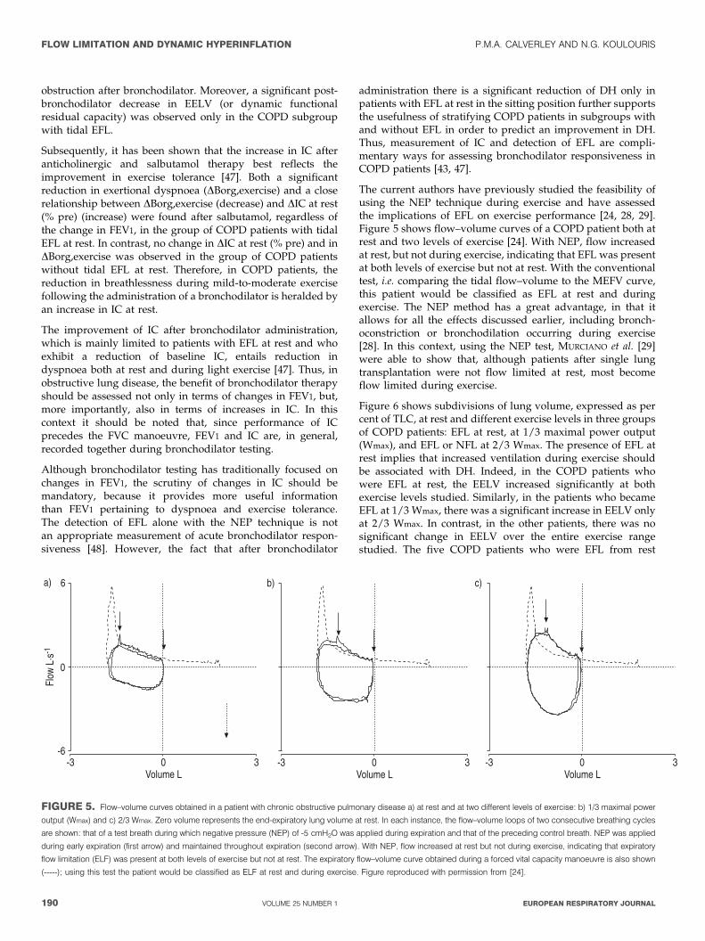
The current authors have previously studied the feasibility ofusing the NEP technique during exercise and have assessedthe implications of EFL on exercise performance [24, 28, 29].Figure 5 shows flow–volume curves of a COPD patient both atrest and two levels of exercise [24]. With NEP, flow increasedat rest, but not during exercise, indicating that EFL was presentat both levels of exercise but not at rest. With the conventionaltest, i.e. comparing the tidal flow–volume to the MEFV curve,this patient would be classified as EFL at rest and duringexercise. The NEP method has a great advantage, in that itallows for all the effects discussed earlier, including bronch-oconstriction or bronchodilation occurring during exercise[28]. In this context, using the NEP test, MURCIANO et al. [29]were able to show that, although patients after single lungtransplantation were not flow limited at rest, most becomeflow limited during exercise.
Figure 6 shows subdivisions of lung volume, expressed as percent of TLC, at rest and different exercise levels in three groupsof COPD patients: EFL at rest, at 1/3 maximal power output(Wmax), and EFL or NFL at 2/3 Wmax. The presence of EFL atrest implies that increased ventilation during exercise shouldbe associated with DH. Indeed, in the COPD patients whowere EFL at rest, the EELV increased significantly at bothexercise levels studied. Similarly, in the patients who becameEFL at 1/3 Wmax, there was a significant increase in EELV onlyat 2/3 Wmax. In contrast, in the other patients, there was nosignificant change in EELV over the entire exercise rangestudied. The five COPD patients who were EFL from rest
�
�
%�
+��"
�#$�%�
%( � (�������#
�& '&
%( � (�������#
�&
%( � (�������#
FIGURE 5. Flow–volume curves obtained in a patient with chronic obstructive pulmonary disease a) at rest and at two different levels of exercise: b) 1/3 maximal power
output (Wmax) and c) 2/3 Wmax. Zero volume represents the end-expiratory lung volume at rest. In each instance, the flow–volume loops of two consecutive breathing cycles
are shown: that of a test breath during which negative pressure (NEP) of -5 cmH2O was applied during expiration and that of the preceding control breath. NEP was applied
during early expiration (first arrow) and maintained throughout expiration (second arrow). With NEP, flow increased at rest but not during exercise, indicating that expiratory
flow limitation (ELF) was present at both levels of exercise but not at rest. The expiratory flow–volume curve obtained during a forced vital capacity manoeuvre is also shown
(-----); using this test the patient would be classified as ELF at rest and during exercise. Figure reproduced with permission from [24].
FLOW LIMITATION AND DYNAMIC HYPERINFLATION P.M.A. CALVERLEY AND N.G. KOULOURIS
190 VOLUME 25 NUMBER 1 EUROPEAN RESPIRATORY JOURNAL
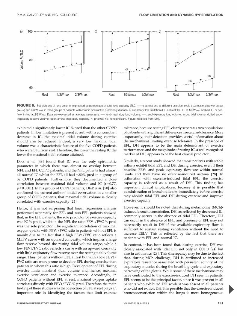
exhibited a significantly lower IC % pred than the other COPDpatients. If flow limitation is present at rest, with a concomitantdecrease in IC, the maximal tidal volume during exerciseshould also be reduced. Indeed, a very low maximal tidalvolume was a characteristic feature of the five COPD patientswho were EFL from rest. Therefore, the lower the resting IC thelower the maximal tidal volume attained.
DIAZ et al. [49] found that IC was the only spirometricparameter in which there was almost no overlap betweenNFL and EFL COPD patients, and the NFL patients had almostall normal IC whilst the EFL all had ,80% pred in a group of52 COPD patients. Furthermore, they documented a closecorrelation between maximal tidal volume and IC (r50.77,p,0.0001). In his group of COPD patients, DIAZ et al. [50] alsoconfirmed the current authors’ initial observation in a smallergroup of COPD patients, that maximal tidal volume is closelycorrelated with exercise capacity [24].
Hence, it was not surprising that linear regression analysisperformed separately for EFL and non-EFL patients showedthat, in the EFL patients, the sole predictor of exercise capacitywas IC % pred, whilst in the NFL the ratio FEV1/FVC % predwas the sole predictor. The significant correlation of maximaloxygen uptake with FEV1/FVC ratio in patients without EFL ismainly due to the fact that a high FEV1/FVC ratio reflects aMEFV curve with an upward convexity, which implies a largeflow reserve beyond the resting tidal volume range, while alow FEV1/FVC ratio reflects a curve with an upward concavitywith little expiratory flow reserve over the resting tidal volumerange. Thus, patients without EFL at rest but with a low FEV1/FVC ratio are more prone to develop EFL during exercise thanpatients in whom this ratio is high. Development of EFL duringexercise limits maximal tidal volume and, hence, maximalexercise ventilation and exercise tolerance. Accordingly, inCOPD patients without EFL at rest, maximal oxygen uptakecorrelates directly with FEV1/FVC % pred. Therefore, the mainfinding of these studies was that detection of EFL at rest plays animportant role in identifying the factors that limit exercise
tolerance, because resting EFL clearly separates two populationsof patients with significant differences in exercise tolerance. Moreimportantly, their detection provides useful information aboutthe mechanisms limiting exercise tolerance. In the presence ofEFL, DH appears to be the main determinant of exerciseperformance, and the magnitude of resting IC, a well recognisedmarker of DH, appears to be the best clinical predictor.
Similarly, a recent study showed that most patients with stableasthma exhibit tidal EFL and DH during exercise, even if theirbaseline FEV1 and peak expiratory flow are within normallimits and they have no exercise-induced asthma [28]. Inasthmatics with exercise-induced tidal EFL, the exercisecapacity is reduced as a result of DH. This finding hasimportant clinical implications, because it is possible thatadministration of bronchodilators immediately before exercisemay abolish tidal EFL and DH during exercise and improveexercise capacity.
However, it should be noted that during metacholine (MCh)-induced bronchoconstriction, DH, as reflected by decreased IC,commonly occurs in the absence of tidal EFL. Therefore, DHcan occur in the absence of EFL, and presence of EFL may notnecessarily result in DH if the available expiratory flow issufficient to sustain resting ventilation without the need toincrease EELV. This is reflected by the fact that there arepatients with EFL and normal IC.
In contrast, it has been found that, during exercise, DH wasclosely associated with tidal EFL not only in COPD [24] butalso in asthmatics [28]. This discrepancy may be due to the factthat, during MCh challenge, DH is attributed to increasedexpiratory resistance associated with persistent activity of theinspiratory muscles during the breathing cycle and expiratorynarrowing of the glottis. While some of these mechanisms mayhave contributed to the exercise-induced DH seen in patients,EFL seems to be the principal factor, since it was present in allpatients who exhibited DH while it was absent in all patientswho did not exhibit DH. It is possible that the exercise-inducedbronchoconstriction within the lungs is more homogeneous
���
�,(-��
��
��
�������.�/#0
�&
��
�
�
�
�
��
�,(-�� ��� �,(-��
'&
���
�
�
�
�
�,(-�� ���
12
�,(-��
�&
�
��
�
�
�
�,(-�� ���
12
12
FIGURE 6. Subdivisions of lung volume, expressed as percentage of total lung capacity (TLC; ??????), at rest and at different exercise levels (1/3 maximal power output
(Wmax) and 2/3 Wmax), in three groups of patients with chronic obstructive pulmonary disease: a) expiratory flow limitation (EFL) at rest; b) EFL at 1/3 Wmax; and c) EFL or non-
flow limited at 2/3 Wmax. Data are expressed as average values¡SE. –––: end-inspiratory lung volume; -----: end-expiratory lung volume; arrow: tidal volume; dotted arrow:
inspiratory reserve volume; open arrow: inspiratory capacity. *: p,0.05; NS: nonsignificant. Figure modified from [24].
P.M.A. CALVERLEY AND N.G. KOULOURIS FLOW LIMITATION AND DYNAMIC HYPERINFLATION
cEUROPEAN RESPIRATORY JOURNAL VOLUME 25 NUMBER 1 191
than MCh-induced bronchoconstriction. With nonhomoge-neous bronchoconstriction, some regions may develop EFLwith concurrent DH, while others empty normally; hence,overall EFL (as measured with NEP) may be absent. In suchcases, IC may be decreased in the absence of overall EFL. Incontrast, with homogeneous bronchoconstriction, overall EFLand DH should reflect the homogeneously distributedmechanical impairment within the lungs [28].
The NEP technique has also been used to detect flow limitationin mechanically ventilated patients [7, 8, 23, 30–32]. In fact, atfirst, the NEP method was applied and validated duringmechanical ventilation in different body postures [23]. It wasfound that almost all COPD patients who require mechanicalventilation are flow limited over the entire range of tidalexpiration and that the supine posture promotes flow limita-tion. It should be noted that flow limitation is reversed inlateral decubitus and on hands and knees positions inspontaneously breathing COPD patients [27]. Other studieshave shown that most patients with acute respiratory failure ofpulmonary origin present tidal EFL, whilst ones with acuterespiratory failure of extra-pulmonary origin did not [30]. Thesame authors found that most acute respiratory distresssyndrome (ARDS) patients exhibit EFL, probably associatedwith small airways closure and a concomitant PEEPi [7]. Thepresence of EFL, which implies concurrent cyclic dynamiccompression and re-expansion of the airways, increases therisk of low lung volume injury. In all ARDS patients, duringapplication of 10 cmH2O of PEEP, EFL was abolished and thearterial oxygenation was improved satisfactorily because ofalveolar recruitment in NFL patients and reduced intrapul-monary PEEPi inequality in EFL patients [8].
Tidal EFL and PEEPi are also common in supine morbidlyobese sedated-paralysed subjects after abdominal surgery [32].This implies that the therapeutic administration of externalPEEP to such patients must be monitored with concurrentassessment of EFL and PEEPi. The presence of EFL andperipheral airway closure implies a possible risk of lowvolume injury. Accordingly, it seems prudent to apply PEEPalso in order to avoid peripheral airway closure and EFL.
Therefore, the assessment of EFL in mechanically ventilatedpatients with the NEP technique is a potentially useful bedsideapproach to provide information concerning respiratorymechanics.
In the past, there was no online method available to assesswhether the flows during the FVC manoeuvres were maximalor not. Recently, however, a simple method to assess FVCperformance has been developed [26, 51]. It is based on avariation of the NEP technique, i.e. application of short NEPpulses of -10 cmH2O during the FVC manoeuvre. If theexpiratory flow increases during the application of the NEPpulse, the expiratory flow is sub-maximal. In contrast, if flowdoes not increase with the negative pressure, EFL has beenreached. Thus, with this method, it is possible to determinewhether the maximal flows are low as a result of insufficientrespiratory effort (e.g. weak respiratory muscles, lack ofcoordination, malingering) or the presence of a lung disorder.
In conclusion, the NEP technique has been used clinically instudies with the following: 1) COPD (during mechanical
ventilation and exercise, correlation with dyspnoea, orthop-noea, and other lung function indexes, before and afterbronchodilatation, various postures) [22–25, 27, 33, 42, 43, 49,50]; 2) asthma (stable asthma, during MCh bronchocostriction,and during exercise) [28, 44–46]; 3) cystic fibrosis [52, 53] andbronchiectasis [54]; 4) restrictive lung disease [33, 37]; 5)obesity [32, 55, 56]; 6) mechanically ventilated with acuterespiratory failure and ARDS [7, 8, 23, 30–32]; 7) left heartfailure [57]; 8) after single lung transplantation [29, 58]; 9)euthyroid goitre [59]; and 10) assessment of bronchialhyperreactivity [60]. It appears that the use of the NEPtechnique during tidal flow–volume analysis studies has ledto the realisation of the important role of EFL in exertionaldyspnoea and ventilatory impairment for a surprisingly widerange of clinical circumstances [61]. Therefore, the NEPtechnique should be regarded as a new useful research andclinical lung function tool.
In conclusion: 1) application of the NEP technique provides asimple, rapid, noninvasive and reliable test to detect tidal EFL[61–63]; 2) it does not require a body-box or any cooperation onthe part of the patient; 3) it can be applied in any body position,during mechanical ventilation and during exercise; 4) it mayprovide new insights in the physiology and pathophysiologyof several diseases and the symptom of dyspnoea.
Alternative approachesAlthough the NEP technique is the most widely usednoninvasive test of EFL, it is not the only one available.Workers in Brussels, Belgium, have shown that manualcompression of the abdomen coinciding with the onset ofexpiration can be used as a simple way of detecting flowlimitation at rest and during exercise [64, 65]. With one handplaced on the lower back of the patient and other applied withthe palm at the level of the umbilicus, perpendicular to the axisbetween the xiphoid process and the pubis, the operator firstdetects a respiratory rhythm by gentle palpation and then,after warning, the subject applies a forceful pressure at theonset of expiration. As in the NEP technique, the resultingexpiratory flow–volume loop recorded at the mouth is super-imposed on the preceding tidal breath. Failure to increaseexpiratory flow indicates EFL. This technique produces cleardifferences between normal subjects and patients with COPD.The presence of EFL detected during exercise in COPD patientswas associated with increases in the EELV. Interestingly, notall subjects with COPD exhibited EFL when lung volumechanged, a finding which requires confirmation in otherseries. The method is appealingly simple, overcomesproblems with the preceding volume history of the testbreath and is not influenced by the upper airway compli-ance. Despite initial concerns about the possibility that gascompression in the alveoli would produce false-positiveresults, this does not seem to be a practical problem.However, it can be extremely difficult to determine whetherEFL is occurring for the whole or part of the precedingbreath, unless the timing of the technique is very precise.Like the NEP approach, breath-to-breath variation in EELVcan produce contradictory results, as the method assumesthat EELV is always constant. Thus far, this technique hasnot been widely applied despite its relative simplicity.
FLOW LIMITATION AND DYNAMIC HYPERINFLATION P.M.A. CALVERLEY AND N.G. KOULOURIS
192 VOLUME 25 NUMBER 1 EUROPEAN RESPIRATORY JOURNAL
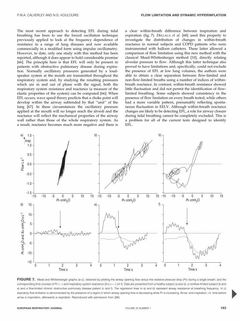
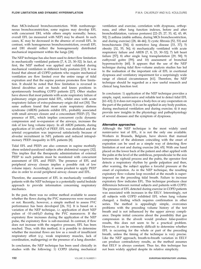
The most recent approach to detecting EFL during tidalbreathing has been to use the forced oscillation techniquepreviously applied to look at the frequency dependence ofresistance in a range of lung diseases and now availablecommercially in a modified form using impulse oscillometry.However, to date, only one study with this method has beenreported, although it does appear to hold considerable promise[66]. The principle here is that EFL will only be present inpatients with obstructive pulmonary disease during expira-tion. Normally oscillatory pressures generated by a loud-speaker system at the mouth are transmitted throughout therespiratory system and, by studying the resulting pressureswhich are in and out of phase with the signal, both therespiratory system resistance and reactance (a measure of theelastic properties of the system) can be computed [66]. WhenEFL occurs, wave speed theory predicts that a choke point willdevelop within the airway subtended by that ‘‘unit’’ of thelung [67]. In these circumstances the oscillatory pressureapplied at the mouth will no longer reach the alveoli and thereactance will reflect the mechanical properties of the airwaywall rather than those of the whole respiratory system. Asa result, reactance becomes much more negative and there is
a clear within-breath difference between inspiration andexpiration (fig. 7). DELLACA et al. [68] used this property toinvestigate the distribution of changes in within-breathreactance in normal subjects and COPD patients who wereinstrumented with balloon catheters. These latter allowed acomparison of flow limitation using this new method with theclassical Mead-Whittenberger method [10], directly relatingalveolar pressure to flow. Although this latter technique alsoproved to have limitations and, specifically, could not excludethe presence of EFL at low lung volumes, the authors wereable to obtain a clear separation between flow-limited andnon-flow-limited breaths using a number of indices of within-breath reactance. In contrast, within-breath resistance showedlittle fluctuation and did not permit the identification of flow-limited breathing. Some subjects showed consistency in thepresence of flow limitation on every breath tested, while othershad a more variable pattern, presumably reflecting sponta-neous fluctuation in EELV. Although within-breath reactancechanges are likely to be detecting EFL, a role for airway closureduring tidal breathing cannot be completely excluded. This isa problem for all of the current tests designed to identifyEFL.
��)
���
��)
���
%��)
%���
%��)
+��"
�#$�%�
�&
3��
�)��)�%)%��%�)�������*
'&
�)��)�%)%��%�)�������*
'&
�)��)�%)%��%�)�������*
����
�� �
*���
������� �
*$�$#
%�
�&
/������ � � ( �
%�)
%��
%)
�
)
��
�) �&
/������ � � ( �
�&
/������ � � ( �
FIGURE 7. Mead and Whittenberger graphs (a–c), obtained by plotting the airway opening flow versus the resistive pressure drop (Pfr) during a single breath, and the
corresponding time courses of Pfr (––) and respiratory system reactance (Xrs) (-----) (d–f). Data are presented from a healthy subject (a and d), a nonflow-limited subject (b and
e) and a flow-limited chronic obstructive pulmonary disease patient (c and f). The regression lines in a) and b) represent airway resistance at breathing frequency. In c)
expiratory flow limitation is demonstrated by the presence of a region in which airway opening flow is decreasing while Pfr is increasing. Arrow: end inspiration, i.e. time before
arrow is inspiration, afterwards is expiration. Reproduced with permission from [66].
P.M.A. CALVERLEY AND N.G. KOULOURIS FLOW LIMITATION AND DYNAMIC HYPERINFLATION
cEUROPEAN RESPIRATORY JOURNAL VOLUME 25 NUMBER 1 193
Like the other methods, this technique is independent of theprevious volume history of the breath tested; however, unlikethem it can give breath-by-breath data continuously andprovide an aggregate estimate of the probability of flowresistance being present in an individual. It can be used easilyduring exercise and, perhaps most importantly of all, can beautomated, which offers more widespread application for thesimple detection of expiratory flow resistance in the intensivecare unit and routine physiology laboratory.
Clearly, comparison between these different methods will beneeded before the best combination of testing methods can bedetermined. All of them represent a substantial advance ontraditional approaches, which compared tidal and maximalflow–volume loops or even the more robust but time-consuming method of determining partial expiratory flow–volume loops. By freeing both the doctor and the patient fromthe confines of the body plethysmograph, a new era has beenopened up in the understanding of the important principles offlow limitation in a wide variety of settings.
DYNAMIC HYPERINFLATIONAlthough many people are now familiar with the term DH,there is, as yet, no rigorous definition of exactly what this termreally means. The observations which led to the study of DHin recent times began when it was noted that patients withairflow obstruction who were being mechanically ventilatedwould continue to exhale for a longer period of time than thatdetermined by the ventilator settings [4]. Initially, attentionfocussed on the deleterious effects of this on cardiac outputand, by analogy with adding PEEP, this phenomenalwas termed PEEPi. A long and sometimes heated argumentfollowed about the optimal way in which this could bemeasured in patients whose abdominal muscles were alsoactive during expiration [69, 70]. Calculations of the increasedwork of breathing due to this phenomena indicated that therewas a substantial elastic burden being placed on therespiratory muscles [71] and this was also present whenPEEPi was observed in spontaneously breathing COPDpatients. Subsequently, the degree of PEEPi was found torelate to the severity of resting hypercapnia [72].
More recently, the focus of attention has shifted to the increasein EELV that accompanies this phenomenon in spontaneouslybreathing patients with obstructive lung disease. This wasnoted to be a common finding in patients with COPD duringexercise [73, 74] and one that related to the intensity of theirbreathlessness during exercise [75]. These studies were madepossible by the development of reliable methods of measuringIC during exercise [76], when TLC appears to be constant [77].In these circumstances, any change in IC should reflect achange in EELV. Usually, EELV falls at the onset of exercise toallow the respiratory system to remain on the steeper portionof the pressure–volume relationship and avoid the flatterupper part of this relationship, where any further increase inpressure no longer generates volume change [78]. This doesnot occur in COPD patients during exercise (fig. 6) with aseries of deleterious consequences. These include mechanicallimitation of the ability to increase tidal volume and also theneed for the respiratory system to operate at a higher lungvolume, both of which require a greater percentage of theinspiratory reserve capacity of the respiratory muscles to
sustain. The inspiratory muscles can no longer develop force aseffectively as at lower lung volumes because of shortening oftheir initial operating length and they are more likely todevelop inspiratory muscle fatigue or approach that state [79],which itself increases volume-related increases in the sensationof breathlessness [80]. As metabolic drive increases withexercise and tidal volume is constrained, the only strategyavailable is to increase respiratory frequency, which, unfortu-nately, further decreases expiratory time and reduces thepatient’s ability to achieve the elastic equilibrium volume.Hence, a vicious cycle is set up, which eventually leads toexercise stopping prematurely, primarily because of exertionalbreathlessness [81].
Changes in self-reported breathlessness in this setting relateclosely to changes in IC and the relationship between the two islikely to be causal [82]. Certainly, bronchodilator drugs whichreduce resting IC also delay the time to peak breathlessness[83, 84]. This is achieved primarily by their effect on resting ICrather than a change in the slope of the IC–breathlessnessrelationship, which is now recognised to be hyperbolic [84].Thus, the term DH now encompasses two slightly differentprocesses: 1) the inability to achieve the true relaxation volumeof the respiratory system, whether at rest or during exercise;and 2) the change in EELV which accompanies exercise incertain patients and contributes to exercise limitation anddyspnoea. Clearly these two are closely related but they are notnecessarily present in all patients with obstructive lung diseaseto the same degree.
Measuring EELV indirectly by recording the IC has anappealing simplicity and is an option available in manycomputerised cardiopulmonary exercise systems. It does notinvolve complex instrumentation, but the measurement pro-tocol should be practiced at rest before exercise begins. The
4�����������������������������"���
/�������������������������"�����������
����������������
(%5�����������������������������
��6��
7�������!��
FIGURE 8. Schematic diagram indicating components steps in the analysis of
chest wall volume by optoelectronic plethysmography, as applied during exercise.
The markers applied to the chest wall permit the construction of the three-
dimensional (3-D) series of interlinked triangles, which allow the volume of the
space they enclose to be calculated.
FLOW LIMITATION AND DYNAMIC HYPERINFLATION P.M.A. CALVERLEY AND N.G. KOULOURIS
194 VOLUME 25 NUMBER 1 EUROPEAN RESPIRATORY JOURNAL
system should report breath-by-breath data and/or have ahard copy printout of the original signal, usually obtained byintegrating flow from a pneumotachygraph. After a period ofstable breathing, the subject is instructed to take as deep abreath in as possible and then breath out normally, the IC beingderived directly as the volume changes from end-expiration. Insome systems the same data can be obtained by asking thepatient to fully breath out (to calculate expiratory reservevolume) and then perform an inspiratory vital capacity; ICbeing the difference between these two results. The formerapproach is now more common, but errors can occur ifinspiration begins before the patients normal EELV isachieved. The most consistent and probably realistic valuesare achieved if the mean EELV of the three preceding breaths isused and the breath when the IC manoeuvre is performed isomitted [85].
Several other points should be noted. Data are normally validonly for the relatively few breaths before the IC manoeuvre, asintegrator drift precludes extended periods of EELV monitor-ing using flow at the mouth to measure volume, a particularproblem at the higher levels of ventilation seen duringexercise. Although most studies assume TLC to beconstant during exercise, this has not been confirmed in all
circumstances, for instance after bronchodilators, although themajor effect here is likely to be on the resting TLC [86]. Finally,the assumption made by most respiratory physiologists thatEELV is relatively constant from breath to breath (which iscertainly true in anaesthetised cats) is less well studied inconscious humans, especially in those whose lung volumes aredynamically regulated and have a degree of PEEPi at rest.Hence, some physiological, as well as measurement-related,variability is to be expected, although the resting value of ICappears to be almost as reproducible as the FEV1, with a varia-tion between tests of ,200 mL being reported [48, 87]. Exerciseposes a harder problem, given the constantly changing EELVin many COPD patients, and usually only one measurement ismade at each workload within a progressive exercise test.Changing EELV can also have implications for the detection oftidal EFL, as the degree of flow limitation may vary with thelung volume adopted and this may be a problem in subjectswith potentially severe EFL [48].
Despite these technical limitations, measurement of EELV hasprovided insight into the mechanisms of exercise-relatedbreathlessness in COPD [73], the effects of bronchodilatorson apparently irreversible airflow obstruction [74], the devel-opment of hypercapnia in COPD patients during exercise [88],
���
���
���
���
���
���
���
%���
���������
�8�
�#
�
� ��
�
��
����
������
��� ��� ���
�&
��
��
��
��
'&
�
�
��
�
�
�
�
�
�&
�
���
���
���
���
���
���
���
���
���
���
%���
%���
���������
�8�
�#
��
��
�
��
�
�&
99
999
:; �- )�. ���.
�
� � �
�� � �
�&
12
99
:; �- )�. ���.
�� �� ��
�
�� �
��
��
�&
999
:; �- )�. ���.
� �
999
-�6�����.����
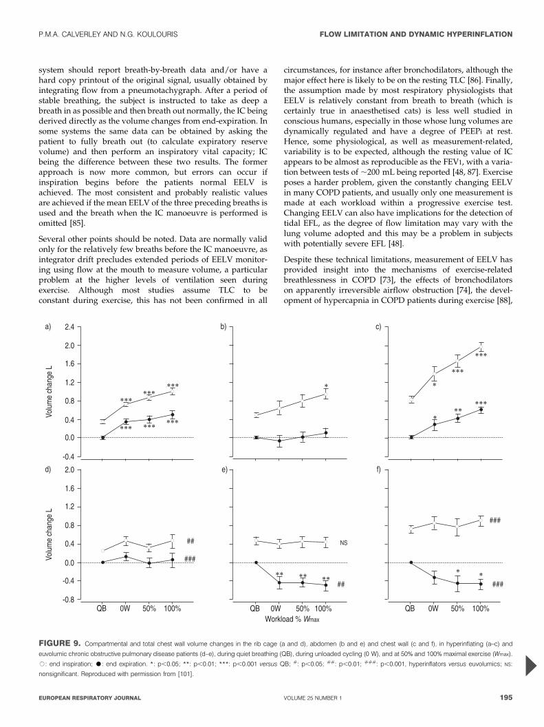
FIGURE 9. Compartmental and total chest wall volume changes in the rib cage (a and d), abdomen (b and e) and chest wall (c and f), in hyperinflating (a–c) and
euvolumic chronic obstructive pulmonary disease patients (d–e), during quiet breathing (QB), during unloaded cycling (0 W), and at 50% and 100% maximal exercise (Wmax).
#: end inspiration; $: end expiration. *: p,0.05; **: p,0.01; ***: p,0.001 versus QB; #: p,0.05; ##: p,0.01; ###: p,0.001, hyperinflators versus euvolumics; NS:
nonsignificant. Reproduced with permission from [101].
P.M.A. CALVERLEY AND N.G. KOULOURIS FLOW LIMITATION AND DYNAMIC HYPERINFLATION
cEUROPEAN RESPIRATORY JOURNAL VOLUME 25 NUMBER 1 195
the mechanisms of action of oxygen and lung volume-reduction surgery in the improving exercise performance[89–91], and, most recently, the effects of breathing helioxmixtures in severe COPD [92]. DH can be detected afterwalking along the corridor [93] and is normally assumed to bethe result of EFL [50], although this is not always the case (seeabove).
Changes in EELV do not capture all the effects of abnormalrespiratory system mechanics on exercise. Studies describingthe qualitative change in chest wall volume have usedmagnetometers to look at the contribution of different chestwall compartments, but these are technically demanding andrequire considerable subject cooperation [94–96]. Recently, adifferent approach that measures the volume of the chest wallhas being applied in healthy subjects [97–99], patients in ICU[100] and those with COPD at rest and during exercise [101–103]. Optoelectronic plethysmography (OEP) is a noninvasivemeasurement based on computing the volume of the chest wallfrom a network of points identified by shining infrared light ata series of reflective markers attached to the ribcage andabdomen [104] (fig. 8). Its ability to track the position of eachpoint in space and compute the volume enclosed and theresulting three-dimensional structure allows it to track chestwall volume during quiet breathing and exercise. Normally,the change in volume at the mouth and those occurring witheach breath derived from the chest wall signal are the same atrest (in health and COPD patients). This remains true duringexercise in healthy subjects, but not if they breathe through aStarling resistor nor is it the case in many COPD patients [99,101]. In this latter group, the difference between the chestwall volume and that at the mouth reflects the effect of gascompression in the lungs, but also the displacement of bloodaway from the thorax and into the abdomen. This powerfulnew tool can track regional chest wall volume change and wasable to confirm that patients with more severe COPD showedDH during incremental exercise [101]. However, others,specifically those with a greater expiratory flow reserve at rest(i.e. less likely to be flow limited during exercise) adopted themore ‘‘normal’’ approach of trying to lower EELV when theyexercised (fig. 9). This proved to be a poor strategy comparedwith DH, as they developed very high intra-abdominalpressures and actually had worse exercise performance. Theexact relationship between EFL and DH and how individualslearn to adopt the latter strategy will require further study, butthe availability of OEP means that this is now possible.
CONCLUSIONIn conclusion, contrary to earlier beliefs, the availability ofpowerful new physiological tools is letting us ask a range ofimportant new questions in patients and settings where itwould have previously been impossible to obtain reliable data.The move beyond the body plethysmograph to instrumenta-tion at the bedside or in the exercise or sleep laboratory servesto emphasise the practical application of these new physiolo-gical approaches. Although they are easier and more reliable toapply than older methods, there is still a need to pay carefulattention to the way the measurement is made and to be awareof their limitations. However, the surprising findings alreadyavailable and, specifically, the contribution that the measure-ment of expiratory flow limitation and dynamic hyperinflation
in explaining symptoms like breathlessness, mean that ourtraditional dependence on the forced expiratory volume in onesecond as the only measurement of respiratory mechanicsworth making is already becoming out of date.
REFERENCES1 Pride NB. Tests of forced expiration and inspiration.
In: Hughes JMB, Pride NB, eds. Lung Function Tests:Physiological Principles and Clinical Applications.London, WB Saunders, 1999: pp. 3–25.
2 Leaver DG, Pride NB. Flow-volume curves and expira-tory pressures during exercise in patients with chronicairways obstruction. Scan J Respir Dis 1971; 77: 23–27.
3 Hyatt RE. The interrelationship of pressure, flow andvolume during various respiratory maneuvers in normaland emphysematous patients. Am Rev Respir Dis 1961; 83:676–683.
4 Pepe PE, Marini JJ. Occult positive end-expiratorypressure in mechanically ventilated patients with airflowobstruction: the auto-PEEP effect. Am Rev Respir Dis1982; 126: 166–170.
5 O’Donnell DE, Sanii R, Anthonisen NR, Younes M. Effectof dynamic airway compression on breathing pattern andrespiratory sensation in severe chronic obstructivepulmonary disease. Am Rev Respir Dis 1987; 135: 912–918.
6 Milic-Emili J. Provocative hypothesis: does mechanicalinjury of the peripheral airways play a role in the genesisof COPD in smokers? COPD: J Chron Obstruc Pulm Dis2004; 1: 1–8.
7 Koutsoukou A, Armaganidis A, Stavrakaki-Kalergi C,et al. Expiratory flow limitation and intrinsic positiveend-expiratory pressure at zero positive end-expiratorypressure in patients with adult respiratory distresssyndrome. Am J Respir Crit Care Med 2000; 161: 1590–1596.
8 Koutsoukou A, Bekos B, Sotiropoulou Ch, Koulouris NG,Roussos Ch, Milic-Emili J. Effects of positive end-expiratory pressure on gas exchange and expiratory flowlimitation in adult respiratory distress syndrome. CritCare Med 2002; 30: 1941–1949.
9 D’Angelo E, Pecchiari M, Baraggia P, Saetta M, Balestro E,Milic-Emili J. Low volume ventilation induces peripheralairways injury and increased airway resistance in normalopen chest rabbits. J Appl Physiol 2002; 92: 949–956.
10 Mead J, Whittenberger JL. Physical properties of humanlungs measured during spontaneous respiration. J ApplPhysiol 1953; 5: 779–796.
11 Rodarte J. Invited editorial on ‘‘Detection of expiratoryflow limitation during exercise in COPD patients’’. J ApplPhysiol 1997; 82: 721–722.
12 Ingram RH Jr, Schilder DP. Effect of gas compression onpulmonary pressure, flow, and volume relationship.J Appl Physiol 1966; 21: 1821–1826.
13 Stubbing DG, Pengelly LD, Morse JLC, Jones NL.Pulmonary mechanics during exercise in subjects withchronic airflow obstruction. J Appl Physiol 1980; 49: 511–515.
14 Younes M, Kivinen G. Respiratory mechanics andbreathing pattern during and following maximal exer-cise. J Appl Physiol 1984; 57: 1773–1782.
FLOW LIMITATION AND DYNAMIC HYPERINFLATION P.M.A. CALVERLEY AND N.G. KOULOURIS
196 VOLUME 25 NUMBER 1 EUROPEAN RESPIRATORY JOURNAL
15 D’Angelo E, Prandi E, Milic-Emili J. Dependence ofmaximal flow-volume curves on time-course of preced-ing inspiration. J Appl Physiol 1993; 75: 1155–1159.
16 D’Angelo E, Prandi E, Marrazzini L, Milic-Emili J.Dependence of maximal flow-volume curves on timecourse of preceding inspiration in patients with chronicobstructive lung disease. Am J Respir Crit Care Med 1994;150: 1581–1586.
17 Koulouris NG, Rapakoulias P, Rassidakis A, et al.Dependence of FVC manoeuvre on time course ofpreceding inspiration in patients with restrictive lungdisease. Eur Respir J 1997; 10: 2366–2370.
18 Melissinos CG, Webster P, Tien YK, Mead J. Timedependence of maximum flow as an index of nonuniformemptying. J Appl Physiol 1979; 47: 1043–1050.
19 Fairshter RD. Airway hysteresis in normal subjects andindividuals with chronic airflow obstruction. J ApplPhysiol 1985; 58: 1505–1510.
20 Wellman JJ, Brown R, Ingram RH Jr, Mead J,McFadden ER. Effect of volume history on successivepartial expiratory maneuvers. J Appl Physiol 1976; 41:153–158.
21 Beck KC, Offord KP, Scanlon PD. Bronchoconstrictionoccurring during exercise in asthmatic patients. Am JRespir Crit Care Med 1994; 149: 352–357.
22 Koulouris NG, Valta P, Lavoie A, et al. A simple methodto detect expiratory flow limitation during spontaneousbreathing. Eur Respir J 1995; 8: 306–313.
23 Valta P, Corbeil C, Lavoie A, et al. Detection of expiratoryflow limitation during mechanical ventilation. Am JRespir Crit Care Med 1994; 150: 1311–1317.
24 Koulouris NG, Dimopoulou I, Valta P, Finkelstein R,Cosio MG, Milic-Emili J. Detection of expiratory flowlimitation during exercise in COPD patients. J ApplPhysiol 1997; 82: 723–731.
25 Eltayara L, Becklake MR, Volta CA, Milic-Emili J.Relationship between chronic dyspnoea and expiratoryflow limitation in patients with chronic obstructivepulmonary disease. Am J Respir Crit Care Med 1996; 154:17260–17234.
26 Jones MH, Davies SD, Kisling JA, Howard JM, Castile R,Tepper RS. Flow limitation in infants assessed bynegative expiratory pressure. Am J Respir Crit Care Med2000; 161: 713–717.
27 Dimitroulis J, Bisirtzoglou D, Retsou S, et al. Effect ofposture on expiratory flow limitation in spontaneouslybreathing stable COPD patients. Am J Respir Crit Care Med2001; 163: A410.
28 Kosmas EN, Milic-Emili J, Polychronaki A, et al. Exercise-induced flow limitation, dynamic hyperinflation andexercise capacity in patients with bronchial asthma. EurRespir J 2004; 24: 378–384.
29 Murciano D, Ferretti A, Boczkowski J, Sleiman C,Fournier M, Milic-Emili J. Flow limitation and dynamichyperinflation during exercise in COPD patients aftersingle lung transplantation. Chest 2000; 118: 1248–1254.
30 Armaganidis A, Stavrakaki-Kalergi K, Koutsoukou A,Lymberis A, Milic-Emili J, Roussos Ch. Intrinsic positiveend-expiratory pressure in mechanically ventilatedpatients with and without tidal expiratory flow limita-tion. Crit Care Med 2000; 28: 3837–3842.
31 Alvisi V, Romanello A, Badet M, Gaillard S, Philit F,Guerin C. Time course of expiratory flow limitation inCOPD patients during acute respiratory failure requiringmechanical ventilation. Chest 2003; 123: 1625–1632.
32 Koutsoukou A, Koulouris N, Bekos B, et al. Expiratoryflow limitation in morbidly obese postoperativemechanically ventilated patients. AnesthesiologicaScandinavica 2004; 48: 1080–1088.
33 Baydur A, Milic-Emili J. Expiratory flow limitationduring spontaneous breathing. Comparison of patientswith restrictive and obstructive respiratory disorders.Chest 1997; 112: 1017–1023.
34 Tantucci C, Duguet A, Ferretti A, et al. Effect of negativeexpiratory pressure on respiratory system flow resistancein awake snorers and nonsnorers. J Appl Physiol 1999; 87:969–976.
35 Liistro G, Veritier C, Dury M, Aubert G, Stanescu D.Expiratory flow limitation in awake sleep-disorderedbreathing subjects. Eur Respir J 1999; 14: 185–190.
36 Verin E, Tardif C, Portier F, Similowski T, Pasquis P,Muir JF. Evidence for expiratory flow limitation ofextrathoracic origin in patients with obstructive sleepapnoea. Thorax 2002; 57: 423–428.
37 Baydur A, Wilkinson L, Mehdian R, Bains B, Milic-Emili J.Extrathoracic expiratory flow limitation in obesity andobstructive and restrictive disorders; effects of increasingnegative expiratory pressure. Chest 2004; 125: 98–105.
38 Tauber E, Fazekas T, Eichler I, et al. Negative expiratorypressure: A new tool for evaluating lung function inchildren? Pediatr Pulmonol 2003; 35: 162–168.
39 Grippo A, Carrai R, Romagnoli I, Pinto F, Sanna A.Respiratory-related evoked potential and upper airwaytransmural pressure change by using the negativeexpiratory pressure (NEP) device. Clin Neurophysiol2003; 114: 636–642.
40 Vanpee D, Swine Ch, Delwich JP, Jamart J, Delanois L.Does negative expiratory pressure influence perfor-mances of spirometry in older patients? Eur Respir J2002; 20: 674–678.
41 Vanpee D, Swine Ch, Delwich JP, Delanois L. Evaluationof flow limitation in elderly patients unable to perform aforced expiratory maneuver. Aging Clin Exp Res 2002; 14:208–211.
42 Eltayara L, Ghezzo H, Milic-Emili J. Orthopnea and tidalexpiratory flow limitation in patients with stable COPD.Chest 2001; 119: 99–104.
43 Tantucci C, Duguet A, Similowski T, Zelter M,Derenne JP, Milic-Emili J. Effect of salbutamol ondynamic hyperinflation in chronic obstructive pulmon-ary disease patients. Eur Respir J 1998; 12: 799–804.
44 Boczkowski J, Murciano D, Pichot M-H, Ferretti A,Pariente R, Milic-Emili J. Expiratory flow limitation instable asthmatic patients during resting breathing. Am JRespir Crit Care Med 1997; 156: 752–757.
45 Tantucci C, Ellaffi M, Duguet A, et al. Dynamichyperinflation and flow limitation during methacholine-induced bronchoconstriction in asthma. Eur Respir J 1999;14: 295–301.
46 Sulc J, Volta CA, Ploysongsang Y, Eltayara L,Olivenstein R, Milic-Emili J. Flow limitation and
P.M.A. CALVERLEY AND N.G. KOULOURIS FLOW LIMITATION AND DYNAMIC HYPERINFLATION
cEUROPEAN RESPIRATORY JOURNAL VOLUME 25 NUMBER 1 197
dyspnoea in healthy supine subjects during methacholinechallenge. Eur Respir J 1999; 14: 1326–1331.
47 Boni E, Corda L, Franchini D, et al. Volume effect andexertional dyspnoea after bronchodilator in patients withCOPD with and without expiratory flow limitation atrest. Thorax 2002; 57: 528–532.
48 Hadcroft J, Calverley PMA. Alternative method forassessing bronchodilator reversibility in chronic obstruc-tive pulmonary disease. Thorax 2001; 56: 713–720.
49 Diaz O, Villafranca C, Ghezzo H, et al. Role of inspiratorycapacity on exercise tolerance in COPD patients with andwithout tidal expiratory flow limitation at rest. Eur Respir J2000; 16: 269–275.
50 Diaz O, Villafranca C, Ghezzo H, et al. Breathing patternand gas exchange at peak exercise in COPD patients withand without tidal expiratory flow limitation at rest. EurRespir J 2001; 17: 1120–1127.
51 Volta CA, Ploysongsang Y, Eltayara L, Sulc J, Milic-Emili J. A simple method to monitor performance offorced vital capacity. J Appl Physiol 1996; 80: 693–8.
52 Braggion C, Polese G, Fenzi V, Carli MV, Pradal U, Milic-Emili J. Detection of tidal expiratory flow limitation ininfants with cystic fibrosis. Paediatr Pulmonol 1998; 25:213–215.
53 Goetghebeur D, Sarni D, Grossi Y, et al. Tidal expiratoryflow limitation and chronic dyspnoea in patients withcystic fibrosis. Eur Respir J 2002; 19: 492–498.
54 Koulouris NG, Retsou S, Kosmas E, et al. Tidal expiratoryflow limitation, dyspnoea, and exercise capacity inpatients with bilateral bronchiectasis. Eur Respir J 2003;21: 743–748.
55 Pankow W, Podszus T, Gutheil T, Penzel T, Peter JH, VonWichert P. Expiratory flow limitation and intrinsicpositive end-expiratory pressure in obesity. J ApplPhysiol 1998; 85: 1236–1243.
56 Ferretti A, Giampiccolo P, Cavalli A, Milic-Emili J,Tantucci C. Expiratory flow limitation and orthopnea inmassively obese subjects. Chest 2001; 119: 1401–1408.
57 Duguet A, Tantucci C, Lozinguez O, et al. Expiratory flowlimitation as a determinant of orthopnea in acute leftheart failure. J Am Coll Cardiol 2000; 35: 690–700.
58 Murciano D, Pichot ME, Boczkowski J, Sleiman C,Pariente R, Milic-Emili J. Expiratory flow-limitation inCOPD patients after single lung transplantation. Am JRespir Crit Care Med 1997; 155: 1036–1047.
59 Torchio R, Gulotta C, Perboni A, et al. Orthopnea andtidal expiratory flow limitation in patients with euthyroidgoiter. Chest 2003; 124: 133–140.
60 Wang PH, Kuo PH, Hsu CH, et al. Diagnostic value ofnegative expiratory pressure for airway hyperreactivity.Chest 2003; 124: 1762–1767.
61 Dueck R. Assessment and monitoring of flow limitationand other parameters fro flow/volume loops. J ClinMonit Comput 2000; 16: 425–432.
62 Milic-Emili J, Koulouris NG, D’Angelo E. Spirometry andflow-volume loops. Eur Respir Mon 1999; 12: 20–32.
63 Johnson BD, Beck KC, Zeballos RJ, Weisman IM.Advances in pulmonary laboratory testing. Chest 1999;116: 1377–1387.
64 Ninane V, Leduc D, Kafi SA, Nasser M, Houa M,Sergysels R. Detection of expiratory flow limitation by
manual compression of the abdominal wall. Am J RespirCrit Care Med 2001; 163: 1326–1330.
65 Abdel KS, Serste T, Leduc D, Sergysels R, Ninane V.Expiratory flow limitation during exercise in COPD:detection by manual compression of the abdominal wall.Eur Respir J 2002; 19: 919–927.
66 Dellaca RL. Measurement of respiratory system impe-dances. In: Aliverti A, Brusasco V, Macklem PT, PedottiA, eds. Mechanics of Breathing. Milan, Springer, 2002;pp. 157–171.
67 Dawson SV, Elliott EA. Wave-speed limitation onexpiratory flow-a unifying concept. J Appl Physiol 1977;43: 498–515.
68 Dellaca RL, Santus P, Aliverti A, et al. Detection ofexpiratory flow limitation in COPD using the forcedoscillation technique. Eur Respir J 2004; 23: 232–240.
69 Ninane V, Yernault JC, De Troyer A. Intrinsic PEEP inpatients with chronic obstructive pulmonary disease.Role of expiratory muscles. Am Rev Respir Dis 1993; 148:1037–1042.
70 Zakynthinos SG, Vassilakopoulos T, Zakynthinos E,Roussos C. Accurate measurement of intrinsic positiveend-expiratory pressure: How to detect and correct forexpiratory muscle activity. Eur Respir J 1997; 10: 522–529.
71 Pride NB, Milic-Emili J. Lung mechanics. In: CalverleyPMA, Pride NB, eds. Chronic Obstructive PulmonaryDisease. London, Edward Arnold, 1995; pp. 69–92.
72 Haluszka J, Chartrand DA, Grassino AE, Milic-Emili J.Intrinsic PEEP and arterial PCO2 in stable patients withchronic obstructive pulmonary disease. Am Rev Respir Dis1990; 141: 1194–1197.
73 Belman MJ, Botnick WC, Shin JW. Inhaled bronchodila-tors reduce dynamic hyperinflation during exercise inpatients with chronic obstructive pulmonary disease. AmJ Respir Crit Care Med 1996; 153: 967–975.
74 O’Donnell DE, Revill SM, Webb KA. Dynamic hyperin-flation and exercise intolerance in chronic obstructivepulmonary disease. Am J Respir Crit Care Med 2001; 164:770–777.
75 O’Donnell DE, Lam M, Webb KA. Measurement ofsymptoms, lung hyperinflation, and endurance duringexercise in chronic obstructive pulmonary disease. Am JRespir Crit Care Med 1998; 158: 1557–1565.
76 Yan S, Kaminski D, Sliwinski P. Reliability of inspiratorycapacity for estimating end-expiratory lung volumechanges during exercise in patients with chronic obstruc-tive pulmonary disease. Am J Respir Crit Care Med 1997;156: 55–59.
77 O’Donnell DE, Webb KA. Exertional breathlessness inpatients with chronic airflow limitation. The role of lunghyperinflation. Am Rev Respir Dis 1993; 148: 1351–1357.
78 Johnson BD, Reddan WG, Pegelow DF, Seow KC,Dempsey JA. Flow limitation and regulation of functionalresidual capacity during exercise in a physically activeaging population. Am Rev Respir Dis 1991; 143: 960–967.
79 Mador MJ, Kufel TJ, Pineda LA, Sharma GK.Diaphragmatic fatigue and high-intensity exercise inpatients with chronic obstructive pulmonary disease.Am J Respir Crit Care Med 2000; 161: 118–123.
80 Yan S. Sensation of inspiratory difficulty during inspira-tory threshold and hyperinflationary loadings. Effect of
FLOW LIMITATION AND DYNAMIC HYPERINFLATION P.M.A. CALVERLEY AND N.G. KOULOURIS
198 VOLUME 25 NUMBER 1 EUROPEAN RESPIRATORY JOURNAL
inspiratory muscle strength. Am J Respir Crit Care Med1999; 160: 1544–1549.
81 Man WD, Soliman MG, Gearing J, et al. Symptoms andquadriceps fatigability after walking and cycling inchronic obstructive pulmonary disease. Am J Respir CritCare Med 2003; 168: 562–567.
82 O’Donnell DE, Bertley JC, Chau LK, Webb KA.Qualitative aspects of exertional breathlessness in chronicairflow limitation: pathophysiologic mechanisms. Am JRespir Crit Care Med 1997; 155: 109–115.
83 O’Donnell DE, Voduc N, Fitzpatrick M, Webb KA. Effectof salmeterol on the ventilatory response to exercise inchronic obstructive pulmonary disease. Eur Respir J 2004;24: 86–94.
84 O’Donnell DE, Fluge T, Gerken F, et al. Effects oftiotropium on lung hyperinflation, dyspnoea andexercise tolerance in COPD. Eur Respir J 2004; 23: 832–840.
85 Dolmage TE, Goldstein RS. Repeatability of inspiratorycapacity during incremental exercise in patients withsevere COPD. Chest JID - 0231335 2002; 121: 708–714.
86 Newton MF, O’Donnell DE, Forkert L. Response of lungvolumes to inhaled salbutamol in a large population ofpatients with severe hyperinflation. Chest 2002; 121: 1042–1050.
87 O’Donnell DE, Lam M, Webb KA. Spirometric correlatesof improvement in exercise performance after anti-cholinergic therapy in chronic obstructive pulmonarydisease. Am J Respir Crit Care Med 1999; 160: 542–549.
88 O’Donnell DE, D’Arsigny C, Fitzpatrick M, Webb KA.Exercise hypercapnia in advanced chronic obstructivepulmonary disease: the role of lung hyperinflation. Am JRespir Crit Care Med 2002; 166: 663–668.
89 O’Donnell DE, D’Arsigny C, Webb KA. Effects ofhyperoxia on ventilatory limitation during exercise inadvanced chronic obstructive pulmonary disease. Am JRespir Crit Care Med 2001; 163: 892–898.
90 O’Donnell DE, Webb KA, Bertley JC, Chau LK,Conlan AA. Mechanisms of relief of exertional breath-lessness following unilateral bullectomy and lungvolume reduction surgery in emphysema. Chest 1996;110: 18–27.
91 Stevenson NJ, Calverley PM. Effect of oxygen onrecovery from maximal exercise in patients with chronicobstructive pulmonary disease. Thorax 2004; 59: 668–672.
92 Palange P, Valli G, Onorati P, et al. Effect of Heliox onlung dynamic hyperinflation, dyspnoea and exerciseendurance capacity in COPD patients. J Appl Physiol2004; 97: 1637–1642.
93 Marin JM, Carrizo SJ, Gascon M, Sanchez A, Gallego B,Celli BR. Inspiratory capacity, dynamic hyperinflation,breathlessness, and exercise performance during the 6-minute-walk test in chronic obstructive pulmonarydisease. Am J Respir Crit Care Med 2001; 163: 1395–1399.
94 Grimby G, Elgefors B, Oxhoj H. Ventilatory levels andchest wall mechanics during exercise in obstructive lungdisease. Scand J Respir Dis 1973; 54: 45–52.
95 Dodd DS, Brancatisano TP, Engel LA. Effect of abdom-inal strapping on chest wall mechanics during exercise inpatients with severe chronic air-flow obstruction. Am RevRespir Dis 1985; 131: 816–821.
96 Dodd DS, Brancatisano T, Engel LA. Chest wallmechanics during exercise in patients with severe chronicair-flow obstruction. Am Rev Respir Dis 1984; 129: 33–38.
97 Aliverti A, Cala SJ, Duranti R, et al. Human respiratorymuscle actions and control during exercise. J Appl Physiol1997; 83: 1256–1269.
98 Dellaca RL, Aliverti A, Pelosi P, et al. Estimation of end-expiratory lung volume variations by optoelectronicplethysmography. Crit Care Med 2001; 29: 1807–1811.
99 Iandelli I, Aliverti A, Kayser B, et al. Determinants ofexercise performance in normal men with externallyimposed expiratory flow limitation. J Appl Physiol 2002;92: 1943–1952.
100 IAliverti A, Dellaca R, Pelosi P, Chiumello D, Pedotti A,Gattinoni L. Optoelectronic plethysmography in inten-sive care patients. Am J Respir Crit Care Med 2000; 161:1546–1552.
101 Aliverti A, Stevenson N, Dellaca RL, Lo MA, Pedotti A,Calverley PM. Regional chest wall volumes duringexercise in chronic obstructive pulmonary disease.Thorax 2004; 59: 210–216.
102 Duranti R, Filippelli M, Bianchi R, et al. Inspiratorycapacity and decrease in lung hyperinflation withalbuterol in COPD. Chest 2002; 122: 2009–2014.
103 Bianchi R, Gigliotti F, Romagnoli I, et al. Chest wallkinematics and breathlessness during pursed-lip breath-ing in patients with COPD. Chest 2004; 125: 459–465.
104 Carnevali P, Ferrigno G, Aliverti A, Pedotti A. A newmethod for 3D optical analysis of chest wall motion.Technol Health Care 1996; 4: 43–65.
P.M.A. CALVERLEY AND N.G. KOULOURIS FLOW LIMITATION AND DYNAMIC HYPERINFLATION
EUROPEAN RESPIRATORY JOURNAL VOLUME 25 NUMBER 1 199