Embed Size (px)
Citation preview
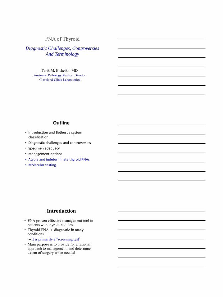
FNA of Thyroid
Tarik M. Elsheikh, MD Anatomic Pathology Medical Director
Cleveland Clinic Laboratories
Diagnostic Challenges, Controversies
And Terminology
Outline
• Introduction and Bethesda system classification
• Diagnostic challenges and controversies
• Specimen adequacy
• Management options
• Atypia and indeterminate thyroid FNAs
• Molecular testing
Introduction
• FNA proven effective management tool in patients with thyroid nodules
• Thyroid FNA is diagnostic in many conditions – It is primarily a “screening test”
• Main purpose is to provide for a rational approach to management, and determine extent of surgery when needed
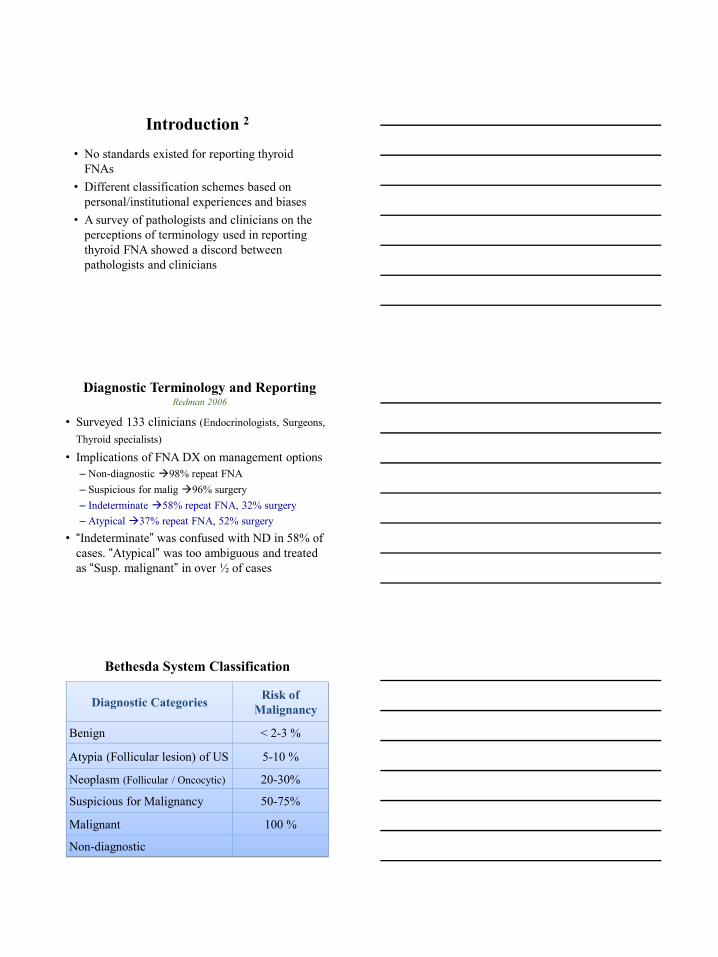
Introduction 2
• No standards existed for reporting thyroid FNAs
• Different classification schemes based on personal/institutional experiences and biases
• A survey of pathologists and clinicians on the perceptions of terminology used in reporting thyroid FNA showed a discord between pathologists and clinicians
Diagnostic Terminology and Reporting Redman 2006
• Surveyed 133 clinicians (Endocrinologists, Surgeons, Thyroid specialists)
• Implications of FNA DX on management options – Non-diagnostic 98% repeat FNA – Suspicious for malig 96% surgery – Indeterminate 58% repeat FNA, 32% surgery – Atypical 37% repeat FNA, 52% surgery
• “Indeterminate” was confused with ND in 58% of cases. “Atypical” was too ambiguous and treated as “Susp. malignant” in over ½ of cases
Bethesda System Classification
Diagnostic Categories Risk of Malignancy
Benign < 2-3 %
Atypia (Follicular lesion) of US 5-10 %
Neoplasm (Follicular / Oncocytic) 20-30%
Suspicious for Malignancy 50-75%
Malignant 100 %
Non-diagnostic
Justifications for
Bethesda System Classification
• FNA has become the standard of care for initial workup of thyroid nodules
• Most clinicians use FNA results in conjunction with clinical findings to guide treatment
• Clinicians generally utilize FNA to provide a relative risk of malignancy, from which they can base their management decisions
• The proposed diagnostic categories are important in providing a risk of malignancy to clinicians and patients Surgery vs. follow-up
Major Challenges and Controversies
– Assessment of watery/thin colloid • Conventional Paps and ThinPrep
– Terminology • Two diagnostic categories (FLUS & FN) vs. one
– Criteria for diagnosis of FN • Proportion of microfollicles • Overall cellularity
– Suspicious for PTC • How much atypia is enough? • Minimal criteria for PTC
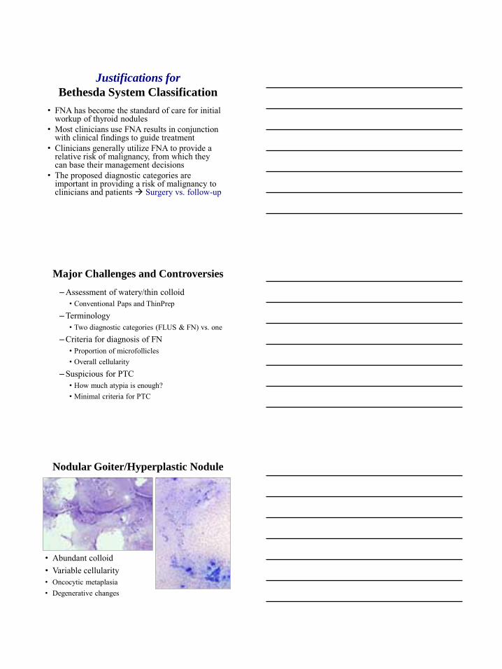
Nodular Goiter/Hyperplastic Nodule
• Abundant colloid • Variable cellularity • Oncocytic metaplasia • Degenerative changes
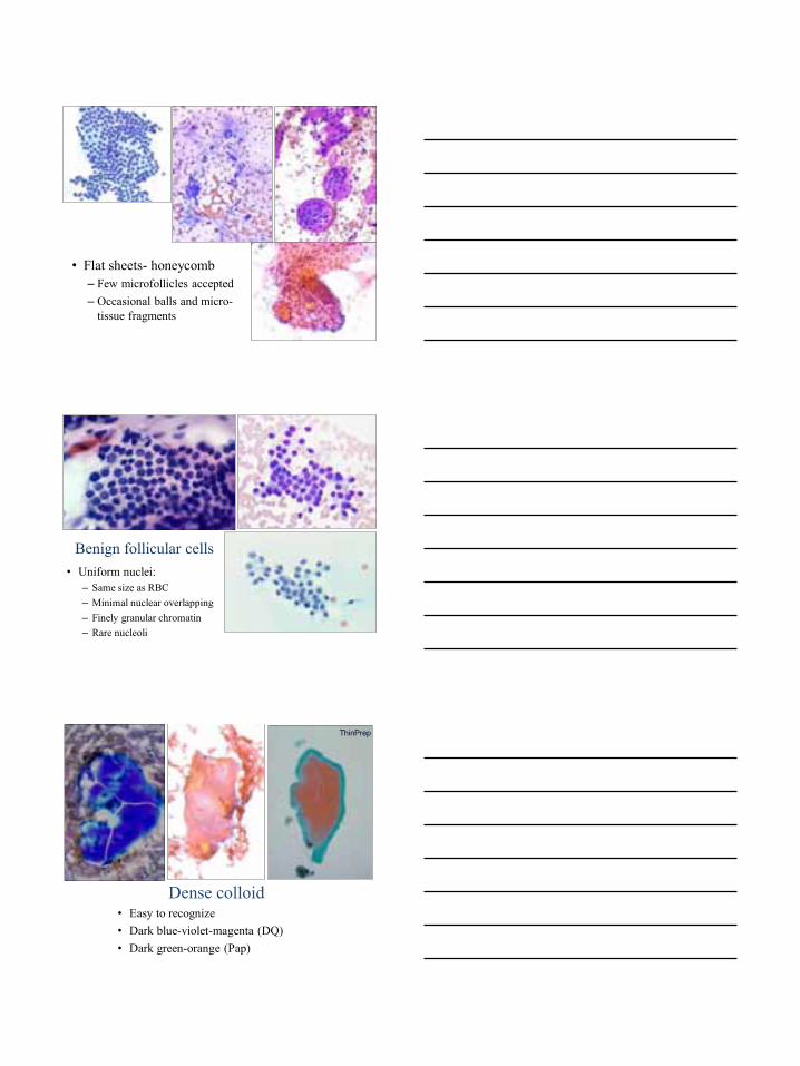
• Flat sheets- honeycomb – Few microfollicles accepted – Occasional balls and micro-
tissue fragments
• Uniform nuclei: – Same size as RBC – Minimal nuclear overlapping – Finely granular chromatin – Rare nucleoli
Benign follicular cells
Dense colloid • Easy to recognize • Dark blue-violet-magenta (DQ) • Dark green-orange (Pap)
ThinPrep
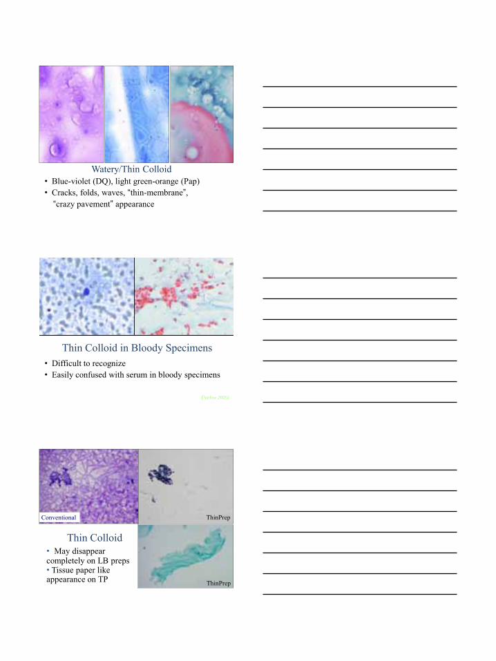
• Blue-violet (DQ), light green-orange (Pap) • Cracks, folds, waves, “thin-membrane”, “crazy pavement” appearance
Watery/Thin Colloid
Thin Colloid in Bloody Specimens • Difficult to recognize • Easily confused with serum in bloody specimens
(Stelow 2005)
ThinPrep
• May disappear completely on LB preps • Tissue paper like appearance on TP
Thin Colloid
Conventional
ThinPrep
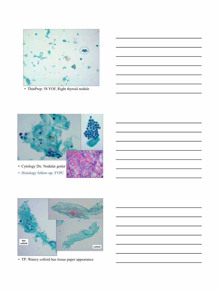
• ThinPrep: 58 YOF, Right thyroid nodule
• Cytology Dx: Nodular goiter
• Histology follow-up: FVPC
• TP: Watery colloid has tissue paper appearance
Not colloid
colloid
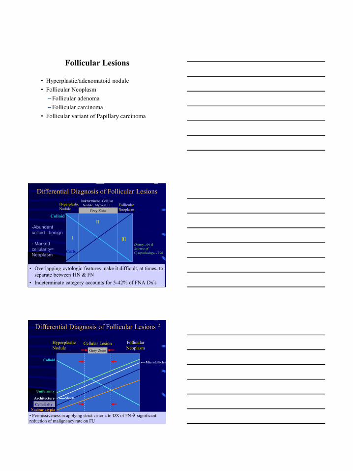
Follicular Lesions
• Hyperplastic/adenomatoid nodule • Follicular Neoplasm
– Follicular adenoma – Follicular carcinoma
• Follicular variant of Papillary carcinoma
Cells
I
II
III
Hyperplastic Nodule Grey Zone
Follicular Neoplasm
Differential Diagnosis of Follicular Lesions
• Overlapping cytologic features make it difficult, at times, to separate between HN & FN
• Indeterminate category accounts for 5-42% of FNA Dx’s
Indeterminate, Cellular Nodule, Atypical FL
Demay. Art & Science of Cytopathology, 1996
-Abundant colloid= benign
- Marked cellularity= Neoplasm
Colloid
Colloid
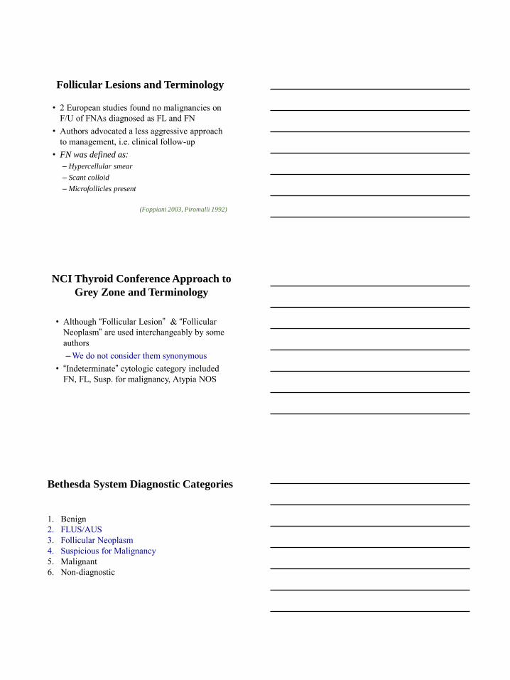
Hyperplastic Nodule
Cellular Lesion Follicular Neoplasm
Sheets
Microfollicles
CellularityArchitecture
Nuclear atypia
Differential Diagnosis of Follicular Lesions 2
Grey Zone
• Permissiveness in applying strict criteria to DX of FN significant reduction of malignancy rate on FU
Uniformity
Follicular Lesions and Terminology
• 2 European studies found no malignancies on F/U of FNAs diagnosed as FL and FN
• Authors advocated a less aggressive approach to management, i.e. clinical follow-up
• FN was defined as:
– Hypercellular smear
– Scant colloid
– Microfollicles present
(Foppiani 2003, Piromalli 1992)
NCI Thyroid Conference Approach to
Grey Zone and Terminology
• Although “Follicular Lesion” & “Follicular Neoplasm” are used interchangeably by some authors – We do not consider them synonymous
• “Indeterminate” cytologic category included FN, FL, Susp. for malignancy, Atypia NOS
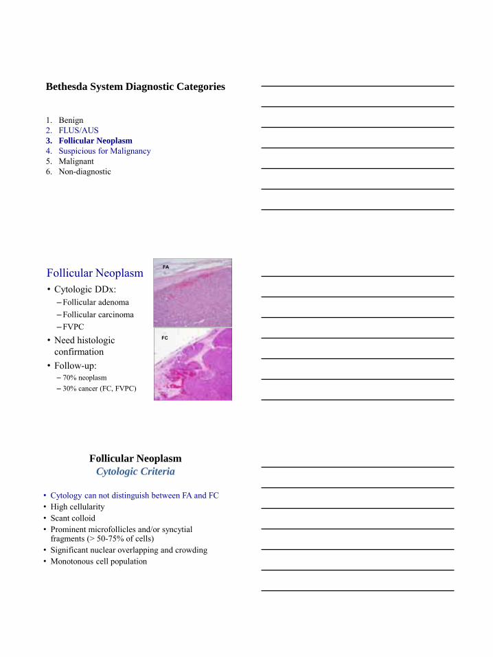
Bethesda System Diagnostic Categories
1. Benign 2. FLUS/AUS 3. Follicular Neoplasm 4. Suspicious for Malignancy 5. Malignant 6. Non-diagnostic
Bethesda System Diagnostic Categories
1. Benign 2. FLUS/AUS 3. Follicular Neoplasm
4. Suspicious for Malignancy 5. Malignant 6. Non-diagnostic
Follicular Neoplasm • Cytologic DDx:
– Follicular adenoma – Follicular carcinoma – FVPC
• Need histologic confirmation
• Follow-up: – 70% neoplasm – 30% cancer (FC, FVPC)
FA
FC
Follicular Neoplasm
Cytologic Criteria
• Cytology can not distinguish between FA and FC • High cellularity • Scant colloid • Prominent microfollicles and/or syncytial
fragments (> 50-75% of cells) • Significant nuclear overlapping and crowding • Monotonous cell population
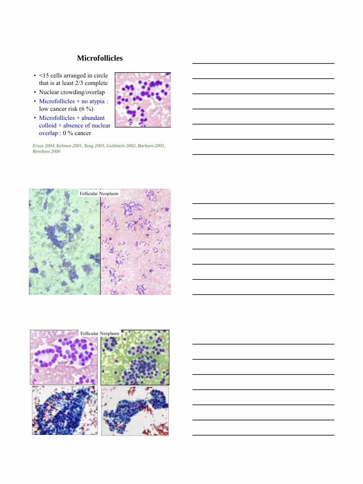
Microfollicles
• <15 cells arranged in circle that is at least 2/3 complete
• Nuclear crowding/overlap • Microfollicles + no atypia :
low cancer risk (6 %) • Microfollicles + abundant
colloid + absence of nuclear overlap : 0 % cancer
Ersoz 2004, Kelman 2001, Yang 2003, Goldstein 2002, Barbaro 2001,
Renshaw 2006
Follicular Neoplasm
Follicular Neoplasm
Follicular Neoplasm
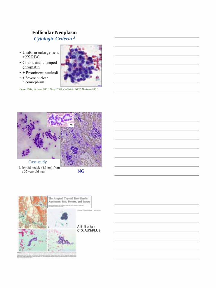
Cytologic Criteria 2
• Uniform enlargement >2X RBC
• Coarse and clumped chromatin
• ± Prominent nucleoli • ± Severe nuclear
pleomorphism
Ersoz 2004, Kelman 2001, Yang 2003, Goldstein 2002, Barbaro 2001
L thyroid nodule (1.3 cm) from a 32 year old man
Case study
NG
FN
A,B: Benign C,D: AUS/FLUS
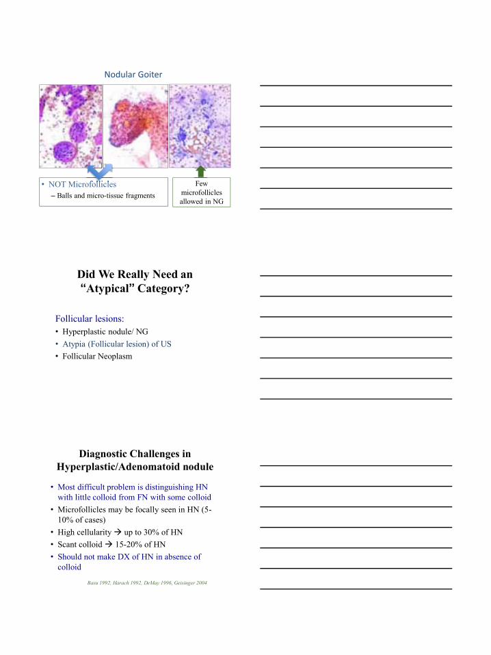
• NOT Microfollicles – Balls and micro-tissue fragments
Microfollicles Few microfollicles allowed in NG
Nodular Goiter
Did We Really Need an “Atypical” Category?
Follicular lesions: • Hyperplastic nodule/ NG • Atypia (Follicular lesion) of US • Follicular Neoplasm
Diagnostic Challenges inHyperplastic/Adenomatoid nodule
• Most difficult problem is distinguishing HN with little colloid from FN with some colloid
• Microfollicles may be focally seen in HN (5-10% of cases)
• High cellularity up to 30% of HN • Scant colloid 15-20% of HN • Should not make DX of HN in absence of
colloid
Basu 1992, Harach 1992, DeMay 1996, Geisinger 2004
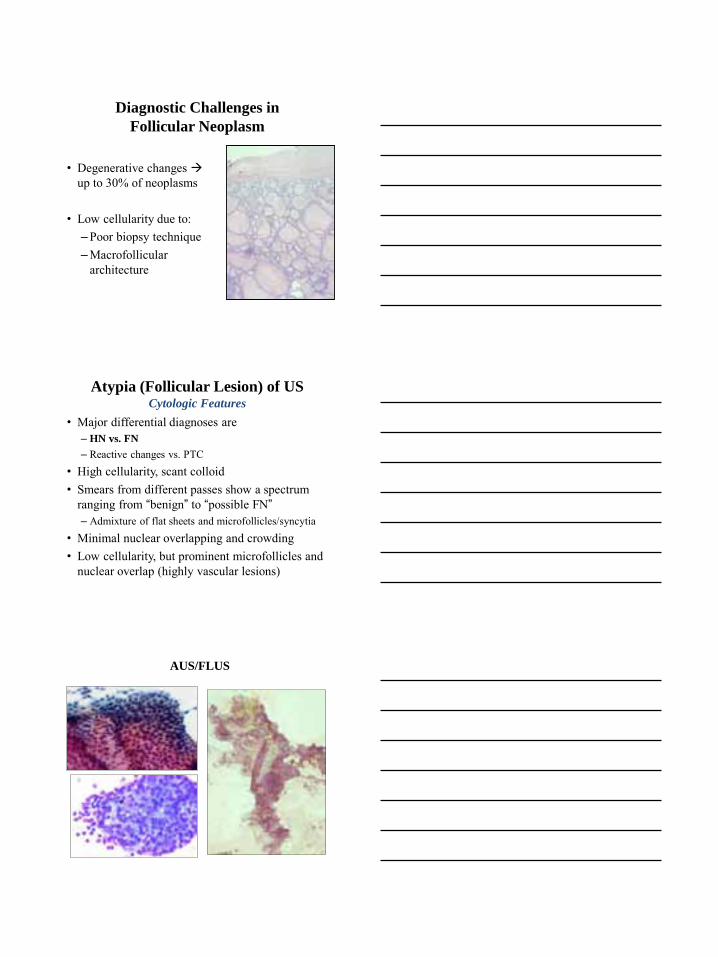
Diagnostic Challenges in
Follicular Neoplasm
• Degenerative changes up to 30% of neoplasms
• Low cellularity due to: – Poor biopsy technique – Macrofollicular
architecture
Atypia (Follicular Lesion) of US Cytologic Features
• Major differential diagnoses are – HN vs. FN
– Reactive changes vs. PTC • High cellularity, scant colloid • Smears from different passes show a spectrum
ranging from “benign” to “possible FN” – Admixture of flat sheets and microfollicles/syncytia
• Minimal nuclear overlapping and crowding • Low cellularity, but prominent microfollicles and
nuclear overlap (highly vascular lesions)
AUS/FLUS
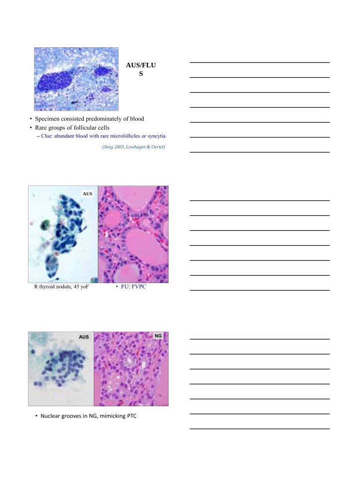
AUS/FLU
S
• Specimen consisted predominately of blood • Rare groups of follicular cells
– Clue: abundant blood with rare microfollicles or syncytia (Yang 2003, Lowhagen & Oertel)
• FU: FVPC
AUS
R thyroid nodule, 45 yoF
• Nuclear grooves in NG, mimicking PTC
AUS NG
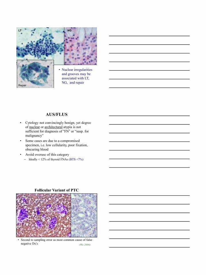
• Nuclear irregularities and grooves may be associated with LT, NG, and repair
LT NG
Repair
AUS/FLUS
• Cytology not convincingly benign, yet degree of nuclear or architectural atypia is not sufficient for diagnosis of “FN” or “susp. for malignancy”
• Some cases are due to a compromised specimen, i.e. low cellularity, poor fixation, obscuring blood
• Avoid overuse of this category – Ideally < 12% of thyroid FNAs (BTS <7%)
Follicular Variant of PTC
• Second to sampling error as most common cause of false negative Dx’s (Wu 2006)
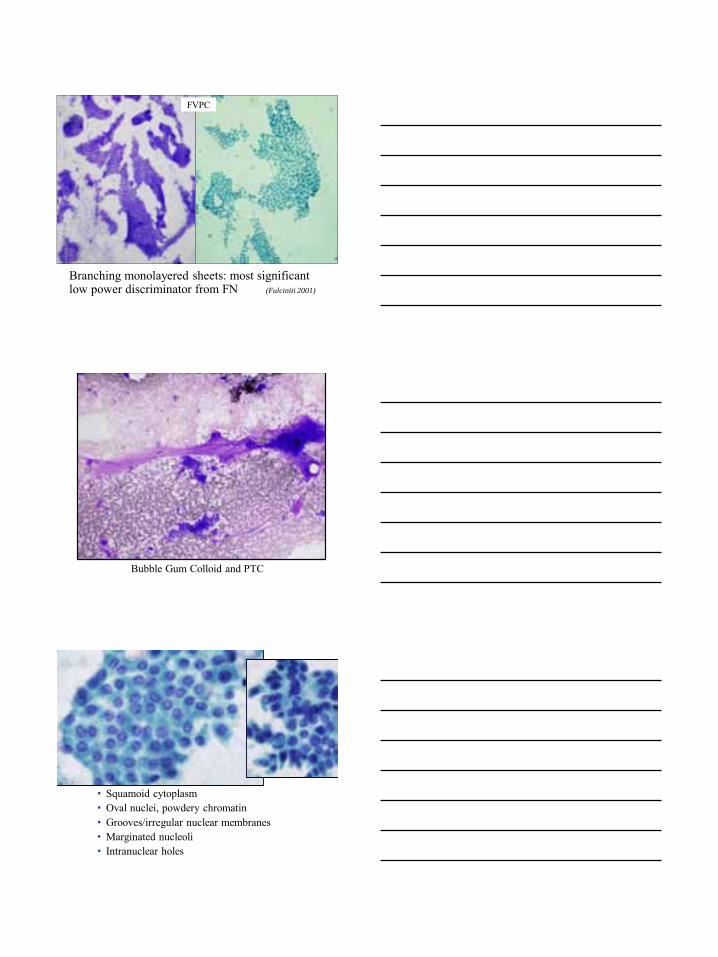
Branching monolayered sheets: most significant low power discriminator from FN (Fulciniti 2001)
FVPC
Bubble Gum Colloid and PTC
• Squamoid cytoplasm • Oval nuclei, powdery chromatin • Grooves/irregular nuclear membranes • Marginated nucleoli • Intranuclear holes
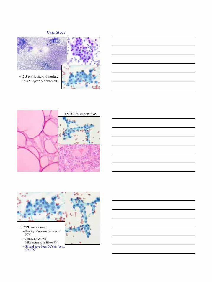
Case Study
• 2.5 cm R thyroid nodule in a 56 year old woman
FVPC, false negative
• FVPC may show: – Paucity of nuclear features of
PTC – Abundant colloid – Misdiagnosed as B9 or FN – Should have been Dx’d as “susp.
for PTC”
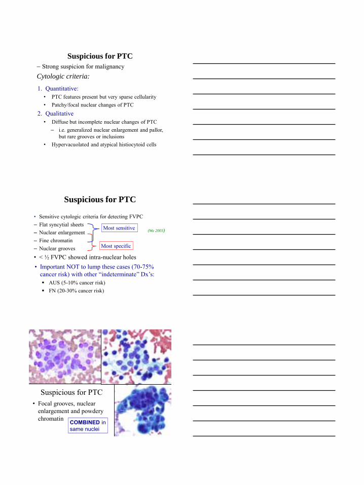
Suspicious for PTC
– Strong suspicion for malignancy Cytologic criteria:
1. Quantitative: • PTC features present but very sparse cellularity • Patchy/focal nuclear changes of PTC
2. Qualitative • Diffuse but incomplete nuclear changes of PTC
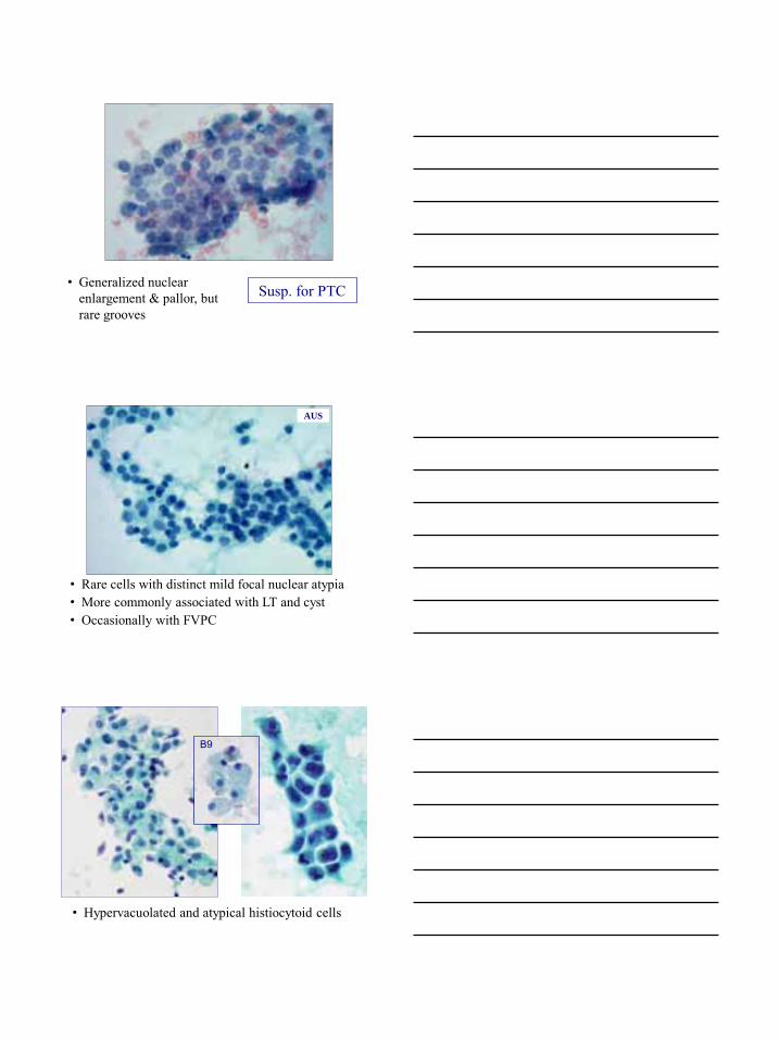
– i.e. generalized nuclear enlargement and pallor, but rare grooves or inclusions
• Hypervacuolated and atypical histiocytoid cells
Suspicious for PTC
• Sensitive cytologic criteria for detecting FVPC – Flat syncytial sheets – Nuclear enlargement – Fine chromatin – Nuclear grooves • < ½ FVPC showed intra-nuclear holes
Most sensitive
Most specific
(Wu 2003)
• Important NOT to lump these cases (70-75% cancer risk) with other “indeterminate” Dx’s: AUS (5-10% cancer risk) FN (20-30% cancer risk)
Suspicious for PTC • Focal grooves, nuclear
enlargement and powdery chromatin COMBINED in
same nuclei
Susp. for PTC • Generalized nuclear enlargement & pallor, but rare grooves
• Rare cells with distinct mild focal nuclear atypia • More commonly associated with LT and cyst • Occasionally with FVPC
AUS
• Hypervacuolated and atypical histiocytoid cells
B9
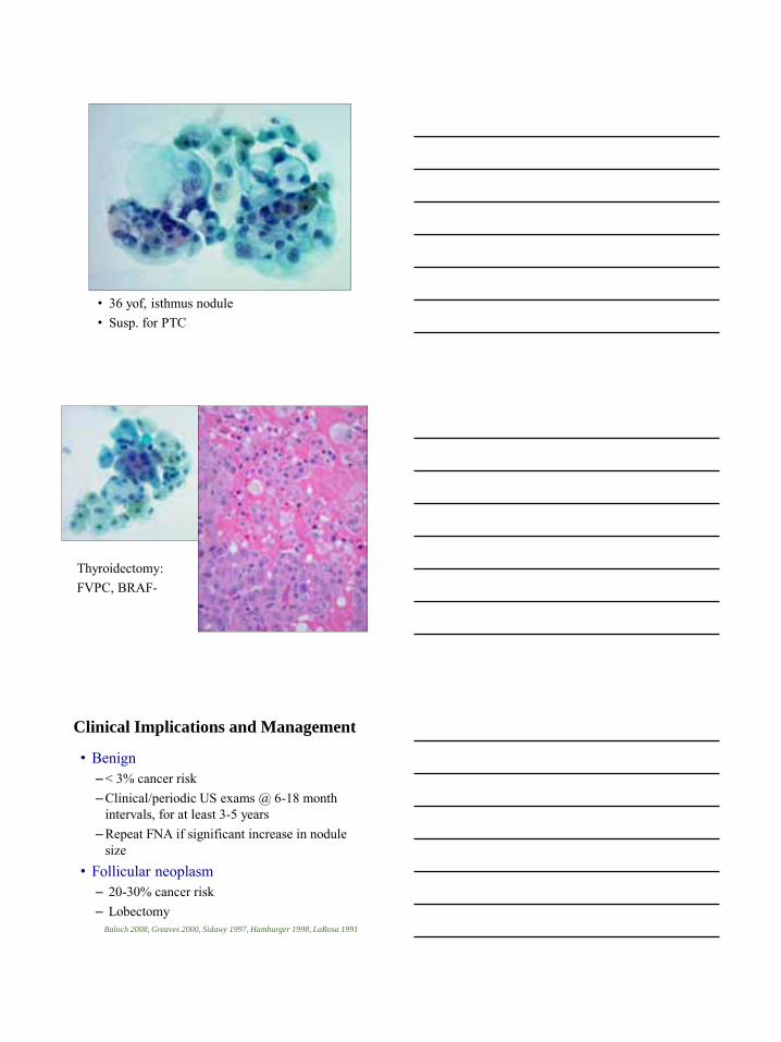
• 36 yof, isthmus nodule • Susp. for PTC
Thyroidectomy: FVPC, BRAF-
Clinical Implications and Management
• Benign – < 3% cancer risk – Clinical/periodic US exams @ 6-18 month
intervals, for at least 3-5 years – Repeat FNA if significant increase in nodule
size • Follicular neoplasm
– 20-30% cancer risk – Lobectomy
Baloch 2008, Greaves 2000, Sidawy 1997, Hamburger 1998, LaRosa 1991
Clinical Implications and Management 2
• AUS/FLUS – Approximately 10% cancer risk – Repeat FNA in 3-6 months, correlate with
clinical and radiologic findings – If repeat FNA is “Atypical” or worse
consider surgery – NOT equivalent to “Susp. for malignancy”
Baloch 2008, Greaves 2000, Sidawy 1997, Hamburger 1998, LaRosa 1991
Clinical Implications and Management 3
• Suspicious for PTC – 50-75% cancer risk Options:
1. Lobectomy 2. Lobectomy + intra-operative consult
• Helpful in additional 30% of cases (Baloch 2002)
3. Total thyroidectomy
Bethesda System Diagnostic Categories
1. Benign 2. Atypia (Follicular lesion) of US 3. Follicular/Oncocytic Neoplasm 4. Suspicious for Malignancy 5. Malignant 6. Non-diagnostic
Specimen Adequacy Criteria
General Principles
• Main purpose is to minimize # of false negative diagnoses
• Provide a meaningful interpretation that is clinically useful – A diagnosis of “rare/few benign follicular
cells” without qualification, is not considered meaningful
• An adequate sample should be representative of the lesion (appropriate cellularity) and technically well prepared, i.e. good fixation, thin smear, adequate staining
Adequacy Criteria NCI Thyroid Conference Conclusions
• Specimen processed and examined, but “Non-diagnostic” due to:
• No follicular cells or limited cellularity • Poor fixation and preservation
• Optimal # of passes: 2-5 • Any significant cytologic atypia precludes the
interpretation of “ND” • Solid nodules: minimum of 5-6 groups (at least
10 cells/group), preferably on a single slide • A repeat FNA can be recommended
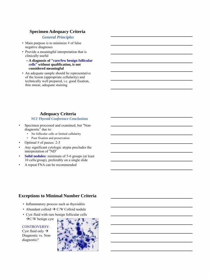
Exceptions to Minimal Number Criteria
• Inflammatory process such as thyroiditis • Abundant colloid C/W Colloid nodule • Cyst fluid with rare benign follicular cells C/W benign cyst
CONTROVERSY: Cyst fluid only Diagnostic vs. Non-diagnostic?
Thyroid Cysts and Malignancy
• Cysts most commonly due to cystic degeneration in NG
• Any residual mass after aspiration should be sampled
• Risk of malignancy – Of all aspirated cysts, as low as 1% are malignant – Simple, non-complex cysts = 1- 4% cancer risk – Mixed solid-cystic nodules, large cysts (>3cm)
and recurring cysts = Up to 14% cancer risk
Bethesda Conference Conclusions
• Cystic lesions, lacking follicular cells, that collapse completely following aspiration “Cyst fluid only”
• Include under “Non-diagnostic” (majority agreement) or “Benign”
• Options: • Recommend correlation with cyst size,
complexity and US features • Disclaimer that cystic CA cannot be entirely
excluded
Atypia and
Indeterminate Lesions
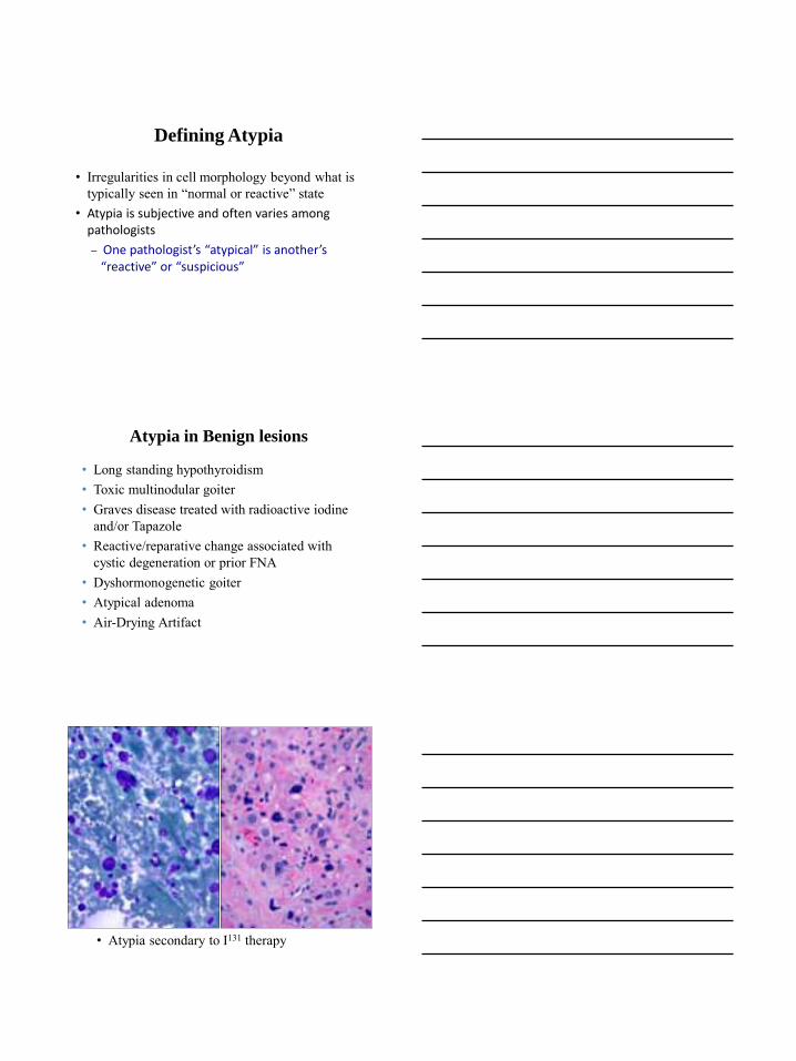
Defining Atypia
• Irregularities in cell morphology beyond what is typically seen in “normal or reactive” state
• Atypia is subjective and often varies among pathologists
– One pathologist’s “atypical” is another’s “reactive” or “suspicious”
Atypia in Benign lesions
• Long standing hypothyroidism • Toxic multinodular goiter • Graves disease treated with radioactive iodine
and/or Tapazole • Reactive/reparative change associated with
cystic degeneration or prior FNA • Dyshormonogenetic goiter • Atypical adenoma • Air-Drying Artifact
• Atypia secondary to I131 therapy
Atypical Repair in cyst
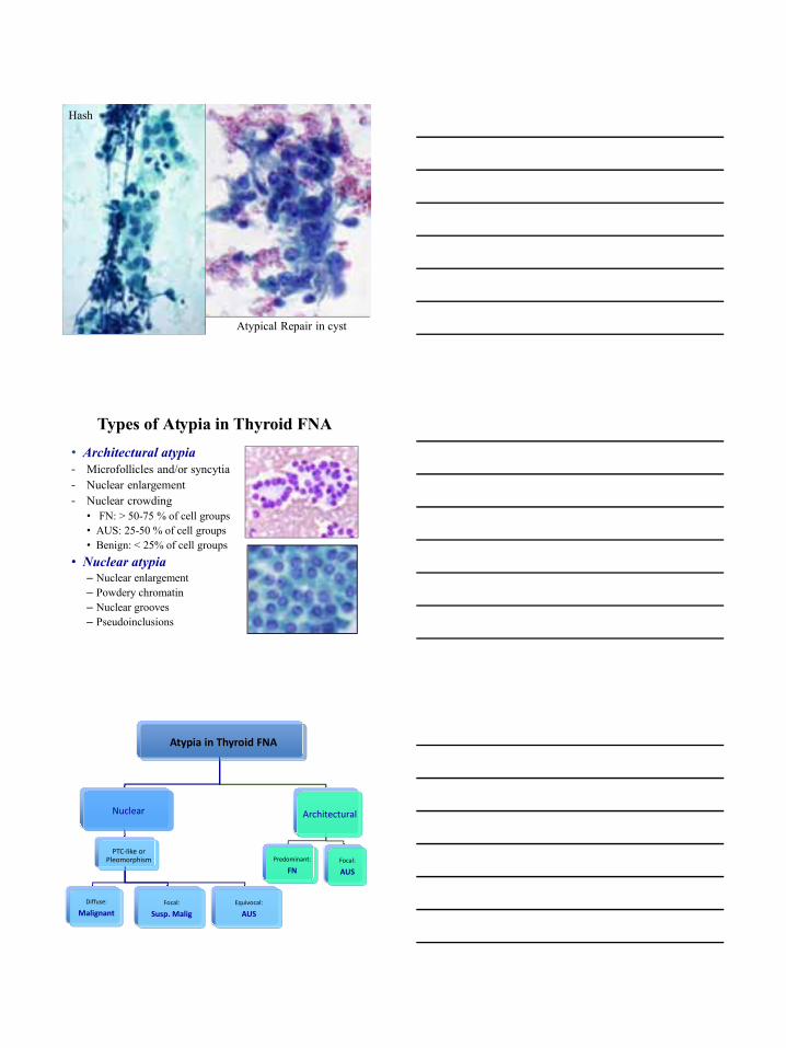
Hash
• Architectural atypia- Microfollicles and/or syncytia - Nuclear enlargement - Nuclear crowding
• FN: > 50-75 % of cell groups • AUS: 25-50 % of cell groups • Benign: < 25% of cell groups
• Nuclear atypia– Nuclear enlargement – Powdery chromatin – Nuclear grooves – Pseudoinclusions
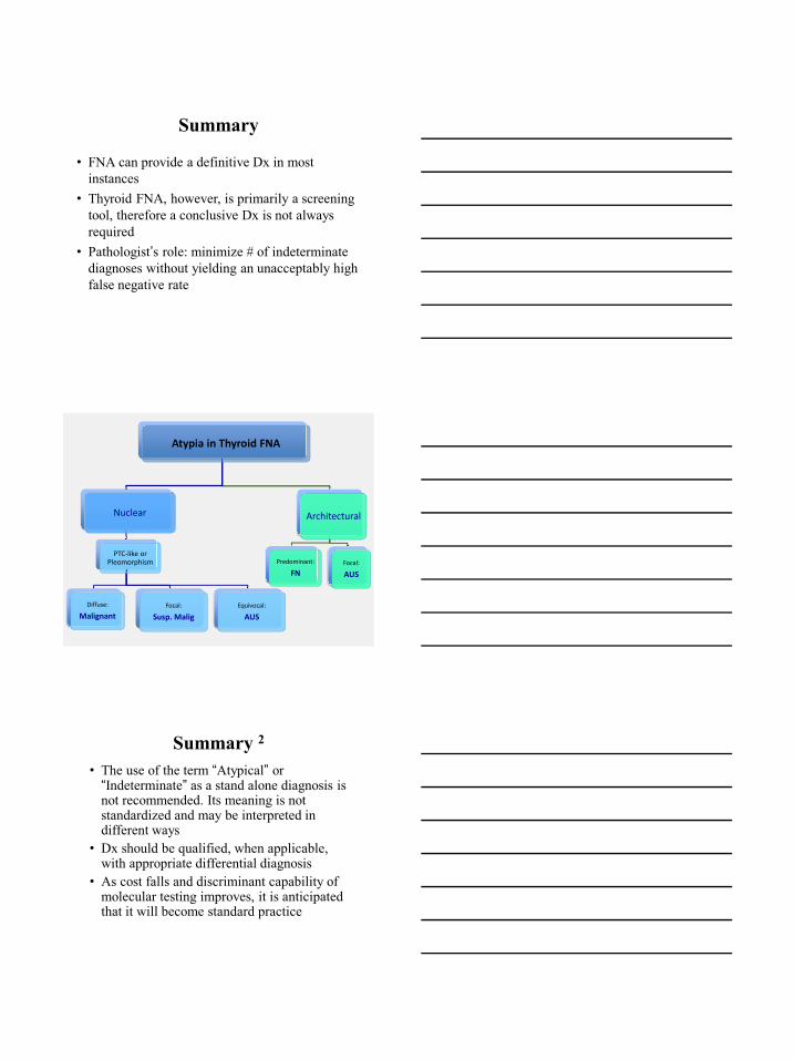
Types of Atypia in Thyroid FNA
Atypia in Thyroid FNA
Nuclear
PTC-like or Pleomorphism
Diffuse:
Malignant
Focal:
Susp. Malig
Equivocal:
AUS
Architectural
Predominant:
FNFocal:
AUS
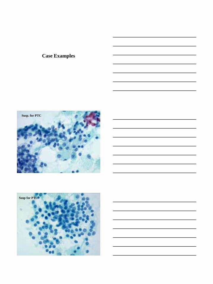
Case Examples
Susp. for PTC
Susp for PTC
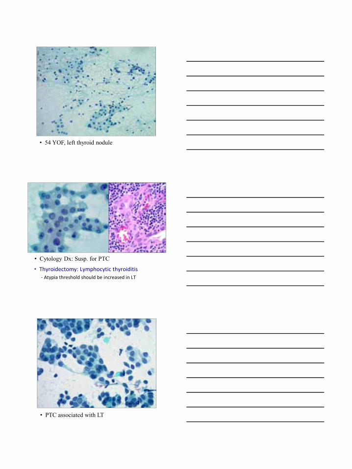
• 54 YOF, left thyroid nodule
• Cytology Dx: Susp. for PTC
• Thyroidectomy: Lymphocytic thyroiditis
- Atypia threshold should be increased in LT
• PTC associated with LT
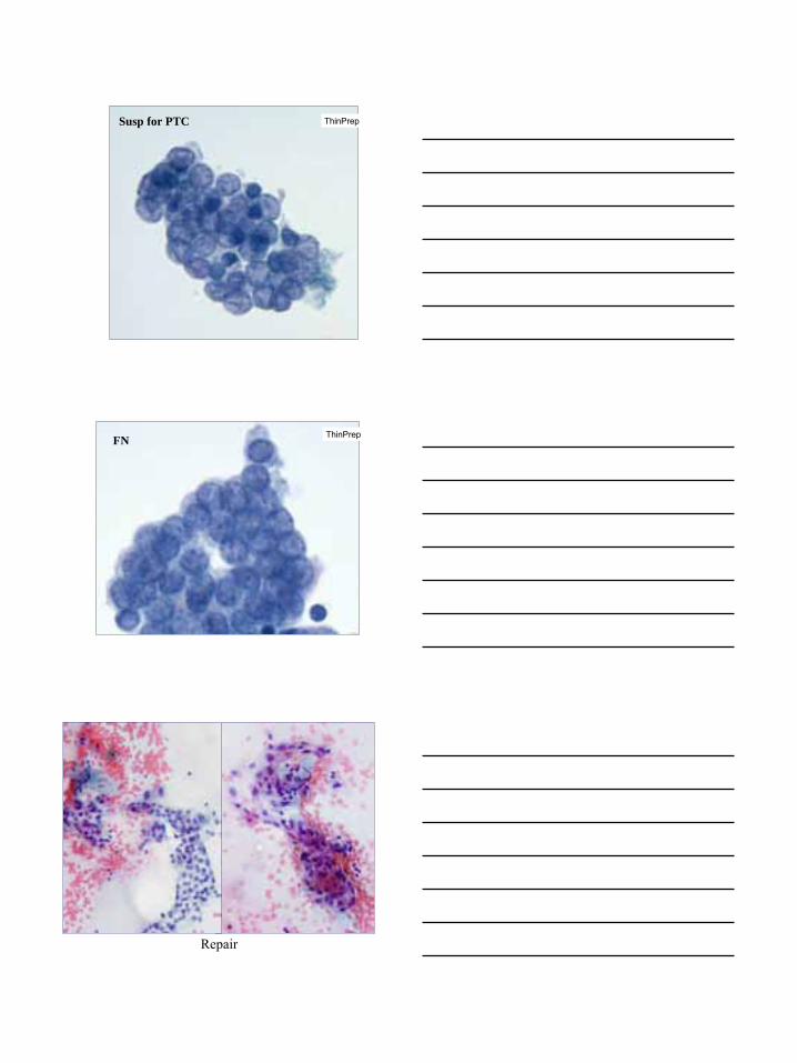
ThinPrep Susp for PTC
FN ThinPrep
Repair
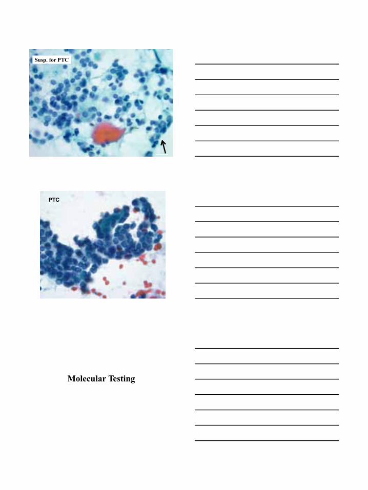
FN or Susp?Susp. for PTC
PTC
Molecular Testing
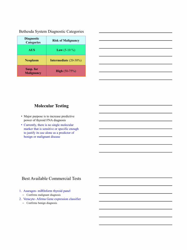
Bethesda System Diagnostic Categories
1. Benign2. Atypia of US 3. Follicular Neoplasm 4. Suspicious for Malignancy 5. Malignant6. Non-diagnostic
Follicular NeoplasmSuspicious for Malignancy
Indeterminate ≈ 20-30% of
cases
Diagnostic Categories Risk of Malignancy
AUS Low (5-10 %)
Neoplasm Intermediate (20-30%)
Susp. for Malignancy High (50-75%)
Molecular Testing
• Major purpose is to increase predictive power of thyroid FNA diagnosis
• Currently, there is no single molecular marker that is sensitive or specific enough to justify its use alone as a predictor of benign or malignant disease
Best Available Commercial Tests
1. Asuragen- miRInform thyroid panel – Confirms malignant diagnosis
2. Veracyte- Afirma Gene expression classifier – Confirms benign diagnosis
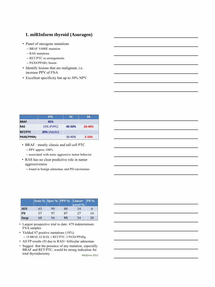
1. miRInform thyroid (Asuragen)
• Panel of oncogene mutations – BRAF V600E mutation – RAS mutations – RET/PTC re-arrangements – PAX8/PPARγ fusion
• Identify lesions that are malignant, i.e. increase PPV of FNA
• Excellent specificity but up to 30% NPV
PTC FC FA
BRAF 45%
RAS 15% (FVPC) 40-50% 20-40%
RET/PTC 20% (Adults)
PAX8/PPAR 30-40% 2-10%
• BRAF : mostly classic and tall cell PTC – PPV approx 100% – associated with more aggressive tumor behavior
• RAS has no clear predictive role in tumor aggressiveness – found in benign adenomas and PD carcinomas
Nikiforov 2011
• Largest prospective trial to date: 479 indeterminate FNA samples
• Yielded 87 positive mutations (18%) – 19 BRAF, 62 RAS, 1 RET/PTC, 5 PAX8/PPARg
• All FP results (9) due to RAS+ follicular adenomas • Suggest that the presence of any mutation, especially
BRAF and RET/PTC, would be strong indication for total thyroidectomy
Sens % Spec % PPV % Cancer prevl %
FN %
AUS 63 99 88 14 6
FN 57 97 87 27 14
Susp 68 96 95 54 28
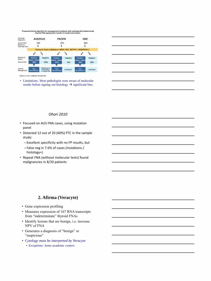
Proposed clinical algorithm for management of patients with cytologically indeterminate thyroid FNA applying the results of mutational analysis.
Nikiforov Y E et al. JCEM 2011;96:3390-3397
• Limitations: Most pathologist were aware of molecular results before signing out histology significant bias
Ohori 2010
• Focused on AUS FNA cases, using mutation panel
• Detected 12 out of 20 (60%) PTC in the sample study:
– Excellent specificity with no FP results, but
– False neg in 7.6% of cases (mutations-/ histology+)
• Repeat FNA (without molecular tests) found malignancies in 8/20 patients
2. Afirma (Veracyte)
• Gene expression profiling • Measures expression of 167 RNA transcripts
from “indeterminate” thyroid FNAs • Identify lesions that are benign, i.e. increase
NPV of FNA • Generates a diagnosis of “benign” or
“suspicious” • Cytology must be interpreted by Veracyte
• Exceptions: Some academic centers
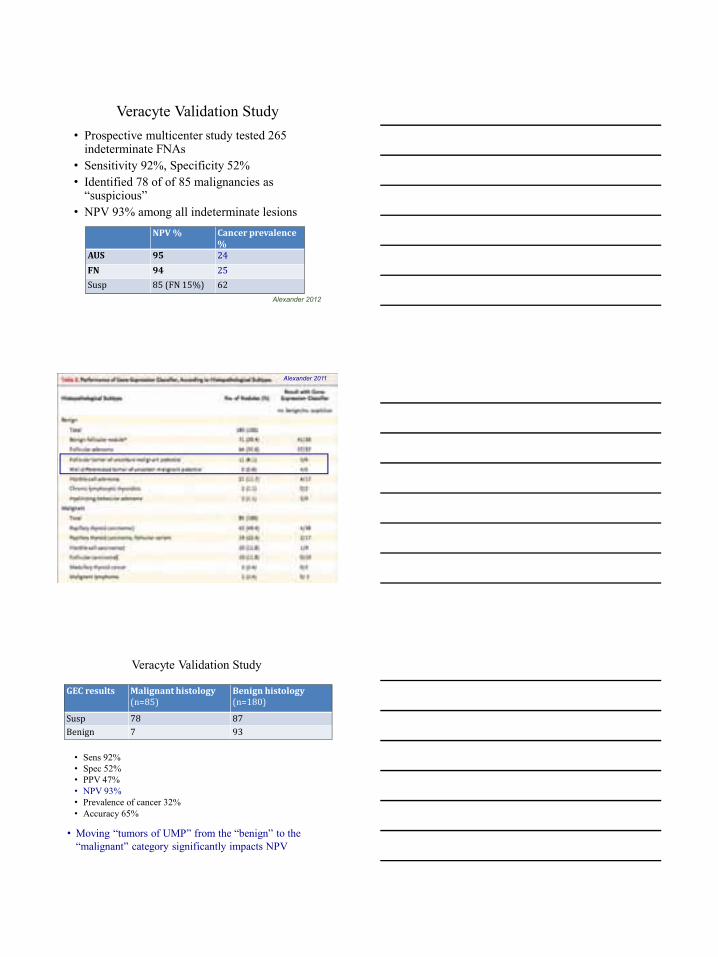
Veracyte Validation Study • Prospective multicenter study tested 265
indeterminate FNAs • Sensitivity 92%, Specificity 52% • Identified 78 of of 85 malignancies as
“suspicious” • NPV 93% among all indeterminate lesions
Alexander 2012
NPV % Cancer prevalence %
AUS 95 24
FN 94 25
Susp 85 (FN 15%) 62
Alexander 2011
Veracyte Validation Study
GEC results Malignant histology (n=85)
Benign histology (n=180)
Susp 78 87
Benign 7 93
• Sens 92% • Spec 52% • PPV 47% • NPV 93% • Prevalence of cancer 32% • Accuracy 65%
• Moving “tumors of UMP” from the “benign” to the “malignant” category significantly impacts NPV
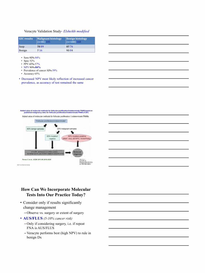
Veracyte Validation Study- Elsheikh modified
GEC results Malignant histology (n=85) 105
Benign histology (n=180) 160
Susp 78 89 87 76
Benign 7 16 93 84
• Sens 92% 84% • Spec 52% • PPV 47% 57% • NPV 93% 84% • Prevalence of cancer 32% 39% • Accuracy 65%
• Decreased NPV most likely reflection of increased cancer prevalence, as accuracy of test remained the same
Added value of molecular methods for follicular proliferation/indeterminate FNAB based on published malignancy rates for follicular proliferation/indeterminate FNAB of 20%
Ferraz C et al. JCEM 2011;96:2016-2026
©2011 by Endocrine Society
How Can We Incorporate Molecular Tests Into Our Practice Today?
• Consider only if results significantly change management – Observe vs. surgery or extent of surgery
• AUS/FLUS (5-10% cancer risk)– Only if considering surgery, i.e. if repeat
FNA is AUS/FLUS – Veracyte performs best (high NPV) to rule in
benign Dx
How Can We Incorporate Molecular
Tests Into Our Practice Today?
• Neoplasm (20-30% cancer risk)
– Veracyte if clinically low risk disease – Asuragen if clinically high risk disease – Perhaps combination of both. Cost?? ($2200-
3000 per panel)
• Susp. For malignancy (60-75% cancer risk)
– Limited to no value – Asuragen performs best (high PPV) – May help confirm extent of surgery, i.e. BRAF+
micro RNA expression
• Initial studies showed predictive diagnostic accuracies ranging from 76-90%
• No large scale prospective trial has been performed yet
• Not commercially available
• Available only through research protocols
Future Trends in Molecular Testing
• Ultimate goal is effectively differentiating benign from malignant thyroid lesions
• This may eventually be achieved by:
1. Combination of cancer mutations and GEP
2. Identification of new mutations and additional miRNA expression profiles
3. Next-generation sequencing analysis of benign and malignant lesions
Summary
• FNA can provide a definitive Dx in most instances
• Thyroid FNA, however, is primarily a screening tool, therefore a conclusive Dx is not always required
• Pathologist’s role: minimize # of indeterminate diagnoses without yielding an unacceptably high false negative rate
Atypia in Thyroid FNA
Nuclear
PTC-like or Pleomorphism
Diffuse:
Malignant
Focal:
Susp. Malig
Equivocal:
AUS
Architectural
Predominant:
FN Focal:
AUS
Summary 2• The use of the term “Atypical” or
“Indeterminate” as a stand alone diagnosis is not recommended. Its meaning is not standardized and may be interpreted in different ways
• Dx should be qualified, when applicable, with appropriate differential diagnosis
• As cost falls and discriminant capability of molecular testing improves, it is anticipated that it will become standard practice
Thank You