Embed Size (px)
Citation preview
doi:10.1182/blood-2002-09-2783Prepublished online March 20, 2003;
Guilio Gabbiani, Torsten Schwede, Thomas Matthes, Stylianos E Antonarakis and Photis BerisSamuel Deutsch, Alexandra Rideau, Marie-Luce Bochaton-Piallat, Giuseppe Merla, Antoine Geinoz, for the phenotypes in May-Hegglin anomaly/Fechtner syndromeThe D1424N MYH9 mutation results in an unstable protein responsible
(2497 articles)Hemostasis, Thrombosis, and Vascular Biology � (1086 articles)Gene Expression �
Articles on similar topics can be found in the following Blood collections
http://bloodjournal.hematologylibrary.org/site/misc/rights.xhtml#repub_requestsInformation about reproducing this article in parts or in its entirety may be found online at:
http://bloodjournal.hematologylibrary.org/site/misc/rights.xhtml#reprintsInformation about ordering reprints may be found online at:
http://bloodjournal.hematologylibrary.org/site/subscriptions/index.xhtmlInformation about subscriptions and ASH membership may be found online at:
articles must include the digital object identifier (DOIs) and date of initial publication. priority; they are indexed by PubMed from initial publication. Citations to Advance online prior to final publication). Advance online articles are citable and establish publicationyet appeared in the paper journal (edited, typeset versions may be posted when available Advance online articles have been peer reviewed and accepted for publication but have not
Copyright 2011 by The American Society of Hematology; all rights reserved.Washington DC 20036.by the American Society of Hematology, 2021 L St, NW, Suite 900, Blood (print ISSN 0006-4971, online ISSN 1528-0020), is published weekly
only.For personal use at PENN STATE UNIVERSITY on February 20, 2013. bloodjournal.hematologylibrary.orgFrom
The D1424N MYH9 mutation results in an unstable protein responsible for the phenotypes in May-Hegglin
anomaly/Fechtner syndrome.
Samuel Deutsch*1,5, Alexandra Rideau*2,5, Marie-Luce Bochaton-Piallat3, Giuseppe Merla1, Antoine Geinoz3, Giulio Gabbiani3, Torsten Schwede4, Thomas Matthes2, Stylianos E. Antonarakis1,, Photis Beris2.
1. Division of Medical Genetics, Faculty of Medicine, University of Geneva. CH-1211 Geneva, Switzerland.2. Division of Hematology, Geneva University Hospital,CH-1206 Geneva, Switzerland.3. Department of Pathology, Faculty of Medicine, University of Geneva. CH- 1211 Geneva, Switzerland.4.Biozentrum der Universität Basel and Swiss Institute of Bioinformatics, CH-4056 Basel, Switzerland.5. Graduate Program of Molecular and Cellular Biology, Faculty of Medicine, University of Geneva. CH-1211 Geneva, Switzerland.
* These authors contributed equally to this work.
Corresponding author :
Photis Beris, MDDivision of HematologyGeneva University HospitalCH-1211 Geneva, SwitzerlandPhone : +41223723928Fax : +41223737288E-mail : [email protected]
Short Title: May-Hegglin anomaly/Fechtner syndrome.
Keywords: May-Hegglin anomaly, Fechtner syndrome, Sebastian syndrome, Epstein syndrome, DFNA17, Macrothrombocytopenia, Doehle bodies, MYH9, NMMHC, Pyrosequencing.
Word Count : 3500
Category :Hemostasis
This work was supported by grants from the Swiss National Foundation for Scientific Research #32-61845.00 to PhB and # 31- 57149.99 to SEA
Copyright (c) 2003 American Society of Hematology
Blood First Edition Paper, prepublished online March 20, 2003; DOI 10.1182/blood-2002-09-2783
only.For personal use at PENN STATE UNIVERSITY on February 20, 2013. bloodjournal.hematologylibrary.orgFrom
2
Abstract
May-Hegglin anomaly (MHA), Fechtner syndrome (FTNS), Sebastian syndrome
(SBS), and Epstein syndrome (EPS) are a group of rare, autosomal dominant
disorders characterized by thrombocytopenia, giant platelets and Doehle-like
inclusion bodies, together with variable manifestations of Alport-like symptoms which
include high tone sensorineural deafness, cataracts and nephritis. These disorders
result from mutations in the MYH9 gene, which encodes for the non-muscle myosin
heavy chain A protein (also known as NMMHC-A). To date 20 different mutations
have been characterized for this gene, but no clear phenotype-genotype correlation
has been established, and very little is known regarding the molecular pathogenesis
of this group of diseases.
In this paper we describe two new families with MHA/FTNS phenotypes which have
been characterized in terms of their mutations, protein localization in
megakaryocytes, protein expression and mRNA stability. Our findings suggest that,
at least for the D1424N mutation in the MYH9 gene, the phenotypes result from a
highly unstable protein. No abnormalities in protein localization or mRNA stability
were observed. We hypothesize that haploinsufficiency of the MYH9 results in a
failure to properly reorganize the cytoskeleton in megakaryocytes as required for
efficient platelet production.
only.For personal use at PENN STATE UNIVERSITY on February 20, 2013. bloodjournal.hematologylibrary.orgFrom

3
Acknowledgements
We thank Dr. A. Reymond for advice and critical comments, Dr. R Lyle for assistance
with the Taqman related experiments, Dr. C. Rossier for assistance with the
sequencing, M. Papasavvas and U. Choudhury for technical support and J. Ringrose
for secretarial help.
only.For personal use at PENN STATE UNIVERSITY on February 20, 2013. bloodjournal.hematologylibrary.orgFrom
4
Introduction
Familial macrothrombocytopenias with leukocyte inclusion bodies are a group of rare
autosomal dominant disorders characterized by mild bleeding symptoms, giant
platelets and Doehle-like inclusion bodies in peripheral blood granulocytes. These
disorders, which include the May-Hegglin anomaly (MHA OMIM: #155100),
Sebastian syndrome (SBS OMIM :#605249), Fechtner syndrome (FTNS OMIM :#
#153640), and Epstein syndrome (EPS OMIM :#153650) all have largely overlapping
phenotypes, but were previously considered as separate clinical entities 1-4.
Biochemical analysis of platelets from MHA patients revealed no abnormalities in the
function of these cells,5,6 leading to the hypothesis that the hematological phenotype
in patients may result from a deficit in the demarcation membranes in
megakaryocytes prior to platelet formation7.
MHA anomaly and SBS are distinguished from each other by small differences in
their inclusion bodies revealed by electron microscopy examination8. FTNS and EPS,
on the other hand, manifest a number of non hematological traits similar to those
observed in Alport syndrome cases, such as nephritis, high-tone sensorineural
deafness and bilateral cataracts, all of which are present with variable
expressivity1,9,10.
The recent discovery that the genetic loci for all of these syndromes mapped to
chromosome 22q12.3 –q13.2 11-13, and the identification of mutations in the MYH9
gene for each of them, showed that this group of pathologies represent allelic
variations of a single genetic disorder14-18.
MYH9, a 5.8kb mRNA transcript, encodes for the non muscle myosin heavy chain A
(also known as NMMHC-A), a large cytoplasmic protein which forms part of the
myosin II hexameric complex19,20. MYH9 consists of an ATPase globular head
domain at its N-terminus, and a C-terminal tail domain which forms a coiled coil
structure upon dimerisation.
The function of non muscular myosin II (MYH9 containing) has not been fully
characterized21. It has been shown to form clusters of minifilaments in the cytoplasm,
which concentrate in stress fibers near the periphery of cells and in the cleavage
furrow of dividing cells22. Non-muscular myosin is involved in processes such as
phagocytosis23and cytokinesis, and in the latter it is thought to drive constriction of
only.For personal use at PENN STATE UNIVERSITY on February 20, 2013. bloodjournal.hematologylibrary.orgFrom
5
the cleavage furrow, as shown elegantly in Dictyostelium discoideum, where myosin-
II-null cells fail to divide24. These data are consistent with the originally proposed
disease mechanism of impaired thrombopoiesis due to defects in cytoskeleton
rearrangement in megakaryocytes7, although this is still a poorly understood process.
The MYH9 gene has subsequently been found to be involved in two further
disorders, DFNA17(OMIM :#603622), an autosomal dominant non-syndromic
deafness with no hematological abnormalities25, and APSM(OMIM :#153650) a
variant of Alport syndrome with macrothrombocytopenia16.
To date, 20 different disease-associated mutations have been found in the MYH9
gene, covering the range of phenotypes represented by the 6 clinical manifestations
described in the literature. Four of these mutations have been reported to be
recurrent, as evidenced by their de-novo origin and haplotype background16,26,27.
To date, no clear phenotype/genotype correlation has been established. Some
mutations result in variable phenotypes in different individuals (E1841K, R1933X,
D1424N, D1424H, R1165L, R1165C), some seem to be associated with purely
hematological manifestations (T1155I, N93K, K371N, A95T, D1424Y, del L1205-
Q1207, and 3 frameshift mutations: 5779delC, 5774delA and 5828delG) and some
are always associated with the more severe hematological and Alport like features
(R702C, R702H, S114P, S96L)14,16,26,27. However, not enough families are available
to make strong conclusions. More research is needed to clarify the mechanisms that
lead to one variant or another of the disorder, and to provide some insight into the
pathophysiology of the disease in the different tissues involved. Another as yet
unresolved question is whether the phenotypes result from haploinsufficiency,
dominant negative or gain of function effects of the mutations.
We have studied two new families presenting FTNS-like phenotypes, and
characterized the nucleotide mutations; we subsequently studied MYH9 protein
localization in megakaryocytes, MYH9 protein expression in platelets and MYH9
mRNA stability, in order to better understand the pathophysiology of this group of
disorders.
only.For personal use at PENN STATE UNIVERSITY on February 20, 2013. bloodjournal.hematologylibrary.orgFrom
6
Study design
Patients
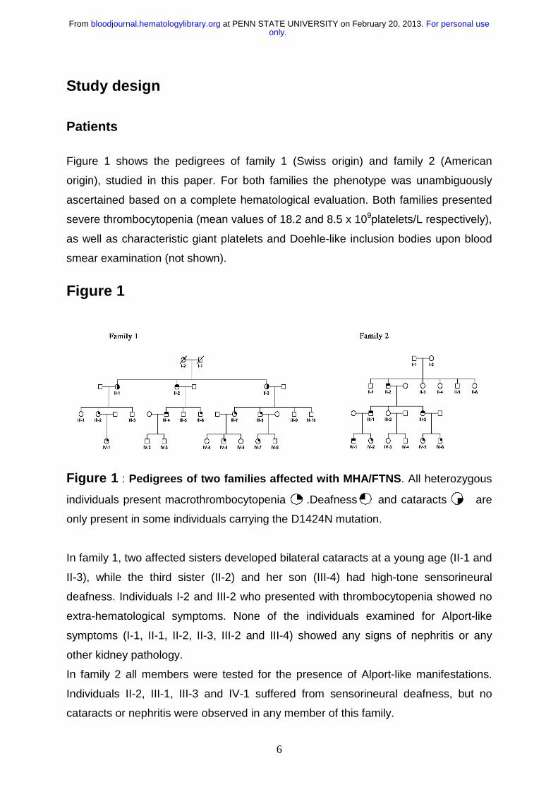
Figure 1 shows the pedigrees of family 1 (Swiss origin) and family 2 (American
origin), studied in this paper. For both families the phenotype was unambiguously
ascertained based on a complete hematological evaluation. Both families presented
severe thrombocytopenia (mean values of 18.2 and 8.5 x 10⁹platelets/L respectively),
as well as characteristic giant platelets and Doehle-like inclusion bodies upon blood
smear examination (not shown).
Figure 1
Figure 1 : Pedigrees of two families affected with MHA/FTNS. All heterozygous
individuals present macrothrombocytopenia .Deafness and cataracts are
only present in some individuals carrying the D1424N mutation.
In family 1, two affected sisters developed bilateral cataracts at a young age (II-1 and
II-3), while the third sister (II-2) and her son (III-4) had high-tone sensorineural
deafness. Individuals I-2 and III-2 who presented with thrombocytopenia showed no
extra-hematological symptoms. None of the individuals examined for Alport-like
symptoms (I-1, II-1, II-2, II-3, III-2 and III-4) showed any signs of nephritis or any
other kidney pathology.
In family 2 all members were tested for the presence of Alport-like manifestations.
Individuals II-2, III-1, III-3 and IV-1 suffered from sensorineural deafness, but no
cataracts or nephritis were observed in any member of this family.
only.For personal use at PENN STATE UNIVERSITY on February 20, 2013. bloodjournal.hematologylibrary.orgFrom
7
Nucleotide sequence analysis
PCR primers were designed to amplify all coding exons as well as the 3’UTR,
together with their intron / exon boundaries. We PCR-amplified all exons in one
affected individual from each pedigree and an unaffected control, in a volume of 25 µl
using 30 ng of genomic DNA per reaction, 1 unit of Taq (Amersham Biosciences,
Buckinghamshire, England) and standard PCR conditions. PCR products were
verified by standard agarose electrophoresis, and purified using the Concert system
(Invitrogen , San Diego, California). Sequencing was performed with an ABI 377
system using the Big Dye terminator sequencing kit (Applied Biosystems, Foster City,
California).
Sequences were then aligned and analyzed using Sequencher 4.0.5 (Gene Codes
Corporation, Ann Arbor, Michigan).
Potential mutations were verified and tested in the population using the
pyrosequencing system 28(Pyrosequencing AB, Uppsala, Sweden). For this 8 µl of
Dynabeads were used for PCR immobilization in a final volume of 40 µl. The rest of
the protocol was performed according to manufacturer's instructions.
Microsatellite Genotyping
Polymorphic markers were analyzed by PCR using radiolabelled primers. One
oligonucleotide primer of each marker was labelled with 5 µCi of γ32P-ATP with T4
polynucleotide kinase. PCR was performed using standard conditions.
Amplification products were separated by electrophoresis in a 6 % denaturing urea /
polyacrylamide gel and genotypes were independently scored by two different
investigators after autoradiography.
Immunohistochemistry
For immunofluorescence staining, bone marrow smears from 5 different donors and
from the MHA-patient (family 1 III-2) were first fixed in 4 % paraformaldehyde (10 min
only.For personal use at PENN STATE UNIVERSITY on February 20, 2013. bloodjournal.hematologylibrary.orgFrom
8
at RT), followed by absolute acetone (3 min at -20 oC). We used an affinity purified
rabbit polyclonal IgG which recognizes only MYH9 (NMMHC-A) and MYH10
(NMMHC-B)29 (Biomedical Technologies Inc., Stoughton, MA). This was followed by
a TRITC-conjugated swine anti-rabbit IgG (Dako, Glostrup, Denmark).The secondary
antibody alone was used as a negative control.
The stained cells were analyzed using a confocal laser scanning fluorescence
inverted microscope (LSM 410; Carl Zeiss, Jena, Germany) equipped with a helium-
neon (He-Ne) laser (excitation wavelength at 543 nm)30. Cells were observed through
an oil immersion planeofluar X63/1.4 objective, and the visual field was enhanced by
zooming in two times.
SDS-PAGE and immunoblotting
For SDS-PAGE, platelet extracts of controls (n=4) and patients (n=3) were
suspended in 0.4 M Tris HCl, PH = 6.8, containing 1 % SDS, 1 % dithiothreitol, 1 mM
phenylmethyl sulfonyl fluoride, 1 mM Nα-p-tosyl-L-arginine methyl ester and boiled
for 3 minutes. Protein content was determined according to Bradford31. Forty µg of
proteins were electrophoresed on a 5-20 % gradient gel and stained with Coomassie
blue. For quantification of total actin, gels were scanned with a computerized scanner
(Arcus II, AGFA, Mortsel, Belgium).
Samples were loaded according to their total actin content and were electrophoresed
on a 5-20 % gradient gel. Western blotting was performed using the same MYH9
antibody as above, a monoclonal antibody recognizing all actin isoforms (clone IC4,
Sigma, St-Louis, Missouri) and a monoclonal antibody specific for α-tubulin (Sigma,
St-Louis, Missouri). Separated proteins were transferred to nitrocellulose filters32
which were incubated with anti-MYH9 (1:1000), anti-total actin (1:10000) and anti-α-
tubulin (1:500) for two hours. After three washes, a second incubation for one hour
was performed with goat anti-rabbit (for MYH9) or anti-mouse (for actin and α-
tubulin) IgG labelled with peroxidase. Enhanced chemiluminescence was used for
detection (Amersham Biosciences, Buckinghamshire, England). Total MYH9 and α-
tubulin expression were evaluated by densitometric scanning of the Western blots
and expressed as mean percentage of control conditions.
only.For personal use at PENN STATE UNIVERSITY on February 20, 2013. bloodjournal.hematologylibrary.orgFrom
9
Statistical comparison between different sample groups was performed using an
unpaired two tailed student T test.
mRNA stability
Total RNA was extracted from blood of three MHA/FTNS subjects, using the Trizol
Reagent (Invitrogen, San Diego, California), and cDNA was produced using the
Superscript II enzyme (Invitrogen, San Diego, California) and an oligo dT primer.
PCR using cDNA specific primers (spanning an intron in genomic DNA; 5'
TGCTGAGGAGGTGAAGAGGA, 3' GCGACAGAGCCTTGGTCTC), in which the
forward primer was labelled with 5’ biotin, was performed under standard conditions,
and the product analysed by the pyrosequencing method. Briefly, this method
(www.pyrosequencing.com) works by a series of four enzymatic reactions in which
the number of nucleotide incorporations is quantitatively measured. Each time a
nucleotide is incorporated by DNA polymerase, a pyrophosphate is released (hence
the term pyrosequencing) which is detected by the sulphorylase and luciferase
enzymes, that generate a signal proportional to the number of nucleotides
incorporated.
For pyrosequencing, an internal primer (CCAGGTCCACCAGCAGG) was designed
two nucleotides before the mutation site, so that the two mRNA populations could be
assayed by quantifying the relative amounts of each allele present in the PCR
product. DNA samples from normal and affected subjects were used as controls.
PCR products were immobilized through the 5' biotin in the forward primer with
Dynabeads (Dynal, Oslo, Norway) by a 15 min, 65oC incubation in a buffer containing
10 mM Tris-HCl, 2 M NaCl, 1 mM EDTA and 0.1 % Tween 20. PCR products were
then removed from solution using magnetic separation, denatured with NaOH 0.5 M
and washed with 200 mM Tris-Acetate, 50 mM Magnesium Acetate. The remaining
single stranded DNA was then hybridized with the internal ‘sequencing’ primer, by
heating the mix to 80oC, and slowly cooling it down to room temperature. Enzyme
and substrate mixes were then automatically added to each well, and the reactions
proceeded at 28oC, with the sequential addition of single nucleotides in a
predetermined order. Luciferase peak heights are proportional to the number of
nucleotide incorporations, which has been shown to be very quantitative (5% error
rate) in a number of experimental settings33,34.
only.For personal use at PENN STATE UNIVERSITY on February 20, 2013. bloodjournal.hematologylibrary.orgFrom
10
As an alternative method for measuring the stability of the mutant mRNA, we used
real-time quantification of the relative proportion of the mutant vs the "wild -type"
allele. For this, we used the ABI 7900 sequence detection system (Applied
Biosystems, Foster City, California).
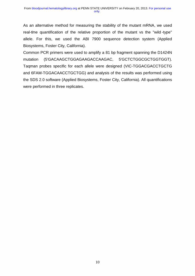
Common PCR primers were used to amplify a 81 bp fragment spanning the D1424N
mutation (5'GACAAGCTGGAGAAGACCAAGAC, 5'GCTCTGGCGCTGGTGGT).
Taqman probes specific for each allele were designed (VIC-TGGACGACCTGCTG
and 6FAM-TGGACAACCTGCTGG) and analysis of the results was performed using
the SDS 2.0 software (Applied Biosystems, Foster City, California). All quantifications
were performed in three replicates.
only.For personal use at PENN STATE UNIVERSITY on February 20, 2013. bloodjournal.hematologylibrary.orgFrom
11
Results
Mutation analysis
All 40 exons of the MYH9 gene including intron / exon junctions and the full 3'UTR
were sequenced in one affected individual from each pedigree, as well as in an
unaffected Caucasian control. Analysis of aligned sequences revealed a missense
mutation in exon 30 of the gene c.4270G>A, which causes a conservative amino acid
substitution D1424N in the rod-like tail domain (data not shown). This mutation, which
surprisingly was present in heterozygosity in both pedigrees, was shown to co-
segregate with the disease phenotype, and to be absent from the general population
as revealed by pyrosequencing analysis of 100 population matched controls .
In order to determine whether the two mutations were of independent origin we
genotyped 5 microsatellite markers (D22S1147, D22S1142, D22S683, D22S283,
D22S445) surrounding an approximate 2Mb region around the mutation (data not
shown).
We concluded that the two mutations are of independent origin since, (i) the mutation
in Pedigree 2 is a de-novo event in individual II-2, as some of his Sibs which share
the same haplotype identical by descent do not carry the mutation; (ii) the mutation in
each pedigree is present on totally different haplotype backgrounds on either side of
the pathogenic mutation.
The D1424N mutation has been previously described in the literature in a pedigree of
Japanese origin, and two pedigrees of American origin, most likely the result of
independent mutation events16,17,27.
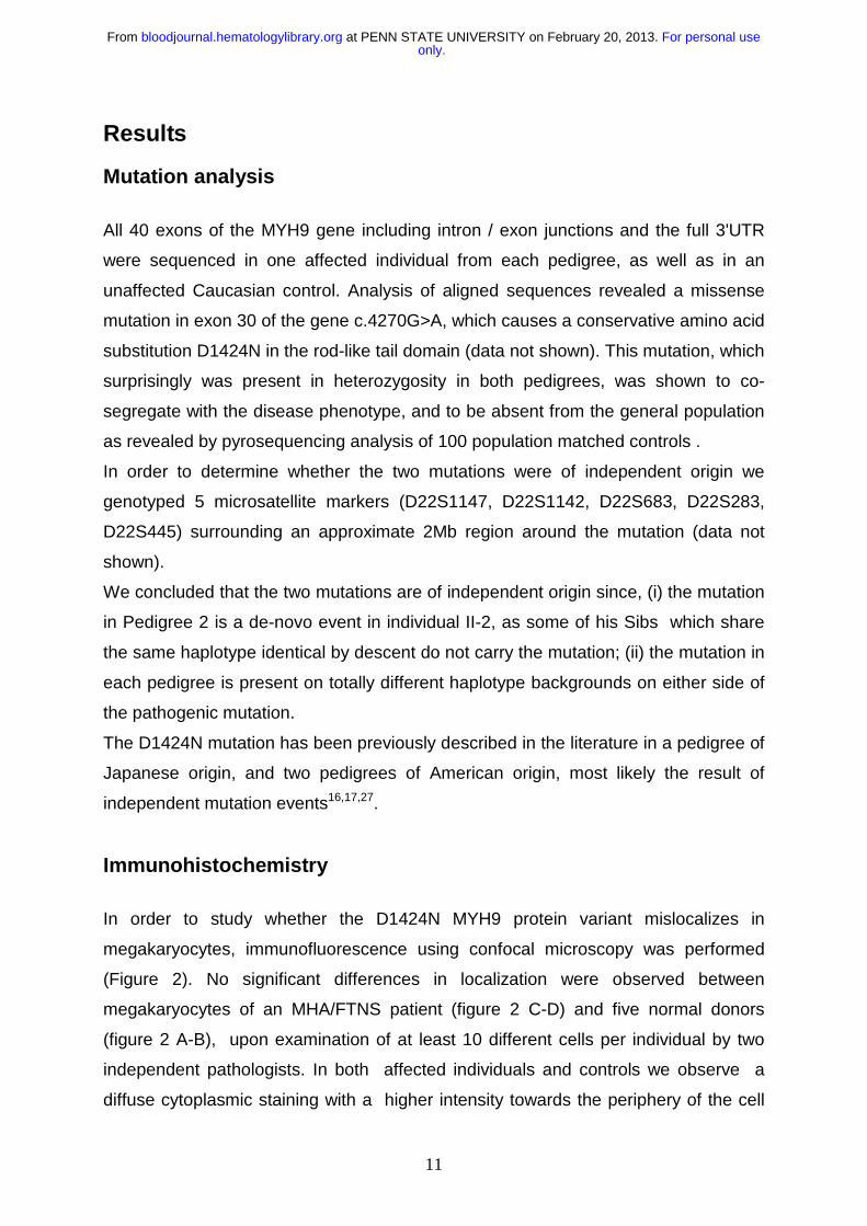
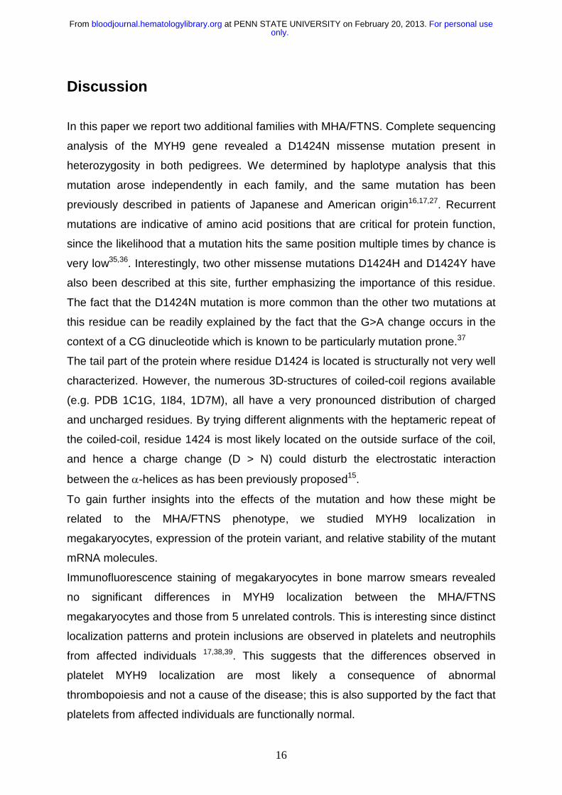
Immunohistochemistry
In order to study whether the D1424N MYH9 protein variant mislocalizes in
megakaryocytes, immunofluorescence using confocal microscopy was performed
(Figure 2). No significant differences in localization were observed between
megakaryocytes of an MHA/FTNS patient (figure 2 C-D) and five normal donors
(figure 2 A-B), upon examination of at least 10 different cells per individual by two
independent pathologists. In both affected individuals and controls we observe a
diffuse cytoplasmic staining with a higher intensity towards the periphery of the cell
only.For personal use at PENN STATE UNIVERSITY on February 20, 2013. bloodjournal.hematologylibrary.orgFrom
12
and the cell membrane. There is thus no evidence that the D1424N mutation causes
changes in localization that could be related to the hematological pathology observed
in MHA/FTNS patients.
Figure 2
Figure 2: MYH9 localization in megakaryocytes. Immunofluorescence
micrographs of the bone marrow from a normal donor (A,B) and the D1424N patient
(C,D). Shown are two megakaryocytes with a typical staining of their cytoskeleton
with MYH9/MYH10 antibodies in one of the 5 donors studied and the patient
respectively.
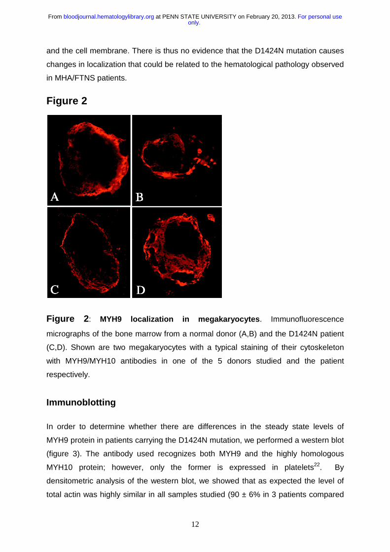
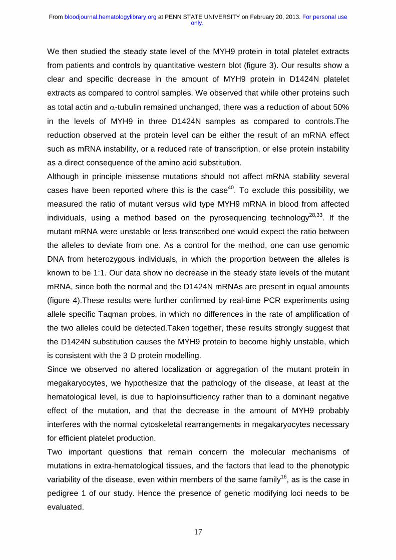
Immunoblotting
In order to determine whether there are differences in the steady state levels of
MYH9 protein in patients carrying the D1424N mutation, we performed a western blot
(figure 3). The antibody used recognizes both MYH9 and the highly homologous
MYH10 protein; however, only the former is expressed in platelets22. By
densitometric analysis of the western blot, we showed that as expected the level of
total actin was highly similar in all samples studied (90 ± 6% in 3 patients compared
only.For personal use at PENN STATE UNIVERSITY on February 20, 2013. bloodjournal.hematologylibrary.orgFrom
13
to the 4 controls, P=0.5). Similarly, the α-tubulin levels did not significantly vary
across the samples (129 ± 13 % in patients compared to controls, P=0.4). The level
of MYH9 however markedly decreased, revealing a specific reduction of 49 ± 9% in
the amount of steady state MYH9 protein in platelets of individuals with the D1424N
mutation as compared to controls. (Patients [n=3], Controls [n=4], P<0.01).
Figure 3
Figure 3 : Immunoblotting. SDS-PAGE of total protein extracts (A,B) and
immunoblots showing MYH9, total actin and α-tubulin expression (C,D) in a
representative control (A,C) and a patient with the D1424N mutation (B,D). Loading
was normalised to total actin concentration.
mRNA stability
In order to investigate whether the c.4270G>A mutation affects the stability of the
MYH9 mRNA transcript, a relative quantification of mutant versus wild type mRNA
molecules from the cDNA of three affected individuals was performed by
pyrosequencing.
only.For personal use at PENN STATE UNIVERSITY on February 20, 2013. bloodjournal.hematologylibrary.orgFrom
14
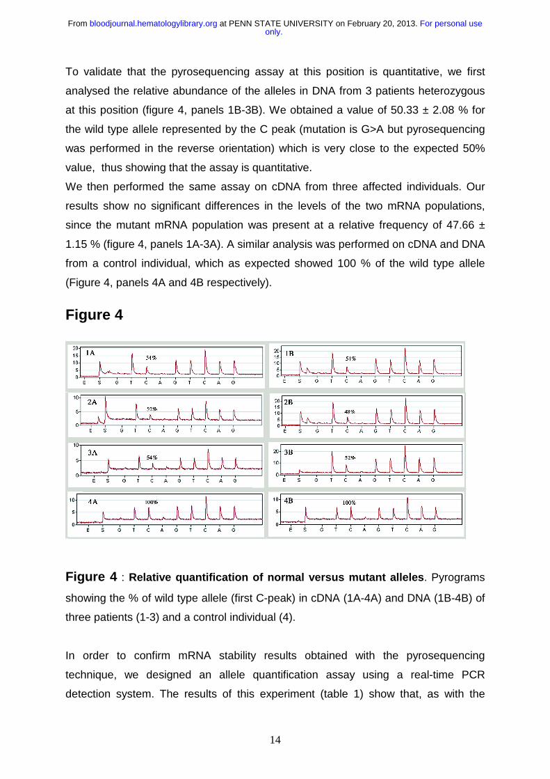
To validate that the pyrosequencing assay at this position is quantitative, we first
analysed the relative abundance of the alleles in DNA from 3 patients heterozygous
at this position (figure 4, panels 1B-3B). We obtained a value of 50.33 ± 2.08 % for
the wild type allele represented by the C peak (mutation is G>A but pyrosequencing
was performed in the reverse orientation) which is very close to the expected 50%
value, thus showing that the assay is quantitative.
We then performed the same assay on cDNA from three affected individuals. Our
results show no significant differences in the levels of the two mRNA populations,
since the mutant mRNA population was present at a relative frequency of 47.66 ±
1.15 % (figure 4, panels 1A-3A). A similar analysis was performed on cDNA and DNA
from a control individual, which as expected showed 100 % of the wild type allele
(Figure 4, panels 4A and 4B respectively).
Figure 4
Figure 4 : Relative quantification of normal versus mutant alleles. Pyrograms
showing the % of wild type allele (first C-peak) in cDNA (1A-4A) and DNA (1B-4B) of
three patients (1-3) and a control individual (4).
In order to confirm mRNA stability results obtained with the pyrosequencing
technique, we designed an allele quantification assay using a real-time PCR
detection system. The results of this experiment (table 1) show that, as with the
only.For personal use at PENN STATE UNIVERSITY on February 20, 2013. bloodjournal.hematologylibrary.orgFrom
15
pyrosequencer, the ratio of the mutant vs the wild type alleles both in DNA and in
cDNA do not significantly deviate from one, demonstrating that their steady state
levels in blood are the same.
Table 1 : Allele ratios quantified by Taqman
Individual RNA DNAFam 1 II-1 1.04± 0.004 1.15± 0.007 Fam 1 III-2 1.04± 0.015 1.15± 0.031 Fam 1 IV-1 1.04± 0.001 1.16± 0.012
only.For personal use at PENN STATE UNIVERSITY on February 20, 2013. bloodjournal.hematologylibrary.orgFrom
16
Discussion
In this paper we report two additional families with MHA/FTNS. Complete sequencing
analysis of the MYH9 gene revealed a D1424N missense mutation present in
heterozygosity in both pedigrees. We determined by haplotype analysis that this
mutation arose independently in each family, and the same mutation has been
previously described in patients of Japanese and American origin16,17,27. Recurrent
mutations are indicative of amino acid positions that are critical for protein function,
since the likelihood that a mutation hits the same position multiple times by chance is
very low35,36. Interestingly, two other missense mutations D1424H and D1424Y have
also been described at this site, further emphasizing the importance of this residue.
The fact that the D1424N mutation is more common than the other two mutations at
this residue can be readily explained by the fact that the G>A change occurs in the
context of a CG dinucleotide which is known to be particularly mutation prone.37
The tail part of the protein where residue D1424 is located is structurally not very well
characterized. However, the numerous 3D-structures of coiled-coil regions available
(e.g. PDB 1C1G, 1I84, 1D7M), all have a very pronounced distribution of charged
and uncharged residues. By trying different alignments with the heptameric repeat of
the coiled-coil, residue 1424 is most likely located on the outside surface of the coil,
and hence a charge change (D > N) could disturb the electrostatic interaction
between the α-helices as has been previously proposed15.
To gain further insights into the effects of the mutation and how these might be
related to the MHA/FTNS phenotype, we studied MYH9 localization in
megakaryocytes, expression of the protein variant, and relative stability of the mutant
mRNA molecules.
Immunofluorescence staining of megakaryocytes in bone marrow smears revealed
no significant differences in MYH9 localization between the MHA/FTNS
megakaryocytes and those from 5 unrelated controls. This is interesting since distinct
localization patterns and protein inclusions are observed in platelets and neutrophils
from affected individuals 17,38,39. This suggests that the differences observed in
platelet MYH9 localization are most likely a consequence of abnormal
thrombopoiesis and not a cause of the disease; this is also supported by the fact that
platelets from affected individuals are functionally normal.
only.For personal use at PENN STATE UNIVERSITY on February 20, 2013. bloodjournal.hematologylibrary.orgFrom
17
We then studied the steady state level of the MYH9 protein in total platelet extracts
from patients and controls by quantitative western blot (figure 3). Our results show a
clear and specific decrease in the amount of MYH9 protein in D1424N platelet
extracts as compared to control samples. We observed that while other proteins such
as total actin and α-tubulin remained unchanged, there was a reduction of about 50%
in the levels of MYH9 in three D1424N samples as compared to controls.The
reduction observed at the protein level can be either the result of an mRNA effect
such as mRNA instability, or a reduced rate of transcription, or else protein instability
as a direct consequence of the amino acid substitution.
Although in principle missense mutations should not affect mRNA stability several
cases have been reported where this is the case40. To exclude this possibility, we
measured the ratio of mutant versus wild type MYH9 mRNA in blood from affected
individuals, using a method based on the pyrosequencing technology28,33. If the
mutant mRNA were unstable or less transcribed one would expect the ratio between
the alleles to deviate from one. As a control for the method, one can use genomic
DNA from heterozygous individuals, in which the proportion between the alleles is
known to be 1:1. Our data show no decrease in the steady state levels of the mutant
mRNA, since both the normal and the D1424N mRNAs are present in equal amounts
(figure 4).These results were further confirmed by real-time PCR experiments using
allele specific Taqman probes, in which no differences in the rate of amplification of
the two alleles could be detected.Taken together, these results strongly suggest that
the D1424N substitution causes the MYH9 protein to become highly unstable, which
is consistent with the 3- D protein modelling.
Since we observed no altered localization or aggregation of the mutant protein in
megakaryocytes, we hypothesize that the pathology of the disease, at least at the
hematological level, is due to haploinsufficiency rather than to a dominant negative
effect of the mutation, and that the decrease in the amount of MYH9 probably
interferes with the normal cytoskeletal rearrangements in megakaryocytes necessary
for efficient platelet production.
Two important questions that remain concern the molecular mechanisms of
mutations in extra-hematological tissues, and the factors that lead to the phenotypic
variability of the disease, even within members of the same family16, as is the case in
pedigree 1 of our study. Hence the presence of genetic modifying loci needs to be
evaluated.
only.For personal use at PENN STATE UNIVERSITY on February 20, 2013. bloodjournal.hematologylibrary.orgFrom
18
In summary, we have characterized the effects of the MYH9 D1424N mutation at the
cellular localization, protein and mRNA level, in order to obtain new insights into the
pathophysiology of MHA/FTNS. We hypothesize that haploinsufficiency resulting
from the high instability of the mutant MYH9 protein could explain the mutation
mechanism, at least at the hematological level.
only.For personal use at PENN STATE UNIVERSITY on February 20, 2013. bloodjournal.hematologylibrary.orgFrom
19
References
1. Epstein CJ, Sahud MA, Piel CF, Goodman JR, Bernfield MR, Kushner JH,
Ablin AR. Hereditary macrothrombocytopathia, nephritis and deafness. Am J Med.
1972;52:299-310.
2. Hegglin R. Gleichzeitige konstitutionelle Veranderungen an Neutrophilen
und Thrombocyten. Helv. Med. Acta. 1945;12:439-440
3. May R. Leukozyteneinschlusse. Dtsch. Arch. Klin. Med. 1909; 96:1-6
4. Peterson LC, Rao KV, Crosson JT, White JG. Fechtner syndrome--a
variant of Alport's syndrome with leukocyte inclusions and macrothrombocytopenia.
Blood. 1985;65:397-406.
5. Coller BS, Zarrabi MH. Platelet membrane studies in the May-Hegglin
anomaly. Blood. 1981;58:279-284.
6. Lusher JM, Schneider J, Mizukami I, Evans RK. The May-Hegglin anomaly:
platelet function, ultrastructure and chromosome studies. Blood. 1968;32:950-961.
7. Godwin HA, Ginsburg AD. May-Hegglin anomaly: a defect in
megakaryocyte fragmentation? Br J Haematol. 1974;26:117-128.
8. Tsurusawa M, Mamiya S. What is the difference between May-Hegglin
anomaly and Sebastian platelet syndrome? International Journal of Hematology.
2000;71:400-401
9. Bernheim J, Dechavanne M, Bryon PA, Lagarde M, Colon S, Pozet N,
Traeger J. Thrombocytopenia, macrothrombocytopathia, nephritis and deafness. Am
J Med. 1976;61:145-150.
10. Eckstein JD, Filip DJ, Watts JC. Hereditary thrombocytopenia, deafness,
and renal disease. Ann Intern Med. 1975;82:639-645.
11. Kunishima S, Kojima T, Tanaka T, Kamiya T, Ozawa K, Nakamura Y,
Saito H. Mapping of a gene for May-Hegglin anomaly to chromosome 22q. Human
Genetics. 1999;105:379-383
12. Kelley MJ, Jawien W, Lin A, Hoffmeister K, Pugh EW, Doheny KF,
Korczak JF. Autosomal dominant macrothrombocytopenia with leukocyte inclusions
(May-Hegglin anomaly) is linked to chromosome 22q12-13. Human Genetics.
2000;106:557-564
only.For personal use at PENN STATE UNIVERSITY on February 20, 2013. bloodjournal.hematologylibrary.orgFrom
20
13. Martignetti JA, Heath KE, Harris J, Bizzaro N, Savoia A, Balduini CL,
Desnick RJ. The gene for May-Hegglin anomaly localizes to a <1-Mb region on
chromosome 22q12.3-13.1. Am J Hum Genet. 2000;66:1449-1454.
14. Kelley MJ, Jawien W, Ortel TL, Korczak JF. Mutation of MYH9, encoding
non-muscle myosin heavy chain A, in May-Hegglin anomaly. Nature Genetics.
2000;26:106-108
15. Seri M, Cusano R, Gangarossa S, Caridi G, Bordo D, Lo Nigro C,
Ghiggeri GM, Ravazzolo R, Savino M, Del Vecchio M, d'Apolito M, Iolascon A,
Zelante LL, Savoia A, Balduini CL, Noris P, Magrini U, Belletti S, Heath KE, Babcock
M, Glucksman MJ, Aliprandis E, Bizzaro N, Desnick RJ, Martignetti JA. Mutations in
MYH9 result in the May-Hegglin anomaly, and Fechtner and Sebastian syndromes.
The May-Heggllin/Fechtner Syndrome Consortium. Nature Genetics. 2000;26:103-
105
16. Heath KE, Campos-Barros A, Toren A, Rozenfeld-Granot G, Carlsson LE,
Savige J, Denison JC, Gregory MC, White JG, Barker DF, Greinacher A, Epstein CJ,
Glucksman MJ, Martignetti JA. Nonmuscle myosin heavy chain IIA mutations define
a spectrum of autosomal dominant macrothrombocytopenias: May-Hegglin anomaly
and Fechtner, Sebastian, Epstein, and Alport-like syndromes. Am J Hum Genet.
2001;69:1033-1045.
17. Kunishima S, Kojima T, Matsushita T, Tanaka T, Tsurusawa M, Furukawa
Y, Nakamura Y, Okamura T, Amemiya N, Nakayama T, Kamiya T, Saito H. Mutations
in the NMMHC-A gene cause autosomal dominant macrothrombocytopenia with
leukocyte inclusions (May-Hegglin anomaly/Sebastian syndrome). Blood.
2001;97:1147-1149
18. Seri M, Savino M, Bordo D, Cusano R, Rocca B, Meloni I, Di Bari F,
Koivisto PA, Bolognesi M, Ghiggeri GM, Landolfi R, Balduini CL, Zelante L,
Ravazzolo R, Renieri A, Savoia A. Epstein syndrome: another renal disorder with
mutations in the nonmuscle myosin heavy chain 9 gene. Hum Genet. 2002;110:182-
186.
19. Simons M, Wang M, McBride OW, Kawamoto S, Yamakawa K, Gdula D,
Adelstein RS, Weir L. Human nonmuscle myosin heavy chains are encoded by two
genes located on different chromosomes. Circ Res. 1991;69:530-539.
20. Toothaker LE, Gonzalez DA, Tung N, Lemons RS, Le Beau MM, Arnaout
MA, Clayton LK, Tenen DG. Cellular myosin heavy chain in human leukocytes:
only.For personal use at PENN STATE UNIVERSITY on February 20, 2013. bloodjournal.hematologylibrary.orgFrom
21
isolation of 5' cDNA clones, characterization of the protein, chromosomal localization,
and upregulation during myeloid differentiation. Blood. 1991;78:1826-1833.
21. Sellers JR. Myosins: a diverse superfamily. Biochim Biophys Acta.
2000;1496:3-22.
22. Maupin P, Phillips CL, Adelstein RS, Pollard TD. Differential localization of
myosin-II isozymes in human cultured cells and blood cells. J Cell Sci.
1994;107:3077-3090.
23. Mansfield PJ, Shayman JA, Boxer LA. Regulation of polymorphonuclear
leukocyte phagocytosis by myosin light chain kinase after activation of mitogen-
activated protein kinase. Blood. 2000;95:2407-2412.
24. Nagasaki A, De Hostos EL, Uyeda TQ. Genetic and morphological
evidence for two parallel pathways of cell- cycle-coupled cytokinesis in Dictyostelium.
J Cell Sci. 2002;115:2241-2251.
25. Lalwani AK, Goldstein JA, Kelley MJ, Luxford W, Castelein CM, Mhatre
AN. Human nonsyndromic hereditary deafness DFNA17 is due to a mutation in
nonmuscle myosin MYH9. Am J Hum Genet. 2000;67:1121-1128.
26. Arrondel C, Vodovar N, Knebelmann B, Grunfeld JP, Gubler MC, Antignac
C, Heidet L. Expression of the nonmuscle myosin heavy chain IIA in the human
kidney and screening for MYH9 mutations in Epstein and Fechtner syndromes. J Am
Soc Nephrol. 2002;13:65-74.
27. Kunishima S, Matsushita T, Kojima T, Amemiya N, Choi YM, Hosaka N,
Inoue M, Jung Y, Mamiya S, Matsumoto K, Miyajima Y, Zhang G, Ruan C, Saito K,
Song KS, Yoon HJ, Kamiya T, Saito H. Identification of six novel MYH9 mutations
and genotype-phenotype relationships in autosomal dominant
macrothrombocytopenia with leukocyte inclusions. J Hum Genet. 2001;46:722-729
28. Alderborn A, Kristofferson A, Hammerling U. Determination of single-
nucleotide polymorphisms by real-time pyrophosphate DNA sequencing. Genome
Res. 2000;10:1249-1258.
29. Benzonana G, Skalli O, Gabbiani G. Correlation between the distribution
of smooth muscle or non muscle myosins and alpha-smooth muscle actin in normal
and pathological soft tissues. Cell Motil Cytoskeleton. 1988;11:260 -274.
30. Bochaton-Piallat ML, Kapetanios AD, Donati G, Redard M, Gabbiani G,
Pournaras CJ. TGF-beta1, TGF-beta receptor II and ED-A fibronectin expression in
myofibroblast of vitreoretinopathy. Invest Ophthalmol Vis Sci. 2000;41:2336-2342.
only.For personal use at PENN STATE UNIVERSITY on February 20, 2013. bloodjournal.hematologylibrary.orgFrom
22
31. Bradford MM. A rapid and sensitive method for the quantitation of
microgram quantities of protein utilizing the principle of protein-dye binding. Anal
Biochem. 1976;72:248-254.
32. Towbin H, Staehelin T, Gordon J. Electrophoretic transfer of proteins from
polyacrylamide gels to nitrocellulose sheets: procedure and some applications. Proc
Natl Acad Sci U S A. 1979;76:4350-4354.
33. Neve B, Froguel P, Corset L, Vaillant E, Vatin V, Boutin P. Rapid SNP
allele frequency determination in genomic DNA pools by pyrosequencing.
Biotechniques. 2002;32:1138-1142.
34. Wasson J, Skolnick G, Love-Gregory L, Permutt MA. Assessing allele
frequencies of single nucleotide polymorphisms in DNA pools by pyrosequencing
technology. Biotechniques. 2002;32:1144-1146, 1148, 1150 passim.
35. Dragich J, Houwink-Manville I, Schanen C. Rett syndrome: a surprising
result of mutation in MECP2. Hum Mol Genet. 2000;9:2365-2375.
36. Shiang R, Thompson LM, Zhu YZ, Church DM, Fielder TJ, Bocian M,
Winokur ST, Wasmuth JJ. Mutations in the transmembrane domain of FGFR3 cause
the most common genetic form of dwarfism, achondroplasia. Cell. 1994;78:335-342.
37. Stylianos E. Antonarakis MK, David N. Cooper. The Nature and
Mechanisms of Human Gene Mutation. In: Charles R. Scriver ALB, William S. Sly,
David Valle., ed. The Metabolic & Molecular Bases of Inherited Disease. Vol. I (ed
eight edition): McGraw-Hill; 2001:343-377
38. Pecci A, Noris P, Invernizzi R, Savoia A, Seri M, Ghiggeri GM, Sartore S,
Gangarossa S, Bizzaro N, Balduini CL. Immunocytochemistry for the heavy chain of
the non-muscle myosin IIA as a diagnostic tool for MYH9-related disorders. Br J
Haematol. 2002;117:164-167.
39. Kunishima S, Matsushita T, Kojima T, Sako M, Kimura F, Jo EK, Inoue C,
Kamiya T, Saito H. Immunofluorescence Analysis of Neutrophil Nonmuscle Myosin
Heavy Chain-A in MYH9 Disorders: Association of Subcellular Localization with
MYH9 Mutations. Lab Invest. 2003;83:115-122.
40. Cartegni L, Chew SL, Krainer AR. Listening to silence and understanding
nonsense: exonic mutations that affect splicing. Nat Rev Genet. 2002;3:285-298.
only.For personal use at PENN STATE UNIVERSITY on February 20, 2013. bloodjournal.hematologylibrary.orgFrom