Embed Size (px)
Citation preview
Listen to this manuscript’s
audio summary by
JACC Editor-in-Chief
Dr. Valentin Fuster.
J O U R N A L O F T H E AM E R I C A N C O L L E G E O F C A R D I O L O G Y V O L . 7 2 , N O . 2 0 , 2 0 1 8
ª 2 0 1 8 T H E A U T H O R S . P U B L I S H E D B Y E L S E V I E R O N B E H A L F O F T H E A M E R I C A N
C O L L E G E O F C A R D I O L O G Y F OU N D A T I O N . T H I S I S A N O P E N A C C E S S A R T I C L E U N D E R
T H E C C B Y - N C - N D L I C E N S E ( h t t p : / / c r e a t i v e c o mm o n s . o r g / l i c e n s e s / b y - n c - n d / 4 . 0 / ) .
Formin Homology 2 Domain Containing 3(FHOD3) Is a Genetic Basis forHypertrophic Cardiomyopathy
Juan Pablo Ochoa, MD,a,b María Sabater-Molina, MD, PHD,c José Manuel García-Pinilla, MD, PHD,dJens Mogensen, MD, PHD,e Alejandra Restrepo-Córdoba, MD,f,g Julián Palomino-Doza, MD, PHD,h
Eduardo Villacorta, MD,i Marina Martinez-Moreno, MD,j Javier Ramos-Maqueda, MD,k Esther Zorio, MD, PHD,l
Maria L. Peña-Peña, MD,b,m Pablo E. García-Granja, MD,n José F. Rodríguez-Palomares, MD, PHD,o
Ivonne J. Cárdenas-Reyes, MD,a María M. de la Torre-Carpente, MD, PHD,p Alicia Bautista-Pavés, MD,q
Mohammed M. Akhtar, MD,r,g Marcos N. Cicerchia, MD,a Raquel Bilbao-Quesada, MD,s
Maria Victoria Mogollón-Jimenez, MD,t Joel Salazar-Mendiguchía, MD,a,u José M. Mesa Latorre, MD,v
Blanca Arnaez, MD,w Ivan Olavarri-Miguel, MD,x María E. Fuentes-Cañamero, MD,y Arsonval Lamounier JR, MD,a,b
José María Pérez Ruiz, MD,z Vicente Climent-Payá, MD,aa,bb Inmaculada Pérez-Sanchez, MD, PHD,c
Juan P. Trujillo-Quintero, MD, PHD,a Luis R. Lopes, MD PHD,r,g,cc Alfredo Repáraz-Andrade, MD,dd
Rosario Marín-Iglesias, MD,ee Alejandro Rodriguez-Vilela, MD,ff María Sandín-Fuentes, MD,n
Jose A. Garrote, MD, PHD,gg Alejandro Cortel-Fuster, MD,hh Miguel Lopez-Garrido, MD,c Ana Fontalba-Romero, MD,ii
Tomás Ripoll-Vera, MD, PHD,jj Isabel Llano-Rivas, MD,kk Xusto Fernandez-Fernandez, MD,a
María Isidoro-García, MD, PHD,ll,mm Diego Garcia-Giustiniani, MD,a Roberto Barriales-Villa, MD, PHD,nn,oo
Martín Ortiz-Genga, MD,a,b Pablo García-Pavía, MD, PHD,f,g Perry M. Elliott, MD, PHD,g,r,cc
Juan R. Gimeno, MD, PHD,c,g Lorenzo Monserrat, MD, PHDa
ABSTRACT
ISS
BACKGROUND The genetic cause of hypertrophic cardiomyopathy remains unexplained in a substantial proportion of
cases. Formin homology 2 domain containing 3 (FHOD3) may have a role in the pathogenesis of cardiac hypertrophy but
has not been implicated in hypertrophic cardiomyopathy.
OBJECTIVES This study sought to investigate the relation between FHOD3 mutations and the development of hy-
pertrophic cardiomyopathy.
METHODS FHOD3 was sequenced by massive parallel sequencing in 3,189 hypertrophic cardiomyopathy unrelated
probands and 2,777 patients with no evidence of cardiomyopathy (disease control subjects). The authors evaluated
protein-altering candidate variants in FHOD3 for cosegregation, clinical characteristics, and outcomes.
RESULTS The authors identified 94 candidate variants in 132 probands. The variants’ frequencies were significantly
higher in patients with hypertrophic cardiomyopathy (74 of 3,189 [2.32%]) than in disease control subjects (18 of 2,777
[0.65%]; p < 0.001) or in the gnomAD database (1,049 of 138,606 [0.76%]; p < 0.001). FHOD3 mutations cosegre-
gated with hypertrophic cardiomyopathy in 17 families, with a combined logarithm of the odds score of 7.92, indicative of
very strong segregation. One-half of the disease-causing variants were clustered in a small conserved coiled-coil domain
(amino acids 622 to 655); odds ratio for hypertrophic cardiomyopathy was 21.8 versus disease control subjects
(95% confidence interval: 1.3 to 37.9; p < 0.001) and 14.1 against gnomAD (95% confidence interval: 6.9 to 28.7; p <
0.001). Hypertrophic cardiomyopathy patients carrying (likely) pathogenic mutations in FHOD3 (n ¼ 70) were diagnosed
after age 30 years (mean 46.1� 18.7 years), and two-thirds (66%)weremales. Of the patients, 82%had asymmetric septal
hypertrophy (mean 18.8 � 5 mm); left ventricular ejection fraction <50% was present in 14% and hypertrabeculation in
16%. Events were rare before age 30 years, with an annual cardiovascular death incidence of 1% during follow-up.
CONCLUSIONS FHOD3 is a novel disease gene in hypertrophic cardiomyopathy, accounting for approximately 1% to 2%
of cases. The phenotype and the rate of cardiovascular events are similar to those reported in unselected cohorts. The
FHOD3 gene should be routinely included in hypertrophic cardiomyopathy genetic testing panels. (J Am Coll Cardiol
2018;72:2457–67) © 2018 The Authors. Published by Elsevier on behalf of the American College of Cardiology Foundation.
This is an open access article under the CC BY-NC-ND license (http://creativecommons.org/licenses/by-nc-nd/4.0/).
N 0735-1097 https://doi.org/10.1016/j.jacc.2018.10.001

ABBR EV I A T I ON S
AND ACRONYMS
DCM = dilated cardiomyopathy
FHOD3 = formin homology 2
domain containing 3 gene
GWAS = genome-wide
association studies
HCM = hypertrophic
cardiomyopathy
LOD = logarithm of the odds
MAF = minor allele frequency
SCD = sudden cardiac death
From the
Research G
Departmen
Cardiac Dis
de Hierro
Network o
UniversitarkHospital U
Universitar
Spain; oHoqHospital U
United Kin
diology, CvHospital U
relavega, S
Cristina, Ca
Universitar
FoundatioddComplex
Mar, Cádiz
Molecular
versitario M
Palma de
Medicine,
Spain; nnIn
versitario A
Research U
tigación Bi
Dr. Garcia-
Biobank PT
Cicerchia,
sonal fees
Monserrat
to the cont
Manuscrip
Ochoa et al. J A C C V O L . 7 2 , N O . 2 0 , 2 0 1 8
FHOD3 is an HCM-Causing Gene N O V E M B E R 1 3 / 2 0 , 2 0 1 8 : 2 4 5 7 – 6 7
2458
H ypertrophic cardiomyopathy(HCM) is the most commoninherited cardiomyopathy and is
characterized by clinical variability and ge-netic heterogeneity (1). The introduction ofmassive parallel sequencing has improvedthe understanding of the disease, but >40%of genetic studies reveal no pathogenicmutation, suggesting that new disease-associated genes remain to be discovered.One potential candidate is formin homology2 domain containing 3 (FHOD3) gene.
SEE PAGE 2468
Formins are a family of proteins containing aseries of conserved domains and functional motifsthat regulate actin dynamics (2). FHOD3 protein isexpressed exclusively in the heart and plays animportant role in sarcomere organization, myofi-brillogenesis, and maintenance of the contractileapparatus in cardiomyocytes (3,4). Functional andgenome-wide association studies (GWAS) havesuggested a potential role of FHOD3 in the
aHealth in Code S.L., Scientific Department, A Coruña, Spain; b
roup), A Coruña, Spain; cHospital Clínico Universitario Virgen
t of Cardiology, Murcia, Spain; dHospital Universitario Virgen de
eases Unit, Málaga, Spain; eOdense Universitetshospital, Cardiolo
Majadahonda, Cardiology, Heart Failure and Inherited Cardiac
n Rare and Complex Diseases of the Heart; hHospital Universitario
io de Salamanca, Cardiology, Salamanca, Spain; jHospital Gener
niversitario Virgen de Valme, Cardiology, Sevilla, Spain; lHosp
io Virgen del Rocío, Cardiology, Sevilla, Spain; nHospital Clínico
spital Vall d’Hebron, Cardiology, Barcelona, Spain; pHospital Univ
niversitario San Cecilio, Cardiology, Granada, Spain; rSaint Bar
gdom; sComplexo Hospitalario Universitario de Vigo, Cardiology,
áceres, Spain; uUniversitat Autónoma de Barcelona, Departamen
niversitario Príncipe de Asturias, Clinical Genetics, Alcalá de He
pain; xHospital Universitario Marqués de Valdecilla, Cardiology
rdiology, Badajoz, Spain; zHospital Regional Universitario “Carlos
io de Alicante, Cardiology, Alicante, Spain; bbAlicante Institute for
n), Alicante, Spain; ccUniversity College London Institute for
o Hospitalario Universitario de Vigo, Genetics and Molecular Patho
, Spain; ffComplexo Hospitalario Arquitecto Marcide, Cardiology, E
Genetics Laboratory, Valladolid, Spain; hhHospital Provincial Cas
arqués de Valdecilla, Genetics, Santander, Spain; jjHospital Son L
Mallorca, Spain; kkHospital Universitario Cruces, Clinical Genet
Salamanca, Spain; mmHospital Universitario de Salamanca, Mol
stituto de Investigación Biomédica de A Coruña (INIBIC), A Co
Coruña, Cardiology, A Coruña, Spain. This study was support
niversity College London Hospitals Biomedical Research Centre
omédica en Red (CIBERCV), “Instituto de Salud Carlos III,” CB16/1
Pavia); “Instituto de Salud Carlos III,” FEDER “Union Europea, U
17/0015/0043) (to Dr. Molina) and PI17/01941 (to Dr. Garcia-Pavia
García-Giustiniani, Trujillo, and Ortiz-Genga are employees of He
from Health in Code SL. Dr. Fernandez-Fernandez is an emplo
is a stakeholder and CEO of Health in Code SL. All other authors ha
ents of this paper to disclose.
t received February 25, 2018; revised manuscript received August
pathogenesis of cardiac hypertrophy (5), but nopathogenic variants clearly associated with HCM havebeen reported.
At the beginning of 2014, we included FHOD3 ingenetic panels used to screen patients with inheritedcardiomyopathies. After detecting an FHOD3 variantthat segregated with HCM in a large Spanish family,we performed a systematic evaluation of FHOD3mutations in a larger cohort of patients and in acontrol population.
METHODS
From February 2014 to August 2017, FHOD3 wassequenced using next-generation sequencing in7,881 consecutive unrelated probands with a diag-nosis of different inherited cardiac conditionsreferred to our center for molecular genetic diag-nosis. The phenotypes were established by eachcenter prior to the genetic studies. Patients’ sampleswere referred mainly from centers from Spain, fol-lowed by centers from the United Kingdom,Denmark, United States, Germany, and Argentina.
Universidade da Coruña, GRINCAR (Cardiovascular
de la Arrixaca, Inherited Cardiac Diseases Unit,
la Victoria, Cardiology, Heart Failure and Inherited
gy, Odense, Denmark; fHospital Universitario Puerta
Diseases Unit, Madrid, Spain; gEuropean Reference
12 de Octubre, Cardiology, Madrid, Spain; iHospital
al Universitario de Elche, Cardiology, Elche, Spain;
ital Universitario La Fe, Valencia, Spain; mHospital
Universitario de Valladolid, Cardiology, Valladolid,
ersitario Rio Hortega, Cardiology, Valladolid, Spain;
tholomew’s Hospital, Barts Heart Centre, London,
Vigo, Spain; tHospital San Pedro de Alcántara, Car-
t de Genetica i de Microbiologia, Barcelona, Spain;
nares, Spain; wHospital Sierrallana, Cardiology, Tor-
, Santander, Spain; yHospital Universitario Infanta
Haya,” Cardiology, Málaga, Spain; aaHospital General
Health and Biomedical Research (ISABIAL-FIDABIO
Cardiovascular Science, London, United Kingdom;
logy, Vigo, Spain; eeHospital Universitario Puerta del
l Ferrol, Spain; ggHospital Universitario Rio Hortega,
tellón, Cardiology, Castellon, Spain; iiHospital Uni-
latzer, Cardiology, Inherited Cardiomyopathies Unit,
ics, Barakaldo, Spain; llUniversidad de Salamanca,
ecular Genetics and Pharmacogenetics, Salamanca,
ruña, Spain; and the ooComplexo Hospitalario Uni-
ed by grants from the National Institute for Health
(to Drs. Elliot, Akhtar, and Lopes); Centro de Inves-
1/00425 (to Dr. Barriales-Villa) and CB16/11/00432 (to
na forma de hacer Europa” (PI14/01477 and La Fe
). Drs. Ochoa, Cárdenas-Reyes, Salazar-Mendiguchía,
alth in Code SL. Dr. Barriales-Villa has received per-
yee of and stakeholder of Health In Code SL. Dr.
ve reported that they have no relationships relevant
6, 2018, accepted August 14, 2018.
J A C C V O L . 7 2 , N O . 2 0 , 2 0 1 8 Ochoa et al.N O V E M B E R 1 3 / 2 0 , 2 0 1 8 : 2 4 5 7 – 6 7 FHOD3 is an HCM-Causing Gene
2459
Of the probands, 3,189 had a diagnosis of HCM. Anadditional 2,777 index cases with no evidence ofstructural cardiac disease (mainly channelopathiesand aortic diseases) were used as control subjects.The predominant ethnicity was European (>90% ofthe probands), and there were no differences be-tween HCM probands and control subjects. Thevariants’ frequencies in the general population wereextracted from the gnomAD database version r2.0,August 2017 (6).
In the initial screening phase, candidate variants inFHOD3 were identified and their frequenciescompared in HCM versus control subgroups. Weapplied an MAF (minor allele frequency) threshold of5 � 10�5 to consider a variant a candidate, followingthe same arguments used by Walsh et al. (7) to reas-sess gene pathogenicity in cardiomyopathies; we alsoexcluded variants with an MAF $1� 10�4 in any sub-population of gnomAD to avoid variants detected incases could be enriched in 1 specific population. Onlyunrelated index cases were included in the screeningphase of the study. Patients with cardiomyopathiesother than HCM and sudden cardiac death (SCD) vic-tims were excluded from the analysis.
In the second phase, HCM probands carryingcandidate variants in FHOD3 were invited to partici-pate in segregation studies. Clinical and genetic fa-milial cascade screening was performed followingwritten informed consent in those who agreed toparticipate. Carriers of variants in other sarcomericgenes that could be related to the phenotype (path-ogenic, likely pathogenic, or of uncertain significancein a priority gene) were excluded. The clinical char-acteristics and outcomes in carriers of pathogenic orlikely pathogenic variants in FHOD3 with HCM(including probands and relatives) were assessed. Thestudy protocol was approved by the Research EthicsCommittee of A Coruña-Ferrol (registry code 2015/576).
GENETIC STUDIES, VARIANT FILTERING, AND
VARIANT CLASSIFICATION. Coding exons andintronic boundaries of 213 genes related to inheritedcardiovascular diseases and SCD (Online Table 1)were captured using a custom probe library (Sure-Select Target Enrichment Kit for Illumina paired-endmultiplexed sequencing method, Agilent Technolo-gies, Santa Clara, California) and sequenced usingthe HiSeq 1500 platform (Illumina, San Diego, Cali-fornia) following lllumina protocols. The read depth(number of times that a base was sequenced byindependent reads) of every nucleotide of genesrelated to the referring phenotype (including FHOD3)was >30� (mean 250� to 400�). Exons that did
not fulfill this standard were complementarysequenced using the Sanger method. Only likelyprotein-altering variants (missense, in-frame in-sertions/deletions, frameshift, nonsense, andconsensus splice site mutations) in the most rele-vant, longest transcript of the FHOD3 gene(NM_001281740.1; 1,644 amino acids) were analyzed.Bioinformatics analysis was performed by means of acustom pipeline including software for variant call-ing, genotyping, and annotation.
To establish the pathogenicity of identified vari-ants, we developed a customized classificationscheme based on the recommendations of the Amer-ican College of Medical Genetics and Genomics(Online Table 2) (8); the final classification of eachvariant was agreed by consensus between 2 cardiol-ogists with experience in interpretation of geneticvariants.
STATISTICAL ANALYSIS. Continuous variables wereexpressed as mean � SD, and comparison betweengroups was performed using the Student’s t-test orthe Mann-Whitney U test according to values distri-bution. Noncontinuous variables were expressed asan integer number (percent of total) and comparedusing the chi-square test or Fisher exact test, asappropriate. A 2-sided p value <0.05 was consideredto indicate statistical significance. Analysis was per-formed using R version 3.4.3 (R Foundation for Sta-tistical Computing, Vienna, Austria).
LOGARITHMS OF ODDS SCORE CALCULATION. Wecalculated 2-point logarithm of the odds (LOD) scorefor informative families by using the PARAMLINKpackage for R software (9). The model was set withq ¼ 0, phenocopy rate ¼ 0.005, and 2 differentpenetrance values: 0.80 and 0.95. An indeterminatestatus was assigned to family members with a con-founding cardiac diagnosis, as well as to men youngerthan age 45 years and women younger than age 50years who did not meet clinical criteria for HCM andcould develop the disease afterwards.
SURVIVAL ANALYSIS. The cumulative probability ofcardiovascular death in carriers of disease-causingmutations in FHOD3 after a diagnosis of HCM(follow-up) was estimated using the Kaplan-Meiermethod. Cardiovascular death was defined as thepresence of sudden cardiac death, appropriate defi-brillator shock, heart failure death, or heart trans-plantation. The beginning of the follow-up wasestablished as the first clinical visit when the patienthad a diagnosis of the disease (unaffected carriers andSCD cases in which HCM was diagnosed postmortemwere excluded from this analysis).
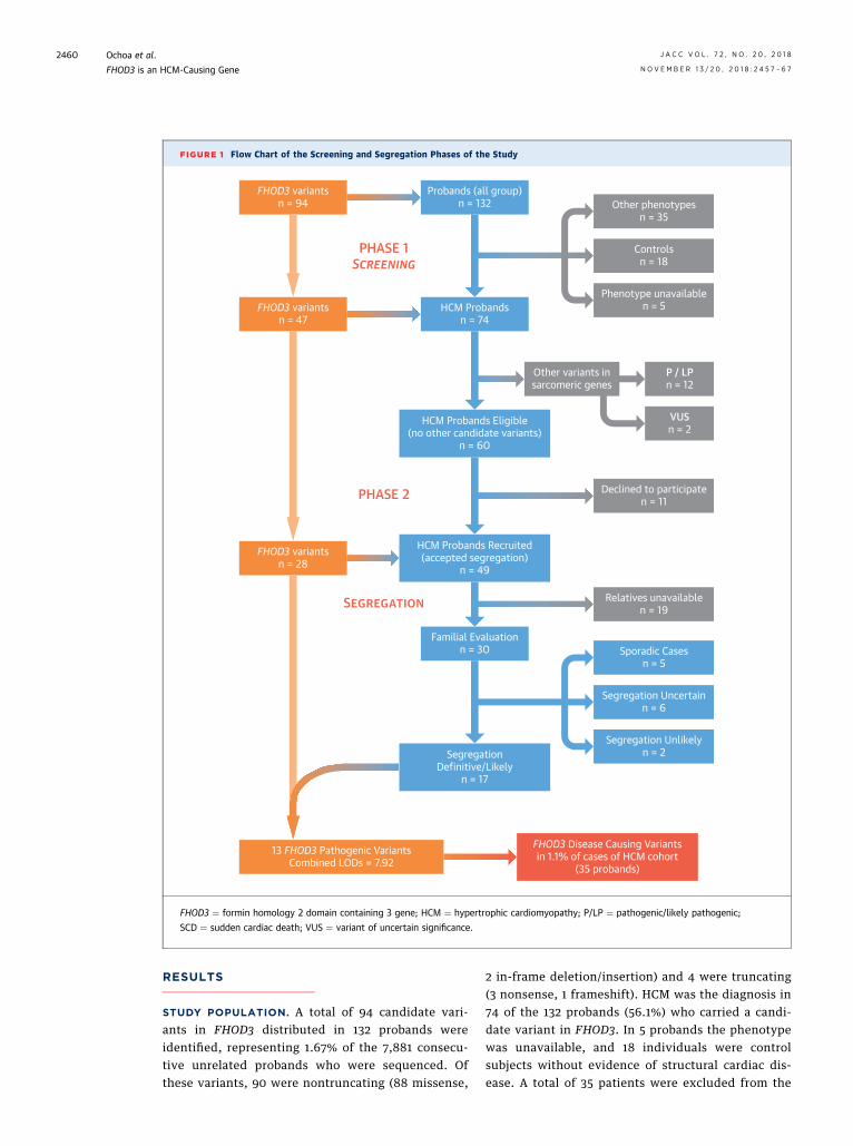
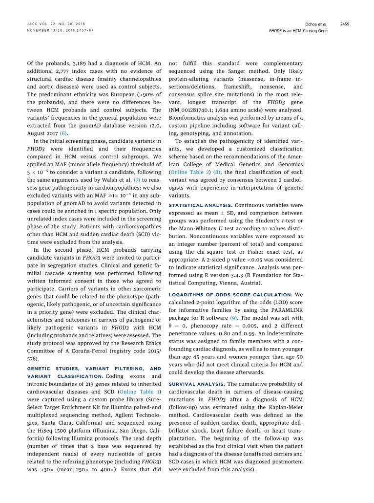
FIGURE 1 Flow Chart of the Screening and Segregation Phases of the Study
FHOD3 variantsn = 94
FHOD3 variantsn = 47
Probands (all group)n = 132
HCM Probandsn = 74
Familial Evaluationn = 30
Other phenotypesn = 35
Controlsn = 18
Phenotype unavailablen = 5
Sporadic Casesn = 5
Segregation Uncertainn = 6
Segregation Unlikelyn = 2
P / LPn = 12
Other variants insarcomeric genes
PHASE 1SCREENING
SEGREGATION
PHASE 2
VUSn = 2
Declined to participaten = 11
Relatives unavailablen = 19
HCM Probands Eligible(no other candidate variants)
n = 60
HCM Probands Recruited(accepted segregation)
n = 49
SegregationDefinitive/Likely
n = 17
FHOD3 Disease Causing Variantsin 1.1% of cases of HCM cohort
(35 probands)
FHOD3 variantsn = 28
13 FHOD3 Pathogenic VariantsCombined LODs = 7.92
FHOD3 ¼ formin homology 2 domain containing 3 gene; HCM ¼ hypertrophic cardiomyopathy; P/LP ¼ pathogenic/likely pathogenic;
SCD ¼ sudden cardiac death; VUS ¼ variant of uncertain significance.
Ochoa et al. J A C C V O L . 7 2 , N O . 2 0 , 2 0 1 8
FHOD3 is an HCM-Causing Gene N O V E M B E R 1 3 / 2 0 , 2 0 1 8 : 2 4 5 7 – 6 7
2460
RESULTS
STUDY POPULATION. A total of 94 candidate vari-ants in FHOD3 distributed in 132 probands wereidentified, representing 1.67% of the 7,881 consecu-tive unrelated probands who were sequenced. Ofthese variants, 90 were nontruncating (88 missense,
2 in-frame deletion/insertion) and 4 were truncating(3 nonsense, 1 frameshift). HCM was the diagnosis in74 of the 132 probands (56.1%) who carried a candi-date variant in FHOD3. In 5 probands the phenotypewas unavailable, and 18 individuals were controlsubjects without evidence of structural cardiac dis-ease. A total of 35 patients were excluded from the
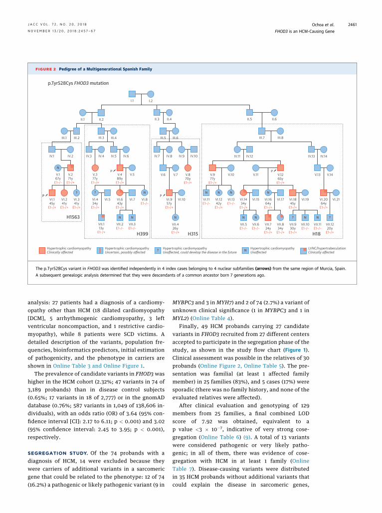
FIGURE 2 Pedigree of a Multigenerational Spanish Family
I.1
II.1
III.1
IV.1
V.167y
E1–/–
VI.145y
E1–/+
PVI.241y
E1–/+
H1563
p.Tyr528Cys FHOD3 mutation
H399 H315 H18
VI.341y
E1–/+
?
N
? N
N N
N N N
N N N N? ?
N
N
VI.434y
E1–/+
VI.5 VI.7
VII.113y
E1–/+
VII.2E1–/–
VII.3E1–/–
VI.643y
E1–/+
VI.8E1–/–
VI.957y
E1–/+
VI.1242y
E1–/+
VI.1434y
E1–/+
VI.1664y
E1–/+
VI.1845y
E1–/+
VI.2064y
E1–/+
VI.11E1–/–
VI.13E1–/–
VII.426y
E1–/+
VII.724y
E1–/+
VII.834y
E1–/+
VII.930y
E1–/+
VII.1220y
E1–/+
VII.5E1–/–
VII.6E1–/–
VII.10E1–/–
VII.11E1–/–
VI.10 VI.15 VI.17 VI.19 VI.21
V.271y
E1–/+
V.377y
E1–/+
V.489y
E1–/+
V.5 V.870y
E1–/+
V.7V.6 V.977y
E1–/+
V.1265y
E1–/+
V.10 V.11 V.13 V.14
IV.2 IV.3 IV.4 IV.5 IV.6 IV.7 IV.8 IV.9 IV.10
III.2 III.3 III.4 III.5 III.6
II.2 II.3 II.4 II.5 II.6
Hypertrophic cardiomyopathyClinically affected ? Hypertrophic cardiomyopathy
Unaffected, could develop the disease in the future N Hypertrophic cardiomyopathyUnaffected
III.7
IV.11 IV.12 IV.13 IV.14
III.8
I.2
LVNC/hypertrabeculationClinically affected
P P
P
Hypertrophic cardiomyopathyUncertain, possibly affected
The p.Tyr528Cys variant in FHOD3 was identified independently in 4 index cases belonging to 4 nuclear subfamilies (arrows) from the same region of Murcia, Spain.
A subsequent genealogic analysis determined that they were descendants of a common ancestor born 7 generations ago.
J A C C V O L . 7 2 , N O . 2 0 , 2 0 1 8 Ochoa et al.N O V E M B E R 1 3 / 2 0 , 2 0 1 8 : 2 4 5 7 – 6 7 FHOD3 is an HCM-Causing Gene
2461
analysis: 27 patients had a diagnosis of a cardiomy-opathy other than HCM (18 dilated cardiomyopathy[DCM], 5 arrhythmogenic cardiomyopathy, 3 leftventricular noncompaction, and 1 restrictive cardio-myopathy), while 8 patients were SCD victims. Adetailed description of the variants, population fre-quencies, bioinformatics predictors, initial estimationof pathogenicity, and the phenotype in carriers areshown in Online Table 3 and Online Figure 1.
The prevalence of candidate variants in FHOD3 washigher in the HCM cohort (2.32%; 47 variants in 74 of3,189 probands) than in disease control subjects(0.65%; 17 variants in 18 of 2,777) or in the gnomADdatabase (0.76%; 587 variants in 1,049 of 138,606 in-dividuals), with an odds ratio (OR) of 3.64 (95% con-fidence interval [CI]: 2.17 to 6.11; p < 0.001) and 3.02(95% confidence interval: 2.45 to 3.95; p < 0.001),respectively.
SEGREGATION STUDY. Of the 74 probands with adiagnosis of HCM, 14 were excluded because theywere carriers of additional variants in a sarcomericgene that could be related to the phenotype: 12 of 74(16.2%) a pathogenic or likely pathogenic variant (9 in
MYBPC3 and 3 in MYH7) and 2 of 74 (2.7%) a variant ofunknown clinical significance (1 in MYBPC3 and 1 inMYL2) (Online Table 4).
Finally, 49 HCM probands carrying 27 candidatevariants in FHOD3 recruited from 27 different centersaccepted to participate in the segregation phase of thestudy, as shown in the study flow chart (Figure 1).Clinical assessment was possible in the relatives of 30probands (Online Figure 2, Online Table 5). The pre-sentation was familial (at least 1 affected familymember) in 25 families (83%), and 5 cases (17%) weresporadic (there was no family history, and none of theevaluated relatives were affected).
After clinical evaluation and genotyping of 129members from 25 families, a final combined LODscore of 7.92 was obtained, equivalent to ap value <3 � 10�7, indicative of very strong cose-gregation (Online Table 6) (9). A total of 13 variantswere considered pathogenic or very likely patho-genic; in all of them, there was evidence of cose-gregation with HCM in at least 1 family (OnlineTable 7). Disease-causing variants were distributedin 35 HCM probands without additional variants thatcould explain the disease in sarcomeric genes,
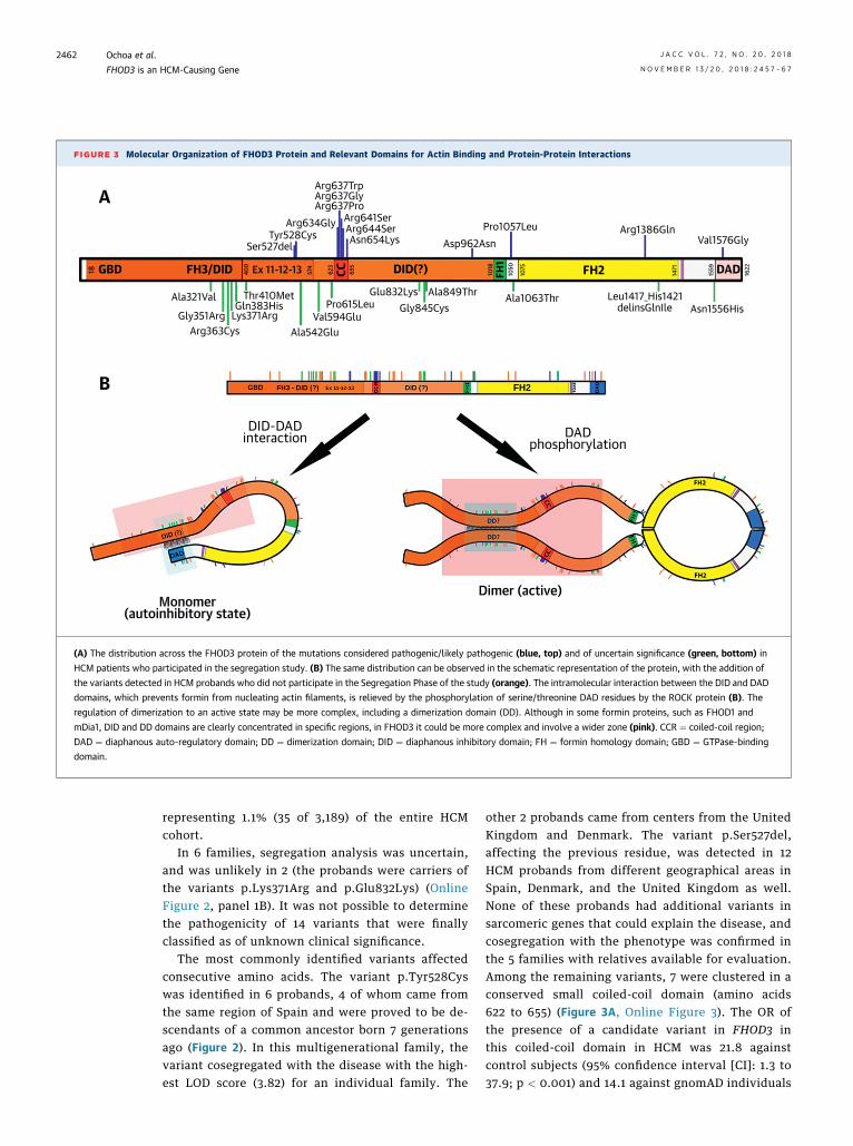
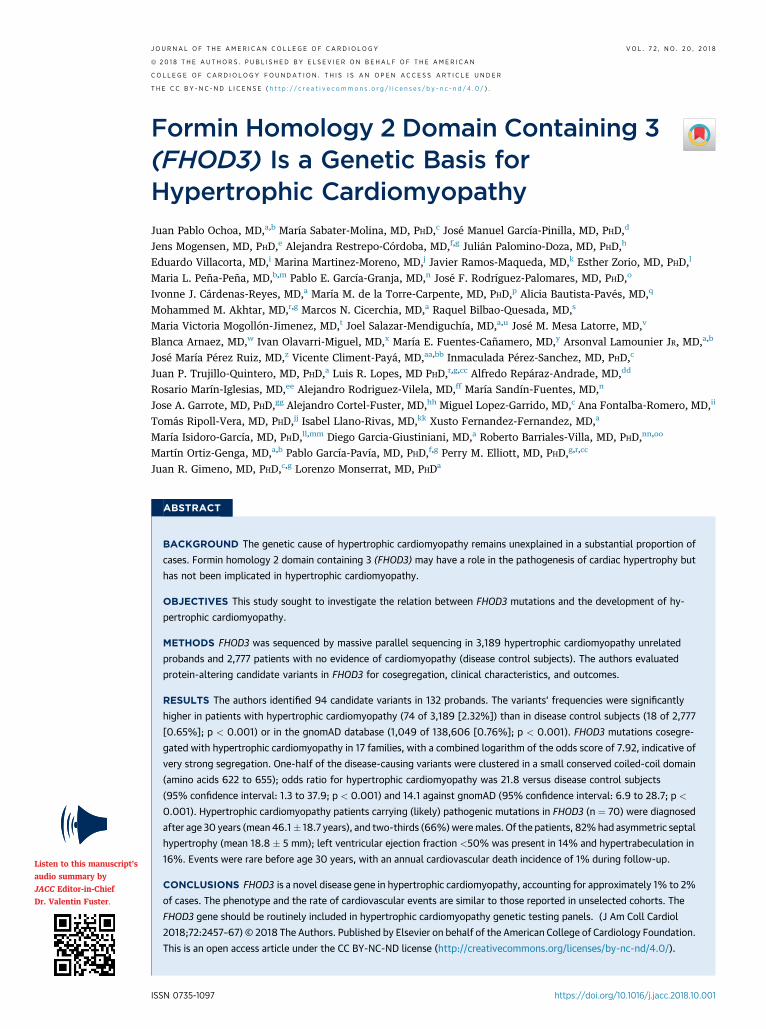
FIGURE 3 Molecular Organization of FHOD3 Protein and Relevant Domains for Actin Binding and Protein-Protein Interactions
A
B
DID-DADinteraction
Dimer (active)Monomer
(autoinhibitory state)
DADphosphorylation
18 400
574
623
655
1018
1050
1075
1471
1559
1622GBD FH3/DID Ex 11-12-13 CC DID(?) FH2FH1 DAD
Arg637TrpArg637GlyArg637Pro
Arg634GlyTyr528Cys
Ser527del
Arg641SerArg644SerAsn654Lys Asp962Asn
Pro1057Leu Arg1386GlnVal1576Gly
Ala321Val
Gly351ArgArg363Cys
Lys371ArgGln383His
Thr410Met
Ala542GluVal594Glu
Pro615LeuGlu832Lys
Gly845CysAla849Thr Ala1063Thr Leu1417_His1421
delinsGlnIle Asn1556His
(A) The distribution across the FHOD3 protein of the mutations considered pathogenic/likely pathogenic (blue, top) and of uncertain significance (green, bottom) in
HCM patients who participated in the segregation study. (B) The same distribution can be observed in the schematic representation of the protein, with the addition of
the variants detected in HCM probands who did not participate in the Segregation Phase of the study (orange). The intramolecular interaction between the DID and DAD
domains, which prevents formin from nucleating actin filaments, is relieved by the phosphorylation of serine/threonine DAD residues by the ROCK protein (B). The
regulation of dimerization to an active state may be more complex, including a dimerization domain (DD). Although in some formin proteins, such as FHOD1 and
mDia1, DID and DD domains are clearly concentrated in specific regions, in FHOD3 it could be more complex and involve a wider zone (pink). CCR ¼ coiled-coil region;
DAD ¼ diaphanous auto-regulatory domain; DD ¼ dimerization domain; DID ¼ diaphanous inhibitory domain; FH ¼ formin homology domain; GBD ¼ GTPase-binding
domain.
Ochoa et al. J A C C V O L . 7 2 , N O . 2 0 , 2 0 1 8
FHOD3 is an HCM-Causing Gene N O V E M B E R 1 3 / 2 0 , 2 0 1 8 : 2 4 5 7 – 6 7
2462
representing 1.1% (35 of 3,189) of the entire HCMcohort.
In 6 families, segregation analysis was uncertain,and was unlikely in 2 (the probands were carriers ofthe variants p.Lys371Arg and p.Glu832Lys) (OnlineFigure 2, panel 1B). It was not possible to determinethe pathogenicity of 14 variants that were finallyclassified as of unknown clinical significance.
The most commonly identified variants affectedconsecutive amino acids. The variant p.Tyr528Cyswas identified in 6 probands, 4 of whom came fromthe same region of Spain and were proved to be de-scendants of a common ancestor born 7 generationsago (Figure 2). In this multigenerational family, thevariant cosegregated with the disease with the high-est LOD score (3.82) for an individual family. The
other 2 probands came from centers from the UnitedKingdom and Denmark. The variant p.Ser527del,affecting the previous residue, was detected in 12HCM probands from different geographical areas inSpain, Denmark, and the United Kingdom as well.None of these probands had additional variants insarcomeric genes that could explain the disease, andcosegregation with the phenotype was confirmed inthe 5 families with relatives available for evaluation.Among the remaining variants, 7 were clustered in aconserved small coiled-coil domain (amino acids622 to 655) (Figure 3A, Online Figure 3). The OR ofthe presence of a candidate variant in FHOD3 inthis coiled-coil domain in HCM was 21.8 againstcontrol subjects (95% confidence interval [CI]: 1.3 to37.9; p < 0.001) and 14.1 against gnomAD individuals
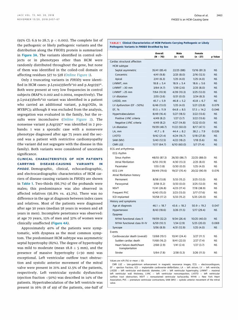
TABLE 1 Clinical Characteristics of HCM Patients Carrying Pathogenic or Likely
Pathogenic Variants in FHOD3 Stratified by Sex
Overall(N ¼ 70)
Male(n ¼ 43)
Female(n ¼27) p Value
Cardiac structural affection
HCM subtype
Septal asymmetric 35/41 (85.4) 22/25 (88) 13/16 (81.3) NS
Concentric 4/41 (9.8) 2/25 (8.0) 2/16 (12.5) NS
Apical 2/41 (6.3) 1/25 (4.0) 1/25 (4.0) NS
LVMWT, mm 18.8 � 5.4 18.9 � 5.4 18.6 � 5.6 NS
LVMWT >30 mm 3/64 (4.7) 1/39 (2.6) 2/25 (8.0) NS
LVMWT >25 mm 7/64 (10.9) 4/39 (10.3) 3/25 (12.0) NS
LV dilatation 2/55 (3.6) 0/31 (0.0) 2/24 (8.3) NS
LVEDD, mm 45.7 � 5.9 46.8 � 5.2 43.8 � 6.7 NS
LV dysfunction (EF <50%) 6/46 (13.0) 1/25 (4.0) 5/21 (23.8) 0.079
EF, % 61.5 � 11.9 64.8 � 8.5 57.5 � 14.2 0.046
Hypertrabeculation 8/49 (16.4) 5/27 (18.5) 3/22 (13.6) NS
Positive LVNC criteria 4/49 (8.2) 1/27 (3.7) 3/22 (13.6) NS
Negative LVNC criteria 4/49 (8.2) 4/27 (14.8) 0/22 (0.0) NS
LA dilatation 19/39 (48.7) 11/22 (50.0) 8/17 (47.1) NS
LA, mm 41.7 � 8 44.4 � 8.2 38.2 � 7.9 0.026
LVOTO 9/42 (21.4) 4/24 (16.7) 5/18 (27.8) NS
SAM (MV) 5/40 (12.5) 4/22 (18.2) 1/18 (5.6) NS
CMR LGE 11/17 (64.7) 6/10 (60.0) 5/7 (71.4) NS
ECG and arrhythmias
ECG rhythm
Sinus rhythm 48/55 (87.3) 26/30 (86.7) 22/25 (88.0) NS
Atrial fibrillation 6/55 (10.9) 4/30 (13.3) 2/25 (8.0) NS
Pacemaker 1/55 (1.8) 0/30 (0.0) 1/25 (4.0) NS
ECG LVH 39/49 (79.6) 19/27 (70.4) 20/22 (90.9) 0.076
Atrial fibrillation history
Permanent 8/58 (13.8) 5/33 (15.2) 3/25 (12.0) NS
Paroxysmal 3/58 (5.2) 0/33 (0.0) 3/25 (12.0) NS
NSVT 11/41 (26.8) 4/23 (17.4) 7/18 (38.9) NS
Frequent PVC 6/40 (15.0) 3/23 (13.0) 3/17 (17.6) NS
ICD implanted 10/58 (17.2) 5/33 (15.2) 5/25 (20.0) NS
History and symptoms
Age at diagnosis 46.1 � 18.7 43.6 � 18.2 50.9 � 19.2 0.047
Hypertension 8/43 (18.6) 3/26 (11.5) 5/17 (29.4) NS
Dyspnea
NYHA functional class II 19/59 (32.2) 9/34 (26.4) 10/25 (40.0) NS
NYHA functional class III–IV 6/59 (10.1) 1/34 (2.9) 5/25 (20.0) 0.069
Syncope 5/56 (8.9) 4/31 (12.9) 1/25 (4.0) NS
Events
Cardiovascular death (overall) 13/68 (19.1) 10/41 (24.4) 3/27 (11.1) NS
Sudden cardiac death 11/68 (16.2) 9/41 (22.0) 2/27 (7.4) NS
Heart failure death/hearttransplantation
2/68 (2.9) 1/41 (2.4) 1/27 (3.7) NS
Stroke 5/64 (7.8) 2/38 (5.3) 3/26 (11.5) NS
Values are n/N (%) or mean � SD.
CMR LGE ¼ late-gadolinium enhancement in magnetic resonance images; ECG ¼ electrocardiogram;EF ¼ ejection fraction; ICD ¼ implantable cardioverter-defibrillator; LA ¼ left atrium; LV ¼ left ventricle;LVEDD ¼ left ventricular end-diastolic diameter; LVH ¼ left ventricular hypertrophy; LVMWT ¼ maximalleft ventricular wall thickness; LVNC ¼ left ventricular noncompaction; LVOTO ¼ left ventricularoutflow tract obstruction; NSVT ¼ nonsustained ventricular tachycardia; NYHA ¼ New York HeartAssociation; PVC ¼ premature ventricular contractions; SAM (MV) ¼ systolic anterior movement of the mitralvalve.
J A C C V O L . 7 2 , N O . 2 0 , 2 0 1 8 Ochoa et al.N O V E M B E R 1 3 / 2 0 , 2 0 1 8 : 2 4 5 7 – 6 7 FHOD3 is an HCM-Causing Gene
2463
(95% CI: 6.9 to 28.7; p < 0.001). The complete list ofthe pathogenic or likely pathogenic variants and thedistribution along the FHOD3 protein is summarizedin Figure 3A. The variants identified in control sub-jects or in phenotypes other than HCM wererandomly distributed throughout the gene, but noneof them was identified in the coiled-coil domain oraffecting residues 527 to 528 (Online Figure 1).
Only 2 truncating variants in FHOD3 were identi-fied in HCM cases: p.Lys1433Serfs*10 and p.Arg1597*.Both were present at very low frequencies in controlsubjects (MAF% 0.002 and 0.0004, respectively). Thep.Lys1433Serfs*10 variant was identified in a patientwho carried an additional variant, p.Arg17Gln, inMYBPC3; although it was excluded from the analysis,segregation was evaluated in the family, but the re-sults were inconclusive (Online Figure 2). Thenonsense variant p.Arg1597* was identified in 2 pro-bands: 1 was a sporadic case with a nonseverephenotype diagnosed after age 75 years and the sec-ond was a patient with restrictive cardiomyopathy(the variant did not segregate with the disease in thisfamily). Both variants were considered of uncertainsignificance.CLINICAL CHARACTERISTICS OF HCM PATIENTS
CARRYING DISEASE-CAUSING VARIANTS IN
FHOD3. Demographic, clinical, echocardiographic,and electrocardiographic characteristics of HCM car-riers of disease-causing variants in FHOD3 are shownin Table 1. Two-thirds (66.7%) of the probands weremales; this predominance was also observed inaffected relatives (56.8% vs. 43.2%). There was nodifference in the age at diagnosis between index casesand relatives. Most of the patients were diagnosedafter age 30 years (median 58 years in women and 48years in men). Incomplete penetrance was observed:at age 70 years, 15% of men and 32% of women wereclinically unaffected (Figure 4A).
Approximately 40% of the patients were symp-tomatic, with dyspnea as the most common symp-tom. The predominant HCM subtype was asymmetricseptal hypertrophy (82%). The degree of hypertrophywas mild to moderate (mean 18.8 � 5 mm), and thepresence of massive hypertrophy ($30 mm) wasexceptional. Left ventricular outflow tract obstruc-tion and systolic anterior movement of the mitralvalve were present in 21% and 12.5% of the patients,respectively. Left ventricular systolic dysfunction(ejection fraction <50%) was described in 14% of thepatients. Hypertrabeculation of the left ventricle waspresent in 16% (8 of 49) of the patients, one-half of
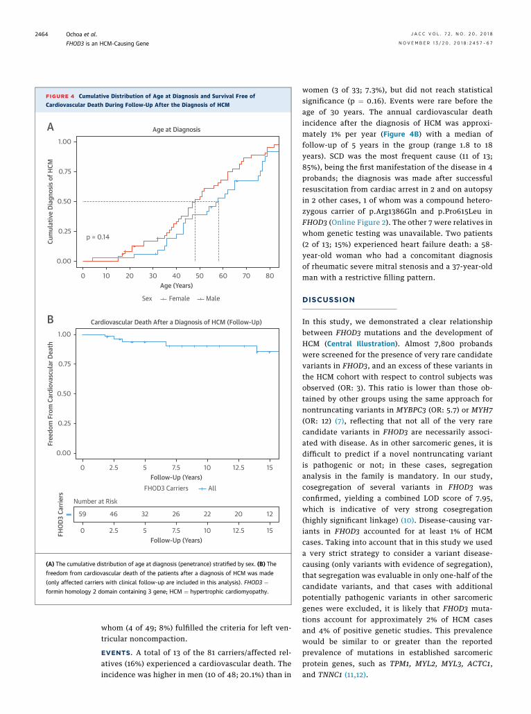
FIGURE 4 Cumulative Distribution of Age at Diagnosis and Survival Free of
Cardiovascular Death During Follow-Up After the Diagnosis of HCM
Cum
ulat
ive
Diag
nosis
of H
CM
0 10
p = 0.14
20
FemaleSex Male
Age (Years)
Age at Diagnosis
30 40 50 60 70 80
0.00
0.75
0.50
0.25
1.00
A
AllFHOD3 Carriers
Free
dom
Fro
m C
ardi
ovas
cula
r Dea
th
0 2.5 5Follow-Up (Years)
Cardiovascular Death After a Diagnosis of HCM (Follow-Up)
7.5 10 12.5 15
0.00
0.75
0.50
0.25
1.00
B
FHOD
3 Ca
rrie
rs
0 2.5 5Follow-Up (Years)
7.5 10 12.5 15
59
Number at Risk
46 32 26 22 20 12
(A) The cumulative distribution of age at diagnosis (penetrance) stratified by sex. (B) The
freedom from cardiovascular death of the patients after a diagnosis of HCM was made
(only affected carriers with clinical follow-up are included in this analysis). FHOD3 ¼formin homology 2 domain containing 3 gene; HCM ¼ hypertrophic cardiomyopathy.
Ochoa et al. J A C C V O L . 7 2 , N O . 2 0 , 2 0 1 8
FHOD3 is an HCM-Causing Gene N O V E M B E R 1 3 / 2 0 , 2 0 1 8 : 2 4 5 7 – 6 7
2464
whom (4 of 49; 8%) fulfilled the criteria for left ven-tricular noncompaction.
EVENTS. A total of 13 of the 81 carriers/affected rel-atives (16%) experienced a cardiovascular death. Theincidence was higher in men (10 of 48; 20.1%) than in
women (3 of 33; 7.3%), but did not reach statisticalsignificance (p ¼ 0.16). Events were rare before theage of 30 years. The annual cardiovascular deathincidence after the diagnosis of HCM was approxi-mately 1% per year (Figure 4B) with a median offollow-up of 5 years in the group (range 1.8 to 18years). SCD was the most frequent cause (11 of 13;85%), being the first manifestation of the disease in 4probands; the diagnosis was made after successfulresuscitation from cardiac arrest in 2 and on autopsyin 2 other cases, 1 of whom was a compound hetero-zygous carrier of p.Arg1386Gln and p.Pro615Leu inFHOD3 (Online Figure 2). The other 7 were relatives inwhom genetic testing was unavailable. Two patients(2 of 13; 15%) experienced heart failure death: a 58-year-old woman who had a concomitant diagnosisof rheumatic severe mitral stenosis and a 37-year-oldman with a restrictive filling pattern.
DISCUSSION
In this study, we demonstrated a clear relationshipbetween FHOD3 mutations and the development ofHCM (Central Illustration). Almost 7,800 probandswere screened for the presence of very rare candidatevariants in FHOD3, and an excess of these variants inthe HCM cohort with respect to control subjects wasobserved (OR: 3). This ratio is lower than those ob-tained by other groups using the same approach fornontruncating variants in MYBPC3 (OR: 5.7) or MYH7(OR: 12) (7), reflecting that not all of the very rarecandidate variants in FHOD3 are necessarily associ-ated with disease. As in other sarcomeric genes, it isdifficult to predict if a novel nontruncating variantis pathogenic or not; in these cases, segregationanalysis in the family is mandatory. In our study,cosegregation of several variants in FHOD3 wasconfirmed, yielding a combined LOD score of 7.95,which is indicative of very strong cosegregation(highly significant linkage) (10). Disease-causing var-iants in FHOD3 accounted for at least 1% of HCMcases. Taking into account that in this study we useda very strict strategy to consider a variant disease-causing (only variants with evidence of segregation),that segregation was evaluable in only one-half of thecandidate variants, and that cases with additionalpotentially pathogenic variants in other sarcomericgenes were excluded, it is likely that FHOD3 muta-tions account for approximately 2% of HCM casesand 4% of positive genetic studies. This prevalencewould be similar to or greater than the reportedprevalence of mutations in established sarcomericprotein genes, such as TPM1, MYL2, MYL3, ACTC1,and TNNC1 (11,12).
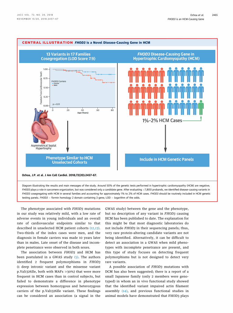
CENTRAL ILLUSTRATION FHOD3 is a Novel Disease-Causing Gene in HCM
Ochoa, J.P. et al. J Am Coll Cardiol. 2018;72(20):2457–67.
Diagram illustrating the results and main messages of the study. Around 50% of the genetic tests performed in hypertrophic cardiomyopathy (HCM) are negative;
FHOD3 plays a role in sarcomere organization, but was considered only a candidate gene. After evaluating >7,800 probands, we identified disease-causing variants in
FHOD3 cosegregating with HCM in several families and accounting for approximately 1% to 2% of HCM cases. FHOD3 should be routinely included in HCM genetic
testing panels. FHOD3 ¼ formin homology 2 domain containing 3 gene; LOD ¼ logarithm of the odds.
J A C C V O L . 7 2 , N O . 2 0 , 2 0 1 8 Ochoa et al.N O V E M B E R 1 3 / 2 0 , 2 0 1 8 : 2 4 5 7 – 6 7 FHOD3 is an HCM-Causing Gene
2465
The phenotype associated with FHOD3 mutationsin our study was relatively mild, with a low rate ofadverse events in young individuals and an overallrate of cardiovascular endpoints similar to thatdescribed in unselected HCM patient cohorts (12,13).Two-thirds of the index cases were men, and thediagnosis in female carriers was made 10 years laterthan in males. Late onset of the disease and incom-plete penetrance were observed in both sexes.
The association between FHOD3 and HCM hasbeen postulated in a GWAS study (5). The authorsidentified 2 frequent polymorphisms in FHOD3(1 deep intronic variant and the missense variantp.Val1326Ile, both with MAFs $30%) that were morefrequent in HCM cases than in control subjects, butfailed to demonstrate a difference in phenotypeexpression between homozygous and heterozygouscarriers of the p.Val1326Ile variant. These findingscan be considered an association (a signal in the
GWAS study) between the gene and the phenotype,but no description of any variant in FHOD3 causingHCM has been published to date. The explanation forthis might be that most diagnostic laboratories donot include FHOD3 in their sequencing panels; thus,very rare protein-altering candidate variants are notbeing identified. Alternatively, it can be difficult todetect an association in a GWAS when mild pheno-types with incomplete penetrance are present, andthis type of study focuses on detecting frequentpolymorphisms but is not designed to detect veryrare variants.
A possible association of FHOD3 mutations withDCM has also been suggested; there is a report of asmall Japanese family (only 2 members were geno-typed) in whom an in vivo functional study showedthat the identified variant impaired actin filamentassembly (14), and previous functional studies inanimal models have demonstrated that FHOD3 plays
Ochoa et al. J A C C V O L . 7 2 , N O . 2 0 , 2 0 1 8
FHOD3 is an HCM-Causing Gene N O V E M B E R 1 3 / 2 0 , 2 0 1 8 : 2 4 5 7 – 6 7
2466
a role in heart development (15). Similarly, an exome-wide association study identified FHOD3 as 1 of 8 lociindependently associated with sporadic DCM (16).Our study was not designed to determine the possiblecausal relationship between FHOD3 and DCM. Of the130 probands carrying candidate variants in FHOD3,18 had this phenotype (they were excluded in thescreening phase of the study). These variants werenot clustered in any particular region of the gene, andmost of them were classified as of uncertain signifi-cance at initial evaluation. Further studies are neededto establish the clinical relevance of FHOD3 muta-tions and their relationship to DCM.
The relationship between FHOD3 and ventricularhypertrophy has been recently explored in a study ofangiotensin II–induced cardiac hypertrophy (17).Under basal conditions, the FHOD3 protein exists inan auto-inhibited state due to the interaction be-tween its diaphanous autoregulatory domain and thediaphanous inhibitory domain (Figure 3B). Thephosphorylation of specific phospho-acceptor resi-dues located in the autoregulatory domain (Ser1590,Ser1596, and Thr1600) by the RhoA/ROCK pathwayseems to inhibit the interaction with the inhibitorydomain, allowing the FHOD3 protein to form activedimers that enable actin nucleation and assemblyof myofibrils in cardiomyocytes, thereby causingcellular hypertrophy (18). Several FHOD3 candidatevariants in this study were located in the autor-egulatory and inhibitory domains or their surround-ing residues (Figure 3A, Online Figure 1); 1hypothesis is that these mutations alter the normalinteraction, leading to a predominance of FHOD3protein in an activated state, a mechanism that hasbeen described for mutations in the paralogueFHOD1 (19,20).
FHOD3 protein dimerization to an activated statemay be more complex, including a dimerizationdomain and a helical region (coiled-coil domain) thatforms a cross-bridge between the opposing chainsand provides the conformational flexibility requiredfor the stair-stepping actin polymerization mecha-nism (21). One-half of the disease-causing variantsfound in our study were located in the coiled-coileddomain that seems to be specific for a HCM pheno-type (no disease controls or patients with cardiomy-opathies other than HCM where identified in it). Theexact location and functional characterization of theinhibitory and dimerization domains in FHOD3 arestill not well understood; 2 clearly pathogenic vari-ants that cosegregated with HCM in several families(p.Ser527del and p.Tyr528Cys) were found near thesedomains. They also affect an exon that is only presentin the longest transcript of FHOD3, which is currently
considered the most important isoform in the adultventricular myocardium, because it contains anadditional exon that is required for targeting theFHOD3 protein to the myofibrils in cardiomyocytes(22). Our findings support the relevance of this iso-form in the pathogenesis of HCM, although functionalstudies are needed to determine the exact mecha-nisms underlying the development of HCM in FHOD3mutation carriers.
STUDY LIMITATIONS. One of the limitations of ourstudy is that segregation was limited in some familiesbecause of the small number of relatives available forscreening, and clinical assessment was incomplete insome carriers. Another possible limitation is that pa-tients were screened only for genes previously asso-ciated with inherited cardiac conditions; therefore,the presence of mutations in other genes contributingto the phenotype cannot be ruled out.
CONCLUSIONS
In this study, we have demonstrated that FHOD3 is anovel disease-causing gene in HCM. Pathogenic mu-tations would account for approximately 1% to 2% ofHCM cases, a prevalence that is similar or greater tothat described in other secondary sarcomeric proteingenes. The associated phenotype and the rate ofcardiovascular events are similar to that described forunselected cohorts of patients with HCM; thus, aclinical follow-up is recommended in affected car-riers. The FHOD3 gene should be routinely includedin genetic testing panels for HCM.
ACKNOWLEDGMENTS The authors thank PilarMolina (Hospital Universitario y Politécnico La Fe,Valencia, Spain), Petros Syrris (UCL Institute of Car-diovascular Science, London, United Kingdom),Carmen Benito-Lopez y Sara Franco-Freire (HospitalRegional Universitario “Carlos Haya”), Juan de D.García-Diaz (Hospital Universitario Príncipe deAsturias, Alcalá de Henares, Madrid, Spain), CristinaGomez-Ramirez, (Hospital Universitario Cruces,Barakaldo, Vizcaya, Spain), Iria C. Duro (HospitalClínico Universitario de Valladolid, Valladolid,Spain), and Javier Limeres-Freire (Hospital Vald’Hebron, Barcelona, Spain), as well as Alicia PallasLozano and Radek Suchac for their help with Englishediting.
ADDRESS FOR CORRESPONDENCE: Dr. Juan PabloOchoa, Health in Code SL, As Xubias s/n, Edificio OFortín, Hospital Marítimo de Oza (15006), A Coruña,Spain. E-mail: [email protected]: @OchoaJP, @_healthincode.
PERSPECTIVES
COMPETENCY IN MEDICAL KNOWLEDGE: FHOD3
accounts for approximately 1% to 2% of cases of HCM,
and its prevalence is similar to or greater than that of
mutations in established sarcomeric protein genes.
TRANSLATIONAL OUTLOOK: Collaborative studies of
a larger number of patients are necessary to identify an
association with other phenotypes and expose mecha-
nisms underlying the development of HCM in carriers of
FHOD3 disease-causing variants.
J A C C V O L . 7 2 , N O . 2 0 , 2 0 1 8 Ochoa et al.N O V E M B E R 1 3 / 2 0 , 2 0 1 8 : 2 4 5 7 – 6 7 FHOD3 is an HCM-Causing Gene
2467
RE F E RENCE S
1. Marian AJ, Braunwald E. Hypertrophic cardio-myopathy: genetics, pathogenesis, clinical mani-festations, diagnosis, and therapy. Circ Res 2017;121:749–70.
2. Schönichen A, Geyer M. Fifteen formins for anactin filament: a molecular view on the regulationof human formins. Biochim Biophys Acta 2010;1803:152–63.
3. Taniguchi K, Takeya R, Suetsugu S, et al.Mammalian formin fhod3 regulates actin assemblyand sarcomere organization in striated muscles.J Biol Chem 2009;284:29873–81.
4. Ushijima T, Fujimoto N, Matsuyama S, et al. Theactin-organizing formin protein Fhod3 is requiredfor postnatal development and functional main-tenance of the adult heart in mice. J Biol Chem2018;293:148–62.
5. Wooten EC, Hebl VB, Wolf MJ, et al. Forminhomology 2 domain containing 3 variants associ-ated with hypertrophic cardiomyopathy. Circ Car-diovasc Genet 2013;6:10–8.
6. Lek M, Karczewski KJ, Minikel EV, et al. Analysisof protein-coding genetic variation in 60,706humans. Nature 2016;536:285–91.
7. Walsh R, Thomson KL, Ware JS, et al. Reas-sessment of Mendelian gene pathogenicity using7,855 cardiomyopathy cases and 60,706 refer-ence samples. Genet Med 2017;19:192–203.
8. Richards S, Aziz N, Bale S, Bick D, Das S. Stan-dards and guidelines for the interpretation ofsequence variants: a joint consensus recommen-dation of the American College of Medical Ge-netics and Genomics and the Association forMolecular Pathology. Genet Med 2015;17:405–24.
9. Egeland T, Pinto N, Vigeland MD. A generalapproach to power calculation for relationshiptesting. Forensic Sci Int Genet 2014;9:186–90.
10. Nyholt DR. All LODs are not created equal. AmJ Hum Genet 2000;67:282–8.
11. Sabater-Molina M, Pérez-Sánchez I, Hernándezdel Rincón JP, Gimeno JR. Genetics of hypertro-phic cardiomyopathy: a review of current state.Clin Genet 2018;93:3–14.
12. Elliott PM, Anastasakis A, Borger MA, et al.2014 ESC guidelines on diagnosis andmanagementof hypertrophic cardiomyopathy: The Task Forcefor the Diagnosis andManagement of HypertrophicCardiomyopathy of the European Society of Cardi-ology (ESC). Eur Heart J 2014;35:2733–79.
13. Ho CY, Charron P, Richard P, Girolami F, VanSpaendonck-Zwarts KY, Pinto Y. Genetic advancesin sarcomeric cardiomyopathies: state of the art.Cardiovasc Res 2015;105:397–408.
14. Arimura T, Takeya R, Ishikawa T, et al. Dilatedcardiomyopathy-associated FHOD3 variant impairsthe ability to induce activation of transcription factorserum response factor. Circ J 2013;77:2990–6.
15. Kan-O M, Takeya R, Abe T, et al. Mammalianformin Fhod3 plays an essential role in cardio-genesis by organizing myofibrillogenesis. BiolOpen [Internet] 2012;1:889–96.
16. Esslinger U, Garnier S, Korniat A, et al. Exome-wide association study reveals novel susceptibilitygenes to sporadic dilated cardiomyopathy. PLoSOne 2017;12:e0172995.
17. Zhou Q, Wei S-S, Wang H, et al. Crucial role ofROCK2-mediated phosphorylation and upregulationof FHOD3 in the pathogenesis of angiotensin
II-induced cardiac hypertrophy. Hypertens 2017;69:1070–83.
18. Iskratsch T, Reijntjes S, Dwyer J, et al. Twodistinct phosphorylation events govern the func-tion of muscle FHOD3. Cell Mol Life Sci 2013;70:893–908.
19. Schulte A, Stolp B, Schönichen A, et al. Thehuman formin FHOD1 contains a bipartite struc-ture of FH3 and GTPase-binding domainsrequired for activation. Structure 2008;16:1313–23.
20. Nezami AG, Poy F, Eck MJ. Structure of theautoinhibitory switch in formin mDia1. Structure2006;14:257–63.
21. Higgs HN. Formin proteins: a domain-basedapproach. Trends Biochem Sci 2005;30:342–53.
22. Iskratsch T, Lange S, Dwyer J, Kho AL, DosRemedios C, Ehler E. Formin follows function: amuscle-specific isoform of FHOD3 is regulated byCK2 phosphorylation and promotes myofibrilmaintenance. J Cell Biol 2010;191:1159–72.
KEY WORDS cardiomyopathies, FHOD3,formins, genetics, hypertrophiccardiomyopathy, sudden death
APPENDIX For a list of the recruiting hospi-tals of the GENESCOPIC research group andresearch centers (genetics, molecular biology,and data-analysis) of the GENESCOPIC researchgroup as well as supplemental tables andfigures, please see the online version of thispaper.