Embed Size (px)
Citation preview
FORMULATION, CHARACTERIZATION, AND IN VIVO REMODELING
MECHANISMS OF POLYURETHANE BIOCOMPOSITES FOR BONE TISSUE
ENGINEERING
By
Edna Margarita Prieto-Ballengee
Dissertation
Submitted to the Faculty of the
Graduate School of Vanderbilt University
in partial fulfillment of the requirements
for the degree of
DOCTOR OF PHILOSOPHY
in
Chemical Engineering
December, 2013
Nashville, Tennessee
Approved:
Professor Scott A. Guelcher
Professor Jamey Young
Professor Matthew Lang
Professor Hak-Joon Sung

Copyright © 2013 by Edna Margarita Prieto-Ballengee
All rights reserved

iii
To my family, the engine of my life:
both the family that made me who I am,
and the family we have just started.
iv
ACKNOWLEDGEMENTS
I would like to start by gratefully acknowledging the different funding sources that have
supported the work presented in this dissertation. These include the Orthopaedic Extremity
Trauma Research Program, the National Science Foundation, the National Institutes of Health,
the Center for Military Biomaterials Research, and the Armed Forces Institute of Regenerative
Medicine.
I also want to express great gratitude to my research advisor Dr. Scott A. Guelcher who
allowed me to be part of an exciting research field by welcoming me in his lab. His support has
provided me with opportunities to explore different professional avenues, and participate in
several collaborative teams to develop not only my research, but also many other professional
skills. Thank you for believing in my work and mentoring me to become a better researcher and
team player. In a similar way, I want to acknowledge the help I have received from my committee
members over these years: Dr. Jamey Young, Dr. Matthew Lang, and Dr. Hak-Joon Sung. Their
advice after each meeting, and support in the lab also enabled me to accomplish my research
objectives.
Vanderbilt has been an excellent place to conduct interdisciplinary research. I want to
thank our collaborators at the Vanderbilt Center for Bone Biology, especially Dr. Julie Sterling
and Dr. James Edwards, for guiding me through the biology of bone; to Alyssa Merkel for always
having a helping hand with the mice and cell culture techniques; and to Javier Esparza for
teaching me how to work with animals. Also thank you to the people at the Biomechanics Lab
(Dr. Jeffrey Nyman, Sasidhar Uppuganti, Matthew Murry), the Vanderbilt Institute of Nanoscale
Science and Engineering (VINSE, Dr. Tony Hmelo and Dr. Ben Schmidt), the National Research
Resource for Imaging Mass Spectroscopy (Dr. Jeremy Norris), the Vanderbilt University Institute
of Imaging Science (VUIIS, Dr. Daniel Perrien), the Flow Cytometry Shared Resource (David

v
Flaherty), and to the Cell Imaging Shared Resource (CISR, Carol Ann Bonner) for providing
access to and training on instruments used in this work. I am also very grateful to our external
collaborators who have brought views from outside academia into our research, in this way
keeping us focused on the patients that need improved treatments. Thank you in particular to Dr.
Joseph Wenke (USAISR) for his support and for introducing me to the interesting problem of
infection.
So many other people have supported my work along the way. Thank you to the
Chemical and Biomolecular Engineering staff (Mary Gilleran, Rae Uson, and Mark Holmes) for
helping me to successfully navigate administrative and laboratory hurdles. I am infinitely grateful
for the help and friendships I have found in my fellow lab members (past and present). Thank you
to Katarzyna Zienkiewicz for being incredibly supportive, always our right hand in the lab, and a
great example of dedicated hard work. Thank you also to the undergraduates (Nicholas Gould,
Erica Von Stein, Brian Shen, David Harris), high school students (Anna Claire Brakefield,
Jonathan Davies), and teacher (Melinda Higgins) who made contributions to this research along
the way.
Leaving the most important for last, I must acknowledge the help and support I have
received from my family and friends. Thank you to my parents, Edna Margarita and Gabriel
Omar, and my siblings, Camila, Gabriel, and Omar, for their love, support, and for always
believing and being part of my dreams and goals. Thank you also to my grandparents, aunts and
uncles, cousins, and friends back in Colombia, who even far away have always managed to be
next to me every step of the way. I am immensely grateful to all the friends I met while in
Vanderbilt; in particular thank you to Stijn, Will, Doris, Svenja, Silvia, Carlos, Sebastian, Juan,
Angela, Juanita, Christi, Elizabeth, Neil, and Taylor for becoming my Nashville family! And of
course, thank you to my best friend and husband Jason Ballengee. You have been there from the
first day of the PhD journey, and all this work would not have been possible without your
unconditional support, constructive feedback, and loving care.
vi
TABLE OF CONTENTS
Page
DEDICATION .................................................................................................................. iii
ACKNOWLEDGEMENTS ............................................................................................... iv
LIST OF TABLES ............................................................................................................. ix
LIST OF FIGURES ........................................................................................................... xi
Chapter
I. INTRODUCTION ...................................................................................................1
References ..........................................................................................................6
II. BACKGROUND ......................................................................................................8
General principles of bone tissue engineering ...................................................8
Characterization of bone scaffolds.....................................................................9
Scaffolding materials for bone tissue engineering ...........................................12
References ........................................................................................................26
III. EFFECTS OF LOCAL DELIVERY OF D-AMINO ACIDS
FROM BIOFILM-DISPERSIVE SCAFFOLDS ON
INFECTION IN CONTAMINATED RAT SEGMENTAL
DEFECTS ........................................................................................................35
Introduction ......................................................................................................35
Experimental ....................................................................................................37
Results ..............................................................................................................46
Discussion ........................................................................................................56
Conclusions ......................................................................................................62
References ........................................................................................................63
IV. SURFACE MODIFICATION OF β-TRI-CALCIUM
PHOSPHATE IMPROVES THE MECHANICAL
PERFORMANCE OF SETTABLE CALCIUM
PHOSPHATE/POLYURETHANE BIOCOMPOSITES FOR
BONE TISSUE ENGINEERING ....................................................................68
Introduction ......................................................................................................68
Experimental ....................................................................................................70
Results ..............................................................................................................79
vii
Discussion ........................................................................................................87
Conclusions ......................................................................................................95
References ........................................................................................................96
V. BALANCING THE RATE OF NEW BONE FORMATION AND
POLYMER DEGRADATION ENHANCES HEALING OF
WEIGHT-BEARING ALLOGRAFT/POLYURETHANE
COMPOSITES IN RABIT FEMORAL DEFECTS ......................................100
Introduction ....................................................................................................100
Experimental ..................................................................................................102
Results ............................................................................................................110
Discussion ......................................................................................................123
Conclusions ....................................................................................................130
References ......................................................................................................132
VI. EFFECT OF PARTICLE SIZE, LOADING, AND MINERAL
CONTENT ON THE IN VIVO REMODELING OF
SETTABLE ALLOGRAFT BONE/POLYMER
COMPOSITES ..............................................................................................137
Introduction ....................................................................................................137
Experimental ..................................................................................................139
Results ............................................................................................................146
Discussion ......................................................................................................157
Conclusions ....................................................................................................162
References ......................................................................................................164
VII. EFFECT OF SUBSTRATE COMPOSISTION ON THE IN
VITRO DIFFERENTIATION OF OSTEOCLAST
PRECURSORS AND RESOPRTIVE ACTIVITY OF
OSTEOCLASTS ............................................................................................167
Introduction ....................................................................................................167
Experimental ..................................................................................................170
Results ............................................................................................................178
Discussion ......................................................................................................189
Conclusions ....................................................................................................193
References ......................................................................................................194
VIII. CONCLUSIONS ..............................................................................................198
References ......................................................................................................203
IX. SUGGESTIONS FOR FUTURE WORK ..........................................................204
viii
References ......................................................................................................211
Appendix
A. EXPERIMENTAL PROTOCOLS .......................................................................213
Grafting of polycaprolactone to the surface of -TCP particles ....................214
Measuring the Euler number of a histology section ......................................216
Preparation of bioactive glass disks and surface modified
bioactive glass disks for cell culture ..............................................................217
Sample sterilization for cell culture ...............................................................218
Extraction of murine bone marrow cells ........................................................219
Bone marrow cell purification (CD11b+ selection using
microbeads) ....................................................................................................221
Preparation of cell culture samples for SEM imaging ...................................223
Actin staining of cultured osteoclasts ............................................................224
Differentiation of osteoclast precursors on different matrices .......................225
RNA extraction from cells cultured on different matrices
(RNeasy kit) ...................................................................................................226
cDNA synthesis and real time qPCR (SYBR green) .....................................228
Intracellular TRAP staining ...........................................................................230
Naphthol AS-BI analysis of TRAP activity in culture
supernatant .....................................................................................................231
Measurement of volumetric resorptive rates in vitro .....................................234
ix
LIST OF TABLES
Table Page
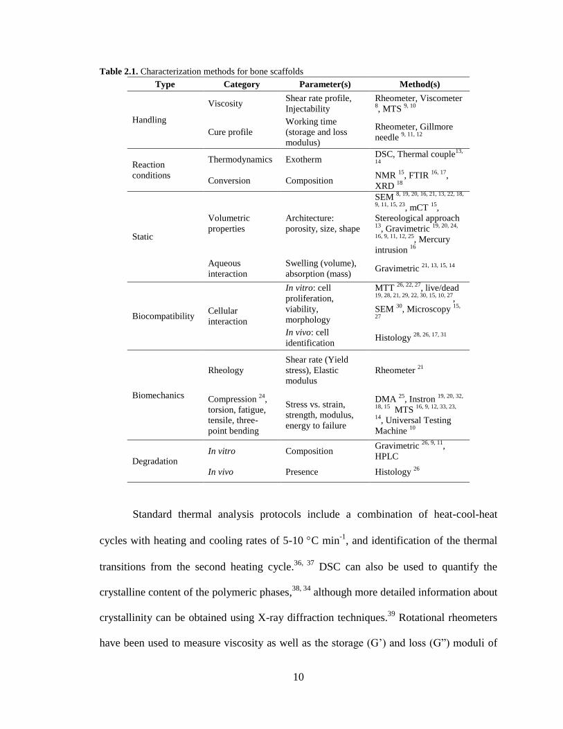
2.1. Characterization methods for bone scaffolds. ...........................................................10
3.1. Description of S. aureus strains invesitigated in this study. .....................................38
3.2. Study design investigating the ability of biofilm-dispersive scaffolds
to reduce bacterials contamination in vitro. Scaffolds were contaminated
with 107 CFU/ml S. aureus for 2 h and outcome were assessed after 24 h
incubation time in PBS (n = 4). .........................................................................................43
3.3. In vivo study design investigating the ability of biofilm-dispersive
scaffolds to reduce contamincation in 6-mm segmental defect in rat femor
contaminated with 102 CFU of S. aureus UAMS-A or Xen36. Outcomes
were assessed at 2 weeks (n=10). ......................................................................................44
4.1. Torsion mechanical properties for TCP and TCP-PCL composites.. .........................84
4.2. Interaction parameters between the ceramic and polymer network. ...........................90
5.1. Treatment groups evaluated in the rabbit femoral condyle plug defect
study. ................................................................................................................................104
5.2. Mechanical properties of the injec5.biocomposites (BC) and the
calcium phosphate cement (CPC) measured under compressive and
torsional loads. Data are reported as the mean ± Standard error of the
mean (SEM). ....................................................................................................................112
5.3. Fitting parameters for bone (B) histomorphometry data, which were
fit to area% B = at/(1+bt). ................................................................................................120
5.4. Fitting parameters for allograft (A) histomorphometry data, which
were fit to area% A = AA,i - at/(1+bt) for the BC group. The parameter AA,i
is the initial area% allograft measured by histomorphometry. Data for
BC+BMP-L and BC+BMP-H groups could not be accurately fit to the
exponential or rational functions. ....................................................................................121
5.5. Fitting parameters for polymer (P) histomorphometry data, which
were fit to area% P = AP,i - at/(1+bt) for BC+BMP-L and BC+BMP-H
groups and to area% P = AP,i – αexp(βt) for the BC group. The parameter
AP,i is the initial area% polymer measured by histomorphometry. ..................................122

x
6.1. Composition and physical properties of injectable and moldable
composites........................................................................................................................140
7.1. Primer sequences for osteoclastic genes ...................................................................175
xi
LIST OF FIGURES
Figure Page
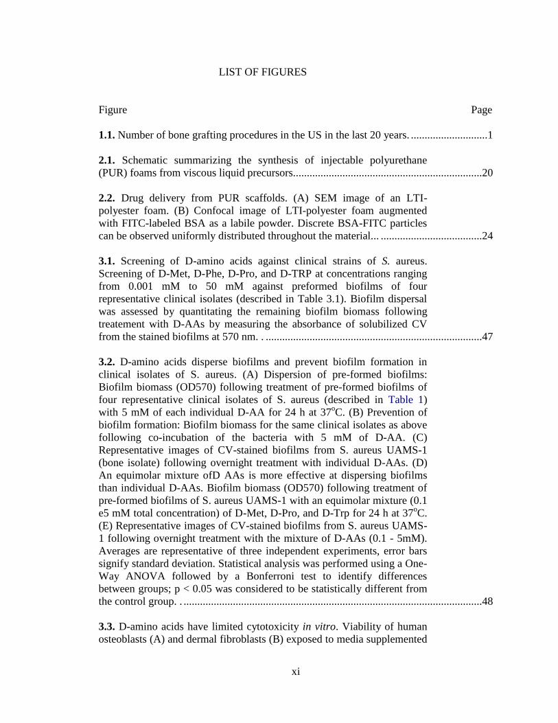
1.1. Number of bone grafting procedures in the US in the last 20 years. ............................1
2.1. Schematic summarizing the synthesis of injectable polyurethane
(PUR) foams from viscous liquid precursors.....................................................................20
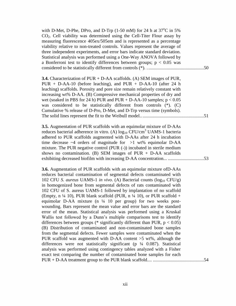
2.2. Drug delivery from PUR scaffolds. (A) SEM image of an LTI-
polyester foam. (B) Confocal image of LTI-polyester foam augmented
with FITC-labeled BSA as a labile powder. Discrete BSA-FITC particles
can be observed uniformly distributed throughout the material... .....................................24
3.1. Screening of D-amino acids against clinical strains of S. aureus.
Screening of D-Met, D-Phe, D-Pro, and D-TRP at concentrations ranging
from 0.001 mM to 50 mM against preformed biofilms of four
representative clinical isolates (described in Table 3.1). Biofilm dispersal
was assessed by quantitating the remaining biofilm biomass following
treatement with D-AAs by measuring the absorbance of solubilized CV
from the stained biofilms at 570 nm. . ...............................................................................47
3.2. D-amino acids disperse biofilms and prevent biofilm formation in
clinical isolates of S. aureus. (A) Dispersion of pre-formed biofilms:
Biofilm biomass (OD570) following treatment of pre-formed biofilms of
four representative clinical isolates of S. aureus (described in Table 1)
with 5 mM of each individual D-AA for 24 h at 37oC. (B) Prevention of
biofilm formation: Biofilm biomass for the same clinical isolates as above
following co-incubation of the bacteria with 5 mM of D-AA. (C)
Representative images of CV-stained biofilms from S. aureus UAMS-1
(bone isolate) following overnight treatment with individual D-AAs. (D)
An equimolar mixture ofD AAs is more effective at dispersing biofilms
than individual D-AAs. Biofilm biomass (OD570) following treatment of
pre-formed biofilms of S. aureus UAMS-1 with an equimolar mixture (0.1
e5 mM total concentration) of D-Met, D-Pro, and D-Trp for 24 h at 37oC.
(E) Representative images of CV-stained biofilms from S. aureus UAMS-
1 following overnight treatment with the mixture of D-AAs (0.1 - 5mM).
Averages are representative of three independent experiments, error bars
signify standard deviation. Statistical analysis was performed using a One-
Way ANOVA followed by a Bonferroni test to identify differences
between groups; p < 0.05 was considered to be statistically different from
the control group. . .............................................................................................................48
3.3. D-amino acids have limited cytotoxicity in vitro. Viability of human
osteoblasts (A) and dermal fibroblasts (B) exposed to media supplemented
xii
with D-Met, D-Phe, DPro, and D-Trp (1-50 mM) for 24 h at 37oC in 5%
CO2. Cell viability was determined using the Cell-Titer Flour assay by
measuring fluorescence 405ex/505em and is represented as a percentage
viability relative to non-treated controls. Values represent the average of
three independent experiments, and error bars indicate standard deviation.
Statistical analysis was performed using a One-Way ANOVA followed by
a Bonferroni test to identify differences between groups; p < 0.05 was
considered to be statistically different from controls (*). . ................................................50
3.4. Characterization of PUR + D-AA scaffolds. (A) SEM images of PUR,
PUR + D-AA-10 (before leaching), and PUR + D-AA-10 (after 24 h
leaching) scaffolds. Porosity and pore size remain relatively constant with
increasing wt% D-AA. (B) Compressive mechanical properties of dry and
wet (soaked in PBS for 24 h) PUR and PUR + D-AA-10 samples; p < 0.05
was considered to be statistically different from controls (*). (C)
Cumulative % release of D-Pro, D-Met, and D-Trp versus time (symbols).
The solid lines represent the fit to the Weibull model.. .....................................................51
3.5. Augmentation of PUR scaffolds with an equimolar mixture of D-AAs
reduces bacterial adherence in vitro. (A) log10 CFU/cm3 UAMS-1 bacteria
adhered to PUR scaffolds augmented with D-AAs after 24 h incubation
time decrease ~4 orders of magnitude for >1 wt% equimolar D-AA
mixture. The PUR negative control (PUR (-)) incubated in sterile medium
shows no contamination. (B) SEM images of PUR + D-AA scaffolds
exhibiting decreased biofilm with increasing D-AA concentration... ...............................53
3.6. Augmentation of PUR scaffolds with an equimolar mixture ofD-AAs
reduces bacterial contamination of segmental defects contaminated with
102 CFU S. aureus UAMS-1 in vivo. (A) Bacterial counts (log10 CFU/g)
in homogenized bone from segmental defects of rats contaminated with
102 CFU of S. aureus UAMS-1 followed by implantation of no scaffold
(Empty, n ¼ 10), PUR blank scaffold (PUR, n ¼ 10), or PUR scaffold +
equimolar D-AA mixture (n ¼ 10 per group) for two weeks post-
wounding. Bars represent the mean value and error bars are the standard
error of the mean. Statistical analysis was performed using a Kruskal
Wallis test followed by a Dunn’s multiple comparisons test to identify
differences between groups (* significantly different than PUR, p < 0.05)
(B) Distribution of contaminated and non-contaminated bone samples
from the segmental defects. Fewer samples were contaminated when the
PUR scaffold was augmented with D-AA content >5 wt%, although the
differences were not statistically significant (p ¼ 0.087). Statistical
analysis was performed using contingency tables analyzed with a Fisher
exact test comparing the number of contaminated bone samples for each
PUR + D-AA treatment group to the PUR blank scaffold.... ............................................54
xiii
3.7. Low- and high-magnification SEM images of biofilms on PUR and
PUR + D-AA-10 scaffolds implanted in contaminated femoral segmental
defects in rats for 2 weeks show reduced bacterial adhesion for the
scaffold augmented with 10 wt% of the equimolar mixture of D-AAs.
PUR scaffolds implanted in sterile defects (PUR (-) negative control)
show minimal bacterial adhesion..... ..................................................................................55
4.1. Characterization of matrix particles. (A) Matrix particle properties.
(B) Frequency particle size distributions measured for TCP, p-TCP, and
TCP-PCL. (C) Relative atomic composition at the surface of TCP, p-TCP,
and TCP-PCL measured by XPS. (D) Thermogravimetric analysis (TGA)
of TCP, TCP-PCL, and PCL. ............................................................................................79
4.2. SEM images of TCP, p-TCP, and TCP-PCL composites. Scale bars
correspond to 300 m. .......................................................................................................80
4.3. Wet mechanical properties of composites at different indexes A)
modulus, B) compressive strength. Statistical analysis compares
composites with the same filler but different indexes at each filler load; *
p<0.05; ** p=0.11. .............................................................................................................82
4.4. Compressive mechanical properties of scaffolds with index 140 as a
function of matrix composition and loading. (A) Water uptake. (B)
Compressive modulus under dry and wet conditions. (C) Compressive
strength under dry and wet conditions. .............................................................................83
4.5. Osteoblastic cell culture of TCP, and TCP-PCL composites. (A) Live-
Dead staining of MC3T3 cells seeded on composites after 2 days of
culture (>95% viability). (B) The biocomposites support the
differentiation of MC3T3 cells after 7 days of culture in differentiation
media. White scale bars represent 200 um, * p<0.05 compared to Day 2.. .......................85
4.6. Osteoclastic cell culture of TCP, and TCP-PCL composites. (A)
Positive TRAP staining of bone marrow cells cultured on the
biocomposites for 15 days. (B) TRAP secreted into the media as a
measure of osteoclastic resorption activity. * Significantly different from
dentin at the same time point (p<0.05). .............................................................................86
4.7. Compressive strength of calcium phosphates and calcium
phosphates/polymer composites. Blue area: Cortical bone properties, Red
area: Trabecular bone properties, Yellow area: properties of TCP-PCL
composites developed in this study....................................................................................93
5.1. Rheological properties of the biocomposite (BC) measured by
rheometry. (A) G’ represents the storage modulus and G” the loss modulus.
The G’-G” crossover point is assumed to be the working time of the BC.
xiv
(B) Viscosity vs. shear rate plot illustrating calculation of the yield stress
using the Herschel-Bulkley model. ..................................................................................110
5.2. Representative stress-strain curves for the BCs and CaP cement under
compression and torsion.. ................................................................................................111
5.3. Representative images of the empty defects filled with the allograft
bone particles, BC, BC+BMP-L, BC+BMP-H at 6 and 12 weeks.
Longitudinal sections of the femur and 3D reconstructions of the
cylindrical defect region of interest.
..........................................................................................................................................113
5.4. Radial distribution of morphometric parameters measured by CT at
6 and 12 weeks. (A) Representative image highlighting subdivision of the
defect into four annular shells each 1mm thick. (B)-(H) Morphometric
parameters evaluated are plotted versus the mean radial distance from the
core (Rm). ........................................................................................................................115
5.5. Low- (1.25x) and high- (20x) magnification images of histological
sections of untreated (empty, top) and allograft-filled defects (bottom) at 6
weeks. CI: cellular infiltration, NB: new bone, A: allograft particles. ............................117
5.6. (A) Low (1.25x) and high (20x) magnification images of histological
sections of the biocomposite treatment groups at 6 weeks. P: residual
polymer, O: Osteoid, NB: New bone formation.. ............................................................117
5.6. (continued) (B) Low (1.25x) and high (20x) magnification images of
histological sections of the biocomposite treatment groups at 12 weeks. P:
residual polymer, O: Osteoid, NB: New bone formation... .............................................118
5.7. Histomorphometric evaluation of remodeling of the biocomposite
scaffolds. (A) Diagram showing the areas of interest. Area% new bone,
Area % remaining allograft particles, and Area% remaining polymer for
(B,E,H) BC, (C,F,I) BC+BMP-L, and (D,G,J) BC+BMP-H... ........................................120
5.8. Analysis of histomorphometric data. The ratio of the rate of new bone
formation (rB) to that of polymer degradation (rP) was calculated for each
group by differentiating the equations expressing area% new bone and
area% polymer as a function of time. Representative images of
histological sections with highlighted areas of interest. (A-B) BC, (C-D)
BC+BMP-L, and (E-F) BC+BMP-H... ............................................................................128
6.1. Initial viscosity of PUR/allograft composites. Non-reactive
formulations were poured between 40mm cross-hatched parallel plates,
compressed to a gap of 1.5 mm, and subjected to a dynamic frequency
sweep (0.1 to 100 rad sec-1
) at 25C with controlled strain amplitude of
xv
0.02% (n=3). Dynamic data was transformed steady state viscosity (h,
Pa*s) as a function of shear rate (g, s-1
) using a Cox-Merz transformation
(geometric figures). Data were fit to the Herschel-Bulkley model (lines) to
determine yield stress in the case of the IDL, MS, and ML formulations. ......................147
6.2. Scanning electron microscopy images and porosity of ABP-PUR
biocomposites. Scale bars represent 750 m. Statistical significant
differences between groups with different letter. ............................................................148
6.3. Compressive strength and modulus of settable ABP-PUR
biocomposites. No statistical significant difference between groups with
the same letter. (a) strength, (b) modulus. .......................................................................149
6.4. μCT analysis of mineralized tissue in ABP-PUR biocomposites. (A)
Representative images at 6 and 12 weeks. (B) Morphometric parameters:
BV/TV, Tb.N., Tb.Th., and Tb.Sp., all measured as a function of radial
distance from the center line of the defect at 6 and 12 weeks and compared
to host bone values measured separately. ........................................................................150
6.5. Low-magnification images of histological sections of ABP-PUR
biocomposites at 6 and 12 weeks. Goldner’s trichrome stain, original
maginification 1X. ...........................................................................................................153
6.6. High-magnification images of histological sections of defects treated
with injectable ABP-PUR biocomposites. All of the images show sections
stained with Goldner’s trichrome, except for IDS at 6 weeks, which is
stained with H&E. Original magnification 70X. With Goldner’s trichrome
stain: white- remaining polymer; light green particles with angled shapes-
remaining allograft particles; green with cells inside- new mineralized
bone with osteocytes; red- osteoid; orange- red blood cells; purple- nuclei;
BM- bone marrow. Arrows in the IDS-6 weeks section point to giant cells. ..................154
6.7. High-magnification images of histological sections of empty defects
and defects treated with moldable ABP-PUR biocomposites. Goldner’s
trichrome stain. Original magnification reported in each image. With
Goldner’s trichrome stain: white- remaining polymer; light green particles
with angled shapes- remaining allograft particles; green with cells inside-
new mineralized bone with osteocytes; red- osteoid; orange- red blood
cells; purple- nuclei; BM- bone marrow. ........................................................................154
6.8. Histomorphometric analysis of new bone formation of ABP-PUR
biocomposites at 6 and 12 weeks. Mineral content (including new bone
and allograft), New bone (NB), remaining allograft (A), and polymer (P)
data are plotted as a function of radial distance from the center line of the
defect. ...............................................................................................................................156
xvi
7.1. Intracellular TRAP staining of osteoclast precursors cultured for 28
days on A,B,D) on tissue culture plastic, and C) TCP. Cell culture media
consisted of complete media supplemented with MCSF (25ng/ml) and
RANKL (50ng/ml) for A-C, or MCSF (25ng/ml) for D. ................................................179
7.2. Longitudinal characterization of the differentiation of osteoclast
precursors cultured on tissue culture plastic (well), dentin, and BG. A)
Gene expression, B) Quantification of secreted TRAP. ..................................................180
7.3. Longitudinal characterization of the differentiation of osteoclast
precursors cultured on dentin, CBG, SBG and TCP with and without
addition of exogenous BMP-2. A) Gene expression quantified in run 1, B)
Gene expression quantified in run 2, C) Quantification secreted TRAP in
run 1 and 2. ......................................................................................................................181
7.4. Differentiation of non-adherent bone marrow osteoclast precursors.
Day 0-8: Representative images of the cells during the differentiation
process. White arrowheads point to an example of multinucleated cells in
each image. Scale bars represent 100 um. Graph: summarizes the number
and average size of multinucleated cells during the differentiation process.
These variables were monitored to decide when to stop the experiment to
collect differentiated osteoclasts. * Significantly different (p<0.05) from
the value quantified the day before. .................................................................................182
7.5. Confocal images of osteoclasts seeded on dentin .....................................................183
7.6. Intracellular TRAP stain of osteoclasts seeded on different matrices.
Inset graph presents size of the +TRAP cells as a function of material.
Scale bars represent 200m. ............................................................................................184
7.7. Morphology of osteoclasts cultured on different materials observed by
SEM. ................................................................................................................................185
7.8. Resorption for 2 days with BMP2. (A) Roughness of the surface
before and after cell culture, (B) 3D view of dentin surface after resorption,
(C) Characteristics of the resorption pits generated by osteoclasts on
dentin, (D) Quantification of dentin’s volumetric resorption rate. ..................................188
7.9. Effect of BMP-2 on the volumetric resorption of dentin ..........................................188
9.1. Histomorphometric evaluation of remodeling in the injectable
scaffolds described in Chapter VI, as a function of time and location. ...........................207
9.2. Histomorphometric evaluation of remodeling in the moldable
scaffolds described in Chapter VI, as a function of time and location. ...........................207

xvii
9.3. Relative rates of new bone formation and polymer degradation for the
injectable and moldable formulations described in Chapter VI, as a
function of fraction healed and location. E corresponds to the Euler
number calculated from a single section for each group at 6 weeks.. .............................208
1
CHAPTER I
INTRODUCTION
The treatment of bone defects remains a challenging clinical problem. Trauma,
disease, primary tumor resection, and/or congenital deformation often result in large bone
losses requiring bone grafting procedures.1-4
Bone is the second most commonly
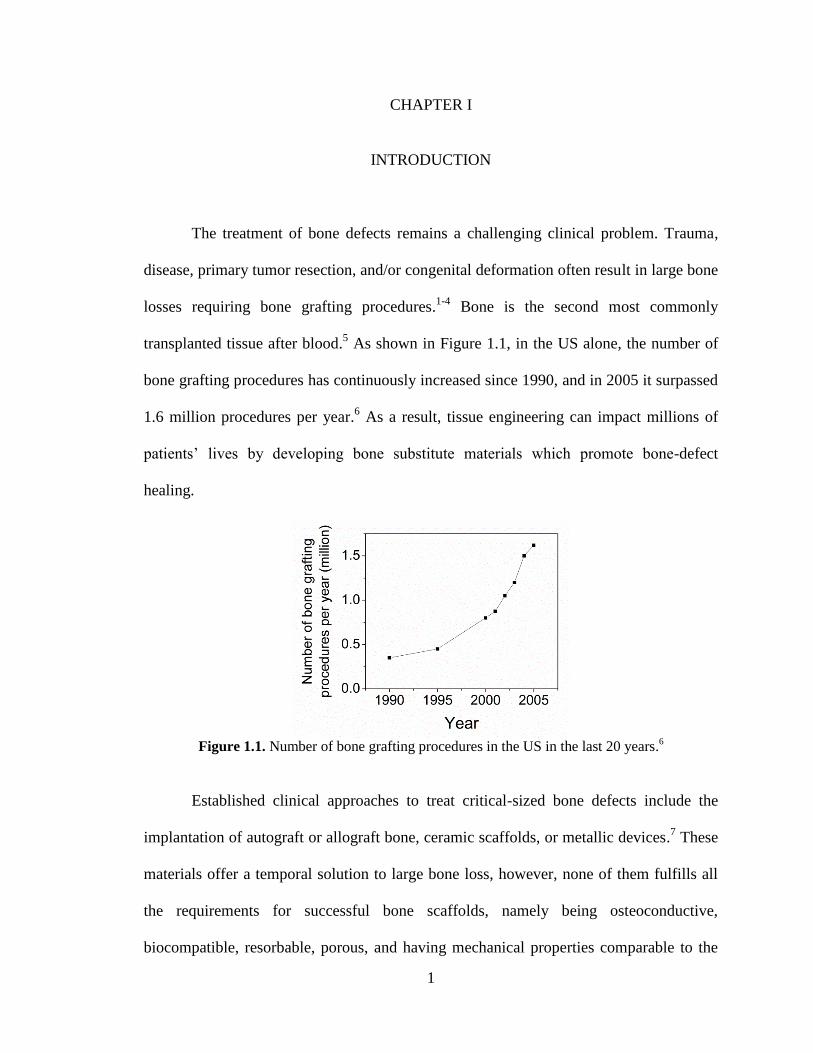
transplanted tissue after blood.5 As shown in Figure 1.1, in the US alone, the number of
bone grafting procedures has continuously increased since 1990, and in 2005 it surpassed
1.6 million procedures per year.6 As a result, tissue engineering can impact millions of
patients’ lives by developing bone substitute materials which promote bone-defect
healing.
Figure 1.1. Number of bone grafting procedures in the US in the last 20 years.
6
Established clinical approaches to treat critical-sized bone defects include the
implantation of autograft or allograft bone, ceramic scaffolds, or metallic devices.7 These
materials offer a temporal solution to large bone loss, however, none of them fulfills all
the requirements for successful bone scaffolds, namely being osteoconductive,
biocompatible, resorbable, porous, and having mechanical properties comparable to the
2
surrounding host bone.8-10
Biomaterials research continues to focus on developing
alternative bone graft formulations that satisfy these characteristics with the dual
objectives of reducing revision surgeries’ frequency and the rate of secondary
complications in the clinic.11
Biodegradable, lysine-derived polyurethane (PUR) networks have been developed
for regenerative medicine applications.12
When implanted in vivo, these materials
generate a minimal and transient inflammatory response, support cellular infiltration, and
degrade to non-cytotoxic compounds.13, 14
Porous, injectable formulations have been used
as delivery vehicles for biologically active small molecules such as growth factors,15, 16
antibiotics,17
and small molecule drugs.18
For bone applications, porous and non-porous
composites incorporating mineralized fillers into a polyurethane (PUR) network have
been developed and tested in femoral plug bone defects in rats,19, 20
rabbits,21-23
and
sheep.24
The mineralized matrix provides osteoconductivity to the polymer by acting as a
porogen that guides cellular infiltration21
, and the polymer reduces the brittle behavior of
the ceramics. While pre-clinical testing positioned PUR composites as suitable
biomaterials for bone regeneration, further formulation development was needed for
moldable scaffolds to approach weight-bearing capabilities.
The goal of this dissertation was to formulate and characterize PUR
biocomposites that participate actively and predictably in the bone healing process.
Building on previous work in the Guelcher lab, high and low porosity formulations were
developed to improve the healing outcomes of contaminated and femoral plug bone
defects. In vitro characterization of the formulations included physical, rheological,
mechanical and cell culture analyses. In vivo implantation and analysis of healing
3
progression provided information about the remodeling mechanisms of the different PUR
formulations. In order to fully characterize the resorptive component of graft remodeling,
an in vitro osteoclastic culture protocol was developed to enable quantitation and direct
comparison between volumetric resorptive rates of synthetic matrices. Each chapter in
this dissertation focuses on a different PUR formulation and its detailed characterization.
Combined, they offer design guidelines for the development of PUR scaffolds that a)
protect scaffolds from contamination, and b) maintain bone-like strength while actively
remodeling.
Chapter III describes the formulation, characterization, and in vivo evaluation of a
PUR biofilm-dispersive graft.25
Infectious complications in open fractures are a major
factor contributing to osseous non-union and extremity amputation.26
The persistence of
bacteria within biofilms, despite meticulous debridement and antibiotic therapy, is
believed to be a major cause of chronic infection.27
As a therapeutic strategy for the
reduction and prevention of biofilm-associated infections, D-Amino Acids (D-AA) with
biofilm dispersal capabilities were incorporated into porous PUR scaffolds. Detailed in
vitro characterization of the D-AA effect over the formation and dispersal of clinical
bacterial strain biofilms is presented. The scaffold’s porosity, mechanical properties and
D-AA release profile are reported. The in vivo biofilm dispersal potential of the
augmented PUR scaffolds was evaluated in contaminated rat segmental bone defects.
Chapter IV continues to present a settable PUR biocomposite formulation
containing surface modified -tricalcium phosphate (TCP) particles. Previous studies in
our lab have developed compression molded TCP-PUR composites that supported
cellular infiltration and remodeling when implanted in femoral condyle defects in rats.20
4
Due to the brittle nature of TCP, high pressure was required to obtain final mechanical
properties matching those of bone. However, since the compressed composites are
molded outside the body, their application is limited by the defect geometry. As a result,
Chapter IV describes the development of a settable TCP-PUR formulation in which the
surface of the ceramic filler was modified to improve the interactions with the PUR
binder. The effect of surface modification was evaluated on the mechanical and in vitro
biological performance of the biocomposites.
In contrast to slow resorbing TCP, allograft bone particles (ABP) remodel by
osteoclastic resorption followed by osteoblastic new bone deposition.28
Thus, ABPs were
included in PUR composites to study the effect of design parameters on the healing of
settable bone grafts in vivo. The design parameters, studied in Chapter V, are the rates of
new bone formation, matrix resorption, and polymer degradation. Biocomposites were
augmented with recombinant human bone morphogenetic protein-2 (rhBMP-2) to
modulate the rates of new bone formation and polymer degradation. Instead, in Chapter
VI, the influence of filler particle size, loading, and mineral content on the ABP-PUR
biocomposites’ remodeling mechanisms are considered. Each chapter reports the
handling and mechanical properties of the appropriate formulations. The composites were
delivered to cylindrical defects in the femoral condyle of New Zealand White rabbits, and
the remodeling progression was evaluated by X-
and histomorphometry at 0, 6, and 12 weeks.
In order for biocomposites to be effectively integrated into the bone remodeling
cycle they must undergo active resorption at a similar rate to that of new bone
formation.29
Consequently, detailed understanding of the osteoclastic effect on different
5
materials is valuable information for the designing functional biocomposites which
promote bone regeneration. Chapter VII introduces an in vitro cell culture protocol to
study the differentiation and resorptive activity of murine bone marrow cells in the
presence of biomaterials. Implementation of the protocol is described to quantify the
volumetric resorption rate of dentin in the presence and absence of rhBMP-2.
To conclude, Chapter VIII summarizes the main findings of this dissertation and
Chapter IX presents suggestions for future work. As a whole, this dissertation presents a
compelling argument for wider use and further development of PUR biocomposites.
6
References
1 Timothy A. Lew, John A. Walker, Joseph C. Wenke, Lorne H. Blackbourne, and Robert G. Hale,
'Characterization of Craniomaxillofacial Battle Injuries Sustained by United States Service
Members in the Current Conflicts of Iraq and Afghanistan', Journal of Oral and Maxillofacial
Surgery, 68 (2010), 3-7.
2 D. Ghysen, L. Ozsarlak, L. van den Hauwe, J. Van Goethem, AM. De Schepper, and PM. Parizel,
'Maxillo-Facial Trauma', JBR-BTR, 83 (2000), 181-97.
3 BB. Ward, SE. Brown, and PH. Krebsbach, 'Bioengineering Strategies for Regeneration of
Craniofacial Bone: A Review of Emerging Technologies', Oral diseases, doi:10.1111/j.1601-
0825.2010.01682.x (2010).
4 BJ. Costello, G. Shah, P. Kumta, and CS. Sfeir, 'Regenerative Medicine for Craniomaxillofacial
Surgery', Oral Maxillofac Surg Clin North Am., 22 (2010), 33-42.
5 P. Giannoudis, H. Dinopoulos, and E. Tsiridis, 'Bone Substitues: An Update', Injury, Int. J. Care
Injured, 36S (2005), S20-S27.
6 United States Census Bureau, 'Organ Transplants and Grafts: 1990 to 2007', (2007).
7 M. A. Woodruff, C. Lange, J. Reichert, A. Berner, F. L. Chen, P. Fratzl, J. T. Schantz, and D. W.
Hutmacher, 'Bone Tissue Engineering: From Bench to Bedside', Materials Today, 15 (2012), 430-
35.
8 K. Rezwan, Q. Z. Chen, J. J. Blaker, and A. R. Boccaccini, 'Biodegradable and Bioactive Porous
Polymer/Inorganic Composite Scaffolds for Bone Tissue Engineering', Biomaterials, 27 (2006),
3413-31.
9 S. Scaglione, E. Lazzarini, C. Ilengo, and R. Quarto, 'A Composite Material Model for Improved
Bone Formation', Journal of Tissue Engineering and Regenerative Medicine, 4 (2010), 505-13.
10 S. Bose, M. Roy, and A. Bandyopadhyay, 'Recent Advances in Bone Tissue Engineering
Scaffolds', Trends in Biotechnology, 30 (2012), 546-54.
11 MS. Gilardino, DS. Cabiling, and SP. Bartlett, 'Long-Term Follow-up Experience with
Carbonated Calcium Phosphate Cement (Norian) for Cranioplasty in Children and Adults.', Plast
Reconstr Surg., 123 (2009), 983-94.
12 S. A. Guelcher, 'Biodegradable Polyurethanes: Synthesis and Applications in Regenerative
Medicine', Tissue Engineering Part B-Reviews, 14 (2008), 3-17.
13 A. Hafeman, K. Zienkiewicz, A. Zachman, H.J. Sung, L.B. Nanney, J.M. Davidson, and S.A.
Guelcher, 'Characterization of the Degradation Mechanisms of Lysine-Derived and Aliphatic
Poly(Ester Urethane) Scaffolds', Biomaterials, In review (2010).
14 J. M. Page, E. M. Prieto, J. E. Dumas, K. J. Zienkiewicz, J. C. Wenke, P. Brown-Baer, and S. A.
Guelcher, 'Biocompatibility and Chemical Reaction Kinetics of Injectable, Settable
Polyurethane/Allograft Bone Biocomposites', Acta Biomaterialia, 8 (2012), 4405-16.
15 B. Li, T. Yoshii, A. E. Hafeman, J. S. Nyman, J. C. Wenke, and S. A. Guelcher, 'The Effects of
Rhbmp-2 Released from Biodegradable Polyurethane/Microsphere Composite Scaffolds on New
Bone Formation in Rat Femora', Biomaterials, 30 (2009), 6768-79.
16 AE Hafeman, B Li, T Yoshii, KL Zienkiewicz, JM Davidson, and SA Guelcher, 'Injectable
Biodegradable Polyurethane Scaffolds with Release of Platelet-Derived Growth Factor for Tissue
Repair and Regeneration', Pharm Res, 25 (2008), 2387-99.
7
17 B. Li, K. V. Brown, J. C. Wenke, and S. A. Guelcher, 'Sustained Release of Vancomycin from
Polyurethane Scaffolds Inhibits Infection of Bone Wounds in a Rat Femoral Segmental Defect
Model', Journal of Controlled Release, 145 (2010), 221-30.
18 T Yoshii, A. Hafeman, J Nyman, J Esparza, K Shinomiya, D. M. Spengler, Gregory R. Mundy, G.
Gutierrez, and S A Guelcher 'A Sustained Release of Lovastatin from Biodegradable, Elastomeric
Polyurethane Scaffolds for Enhanced Bone Regeneration', Tissue Eng Part A, 16 (2010).
19 J. E. Dumas, K. Zienkiewicz, S. A. Tanner, E. M. Prieto, S. Bhattacharyya, and S. A. Guelcher,
'Synthesis and Characterization of an Injectable Allograft Bone/Polymer Composite Bone Void
Filler with Tunable Mechanical Properties', Tissue Engineering Part A, 16 (2010), 2505-18.
20 T. Yoshii, J. E. Dumas, A. Okawa, D. M. Spengler, and S. A. Guelcher, 'Synthesis,
Characterization of Calcium Phosphates/Polyurethane Composites for Weight-Bearing Implants',
Journal of Biomedical Materials Research Part B-Applied Biomaterials, 100B (2012), 32-40.
21 J. E. Dumas, T. Davis, G.E. Holt, T. Yoshii, D. S. Perrien, J. S. Nyman, T. Boyce, and S.A.
Guelcher, 'Synthesis, Characterization, and Remodeling of Weight-Bearing Allograft
Bone/Polyurethane Composites in the Rabbit', Acta Biomaterialia, 6 (2010), 2394-406.
22 J. E. Dumas, E. M. Prieto, K.J. Zienkiewicz, T. Guda, J. C. Wenke, J. Bible, G.E. Holt, and S.A.
Guelcher, 'Balancing the Rates of New Bone Formation and Polymer Degradation Enhances
Healing of Weight-Bearing Allograft/Polyurethane Composites in Rabbit Femoral Defects', Tissue
Eng Part A, Not available - ahead of print (2013).
23 J.E. Dumas, P.B. BrownBaer, E.M. Prieto, T. Guda, R.G. Hale, J.C. Wenke, and S.A. Guelcher,
'Injectable Reactive Biocomposites for Bone Healing in Critical-Size Rabbit Calvarial Defects',
Biomedical Materials, 7 (2012), 024112.
24 R. Adhikari, P. A. Gunatillake, I. Griffiths, L. Tatai, M. Wickramaratna, S. Houshyar, T. Moore,
R. T. M. Mayadunne, J. Field, M. McGee, and T. Carbone, 'Biodegradable Injectable
Polyurethanes: Synthesis and Evaluation for Orthopaedic Applications', Biomaterials, 29 (2008),
3762-70.
25 Carlos J. Sanchez Jr, Edna M. Prieto, Chad A. Krueger, Katarzyna J. Zienkiewicz, Desiree R.
Romano, Catherine L. Ward, Kevin S. Akers, Scott A. Guelcher, and Joseph C. Wenke, 'Effects of
Local Delivery of D-Amino Acids from Biofilm-Dispersive Scaffolds on Infection in
Contaminated Rat Segmental Defects', Biomaterials, 34 (2013), 7533-43.
26 J. Huh, D. J. Stinner, T. C. Burns, and J. R. Hsu, 'Infectious Complications and Soft Tissue Injury
Contribute to Late Amputation after Severe Lower Extremity Trauma', J Trauma, 71 (2011), S47-
51.
27 D. P. Lew, and F. A. Waldvogel, 'Osteomyelitis', Lancet, 364 (2004), 369-79.
28 Leandro Eduardo Klüppel, Fernando Antonini, Sérgio Olate, Frederico Felipe Nascimento, José
Ricardo Albergaria-Barbosa, and Renato Mazzonetto, 'Bone Repair Is Influenced by Different
Particle Sizes of Anorganic Bovine Bone Matrix: A Histologic and Radiographic Study in Vivo',
Journal of Craniofacial Surgery, 24 (2013), 1074-77 10.97/SCS.0b013e318286a0a3.
29 ArndtF Schilling, Sandra Filke, Silja Brink, Heike Korbmacher, Michael Amling, and JohannesM
Rueger, 'Osteoclasts and Biomaterials', European Journal of Trauma, 32 (2006), 107-13.
8
CHAPTER II
BACKGROUND
General principles of bone tissue engineering
Tissue engineering is “an interdisciplinary field of research that applies the
principles of engineering and the life sciences towards the development of biological
substitutes that restore, maintain, or improve tissue function’’.1 As such, bone tissue
engineering aims to develop scaffolds that promote the healing of bone defects generated
by trauma, disease, primary tumor resection, and/or congenital deformation.2-5
Bone is a dynamic tissue under constant remodeling in the body. This is achieved
by the combined action of 3 cell types: osteoblasts (bone forming cells), osteoclasts (bone
resorbing cells), and osteocytes (mechanosensor cells entrapped in bone). As a material,
bone is a natural composite of an organic phase (mainly collagen) and hydroxycarbonate
apatite.6 It is organized in two architectural forms: trabecular or porous bone, and cortical
or compact bone.7 Depending on the location in the body, the architectural organization
determines target bone mechanical properties.
Several requirements have been identified for bone tissue engineering scaffolds.
These focus on achieving mechanical as well as biological properties that will favor
scaffold replacement by new and functional bone in time. As a result, successful bone
scaffolds must:
(a) be biocompatible, or support normal cellular activity without any local and
systematic toxic effects to the host tissue,6
9
(b) be osteoconductive, or provide a surface for bone cells to adhere, proliferate,
migrate, and deposit new bone,
(c) be osteoinductive, or capable of inducing new bone formation through the
recruitment of osteoprogenitor cells,
(d) have mechanical properties similar to those of the surrounding host bone,
(e) have interconnected pores to facilitate diffusion of nutrients and oxygen required
for the survival of cells, and
(f) be bioresorbable or degradable in a controlled manner such that they are replaced
by new bone.
Characterization of bone scaffolds
Proper characterization of bone scaffolds encompasses multiple strategies from
several design perspectives. Table 2.1 lists various design perspectives, corresponding
parameters, and utilized methods to characterize bone scaffolds. The list is organized
generally as proceeding from fabrication (top) to implementation (bottom). This section
provides a summary of some techniques and protocols listed. A detailed discussion about
how the techniques work is not provided since this falls outside the scope of the chapter,
but citations to previous work applying these techniques have been included as reference.
Characterization of polymer scaffolds often requires analysis of polymer
composition and structure since these parameters influence scaffold processability and
final performance. Molecular weight distribution is usually quantified using gas
permeation chromatography.34, 35
Differential Scanning Calorimetry (DSC) is used to
identify thermal transitions such as the glass transition and melting temperatures.
10
Table 2.1. Characterization methods for bone scaffolds
Type Category Parameter(s) Method(s)
Handling
Viscosity Shear rate profile,
Injectability
Rheometer, Viscometer 8, MTS
9, 10
Cure profile
Working time
(storage and loss
modulus)
Rheometer, Gillmore
needle 9, 11, 12
Reaction
conditions
Thermodynamics Exotherm DSC, Thermal couple
13,
14
Conversion Composition NMR
15, FTIR
16, 17,
XRD 18
Static
Volumetric
properties
Architecture:
porosity, size, shape
SEM 8, 19, 20, 16, 21, 13, 22, 18,
9, 11, 15, 23, mCT
15,
Stereological approach 13
, Gravimetric 19, 20, 24,
16, 9, 11, 12, 25, Mercury
intrusion 16
Aqueous
interaction
Swelling (volume),
absorption (mass) Gravimetric
21, 13, 15, 14
Biocompatibility Cellular
interaction
In vitro: cell
proliferation,
viability,
morphology
MTT 26, 22, 27
, live/dead 19, 28, 21, 29, 22, 30, 15, 10, 27
,
SEM 30
, Microscopy 15,
27
In vivo: cell
identification Histology
28, 26, 17, 31
Biomechanics
Rheology
Shear rate (Yield
stress), Elastic
modulus
Rheometer 21
Compression 24
,
torsion, fatigue,
tensile, three-
point bending
Stress vs. strain,
strength, modulus,
energy to failure
DMA 25
, Instron 19, 20, 32,
18, 15 MTS
16, 9, 12, 33, 23,
14, Universal Testing
Machine 10
Degradation In vitro Composition
Gravimetric 26, 9, 11
,
HPLC
In vivo Presence Histology 26
Standard thermal analysis protocols include a combination of heat-cool-heat
cycles with heating and cooling rates of 5-10 C min-1
, and identification of the thermal
transitions from the second heating cycle.36, 37
DSC can also be used to quantify the
crystalline content of the polymeric phases,38, 34
although more detailed information about
crystallinity can be obtained using X-ray diffraction techniques.39
Rotational rheometers
have been used to measure viscosity as well as the storage (G’) and loss (G”) moduli of
11
flowable formulations.40
Additional protocols to measure viscosity include the use of
more traditional Ubbelohde viscometers.41, 42
The porous architecture of bone scaffolds has a direct impact on the mechanical
properties, degradation rates, and cellular infiltration of the materials. Although high
porosity and interconnectivity are desired to support cellular infiltration, mechanical
properties decrease with porosity squared, while the effect on degradation rates varies
depending on the material. Pore size distribution and morphology can be evaluated using
Scanning Electron Microscopy (SEM). To do so, a thin section of the material is gold
sputter-coated and imaged at different locations. Images are used to measure pore
diameter which is reported either as an average (n>100) or after applying a statistical
correction to account for non-ideal spherical pores.40,42,43
Porosity is commonly
determined gravimetrically by comparing dry scaffold density (F) with the density of
bulk material () according to: .44, 45
X-Ray microtomography (CT)
has also been used to accurately quantify porosity. In this case, dry scaffolds are scanned
in a CT system at high resolution modes, and porosity values are obtained after
thresholding the reconstructed image.46-48
Pore size distribution, total pore volume,
surface area, and density can also be obtained using mercury intrusion porosimetry
(MIP),49, 34
although interconnected pores are required in order to obtain representative
results using this technique.
In vitro degradation profiles of scaffolds are determined by incubating samples at
37C in phosphate-buffered saline (PBS) at various pH levels, enzyme-containing media,
or oxidative media.34,50,47,39
At each time point, the media with degradation products is
collected and the dry sample mass is measured and compared to the initial sample mass.
12
In addition to reporting the mass loss in time, the media collected can be analyzed with
techniques such as high-performance liquid chromatography (HPLC) to determine the
nature and concentration of the degradation products.
Depending on the specific application of the bone scaffold, specific mechanical
requirements must be achieved under tension, compression, and/or torsion. Mechanical
testing of the samples resulting in stress-strain data is conducted with protocols modeled
by standard methods.51-53
Reported properties usually include compressive and tensile
modulus and strength.50,37,44,38,34
Scaffolding materials for bone tissue engineering
Autologous bone remains the standard of care for the treatment of bone defects
due to its ideal osteogenic, osteoinductive and osteoconductive properties for bone defect
healing. However, limited availability, postsurgical morbidity at the donor site, increased
lengths of surgery and higher costs associated with autograft use54
have increased the
demand for alternative materials, such as allograft bone. In 2002, more than 980,000
bone allografts were distributed in the US and their usage was reported to be increasing.55
Even though sterilization and viral inactivation of allograft bone render the material
suitable for clinical use, the available shapes are limited by the donor source.56
As a result,
bone tissue engineering efforts have focused on the development of synthetic grafts that
promote healing of bone defects. The following sections discuss the characteristics and
performance of several of these formulations.
13
Low porosity calcium phosphate cements
Since their initial discovery in 1982,57
injectable calcium phosphate cements
(CPCs) have been successfully introduced into the clinic for a number or orthopaedic and
craniomaxillofacial applications, including repair of tibial plateau fractures and calvarial
defects. CPCs have been investigated extensively as injectable bone replacement
biomaterials due to their similar chemical composition to the mineral component of bone,
biocompatibility,58
osteoconductivity, and fast setting times (< 5 min).59
These
biomaterials set at a physiological pH with minimal reaction exotherm and do not release
toxic monomers or solvents.60, 18
Apatite, which has low solubility and resorbs slowly,
and brushite, which has higher solubility than apatite and resorbs more rapidly, comprise
the two primary classes of CPCs.61
While CPCs set by an acid/base reaction that can
reduce the pH of the paste to values as low as 3,32
a number of in vivo studies have
reported favorable host responses after setting.24, 17
In a recent study, the mechanism of
cell-mediated degradation of brushite CPCs was investigated by culturing RAW264.7
cells on the cements in vitro.30
The RAW264.7 cells differentiated to macrophages,
multinucleated giant cells, as well as osteoclast-like cells, on the brushite CPCs. SEM
analysis of the ultra-structure of osteoclast-like cells revealed characteristics associated
with the osteoclast phenotype, such as formation of a sealing zone and ruffled border.
Furthermore, osteoclast-like cells were observed to penetrate deep into the interior of the
cements, which suggests that brushite CPCs are demineralized by osteoclast-mediated
resorption in vivo.
In a promising area of new research, the effects of nanometer-scale calcium
phosphate (CaP) crystals on cellular interactions are under investigation.62
Coating of
14
mesenchymal stem cells with CaP nanorods has been reported to exhibit enhanced
osteoblastic differentiation and production of extracellular matrix production relative to
uncoated cells,63
which suggests that nanoscale injectable CPCs may be desirable carriers
for stem cells. However, while nanostructured CaPs enhance osteoblast differentiation,
the rate of cellular infiltration into monolithic cements is limited by osteoclast-mediated
resorption. This rate of infiltration is relatively slow (i.e., 0.02 μm3 μm
-2 day
-1), in
contrast to migration and proliferation of cells through interconnected pores.64
Thus, a
number of recent studies have investigated new methods for introducing macropores into
injectable CPCs to enhance cellular infiltration and remodeling.
Porous calcium phosphate cements
Primary limitations of first-generation low porosity CPCs restricting their use in
the clinic include brittle mechanical properties, which lead to low shear strength and
fracture toughness; slow degradation in vivo;65
and the small pore size (0.1 – 10 m),
which results in slow cellular infiltration and ingrowth of new bone.58,66
Both low
porosity as well as small pore size in CPCs contribute to slow remodeling and ingrowth
of new bone, and have thus been suggested as a root cause for the failure of CPCs in
periodontal bone repair.67
Slow remodeling of CPCs has also been suggested to
contribute to their failure in cranioplasty applications.68
Therefore, recent studies have
focused on introducing macropores into CPCs and improving their mechanical properties
while preserving their favorable biocompatibility in order to improve their clinical
performance.
15
Interconnected macropores >10 μm are essential for promoting infiltration of cells
and ingrowth of blood vessels and new bone into CPCs,69, 70
as well as for polymeric
scaffolds such as hydrogels.15
In order to preserve the excellent biocompatibility and
osteoconductivity of CPCs, adjuvants added to create macropores must be non-cytotoxic.
In one approach, multi-phasic cements have been prepared by combining soluble ceramic
particles, such as calcium sulphate, with less soluble particles, such as triccalcium
phosphate or hydroxyapatite.71,23
A bi-phasic CPC was fabricated by mixing α-tricalcium
phosphate (α-TCP), calcium sulphate hemihydrate (CSH), and an aqueous solution
containing 2.5 wt% Na2HPO4.33
The resulting cement comprised an apatitic phase
(calcium-deficient hydroxyapatite, CDHA) and a soluble phase (calcium sulphate
dihydrate, CSD). After setting, the soluble CSD and CSH phases dissolved, resulting in
pores that facilitated the ingrowth of new bone in a rat on-lay graft model.31
The
technology has been licensed to BoneSupport and is marketed as CERAMENT™ bone
void filler for spinal and orthopaedic indications. Recent clinical studies have highlighted
the potential of the partially resorbable biphasic (60% calcium sulphate/40%
hydroxyapatite) CERAMENT™ CPC as an effective substitute for non-resorbable
poly(methyl methacrylate) (PMMA) for treatment of osteoporotic and traumatic vertebral
fractures.72, 73
Composites with calcium phosphates
A number of other strategies have been investigated for fabricating injectable
macroporous CPCs. Foaming agents such as surfactants,12
hydrogen peroxide solution,16
carbon dioxide,74
or hydrophobic liquids,75
have been utilized to fabricate injectable
16
calcium phosphate foams. In one study, the surfactant sodium dodecyl sulphate (SDS)
was used as an air-entraining agent to create macroporous CPCs.12
Micro- and
macroporosity were controlled by the liquid-to-powder ratio and the concentration of
SDS, which stabilized the bubbles formed by the setting reaction of the cement.
Alternatively, biocompatible and degradable polymeric microspheres such as poly(lactic-
co-glycolic acid) (PLGA),76
poly(trimethyl carbonate), or gelatin9, 11
have been
investigated to synthesize macroporous CPCs. CPCs incorporating degradable porogens
offer the additional advantage of local delivery of drugs or growth factors, since biologics
can be encapsulated in the porogens prior to embedding in the calcium phosphate
matrix.77
While incorporation of PLGA microspheres in CPCs has been suggested to
improve the initial strength of the composites, the strength decreases upon dissolution of
the microspheres and does not increase until new bone starts to grow into the
macropores.78
To improve the injectability of CPC incorporating 30 wt% PLGA
microspheres, sodium citrate was added to the aqueous solution, which reduced the
viscosity of the paste. Due to their macroporosity, injectable CPCs incorporating a
porogen have significant advantages compared to monolithic CPCs. Thus, macroporous
CPCs are anticipated to be considered a preferred choice for healing of bony defects after
regulatory approval.65
In an alternative approach, CaP granules suspended in aqueous
solutions of hydroxy-propylmethyl-cellulose promoted faster initial ingrowth of new
bone at the surface of the material, relative to macroporous CPCs.28, 79
It has been
suggested that the observed early apposition of new bone could potentially enhance the
interfacial bonding between host bone and the CPC, thus reinforcing the material for
weight-bearing applications.65
17
Polymeric bone scaffolds
Polyesters are among the most studied biodegradable materials for biomedical
applications.80-83
The degradation profiles and mechanical properties of polyesters can be
tailored according to the final application by modifying the backbone composition. Due
to this versatility, polyester scaffolds have been developed for bone, cartilage, and
meniscus among other applications.84, 82
The main degradation mechanism of polyesters
is hydrolytic chain scission of the ester bonds in the backbone.85, 82, 86, 80
Degradation
products in vivo are incorporated into the tricarboxylic acid cycle (TCA) and secreted
from the body.82, 85
Poly(propylene fumarate) (PPF) is a linear, unsaturated, cross-linkable, and
biodegradable polyester.87, 88, 35, 42
used in bone scaffolds. Physical, mechanical, and
degradation properties of PPF networks can be tuned by modifying one or several
formulation parameters such as macromer molecular weight, cross-linking density, nature
of the cross-linking agents, and porosity. Increased molecular weight of the linear PPF
has been suggested to increase glass transition temperatures and viscosity.88
Increased
viscosity allows PPF to be injected into irregular shaped defects to later be cross-linked in
situ. PPF networks exhibit a wide range of mechanical properties, which increase with
higher cross-linking densities.89
PPF degrades by hydrolysis of the ester bonds into
propylene glycol and fumaric acid.87
Fumaric acid is incorporated into the TCA cycle,
while propylene glycol, which is a commonly used food additive, can be secreted by the
body without generating any toxic reaction. Techniques to produce porous PPF-based
scaffolds include crosslinking in combination with salt-leaching,90
emulsion templating,44,
42 gas foaming,
91 and a combination of 3D printing and injection molding.
92
18
Tyrosine-derived polymers have been developed as materials with significant
biological compatibility due to the presence of naturally occurring building blocks and
degradable bonds, as well as high mechanical properties provided by the aromatic rings
in their backbone. The main building block of these polymers is the diphenolic monomer
Desaminotyrosyl-L-Tyrosine alkyl ester (DTR, where R represents the alkyl pendant
group) which is obtained from the carbodiimide-mediated reaction between L-tyrosine
alkyl ester and desaminotyrosine (DAT).93
DTR can be polymerized with different
reagents to create tyrosine derived polymers with a range of mechanical properties and
degradation profiles. Reaction with phosgene, dicarboxylic acids, or alkyl or aryl
dichlorophosphates generates poly(DTR carbonates),36
poly(DTR arylates), or poly(DTR
phosphate esters) respectively.93
Tyrosine-derived polycarbonate foams have been
prepared using a combination of solvent casting/porogen leaching techniques.36
Polymeric scaffolds for bone tissue engineering have been developed
incorporating the materials described above. PPF foams prepared by solvent casting
followed by salt leaching have been tested in calvarial defects in vivo and shown to be
biocompatible and support new bone formation after 8 weeks.90
Tyrosine-derived
polycarbonates containing DTR, DT, and PEG, prepared using a combination of solvent
casting, porogen leaching, and phase separation techniques have also been tested both in
vitro and in vivo. The scaffolds had 85% porosity, a bimodal pore size distribution with
macropores >200 m and micropores <20 m,36
and a compressive modulus >0.5 MPa
(minimal requirement for bone graft substitutes) after 6 weeks of incubation in PBS at
37C.94
When tested in vivo using a critical size calvarial defect in New Zealand White
rabbits, the materials generated minimal inflammatory response and degraded faster than
19
under in vitro conditions. New bone formation was promoted when the scaffolds were
loaded with 50 g of recombinant human bone morphogenetic protein-2 (rhBMP-2) or
coated with calcium phosphate.94
Recently, PLGA porous scaffolds with pore sizes in the
range of 200-600 m were prepared via thermal sintering of PLGA spheres and porogen
leaching.48
The scaffolds had initial mechanical properties in the range of human
trabecular bone, and improved oxygen diffusion across the scaffold. This last property is
of interest for the treatment of large-area bone defects to support cellular infiltration deep
into the scaffolds. In general, the in vivo performance of polymeric scaffolds in bone
defects has shown to be improved with the incorporation of ceramic fillers or biologics
which provide osteoinductive properties to the already biocompatible and
osteoconductive materials. Examples of ceramic fillers include calcium phosphates,95-97
allograft bone,98
and bioactive glass,99, 100
while biologics include rhBMP-2,94, 101
lovastatin,102
and TGF-β.103
Biodegradable polyurethanes
Polyurethanes (PUR104
) comprise a diverse family of materials including cast
elastomers, thermoplastic elastomers, and flexible foams. Due to their toughness,
durability, biocompatibility, and improved biostability, they have been incorporated in a
wide variety of implantable biomedical devices. Biodegradable polyurethanes prepared
from biocompatible intermediates, such as polyester polyols and aliphatic and lysine-
derived polyisocyanates, have attracted considerable interest recently for use as tissue
scaffolds and drug delivery systems.104, 105
Their ability to be processed by reactive liquid
molding renders polyurethanes useful as injectable biomaterials for minimally invasive
20
surgeries, in which the material is injected as a reactive viscous liquid that cures in situ to
form a viscoelastic solid scaffold. Furthermore, macropores that facilitate cellular
infiltration can be generated within the scaffold by the process of gas foaming through
the reaction of isocyanate groups with water106-108
or by encapsulation of porogens.109
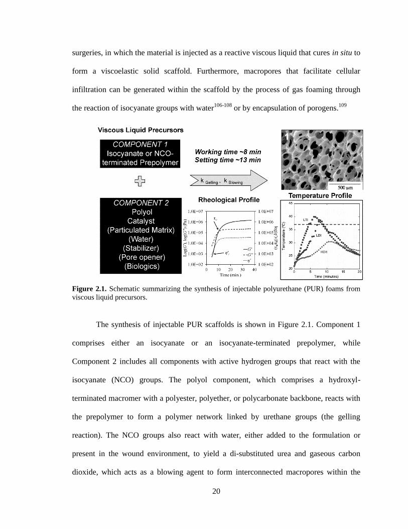
Figure 2.1. Schematic summarizing the synthesis of injectable polyurethane (PUR) foams from
viscous liquid precursors.
The synthesis of injectable PUR scaffolds is shown in Figure 2.1. Component 1
comprises either an isocyanate or an isocyanate-terminated prepolymer, while
Component 2 includes all components with active hydrogen groups that react with the
isocyanate (NCO) groups. The polyol component, which comprises a hydroxyl-
terminated macromer with a polyester, polyether, or polycarbonate backbone, reacts with
the prepolymer to form a polymer network linked by urethane groups (the gelling
reaction). The NCO groups also react with water, either added to the formulation or
present in the wound environment, to yield a di-substituted urea and gaseous carbon
dioxide, which acts as a blowing agent to form interconnected macropores within the
21
PUR network 107, 108, 106
as shown in the SEM image (Figure 2.1). The working time,
identified by the G’-G” crossover point (rheological profile in Figure 2.1) where the foam
transitions from a viscous liquid to an elastic solid, can be tuned to targeted values using
urethane catalysts, such as tertiary amines110
or organometallic compounds. Amine-based
catalysts have low cytotoxicity111
and selectively catalyze the water-isocyanate blowing
reaction,112
while the more cytotoxic organometallic catalysts selectively catalyze the
polyol-isocyanate gelling reaction. In contrast to the temperatures during cure exceeding
100oC observed for aromatic isocyanates, the exotherm for lysine-derived PUR foams is
<20oC (temperature profile in Figure 2.1). Other additives, such as stabilizers and pore
openers to control miscibility and pore size,106
matrix particles to control expansion and
increase mechanical properties, or biologics to enhance bioactivity,113
can also be
included in component 2.
While biodegradable PUR foams have been synthesized from aliphatic
polyisocyanates,114
lysine-derived polyisocyanates have shown the greatest promise for
injectable and settable systems.107, 115-117, 108, 110, 112, 118
Reactive PUR scaffolds prepared
from lysine-derived polyisocyanates, including lysine methyl ester diisocyanate (LDI)
and lysine triisocyanate (LTI), and tertiary amine catalysts, have been reported to induce
a minimal inflammatory response on host tissue in both cutaneous and bone defects in
vivo.115, 108, 112, 118
Furthermore, in vitro cytotoxicity testing of the leachates from the
reactive PUR has shown that the reactive PUR components are non-toxic.112
These
studies highlight the potential of injectable and settable lysine-derived PURs for both soft
and hard tissue regeneration.
22
Polyurethane scaffolds for bone regeneration
PUR biocomposites derived from LDI108, 119
and LTI110, 120, 121
have been
investigated as injectable bone void fillers and cements. An extracellular matrix
component, such as demineralized bone matrix (DBM), calcium phosphate particles, or
allograft bone particles, is blended with the reactive PUR to increase the
osteoconductivity and mechanical properties of the graft and also to reduce its volumetric
expansion in situ. Lysine-derived PUR composites are injectable, set within clinically
relevant working times (e.g., 5-10 min) to form grafts with mechanical strength
approaching that of host bone, and remodel and heal to form new bone. In an early study,
a four-armed poly(dioxanone-co-glycolide) prepolymer capped with LDI was mixed with
calcium phosphate particles in the presence of an diethanolamine catalyst and water to
form a reactive putty that expanded to form a foam with interconnected pores.108
When
implanted into the pectoralis muscles of rats for up to 42 days, the putty exhibited a
transient inflammatory response that resolved after 3 days. At longer time points, the
foams were well integrated with host tissue and showed no evidence of a chronic
inflammatory reaction.
In a similar study in bony defects, a settable foam was prepared by reacting an
LDI-pentaerythritol prepolymer with a poly(lactide-co-glycolide) polyol (functionality of
4).119
The two reactive liquid components were mixed with stannous octoate catalyst,
water, and β-tricalcium phosphate particles (10 μm) and injected into femoral condyle
plug defects in sheep, where they cured in ~10 min and expanded for form porous foam.
Defects showed progressive healing from 6 to 24 weeks, characterized by the formation
of new cortex, polymer degradation, and appositional bone growth with no signs of acute
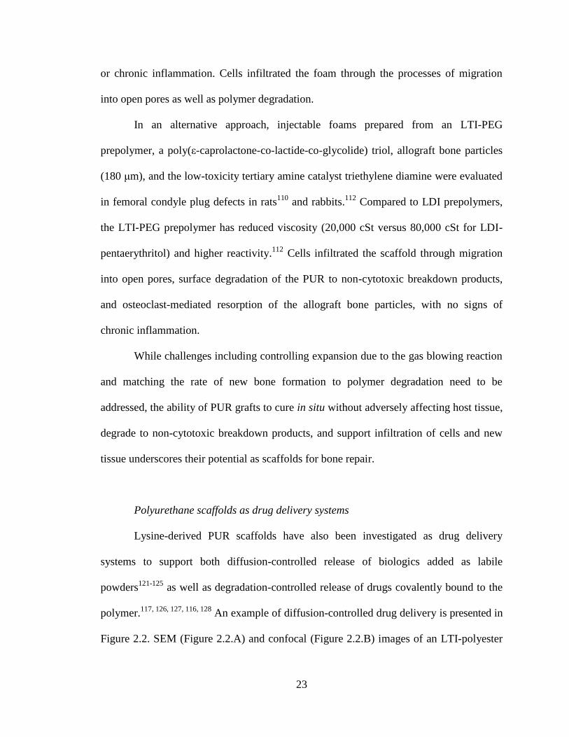
23
or chronic inflammation. Cells infiltrated the foam through the processes of migration
into open pores as well as polymer degradation.
In an alternative approach, injectable foams prepared from an LTI-PEG
prepolymer, a poly(ε-caprolactone-co-lactide-co-glycolide) triol, allograft bone particles
(180 μm), and the low-toxicity tertiary amine catalyst triethylene diamine were evaluated
in femoral condyle plug defects in rats110
and rabbits.112
Compared to LDI prepolymers,
the LTI-PEG prepolymer has reduced viscosity (20,000 cSt versus 80,000 cSt for LDI-
pentaerythritol) and higher reactivity.112
Cells infiltrated the scaffold through migration
into open pores, surface degradation of the PUR to non-cytotoxic breakdown products,
and osteoclast-mediated resorption of the allograft bone particles, with no signs of
chronic inflammation.
While challenges including controlling expansion due to the gas blowing reaction
and matching the rate of new bone formation to polymer degradation need to be
addressed, the ability of PUR grafts to cure in situ without adversely affecting host tissue,
degrade to non-cytotoxic breakdown products, and support infiltration of cells and new
tissue underscores their potential as scaffolds for bone repair.
Polyurethane scaffolds as drug delivery systems
Lysine-derived PUR scaffolds have also been investigated as drug delivery
systems to support both diffusion-controlled release of biologics added as labile
powders121-125
as well as degradation-controlled release of drugs covalently bound to the
polymer.117, 126, 127, 116, 128
An example of diffusion-controlled drug delivery is presented in
Figure 2.2. SEM (Figure 2.2.A) and confocal (Figure 2.2.B) images of an LTI-polyester
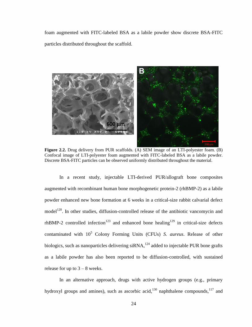
24
foam augmented with FITC-labeled BSA as a labile powder show discrete BSA-FITC
particles distributed throughout the scaffold.
Figure 2.2. Drug delivery from PUR scaffolds. (A) SEM image of an LTI-polyester foam. (B)
Confocal image of LTI-polyester foam augmented with FITC-labeled BSA as a labile powder.
Discrete BSA-FITC particles can be observed uniformly distributed throughout the material.
In a recent study, injectable LTI-derived PUR/allograft bone composites
augmented with recombinant human bone morphogenetic protein-2 (rhBMP-2) as a labile
powder enhanced new bone formation at 6 weeks in a critical-size rabbit calvarial defect
model120
. In other studies, diffusion-controlled release of the antibiotic vancomycin and
rhBMP-2 controlled infection123
and enhanced bone healing129
in critical-size defects
contaminated with 105 Colony Forming Units (CFUs) S. aureus. Release of other
biologics, such as nanoparticles delivering siRNA,124
added to injectable PUR bone grafts
as a labile powder has also been reported to be diffusion-controlled, with sustained
release for up to 3 – 8 weeks.
In an alternative approach, drugs with active hydrogen groups (e.g., primary
hydroxyl groups and amines), such as ascorbic acid,130
naphthalene compounds,117
and
25
the anti-tumor drug 7-tert-butyldimethylsilyl-10-hydroxy-camptothecin (DB-67),128
were
dissolved in the reactive PUR prior to injection and subsequently released in biologically
active form by degradation of the polymer. Recent studies have reported that the release
kinetics can be controlled by the composition of the drug,117
the choice of catalyst,116
or
the addition of ionic ligands,126
which offer greater tunability compared to diffusion-
controlled release. Potential advantages of covalently binding drugs to PUR scaffolds
include the ability to increase drug loading and control release to achieve therapeutically
relevant doses. For example, clinical evaluation of DB-67 has been limited by the
inability to deliver it in sufficient quantities to slow tumor growth and disease
progression. When DB-67 was covalently incorporated into LDI-glycerol PUR scaffolds,
it was released in vitro at a rate suitable for delivering therapeutic concentrations of the
drug and inhibited cellular proliferation by 50% for several malignant glioma cell
lines.128, 126
While the degradation-controlled release mechanism has proven successful
for small molecule synthetic drugs, another study has reported that recombinant human
proteins dissolved in the aqueous phase prior to foaming are not released from the cured
foam in a biologically active form.121
26
References
1 Robert Langer, and Joseph P. Vacanti, 'Tissue Engineering', Science, 260 (1993), 920-26.
2 Timothy A. Lew, John A. Walker, Joseph C. Wenke, Lorne H. Blackbourne, and Robert G. Hale,
'Characterization of Craniomaxillofacial Battle Injuries Sustained by United States Service
Members in the Current Conflicts of Iraq and Afghanistan', Journal of Oral and Maxillofacial
Surgery, 68 (2010), 3-7.
3 D. Ghysen, L. Ozsarlak, L. van den Hauwe, J. Van Goethem, AM. De Schepper, and PM. Parizel,
'Maxillo-Facial Trauma', JBR-BTR, 83 (2000), 181-97.
4 BB. Ward, SE. Brown, and PH. Krebsbach, 'Bioengineering Strategies for Regeneration of
Craniofacial Bone: A Review of Emerging Technologies', Oral diseases, doi:10.1111/j.1601-
0825.2010.01682.x (2010).
5 BJ. Costello, G. Shah, P. Kumta, and CS. Sfeir, 'Regenerative Medicine for Craniomaxillofacial
Surgery', Oral Maxillofac Surg Clin North Am., 22 (2010), 33-42.
6 S. Bose, M. Roy, and A. Bandyopadhyay, 'Recent Advances in Bone Tissue Engineering
Scaffolds', Trends in Biotechnology, 30 (2012), 546-54.
7 António J. Salgado, Olga P. Coutinho, and Rui L. Reis, 'Bone Tissue Engineering: State of the Art
and Future Trends', Macromolecular Bioscience, 4 (2004), 743-65.
8 Elizabeth M. Christenson, Wafa Soofi, Jennifer L. Holm, Neil R. Cameron, and Antonios G.
Mikos, 'Biodegradable Fumarate-Based Polyhipes as Tissue Engineering Scaffolds',
Biomacromolecules, 8 (2007), 3806-14.
9 W. J. Habraken, L. T. de Jonge, J. G. Wolke, L. Yubao, A. G. Mikos, and J. A. Jansen,
'Introduction of Gelatin Microspheres into an Injectable Calcium Phosphate Cement', J Biomed
Mater Res A, 87 (2008), 643-55.
10 Liang Zhao, Michael D Weir, and Hockin HK Xu, 'An Injectable Calcium Phosphate-Alginate
Hydrogel-Umbilical Cord Mesenchymal Stem Cell Paste for Bone Tissue Engineering',
Biomaterials, 31 (2010), 6502-10.
11 W. J. Habraken, Z. Zhang, J. G. Wolke, D. W. Grijpma, A. G. Mikos, J. Feijen, and J. A. Jansen,
'Introduction of Enzymatically Degradable Poly(Trimethylene Carbonate) Microspheres into an
Injectable Calcium Phosphate Cement', Biomaterials, 29 (2008), 2464-76.
12 S. Sarda, M. Nilsson, M. Balcells, and E. Fernandez, 'Influence of Surfactant Molecules as Air-
Entraining Agent for Bone Cement Macroporosity', J Biomed Mater Res A, 65 (2003), 215-21.
13 E. Behravesh, S. Jo, K. Zygourakis, and A. G. Mikos, 'Synthesis of in Situ Cross-Linkable
Macroporous Biodegradable Poly(Propylene Fumarate-Co-Ethylene Glycol) Hydrogels',
Biomacromolecules, 3 (2002), 374-81.
14 Shulin He, Michael J. Yaszemski, Alan W. Yasko, Paul S. Engel, and Antonios G. Mikos,
'Injectable Biodegradable Polymer Composites Based on Poly(Propylene Fumarate) Crosslinked
with Poly(Ethylene Glycol)-Dimethacrylate', Biomaterials, 21 (2000), 2389-94.
15 Sidi A Bencherif, R Warren Sands, Deen Bhatta, Praveen Arany, Catia S Verbeke, David A
Edwards, and David J Mooney, 'Injectable Preformed Scaffolds with Shape-Memory Properties',
Proceedings of the National Academy of Sciences, 109 (2012), 19590-95.
27
16 A. Almirall, G. Larrecq, J. A. Delgado, S. Martinez, J. A. Planell, and M. P. Ginebra, 'Fabrication
of Low Temperature Macroporous Hydroxyapatite Scaffolds by Foaming and Hydrolysis of an
Alpha-Tcp Paste', Biomaterials, 25 (2004), 3671-80.
17 B. R. Constantz, B. M. Barr, I. C. Ison, M. T. Fulmer, J. Baker, L. McKinney, S. B. Goodman, S.
Gunasekaren, D. C. Delaney, J. Ross, and R. D. Poser, 'Histological, Chemical, and
Crystallographic Analysis of Four Calcium Phosphate Cements in Different Rabbit Osseous Sites',
J Biomed Mater Res, 43 (1998), 451-61.
18 E. Fernandez, M. P. Ginebra, M. G. Boltong, F. C. Driessens, J. Ginebra, E. A. De Maeyer, R. M.
Verbeeck, and J. A. Planell, 'Kinetic Study of the Setting Reaction of a Calcium Phosphate Bone
Cement', J Biomed Mater Res, 32 (1996), 367-74.
19 Robert S. Moglia, Jennifer L. Holm, Nicholas A. Sears, Caitlin J. Wilson, Dawn M. Harrison, and
Elizabeth Cosgriff-Hernandez, 'Injectable Polyhipes as High-Porosity Bone Grafts',
Biomacromolecules, 12 (2011), 3621-28.
20 J. M. Williams, A. J. Gray, and M. H. Wilkerson, 'Emulsion Stability and Rigid Foams from
Styrene or Divinylbenzene Water-in-Oil Emulsions', Langmuir, 6 (1990), 437-44.
21 E. Hill, T. Boontheekul, and D. J. Mooney, 'Designing Scaffolds to Enhance Transplanted
Myoblast Survival and Migration', Tissue Engineering, 12 (2006), 1295-304.
22 Jun Liu, Hockin H. K. Xu, Hongzhi Zhou, Michael D. Weir, Qianming Chen, and Carroll Ann
Trotman, 'Human Umbilical Cord Stem Cell Encapsulation in Novel Macroporous and Injectable
Fibrin for Muscle Tissue Engineering', Acta Biomaterialia, 9 (2013), 4688-97.
23 E. Fernandez, M. D. Vlad, M. M. Gel, J. Lopez, R. Torres, J. V. Cauich, and M. Bohner,
'Modulation of Porosity in Apatitic Cements by the Use of Alpha-Tricalcium Phosphate-Calcium
Sulphate Dihydrate Mixtures', Biomaterials, 26 (2005), 3395-404.
24 P. Frayssinet, L. Gineste, P. Conte, J. Fages, and N. Rouquet, 'Short-Term Implantation Effects of
a Dcpd-Based Calcium Phosphate Cement', Biomaterials, 19 (1998), 971-7.
25 Diederik H. R. Kempen, Lichun Lu, Choll Kim, Xun Zhu, Wouter J. A. Dhert, Bradford L.
Currier, and Michael J. Yaszemski, 'Controlled Drug Release from a Novel Injectable
Biodegradable Microsphere/Scaffold Composite Based on Poly(Propylene Fumarate)', Journal of
Biomedical Materials Research Part A, 77A (2006), 103-11.
26 Jaclyn A. Shepard, Farrukh R. Virani, Ashley G. Goodman, Timothy D. Gossett, Seungjin Shin,
and Lonnie D. Shea, 'Hydrogel Macroporosity and the Prolongation of Transgene Expression and
the Enhancement of Angiogenesis', Biomaterials, 33 (2012), 7412-21.
27 Qingchun Zhang, Ke Tan, Zhaoyang Ye, Yan Zhang, Wensong Tan, and Meidong Lang,
'Preparation of Open Porous Polycaprolactone Microspheres and Their Applications as Effective
Cell Carriers in Hydrogel System', Materials Science and Engineering: C, 32 (2012), 2589-95.
28 C. Trojani, F. Boukhechba, J. C. Scimeca, F. Vandenbos, J. F. Michiels, G. Daculsi, P. Boileau, P.
Weiss, G. F. Carle, and N. Rochet, 'Ectopic Bone Formation Using an Injectable Biphasic Calcium
Phosphate/Si-Hpmc Hydrogel Composite Loaded with Undifferentiated Bone Marrow Stromal
Cells', Biomaterials, 27 (2006), 3256-64.
29 Hongzhi Zhou, and Hockin H. K. Xu, 'The Fast Release of Stem Cells from Alginate-Fibrin
Microbeads in Injectable Scaffolds for Bone Tissue Engineering', Biomaterials, 32 (2011), 7503-
13.

28
30 Z Xia, L M Groverb, Y Huang, I E Adamopoulosa, U Gbureck, J T Triffitt, R M Sheltone, and J E
Barralet, 'In Vitro Biodegradation of Three Brushite Calcium Phosphate Cements by a
Macrophage Cell-Line', Biomaterials, 27 (2006), 4557-65.
31 A. Truedsson, J. S. Wang, P. Lindberg, M. Gordh, B. Sunzel, and G. Warfvinge, 'Bone Substitute
as an on-Lay Graft on Rat Tibia', Clin Oral Implants Res, 21 (2010), 424-9.
32 M. Bohner, P. Van Landuyt, H. P. Merkle, and J. Lemaitre, 'Composition Effects on the Ph of a
Hydraulic Calcium Phosphate Cement', J Mater Sci Mater Med, 8 (1997), 675-81.
33 M. Nilsson, E. Fernandez, S. Sarda, L. Lidgren, and J. A. Planell, 'Characterization of a Novel
Calcium Phosphate/Sulphate Bone Cement', J Biomed Mater Res, 61 (2002), 600-7.
34 Lichun Lu, Susan J. Peter, Michelle D. Lyman, Hui Lai, Lin, Susan M. Leite, Janet A. Tamada,
Joseph Vacanti, P., Rober Langer, and Antonios G. Mikos, 'In Vitro Degradation of Porous
Poly(L-Lactic Acid) Foams', Biomaterials, 21 (2000), 1595-605.
35 F Kurtis Kasper, Kazuhiro Tanahashi, John P. Risher, and Antonios G. Mikos, 'Synthesis of
Poly(Propylene Fumarate)', Nature Protocols, 4 (2009), 518-25.
36 MHR. Magno, J Kim, A Srinivasan, S McBride, D Bolikal, A Darr, JO Hollinger, and J Kohn,
'Synthesis, Degradation and Biocompatibility of Tyrosine-Derived Polycarbonate Scaffolds',
Journal of Materials Chemistry, 20 (2010), 8885-93.
37 A.S. Rowlands, S.A. Lim, D. Martin, and J.J. Cooper-White, 'Polyurethane/Poly(Lactic-Co-
Glycolic) Acid Composite Scaffolds Fabricated by Thermally Induced Phase Separation',
Biomaterials, 28 (2007), 2109-21.
38 Peter X. Ma, and Ji-Won Choi, 'Biodegradable Polymer Scaffolds with Well-Defined
Interconnected Spherical Pore Network', Tissue Engineering, 7 (2001), 23-49.
39 F Carfi Pavia, V La Carrubba, S Piccarolo, and V Brucato, 'Polymeric Scaffolds Prepared Via
Thermally Induced Phase Separation: Tuning of Structure and Morphology', J Biomed Mater Res
Part A, 86A (2008), 459-66.
40 Aurelio Salerno, Salvatore Iannace, and Paolo A. Netti, 'Open-Pore Biodegradable Foams
Prepared Via Gas Foaming and Microparticulate Templating', Macromolecular Bioscience, 8
(2008), 655-64.
41 ASTM, 'D445: Standard Test Method for Kinematic Viscosity of Transparent and Opaque Liquids
(and Calculation of Dynamic Viscosity)', (1997).
42 Elizabeth M. Christenson, Wafa Soofi, Jennifer Holm, L., Neil Cameron, R., and Antonios G.
Mikos, 'Biodegradable Fumarate-Based Polyhipes as Tissue Engineering Scaffolds',
Biomacromolecules, 8 (2007), 3806-14.
43 ASTM, 'D3576: Standard Test Method for Cell Size of Rigid Cellular Plastics', (2004).
44 Robert S. Moglia, Jennifer L. Holm, Nicholas A. Sears, Caitlin J. Wilson, Dawn Harrison, and
Cosgriff-Hernandez, 'Injectable Polyhipes as High-Porosity Bone Grafts', Biomacromolecules, 12
(2011), 3621-28.
45 SA Guelcher, V Patel, K Gallagher, S Connonlly, JE Didier, JS Doctor, and JO Hollinger,
'Synthesis and in Vitro Biocompatibility of Injectable Polyurethane Foam Scaffolds', Tissue
engineering, 12 (2006b), 1247-59.
29
46 Leon J.M. Jacobs, Maartje F. Kemmere, and Jos T.F. Keurentjes, 'Sustainable Polymer Foaming
Using High Pressure Carbon Dioxide: A Review on Fundamentals, Processes and Applications',
Green Chemistry, 10 (2008), 731-38.
47 Joanne. E McBane, Soroor Sharifpoor, Cai Kuihua, Rosalind S. Labow, and J. Paul Santerre,
'Biodegradation and in Vivo Biocompatibility of a Degradable, Polar/Hydrophobic/Ionic
Polyurethane for Tissue Engineering Applications', Biomaterials, 32 (2011), 6034-44.
48 AR. Amini, DJ. Adams, CT. Laurencin, and SP Nukavarapu, 'Optimally Porous and
Biomechanically Compatible Scaffolds for Large-Area Bone Regeneration', Tissue Engineering:
Part A, 18 (2012), 1376-88.
49 Yoon Sung Nam, and Tae Gwan Park, 'Porous Biodegradable Polymeric Scaffolds Prepared by
Thermally Induced Phase Separation', J Biomed Mater Res, 47 (1999), 8-17.
50 AE Hafeman, KJ Zienkiewicz, AL Zachman, HJ Sung, LB Nanney, JM Davidson, and SA
Guelcher, 'Characterization of the Degradation Mechanisms of Lysine-Derived Aliphatic
Poly(Ester Urethane) Scaffolds', Biomaterials, 32 (2010), 419-29.
51 ASTM, 'D-3574: Standard Test Methods for Flexible Cellular Materials- Slab, Bonded, and
Molded Urethane Foams', (2001).
52 ASTM, 'D695: Standard Test Method for Compressive Properties of Rigid Plastics', (2002).
53 ASTM, 'D-1621: Standard Test Method for Compressive Properties of Rigid Cellular Plastics',
(2010).
54 S.N. Khan, F.P. Cammisa, H.S. Sandbu, A.D. Diwan, F.P. Girardi, and J.M. Lane, 'The Biology of
Bone Grafting', Journal of the American Academy of Orthopaedic Surgeons, 13 (2005), 77-86.
55 K.S. Beebe, J. Benevenia, B.E. Tuy, C.A. DePaula, R.D. Harten, and W.F. Enneking, 'Effects of a
New Allograft Processing Procedure on Garfting Healing in a Canine Model', Clin Orthop Relat
Res, 467 (2009), 273-80.
56 J. E. Dumas, K. Zienkiewicz, S. Tanner, E.M. Prieto, S. Battacharyya, and S.A. Guelcher,
'Synthesis and Characterization of an Injectable Allograft Bone/Polymer Composite Bone Void
Filler with Tunable Mechanical Properties', Tissue Engineering: Part A, 16 (2010), 2505-18.
57 R. Z. Legeros, A. Chohayeb, and A. Schulman, 'Apatitic Calcium Phosphates Possible Dental
Restorative Materials', Journal of Dental Research, 61 (1982), 343.
58 K. L. Low, S. H. Tan, S. H. Zein, J. A. Roether, V. Mourino, and A. R. Boccaccini, 'Calcium
Phosphate-Based Composites as Injectable Bone Substitute Materials: A Review', J Biomed Mater
Res B Appl Biomater.
59 Harvey Chim, and Arun K. Gosain, 'Biomaterials in Craniofacial Surgery Experimental Studies
and Clinical Application', Journal of Craniofacial Surgery, 20 (2009), 29-33.
60 C. Liu, W. Wang, W. Shen, T. Chen, L. Hu, and Z. Chen, 'Evaluation of the Biocompatibility of a
Nonceramic Hydroxyapatite', J Endod, 23 (1997), 490-3.
61 M Bohner, 'Calcium Orthophosphates in Medicine: From Ceramics to Calcium Phosphate
Cements', Injury, 31 (2000), S-D37-47.
62 Thomas J. Webster, Celaletdin Ergun, Robert H. Doremus, Richard W. Siegel, and Rena Bizios,
'Enhanced Functions of Osteoblasts on Nanophase Ceramics', Biomaterials, 21 (2000), 1803-10.
30
63 R Gonzalez-McQuire, D W Green, K. A. Partridge, R. O. Oreffo, S Mann, and S A Davis,
'Coating of Human Mesenchymal Cells in 3d Culture with Bioinorganic Nanoparticles Promotes
Osteoblastic Differentiation and Gene Transfection', Adv Mater, 19 (2007), 2236-40.
64 T. Winkler, E. Hoenig, R. Gildenhaar, G. Berger, D. Fritsch, R. Janssen, M. M. Morlock, and A.
F. Schilling, 'Volumetric Analysis of Osteoclastic Bioresorption of Calcium Phosphate Ceramics
with Different Solubilities', Acta Biomaterialia, 6 (2010), 4127-35.
65 J. D. Kretlow, S. Young, L. Klouda, M. Wong, and A. G. Mikos, 'Injectable Biomaterials for
Regenerating Complex Craniofacial Tissues', Adv Mater Deerfield, 21 (2009), 3368-93.
66 M. L. Shindo, P. D. Costantino, C. D. Friedman, and L. C. Chow, 'Facial Skeletal Augmentation
Using Hydroxyapatite Cement', Arch Otolaryngol Head Neck Surg, 119 (1993), 185-90.
67 G. D. Brown, B. L. Mealey, P. V. Nummikoski, S. L. Bifano, and T. C. Waldrop, 'Hydroxyapatite
Cement Implant for Regeneration of Periodontal Osseous Defects in Humans', J Periodontol, 69
(1998), 146-57.
68 C S Goldberg, O Antonyshyn, R Midha, and J A Fialkov, 'Measuring Pulsatile Forces on the
Human Cranium', J Craniofacial Surgery, 16 (2005), 134 - 39.
69 T. Livingston, P. Ducheyne, and J. Garino, 'In Vivo Evaluation of a Bioactive Scaffold for Bone
Tissue Engineering', J Biomed Mater Res, 62 (2002), 1-13.
70 R. M. Pilliar, M. J. Filiaggi, J. D. Wells, M. D. Grynpas, and R. A. Kandel, 'Porous Calcium
Polyphosphate Scaffolds for Bone Substitute Applications -- in Vitro Characterization',
Biomaterials, 22 (2001), 963-72.
71 J. E. Barralet, L. Grover, T. Gaunt, A. J. Wright, and I. R. Gibson, 'Preparation of Macroporous
Calcium Phosphate Cement Tissue Engineering Scaffold', Biomaterials, 23 (2002), 3063-72.
72 S. Marcia, C. Boi, M. Dragani, S. Marini, M. Marras, E. Piras, G. C. Anselmetti, and S. Masala,
'Effectiveness of a Bone Substitute (Cerament) as an Alternative to Pmma in Percutaneous
Vertebroplasty: 1-Year Follow-up on Clinical Outcome', Eur Spine J, 21 Suppl 1 (2012), S112-8.
73 S. Masala, G. Nano, S. Marcia, M. Muto, F. P. Fucci, and G. Simonetti, 'Osteoporotic Vertebral
Compression Fracture Augmentation by Injectable Partly Resorbable Ceramic Bone Substitute
(Cerament|Spinesupport): A Prospective Nonrandomized Study', Neuroradiology, 54 (2012),
1245-51.
74 W. Chen, Hongzhi Zhou, M. Tang, Michael D Weir, C. Bao, and Hockin H. K. Xu, 'Gas-Foaming
Calcium Phosphate Cement Scaffold Encapsulating Human Umbilical Cord Stem Cells.', Tissue
Engineering Part A, 18 (2012), 816-27.
75 M Bohner, 'Calcium Phosphate Emulsions: Possible Applications', Key Eng Mater, 192-195
(2001), 765-68.
76 D. P. Link, J. van den Dolder, W. J. Jurgens, J. G. Wolke, and J. A. Jansen, 'Mechanical
Evaluation of Implanted Calcium Phosphate Cement Incorporated with Plga Microparticles',
Biomaterials, 27 (2006), 4941-7.
77 P. Q. Ruhe, O. C. Boerman, F. G. Russel, P. H. Spauwen, A. G. Mikos, and J. A. Jansen,
'Controlled Release of Rhbmp-2 Loaded Poly(Dl-Lactic-Co-Glycolic Acid)/Calcium Phosphate
Cement Composites in Vivo', J Control Release, 106 (2005), 162-71.
78 R B Martin, M W Chapman, R E Holmes, D J Sartoris, E C Shors, J E Gordon, D O Heitter, N A
Sharkey, and A G Zissimos, 'Effects of Bone Ingrowth on the Strength and Non-Invasive
Assessment of a Coralline Hydroxyapatite Material', Biomaterials, 10 (1989), 481-88.
31
79 O. Gauthier, R. Muller, D. von Stechow, B. Lamy, P. Weiss, J. M. Bouler, E. Aguado, and G.
Daculsi, 'In Vivo Bone Regeneration with Injectable Calcium Phosphate Biomaterial: A Three-
Dimensional Micro-Computed Tomographic, Biomechanical and Sem Study', Biomaterials, 26
(2005), 5444-53.
80 C. Mauli Agrawal, and Robert B. Ray, 'Biodegradable Polymeric Scaffolds for Musculoskeletal
Tissue Engineering', Journal of Biomed Mater Res, 55 (2001), 141-50.
81 Zhen Pan, and Jiandong Ding, 'Poly(Lactide-Co-Glycolide) Porous Scaffolds for Tissue
Engineering and Regenerative Medicine', Interface Focus, 2 (2012), 366-77.
82 Lakshimi S. Nair, and Cato T. Laurencin, 'Biodegradable Polymers as Biomaterials', Progress in
Polymer Science, 32 (2007), 762-98.
83 Xiaohua Liu, and Peter X. Ma, 'Polymeric Scaffolds for Bone Tissue Engineering', Annals of
Biomedical Engineering, 32 (2004), 477-86.
84 E Farng, and O Sherman, 'Meniscal Repair Devices: A Clinical and Biomechanical Literature
Review', J Arthrosc Relat Surg, 20 (2004), 273-86.
85 C. Vaca-Garcia, 'Biomaterials', in Introduction to Chemicals from Biomass, ed. by JH Clark and
FEI Deswarte (Chichester: Wiley, 2008), pp. 103-42.
86 L. Brannon-Peppas, and M. Vert, 'Polylactic and Polyglycolic Acids as Drug Delivery Carriers', in
Handbook of Pharmaceutical Controlled Release Technology, ed. by DL Wise (New York:
Marcel Dekker, 2000), pp. 99-130.
87 X Shi, and AG Mikos, 'Poly(Propylene Fumarate)', in An Introduction to Biomaterials, ed. by SA
Guelcher and JO Hollinger (Boca Raton: CRC Press, 2006), pp. 205-18.
88 Shanfeng Wang, Lichun Lu, and Michael J. Yaszemski, 'Bone Tissue-Engineering Material
Poly(Propylene Fumarate): Correlation between Molecular Weight, Chain Dimensions, and
Physical Properties', Biomacromolecules, 7 (2006), 1976-82.
89 MD Timmer, C Carter, CG Ambrose, and AG Mikos, 'Fabrication of Poly(Propylene Fumarate)-
Based Orthopaedic Implants by Photo-Crosslinking through Transparent Silicone Molds',
Biomaterials, 24 (2003), 4707.
90 JP Fisher, JWM Vehof, D Dean, JPCM van der Waerden, TA Holland, A Mikos, and JA Jansen,
'Soft and Hard Tissue Response to Photocrosslinked Poly(Propylene Fumarate) Scaffolds in a
Rabbit Model', J Biomed Mater Res, 59 (2002), 547-56.
91 D.H. Kempen, L Lu, C. Kim, X. Zhu, W.J.A. Dhert, B.L. Currier, and M.J. Yaszemski,
'Controlled Drug Release from a Novel Injectable Biodegradable Microsphere/Scaffold Composite
Based on Poly(Propylene Fumarate)', J Biomed Mater Res A., 77 (2006), 103-11.
92 K.W. Lee, S. Wang, L. Lu, E. Jabbari, B.L. Currier, and M.J. Yaszemski, 'Fabrication and
Characterization of Poly(Propylene Fumarate) Scaffolds with Controlled Pore Structures Using 3-
Dimensional Printing and Injection Molding', Tissue engineering, 12 (2006), 2801-11.
93 J Kohn, and J Schut, 'Polymers Derived from L-Tyrosine', in An Introduction to Biomaterials, ed.
by SA Guelcher and JO Hollinger (Boca Raton: CRC Press, 2006), pp. 185-204.
94 J Kim, MHR Magno, H Waters, B Doll, S McBride, P Alvarez, A Darr, A Vasanji, J Kohn, and
JO Hollinger, 'Bone Regeneration in a Rabbit Critical-Sized Calvarial Model Using Tyrosine-
Derived Polycarbonate Scaffolds', Tissue Engineering: Part A, 18 (2012), 1132-39.
32
95 R Adhikari, PA Gunatillake, I Griffiths, L Tatai, M Wickramaratna, S Houshyar, T Moore, RTM
Mayadunne, J Field, M McGee, and T Carbone, 'Biodegradable Injectable Polyurethanes:
Synthesis and Evaluation for Orthopaedic Applications', Biomaterials, 29 (2008), 3762-70.
96 S Bennett, K Connolly, DR Lee, Y Jiang, D Buck, JO Hollinger, and EA Gruskin, 'Initial
Biocompatibility Studies of a Novel Degradable Polymeric Bone Substitute That Hardens in Situ',
Bone, 19 (1996), 1015.
97 MJ Yaszemski, RG Payne, WC Hayes, R Langer, TB Aufdemorte, and AG Mikos, 'The Ingrowth
of New Bone Tissue and Initial Mechanical Properties of a Degrading Polymeric Composite
Scaffold', Tissue engineering, 1 (1995), 41.
98 JE Dumas, K Zienkiewicz, SA Tanner, EM Prieto, S Battacharyya, and SA Guelcher, 'Synthesis
and Characterization of an Injectable Allograft Bone/Polymer Composite Bone Void Filler with
Tunable Mechanical Properties', Tissue engineering: Part A, 16 (2010), 2505-18.
99 P Gentile, M Mattioli-Belmonte, V Chiono, C Ferretti, F Baino, C Tonda-Turo, C Vitale-
Brovarone, I Pashkuleva, RL Reis, and G Ciardelli, 'Bioactive Glass/Polymer Composite Scaffolds
Mimicking Bone Tissue', J Biomed Mater Res Part A, 100A (2012), 2654-67.
100 HH Lu, SF El-Amin, KD Scott, and CT Laurencin, 'Three-Dimensional, Bioactive, Biodegradable,
Polymer-Bioactive Glass Composite Scaffolds with Improved Mechanical Properties Support
Collagen Synthesis and Mineralization of Human Osteoblast-Like Cells in Vitro', J Biomed Mater
Res A, 64 (2003), 465-74.
101 B Li, T Yoshii, AE Hafeman, JS Nyman, JC Wenke, and SA Guelcher, 'The Effects of Rhbmp-2
Released from Biodegradable Polyurethane/Microsphere Composite Scaffolds on New Bone
Formation in Rat Femora', Biomaterials, 30 (2009), 6768-79.
102 T Yoshii, AE Hafeman, JS Nyman, JM Esparza, K Shinomiya, DM Spengler, GR Mundy, GE
Gutierrez, and SA Guelcher, 'A Sustained Release of Lovastatin from Biodegradable, Elastomeric
Polyurethane Scaffolds for Enhanced Bone Regeneration', Tissue engineering: Part A, 16 (2010),
2396-79.
103 SJ Peter, L Lu, DJ Kim, GN Stamatas, MJ Miller, MJ Yaszemski, and AG Mikos, 'Effects of
Transforming Growth Factor B1 Released from Biodegradable Polymer Microparticles on
Marrow Stromal Osteoblasts Cultured on Poly(Propylene Fumarate) Substrates', J. Biomed. Mater.
Res., 50 (2000), 452-62.
104 S. A. Guelcher, 'Biodegradable Polyurethanes: Synthesis and Applications in Regenerative
Medicine', Tissue Engineering Part B-Reviews, 14 (2008), 3-17.
105 J. P. Santerre, K. Woodhouse, G. Laroche, and R. S. Labow, 'Understanding the Biodegradation of
Polyurethanes: From Classical Implants to Tissue Engineering Materials', Biomaterials, 26 (2005),
7457-70.
106 S. Guelcher, A. Srinivasan, A. Hafeman, K. Gallagher, J. Doctor, S. Khetan, S. McBride, and J.
Hollinger, 'Synthesis, in Vitro Degradation, and Mechanical Properties of Two-Component
Poly(Ester Urethane)Urea Scaffolds: Effects of Water and Polyol Composition', Tissue
Engineering, 13 (2007), 2321-33.
107 J. Y. Zhang, E. J. Beckman, N. P. Piesco, and S. Agarwal, 'A New Peptide-Based Urethane
Polymer: Synthesis, Biodegradation, and Potential to Support Cell Growth in Vitro', Biomaterials,
21 (2000), 1247-58.
108 S. Bennett, K. Connolly, D. R. Lee, Y. Jiang, D. Buck, J. O. Hollinger, and E. A. Gruskin, 'Initial
Biocompatibility Studies of a Novel Degradable Polymeric Bone Substitute That Hardens in Situ',
Bone, 19 (1996), S101-S07.
33
109 P. Bruin, G. J. Veenstra, A. J. Nijenhuis, and A. J. Pennings, 'Design and Synthesis of
Biodegradable Poly(Ester-Urethane) Elastomer Networks Composed of Non-Toxic Building-
Blocks', Makromolekulare Chemie-Rapid Communications, 9 (1988), 589-94.
110 J. E. Dumas, K. Zienkiewicz, S. A. Tanner, E. M. Prieto, S. Bhattacharyya, and S. A. Guelcher,
'Synthesis and Characterization of an Injectable Allograft Bone/Polymer Composite Bone Void
Filler with Tunable Mechanical Properties', Tissue Engineering Part A, 16 (2010), 2505-18.
111 M. C. Tanzi, P. Verderio, M. G. Lampugnani, M. Resnati, E. Dejana, and E. Sturani, 'Cytotoxicity
of Some Catalysts Commonly Used in the Synthesis of Copolymers for Biomedical Use', Journal
of Materials Science-Materials in Medicine, 5 (1994), 393-96.
112 J. M. Page, E. M. Prieto, J. E. Dumas, K. J. Zienkiewicz, J. C. Wenke, P. Brown-Baer, and S. A.
Guelcher, 'Biocompatibility and Chemical Reaction Kinetics of Injectable, Settable
Polyurethane/Allograft Bone Biocomposites', Acta Biomaterialia, 8 (2012), 4405-16.
113 J. Y. Zhang, B. A. Doll, E. J. Beckman, and J. O. Hollinger, 'A Biodegradable Polyurethane-
Ascorbic Acid Scaffold for Bone Tissue Engineering', Journal of Biomedical Materials Research
Part A, 67A (2003), 389-400.
114 K. Gorna, and S. Gogolewski, 'Biodegradable Porous Polyurethane Scaffolds for Tissue Repair
and Regeneration', Journal of Biomedical Materials Research Part A, 79A (2006), 128-38.
115 J. Y. Zhang, E. J. Beckman, J. Hu, G. G. Yang, S. Agarwal, and J. O. Hollinger, 'Synthesis,
Biodegradability, and Biocompatibility of Lysine Diisocyanate-Glucose Polymers', Tissue
Engineering, 8 (2002), 771-85.
116 W. N. Sivak, I. F. Pollack, S. Petoud, W. G. Zamboni, J. Y. Zhang, and E. J. Beckman, 'Catalyst-
Dependent Drug Loading of Ldi-Glycerol Polyurethane Foams Leads to Differing Controlled
Release Profiles', Acta Biomaterialia, 4 (2008), 1263-74.
117 W. N. Sivak, J. Y. Zhang, S. Petoud, and E. J. Beckman, 'Degradative-Release as a Function of
Drug Structure from Ldi-Glycerol Polyurethanes', Bio-Medical Materials and Engineering, 20
(2010), 269-81.
118 E. J. Adolph, A. E. Hafeman, J. M. Davidson, L. B. Nanney, and S. A. Guelcher, 'Injectable
Polyurethane Composite Scaffolds Delay Wound Contraction and Support Cellular Infiltration and
Remodeling in Rat Excisional Wounds', Journal of Biomedical Materials Research Part A, 100A
(2012), 450-61.
119 R. Adhikari, P. A. Gunatillake, I. Griffiths, L. Tatai, M. Wickramaratna, S. Houshyar, T. Moore,
R. T. M. Mayadunne, J. Field, M. McGee, and T. Carbone, 'Biodegradable Injectable
Polyurethanes: Synthesis and Evaluation for Orthopaedic Applications', Biomaterials, 29 (2008),
3762-70.
120 J.E. Dumas, P.B. BrownBaer, E.M. Prieto, T. Guda, R.G. Hale, J.C. Wenke, and S.A. Guelcher,
'Injectable Reactive Biocomposites for Bone Healing in Critical-Size Rabbit Calvarial Defects',
Biomedical Materials, 7 (2012), 024112.
121 B. Li, T. Yoshii, A. E. Hafeman, J. S. Nyman, J. C. Wenke, and S. A. Guelcher, 'The Effects of
Rhbmp-2 Released from Biodegradable Polyurethane/Microsphere Composite Scaffolds on New
Bone Formation in Rat Femora', Biomaterials, 30 (2009), 6768-79.
122 K. V. Brown, B. Li, T. Guda, D. S. Perrien, S. A. Guelcher, and J. C. Wenke, 'Improving Bone
Formation in a Rat Femur Segmental Defect by Controlling Bone Morphogenetic Protein-2
Release', Tissue Engineering Part A, 17 (2011), 1735-46.
34
123 B. Li, K. V. Brown, J. C. Wenke, and S. A. Guelcher, 'Sustained Release of Vancomycin from
Polyurethane Scaffolds Inhibits Infection of Bone Wounds in a Rat Femoral Segmental Defect
Model', Journal of Controlled Release, 145 (2010), 221-30.
124 C. E. Nelson, M. K. Gupta, E. J. Adolph, J. M. Shannon, S. A. Guelcher, and C. L. Duvall,
'Sustained Local Delivery of Sirna from an Injectable Scaffold', Biomaterials, 33 (2012), 1154-61.
125 Carlos J. Sanchez Jr, Edna M. Prieto, Chad A. Krueger, Katarzyna J. Zienkiewicz, Desiree R.
Romano, Catherine L. Ward, Kevin S. Akers, Scott A. Guelcher, and Joseph C. Wenke, 'Effects of
Local Delivery of D-Amino Acids from Biofilm-Dispersive Scaffolds on Infection in
Contaminated Rat Segmental Defects', Biomaterials, 34 (2013), 7533-43.
126 W. N. Sivak, J. Y. Zhang, S. Petoud, and E. J. Beckman, 'Incorporation of Ionic Ligands
Accelerates Drug Release from Ldi-Glycerol Polyurethanes', Acta Biomaterialia, 6 (2010), 144-
53.
127 W.N Sivak, J. Zhang, S. Petroud, and E. J. Beckman, 'Simultaneous Drug Release at Different
Rates from Biodegradable Polyurethane Foams', Acta Biomaterialia, 5 (2009), 2398-408.
128 W. N. Sivak, I. F. Pollack, S. Petoud, W. C. Zamboni, J. Y. Zhang, and E. J. Beckman, 'Ldi-
Glycerol Polyurethane Implants Exhibit Controlled Release of Db-67 and Anti-Tumor Activity in
Vitro against Malignant Gliomas', Acta Biomaterialia, 4 (2008), 852-62.
129 S. A. Guelcher, K. V. Brown, B. Li, T. Guda, B. H. Lee, and J. C. Wenke, 'Dual-Purpose Bone
Grafts Improve Healing and Reduce Infection', Journal of Orthopaedic Trauma, 25 (2011), 477-
82.
130 J. Y. Zhang, B. A. Doll, E. J. Beckman, and J. O. Hollinger, 'Three-Dimensional Biocompatible
Ascorbic Acid-Containing Scaffold for Bone Tissue Engineering', Tissue Engineering, 9 (2003),
1143-57.
35
CHAPTER III
EFFECTS OF LOCAL DELIVERY OF D-AMINO ACIDS FROM BIOFILM-DISPERSIVE
SCAFFOLDS ON INFECTION IN CONTAMINATED RAT SEGMENTAL DEFECTS
Introduction
Despite meticulous treatment, infectious complications of open bone fractures
continue to be a significant factor contributing to patient morbidity and poor healing
outcomes. The ensuing infection of bone by bacteria (osteomyelitis) is characterized by
high levels of inflammation and destruction of viable bone tissue. Often the infection
becomes chronic, resulting in increased rates of surgical revisions, non-union, and
extremity amputation 1-3
. Among the pathogenic microorganisms associated with chronic
osteomyelitis, Staphylococcus aureus is the most frequently isolated organism,
accounting for >50% of all cases 4-8
. In addition to the increasing trend of antimicrobial
resistance among clinical isolates, biofilm formation is a significant contributing factor in
the development of both device and non-device related chronic orthopaedic infections
and a major barrier to wound healing 6, 4
.
Bacterial biofilms are an association of single or multiple species attached to a
surface surrounded by an extracellular polymeric matrix (EPM), which constitutes a
protected mode of growth. Compared to their planktonic counterparts, biofilm-derived
bacteria have distinctive phenotypes in regards to growth, gene expression, and protein
production that confer resistance to antimicrobial agents as well as host mechanisms of
clearance 5, 9
. Importantly, bacterial biofilms have been associated with a broad range of
human infections, including chronic non-device-related infections such as osteomyelitis10,
36
11. Previous studies have reported that staphylococcal biofilms are present within infected
bone of patients with chronic osteomyelitis 6-8
, and that clinical osteomyelitis isolates of S.
aureus are capable of forming biofilms in vitro 12-14
. Furthermore, staphylococcal
biofilms have been implicated as a major cause of osseous non-union 15
. These studies
suggest that staphylococcal biofilms play a critical role in both the development of
chronic osteomyelitis and the sequelae of infectious complications.
Biofilm development is a highly coordinated and reversible process beginning
with the attachment and proliferation of cells on a surface and culminating in the
dispersal of cells from the biofilm into the surrounding environment. The dispersal of
cells is an essential stage of the biofilm life cycle that contributes to survival of the
organism and disease transmission. For both Gram-positive and Gram-negative
microorganisms, biofilm dispersion is mediated by self-produced diffusible factors 16, 17
.
Considering the specificity and effectiveness of these molecules for dispersing biofilms,
the use of biofilm dispersal agents has attracted considerable interest for the treatment of
biofilm-associated infections 18, 19
. Recent studies have shown that use of biofilm
dispersal agents, including bismuth thiols 20
, recombinant DNAses 21
, and diffusible
soluble factors 22, 23
, can disperse biofilms in vitro and improve healing of biofilm-
associated infections in vivo 24, 25
. However, toxicity to viable host tissues (as observed
for bismuth thiols and xylitol), as well as the specificity of these agents for certain
bacterial species and/or strains, may preclude their use as broad therapeutic strategies.
Recent studies have shown that the D-isomers of amino acids (D-AAs) can
prevent and disperse biofilms formed by a diverse range of bacterial species, including S.
aureus and P. aeruginosa 26, 27
. In contrast to other biofilm dispersal agents, D-AAs
37
promote the disassembly of biofilms through multiple mechanisms and have minimal
cellular toxicity 28
. In this study, we investigated the ability of biofilm-dispersive
polyurethane (PUR) scaffolds augmented with D-AAs to protect the scaffold from
contamination from the contiguous wound environment and to reduce microbial burden
within segmental defects in vivo. A mixture of D-AAs with optimal in vitro anti-biofilm
activity was evaluated in a rat contaminated segmental defect model to test our
hypothesis that local delivery of D-AAs would reduce the extent of infection within the
defect. We also investigated the cytotoxicity of D-AAs on host mammalian cells to
further evaluate their therapeutic potential.
Experimental
Materials
D- and L-isomers of amino acids (free base form), including alanine, isoleucine,
leucine, methionine, phenylalanine, proline, tryptophan, tyrosine, and valine, were
purchased from Sigma Aldrich (St. Louis, MO, USA). For use in bacterial and cell
cultures, D-AA stocks were prepared by dissolving powders in 0.5 M HCl at
concentrations between 150-200 mM. Stocks were then diluted into cation-adjusted
Mueller Hinton (MHB-II) broth neutralized to pH 7.4 and stored at -80°C. For
polyurethane scaffold synthesis, ε-caprolactone and stannous octoate were supplied by
Sigma-Aldrich, and glycolide and D,L-lactide were purchased from Polysciences. An
isocyanate-terminated prepolymer (22.7% NCO) comprising polyethylene glycol (PEG)
end-capped with lysine triisocyanate (LTI) at a 2:1 molar ratio of LTI:PEG was supplied
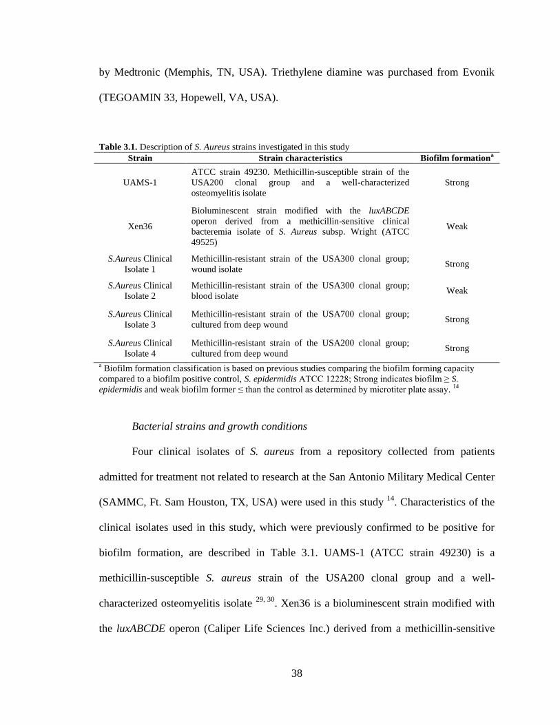
38
by Medtronic (Memphis, TN, USA). Triethylene diamine was purchased from Evonik
(TEGOAMIN 33, Hopewell, VA, USA).
Table 3.1. Description of S. Aureus strains investigated in this study
Strain Strain characteristics Biofilm formationa
UAMS-1
ATCC strain 49230. Methicillin-susceptible strain of the
USA200 clonal group and a well-characterized
osteomyelitis isolate
Strong
Xen36
Bioluminescent strain modified with the luxABCDE
operon derived from a methicillin-sensitive clinical
bacteremia isolate of S. Aureus subsp. Wright (ATCC
49525)
Weak
S.Aureus Clinical
Isolate 1
Methicillin-resistant strain of the USA300 clonal group;
wound isolate Strong
S.Aureus Clinical
Isolate 2
Methicillin-resistant strain of the USA300 clonal group;
blood isolate Weak
S.Aureus Clinical
Isolate 3
Methicillin-resistant strain of the USA700 clonal group;
cultured from deep wound Strong
S.Aureus Clinical
Isolate 4
Methicillin-resistant strain of the USA200 clonal group;
cultured from deep wound Strong
a Biofilm formation classification is based on previous studies comparing the biofilm forming capacity
compared to a biofilm positive control, S. epidermidis ATCC 12228; Strong indicates biofilm ≥ S.
epidermidis and weak biofilm former ≤ than the control as determined by microtiter plate assay. 14
Bacterial strains and growth conditions
Four clinical isolates of S. aureus from a repository collected from patients
admitted for treatment not related to research at the San Antonio Military Medical Center
(SAMMC, Ft. Sam Houston, TX, USA) were used in this study 14
. Characteristics of the
clinical isolates used in this study, which were previously confirmed to be positive for
biofilm formation, are described in Table 3.1. UAMS-1 (ATCC strain 49230) is a
methicillin-susceptible S. aureus strain of the USA200 clonal group and a well-
characterized osteomyelitis isolate 29, 30
. Xen36 is a bioluminescent strain modified with
the luxABCDE operon (Caliper Life Sciences Inc.) derived from a methicillin-sensitive
39
clinical bacteremia isolate of S. aureus subsp. Wright (ATCC 49525). All bacterial
strains were cultured in tryptic soy broth (TSB) with agitation or on blood agar plates
overnight at 37°C.
Biofilm formation in 96 well plates and dispersal assays
Biofilm formation was assessed under static conditions using polystyrene 96-well
plates (Corning, Inc., Corning, NY, USA) as described previously 31, 32
. Briefly, overnight
bacterial cultures were diluted to an OD600 of 0.1 in TSB (~107 CFU/mL), and 20 µL
were added to individual wells filled with 180 µl of media and incubated at 37°C for 48 h.
To assess the biofilm dispersal activity of D-AAs, the culture medium from biofilms was
removed after 48 h and 200 µL fresh medium containing either an individual D-AA or an
equimolar mixture of D-AAs (1:1:1: D-Met:D-Pro:D-Trp) were added at the indicated
concentrations. We chose this particular combination because of the individual D-AAs
broad activity in the in vitro evaluation against clinical isolates. After treatment with D-
AA(s) for 24 h, plates were gently washed with 1X phosphate buffered saline (PBS) to
remove unattached cells, stained with 0.1% (w/v) crystal violet (Sigma Aldrich, St. Louis,
MI, USA) for 10 min, rinsed with PBS, and then solubilized with 80% (v/v) ethanol.
Biofilm biomass was determined by measuring the absorbance of solubilized stain at 570
nm using a microtiter plate reader. For assays measuring the ability of D-AA to block
biofilm formation, cells were grown under biofilm conditions as above in the presence of
media containing D-AAs. Representative images of the plates of CV-stained biofilms
following treatment with D-AA prior to solubilization were taken using a digital camera.
All assays were repeated in triplicate with a minimum of four technical replicates.
40
Cell viability assays
Human dermal fibroblasts and osteoblasts (PromoCell, Heidelberg, Germany)
were maintained in Dulbecco’s Modified Eagle Medium (DMEM; Invitrogen, Grand
Island, NY, USA) supplemented with 10% Fetal Bovine Serum (FBS) and 1X
penicillin/streptomycin at 37°C in 5% CO2. Prior to each assay, cells were seeded at
100% confluence in black-clear bottom 96-well plates. After 24 h cells were exposed to
media containing D-AA (1 nM-50 mM) and incubated for 24 h. Following treatment,
cells were washed, re-suspended in 100 µL of sterile saline, and assessed for viability
using the CellTiter-Fluor Cell Viability Assay (Promega, Madison, WI, USA) following
the manufacturer’s instructions. Viability assays were performed in triplicate with a
minimum of four replicates. Viability was reported as the percentage of viable cells
relative to untreated controls.
Synthesis of polyurethane (PUR) scaffolds
Polyester triols with a molecular weight of 900 g mol-1
and a backbone
comprising 60 wt% ε-caprolactone, 30% glycolide, and 10% lactide (T6C3G1L900) were
synthesized using published techniques 33, 34
. Appropriate amounts of dried glycerol and
ε-caprolactone, glycolide, DL-lactide, and stannous octoate (0.1 wt-%) were mixed in a
100-ml flask and heated under an argon atmosphere with mechanical stirring to 140oC for
24 h. The polyester triol was subsequently washed with hexane and dried. The
appropriate amounts of each D-AA (as received from the vendor) were pre-mixed. Next,
the polyester triol, LTI-PEG prepolymer (excess isocyanate 15%), 2.0 parts per hundred
parts polyol (pphp) tertiary catalyst, 3.0 pphp water, 4.0 pphp calcium stearate pore
41
opener, and the equimolar mixture of D-AAs (0 – 10 wt% total D-AA, 1:1:1 mixture of
D-Met:D-Pro:D-Trp) were loaded into a 20 ml cup and mixed for 1 min using a
Hauschild SpeedMixer DAC 150 FVZ-K vortex mixer (FlackTek). The reactive mixture
was allowed to cure and foam at room temperature for 24 h. Cylindrical samples for in
vivo testing (~3mm diameter x 6.5mm height) were cut using a coring tool and then
sterilized by treating with ethylene oxide (EO).
Characterization of PUR scaffolds
Scaffold density was determined from mass and volume measurements of cured
samples, from which the gravimetric porosity was calculated as the volume fraction of
pores as described previously 35
. After curing, PUR sections were sputter-coated with
gold and imaged using a Hitachi 4200 SEM. Pore size was determined from the SEM
images using MetaMorph 7.1 Image Analysis software (MDS Analytical Technologies).
Compressive mechanical properties of the scaffolds were measured using a TA
Instruments Q800 Dynamic Mechanical Analyzer (DMA, New Castle, DE). Samples
were tested after 24 h or 7 days of incubation in PBS. Stress–strain curves were generated
by compressing wet cylindrical 6 mm x 6 mm samples at 37C at a rate of 0.1 N/min
until they reached 60% strain. The compressive modulus was determined from the slope
of the initial linear region of each stress–strain curve. Since the scaffolds could not be
compressed to failure due to their elasticity, the compressive stress was reported at 50%
strain 36
.
42
Scanning electron microscopy (SEM) analysis of biofilm formation on PUR
scaffold.
SEM analysis was performed to examine the effect of augmentation with D-AAs
on bacterial attachment and biofilm formation on the scaffold in vitro and in vivo. PUR
scaffolds were fixed with 2% (w/v) glutaraldehyde, 2% (w/v) paraformaldehyde (PFA),
0.15M sodium cacodylate, 0.15% (w/v) alcian blue for 3 h, rinsed 3X with 0.15M sodium
cacodylate buffer, and incubated in 1% (v/v) osmium tetroxide in sodium cacodylate for
1 h. Samples were dehydrated with a stepwise gradient of ethanol and then treated with
hexamethyldisilizane prior to drying in a desiccator overnight. Samples were sputter-
coated with gold palladium and viewed with a Hitachi 4200 or JEOL-6610 scanning
electron microscope.
D-AA release kinetics
PUR scaffolds incorporating 10 wt% of a 1:1:1 mixture of D-Met:D-Pro:D-Trp
were incubated in PBS for up to 8 weeks. The medium was sampled twice weekly and
analyzed for D-AAs by HPLC using a system equipped with a Waters 1525 binary pump
and a 2487 Dual-Absorbance Detector at 200 nm. Samples of released D-AAs were
eluted through an Atlantis HILIC Silica column (5 µm particle size, 4.6 mm diameter x
250 mm length) using an isocratic mobile phase flowing at 1mL/min 37
. The mobile
phase contained 2.5 mM potassium dihydrogen phosphate with pH = 2.85 (A) and
Acetonitrile (B) at a ratio of A25:B75. The column oven temperature was maintained at
30C. Sample concentration was determined in reference to an external standard curve
using the Waters Breeze system. Standard curves were prepared in the following
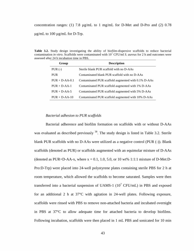
43
concentration ranges: (1) 7.8 µg/mL to 1 mg/mL for D-Met and D-Pro and (2) 0.78
µg/mL to 100 µg/mL for D-Trp.
Table 3.2. Study design investigating the ability of biofilm-dispersive scaffolds to reduce bacterial
contamination in vitro. Scaffolds were contaminated with 107 CFU/ml S. aureus for 2 h and outcomes were
assessed after 24 h incubation time in PBS.
Group Description
PUR (-) Sterile blank PUR scaffold with no D-AAs
PUR Contaminated blank PUR scaffold with no D-AAs
PUR + D-AA-0.1 Contaminated PUR scaffold augmented with 0.1% D-AAs
PUR + D-AA-1 Contaminated PUR scaffold augmented with 1% D-AAs
PUR + D-AA-5 Contaminated PUR scaffold augmented with 5% D-AAs
PUR + D-AA-10 Contaminated PUR scaffold augmented with 10% D-AAs
Bacterial adhesion to PUR scaffolds
Bacterial adherence and biofilm formation on scaffolds with or without D-AAs
was evaluated as described previously 38
. The study design is listed in Table 3.2. Sterile
blank PUR scaffolds with no D-AAs were utilized as a negative control (PUR (-)). Blank
scaffolds (denoted as PUR) or scaffolds augmented with an equimolar mixture of D-AAs
(denoted as PUR+D-AA-x, where x = 0.1, 1.0, 5.0, or 10 wt% 1:1:1 mixture of D-Met:D-
Pro:D-Trp) were placed into 24-well polystyrene plates containing sterile PBS for 2 h at
room temperature, which allowed the scaffolds to become saturated. Samples were then
transferred into a bacterial suspension of UAMS-1 (107 CFU/mL) in PBS and exposed
for an additional 2 h at 37°C with agitation in 24-well plates. Following exposure,
scaffolds were rinsed with PBS to remove non-attached bacteria and incubated overnight
in PBS at 37°C to allow adequate time for attached bacteria to develop biofilms.
Following incubation, scaffolds were then placed in 1 mL PBS and sonicated for 10 min

44
using a low-power bath sonicator. Bacterial CFUs per gram of scaffold were determined
by plating serial dilutions on blood agar plates. Bacterial attachment and biofilm
formation on scaffolds following incubation was also evaluated by SEM analysis. The
sample size was 3 with duplicate runs.
Table 3.3. In vivo study design investigating the ability of biofilm-dispersive scaffolds to reduce
contamination in 6-mm segmental defects in rat femora contaminated with 102 CFU of S.aureus UAMS-1
or Xen36. Outcomes were assessed at 2 weeks.
Group Description No
infection UAMS-1 Xen36
PUR (-) Blank PUR scaffold in a sterile defect 10 0 0
Empty Contaminated defect not grafted with a scaffold 0 10 10
PUR Blank PUR scaffold in a contaminated defect 0 10 10
PUR + D-AA-1 PUR scaffold with 1% D-AAs in a contaminated
defect 0 10 0
PUR + D-AA-5 PUR scaffold with 5% D-AAs in a contaminated
defect 0 10 0
PUR + D-AA-10 PUR scaffold with 10% D-AAs in a contaminated
defect 0 10 10
Rat femoral 6-mm segmental defect model
This study was conducted in compliance with the Animal Welfare Act, the
implementing Animal Welfare Regulations, and the principles of the Guide for the Care
and Use of Laboratory Animals. A previously characterized contaminated critical size
defect in rat (Sprague–Dawley; 373 ± 4.15 g) femurs was utilized as the in vivo model of
infection 39-41
. The study design is listed in Table 3.3. Briefly, a 6-mm segmental defect
was created using a small reciprocating saw blade (MicroAire 1025, MicroAire,
Charlottesville, VA), stabilized with a polyacetyl plate (length 25 mm, width 4 mm and
height 4 mm) and fixed to the surface of the femur using threaded K-wires. Blank PUR
scaffolds implanted in a sterile defect were utilized as a negative control (PUR (-)) and
for SEM analysis to distinguish between host cellular and bacterial infiltration of the
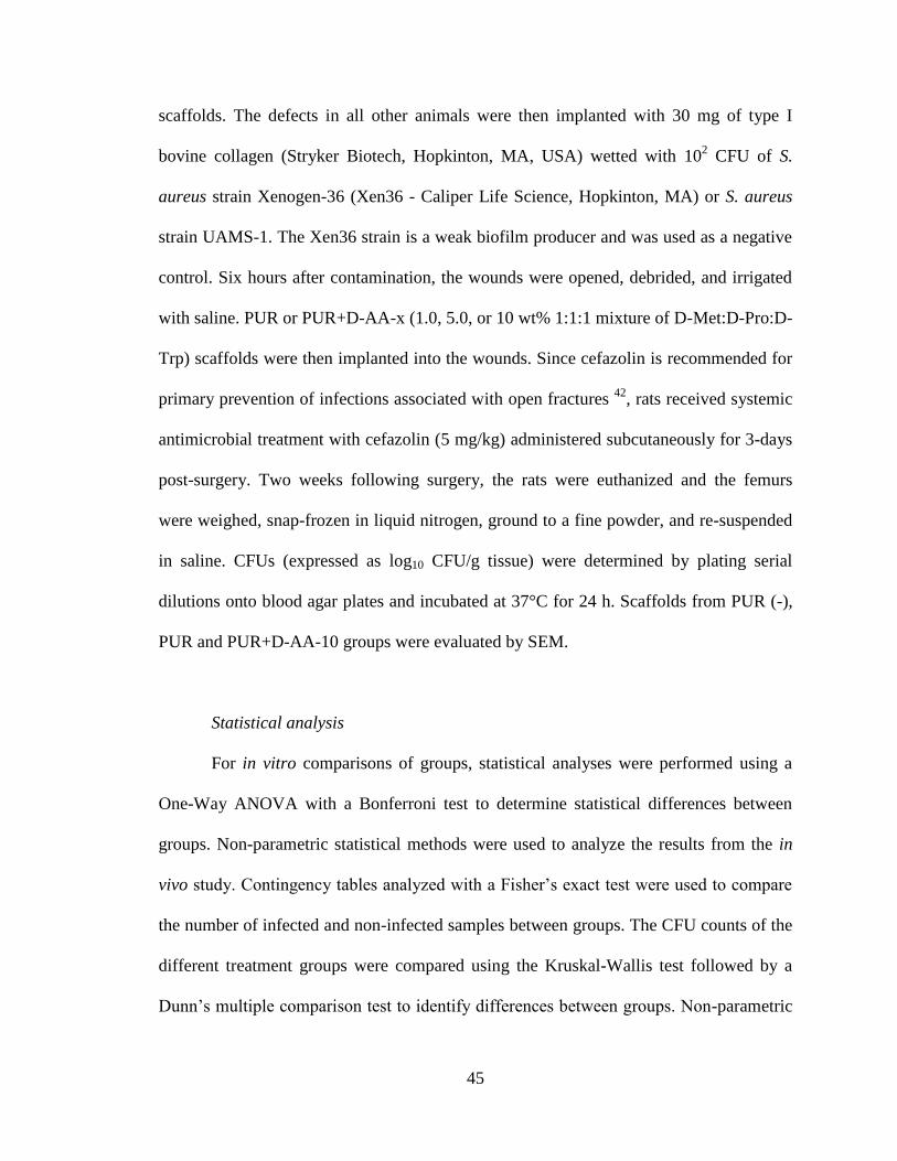
45
scaffolds. The defects in all other animals were then implanted with 30 mg of type I
bovine collagen (Stryker Biotech, Hopkinton, MA, USA) wetted with 102 CFU of S.
aureus strain Xenogen-36 (Xen36 - Caliper Life Science, Hopkinton, MA) or S. aureus
strain UAMS-1. The Xen36 strain is a weak biofilm producer and was used as a negative
control. Six hours after contamination, the wounds were opened, debrided, and irrigated
with saline. PUR or PUR+D-AA-x (1.0, 5.0, or 10 wt% 1:1:1 mixture of D-Met:D-Pro:D-
Trp) scaffolds were then implanted into the wounds. Since cefazolin is recommended for
primary prevention of infections associated with open fractures 42
, rats received systemic
antimicrobial treatment with cefazolin (5 mg/kg) administered subcutaneously for 3-days
post-surgery. Two weeks following surgery, the rats were euthanized and the femurs
were weighed, snap-frozen in liquid nitrogen, ground to a fine powder, and re-suspended
in saline. CFUs (expressed as log10 CFU/g tissue) were determined by plating serial
dilutions onto blood agar plates and incubated at 37°C for 24 h. Scaffolds from PUR (-),
PUR and PUR+D-AA-10 groups were evaluated by SEM.
Statistical analysis
For in vitro comparisons of groups, statistical analyses were performed using a
One-Way ANOVA with a Bonferroni test to determine statistical differences between
groups. Non-parametric statistical methods were used to analyze the results from the in
vivo study. Contingency tables analyzed with a Fisher’s exact test were used to compare
the number of infected and non-infected samples between groups. The CFU counts of the
different treatment groups were compared using the Kruskal-Wallis test followed by a
Dunn’s multiple comparison test to identify differences between groups. Non-parametric
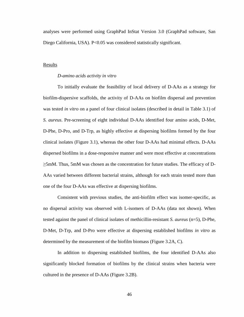
46
analyses were performed using GraphPad InStat Version 3.0 (GraphPad software, San
Diego California, USA). P<0.05 was considered statistically significant.
Results
D-amino acids activity in vitro
To initially evaluate the feasibility of local delivery of D-AAs as a strategy for
biofilm-dispersive scaffolds, the activity of D-AAs on biofilm dispersal and prevention
was tested in vitro on a panel of four clinical isolates (described in detail in Table 3.1) of
S. aureus. Pre-screening of eight individual D-AAs identified four amino acids, D-Met,
D-Phe, D-Pro, and D-Trp, as highly effective at dispersing biofilms formed by the four
clinical isolates (Figure 3.1), whereas the other four D-AAs had minimal effects. D-AAs
dispersed biofilms in a dose-responsive manner and were most effective at concentrations
≥5mM. Thus, 5mM was chosen as the concentration for future studies. The efficacy of D-
AAs varied between different bacterial strains, although for each strain tested more than
one of the four D-AAs was effective at dispersing biofilms.
Consistent with previous studies, the anti-biofilm effect was isomer-specific, as
no dispersal activity was observed with L-isomers of D-AAs (data not shown). When
tested against the panel of clinical isolates of methicillin-resistant S. aureus (n=5), D-Phe,
D-Met, D-Trp, and D-Pro were effective at dispersing established biofilms in vitro as
determined by the measurement of the biofilm biomass (Figure 3.2A, C).
In addition to dispersing established biofilms, the four identified D-AAs also
significantly blocked formation of biofilms by the clinical strains when bacteria were
cultured in the presence of D-AAs (Figure 3.2B).
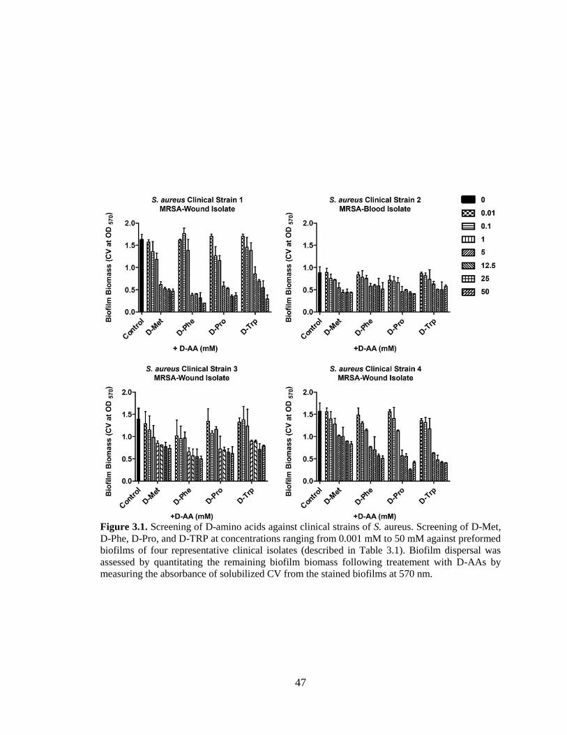
47
Figure 3.1. Screening of D-amino acids against clinical strains of S. aureus. Screening of D-Met,
D-Phe, D-Pro, and D-TRP at concentrations ranging from 0.001 mM to 50 mM against preformed
biofilms of four representative clinical isolates (described in Table 3.1). Biofilm dispersal was
assessed by quantitating the remaining biofilm biomass following treatement with D-AAs by
measuring the absorbance of solubilized CV from the stained biofilms at 570 nm.
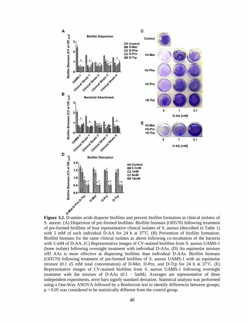
48
Figure 3.2. D-amino acids disperse biofilms and prevent biofilm formation in clinical isolates of
S. aureus. (A) Dispersion of pre-formed biofilms: Biofilm biomass (OD570) following treatment
of pre-formed biofilms of four representative clinical isolates of S. aureus (described in Table 1)
with 5 mM of each individual D-AA for 24 h at 37oC. (B) Prevention of biofilm formation:
Biofilm biomass for the same clinical isolates as above following co-incubation of the bacteria
with 5 mM of D-AA. (C) Representative images of CV-stained biofilms from S. aureus UAMS-1
(bone isolate) following overnight treatment with individual D-AAs. (D) An equimolar mixture
ofD AAs is more effective at dispersing biofilms than individual D-AAs. Biofilm biomass
(OD570) following treatment of pre-formed biofilms of S. aureus UAMS-1 with an equimolar
mixture (0.1 e5 mM total concentration) of D-Met, D-Pro, and D-Trp for 24 h at 37oC. (E)
Representative images of CV-stained biofilms from S. aureus UAMS-1 following overnight
treatment with the mixture of D-AAs (0.1 - 5mM). Averages are representative of three
independent experiments, error bars signify standard deviation. Statistical analysis was performed
using a One-Way ANOVA followed by a Bonferroni test to identify differences between groups;
p < 0.05 was considered to be statistically different from the control group.
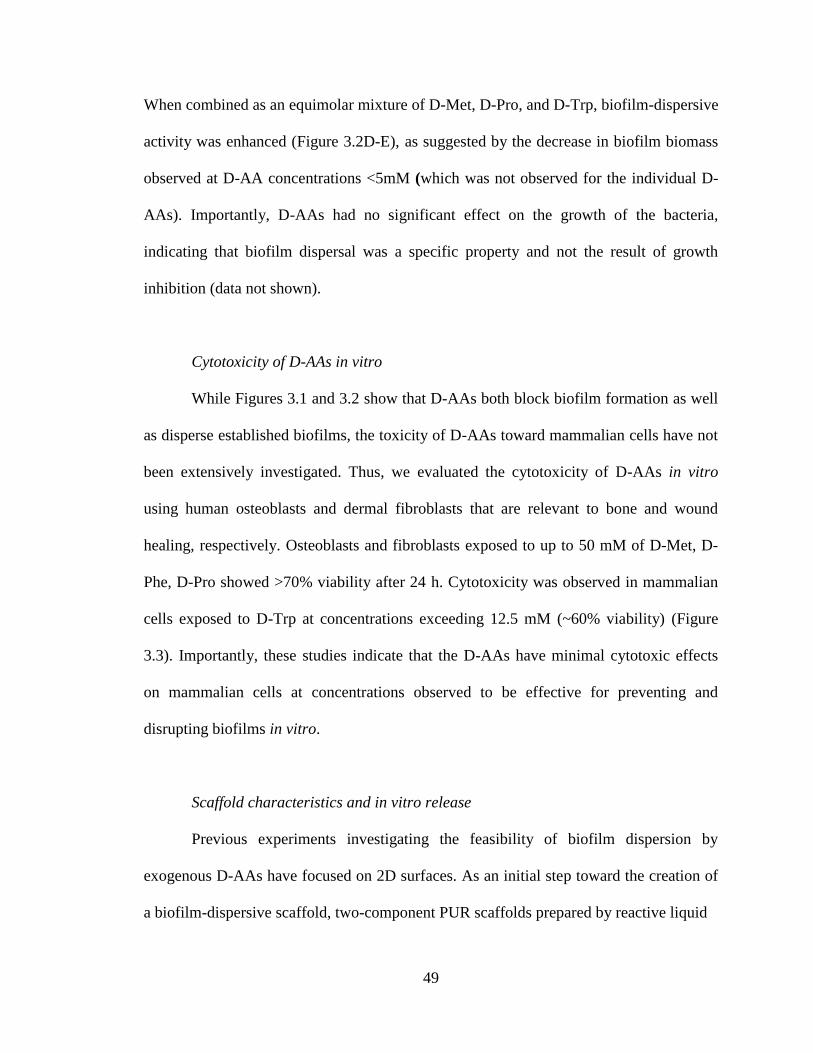
49
When combined as an equimolar mixture of D-Met, D-Pro, and D-Trp, biofilm-dispersive
activity was enhanced (Figure 3.2D-E), as suggested by the decrease in biofilm biomass
observed at D-AA concentrations <5mM (which was not observed for the individual D-
AAs). Importantly, D-AAs had no significant effect on the growth of the bacteria,
indicating that biofilm dispersal was a specific property and not the result of growth
inhibition (data not shown).
Cytotoxicity of D-AAs in vitro
While Figures 3.1 and 3.2 show that D-AAs both block biofilm formation as well
as disperse established biofilms, the toxicity of D-AAs toward mammalian cells have not
been extensively investigated. Thus, we evaluated the cytotoxicity of D-AAs in vitro
using human osteoblasts and dermal fibroblasts that are relevant to bone and wound
healing, respectively. Osteoblasts and fibroblasts exposed to up to 50 mM of D-Met, D-
Phe, D-Pro showed >70% viability after 24 h. Cytotoxicity was observed in mammalian
cells exposed to D-Trp at concentrations exceeding 12.5 mM (~60% viability) (Figure
3.3). Importantly, these studies indicate that the D-AAs have minimal cytotoxic effects
on mammalian cells at concentrations observed to be effective for preventing and
disrupting biofilms in vitro.
Scaffold characteristics and in vitro release
Previous experiments investigating the feasibility of biofilm dispersion by
exogenous D-AAs have focused on 2D surfaces. As an initial step toward the creation of
a biofilm-dispersive scaffold, two-component PUR scaffolds prepared by reactive liquid
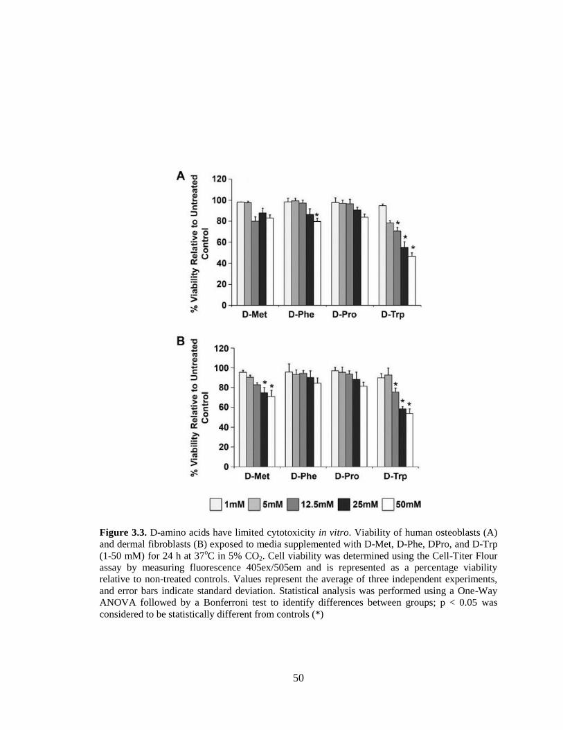
50
Figure 3.3. D-amino acids have limited cytotoxicity in vitro. Viability of human osteoblasts (A)
and dermal fibroblasts (B) exposed to media supplemented with D-Met, D-Phe, DPro, and D-Trp
(1-50 mM) for 24 h at 37oC in 5% CO2. Cell viability was determined using the Cell-Titer Flour
assay by measuring fluorescence 405ex/505em and is represented as a percentage viability
relative to non-treated controls. Values represent the average of three independent experiments,
and error bars indicate standard deviation. Statistical analysis was performed using a One-Way
ANOVA followed by a Bonferroni test to identify differences between groups; p < 0.05 was
considered to be statistically different from controls (*)
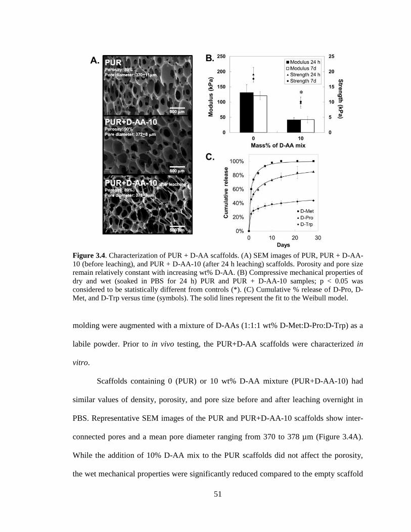
51
Figure 3.4. Characterization of PUR + D-AA scaffolds. (A) SEM images of PUR, PUR + D-AA-
10 (before leaching), and PUR + D-AA-10 (after 24 h leaching) scaffolds. Porosity and pore size
remain relatively constant with increasing wt% D-AA. (B) Compressive mechanical properties of
dry and wet (soaked in PBS for 24 h) PUR and PUR + D-AA-10 samples; p < 0.05 was
considered to be statistically different from controls (*). (C) Cumulative % release of D-Pro, D-
Met, and D-Trp versus time (symbols). The solid lines represent the fit to the Weibull model.
molding were augmented with a mixture of D-AAs (1:1:1 wt% D-Met:D-Pro:D-Trp) as a
labile powder. Prior to in vivo testing, the PUR+D-AA scaffolds were characterized in
vitro.
Scaffolds containing 0 (PUR) or 10 wt% D-AA mixture (PUR+D-AA-10) had
similar values of density, porosity, and pore size before and after leaching overnight in
PBS. Representative SEM images of the PUR and PUR+D-AA-10 scaffolds show inter-
connected pores and a mean pore diameter ranging from 370 to 378 µm (Figure 3.4A).
While the addition of 10% D-AA mix to the PUR scaffolds did not affect the porosity,
the wet mechanical properties were significantly reduced compared to the empty scaffold
52
(Figure 3.4B). There were no differences in the properties of the scaffolds incubated in
PBS for 24 hours or 7 days.
The release kinetics of D-Pro, D-Met, and D-Trp were characterized by an initial
burst followed by a sustained release for up to 21 days (Figure 3.4C). D-Met released the
fastest, characterized by a 60% burst on day 1 and nearly 100% release by day 14. The
release of D-Pro was somewhat slower (45% burst and 85% release by day 28), while D-
Trp released slowly, with only a 25% burst and 44% release after 28 days. The Weibull
equation has been used to identify the mechanism controlling drug release from
polymeric materials 39, 43
:
(3.1)
where Mt corresponds to the mass of drug released in time t, M is the mass of drug
released at infinite time (which corresponds to the initial loading of drug), and a and b are
constants. When b<0.75, Fickian diffusion controls drug release, while a more complex
mechanism involving both diffusion and swelling controls release when b>0.75 44
. The
D-AA release data were fit to the Weibull model and the values of the b parameter for D-
Met, D-Pro, and D-Trp were calculated as 0.56, 0.35, and 0.21 respectively, suggesting
that the release of each D-AA from the scaffolds was diffusion-controlled.
Effects of biofilm-dispersive scaffolds on bacterial contamination in vitro
The effect of local delivery of D-AAs from biofilm-dispersive scaffolds on
bacterial contamination was evaluated in vitro prior to in vivo testing. Incorporation of D-
AA into PUR scaffolds at concentrations ≥ 1 wt% D-AA significantly reduced the
amount of attached bacteria and biofilm formation on the surface compared to the
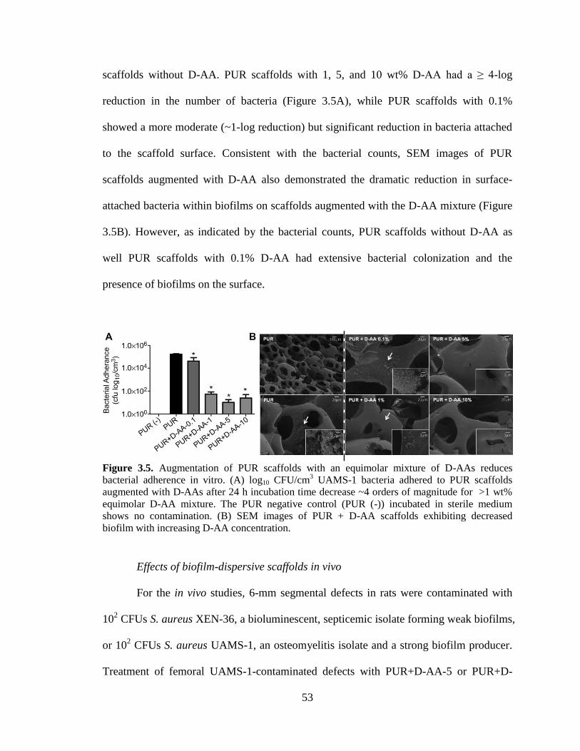
53
scaffolds without D-AA. PUR scaffolds with 1, 5, and 10 wt% D-AA had a ≥ 4-log
reduction in the number of bacteria (Figure 3.5A), while PUR scaffolds with 0.1%
showed a more moderate (~1-log reduction) but significant reduction in bacteria attached
to the scaffold surface. Consistent with the bacterial counts, SEM images of PUR
scaffolds augmented with D-AA also demonstrated the dramatic reduction in surface-
attached bacteria within biofilms on scaffolds augmented with the D-AA mixture (Figure
3.5B). However, as indicated by the bacterial counts, PUR scaffolds without D-AA as
well PUR scaffolds with 0.1% D-AA had extensive bacterial colonization and the
presence of biofilms on the surface.
Figure 3.5. Augmentation of PUR scaffolds with an equimolar mixture of D-AAs reduces
bacterial adherence in vitro. (A) log10 CFU/cm3 UAMS-1 bacteria adhered to PUR scaffolds
augmented with D-AAs after 24 h incubation time decrease ~4 orders of magnitude for >1 wt%
equimolar D-AA mixture. The PUR negative control (PUR (-)) incubated in sterile medium
shows no contamination. (B) SEM images of PUR + D-AA scaffolds exhibiting decreased
biofilm with increasing D-AA concentration.
Effects of biofilm-dispersive scaffolds in vivo
For the in vivo studies, 6-mm segmental defects in rats were contaminated with
102 CFUs S. aureus XEN-36, a bioluminescent, septicemic isolate forming weak biofilms,
or 102 CFUs S. aureus UAMS-1, an osteomyelitis isolate and a strong biofilm producer.
Treatment of femoral UAMS-1-contaminated defects with PUR+D-AA-5 or PUR+D-
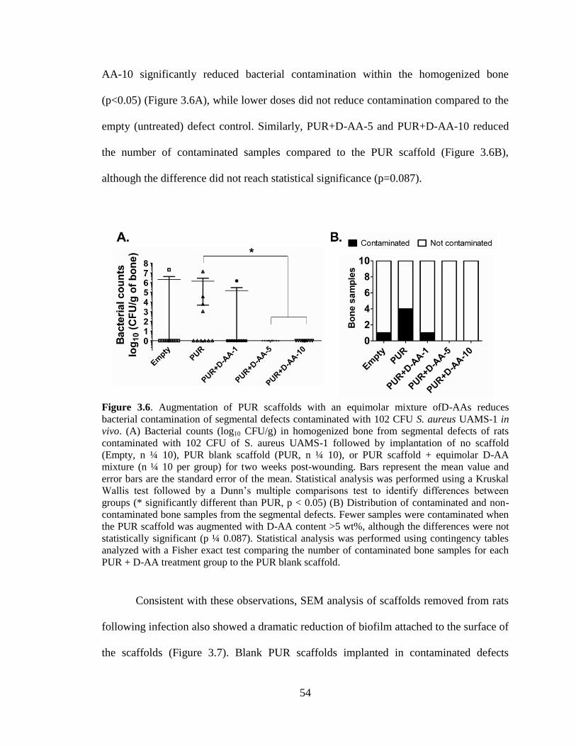
54
AA-10 significantly reduced bacterial contamination within the homogenized bone
(p<0.05) (Figure 3.6A), while lower doses did not reduce contamination compared to the
empty (untreated) defect control. Similarly, PUR+D-AA-5 and PUR+D-AA-10 reduced
the number of contaminated samples compared to the PUR scaffold (Figure 3.6B),
although the difference did not reach statistical significance (p=0.087).
Figure 3.6. Augmentation of PUR scaffolds with an equimolar mixture ofD-AAs reduces
bacterial contamination of segmental defects contaminated with 102 CFU S. aureus UAMS-1 in
vivo. (A) Bacterial counts (log10 CFU/g) in homogenized bone from segmental defects of rats
contaminated with 102 CFU of S. aureus UAMS-1 followed by implantation of no scaffold
(Empty, n ¼ 10), PUR blank scaffold (PUR, n ¼ 10), or PUR scaffold + equimolar D-AA
mixture (n ¼ 10 per group) for two weeks post-wounding. Bars represent the mean value and
error bars are the standard error of the mean. Statistical analysis was performed using a Kruskal
Wallis test followed by a Dunn’s multiple comparisons test to identify differences between
groups (* significantly different than PUR, p < 0.05) (B) Distribution of contaminated and non-
contaminated bone samples from the segmental defects. Fewer samples were contaminated when
the PUR scaffold was augmented with D-AA content >5 wt%, although the differences were not
statistically significant (p ¼ 0.087). Statistical analysis was performed using contingency tables
analyzed with a Fisher exact test comparing the number of contaminated bone samples for each
PUR + D-AA treatment group to the PUR blank scaffold.
Consistent with these observations, SEM analysis of scaffolds removed from rats
following infection also showed a dramatic reduction of biofilm attached to the surface of
the scaffolds (Figure 3.7). Blank PUR scaffolds implanted in contaminated defects
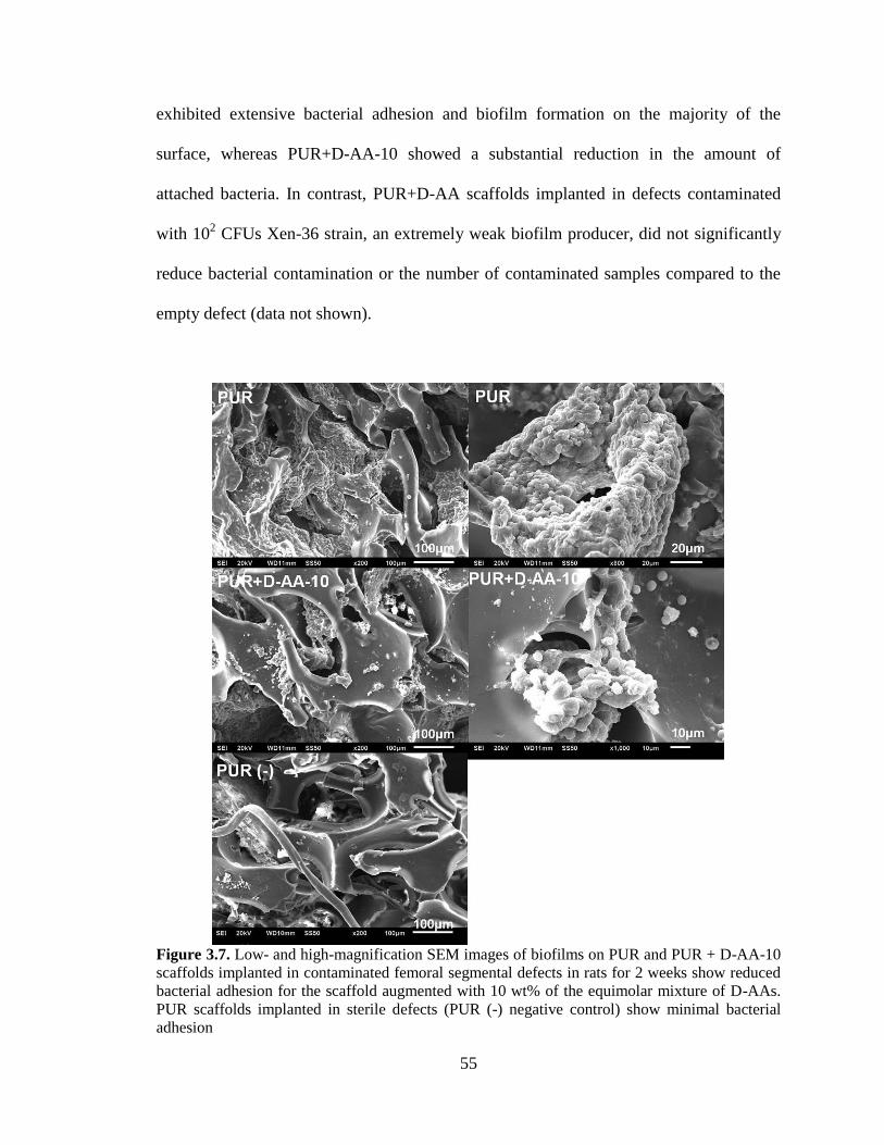
55
exhibited extensive bacterial adhesion and biofilm formation on the majority of the
surface, whereas PUR+D-AA-10 showed a substantial reduction in the amount of
attached bacteria. In contrast, PUR+D-AA scaffolds implanted in defects contaminated
with 102 CFUs Xen-36 strain, an extremely weak biofilm producer, did not significantly
reduce bacterial contamination or the number of contaminated samples compared to the
empty defect (data not shown).
Figure 3.7. Low- and high-magnification SEM images of biofilms on PUR and PUR + D-AA-10
scaffolds implanted in contaminated femoral segmental defects in rats for 2 weeks show reduced
bacterial adhesion for the scaffold augmented with 10 wt% of the equimolar mixture of D-AAs.
PUR scaffolds implanted in sterile defects (PUR (-) negative control) show minimal bacterial
adhesion
56
Discussion
Despite meticulous clinical management including surgical debridement and the
use of systemic antibiotics, contamination rates of open fractures continue to be a
significant cause of non-osseous union, potentially leading to extremity amputation.
Local delivery of antibiotics from bone grafts has been investigated as a strategy to
reduce bacterial contamination and promote osseous union 39, 45-52
. However, the efficacy
of antibiotics against the surface-attached communities of bacteria known as biofilms,
which are considered a major virulence factor in chronic disease 6, 4
, is limited due to the
slow metabolic and growth rates of bacteria within the biofilm 53
. Furthermore, the
avascular bone graft may itself serve as a substrate for bacterial colonization and a nidus
for recurrent infections 54
. Finally, a small population (i.e., 0.1–10%) of “persister cells”
survives antimicrobial therapy and rapidly grows after the cessation of antibiotic therapy,
potentially resulting in recurrent infections 55
. Recent studies have highlighted the
potential of bacterial signaling molecules that trigger biofilm dispersal, such as bismuth
thiols 56, 20
, quorum-sensing inhibitors and analogs 57
, and D-AAs 27, 26
, as therapeutic
agents for treatment of chronic infections. In this study, we have shown that local
delivery of D-AAs from PUR scaffolds inhibits biofilm formation by clinical isolates of S.
aureus both in vitro and in vivo. Dose-response experiments showed that D-AAs
inhibited biofilm formation and dispersed existing biofilms at concentrations ≥5 mM in
vitro. A PUR scaffold augmented with ≥5 wt% D-AAs significantly reduced bacterial
contamination and biofilm formation by the strong biofilm-producing strain S. aureus
UAMS-1 compared to the control scaffold with no D-AAs in a contaminated segmental
defect in rats. Interestingly, with careful scanning of the scaffolds with D-AAs, small
57
colonies of bacteria within a biofilm could be found. Conversely, biofilm formation could
easily be found on the scaffolds without D-AAs that were retrieved from contaminated
defects. Almost the entire scaffold was covered with biofilm, and the colonies were very
robust. Furthermore, D-AAs exhibited relatively low cytotoxicity to mammalian cells at
doses effective at inhibiting biofilm formation.
Considering the many human chronic diseases involving biofilms, the use of
biofilm dispersal agents has gained considerable interest for the treatment of biofilm-
mediated infections. A previous study has reported that D-Phe, D-Pro, and D-Tyr prevent
biofilm formation and trigger biofilm dispersion in S. aureus WT strain SC01 at
concentrations as low as 0.5 mM in vitro 27
. In this study, we have shown that D-Met, D-
Phe, D-Pro, and D-Trp inhibit biofilm formation and disperse established biofilms of
clinical strains of S. aureus at concentrations ≥5 mM (Figure 3.1), which is ~10 times
greater than the previously reported dose. The activity of individual D-AAs varied in
previous studies, with D-Tyr requiring the lowest concentration (3 μM), and D-Met (2
mM), D-Trp (5 mM) and D-Leu (8.5 mM) requiring higher concentrations for anti-
biofilm activity against Bacillus subtilis strain NCIB 3610 and for S. aureus strain SC01
26. Differences between bacterial species as well as strain heterogeneity are likely
contributing factors to the observed discrepancies between studies. Importantly, from our
studies we identified concentrations having effective biofilm-dispersive activity active
against a number of clinical strains. Consistent with a previous study showing that the
equimolar mixture of D-Phe:D-Pro:D-Tyr lowered the effective dose 27
, the data in
Figure 3.2D-E show that an equimolar mixture of D-Met:D-Pro:D-Trp shifted the dose-
response curve toward lower doses compared to the individual D-AAs. Other than for B.
58
subtilis, the mechanisms by which D-AAs disrupt biofilms are not known. However, the
enhanced anti-biofilm activity of the D-AA mixture is suggestive of multiple mechanisms
acting on the bacteria, which warrants further investigation.
In order to be useful as a clinical therapy, D-AAs must exhibit minimal
cytotoxicity at concentrations that are effective at dispersing biofilms. As shown in
Figure 3.3, osteoblasts and fibroblasts treated with D-Phe, D-Pro, or D-Met for 24 h
exhibited >70% viability at concentrations ≤50 mM, while D-Trp exhibited cytotoxic
effects (i.e., <70% viability) at concentrations >12.5 mM. While these data suggest that
individual D-AAs are non-cytotoxic to mammalian cells at concentrations efficacious
against biofilms, they contrast with a previous study reporting cytotoxicity of D-Phe, D-
Met, and D-Trp toward Chinese Hamster Ovary (CHO) and HeLa cells at concentrations
≥10 mM 28
. The discrepancies in D-AA toxicities between the present study and that
reported previously may be attributed to differences among cell lines. In another study,
D-Phe and D-Trp reportedly elicited a chemotactic response in human neutrophils via
activation of GPR109B 58
. Additional in vivo studies investigating the biocompatibility of
D-AAs in sterile defects are needed to further assess their safety, and these studies are
currently ongoing in our laboratories.
As a first step toward the development of a biofilm-dispersive bone graft for
clinical applications, we evaluated the effects of local delivery of D-AAs from
biodegradable PUR scaffolds on infection in a contaminated segmental defect model in
rats. The scaffold investigated in this study has a half-life of 14 weeks in vitro 36
, and
degrades to non-cytotoxic breakdown products such as lysine and -hydroxy acids 59
.
PUR scaffolds are effective delivery vehicles for sustained released of biologics, such as
59
antibiotics 60, 39
and recombinant human bone morphogenic protein-2 (rhBMP-2) 61, 44, 40
.
Furthermore, PUR scaffolds are injectable 62, 63
and thus can be delivered using
minimally invasive surgical techniques. As shown in Figure 3.4, while D-AAs had
minimal effects on the porosity and pore size, PUR+D-AA scaffolds exhibited
approximately 2-3-fold decreases in both Young’s modulus and compressive stress at
50% strain. A previous study reported that PUR scaffolds augmented with 8 wt%
tobramycin had significantly higher porosity and lower modulus than blank PUR
scaffolds after 24 h incubation time in PBS, which was attributed to rapid leaching of
tobramycin and consequent formation of new pores 60
. Surprisingly, in the present study
the modulus decreased significantly even after 24 h, at which time only a fraction of the
D-AAs had released, and did not decrease further for up to 7 days of incubation. These
observations suggest that the decrease in modulus with addition of D-AAs results from
defects in the pore walls of the scaffold caused by the presence of the particles rather than
from the formation of new pores due to leaching of the D-AAs.
As shown in Figure 3.4C, PUR+D-AA scaffolds supported diffusion-controlled
sustained release of the biologically active drug for up to 4 weeks, which is consistent
with previous studies reporting diffusion-controlled release of active antibiotics 60, 39
,
recombinant human growth factors 64, 44
, and siRNA nanoparticles 65
. At each time point,
the order of cumulative release was D-Met>D-Pro>D-Trp, while the order of solubility
was D-Pro>>D-Met>D-Trp 66
. At 4 weeks, >85% of D-Pro and D-Met had been released
and <10% of the scaffold had degraded 36
, which is consistent with the notion that the D-
AA release was diffusion-controlled at early time points. However, <40% of the D-Trp
had been released by 4 weeks, suggesting that degradation of the scaffold may control D-
60
Trp release kinetics at later (>4 weeks) time points. Since the free base form of each D-
AA was used in this study, the release kinetics could be increased by using the more
soluble hydrochloride as reported previously for vancomycin 39
. While antibiotic therapy
for up to 8 weeks is recommended for effective treatment of MRSA osteomyelitis 67
, the
optimal release profile for D-AAs is unknown and thus merits further investigation.
In a proof-of-concept study, we evaluated the ability of biofilm-dispersive PUR
scaffolds augmented with D-AAs to prevent biofilm formation and reduce CFUs in a
contaminated rat segmental defect model. Several recent studies have evaluated the
effects of local delivery of antibiotics on infection using an acute contamination model, in
which the bone graft was placed immediately after contamination of the defect with
bacteria 68, 69
. However, preclinical models with an established chronic infection 40, 70
represent a more rigorous test for the efficacy of biofilm-dispersive grafts, since they
ensure that the bacteria are able to adhere to the surface of the wound and form biofilms
71. In the present study, rat segmental defects were contaminated with 10
2 CFU for 6 h
prior to implantation of the PUR+D-AA scaffolds. Augmentation of the scaffolds with ≥5
wt% D-AAs significantly reduced bacterial contamination within the segmental defects
treated with UAMS-1, an osteomyelitis strain. In contrast, the D-AAs had no significant
effect on defects contaminated with Xen36, which is a weak biofilm producer 14
. These
observations are consistent with the notion that D-AAs reduce contamination in the
defect by preventing the formation of and/or dispersing biofilms, thus limiting the
application of D-AAs as a treatment for biofilm infections. However, numerous studies
indicate that the majority of staphylococcal clinical isolates in vitro as well as those found
in human tissues are strong biofilm producers.
61
Because D-AAs are not bactericidal, they are anticipated to be most effective as
an adjuvant therapy to conventional treatment with systemic antibiotics. In this study, rats
were treated with cefazolin (5 mg/kg) administered subcutaneously for 3-days post
surgery to simulate the clinical scenario 42
. Despite the presence of systemic antibiotics,
one of the untreated rats developed an infection, which increased to four rats when
treated with blank PUR scaffolds. This suggests that placement of an avascular graft into
an open fracture potentiates infection. Treatment with PUR+D-AA-5 or PUR+D-AA-10
scaffolds reduced the number of infected rats to zero, which underscores the potential
clinical utility of biofilm-dispersive bone grafts as an adjuvant therapy to systemic
antibiotics. The broad spectrum of D-AAs will allow their use for preventing infection
without the need of knowing the nature of contaminating bacteria. For chronic infections
such as MRSA osteomyelitis, treatment with systemic antibiotics is recommended for a
minimum of 8 weeks 67
. Importantly, for most patients, the extended duration of systemic
treatment is associated with a number of risks to the patient’s health, including renal
toxicity, and is an economic burden to the patient and healthcare system. Our results
suggest that local delivery of a biofilm dispersion agent alone or co-delivered with
antimicrobial agents represents a potentially efficacious therapy for treatment of chronic
infections, which, when combined with the standard systemic antimicrobial treatment
may reduce the time of treatment and resulting complications and cost for chronic
orthopedic infections.
62
Conclusions
The ability of bacteria to establish biofilms substantially hinders the treatment of
orthopedic infections and is implicated as a significant contributing factor in the sequelae
associated with open fractures. Biofilm-dispersive scaffolds augmented with D-AAs can
be used as a novel therapeutic strategy to reduce microbial burden within wounds and
improve healing outcomes.
63
References
1 D. P. Lew, and F. A. Waldvogel, 'Osteomyelitis', Lancet, 364 (2004), 369-79.
2 L. O. Conterno, and C. R. da Silva Filho, 'Antibiotics for Treating Chronic Osteomyelitis in
Adults', Cochrane Database Syst Rev (2009), CD004439.
3 J. Huh, D. J. Stinner, T. C. Burns, and J. R. Hsu, 'Infectious Complications and Soft Tissue Injury
Contribute to Late Amputation after Severe Lower Extremity Trauma', J Trauma, 71 (2011), S47-
51.
4 J. W. Costerton, 'Biofilm Theory Can Guide the Treatment of Device-Related Orthopaedic
Infections', Clin Orthop Relat Res (2005), 7-11.
5 J. W. Costerton, P. S. Stewart, and E. P. Greenberg, 'Bacterial Biofilms: A Common Cause of
Persistent Infections', Science, 284 (1999), 1318-22.
6 R. A. Brady, J. G. Leid, J. H. Calhoun, J. W. Costerton, and M. E. Shirtliff, 'Osteomyelitis and the
Role of Biofilms in Chronic Infection', FEMS Immunol Med Microbiol, 52 (2008), 13-22.
7 T. J. Marrie, and J. W. Costerton, 'Mode of Growth of Bacterial Pathogens in Chronic
Polymicrobial Human Osteomyelitis', J Clin Microbiol, 22 (1985), 924-33.
8 A. G. Gristina, M. Oga, L. X. Webb, and C. D. Hobgood, 'Adherent Bacterial Colonization in the
Pathogenesis of Osteomyelitis', Science, 228 (1985), 990-3.
9 J. W. Costerton, 'Introduction to Biofilm', Int J Antimicrob Agents, 11 (1999), 217-21; discussion
37-9.
10 J. L. del Pozo, and R. Patel, 'The Challenge of Treating Biofilm-Associated Bacterial Infections',
Clin Pharmacol Ther, 82 (2007), 204-9.
11 L. Hall-Stoodley, J. W. Costerton, and P. Stoodley, 'Bacterial Biofilms: From the Natural
Environment to Infectious Diseases', Nat Rev Microbiol, 2 (2004), 95-108.
12 J. Esteban, D. Molina-Manso, I. Spiliopoulou, J. Cordero-Ampuero, R. Fernandez-Roblas, A.
Foka, and E. Gomez-Barrena, 'Biofilm Development by Clinical Isolates of Staphylococcus Spp.
From Retrieved Orthopedic Prostheses', Acta Orthop, 81 (2010), 674-9.
13 E. O'Neill, C. Pozzi, P. Houston, D. Smyth, H. Humphreys, D. A. Robinson, and J. P. O'Gara,
'Association between Methicillin Susceptibility and Biofilm Regulation in Staphylococcus Aureus
Isolates from Device-Related Infections', J Clin Microbiol, 45 (2007), 1379-88.
14 C. J. Sanchez, Jr., K. Mende, M. L. Beckius, K. S. Akers, D. R. Romano, J. C. Wenke, and C. K.
Murray, 'Biofilm Formation by Clinical Isolates and the Implications in Chronic Infections', BMC
Infect Dis, 13 (2013), 47.
15 M. Palmer, W. Costerton, J. Sewecke, and D. Altman, 'Molecular Techniques to Detect Biofilm
Bacteria in Long Bone Nonunion: A Case Report', Clin Orthop Relat Res, 469 (2011), 3037-42.
16 D. McDougald, S. A. Rice, N. Barraud, P. D. Steinberg, and S. Kjelleberg, 'Should We Stay or
Should We Go: Mechanisms and Ecological Consequences for Biofilm Dispersal', Nat Rev
Microbiol, 10 (2012), 39-50.
17 L. Hall-Stoodley, and P. Stoodley, 'Biofilm Formation and Dispersal and the Transmission of
Human Pathogens', Trends Microbiol, 13 (2005), 7-10.
64
18 J. B. Kaplan, 'Biofilm Dispersal: Mechanisms, Clinical Implications, and Potential Therapeutic
Uses', J Dent Res, 89 (2010), 205-18.
19 A. S. Lynch, and D. Abbanat, 'New Antibiotic Agents and Approaches to Treat Biofilm-
Associated Infections', Expert Opin Ther Pat, 20 (2010), 1373-87.
20 J. P. Folsom, B. Baker, and P. S. Stewart, 'In Vitro Efficacy of Bismuth Thiols against Biofilms
Formed by Bacteria Isolated from Human Chronic Wounds', J Appl Microbiol, 111 (2011), 989-
96.
21 J. B. Kaplan, K. LoVetri, S. T. Cardona, S. Madhyastha, I. Sadovskaya, S. Jabbouri, and E. A.
Izano, 'Recombinant Human Dnase I Decreases Biofilm and Increases Antimicrobial
Susceptibility in Staphylococci', J Antibiot (Tokyo), 65 (2012), 73-7.
22 J. M. Dow, L. Crossman, K. Findlay, Y. Q. He, J. X. Feng, and J. L. Tang, 'Biofilm Dispersal in
Xanthomonas Campestris Is Controlled by Cell-Cell Signaling and Is Required for Full Virulence
to Plants', Proc Natl Acad Sci U S A, 100 (2003), 10995-1000.
23 J. A. Jennings, H. S. Courtney, and W. O. Haggard, 'Cis-2-Decenoic Acid Inhibits S. Aureus
Growth and Biofilm in Vitro: A Pilot Study', Clin Orthop Relat Res, 470 (2012), 2663-70.
24 O. Simonetti, O. Cirioni, R. Ghiselli, G. Goteri, A. Scalise, F. Orlando, C. Silvestri, A. Riva, V.
Saba, K. D. Madanahally, A. Offidani, N. Balaban, G. Scalise, and A. Giacometti, 'Rnaiii-
Inhibiting Peptide Enhances Healing of Wounds Infected with Methicillin-Resistant
Staphylococcus Aureus', Antimicrob Agents Chemother, 52 (2008), 2205-11.
25 G. Brackman, P. Cos, L. Maes, H. J. Nelis, and T. Coenye, 'Quorum Sensing Inhibitors Increase
the Susceptibility of Bacterial Biofilms to Antibiotics in Vitro and in Vivo', Antimicrob Agents
Chemother, 55 (2011), 2655-61.
26 I. Kolodkin-Gal, D. Romero, S. Cao, J. Clardy, R. Kolter, and R. Losick, 'D-Amino Acids Trigger
Biofilm Disassembly', Science, 328 (2010), 627-9.
27 A. I. Hochbaum, I. Kolodkin-Gal, L. Foulston, R. Kolter, J. Aizenberg, and R. Losick, 'Inhibitory
Effects of D-Amino Acids on Staphylococcus Aureus Biofilm Development', J Bacteriol, 193
(2011), 5616-22.
28 N. Ercal, X. Luo, R. H. Matthews, and D. W. Armstrong, 'In Vitro Study of the Metabolic Effects
of D-Amino Acids', Chirality, 8 (1996), 24-9.
29 M. S. Smeltzer, J. R. Thomas, S. G. Hickmon, R. A. Skinner, C. L. Nelson, D. Griffith, T. R. Parr,
Jr., and R. P. Evans, 'Characterization of a Rabbit Model of Staphylococcal Osteomyelitis', J
Orthop Res, 15 (1997), 414-21.
30 E. C. Weiss, A. Zielinska, K. E. Beenken, H. J. Spencer, S. J. Daily, and M. S. Smeltzer, 'Impact
of Sara on Daptomycin Susceptibility of Staphylococcus Aureus Biofilms in Vivo', Antimicrob
Agents Chemother, 53 (2009), 4096-102.
31 J. E. Cassat, C. Y. Lee, and M. S. Smeltzer, 'Investigation of Biofilm Formation in Clinical
Isolates of Staphylococcus Aureus', Methods Mol Biol, 391 (2007), 127-44.
32 G. D. Christensen, W. A. Simpson, J. J. Younger, L. M. Baddour, F. F. Barrett, D. M. Melton, and
E. H. Beachey, 'Adherence of Coagulase-Negative Staphylococci to Plastic Tissue Culture Plates:
A Quantitative Model for the Adherence of Staphylococci to Medical Devices', J Clin Microbiol,
22 (1985), 996-1006.
65
33 S. A. Guelcher, V. Patel, K. M. Gallagher, S. Connolly, J. E. Didier, J. S. Doctor, and J. O.
Hollinger, 'Synthesis and in Vitro Biocompatibility of Injectable Polyurethane Foam Scaffolds',
Tissue Eng, 12 (2006), 1247-59.
34 A. S. Sawhney, and J. A. Hubbell, 'Rapidly Degraded Terpolymers of Dl-Lactide, Glycolide, and
Epsilon-Caprolactone with Increased Hydrophilicity by Copolymerization with Polyethers', J
Biomed Mater Res, 24 (1990), 1397-411.
35 S. Guelcher, A. Srinivasan, A. Hafeman, K. Gallagher, J. Doctor, S. Khetan, S. McBride, and J.
Hollinger, 'Synthesis, in Vitro Degradation, and Mechanical Properties of Two-Component
Poly(Ester Urethane)Urea Scaffolds: Effects of Water and Polyol Composition', Tissue
Engineering, 13 (2007), 2321-33.
36 AE Hafeman, B Li, T Yoshii, KL Zienkiewicz, JM Davidson, and SA Guelcher, 'Injectable
Biodegradable Polyurethane Scaffolds with Release of Platelet-Derived Growth Factor for Tissue
Repair and Regeneration', Pharm Res, 25 (2008), 2387-99.
37 P Bhandare, P Madhavan, B M Rao, and N Someswar Rao, 'Determination of Amino Acid
without Derivatization by Using Hplc-Hilic Column', J. Chem. Pharm. Res., 2 (2010), 372-80.
38 T. J. Kinnari, J. Esteban, N. Z. Martin-de-Hijas, O. Sanchez-Munoz, S. Sanchez-Salcedo, M.
Colilla, M. Vallet-Regi, and E. Gomez-Barrena, 'Influence of Surface Porosity and Ph on Bacterial
Adherence to Hydroxyapatite and Biphasic Calcium Phosphate Bioceramics', J Med Microbiol, 58
(2009), 132-7.
39 B. Li, K. V. Brown, J. C. Wenke, and S. A. Guelcher, 'Sustained Release of Vancomycin from
Polyurethane Scaffolds Inhibits Infection of Bone Wounds in a Rat Femoral Segmental Defect
Model', J Control Release, 145 (2010), 221 - 30.
40 S. A. Guelcher, K. V. Brown, B. Li, T. Guda, B. H. Lee, and J. C. Wenke, 'Dual-Purpose Bone
Grafts Improve Healing and Reduce Infection', Journal of orthopaedic trauma, 25 (2011), 477-82.
41 J.G. Penn-Barwell, C.K. Murray, and J.C. Wenke, 'Early Antibiotics and Debridement
Independently Reduce Infection in an Open Fracture Model', J Bone Joint Sur Br, 94-B (2012), 1-
6.
42 D. R. Hospenthal, C. K. Murray, R. C. Andersen, R. B. Bell, J. H. Calhoun, L. C. Cancio, J. M.
Cho, K. K. Chung, J. C. Clasper, M. H. Colyer, N. G. Conger, G. P. Costanzo, H. K. Crouch, T. K.
Curry, L. C. D'Avignon, W. C. Dorlac, J. R. Dunne, B. J. Eastridge, J. R. Ficke, M. E. Fleming,
M. A. Forgione, A. D. Green, R. G. Hale, D. K. Hayes, J. B. Holcomb, J. R. Hsu, K. E. Kester, G.
J. Martin, L. E. Moores, W. T. Obremskey, K. Petersen, E. M. Renz, J. R. Saffle, J. S. Solomkin,
D. E. Sutter, D. R. Tribble, J. C. Wenke, T. J. Whitman, A. R. Wiesen, and G. W. Wortmann,
'Guidelines for the Prevention of Infections Associated with Combat-Related Injuries: 2011
Update: Endorsed by the Infectious Diseases Society of America and the Surgical Infection
Society', J Trauma, 71 (2011), S210-34.
43 V. Papadopoulou, K. Kosmidis, M. Vlachou, and P. Macheras, 'On the Use of the Weibull
Function for the Discernment of Drug Release Mechanisms', Int J Pharm, 309 (2006), 44-50.
44 B. Li, T. Yoshii, A. E. Hafeman, J. S. Nyman, J. C. Wenke, and S. A. Guelcher, 'The Effects of
Rhbmp-2 Released from Biodegradable Polyurethane/Microsphere Composite Scaffolds on New
Bone Formation in Rat Femora', Biomaterials, 30 (2009), 6768-79.
45 A. A. Beardmore, D. E. Brookds, J. C. Wenke, and D. B. Thomas, 'Effectiveness of Local
Antibiotic Delivery with an Osteoinductive and Osteoconductive Bone-Graft Substitute', J Bone
Joint Sur, 87A (2005), 107-12.
66
46 MD McKee, LM Wild, EH Schemitsch, and JP Waddell, 'The Use of an Antibioticimpregnated,
Osteoconductive, Bioabsorbable Bone Substitute in the Treatment of Infected Long Bone Defects:
Early Results of a Prospective Trial. ', J Orthop Trauma, 16 (2002), 622-7.
47 S. Radin, P. Ducheyne, T. Kamplain, and B. H. Tan, 'Silica Sol-Gel for the Controlled Release of
Antibiotics. I. Synthesis, Characterization, and in Vitro Release', J Biomed Mater Res, 57 (2001),
313-20.
48 S. P. Noel, H. S. Courtney, J. D. Bumgardner, and W. O. Haggard, 'Chitosan Sponges to Locally
Deliver Amikacin and Vancomycin: A Pilot in Vitro Evaluation', Clin Orthop Relat Res, 468
(2010), 2074-80.
49 K. Feng, H. Sun, M. A. Bradley, E. J. Dupler, W. V. Giannobile, and P. X. Ma, 'Novel
Antibacterial Nanofibrous Plla Scaffolds', J Control Release, 146 (2010), 363-9.
50 M. R. Virto, B. Elorza, S. Torrado, L. Elorza Mde, and G. Frutos, 'Improvement of Gentamicin
Poly(D,L-Lactic-Co-Glycolic Acid) Microspheres for Treatment of Osteomyelitis Induced by
Orthopedic Procedures', Biomaterials, 28 (2007), 877-85.
51 S. Radin, T. Chen, and P. Ducheyne, 'The Controlled Release of Drugs from Emulsified, Sol Gel
Processed Silica Microspheres', Biomaterials, 30 (2009), 850-8.
52 X. Shi, Y. Wang, L. Ren, N. Zhao, Y. Gong, and D. A. Wang, 'Novel Mesoporous Silica-Based
Antibiotic Releasing Scaffold for Bone Repair', Acta Biomater, 5 (2009), 1697-707.
53 E. Tuomanen, R. Cozens, W. Tosch, O. Zak, and A. Tomasz, 'The Rate of Killing of Escherichia
Coli by Beta-Lactam Antibiotics Is Strictly Proportional to the Rate of Bacterial Growth', J Gen
Microbiol, 132 (1986), 1297-304.
54 A. G. Gristina, P. Naylor, and Q. Myrvik, 'Infections from Biomaterials and Implants: A Race for
the Surface', Med Prog Technol, 14 (1988), 205-24.
55 S. L. Percival, K. E. Hill, S. Malic, D. W. Thomas, and D. W. Williams, 'Antimicrobial Tolerance
and the Significance of Persister Cells in Recalcitrant Chronic Wound Biofilms', Wound Repair
Regen, 19 (2011), 1-9.
56 M. Alipour, Z. E. Suntres, R. M. Lafrenie, and A. Omri, 'Attenuation of Pseudomonas Aeruginosa
Virulence Factors and Biofilms by Co-Encapsulation of Bismuth-Ethanedithiol with Tobramycin
in Liposomes', J Antimicrob Chemother, 65 (2010), 684-93.
57 M. Hentzer, and M. Givskov, 'Pharmacological Inhibition of Quorum Sensing for the Treatment of
Chronic Bacterial Infections', J Clin Invest, 112 (2003), 1300-7.
58 Y. Irukayama-Tomobe, H. Tanaka, T. Yokomizo, T. Hashidate-Yoshida, M. Yanagisawa, and T.
Sakurai, 'Aromatic D-Amino Acids Act as Chemoattractant Factors for Human Leukocytes
through a G Protein-Coupled Receptor, Gpr109b', Proc Natl Acad Sci U S A, 106 (2009), 3930-4.
59 A. E. Hafeman, K. J. Zienkiewicz, A. L. Zachman, H. J. Sung, L. B. Nanney, J. M. Davidson, and
S. A. Guelcher, 'Characterization of the Degradation Mechanisms of Lysine-Derived Aliphatic
Poly(Ester Urethane) Scaffolds', Biomaterials, 32 (2011), 419-29.
60 A. E. Hafeman, K. J. Zienkiewicz, E. Carney, B. Litzner, C. Stratton, J. C. Wenke, and S. A.
Guelcher, 'Local Delivery of Tobramycin from Injectable Biodegradable Polyurethane Scaffolds',
J Biomater Sci Polym Ed, 21 (2010), 95-112.
61 K. V. Brown, B. Li, T. Guda, D. S. Perrien, S. A. Guelcher, and J. C. Wenke, 'Improving Bone
Formation in a Rat Femur Segmental Defect by Controlling Bone Morphogenetic Protein-2
Release', Tissue engineering. Part A, 17 (2011), 1735-46.
67
62 J. E. Dumas, K. Zienkiewicz, S. A. Tanner, E. M. Prieto, S. Bhattacharyya, and S. Guelcher,
'Synthesis and Characterization of an Injectable Allograft Bone/Polymer Composite Bone Void
Filler with Tunable Mechanical Properties', Tissue Eng Part A, 16 (2010), 2505-18.
63 J. E. Dumas, P. B. Brownbaer, E. M. Prieto, T. Guda, R. G. Hale, J. C. Wenke, and S. A.
Guelcher, 'Injectable Reactive Biocomposites for Bone Healing in Critical-Size Rabbit Calvarial
Defects', Biomed Mater, 7 (2012), 024112.
64 B. Li, J. M. Davidson, and S. A. Guelcher, 'The Effect of the Local Delivery of Platelet-Derived
Growth Factor from Reactive Two-Component Polyurethane Scaffolds on the Healing in Rat Skin
Excisional Wounds', Biomaterials, 30 (2009), 3486-94.
65 C. E. Nelson, M. K. Gupta, E. J. Adolph, J. M. Shannon, S. A. Guelcher, and C. L. Duvall,
'Sustained Local Delivery of Sirna from an Injectable Scaffold', Biomaterials, 33 (2012), 1154-61.
66 Y. Nozaki, and C. Tanford, 'The Solubility of Amino Acids and Two Glycine Peptides in Aqueous
Ethanol and Dioxane Solutions. Establishment of a Hydrophobicity Scale', J Biol Chem, 246
(1971), 2211-7.
67 C. Liu, A. Bayer, S. E. Cosgrove, R. S. Daum, S. K. Fridkin, R. J. Gorwitz, S. L. Kaplan, A. W.
Karchmer, D. P. Levine, B. E. Murray, J. Rybak M, D. A. Talan, and H. F. Chambers, 'Clinical
Practice Guidelines by the Infectious Diseases Society of America for the Treatment of
Methicillin-Resistant Staphylococcus Aureus Infections in Adults and Children: Executive
Summary', Clin Infect Dis, 52 (2011), 285-92.
68 R. L. Stewart, J. T. Cox, D. Volgas, J. Stannard, L. Duffy, K. B. Waites, and T. M. Chu, 'The Use
of a Biodegradable, Load-Bearing Scaffold as a Carrier for Antibiotics in an Infected Open
Fracture Model', J Orthop Trauma, 24 (2010), 587-91.
69 Z. Zheng, W. Yin, J. N. Zara, W. Li, J. Kwak, R. Mamidi, M. Lee, R. K. Siu, R. Ngo, J. Wang, D.
Carpenter, X. Zhang, B. Wu, K. Ting, and C. Soo, 'The Use of Bmp-2 Coupled - Nanosilver-Plga
Composite Grafts to Induce Bone Repair in Grossly Infected Segmental Defects', Biomaterials, 31
(2010), 9293-300.
70 J. C. Wenke, and S. A. Guelcher, 'Dual Delivery of an Antibiotic and a Growth Factor Addresses
Both the Microbiological and Biological Challenges of Contaminated Bone Fractures', Expert
Opin Drug Deliv, 8 (2011), 1555-69.
71 K. V. Brown, J. A. Walker, D. S. Cortez, C. K. Murray, and J. C. Wenke, 'Earlier Debridement
and Antibiotic Administration Decrease Infection', Journal of Surgical Orthopaedic Advances, 19
(2010), 18-22.
68
CHAPTER IV
SURFACE MODIFICATION OF B-TRI-CALCIUM PHOSPHATE IMPROVES THE
MECHANICAL PERFORMANCE OF SETTABLE CALCIUM
PHOSPHATE/POLYURETHANE BIOCOMPOSITES FOR BONE TISSUE
ENGINEERING
Introduction
Due to their natural presence in bone 1, calcium phosphate cements (CPCs) and
scaffolds have been investigated extensively for the treatment of osseous defects. -
tricalcium phosphate (-TCP) is an osteoconductive 2 and biocompatible ceramic which
has been reported to bond directly to bone 3, 4
. However, injectable CPCs derived from -
TCP are susceptible to brittle fracture and graft migration 5-7
. These conditions limit their
application in weight-bearing defects, and increase the probability of additional revision
surgeries for patients.
As alternative materials to overcome the mechanical limitations of CPCs, -
TCP/polymer composites have been investigated. A wide range of polymers have been
used to fabricate composites with calcium phosphates resulting in materials with varying
porosities and mechanical properties 8. In a previous study, we reported that compression-
molded -TCP/polyurethane composites implanted in femoral condyle defects in rats
supported cellular infiltration and appositional remodeling 9. However, few -
TCP/polymer composites are moldable and settable, which precludes their use in
minimally invasive surgical procedures.
69
Lysine-derived polyurethane (PUR) scaffolds are biocompatible, degrade to non-
cytotoxic compounds 10-15
, and have tunable working times which make them suitable for
injectable applications 16, 17, 14
. The reactive nature of the polyurethane gelling process
enables interfacial bonding between the polymer and filler phases with functional
hydroxyl or amine groups at the surface 13
. Low porosity allograft bone/PUR molded
composites with high filler loading (>64 vol.%) have also been shown to support cellular
infiltration and remodel by creeping substitution as cells follow the path provided by the
interconnected particles 18
. The limited availability and risks of disease transmission
associated with allograft bone 19
have generated interest in the development of
composites with similar properties using synthetic matrix particles.
In the present study, we investigated the effects of -TCP particle loading and
surface composition on the mechanical and biological properties of -TCP/PUR
composites. A composite with completely interconnected phases may offer increased
resistance to crack growth and consequently extend the effective loading range supported
by the materials 8. Thus, we hypothesized that surface modification of the -TCP
particles with grafted polymeric chains would improve the compatibility of the ceramic
and polymeric phases by promoting chain entanglements and/or reactivity between the
phases, resulting in enhanced mechanical properties. Poly(-caprolactone) (PCL - a
polymer also present in the backbone of the polyurethane) was covalently grafted to
protonated -TCP particles via ring-opening-polymerization 20
, and composite
mechanical properties were evaluated under dry and wet conditions. Effects of surface
modification on cellular attachment and differentiation were evaluated in vitro.
70
Experimental
Materials
Lysine triisocyanate (LTI) was purchased from Kyowa Hakko USA (New York)
and polyethylene glycol (PEG, MW 200 Da) from Alfa Aesar (Ward Hill, MA). Glycerol,
-caprolactone, stannous octoate, phosphoric acid, chloroform, the reagents for the
alkaline phosphatase assay, and the chemicals for intracellular (Sigma kit 387A) and
extracellular staining of tartrate resistance acid phosphatase (TRAP) were obtained from
Sigma-Aldrich (St. Louis, MO). Glycolide and D,L-lactide were purchased from
Polysciences (Warrington, PA). The tertiary amine catalyst (TEGOAMIN33) was
obtained as a gift from Goldschmidt (Hopewell, VA). -TriCalcium Phosphate particles
(TCP, grain size distribution 100-300 m) were purchased from Berkeley Advanced
Biomaterials (Berkeley, CA). MC3T3 osteoprogenitor cells and c57bl wild type mice
were obtained from Javier Esparza and Dr. James Edwards, respectively, at the
Vanderbilt Center for Bone Biology (VCBB). Dentin pieces were kindly provided by Dr.
Julie Sterling also at the VCBB, and were cut into thin chips as controls for osteoclastic
cell culture. -minimal essential medium (-MEM), fetal bovine serum (FBS),
Penicillin/Streptomycin solution (P/S), Dulbecco’s phosphate-buffered salt without
calcium and magnesium (PBS), and the Pierce BCA Protein kit were purchased from
Fisher Scientific (Pittsburgh, PA). The Live/Dead Viability/Cytotoxicity and CyQUANT
Cell proliferation Assay kits were purchased from Invitrogen. Osteoclast growth factors
(recombinant mouse Macrophage Colony Stimulating Factor [MCSF] and Receptor
Activator of NF-kB Ligand [RANKL]) were purchased from R&D Systems (Minneapolis,
MN). cD11b Microbeads used in the purification of osteoclast precursors were obtained
71
from Miltenyi Biotec (Auburn, CA). Prior to use, glycerol and PEG were dried at 10
mmHg overnight at 80C, and -caprolactone was dried over anhydrous magnesium
sulfate 21
. All other materials were used as received.
Surface modification of TCP
PCL was covalently grafted to TCP particles following the method proposed by
Kunze et. al. 20
. Briefly, the surface of the particles was activated by stirring in 5%
aqueous phosphoric acid for 1 hr, followed by washing with distilled water and vacuum-
drying at 40ºC overnight. The resulting protonated material (p-TCP) was then reacted
with -caprolactone at 150C for 4 days. The molar ratio of TCP to -caprolactone was
1:10. The product (TCP-PCL) was washed with chloroform, filtered, and dried under
vacuum at 60C for 24 h.
Characterization of matrix particles
The skeletal density, which accounts for both the volume of the solid as well as the
blind (e.g., inaccessible) pores of the particles, was measured by gas pycnometry using
nitrogen as the penetrating gas (Micromeritics, Norcross, GA). Particle size distribution
was measured by static image analysis using a Morphologi G3 (Malvern Instruments).
Specific surface area was determined using a Micromeritics ASAP 2020 porosimeter
with nitrogen as the adsorbing gas.
72
Gel Permeation Chromatography (GPC)
Previous studies grafting polystyrene on cellulose surfaces have shown minimal
differences between the molecular weight of resulting grafted and non-grafted polymers
22. Thus, to obtain an approximate molecular weight of the PCL grafted to the surface of
TCP-PCL, the polymer collected after washing with chloroform (non-grafted PCL) was
analyzed using a Waters Breeze GPC (Milford, MA). Two MesoPore 300x7.5 mm
columns (Polymer Laboratories) were used in series with stabilized tetrahydrofuran
(THF) as the mobile phase at a flow rate of 1 mL min-1
at 35°C.
Thermogravimetric Analysis (TGA)
The amount of polymer grafted to the surface of TCP-PCL particles was quantified
using a TGA Q500 (TA Instruments). As a reference material, non-grafted PCL was
analyzed by TGA to identify its degradation temperature. TCP-PCL and non-grafted
PCL samples (~10 mg) were heated from 20ºC to 550ºC at a rate of 20ºC min-1
under a
purge flow of nitrogen. The mass loss taking place at temperatures above the PCL
degradation temperature was determined to be the polymer content on the particles.
X-Ray Photoelectron Spectroscopy (XPS)
The surface composition of the matrix particles was assessed using a PHI 5000
VersaProbe X-Ray Photoelectron Spectrometer with a 25 W monochromatic Al K- X-
ray source and a 100 µm spot size. The matrix particles were compressed (12,000lbs) into
14 mm diameter pellets before analysis. Survey spectra were collected using 187.85 pass
energy. All measurements were performed using a 45º take-off angle and charge
73
neutralization under ultra-high vacuum. Analysis of the data was performed using the
software CasaXPS Version 2.3.14 (© 1999-2008 Neal Fairley).
Contact angle measurements
In order to track changes in wetting properties of the matrix, contact angles
between TCP or TCP-PCL particles and unreactive polymer mixture (no catalyst added)
were measured. Contact angle measurements were completed using sintered TCP pellets
with further surface modification. Compressed pellets (as prepared as for XPS analysis)
were sintered at 1100 ºC for 2 hours. The surface modification method described before
was used to graft PCL to the surface of the pellets. Sessile drops of the polyol-prepolymer
mixture (without catalyst) were placed on the surface of the pellets and the contact angle
was measured using a Rame-Hart (Succasunna, NJ) goniometer.
Prepolymer and polyol synthesis
Previously published techniques were applied to prepare the polyol 23, 21
and the
NCO-terminated prepolymer 13
. A trifunctional polyester polyol (target molecular weight:
300 g/mol, measured OH number: 429) was prepared from a glycerol starter and a 60% -
caprolactone, 30% glycolide, 10% D,L-lactide backbone using stannous octoate catalyst
(0.1 wt%). LTI-PEG prepolymer was prepared from LTI and PEG (200 g/mol) with a
molar ratio of 2:1 (NCO:OH equivalent ratio = 3:1) and NCO number of 22.2%. The OH
number of the polyol (used to verify the molecular weight) and the NCO number of the
pre-polymer were measured by titration using a Metrohm 798 MPT Titrino according to
74
ASTM D4274-99 Method C 24
, and ASTM D2572-97 25
, respectively. The properties of
the prepolymer have been reported previously 14
.
Matrix/PUR biocomposite synthesis
The polyol and prepolymer described above were used to synthesize the composites.
To investigate the effect of PUR composition on the mechanical performance of the
composites, samples with target index (the ratio of isocyanate and hydroxyl equivalents
multiplied by 100) of 105 and 140 were prepared. The actual index was calculated from
the measured OH number for the polyol and %NCO for the prepolymer. The polyol and
the amine catalyst were mixed in a 10 mL cup at 3300 rpm for 1 min using a Hauschild
SpeedMixer mixer. The particles (either unmodified or modified) were then added and
the components hand-mixed for 3 min, followed by addition of the prepolymer and hand-
mixing for another 3 min. The resulting reactive paste was cast into cylindrical molds (6
mm diameter) and a pressure of 330 kPa was applied for a period exceeding the setting
time of the material. The green composite was subsequently cured at 37C for 15 hr.
Scanning electron microscopy (SEM)
SEM was used to observe the morphology of the composites, in particular near the
interface between the matrix and polyurethane phases. After curing, disks were cut from
the cylindrical composites and mounted on conductive carbon tape. Samples were
sputter-coated with gold and analyzed using a Hitachi S-4200 scanning electron
microscope. Digital images were obtained using Quartz PCI image management software
(Hitachi, Pleasanton, CA).

75
Mechanical testing of the biocomposites
Compression testing was performed using an MTS 858 Bionix Servohydraulic Test
System. Cylindrical specimens (6 mm diameter 12 mm high) were conditioned in PBS
at room temperature for 24 hr immediately before testing. The dimensions and mass of
the samples before and after hydration were used to determine dry and wet density as
well as water uptake patterns of the composites. Next, the specimens were pre-loaded to
approximately 12 N, followed by continuous compression until failure at a rate of 25 mm
min-1
. The load and position were recorded every 0.01 sec. The compressive stress was
calculated by dividing the load by the initial cross-sectional area of the wet samples and
reported in MPa. The compressive modulus was calculated as the slope of the initial
linear section of the stress-strain curve and the compressive strength corresponded to the
maximum stress tolerated by the material before failure.
Samples for torsion testing were prepared by potting ~5 mm at each end of 6-mm
diameter cylinders into Technovit 4000 (Heraeus Kulzer) resulting in a central gauge
length of about 10 mm. The potting material was prepared by mixing the powder:syrup
I:syrup II using a ratio of 2:2:1 following the manufacturer’s instructions. After potting,
the torsion samples were hydrated for 24 hrs in PBS and then tested using an Instron
DynaMight 8841 machine equipped with a 1.7 Nm torque cell. The potted ends were
gripped using the Instron clamps in a horizontal setup (such that the residual torque was
minimal). During testing, the clamps were rotated at an angular speed of 2/s until failure.
The measured torque and rotation angle were used to determine torsional stress using the
equation 26, 27
:
= [(dT/d) + 3T]/(2r3) (4.1)
76
where r is the radius of the cylinder, = /L is the rotation angle per gauge unit length,
and T is the measured torque. The derivative dT/d was determined by fitting the T vs.
data to a 5th
order polynomial (between zero up to the maximum torque or ultimate point).
Torsional strain () was determined as = r, and the shear modulus calculated from the
linear region of the stress-strain curve G = / . Toughness was calculated as the
corresponding area under the stress-strain curve from zero to the maximum stress.
Osteoblast precursor cell culture and differentiation
Samples for cell culture were obtained by cutting thin disks from composite
cylinders using an IsoMet Low Speed Saw (Buehler). Samples were sterilized with
ethanol and conditioned with complete media (-MEM with 10%FBS and 1% P/S) in a
48-well plate overnight. MC3T3 cells were cultured in complete media at 37C in a
humidified incubator with 5% CO2 enriched atmosphere. 1.5x10⁴ MC3T3 cells were
seeded on the materials and cultured in the presence of 0.5 ml of complete media. To
determine the viability of MC3T3 cells seeded on the composites, the media was
removed after 2 days of culture, the cells were gently rinsed with PBS, and live-dead
reagent was added to the wells according to manufacturer’s instructions. The surface of
the materials was imaged and the percentage of cells stained green (live) was determined
from the total population (which included dead cells stained red). Differentiation of
MC3T3 cells seeded on the composites was monitored through the expression of active
alkaline phosphatase (ALP). After 2 days of culture, the medium was changed to
differentiating media (-MEM with 2.5% FBS, 1% P/S, 100 g/ml ascorbic acid, and 10
mM -glycerophosphate) and culture continued for up to 7 days, with media change on
77
day 5. At days 2 and 7, cells were washed with PBS, removed from the materials using
trypsin, centrifuged, re-suspended in 100 l of 0.1% Triton-X, and frozen at -80C. The
Triton-X solutions were freeze-thawed 3 times to completely lyse the cells. 20 l of the
lysate were mixed with 100 l of substrate buffer (1.5M alkaline buffer solution pH 10
[Sigma A9226], 16mM magnesium chloride, 2mg/ml phosphatase substrate [Sigma
P4744], de-ionized water) and incubated at 37C for 30 min, followed by measuring the
absorbance of the solution at 405 nm. A standard curve with varying p-nitrophenol
(product from the reaction of ALP with the substrate) concentrations was used to
determine ALP activity at each time point. ALP activity was further normalized by total
protein in the samples, measured using the Pierce BCA kit according to manufacturer’s
instructions.
Osteoclast precursor cell culture and differentiation
Samples for osteoclastic cell culture were cut, sterilized, and conditioned as detailed
in the previous section. Osteoclast precursors were obtained from bone marrow cells
extracted from the tibia and femoral head of 4-7 week old c57bl wild type mice. After
extraction, CD11b+ osteoclast precursors present in the bone marrow 28
were labeled
with microbeads, and separated from the non-labeled cells using an AutoMacs magnetic
separator (Vanderbilt University Flow Cytometry Core). cD11b+ cells were seeded onto
composite and dentin (control) samples at a density of 1.8x10⁵ cells/ml. Complete
medium was supplemented with 50 ng/mL RANKL and 25 ng/mL MCSF. Cells were
cultured for up to 21 days with media changes every 2-3 days.
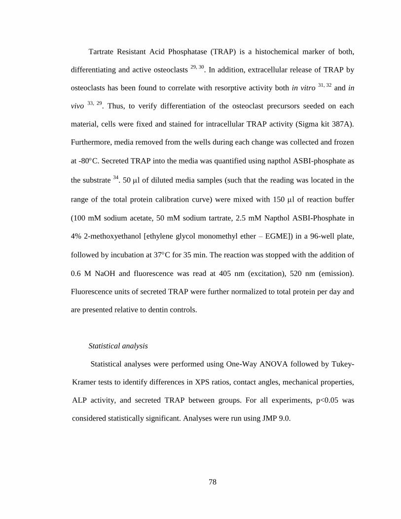
78
Tartrate Resistant Acid Phosphatase (TRAP) is a histochemical marker of both,
differentiating and active osteoclasts 29, 30
. In addition, extracellular release of TRAP by
osteoclasts has been found to correlate with resorptive activity both in vitro 31, 32
and in
vivo 33, 29
. Thus, to verify differentiation of the osteoclast precursors seeded on each
material, cells were fixed and stained for intracellular TRAP activity (Sigma kit 387A).
Furthermore, media removed from the wells during each change was collected and frozen
at -80C. Secreted TRAP into the media was quantified using napthol ASBI-phosphate as
the substrate 34
. 50 l of diluted media samples (such that the reading was located in the
range of the total protein calibration curve) were mixed with 150 l of reaction buffer
(100 mM sodium acetate, 50 mM sodium tartrate, 2.5 mM Napthol ASBI-Phosphate in
4% 2-methoxyethanol [ethylene glycol monomethyl ether – EGME]) in a 96-well plate,
followed by incubation at 37C for 35 min. The reaction was stopped with the addition of
0.6 M NaOH and fluorescence was read at 405 nm (excitation), 520 nm (emission).
Fluorescence units of secreted TRAP were further normalized to total protein per day and
are presented relative to dentin controls.
Statistical analysis
Statistical analyses were performed using One-Way ANOVA followed by Tukey-
Kramer tests to identify differences in XPS ratios, contact angles, mechanical properties,
ALP activity, and secreted TRAP between groups. For all experiments, p<0.05 was
considered statistically significant. Analyses were run using JMP 9.0.
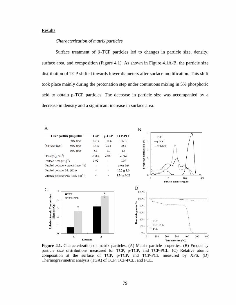
79
Results
Characterization of matrix particles
Surface treatment of -TCP particles led to changes in particle size, density,
surface area, and composition (Figure 4.1). As shown in Figure 4.1A-B, the particle size
distribution of TCP shifted towards lower diameters after surface modification. This shift
took place mainly during the protonation step under continuous mixing in 5% phosphoric
acid to obtain p-TCP particles. The decrease in particle size was accompanied by a
decrease in density and a significant increase in surface area.
Figure 4.1. Characterization of matrix particles. (A) Matrix particle properties. (B) Frequency
particle size distributions measured for TCP, p-TCP, and TCP-PCL. (C) Relative atomic
composition at the surface of TCP, p-TCP, and TCP-PCL measured by XPS. (D)
Thermogravimetric analysis (TGA) of TCP, TCP-PCL, and PCL.
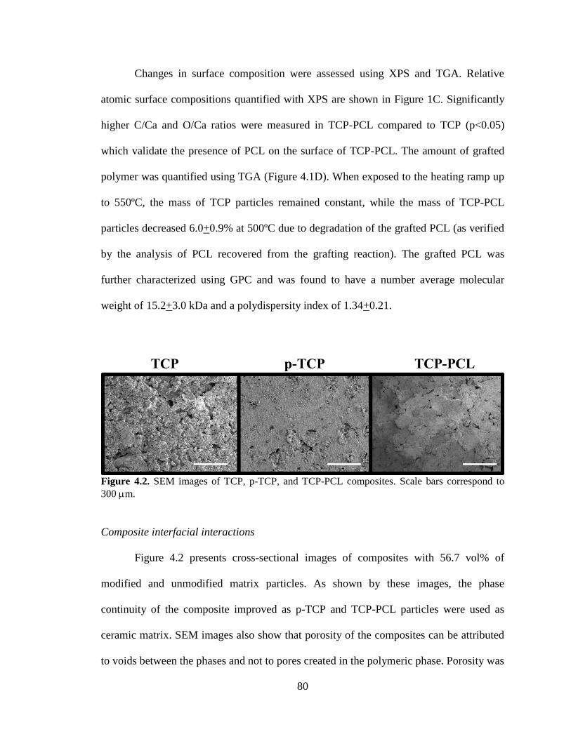
80
Changes in surface composition were assessed using XPS and TGA. Relative
atomic surface compositions quantified with XPS are shown in Figure 1C. Significantly
higher C/Ca and O/Ca ratios were measured in TCP-PCL compared to TCP (p<0.05)
which validate the presence of PCL on the surface of TCP-PCL. The amount of grafted
polymer was quantified using TGA (Figure 4.1D). When exposed to the heating ramp up
to 550ºC, the mass of TCP particles remained constant, while the mass of TCP-PCL
particles decreased 6.0+0.9% at 500ºC due to degradation of the grafted PCL (as verified
by the analysis of PCL recovered from the grafting reaction). The grafted PCL was
further characterized using GPC and was found to have a number average molecular
weight of 15.2+3.0 kDa and a polydispersity index of 1.34+0.21.
Figure 4.2. SEM images of TCP, p-TCP, and TCP-PCL composites. Scale bars correspond to
300 m.
Composite interfacial interactions
Figure 4.2 presents cross-sectional images of composites with 56.7 vol% of
modified and unmodified matrix particles. As shown by these images, the phase
continuity of the composite improved as p-TCP and TCP-PCL particles were used as
ceramic matrix. SEM images also show that porosity of the composites can be attributed
to voids between the phases and not to pores created in the polymeric phase. Porosity was
81
measured gravimetrically 14
and determined to be between 19-27% for the TCP
composite formulations and 7-16% for the composites with modified matrix (p-TCP or
TCP-PCL). TCP composites show evidence of TCP particles and voids of varying size
distributed throughout the material. Although TCP grains with original size (100-300
m) remain, the images also show many smaller particles. These small particles may
have been obtained by crushing during the hand-mixing steps. Due to the significant
decrease in particle size, matrix particles are harder to identify in the p-TCP and TCP-
PCL composites. Though voids were also present in the TCP-PCL composite, higher
continuity of the phases was evident in the images, suggesting improved interaction
between the polymeric and ceramic phases.
Further characterization of this interaction was conducted using contact angle
measurements. The contact angle () between a solid and a liquid can be used to calculate
the work of adhesion (WAd) at the interface according to the Young-Dupré relationship
where corresponds to the liquid surface tension 35
:
(4.2)
An interface with strong interactions has high work of adhesion and low contact angle
(good wetting). Non-reactive polymer was able to wet the TCP pellet surface with a
contact angle of 29+2. The reduced contact angle on TCP-PCL (26+2) suggests a
modest improvement in wetting due to PCL grafting, though no significant differences
were observed between the TCP and TCP-PCL contact angles (n=5, p=0.057). Although
water contact angles are a more common measure of changes in wettability of a surface,
in this case, the treatment of the TCP pellets with acid during the protonation step
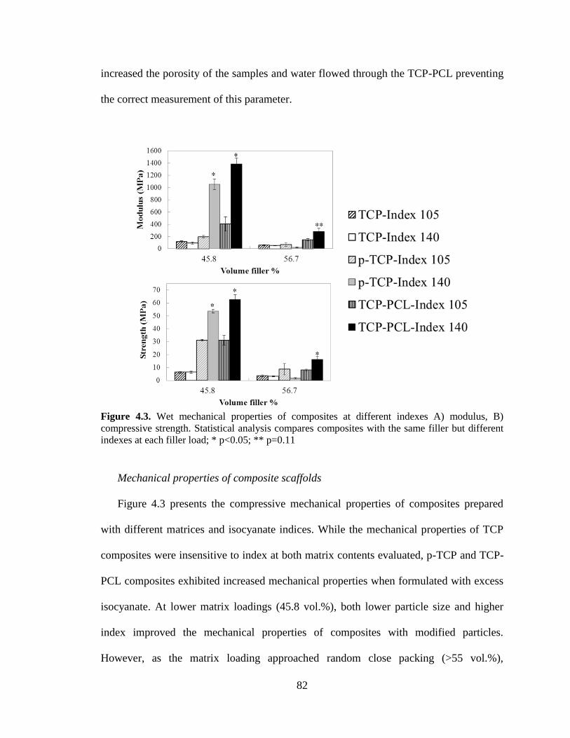
82
increased the porosity of the samples and water flowed through the TCP-PCL preventing
the correct measurement of this parameter.
Figure 4.3. Wet mechanical properties of composites at different indexes A) modulus, B)
compressive strength. Statistical analysis compares composites with the same filler but different
indexes at each filler load; * p<0.05; ** p=0.11
Mechanical properties of composite scaffolds
Figure 4.3 presents the compressive mechanical properties of composites prepared
with different matrices and isocyanate indices. While the mechanical properties of TCP
composites were insensitive to index at both matrix contents evaluated, p-TCP and TCP-
PCL composites exhibited increased mechanical properties when formulated with excess
isocyanate. At lower matrix loadings (45.8 vol.%), both lower particle size and higher
index improved the mechanical properties of composites with modified particles.
However, as the matrix loading approached random close packing (>55 vol.%),
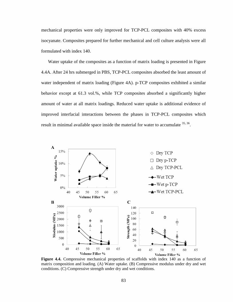
83
mechanical properties were only improved for TCP-PCL composites with 40% excess
isocyanate. Composites prepared for further mechanical and cell culture analysis were all
formulated with index 140.
Water uptake of the composites as a function of matrix loading is presented in Figure
4.4A. After 24 hrs submerged in PBS, TCP-PCL composites absorbed the least amount of
water independent of matrix loading (Figure 4A). p-TCP composites exhibited a similar
behavior except at 61.3 vol.%, while TCP composites absorbed a significantly higher
amount of water at all matrix loadings. Reduced water uptake is additional evidence of
improved interfacial interactions between the phases in TCP-PCL composites which
result in minimal available space inside the material for water to accumulate 35, 36
.
Figure 4.4. Compressive mechanical properties of scaffolds with index 140 as a function of
matrix composition and loading. (A) Water uptake. (B) Compressive modulus under dry and wet
conditions. (C) Compressive strength under dry and wet conditions.
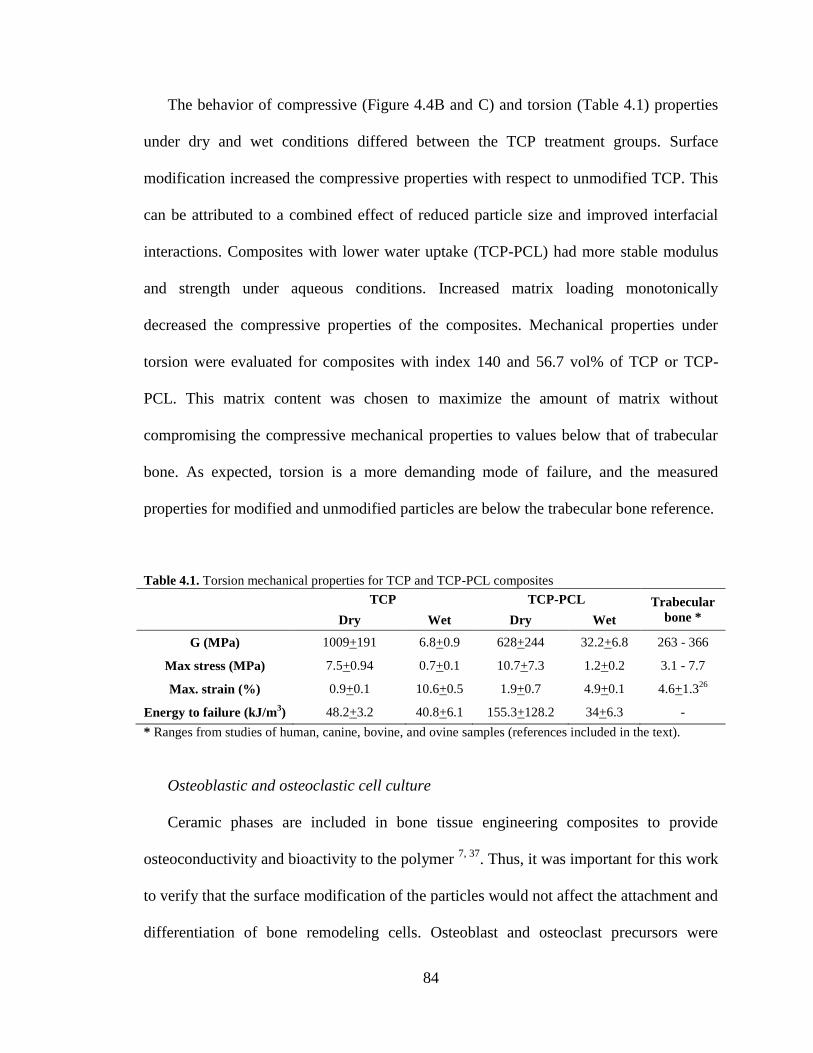
84
The behavior of compressive (Figure 4.4B and C) and torsion (Table 4.1) properties
under dry and wet conditions differed between the TCP treatment groups. Surface
modification increased the compressive properties with respect to unmodified TCP. This
can be attributed to a combined effect of reduced particle size and improved interfacial
interactions. Composites with lower water uptake (TCP-PCL) had more stable modulus
and strength under aqueous conditions. Increased matrix loading monotonically
decreased the compressive properties of the composites. Mechanical properties under
torsion were evaluated for composites with index 140 and 56.7 vol% of TCP or TCP-
PCL. This matrix content was chosen to maximize the amount of matrix without
compromising the compressive mechanical properties to values below that of trabecular
bone. As expected, torsion is a more demanding mode of failure, and the measured
properties for modified and unmodified particles are below the trabecular bone reference.
Table 4.1. Torsion mechanical properties for TCP and TCP-PCL composites
TCP TCP-PCL Trabecular
bone * Dry Wet Dry Wet
G (MPa) 1009+191 6.8+0.9 628+244 32.2+6.8 263 - 366
Max stress (MPa) 7.5+0.94 0.7+0.1 10.7+7.3 1.2+0.2 3.1 - 7.7
Max. strain (%) 0.9+0.1 10.6+0.5 1.9+0.7 4.9+0.1 4.6+1.326
Energy to failure (kJ/m3) 48.2+3.2 40.8+6.1 155.3+128.2 34+6.3 -
* Ranges from studies of human, canine, bovine, and ovine samples (references included in the text).
Osteoblastic and osteoclastic cell culture
Ceramic phases are included in bone tissue engineering composites to provide
osteoconductivity and bioactivity to the polymer 7, 37
. Thus, it was important for this work
to verify that the surface modification of the particles would not affect the attachment and
differentiation of bone remodeling cells. Osteoblast and osteoclast precursors were
85
seeded on TCP and TCP-PCL composites (56.7 vol.% matrix) in vitro, and their
differentiation was monitored over time. It was not possible to seed cells on p-TCP
composites to isolate the effects of changes in particle size, given that the materials
acidified the media and did not allow cells to attach.
Figure 4.5. Osteoblastic cell culture of TCP, and TCP-PCL composites. (A) Live-Dead staining
of MC3T3 cells seeded on composites after 2 days of culture (>95% viability). (B) The
biocomposites support the differentiation of MC3T3 cells after 7 days of culture in differentiation
media. White scale bars represent 200 um, * p<0.05 compared to Day 2.
Both osteoblast and osteoclast precursors attached and differentiated on the surface of
TCP and TCP-PCL composites. Figure 4.5A shows images of live/dead staining of
MC3T3 cells after being cultured 2 days. Viability of the cells was >95% for both
formulations, suggesting minimal toxicity from the scaffolds. Furthermore, increased
ALP activity in the cultured cells (Figure 4.5B) suggests that the cells differentiated into
osteoblasts, and no significant differences were identified between the materials.
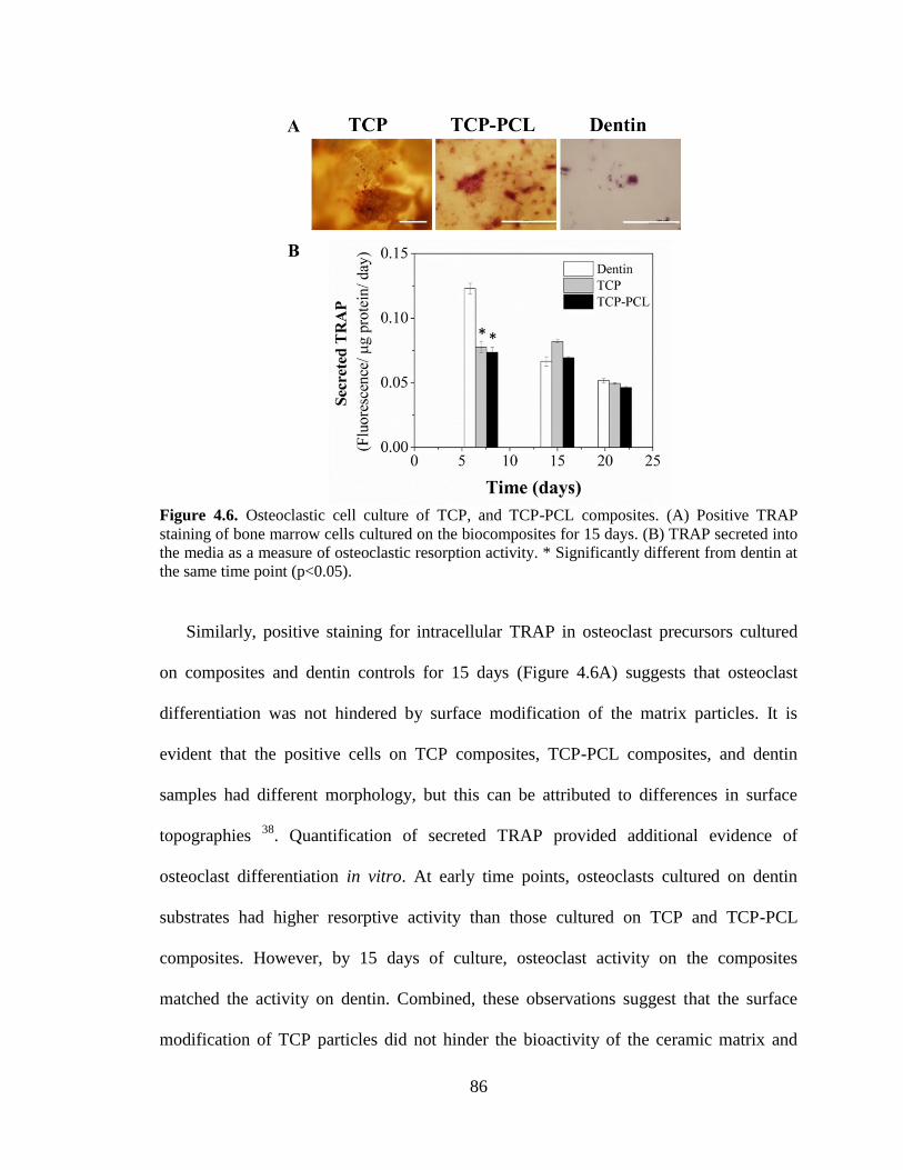
86
Figure 4.6. Osteoclastic cell culture of TCP, and TCP-PCL composites. (A) Positive TRAP
staining of bone marrow cells cultured on the biocomposites for 15 days. (B) TRAP secreted into
the media as a measure of osteoclastic resorption activity. * Significantly different from dentin at
the same time point (p<0.05).
Similarly, positive staining for intracellular TRAP in osteoclast precursors cultured
on composites and dentin controls for 15 days (Figure 4.6A) suggests that osteoclast
differentiation was not hindered by surface modification of the matrix particles. It is
evident that the positive cells on TCP composites, TCP-PCL composites, and dentin
samples had different morphology, but this can be attributed to differences in surface
topographies 38
. Quantification of secreted TRAP provided additional evidence of
osteoclast differentiation in vitro. At early time points, osteoclasts cultured on dentin
substrates had higher resorptive activity than those cultured on TCP and TCP-PCL
composites. However, by 15 days of culture, osteoclast activity on the composites
matched the activity on dentin. Combined, these observations suggest that the surface
modification of TCP particles did not hinder the bioactivity of the ceramic matrix and
87
that the composite formulations developed in this work are candidates to undergo
remodeling in vivo.
Discussion
Development of composite biomaterials for bone tissue engineering, in particular
for load-bearing applications, has received increased attention in the past decade due to
the improvements in handling, mechanical properties, and biological performance that
arise when combining two or more phases 39, 7
. However, a recent review of the
mechanical behavior of calcium phosphate/polymer composites reported that few
formulations have strength and porosity comparable to trabecular bone 8. To overcome
this problem, compression molded composite formulations with polyurethane and
calcium phosphates (hydroxyapatite and -TCP) 9 or allograft bone particles
13 have been
developed and tested in vivo. These low porosity composites achieved mechanical
properties approaching those of cortical bone, and when implanted into femoral defects in
rats and rabbits the materials supported cellular infiltration and promoted new bone
deposition. Although these formulations have the potential to be applied as weight-
bearing implants, they still require compaction with high pressures which limits their use
in operating rooms. In this study, moldable and settable composites incorporating
modified and unmodified -TCP particles and lysine-derived polyurethanes were
developed and characterized in vitro. The formulations can be easily prepared in the
operating room by hand mixing, and delivered to bone defects using a straight-bore
syringe. The -TCP phase was included to increase osteoconductivity of the composite,
88
while the reactive polyurethane was anticipated to improve handling properties and
reduce the brittleness of the calcium phosphate.
Incorporation of -TCP particles into polymers has been reported to decrease the
mechanical strength of composites due to incompatibility between phases 40
. Thus, the
surface of -TCP particles was modified with covalently grafted PCL chains to improve
the interactions between the reactive polyurethane and ceramic phases. TGA and XPS
analysis revealed successful polymer grafting (Figure 4.1C-D) on the surface, with a
decrease in particle size as an additional effect of the protonation step. Contact angle
measurements of non-reactive polyurethane on TCP and TCP-PCL pellets suggested a
slight improvement in the interactions between the phases as evidenced by improved
wetting of the TCP-PCL pellets. However, contact angle measurements were affected by
changes in the surface topography and porosity of the pellets, as evidenced by the
difficulty for measuring water contact angles on TCP-PCL samples. As a result, these
measurements need to be complemented with additional characterization of the polymer-
ceramic interface.
Interactions between polymer-coated particles and the polymeric matrix in
composite materials not only depend on the surface chemistry of the particles but also on
the arrangement of polymeric chains at the interface 41
. An important variable influencing
the conformation of terminally anchored polymer chains is the surface grafting density ( ,
chains/nm2)
42. Previous work studying the conformation of anchored polymer chains in
the presence of a cross-linked network identified a critical value of grafting density:
(4.3)
89
where b corresponds to the Kuhn length of the polymer and N is the number of repeat
units in the chain 43-45
. At low grafting densities ( ) the chains remain close to the
surface without penetrating the network. On the other hand, if higher grafting densities
are achieved ( ), the entropic cost of excluding the chains from the network is
higher than the energy penalty associated to the network swelling due to interdigitation of
the chains 44
. As a result, the chains extend away from the surface creating a brush layer
capable of penetrating the network. The thickness of the resulting brush layer (L) can be
estimated by assuming equilibrium at the interface such that the chemical potential of the
polymer network is the same at the interface as in the bulk 43
. Mean-field theory has been
used to calculate the free energy at the interface and defined the equilibrium condition in
terms of (dimensionless layer thickness) as:
(4.4)
where z (0 under theta-conditions under which the polymer acts as ideal chains) is the
ratio of excluded volume per chain to the volume occupied by the chain at ideal
conditions 43
. is the dimensionless surface density which takes into account
three-body interactions of the chains through , where MWRU is the
molecular weight of the repeating units, v is the specific volume (calculated from density
values) and NA is Avogradro’s number. After solving for , the layer thickness can be
calculated as . Layer thickness has been reported to influence the interfacial
adhesion energy at the interface of a polymeric layer anchored to a surface and an
elastomeric network 41
. Deruelle et. al. characterized this relation using the JKR method
(Johnson, Kendal, Roberts method 46
) and reported the interfacial adhesion energy
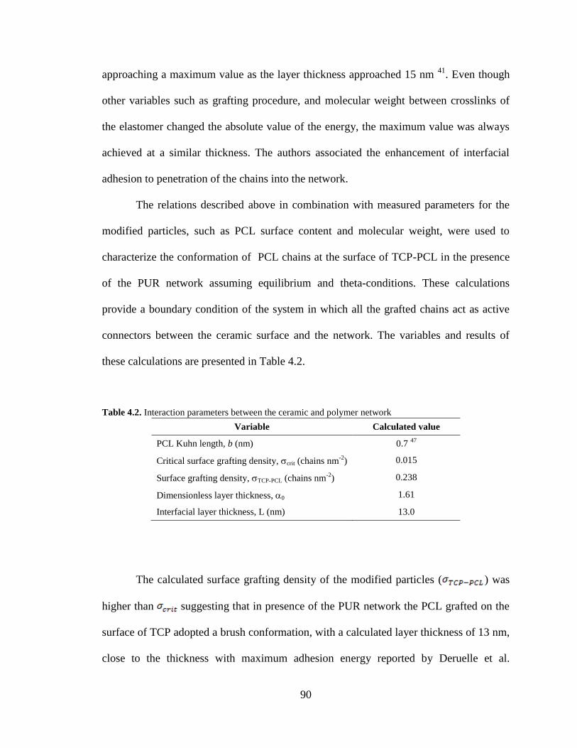
90
approaching a maximum value as the layer thickness approached 15 nm 41
. Even though
other variables such as grafting procedure, and molecular weight between crosslinks of
the elastomer changed the absolute value of the energy, the maximum value was always
achieved at a similar thickness. The authors associated the enhancement of interfacial
adhesion to penetration of the chains into the network.
The relations described above in combination with measured parameters for the
modified particles, such as PCL surface content and molecular weight, were used to
characterize the conformation of PCL chains at the surface of TCP-PCL in the presence
of the PUR network assuming equilibrium and theta-conditions. These calculations
provide a boundary condition of the system in which all the grafted chains act as active
connectors between the ceramic surface and the network. The variables and results of
these calculations are presented in Table 4.2.
Table 4.2. Interaction parameters between the ceramic and polymer network
Variable Calculated value
PCL Kuhn length, b (nm) 0.7 47
Critical surface grafting density, crit (chains nm-2
) 0.015
Surface grafting density, TCP-PCL (chains nm-2
) 0.238
Dimensionless layer thickness, 0 1.61
Interfacial layer thickness, L (nm) 13.0
The calculated surface grafting density of the modified particles ( ) was
higher than suggesting that in presence of the PUR network the PCL grafted on the
surface of TCP adopted a brush conformation, with a calculated layer thickness of 13 nm,
close to the thickness with maximum adhesion energy reported by Deruelle et al.
91
Although the real layer thickness between the phases may have different dimensions,
interpenetration of the chains is also favored by the low elasticity of the network 41
before
the polyurethane completely sets.
SEM images and mechanical properties of the composites provided additional
evidence of improved interactions between the modified TCP-PCL particles and the
polyurethane binder. Compressive properties of formulations with 105 and 140 index
including TCP, p-TCP, and TCP-PCL particles showed that while TCP composites were
insensitive to changes in polyurethane index, p-TCP and TCP-PCL both had improved
mechanical properties when excess isocyanate was present. This suggests that in addition
to the strengthening mechanism of PCL interpenetration into the PUR network discussed
previously, reactivity between hydroxyl groups at the surface of p-TCP and TCP-PCL
with excess isocyanate groups can also improve interfacial interactions. While TCP
composites exhibited a dramatic decrease (15-97%) of mechanical properties after 24 hrs
in PBS, TCP-PCL composites had higher stability under water presenting a decrease in
properties in the range of 35-61%. Reduction of mechanical properties of composites in
an aqueous environment has been associated to a plasticizing effect of the absorbed water,
degradation of the polymeric phase, and/or diffusion of water through the
organic/inorganic interface 35, 40, 36
. Given that the polyurethane binder in our study had
the same characteristics between the unstable TCP and more stable TCP-PCL composites,
it is more likely that increased interfacial interactions in the TCP-PCL composites
reduced water diffusion and enhanced the performance of the materials. In contrast to our
results, previous studies incorporating ~10 vol% of modified TCP particles (with grafted
poly(L-lactide) chains with molecular weight between 390 – 1,050 gmol-1
) in a
92
poly(D,L-lactide) matrix have reported improved coupling between phases (imaged with
SEM) without any improvements in the mechanical properties when compared to
composites with non-modified TCP particles 20
. However, for linear polymers, physical
crosslinks due to chain entanglements result as the molecular weight of the polymer
approaches the molecular weight for entanglements Me 48
, which for poly(lactide) is
~9,000 gmol-1
49
. Furthermore, models predicting the mechanical properties of filled
polymers have shown that tensile strength of composites with significant interfacial
interactions increases only when the filler volume fraction is above 15% 50
. In our case,
the molecular weight of the grafted PCL was similar to the reported Me for PCL (15,000
gmol-1
51
), and the filler loadings evaluated were above 45 vol%. It is important to
highlight that the effect of filler surface modification was significant up to filler contents
of 56.7 vol%. It is possible that above this value particle-particle instead of particle-
polymer interactions become more significant due to changes in the packing distribution
of the filler.
As shown in Figure 4.7, TCP-PCL composites developed in this work have higher
mechanical properties than several calcium phosphate formulations recently reviewed 8.
TCP-PCL composites with high matrix loadings (>55 vol.%) exhibited compressive
properties in the range of trabecular bone: compressive modulus of 100-500 MPa, and
compressive strength of 4-12 MPa 8. Since the mechanical properties of the composites
are expected to decrease as initial degradation of the polymer takes place, TCP-PCL
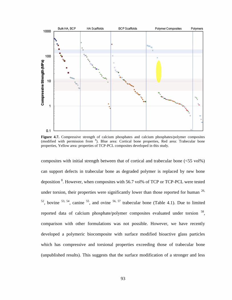
93
Figure 4.7. Compressive strength of calcium phosphates and calcium phosphates/polymer composites
(modified with permission from 8). Blue area: Cortical bone properties, Red area: Trabecular bone
properties, Yellow area: properties of TCP-PCL composites developed in this study.
composites with initial strength between that of cortical and trabecular bone (<55 vol%)
can support defects in trabecular bone as degraded polymer is replaced by new bone
deposition 8. However, when composites with 56.7 vol% of TCP or TCP-PCL were tested
under torsion, their properties were significantly lower than those reported for human 26,
52, bovine
53, 54, canine
55, and ovine
56, 57 trabecular bone (Table 4.1). Due to limited
reported data of calcium phosphate/polymer composites evaluated under torsion 58
,
comparison with other formulations was not possible. However, we have recently
developed a polymeric biocomposite with surface modified bioactive glass particles
which has compressive and torsional properties exceeding those of trabecular bone
(unpublished results). This suggests that the surface modification of a stronger and less
94
brittle ceramic further improves the performance of polyurethane biocomposites under
both, compression and torsion.
Low porosity polyurethane composites have been shown to support cellular
infiltration and remodeling in vivo 9, 13, 18
. Composite formulations developed in this study
are expected to remodel in a similar fashion, according to the in vitro characterization
results which show that surface modification of ceramic surfaces allowed attachment and
differentiation of osteoblast and osteoclast precursors. Even though compared to dentin
controls the TCP and TCP-PCL composites had an initial lag in the activation of
osteoclasts, the lag disappeared after 15 days of culture. Increased surface roughness and
differences in mechanical properties between the composite phases influence the
attachment and morphology of cells 38
. Thus it is possible that osteoclast precursors
seeded on dentin samples were able to attach faster and start the differentiation process
before the cells seeded on TCP-PCL materials. This small lag is not expected to pose
problems for the in vivo remodeling of the composites. A limitation of the in vitro
methods is that during the cutting of the samples for cell culture TCP-PCL grains are also
cut through exposing non-modified surfaces which can promote cellular development. In
vivo studies of hydroxyapatite/PCL composites with high and low levels of embedding of
the ceramic by the polymer showed enhanced osteoconductive behavior in composites
with low embedding of the hydroxyapatite (increased exposed surface) 37
. Thus, further
in vivo studies should be completed to verify the ability of cells to infiltrate the low
porosity composites developed in this study.
95
Conclusions
TCP-PCL/PUR composites with compressive mechanical properties between
those of trabecular and cortical bone were developed. Improved interactions between -
TCP particles and the PUR phase were achieved by covalently grafting PCL to the
surface of the filler. The resulting TCP-PCL composites had higher compressive
mechanical properties and stability under aqueous conditions than TCP composites (non-
modified). No significant effect of surface modification on cell differentiation or activity
was identified in vitro. Thus, surface modification of ceramic particles is a suitable
technique to improve the mechanical performance of settable composites for bone tissue
engineering. The application of this technique with stronger and less brittle fillers is a
promising alternative to develop formulations with initial weight-bearing capabilities.
96
References
1 J. A. Buckwalter, M. J. Glimcher, R. R. Cooper, and R. Recker, 'Bone Biology. I: Structure, Blood
Supply, Cells, Matrix, and Mineralization', Instr Course Lect, 45 (1996), 371-86.
2 P. V. Giannoudis, H. Dinopoulos, and E. Tsiridis, 'Bone Substitutes: An Update', Injury, 36 Suppl
3 (2005), S20-7.
3 S. Kotani, Y. Fujita, T. Kitsugi, T. Nakamura, T. Yamamuro, C. Ohtsuki, and T. Kokubo, 'Bone
Bonding Mechanism of Beta-Tricalcium Phosphate', J Biomed Mater Res, 25 (1991), 1303-15.
4 M. Neo, S. Kotani, Y. Fujita, T. Nakamura, T. Yamamuro, Y. Bando, C. Ohtsuki, and T. Kokubo,
'Differences in Ceramic-Bone Interface between Surface-Active Ceramics and Resorbable
Ceramics: A Study by Scanning and Transmission Electron Microscopy', J Biomed Mater Res, 26
(1992), 255-67.
5 V. James Sammarco, and Laurette Chang, 'Modern Issues in Bone Graft Substitutes and Advances
in Bone Tissue Technology', Foot and Ankle Clinics of North America, 7 (2002), 19-41.
6 Yusuf Khan, Michael J. Yaszemski, Antonios G. Mikos, and Cato T. Laurencin, 'Tissue
Engineering of Bone: Material and Matrix Considerations', J Bone Joint Surg Am, 90 (2008), 36-
42.
7 K. Rezwan, Q. Z. Chen, J. J. Blaker, and Aldo Roberto Boccaccini, 'Biodegradable and Bioactive
Porous Polymer/Inorganic Composite Scaffolds for Bone Tissue Engineering', Biomaterials, 27
(2006), 3413-31.
8 A. J. Wagoner Johnson, and B. A. Herschler, 'A Review of the Mechanical Behavior of Cap and
Cap/Polymer Composites for Applications in Bone Replacement and Repair', Acta Biomater, 7
(2011), 16-30.
9 T. Yoshii, J. E. Dumas, A. Okawa, D. M. Spengler, and S. A. Guelcher, 'Synthesis,
Characterization of Calcium Phosphates/Polyurethane Composites for Weight-Bearing Implants',
Journal of Biomedical Materials Research Part B-Applied Biomaterials, 100B (2012), 32-40.
10 Jian Ying Zhang, Eric J. Beckman, Nicholas P. Piesco, and Sudha Agarwal, 'A New Peptide-
Based Urethane Polymer: Synthesis, Biodegradation, and Potential to Support Cell Growth in
Vitro', Biomaterials, 21 (2000), 1247-58.
11 Jian Han, Bing Chen, Lin Ye, Ai-ying Zhang, Jian Zhang, and Zeng-guo Feng, 'Synthesis and
Characterization of Biodegradable Polyurethane Based on Poly(Ε-Caprolactone) and L-Lysine
Ethyl Ester Diisocyanate', Frontiers of Materials Science in China, 3 (2009), 25-32.
12 S. A. Guelcher, A. Srinivasan, J. E. Dumas, J. E. Didier, S. McBride, and J. O. Hollinger,
'Synthesis, Mechanical Properties, Biocompatibility, and Biodegradation of Polyurethane
Networks from Lysine Polyisocyanates', Biomaterials, 29 (2008), 1762-75.
13 J. E. Dumas, T. Davis, G.E. Holt, T. Yoshii, D. S. Perrien, J. S. Nyman, T. Boyce, and S.A.
Guelcher, 'Synthesis, Characterization, and Remodeling of Weight-Bearing Allograft
Bone/Polyurethane Composites in the Rabbit', Acta Biomaterialia, 6 (2010), 2394-406.
14 J. E. Dumas, K. Zienkiewicz, S. A. Tanner, E. M. Prieto, S. Bhattacharyya, and S. A. Guelcher,
'Synthesis and Characterization of an Injectable Allograft Bone/Polymer Composite Bone Void
Filler with Tunable Mechanical Properties', Tissue Engineering Part A, 16 (2010), 2505-18.
15 Ian C. Bonzani, Raju Adhikari, Shadi Houshyar, Roshan Mayadunne, Pathiraja Gunatillake, and
Molly M. Stevens, 'Synthesis of Two-Component Injectable Polyurethanes for Bone Tissue
Engineering', Biomaterials, 28 (2007), 423-33.
97
16 Raju Adhikari, Pathiraja A. Gunatillake, Ian Griffiths, Lisa Tatai, Malsha Wickramaratna, Shadi
Houshyar, Tim Moore, Roshan T. M. Mayadunne, John Field, Margaret McGee, and Tania
Carbone, 'Biodegradable Injectable Polyurethanes: Synthesis and Evaluation for Orthopaedic
Applications', Biomaterials, 29 (2008), 3762-70.
17 J. E. Dumas, K. Zienkiewicz, S. A. Tanner, E. M. Prieto, S. Bhattacharyya, and S. A. Guelcher,
'Synthesis and Characterization of an Injectable Allograft Bone/Polymer Composite Bone Void
Filler with Tunable Mechanical Properties', Tissue Eng Part A, 16, 2505-18.
18 J. E. Dumas, E. M. Prieto, K.J. Zienkiewicz, T. Guda, J. C. Wenke, J. Bible, G.E. Holt, and S.A.
Guelcher, 'Balancing the Rates of New Bone Formation and Polymer Degradation Enhances
Healing of Weight-Bearing Allograft/Polyurethane Composites in Rabbit Femoral Defects', Tissue
Eng Part A, Not available - ahead of print (2013).
19 F. H. Norman-Taylor, N. Santori, and R. N. Villar, 'The Trouble with Bone Allograft', BMJ, 315
(1997), 498.
20 C. Kunze, T. Freier, E. Helwig, B. Sandner, D. Reif, A. Wutzler, and H. J. Radusch, 'Surface
Modification of Tricalcium Phosphate for Improvement of the Interfacial Compatibility with
Biodegradable Polymers', Biomaterials, 24 (2003), 967-74.
21 S. Guelcher, A. Srinivasan, A. Hafeman, K. Gallagher, J. Doctor, S. Khetan, S. McBride, and J.
Hollinger, 'Synthesis, in Vitro Degradation, and Mechanical Properties of Two-Component
Poly(Ester Urethane)Urea Scaffolds: Effects of Water and Polyol Composition', Tissue
Engineering, 13 (2007), 2321-33.
22 Murat Barsbay, Olgun Güven, Martina H. Stenzel, Thomas P. Davis, Christopher Barner-
Kowollik, and Leonie Barner, 'Verification of Controlled Grafting of Styrene from Cellulose Via
Radiation-Induced Raft Polymerization', Macromolecules, 40 (2007), 7140-47.
23 Guelcher, 'Synthesis, Mechanical Properties, Biocompatibility, and Biodegradation of
Polyurethane Networks from Lysine Polyisocyanates.', Biomaterials (2008).
24 ASTM, 'D4274: Standard Test Methods for Testing Polyurethane Raw Materials: Determination
of Hydroxyl Numbers of Polyols', (1999).
25 ASTM, 'D2572: Standard Test Method for Isocyanate Groups in Urethane Materials or
Prepolymers', (1997).
26 K. Bruyere Garnier, R. Dumas, C. Rumelhart, and M. E. Arlot, 'Mechanical Characterization in
Shear of Human Femoral Cancellous Bone: Torsion and Shear Tests', Med Eng Phys, 21 (1999),
641-9.
27 C. M. Ford, and T. M. Keaveny, 'The Dependence of Shear Failure Properties of Trabecular Bone
on Apparent Density and Trabecular Orientation', J Biomech, 29 (1996), 1309-17.
28 Hidetaka Hayashi, Ken-ichi Nakahama, Takahiro Sato, Takehiko Tuchiya, Yasuyuki Asakawa,
Toshimitu Maemura, Masanobu Tanaka, Mineto Morita, and Ikuo Morita, 'The Role of Mac-1
(Cd11b/Cd18) in Osteoclast Differentiation Induced by Receptor Activator of Nuclear Factor-Κb
Ligand', FEBS Letters, 582 (2008), 3243-48.
29 GregoryR Mundy, 'Regulatory Mechanisms of Osteoclast Differentiation and Function', Journal of
Bone and Mineral Metabolism, 14 (1996), 59-64.
30 Steven L. Teitelbaum, 'Bone Resorption by Osteoclasts', Science, 289 (2000), 1504-08.
31 Anthony J. Janckila, Ranga N. Parthasarathy, Latha K. Parthasarathy, Ratnam S. Seelan, Yi-
Cheung Hsueh, Jukka Rissanen, Sari L. Alatalo, Jussi M. Halleen, and Lung T. Yam, 'Properties
98
and Expression of Human Tartrate-Resistant Acid Phosphatase Isoform 5a by Monocyte-Derived
Cells', Journal of Leukocyte Biology, 77 (2005), 209-18.
32 Barrie Kirstein, Timothy J. Chambers, and Karen Fuller, 'Secretion of Tartrate-Resistant Acid
Phosphatase by Osteoclasts Correlates with Resorptive Behavior', Journal of Cellular
Biochemistry, 98 (2006), 1085-94.
33 J. M. Halleen, H. Ylipahkala, S. L. Alatalo, A. J. Janckila, J. E. Heikkinen, H. Suominen, S.
Cheng, and H. K. Väänänen, 'Serum Tartrate-Resistant Acid Phosphatase 5b, but Not 5a,
Correlates with Other Markers of Bone Turnover and Bone Mineral Density', Calcified Tissue
International, 71 (2002), 20-25.
34 Anthony J. Janckila, Karen Takahashi, Susan Z. Sun, and Lung T. Yam, 'Naphthol-Asbi
Phosphate as a Preferred Substrate for Tartrate-Resistant Acid Phosphatase Isoform 5b', Journal of
Bone and Mineral Research, 16 (2001), 788-93.
35 R. E. Neuendorf, E. Saiz, A. P. Tomsia, and R. O. Ritchie, 'Adhesion between Biodegradable
Polymers and Hydroxyapatite: Relevance to Synthetic Bone-Like Materials and Tissue
Engineering Scaffolds', Acta Biomaterialia, 4 (2008), 1288-96.
36 P. S. Uskokovic, C. Y. Tang, C. P. Tsui, N. Ignjatovic, and D. P. Uskokovic, 'Micromechanical
Properties of a Hydroxyapatite/Poly-L-Lactide Biocomposite Using Nanoindentation and Modulus
Mapping', Journal of the European Ceramic Society, 27 (2007), 1559-64.
37 S. Scaglione, E. Lazzarini, C. Ilengo, and R. Quarto, 'A Composite Material Model for Improved
Bone Formation', Journal of Tissue Engineering and Regenerative Medicine, 4 (2010), 505-13.
38 Karen Fuller, Jade L. Ross, Kinga A. Szewczyk, Raymond Moss, and Tim J. Chambers, 'Bone Is
Not Essential for Osteoclast Activation', PLoS ONE, 5 (2010), e12837.
39 R. Kruger, and J. Groll, 'Fiber Reinforced Calcium Phosphate Cements -- on the Way to
Degradable Load Bearing Bone Substitutes?', Biomaterials, 33 (2012), 5887-900.
40 Blanca Vázquez, María Pau Ginebra, Xavier Gil, Josep Antón Planell, and Julio San Román,
'Acrylic Bone Cements Modified with -Tcp Particles Encapsulated with Poly(Ethylene Glycol)',
Biomaterials, 26 (2005), 4309-16.
41 M. Deruelle, L. Leger, and M. Tirrell, 'Adhesion at the Solid-Elastomer Interface: Influence of the
Interfacial Chains', Macromolecules, 28 (1995), 7419-28.
42 P. G. de Gennes, 'Conformations of Polymers Attached to an Interface', Macromolecules, 13
(1980), 1069-75.
43 W. B. Russel, D. A. Saville, and W. R. Schowalter, Colloidal Dispersions (Australia: Cambridge
University Press, 1999).
44 M. Geoghegan, C. J. Clarke, F. Boué, A. Menelle, T. Russ, and D. G. Bucknall, 'The Kinetics of
Penetration of Grafted Polymers into a Network', Macromolecules, 32 (1999), 5106-14.
45 K. P. O'Connor, and T. C. B. McLeish, '"Molecular Velcro": Dynamics of a Constrained Chain
into an Elastomer Network', Macromolecules, 26 (1993), 7322-25.
46 K. L. Johnson, K. Kendall, and A. D. Roberts, 'Surface Energy and the Contact of Elastic Solids',
Proceedings of the Royal Society of London. A. Mathematical and Physical Sciences, 324 (1971),
301-13.
47 Dinorah Herrera, Jean-Carlos Zamora, Alfredo Bello, Mario Grimau, Estrella Laredo, Alejandro J.
Müller, and Timothy P. Lodge, 'Miscibility and Crystallization in Polycarbonate/Poly(Ε-
99
Caprolactone) Blends: Application of the Self-Concentration Model', Macromolecules, 38 (2005),
5109-17.
48 Hugh R. Brown, and Thomas P. Russell, 'Entanglements at Polymer Surfaces and Interfaces',
Macromolecules, 29 (1996), 798-800.
49 John R. Dorgan, Joshua S. Williams, and David N. Lewis, 'Melt Rheology of Poly(Lactic Acid):
Entanglement and Chain Architecture Effects', Journal of Rheology (1978-present), 43 (1999),
1141-55.
50 Lawrence E. Nielsen, 'Simple Theory of Stress-Strain Properties of Filled Polymers', Journal of
Applied Polymer Science, 10 (1966), 97-103.
51 Alper Gurarslan, Jialong Shen, and Alan E. Tonelli, 'Behavior of Poly(Ε-Caprolactone)S (Pcls)
Coalesced from Their Stoichiometric Urea Inclusion Compounds and Their Use as Nucleants for
Crystallizing Pcl Melts: Dependence on Pcl Molecular Weights', Macromolecules, 45 (2012),
2835-40.
52 M. Halawa, A. J. C. Lee, R. S. M. Ling, and S. S. Vangala, 'Shear-Strength of Trabecular Bone
from Femur, and Some Factors Affecting Shear-Strength of Cement-Bone Interface', Archives of
Orthopaedic and Trauma Surgery, 92 (1978), 19-30.
53 J. L. Stone, G. S. Beaupre, and W. C. Hayes, 'Multiaxial Strength Characteristics of Trabecular
Bone', J Biomech, 16 (1983), 743-52.
54 R. B. Ashman, J. D. Corin, and C. H. Turner, 'Elastic Properties of Cancellous Bone:
Measurement by an Ultrasonic Technique', J Biomech, 20 (1987), 979-86.
55 S. K. Eswaran, M. R. Allen, D. B. Burr, and T. M. Keaveny, 'A Computational Assessment of the
Independent Contribution of Changes in Canine Trabecular Bone Volume Fraction and
Microarchitecture to Increased Bone Strength with Suppression of Bone Turnover', J Biomech, 40
(2007), 3424-31.
56 D. Mitton, C. Rumelhart, D. Hans, and P. J. Meunier, 'The Effects of Density and Test Conditions
on Measured Compression and Shear Strength of Cancellous Bone from the Lumbar Vertebrae of
Ewes', Med Eng Phys, 19 (1997), 464-74.
57 M. Kasra, and M. D. Grynpas, 'On Shear Properties of Trabecular Bone under Torsional Loading:
Effects of Bone Marrow and Strain Rate', J Biomech, 40 (2007), 2898-903.
58 M. Bohner, 'Design of Ceramic-Based Cements and Putties for Bone Graft Substitution', Eur Cells
Mater, 20 (2010), 1-12.
100
CHAPTER V
BALANCING THE RATES OF NEW BONE FORMATION AND POLYMER
DEGRADATION ENHANCES HEALING OF WEIGHT-BEARING
ALLOGRAFT/POLYURETHANE COMPOSITES IN RABBIT FEMORAL DEFECTS
Introduction
Periarticular fractures involve a weight-bearing joint and often have depressed
portions that require extensive open reduction and internal fixation along with
subchondral grafting to maintain articular congruence. Maintenance of articular reduction
and joint congruity are important for both bone healing and articular alignment, since
lack of articular congruence after fractures increases the likelihood of osteoarthritis 1.
While autograft remains the standard of care for bone healing, calcium phosphate (CaP)
cements (CPCs) have proven to be superior to autograft for maintaining articular
congruence of tibial plateau fractures 2. A recent retrospective study has reported that
61% of patients treated with buttress plating and autograft experienced loss of reduction
after one year compared to 23% of patients treated with a hydroxyapatite (HA) bone
cement 3. However, HA bone cement does not extensively remodel and is not replaced by
new bone. Moreover, the brittleness and low shear strength of CPCs adversely impact
their ability to bear mechanical loads, requiring the use of large, mechanically rigid
internal fixation devices (such as buttress plating), which has been suggested to increase
complications 4.
Settable, weight-bearing bone grafts ideally exhibit both initial bone-like
mechanical properties and also remodel with minimal resorption gaps such that the
101
strength of the graft exceeds that of host bone during all stages of healing. Injectable bone
void fillers (BVFs) include non-setting allograft 5 and non-allogenic pastes
6, which are
typically delivered using viscous carriers (e.g., sodium hyaluronate 7, glycerol
8, or
dextran 9) resulting in weak mechanical properties. Resorbable CPCs
10-13, such as -TCP
(slowly resorbed by osteoclasts) or calcium sulfate (CSH) and brushite (undergo
dissolution), have also been used to enhance bone healing. Tri-phasic CPCs comprising
CSH, brushite, and granular -TCP have been shown to heal metaphyseal defects in
preclinical 14
and clinical studies 15
. However, rapid dissolution of CSH and brushite at
rates exceeding that of new bone formation can result in loss of mechanical strength 16
, as
well as the appearance of a fibrous resorption gap between the ingrowing new bone and
the resorbing cement 17, 18
. While the initial compressive strength of CPCs is comparable
to that of trabecular bone, they undergo brittle fracture at strains less than the yield strain
of trabecular bone, resulting in failure due to fatigue under physiologically relevant cyclic
loads where the shear component is significant 19, 6
. Reinforcement with polymeric,
bioactive glass, or ceramic fibers has been reported to enhance strength and ductility of
CPCs, resulting in strengths approaching 50 MPa for poly(lactic-co-glycolic acid)
(PLGA) fibers 20
and 130 MPa for magnesium alloy fibers 21
.
Despite the growing interest in weight-bearing settable bone grafts, there is a
paucity of studies investigating in vivo remodeling in preclinical models. While several
studies have reported that weight-bearing grafts heal and form new bone 22-25
, there is
limited understanding of the mechanisms by which these grafts remodel in vivo, which is
essential for designing grafts that ideally maintain bone-like strength during the entire
healing process 26
. It has been suggested that the degradation rates of the polymer and
102
ceramic phases must be balanced to optimize healing, which is a challenging problem due
to the fundamentally different mechanisms by which polymers and ceramics degrade 26
.
In this study, we investigated the effects of the rates of new bone formation, matrix
resorption, and polymer degradation on healing of settable, weight-bearing polyurethane
(PUR) composites comprising approximately equivalent volume fractions of polymer and
matrix. Based on our previous study reporting that allograft bone particles (ABP)
embedded in a degradable lysine-derived PUR network remodel by creeping substitution
22, we investigated ABP as the reinforcing particulated matrix phase. We hypothesized
that matching the rate of new bone formation to that of polymer degradation would
optimize healing. To test this hypothesis, we augmented the ABP/PUR composites with
either 105 or 420 μg cm-3
recombinant human bone morphogenetic protein-2 (rhBMP-2),
which was anticipated to increase the rate of new bone formation. Further, rhBMP-2 has
been reported to promote zero-order degradation of the PUR network 27
, resulting in more
closely balanced rates of bone formation and PUR degradation. The composites were
injected into cylindrical defects in the femoral condyles of rabbits and evaluated by
micro-computed tomography (μCT) and histomorphometry at 0, 6, and 12 weeks.
Experimental
Materials
LTI-PEG prepolymer (23.6% NCO), poly(-caprolactone-co-D.L-lactide-co-
glycolide) triol [162 mg KOH/g and Mn = 1393 g/mol (measured by gel permeation
chromatography)], and rabbit allograft mineralized bone particles (ABP, 180 ± 70 m)
were received as a gift from Medtronic, Inc. (Memphis, TN) 28
. Triethylenediamine
103
(TEDA) catalyst was received as a gift from Goldschmidt (Hopewell, VA), and
recombinant human bone morphogenetic protein (rhBMP-2) was purchased from R&D
systems (Minneapolis, MN). Trehalose dehydrate, heparin sodium salt, acetonitrile, and
trifluoroacetic acid (TFA) were purchased from Sigma Aldrich (St. Louis, MO).
Preparation of rhBMP-2
The rhBMP-2 was supplied as a solution comprising 35% acetonitrile and 0.1%
TFA. A separate acetonitrile/TFA solution was prepared containing a ratio of 10:1 of
trehalose dehydrate:heparin sodium. The rhBMP-2 and trehalose mixtures were
combined such that the ratio of rhBMP-2 to trehalose was 1:125. The resulting mixture
was distributed in glass vials and frozen at -80oC in preparation for freeze-drying, which
produced a powder.
Synthesis of ABP/PUR Biocomposites
The appropriate amounts of polyester triol, ABP, and LTI-PEG prepolymer were
added to a mixing cup and mixed by hand for 90 s. The index (ratio of NCO:OH
equivalents x 100) was 130. The resulting paste and the catalyst (5500 ppm TEDA) were
then added to the rhBMP-2 vial and mixed for 60 s. The filler content (ABP and rhBMP-
2 powder) was maintained at 70 wt% (56.7 vol%) for each biocomposite (BC) treatment
group (Table 5.1). Two rhBMP-2 doses were investigated: 420 g cm-3
and 105 g cm-3
,
corresponding to 100% and 25% of the dose recommended for the absorbable collagen
sponge (ACS) for rabbits 29
. The porosities of the composites (measured
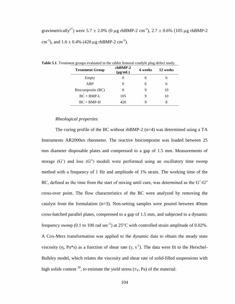
104
gravimetrically27
) were 5.7 ± 2.0% (0 g rhBMP-2 cm-3
), 2.7 ± 0.6% (105 g rhBMP-2
cm-3
), and 1.6 ± 0.4% (420 g rhBMP-2 cm-3
).
Table 5.1. Treatment groups evaluated in the rabbit femoral condyle plug defect study.
Treatment Group rhBMP-2
(µg/mL) 6 weeks 12 weeks
Empty 0 6 6
ABP 0 6 6
Biocomposite (BC) 0 9 10
BC + BMP-L 105 9 10
BC + BMP-H 420 9 8
Rheological properties
The curing profile of the BC without rhBMP-2 (n=4) was determined using a TA
Instruments AR2000ex rheometer. The reactive biocomposite was loaded between 25
mm diameter disposable plates and compressed to a gap of 1.5 mm. Measurements of
storage (G’) and loss (G”) moduli were performed using an oscillatory time sweep
method with a frequency of 1 Hz and amplitude of 1% strain. The working time of the
BC, defined as the time from the start of mixing until cure, was determined as the G’-G”
cross-over point. The flow characteristics of the BC were analyzed by removing the
catalyst from the formulation (n=3). Non-setting samples were poured between 40mm
cross-hatched parallel plates, compressed to a gap of 1.5 mm, and subjected to a dynamic
frequency sweep (0.1 to 100 rad sec-1
) at 25C with controlled strain amplitude of 0.02%.
A Cox-Merz transformation was applied to the dynamic data to obtain the steady state
viscosity (, Pa*s) as a function of shear rate (, s-1
). The data were fit to the Herschel-
Bulkley model, which relates the viscosity and shear rate of solid-filled suspensions with
high solids content 30
, to estimate the yield stress (Y, Pa) of the material:
105
= Y-1
+ K(n-1) (5.1)
where K is the consistency index of the composite (constant) and n is the power-law
index of the suspending polymer, which in this case is the non-reactive mixture of
polyester triol and LTI-PEG prepolymer.
Mechanical properties
Cylindrical (6-mm diameter) samples of each treatment group were prepared for
mechanical testing. The reactive paste was transferred into cylindrical plastic cups and a
1-lb weight (20.7 psi) was placed on the material for 10 min. Cylinders were cured in a
vacuum oven at 37oC overnight, removed from the plastic cups, and cut using a Buehler
saw to a length of 12 mm. After 24 hours of hydration in phosphate buffered saline (PBS),
the samples were tested using an MTS 898 by preloading to 12N followed by
compression at a rate of 25mm/min until failure. The original cross sectional area of the
cylinders was used to calculate compressive stress. The ultimate properties reported in
this article correspond to the point where maximum stress was achieved. To prepare
samples for torsion testing, ~5 mm at each end of 6-mm diameter cylinders was potted
into Technovit 4000 (Heraeus Kulzer) resulting in a central gauge length of about 10 mm.
The potting material was prepared by mixing the powder:syrup I:syrup II using a ratio of
2:2:1 following the manufacturer’s instructions. After potting, the torsion samples were
hydrated for 24 hrs in PBS and then tested using an Instron DynaMight 8841 machine
equipped with a 1.7 Nm torque cell. The potted ends were gripped using the Instron
clamps in a horizontal setup (such that the residual torque was minimal). During testing,
106
the clamps were rotated at an angular speed of 2/s until failure. The measured torque and
rotation angle were used to determine torsional stress using the equation 31, 32
:
= [(dT/d) + 3T]/(2r3) (5.2)
where r is the radius of the cylinder, = /L is the rotation angle per gauge unit length,
and T is the measured torque. The derivative dT/d was determined by fitting the T vs.
data to a 5th
order polynomial (between zero up to the maximum torque or ultimate point).
Torsional strain () was determined as = r, and the shear modulus calculated from the
linear region of the stress-strain curve G = / . Toughness was calculated as the
corresponding area under the stress-strain curve from zero to the maximum stress.
Animal Study
Forty-two New Zealand White (NZW) rabbits weighing between 3.8 and 4.1 kg
were used in this study. All surgical and care procedures were carried out under aseptic
conditions per the approved Institutional Animal Care Use Committee (IACUC) protocol.
The ABP/PUR putty components were gamma irradiated using a dose of ~25 kGY.
Glycopyrrolate was administered at 0.01 mg/kg intramuscularly (IM) followed by
ketamine at 40 mg/kg IM. Bilateral defects of ~6 mm diameter by 11 mm in depth were
drilled in the lateral metaphysis of the distal femurs of each rabbit. ABP/PUR plugs from
each treatment group (Table 5.1) were subsequently injected into each defect using a 1
mL syringe (modified by cutting the needle hub adapter). Treatment groups for each
composite were dispersed randomly among the rabbits with rhBMP-2 treatment groups
paired in each rabbit to eliminate systemic effects. The wound was closed approximately
107
3 minutes after injection. The rabbits were euthanized at both 6 and 12 week time points
using Fatal-plus (2.2 mL/10 kg) intra-venously.
CT Analysis
A CT40 (SCANCO Medical) was used to acquire images of the extracted
femurs post implantation at 6 and 12 weeks. In addition, femurs from NZW rabbits that
were part of other experimental protocols which did not affect the skeleton (same age,
sex, and similar weight to the rabbits used in this study) were scanned and analyzed as
host bone controls. Subsequently, cylindrical defects (6 mm x 11 mm) were created in the
host bone controls and filled with the BC formulation. The injected material was left to
cure under PBS for 24 hrs before submerging the bone in formalin for further scanning as
BC controls (BC, 0w). µCT scans were performed in formalin at 70 kVp energy, 114 µA
source current, 1000 projections per rotation, 300 ms integration time, and an isotropic
voxel size of 36 µm. A 0.1 mm thick aluminum foil filter was employed to reduce beam
hardening. Axial images were reconstructed using manufacturer-provided software.
Further beam hardening correction was performed during image reconstruction using the
manufacturer’s 1200 mg HA cm-3
algorithm. Attenuation values were converted to tissue
mineral density (TMD) through calibration with hydroxyapatite (HA) phantoms with
densities between 0 and 780 mg HA cm-3
(calibration of the instrument was checked
weekly). Using the cortical borders of the defect for alignment, the reconstructed image
stack was re-oriented such that the depth of the defect was parallel to the z-axis. A radial
analysis of the morphometric parameters was conducted from the core of the implant to
the interphase with host bone. Four concentric annular volumes of interest with thickness
108
of 1 mm and approximate length of 11mm (from the outer cortical surface of the femur)
were defined for each sample. The 3 inner regions incorporated the BCs, while the outer
region provided information about the interphase with host bone. Ossified tissue was
segmented from soft tissue using a threshold of 480 mgHAcm-3
and Gaussian noise
filter settings of sigma 0.2 and support 2. The threshold conditions were chosen visually
and kept constant for the analysis of all the samples. Morphometric parameters within the
annular volumes were calculated for each region, grouped by treatment and time point,
and plotted versus the mean radial distance from the core of the defect Rm (Rm=(Ro+Ri)/2,
where Ro and Ri correspond to the outer and inner radius of each region, respectively).
Bone volume/total volume (BV/TV), tissue mineral density (TMD, density of the ossified
tissue), connectivity density (Conn.D.), trabecular number (Tb.N.), trabecular thickness
(Tb.Th.), and trabecular separation (Tb.Sp.) within the regions of interest were computed
using SCANCO’s Medical microCT systems software.
Histology and histomorphometry
Rabbit femora were placed in a solution of 10% formalin for two weeks followed
by a series of ethanol dehydrations. After fixation, the femurs were embedded in
poly(methyl methacrylate) and 200-m sections were cut from the resulting blocks using
an Exakt band saw. The sections were then ground and polished using an Exakt grinding
system to less than 100 m and stained with Sanderson’s rapid bone stain counterstained
with van Gieson. Residual allograft bone particles stained light brown, residual polymer
stained black, new bone stained pink with dark blue osteocytes within the matrix, red
blood cells stained torquiose, and other cells stained a lighter blue. The sections were

109
imaged at 10X magnification with an Olympus camera (DP71) using an Olympus BX60
microscope with and without polarizing the light. Residual ABPs, new bone formation,
and remaining polymer were quantified in an area of interest 1.5 mm high 6 mm wide
located in the center of the defect. To analyze the radial remodeling of the scaffolds in
time, the rectangular area of interest was further subdivided into 3 concentric annular
regions (each 1 mm thick): A1, representing the area of the defect in contact with the host
bone and up to 1 mm away from the edge of the defect; A2, representing the mid-region
of the defect; and A3, representing the core of the defect. Residual allograft was
differentiated from new bone by meeting the following three criteria: (i) acellularity,
(ii)angular in shape, and (iii) ilumination under polarized light. Metamorph (Version
7.0.1) was utilized to perform histomorphometry using a color thresholding and an image
layering technique to quantify the number of pixels of each layer and compare it to the
total pixels in the area of interest.
Statistical analysis
Three-way ANOVA, performed in JMP 9.0, was used to determine whether
statistical differences existed between treatment groups, area, and time. Comparissons of
individual sample groups were performed using a Tukey HSD test. For all experiments
p<0.05 was considered statistically significant.
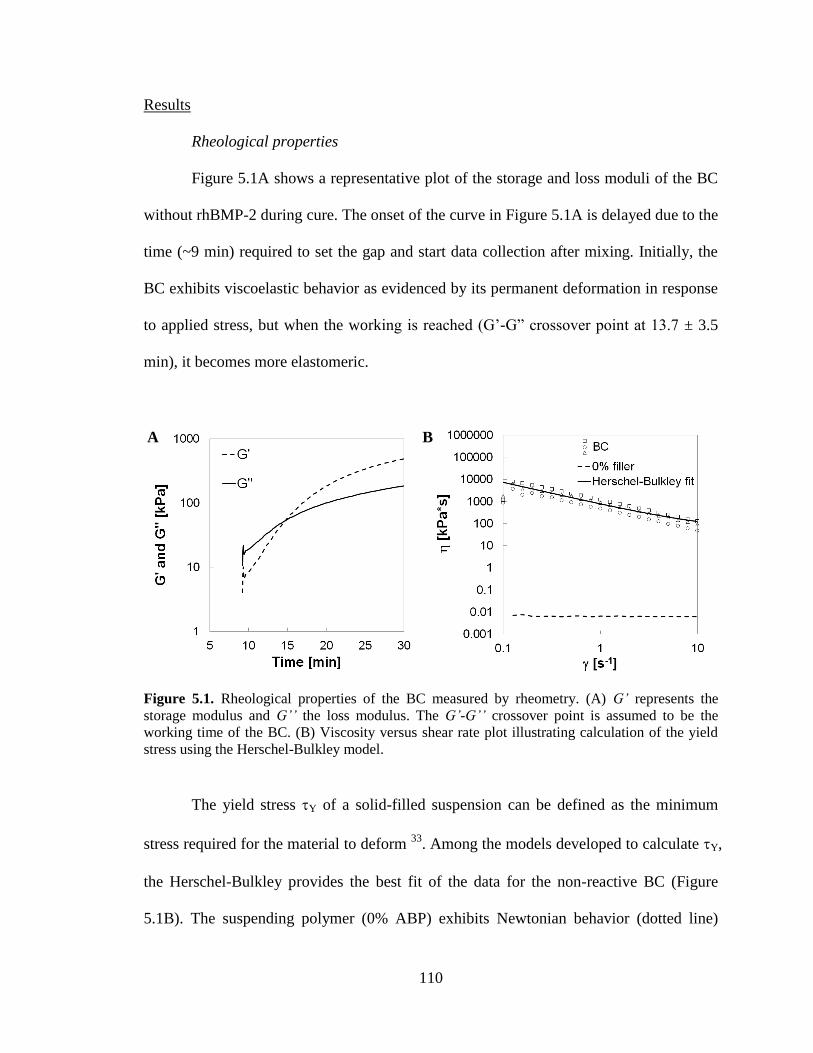
110
Results
Rheological properties
Figure 5.1A shows a representative plot of the storage and loss moduli of the BC
without rhBMP-2 during cure. The onset of the curve in Figure 5.1A is delayed due to the
time (~9 min) required to set the gap and start data collection after mixing. Initially, the
BC exhibits viscoelastic behavior as evidenced by its permanent deformation in response
to applied stress, but when the working is reached (G’-G” crossover point at 13.7 ± 3.5
min), it becomes more elastomeric.
Figure 5.1. Rheological properties of the BC measured by rheometry. (A) G’ represents the
storage modulus and G’’ the loss modulus. The G’-G’’ crossover point is assumed to be the
working time of the BC. (B) Viscosity versus shear rate plot illustrating calculation of the yield
stress using the Herschel-Bulkley model.
The yield stress Y of a solid-filled suspension can be defined as the minimum
stress required for the material to deform 33
. Among the models developed to calculate Y,
the Herschel-Bulkley provides the best fit of the data for the non-reactive BC (Figure
5.1B). The suspending polymer (0% ABP) exhibits Newtonian behavior (dotted line)
A B
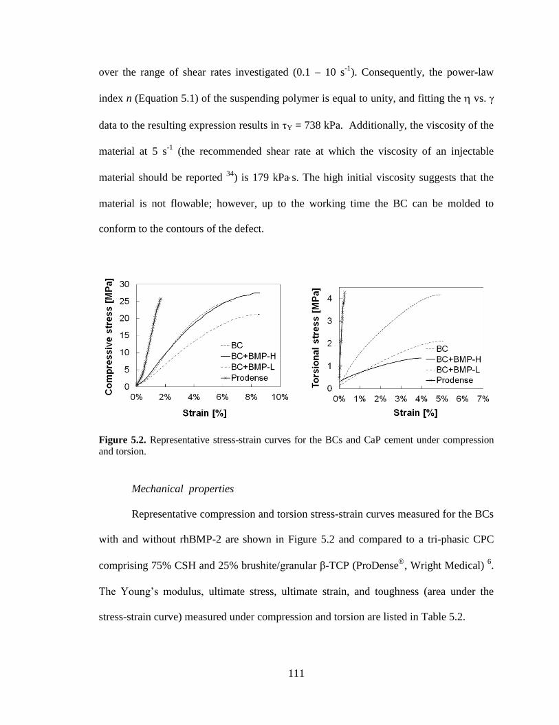
111
over the range of shear rates investigated (0.1 – 10 s-1
). Consequently, the power-law
index n (Equation 5.1) of the suspending polymer is equal to unity, and fitting the vs.
data to the resulting expression results in Y = 738 kPa. Additionally, the viscosity of the
material at 5 s-1
(the recommended shear rate at which the viscosity of an injectable
material should be reported 34
) is 179 kPas. The high initial viscosity suggests that the
material is not flowable; however, up to the working time the BC can be molded to
conform to the contours of the defect.
Figure 5.2. Representative stress-strain curves for the BCs and CaP cement under compression
and torsion.
Mechanical properties
Representative compression and torsion stress-strain curves measured for the BCs
with and without rhBMP-2 are shown in Figure 5.2 and compared to a tri-phasic CPC
comprising 75% CSH and 25% brushite/granular β-TCP (ProDense
, Wright Medical) 6.
The Young’s modulus, ultimate stress, ultimate strain, and toughness (area under the
stress-strain curve) measured under compression and torsion are listed in Table 5.2.
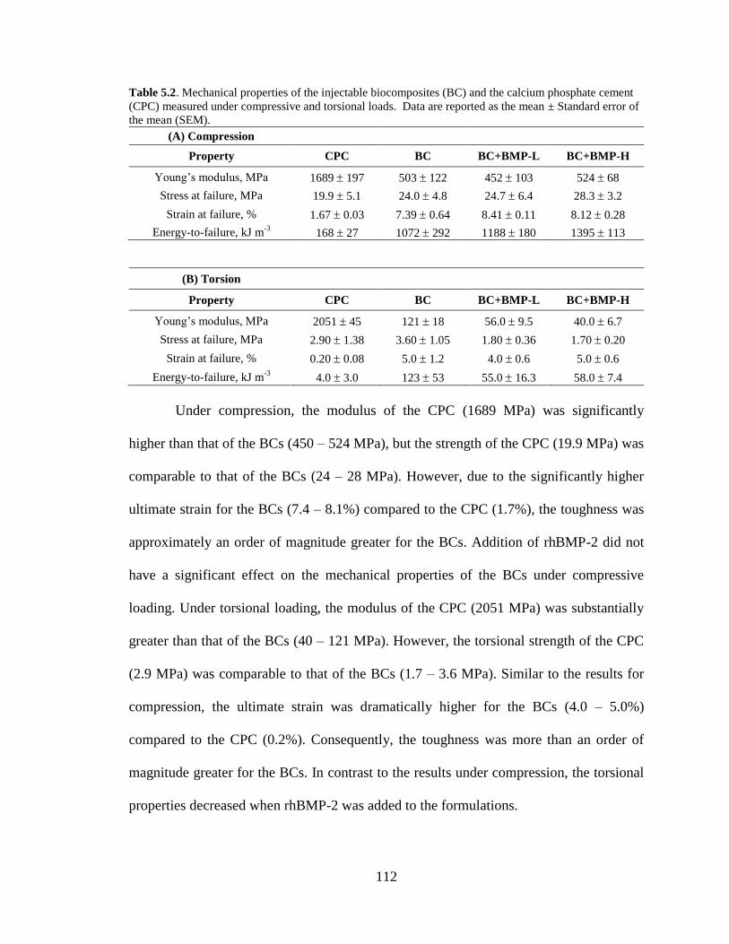
112
Table 5.2. Mechanical properties of the injectable biocomposites (BC) and the calcium phosphate cement
(CPC) measured under compressive and torsional loads. Data are reported as the mean ± Standard error of
the mean (SEM).
(A) Compression
Property CPC BC BC+BMP-L BC+BMP-H
Young’s modulus, MPa 1689 197 503 122 452 103 524 68
Stress at failure, MPa 19.9 5.1 24.0 4.8 24.7 6.4 28.3 3.2
Strain at failure, % 1.67 0.03 7.39 0.64 8.41 0.11 8.12 0.28
Energy-to-failure, kJ m-3
168 27 1072 292 1188 180 1395 113
(B) Torsion
Property CPC BC BC+BMP-L BC+BMP-H
Young’s modulus, MPa 2051 45 121 18 56.0 9.5 40.0 6.7
Stress at failure, MPa 2.90 1.38 3.60 1.05 1.80 0.36 1.70 0.20
Strain at failure, % 0.20 0.08 5.0 1.2 4.0 0.6 5.0 0.6
Energy-to-failure, kJ m-3
4.0 3.0 123 53 55.0 16.3 58.0 7.4
Under compression, the modulus of the CPC (1689 MPa) was significantly
higher than that of the BCs (450 – 524 MPa), but the strength of the CPC (19.9 MPa) was
comparable to that of the BCs (24 – 28 MPa). However, due to the significantly higher
ultimate strain for the BCs (7.4 – 8.1%) compared to the CPC (1.7%), the toughness was
approximately an order of magnitude greater for the BCs. Addition of rhBMP-2 did not
have a significant effect on the mechanical properties of the BCs under compressive
loading. Under torsional loading, the modulus of the CPC (2051 MPa) was substantially
greater than that of the BCs (40 – 121 MPa). However, the torsional strength of the CPC
(2.9 MPa) was comparable to that of the BCs (1.7 – 3.6 MPa). Similar to the results for
compression, the ultimate strain was dramatically higher for the BCs (4.0 – 5.0%)
compared to the CPC (0.2%). Consequently, the toughness was more than an order of
magnitude greater for the BCs. In contrast to the results under compression, the torsional
properties decreased when rhBMP-2 was added to the formulations.
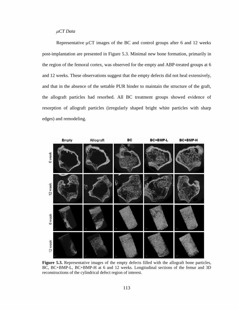
113
CT Data
Representative CT images of the BC and control groups after 6 and 12 weeks
post-implantation are presented in Figure 5.3. Minimal new bone formation, primarily in
the region of the femoral cortex, was observed for the empty and ABP-treated groups at 6
and 12 weeks. These observations suggest that the empty defects did not heal extensively,
and that in the absence of the settable PUR binder to maintain the structure of the graft,
the allograft particles had resorbed. All BC treatment groups showed evidence of
resorption of allograft particles (irregularly shaped bright white particles with sharp
edges) and remodeling.
Figure 5.3. Representative images of the empty defects filled with the allograft bone particles,
BC, BC+BMP-L, BC+BMP-H at 6 and 12 weeks. Longitudinal sections of the femur and 3D
reconstructions of the cylindrical defect region of interest.
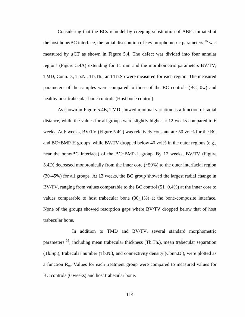
114
Considering that the BCs remodel by creeping substitution of ABPs initiated at
the host bone/BC interface, the radial distribution of key morphometric parameters 35
was
measured by CT as shown in Figure 5.4. The defect was divided into four annular
regions (Figure 5.4A) extending for 11 mm and the morphometric parameters BV/TV,
TMD, Conn.D., Tb.N., Tb.Th., and Tb.Sp were measured for each region. The measured
parameters of the samples were compared to those of the BC controls (BC, 0w) and
healthy host trabecular bone controls (Host bone control).
As shown in Figure 5.4B, TMD showed minimal variation as a function of radial
distance, while the values for all groups were slightly higher at 12 weeks compared to 6
weeks. At 6 weeks, BV/TV (Figure 5.4C) was relatively constant at ~50 vol% for the BC
and BC+BMP-H groups, while BV/TV dropped below 40 vol% in the outer regions (e.g.,
near the bone/BC interface) of the BC+BMP-L group. By 12 weeks, BV/TV (Figure
5.4D) decreased monotonically from the inner core (~50%) to the outer interfacial region
(30-45%) for all groups. At 12 weeks, the BC group showed the largest radial change in
BV/TV, ranging from values comparable to the BC control (51+0.4%) at the inner core to
values comparable to host trabecular bone (30+1%) at the bone-composite interface.
None of the groups showed resorption gaps where BV/TV dropped below that of host
trabecular bone.
In addition to TMD and BV/TV, several standard morphometric
parameters 35
, including mean trabecular thickness (Tb.Th.), mean trabecular separation
(Tb.Sp.), trabecular number (Tb.N.), and connectivity density (Conn.D.), were plotted as
a function Rm. Values for each treatment group were compared to measured values for
BC controls (0 weeks) and host trabecular bone.
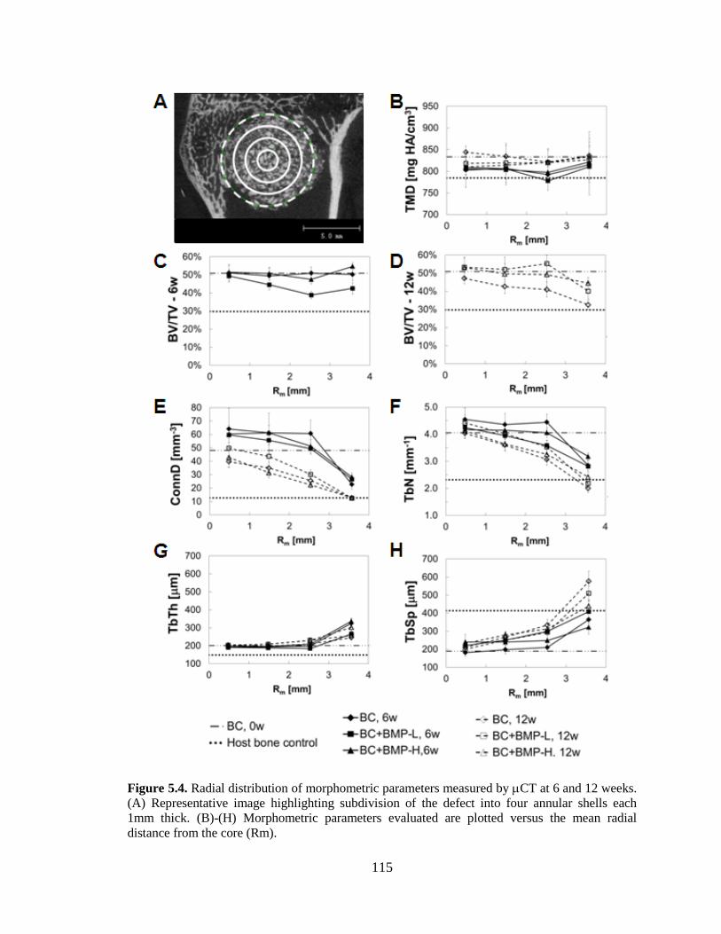
115
Figure 5.4. Radial distribution of morphometric parameters measured by CT at 6 and 12 weeks.
(A) Representative image highlighting subdivision of the defect into four annular shells each
1mm thick. (B)-(H) Morphometric parameters evaluated are plotted versus the mean radial
distance from the core (Rm).
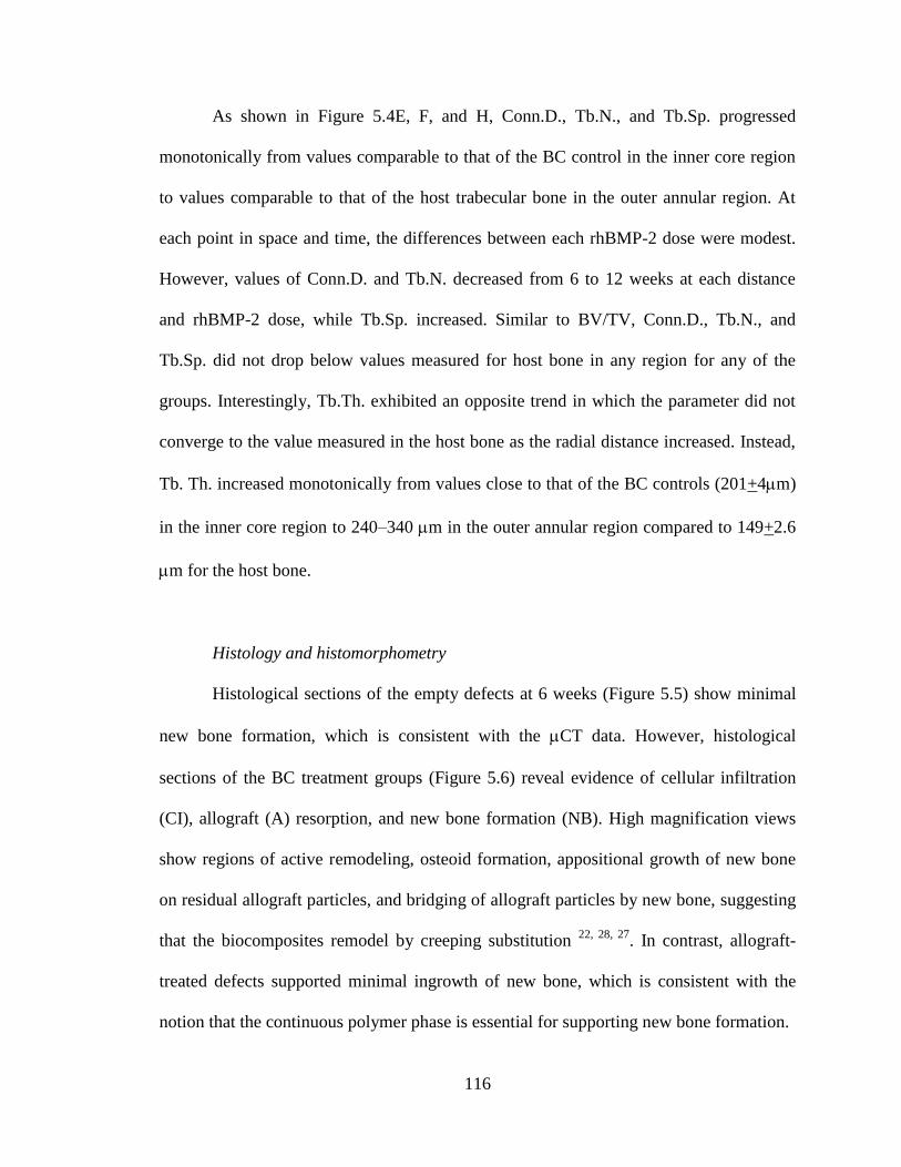
116
As shown in Figure 5.4E, F, and H, Conn.D., Tb.N., and Tb.Sp. progressed
monotonically from values comparable to that of the BC control in the inner core region
to values comparable to that of the host trabecular bone in the outer annular region. At
each point in space and time, the differences between each rhBMP-2 dose were modest.
However, values of Conn.D. and Tb.N. decreased from 6 to 12 weeks at each distance
and rhBMP-2 dose, while Tb.Sp. increased. Similar to BV/TV, Conn.D., Tb.N., and
Tb.Sp. did not drop below values measured for host bone in any region for any of the
groups. Interestingly, Tb.Th. exhibited an opposite trend in which the parameter did not
converge to the value measured in the host bone as the radial distance increased. Instead,
Tb. Th. increased monotonically from values close to that of the BC controls (201+4m)
in the inner core region to 240–340 m in the outer annular region compared to 149+2.6
m for the host bone.
Histology and histomorphometry
Histological sections of the empty defects at 6 weeks (Figure 5.5) show minimal
new bone formation, which is consistent with the CT data. However, histological
sections of the BC treatment groups (Figure 5.6) reveal evidence of cellular infiltration
(CI), allograft (A) resorption, and new bone formation (NB). High magnification views
show regions of active remodeling, osteoid formation, appositional growth of new bone
on residual allograft particles, and bridging of allograft particles by new bone, suggesting
that the biocomposites remodel by creeping substitution 22, 28, 27
. In contrast, allograft-
treated defects supported minimal ingrowth of new bone, which is consistent with the
notion that the continuous polymer phase is essential for supporting new bone formation.
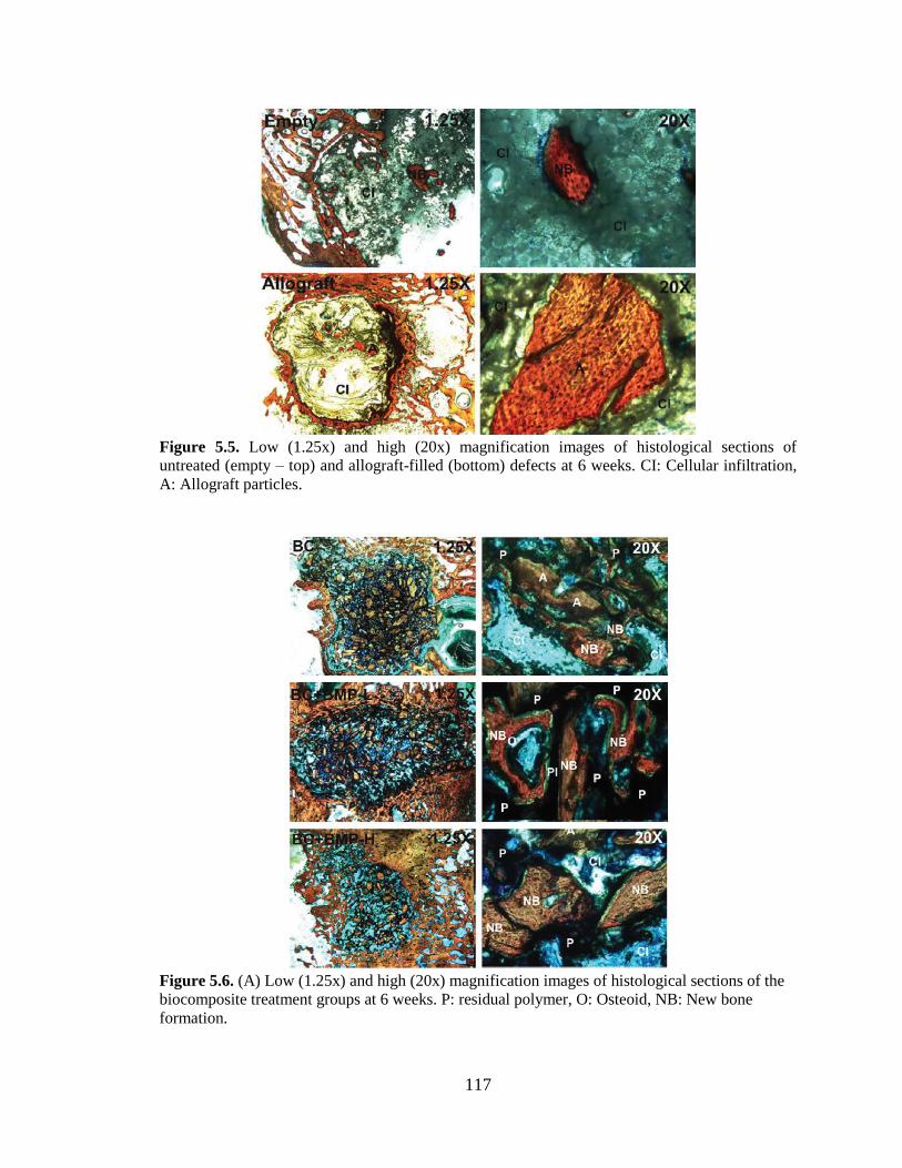
117
Figure 5.5. Low (1.25x) and high (20x) magnification images of histological sections of
untreated (empty – top) and allograft-filled (bottom) defects at 6 weeks. CI: Cellular infiltration,
A: Allograft particles.
Figure 5.6. (A) Low (1.25x) and high (20x) magnification images of histological sections of the
biocomposite treatment groups at 6 weeks. P: residual polymer, O: Osteoid, NB: New bone
formation.
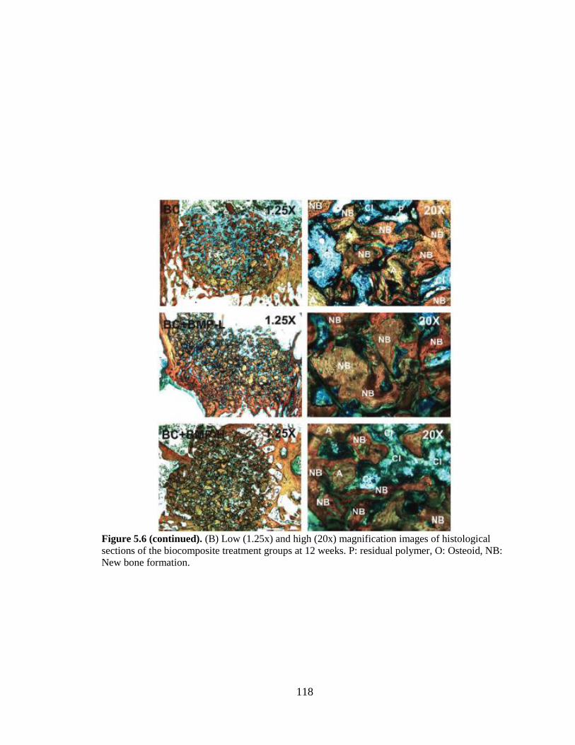
118
Figure 5.6 (continued). (B) Low (1.25x) and high (20x) magnification images of histological
sections of the biocomposite treatment groups at 12 weeks. P: residual polymer, O: Osteoid, NB:
New bone formation.
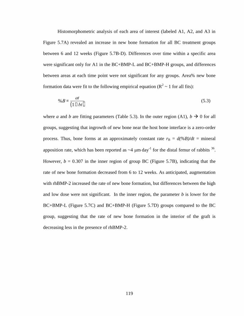
119
Histomorphometric analysis of each area of interest (labeled A1, A2, and A3 in
Figure 5.7A) revealed an increase in new bone formation for all BC treatment groups
between 6 and 12 weeks (Figure 5.7B-D). Differences over time within a specific area
were significant only for A1 in the BC+BMP-L and BC+BMP-H groups, and differences
between areas at each time point were not significant for any groups. Area% new bone
formation data were fit to the following empirical equation (R2 ~ 1 for all fits):
%B =at
1+ bt( ) (5.3)
where a and b are fitting parameters (Table 5.3). In the outer region (A1), b 0 for all
groups, suggesting that ingrowth of new bone near the host bone interface is a zero-order
process. Thus, bone forms at an approximately constant rate rB = d(%B)/dt = mineral
apposition rate, which has been reported as ~4 μmday-1
for the distal femur of rabbits 36
.
However, b = 0.307 in the inner region of group BC (Figure 5.7B), indicating that the
rate of new bone formation decreased from 6 to 12 weeks. As anticipated, augmentation
with rhBMP-2 increased the rate of new bone formation, but differences between the high
and low dose were not significant. In the inner region, the parameter b is lower for the
BC+BMP-L (Figure 5.7C) and BC+BMP-H (Figure 5.7D) groups compared to the BC
group, suggesting that the rate of new bone formation in the interior of the graft is
decreasing less in the presence of rhBMP-2.
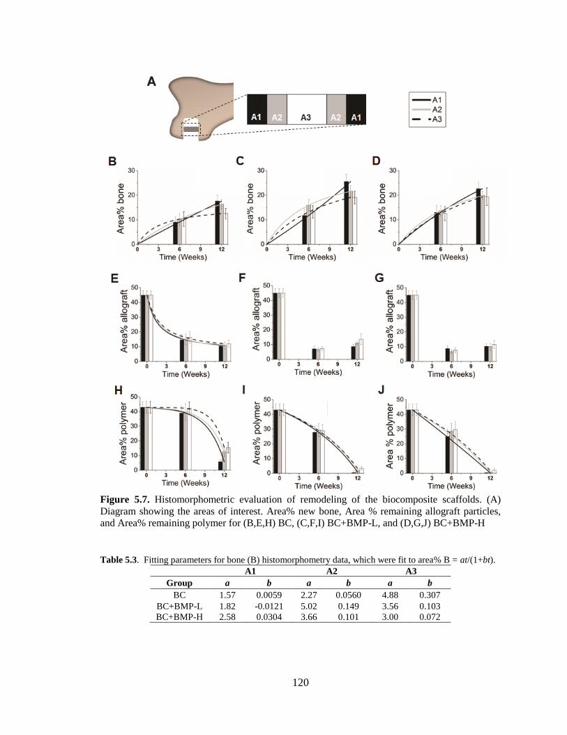
120
Figure 5.7. Histomorphometric evaluation of remodeling of the biocomposite scaffolds. (A)
Diagram showing the areas of interest. Area% new bone, Area % remaining allograft particles,
and Area% remaining polymer for (B,E,H) BC, (C,F,I) BC+BMP-L, and (D,G,J) BC+BMP-H
Table 5.3. Fitting parameters for bone (B) histomorphometry data, which were fit to area% B = at/(1+bt).
A1 A2 A3
Group a b a b a b
BC 1.57 0.0059 2.27 0.0560 4.88 0.307
BC+BMP-L 1.82 -0.0121 5.02 0.149 3.56 0.103
BC+BMP-H 2.58 0.0304 3.66 0.101 3.00 0.072
121
Histomorphometry data for residual allograft are presented for each area in Figure
5.7E-G. Area% allograft (A) data for the BC group (Figure 5.7E) were fit to the
following equation:
(5.4)
where AA,i is the initial area% allograft in the sample at t = 0. Values of the fitting
parameters a and b for each group are listed in Table 5.4. As anticipated, for the BC
group the area% allograft decreased with time due to osteoclast-mediated resorption 27, 22,
28, but the differences between 6 and 12 weeks were not significant for any group. The
half-life varied from 2.3 – 2.9 weeks. In the presence of rhBMP-2, the area% residual
allograft was significantly less than in the BC group at 6 weeks, suggesting that rhBMP-2
accelerated allograft resorption at early time points. Interestingly, no significant changes
in allograft area% were observed in the rhBMP-2 treatment groups from 6 to 12 weeks.
Due to the transient resorption of allograft at 6 weeks, it was not possible to fit the area%
allograft data to eq (5.4) in the presence of rhBMP-2.
Table 5.4. Fitting parameters for allograft (A) histomorphometry data, which were fit to area% A = AA,i -
at/(1+bt) for the BC group. The parameter AA,i is the initial area% allograft measured by histomorphometry.
Data for BC+BMP-L and BC+BMP-H groups could not be accurately fit to the exponential or rational
functions.
A1 A2 A3
Group a b t1/2 (wk) a b t1/2 (wk) a b t1/2 (wk)
BC 22.8 0.583 2.3 0.318 0.380 2.6 0.023 0.590 2.9
Polymer degradation was affected by both time and rhBMP-2 dose (Figure 5.7H-
J). Differences between 6 and 12 weeks were significant within a specific area for all
122
groups, but differences between areas were not significant. Area% polymer (P) data for
the BC group (Figure 5.7H) were fit to the following equation:
(5.5)
where AP,i is the initial area% polymer in the sample at t = 0. The half-life of the polymer
ranged from 10.5 – 11.5 weeks, which is substantially longer than that of the allograft but
slightly shorter than t1/2 = 14 weeks reported in vitro 37
. Similar to observations from in
vitro studies, the degradation profile of the poly(ester urethane) is consistent with an
autocatalytic mechanism 37
and does not follow the first-order kinetics associated with
ester hydrolysis 38
. Local delivery of rhBMP-2 (Figure 5.7I-J) changed the shape of the
area% polymer curve, which was fit to an equation similar to the one used for new bone:
(5.6)
Table 5.5. Fitting parameters for polymer (P) histomorphometry data, which were fit to area% P = AP,i -
at/(1+bt) for BC+BMP-L and BC+BMP-H groups and to area% P = AP,i – αexp(βt) for the BC group. The
parameter AP,i is the initial area% polymer measured by histomorphometry.
A1 A2 A3
Group a (α) b (β) t1/2
(wk) a (α) b (β)
t1/2
(wk) a (α) b (β)
t1/2
(wk)
BC 0.408 0.376 10.5 0.318 0.380 11.0 0.023 0.590 11.5
BC+BMP-L 1.98 -0.0365 6.4 1.69 -0.0425 8.3 1.83 -0.0373 8.2
BC+BMP-H 3.30 -0.0062 6.3 2.44 -0.0258 7.2 2.24 -0.0292 7.5
As evidenced by the values of b 0 (Table 5.5), the process of PUR degradation
in the presence of rhBMP-2 is nearly zero-order (i.e., constant rate). Augmentation with
rhBMP-2 also accelerated PUR degradation, as evidenced by t1/2 = 6.4 – 8.3 weeks for
BC+BMP-L and t1/2 = 6.3 – 7.5 weeks for BC+BMP-H. Differences in residual polymer
were significant between the BC group and the BC+BMP-L and –H groups at 6 weeks,
123
but not at 12 weeks. Thus, rhBMP2 both altered the mechanism of PUR degradation and
also increased the degradation rate.
Discussion
Weight-bearing bone grafts have generated considerable interest in recent years,
but the mechanisms by which they remodel, while maintaining their initial bone-like
strength are poorly understood. In this study, we investigated the effects of the rate of
new bone formation, the rate of reinforcing matrix (allograft) resorption, and the
mechanism of polymer degradation on healing of ABP/PUR composites in femoral
condyle plug defects in rabbits. The initial compressive strength (24–28 MPa) and
toughness (1070–1400 kJm-3
) of the composites were comparable to or exceeding those
of trabecular bone (6 MPa and 1050 kJmm-3
, respectively 39
). When injected into femoral
condyle plug defects in NZW rabbits, new bone formation and healing progressed in both
space and time for up to 12 weeks. Polymer degradation was autocatalytic and new bone
formation was approximately zero-order in the absence of rhBMP-2, resulting in
incomplete healing in the inner core of the graft at later time points. In contrast, local
delivery of rhBMP-2 increased the rate of new bone formation, and promoted
approximately zero-order degradation of the continuous polymer phase, resulting in
improved healing at later time points. Thus, balancing the rates of new bone formation
and polymer degradation was observed to improve healing.
The settable composites exhibited a working time of 13.7 ± 3.5 min, which is
longer than the value of 4.5 min reported for an injectable, flowable bone void filler (36
vol% ABP 27
) and the working times of 6–10 min reported for CPCs 40, 41
. A proposed
124
criterion for injectability of CPCs dictates that the material can be administered from a
syringe by applying a force < 100 N 6. Consequently, a syringe with diameter >13-mm
would be required to inject the BC due to its high yield stress (738 kPa). Similarly, the
viscosity of the BC at 5 s-1
is substantially higher (18×103 Pas) than that of other
injectable bone grafts, such the flowable ABP void filler (170 Pas 27
, 35 vol% ABP) or
flowable CPCs (5 – 10 Pas, 35 – 39 vol% solids 34
). Thus, while the BC has higher initial
viscosity and yield stress compared to other BVFs due to its high solids content (57
vol%), it is moldable and settable within a clinically relevant time frame, which enables
the surgeon to conform the graft to the contours of the bone defect 42
.
The initial compressive strength (24.0 4.8 MPa) and modulus (503 122 MPa)
of the BCs exceed values reported for trabecular bone, which range from 3.7–8.4 MPa
and 104–300 MPa, respectively 43, 44, 39, 45
. Similarly, the ultimate strain of the BCs
(7.30.6%) exceeds that of trabecular bone in human femora (1.05±0.46%) 46
), while the
toughness (1072292 kJm-3
) is comparable to that of trabecular bone (1000 kJm-3
39
).
Under torsional deformation, the strength and modulus of the BCs are 2.9±1.4 MPa and
121±18 MPa, respectively, which are close to values reported for human 31, 47
, bovine 48,
49, canine
43, and ovine
50, 51 trabecular bone ranging from 3.1 to 7.7MPa and 263 to
366MPa, respectively. However, the ultimate strain of the BC (5.0±1.2%) is comparable
to a previous study reporting strain at failure of 4.6±1.3% for trabecular bone 31
. Taken
together, these observations suggest that the initial mechanical properties of the BCs are
comparable to those of trabecular bone under compression and slightly weaker under
torsion.
125
Recent studies have reported that the poor mechanical properties of CPCs under
the physiologically relevant conditions of cyclic loading with a significant shear
component 52
limit their use to non-load-bearing applications 19, 6
. The weak shear and
torsional properties of these materials have been attributed to their inherent brittleness;
however, there are limited toughness and fatigue data. Therefore, we compared the
toughness of the BCs to an injectable tri-phasic CPC that supports bone healing in both
preclinical models 14
and patients 53
. The toughness of the CPC was ~6 times less than
that of the BC and trabecular bone under compression, while under torsion the toughness
of the CPC was ~30 times less than that of the BC. The reduced toughness of the CPC is
attributed in part to its lower ultimate strain compared to the BC and trabecular bone
under compression (1.7%) and torsion (0.2%). While the area under the stress-strain
curve is a convenient estimate of toughness, it is not a true material property 19
, and thus
the fatigue properties, fracture toughness, and/or other measures of reliability (such as the
Weibull modulus) must be characterized to assess the potential of the BC to promote
healing in weight-bearing bone defects.
While incorporation of calcium phosphate has been reported to increase
osteoconductivity for some polymers 54
, a recent review has reported that in most studies
it does not increase the strength of polymer/ceramic composites 19
. As an exception to
this trend, HA/poly(lactic acid) (HA/PLLA) composites incorporating negligible porosity
and ~20 vol% HA particles exhibited bending strengths up to 280 MPa. However, since
bridging between the ceramic particles and host bone was dependent upon the
degradation of PLLA 55
, the rate of remodeling was slow, as evidenced by the fact that
4% of the composite implanted in rabbit femora remained after 7 years 56
. Furthermore,
126
the resorption zone near the host bone interface resulting from phagocytosis of the
implant by histocytes 56
is anticipated to reduce its strength. Compression-molded
ABP/PUR BCs incorporating 68 vol% ABP exhibited compressive strengths ranging
from 100 to 170 MPa and supported extensive cellular infiltration at 6 weeks in rabbit
femoral condyle defects 22
. However, only modest new bone formation (~2%) was
observed at 6 weeks. The grafts remodeled by creeping substitution, a process
characterized by resorption of allograft particles by osteoclasts 27
followed by infiltration
of osteoblasts into the newly formed pore 27, 22, 28
. Subsequent new bone formation was
observed on the surfaces of both the polymer and also the residual allograft particles.
However, as the polymer degraded the grafts filled with fibrous tissue due to the lack of a
continuous surface for new bone growth. In the present study, the ABP control group
with no polymer exhibited extensive resorption of allograft particles and minimal new
bone formation away from the cortex. These observations underscore the need for a
continuous polymer phase to persist throughout the interior of the BC until the defect has
almost fully healed.
To overcome the limitations of previous studies, settable ABP/PUR composites
were designed to incorporate 43% polymer, which formed a continuous phase. New
bone formation in the outer region of all grafts progressed at a nearly constant rate
(Figure 5.7B-D)). However, rB = d(%B)/dt in the inner regions decreased with time,
suggesting that mass-transfer limitations associated with infiltration of cells from the host
bone into the interior of the scaffold slowed bone formation. Recently, a diffusion model
for bone formation in osteoconductive scaffolds has been proposed 57
:
(5.7)
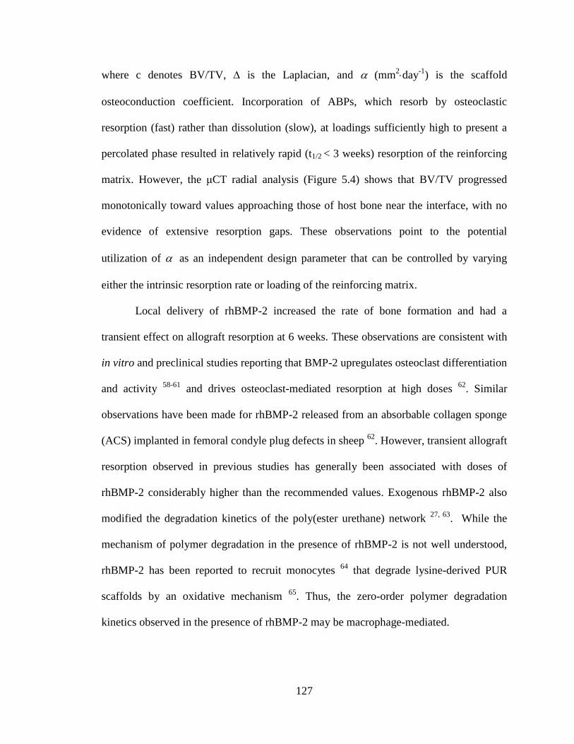
127
where c denotes BV/TV, is the Laplacian, and (mm2day
-1) is the scaffold
osteoconduction coefficient. Incorporation of ABPs, which resorb by osteoclastic
resorption (fast) rather than dissolution (slow), at loadings sufficiently high to present a
percolated phase resulted in relatively rapid (t1/2 < 3 weeks) resorption of the reinforcing
matrix. However, the μCT radial analysis (Figure 5.4) shows that BV/TV progressed
monotonically toward values approaching those of host bone near the interface, with no
evidence of extensive resorption gaps. These observations point to the potential
utilization of as an independent design parameter that can be controlled by varying
either the intrinsic resorption rate or loading of the reinforcing matrix.
Local delivery of rhBMP-2 increased the rate of bone formation and had a
transient effect on allograft resorption at 6 weeks. These observations are consistent with
in vitro and preclinical studies reporting that BMP-2 upregulates osteoclast differentiation
and activity 58-61
and drives osteoclast-mediated resorption at high doses 62
. Similar
observations have been made for rhBMP-2 released from an absorbable collagen sponge
(ACS) implanted in femoral condyle plug defects in sheep 62
. However, transient allograft
resorption observed in previous studies has generally been associated with doses of
rhBMP-2 considerably higher than the recommended values. Exogenous rhBMP-2 also
modified the degradation kinetics of the poly(ester urethane) network 27, 63
. While the
mechanism of polymer degradation in the presence of rhBMP-2 is not well understood,
rhBMP-2 has been reported to recruit monocytes 64
that degrade lysine-derived PUR
scaffolds by an oxidative mechanism 65
. Thus, the zero-order polymer degradation
kinetics observed in the presence of rhBMP-2 may be macrophage-mediated.
128
Figure 5.8. Analysis of histomorphometric data. The ratio of the rate of new bone formation (rB)
to that of polymer degradation (rP) was calculated for each group by differentiating the equations
expressing area% new bone and area% polymer as a function of time. Representative images of
histological sections with highlighted areas of interest. (A-B) BC, (C-D) BC+BMP-L, and (E-F)
BC+BMP-H
The histomorphometry data were further analyzed in Figure 5.8 to gain insight
into the design criteria for weight-bearing bone grafts. The rate of new bone formation rB
relative to the rate of polymer degradation (rP = -d(area% P)/dt) is plotted versus the
parameter fH, which approximates the fractional healing of the defect:
fH =area% bone in defect
area% bone in host bone (5.8)
The area% bone in the host bone was measured by histomorphometry to be
39.3+1.9%. For the BC at early stages of healing, rB/rP >>1 but it decreased dramatically
with time (Figure 5.8A). A representative histological section (Figure 5.8B) shows poor
surface connectivity at 12 weeks, characterized by isolated islands of polymer and
allograft. The consequences of mismatched rates of new bone formation and polymer
degradation are particularly acute in the inner region, where rB/rP 0 at fH = 32% healing,
at which time the surface is likely insufficiently interconnected to continue to support
cellular infiltration and consequent new bone formation. Thus, autocatalytic degradation
129
of the PUR network driven by carboxylic acid groups formed after hydrolysis of ester
bonds does not match the zero-order kinetics of new bone formation 66
, resulting in poor
healing in the inner region.
In the presence of exogenous rhBMP-2, the rates of new bone formation and
polymer degradation were more balanced. Although rB/rP monotonically decreased with
time in the presence of rhBMP-2 (Figure 5.8C,E), the effect was substantially weaker
than in the BC group, and in the outer region rB/rP was nearly constant. The relative rate
rB/rP approached values <0.2 in the inner regions, but by this time (12 weeks) healing had
progressed to fH>50%. The representative histological sections from the BC+BMP-L
(Figure 5.8D) and BC+BMP-H (Figure 5.8F) groups show a more connected surface in
the inner regions (A2 and A3) for continued bone growth at 12 weeks. Taken together,
the data in Figure 5.8 suggest that to optimize bone healing, the polymer must persist
until a sufficient amount of new bone has formed to provide a continuous surface, and
that the rate of degradation of the continuous polymeric phase should be matched to the
rate of new bone formation. While a polyester with a longer half-life 37
is anticipated to
delay the onset of rapid polymer degradation until later stages of healing, autocatalytic
degradation of these materials could result in unpredictable outcomes. In contrast,
polymers that degrade at a constant rate, for example by enzymatic degradation, are
anticipated to more closely match the rate of new bone formation 26
.
Settable weight-bearing bone grafts could potentially transform the management
of tibial plateau fractures, screw augmentation, vertebroplasty, and other orthopaedic
indications, but their success is predicated on the ability to maintain sufficient bone-like
strength while actively remodeling. Weight-bearing composites generally comprise a
130
resorbable reinforcing matrix (e.g., short fibers 20, 21
or particles 22, 56
) dispersed in a
continuous phase (e.g., polymers or ceramics). A recent review highlighting the
challenges associated with the design of weight-bearing composites has suggested that
the rates of degradation of the reinforcing matrix and the continuous phase should be
matched to ensure adequate healing 26
. In this study, we found that early resorption of the
reinforcing matrix does not affect healing at later time points, but the continuous phase
must persist until sufficient new bone has formed. Hydrolytically degradable polymers
undergo autocatalytic degradation, resulting in poor healing in the interior of the graft due
to transport limitations associated with ingrowth of new bone 57
. Closely balancing the
rates of new bone formation and polymer degradation, accomplished by local delivery of
rhBMP-2 in this study, allowed healing to progress to later stages (up to fH=70%). A
limitation of this study is that none of the areas healed >70% over the 12-week time
course. However, our findings point to the importance of both maintaining a continuous
surface for bone healing as well as balancing the rates of new bone formation and
continuous phase degradation as a strategy for maintaining sufficient bone-like strength
over the lifetime of the graft. Polymers that undergo cell-mediated degradation 26
are
anticipated to promote balanced remodeling at all stages of healing, which should be
assessed by long-term studies.
Conclusions
Moldable, settable ABP/PUR biocomposites exhibited initial mechanical
properties comparable to those of trabecular bone. When implanted in plug defects in
rabbit femoral condyles, morphometric analysis of both histological sections and CT
131
reconstructions revealed that new bone formation and healing progressed in both space
and time from the outer region near the host bone interface to the inner core for up to 12
weeks. However, the unbalanced rates of autocatalytic polymer degradation and zero-
order new bone formation resulted in incomplete healing in the interior of the composite.
Augmentation of the composites with rhBMP-2 increased the rate of new bone formation
and induced approximately zero-order degradation of the polymer. The consequent
balance of new bone formation and polymer degradation resulted in more extensive
healing at later time points in all regions of the graft, including the interior. These
observations underscore the importance of both maintaining a continuous polymer
surface for new bone growth, as well as matching the rate of new bone formation to that
of polymer degradation to promote healing of settable weight-bearing bone grafts that
maintain bone-like strength while actively remodeling.
132
References
1 D. D. Anderson, C. Van Hofwegen, J. L. Marsh, and T. D. Brown, 'Is Elevated Contact Stress
Predictive of Post-Traumatic Osteoarthritis for Imprecisely Reduced Tibial Plafond Fractures?', J
Orthop Res, 29 (2011), 33-9.
2 T. A. Russell, and R. K. Leighton, 'Comparison of Autogenous Bone Graft and Endothermic
Calcium Phosphate Cement for Defect Augmentation in Tibial Plateau Fractures. A Multicenter,
Prospective, Randomized Study', J Bone Joint Surg Am, 90 (2008), 2057-61.
3 D. Simpson, and J. F. Keating, 'Outcome of Tibial Plateau Fractures Managed with Calcium
Phosphate Cement', Injury, 35 (2004), 913-8.
4 J. A. Hall, M. J. Beuerlein, and M. D. McKee, 'Open Reduction and Internal Fixation Compared
with Circular Fixator Application for Bicondylar Tibial Plateau Fractures. Surgical Technique', J
Bone Joint Surg Am, 91 Suppl 2 Pt 1 (2009), 74-88.
5 Z. Schwartz, M. Goldstein, E. Raviv, A. Hirsch, D. M. Ranly, and B. D. Boyan, 'Clinical
Evaluation of Demineralized Bone Allograft in a Hyaluronic Acid Carrier for Sinus Lift
Augmentation in Humans: A Computed Tomography and Histomorphometric Study', Clin Oral
Implants Res, 18 (2007), 204-11.
6 M. Bohner, 'Design of Ceramic-Based Cements and Putties for Bone Graft Substitution', Eur Cell
Mater, 20 (2010), 1-12.
7 M. Chazono, T. Tanaka, H. Komaki, and K. Fujii, 'Bone Formation and Bioresorption after
Implantation of Injectable Beta-Tricalcium Phosphate Granules-Hyaluronate Complex in Rabbit
Bone Defects', J Biomed Mater Res A, 70 (2004), 542-9.
8 F. P. Cammisa, Jr., G. Lowery, S. R. Garfin, F. H. Geisler, P. M. Klara, R. A. McGuire, W. R.
Sassard, H. Stubbs, and J. E. Block, 'Two-Year Fusion Rate Equivalency between Grafton Dbm
Gel and Autograft in Posterolateral Spine Fusion: A Prospective Controlled Trial Employing a
Side-by-Side Comparison in the Same Patient', Spine (Phila Pa 1976), 29 (2004), 660-6.
9 C. Chan, I. Thompson, P. Robinson, J. Wilson, and L. Hench, 'Evaluation of Bioglass/Dextran
Composite as a Bone Graft Substitute', Int J Oral Maxillofac Surg, 31 (2002), 73-7.
10 C. D. Friedman, P. D. Constantino, S. Takagi, and L. C Chow, 'Bonesource™ Hydroxyapatite
Cement: A Novel Biomaterial for Craniofacial Skeletal Yissue Engineering and Reconstruction', J
Biomed Mater Res, 43 (1998), 428-32.
11 H. Chim, and A. K. Gosain, ' Biomaterials in Craniofacial Surgery Experimental Studies and
Clinical Application ', J Craniofac Surg, 20 (2009), 29-33.
12 G. Gasparini, R. Boniello, A. Moro, G. Tamburrini, C. Di Rocco, and S. Pelo, 'Cranial Reshaping
Using Methyl Methacrylate:Technical Note', J Craniofac Surg, 20 (2009), 184-90.
13 A. Moreira-Gonzalez, I. T. Jackson, T. Miyawaki, K. Barakat, and V. DiNick, 'Clinical Outcome
in Cranioplasty: Critical Review in Long-Term Follow-Up', J Craniofac Surg, 14 (2003), 144-53.
14 R. M. Urban, T. M. Turner, D. J. Hall, N. Inoue, and S. Gitelis, 'Increased Bone Formation Using
Calcium Sulfate-Calcium Phosphate Composite Graft', Clin Orthop Relat Res, 459 (2007), 110-7.
15 S Gitelis, R. M. Urban, T M Turner, R Heck, and A D Parameswaran, 'Outcomes in the Treatment
of Benign Bone Lesions Using an Engineering Ceramic: Preclinical and Clinical Results', in
Materials and Processes for Medical Devices Conference, ed. by J Gilbert (Minneapolis, MN:
2009).
133
16 M. Ikenaga, P. Hardouin, J. Lemaitre, H. Andrianjatovo, and B. Flautre, 'Biomechanical
Characterization of a Biodegradable Calcium Phosphate Hydraulic Cement: A Comparison with
Porous Biphasic Calcium Phosphate Ceramics', J Biomed Mater Res, 40 (1998), 139-44.
17 R. M. Urban, T. M. Turner, D. J. Hall, S. Infanger, N. Cheema, and T. H. Lim, 'Healing of Large
Defects Treated with Calcium Sulfate Pellets Containing Demineralized Bone Matrix Particles',
Orthopedics, 26 (2003), s581-5.
18 Y. E. Greish, and P. W. Brown, 'Phase Evolution During the Formation of Stoichiometric
Hydroxyapatite at 37.4 Degrees C', J Biomed Mater Res B Appl Biomater, 67 (2003), 632-7.
19 A. J. Wagoner Johnson, and B. A. Herschler, 'A Review of the Mechanical Behavior of Cap and
Cap/Polymer Composites for Applications in Bone Replacement and Repair', Acta Biomater, 7
(2011), 16-30.
20 R. Kruger, and J. Groll, 'Fiber Reinforced Calcium Phosphate Cements -- on the Way to
Degradable Load Bearing Bone Substitutes?', Biomaterials, 33 (2012), 5887-900.
21 R. Kruger, J. M. Seitz, A. Ewald, F. W. Bach, and J. Groll, 'Strong and Tough Magnesium Wire
Reinforced Phosphate Cement Composites for Load-Bearing Bone Replacement', J Mech Behav
Biomed Mater, 20 (2013), 36-44.
22 J E Dumas, T E Davis, T Yoshii, J Nyman, G E Holt, D S Perrien, T M Boyce, and S A Guelcher
'Synthesis of Allograft Bone/Polymer Composites and Evaluation of Remodeling in a Rabbit
Femoral Condyle Model', Acta Biomaterialia, 6 (2010), 2394–406.
23 Q. Lian, D. C. Li, J. K. He, and Z. Wang, 'Mechanical Properties and in-Vivo Performance of
Calcium Phosphate Cement-Chitosan Fibre Composite', Proc Inst Mech Eng H, 222 (2008), 347-
53.
24 J. Kim, S. McBride, M. Fulmer, R. Harten, Z. Garza, D. D. Dean, V. L. Sylvia, B. Doll, T. L.
Wolfgang, E. Gruskin, and J. O. Hollinger, 'Fiber-Reinforced Calcium Phosphate Cement
Formulations for Cranioplasty Applications: A 52-Week Duration Preclinical Rabbit Calvaria
Study', J Biomed Mater Res B Appl Biomater, 100 (2012), 1170-8.
25 Z. Pan, and P. Jiang, 'Assessment of the Suitability of a New Composite as a Bone Defect Filler in
a Rabbit Model', J Tissue Eng Regen Med, 2 (2008), 347-53.
26 J. R. Jones, 'Review of Bioactive Glass: From Hench to Hybrids', Acta Biomater, 9 (2013), 4457-
86.
27 J. E. Dumas, P. B. Brownbaer, E. M. Prieto, T. Guda, R. G. Hale, J. C. Wenke, and S. A.
Guelcher, 'Injectable Reactive Biocomposites for Bone Healing in Critical-Size Rabbit Calvarial
Defects', Biomed Mater, 7 (2012), 024112.
28 J. E. Dumas, K. Zienkiewicz, S. A. Tanner, E. M. Prieto, S. Bhattacharyya, and S. Guelcher,
'Synthesis and Characterization of an Injectable Allograft Bone/Polymer Composite Bone Void
Filler with Tunable Mechanical Properties', Tissue Eng Part A, 16 (2010), 2505-18.
29 D. M. Smith, A. M. Afifi, G. M. Cooper, M. P. Mooney, K. G. Marra, and J. E. Losee, 'Bmp-2-
Based Repair of Large-Scale Calvarial Defects in an Experimental Model: Regenerative Surgery
in Cranioplasty', J Craniofac Surg, 19 (2008), 1315-22.
30 A J Poslinski, Ryan M E, Gupta R K, Seshadri S G, and Frechette F J, 'Rheological Behavior of
Filled Polymeric Systems I. Yield Stress and Shear-Thinning Effects', Journal of Rheology, 32
(1988), 703-35.
134
31 K. Bruyere Garnier, R. Dumas, C. Rumelhart, and M. E. Arlot, 'Mechanical Characterization in
Shear of Human Femoral Cancellous Bone: Torsion and Shear Tests', Med Eng Phys, 21 (1999),
641-9.
32 C. M. Ford, and T. M. Keaveny, 'The Dependence of Shear Failure Properties of Trabecular Bone
on Apparent Density and Trabecular Orientation', J Biomech, 29 (1996), 1309-17.
33 P.J. Carreau, and P. A. Lavoie, 'Rheological Properties of Filled Polymers', Macromolecular
symposia, 108 (1996), 111-26.
34 G. Baroud, E. Cayer, and M. Bohner, 'Rheological Characterization of Concentrated Aqueous
Beta-Tricalcium Phosphate Suspensions: The Effect of Liquid-to-Powder Ratio, Milling Time,
and Additives', Acta Biomater, 1 (2005), 357-63.
35 M. L. Bouxsein, S. K. Boyd, B. A. Christiansen, R. E. Guldberg, K. J. Jepsen, and R. Muller,
'Guidelines for Assessment of Bone Microstructure in Rodents Using Micro-Computed
Tomography', J Bone Miner Res, 25 (2010), 1468-86.
36 A. Cacchioli, F. Ravanetti, L. Soliani, and P. Borghetti, 'Preliminary Study on the Mineral
Apposition Rate in Distal Femoral Epiphysis of New Zealand White Rabbit at Skeletal Maturity',
Anat Histol Embryol, 41 (2012), 163-9.
37 AE Hafeman, B Li, T Yoshii, KL Zienkiewicz, JM Davidson, and SA Guelcher, 'Injectable
Biodegradable Polyurethane Scaffolds with Release of Platelet-Derived Growth Factor for Tissue
Repair and Regeneration', Pharm Res, 25 (2008), 2387-99.
38 T. J. Lehtonen, J. U. Tuominen, and E. Hiekkanen, 'Resorbable Composites with Bioresorbable
Glass Fibers for Load-Bearing Applications. In Vitro Degradation and Degradation Mechanism',
Acta Biomater, 9 (2013), 4868-77.
39 M. R. Allen, H. A. Hogan, W. A. Hobbs, A. S. Koivuniemi, M. C. Koivuniemi, and D. B. Burr,
'Raloxifene Enhances Material-Level Mechanical Properties of Femoral Cortical and Trabecular
Bone', Endocrinology, 148 (2007), 3908-13.
40 G Lewis, 'Percutaneous Vertebroplasty and Kyphoplasty for the Stand-Alone Augmentation of
Osteoporosis-Induced Vertebral Compression Fractures: Present Status and Future Directions', J
Biomed Mater Res Part B : Appl Biomater, 81B (2007), 371-86.
41 O M Clarkin, D Boyd, S Madigan, and M R Towler, 'Comparison of an Experimental Bone
Cement with a Commercial Control, Hydrosettm
', J Mater Sci: Mater Med, 20 (2009), 1563 - 70.
42 J. D. Kretlow, S. Young, L. Klouda, M. Wong, and A. G. Mikos, 'Injectable Biomaterials for
Regenerating Complex Craniofacial Tissues', Adv Mater Deerfield, 21 (2009), 3368-93.
43 S. K. Eswaran, M. R. Allen, D. B. Burr, and T. M. Keaveny, 'A Computational Assessment of the
Independent Contribution of Changes in Canine Trabecular Bone Volume Fraction and
Microarchitecture to Increased Bone Strength with Suppression of Bone Turnover', J Biomech, 40
(2007), 3424-31.
44 E. F. Morgan, and T. M. Keaveny, 'Dependence of Yield Strain of Human Trabecular Bone on
Anatomic Site', J Biomech, 34 (2001), 569-77.
45 C. Du, H. Ma, M. Ruo, Z. Zhang, X. Yu, and Y. Zeng, 'An Experimental Study on the
Biomechanical Properties of the Cancellous Bones of Distal Femur', Biomed Mater Eng, 16
(2006), 215-22.
46 T. A. Burgers, J. Mason, G. Niebur, and H. L. Ploeg, 'Compressive Properties of Trabecular Bone
in the Distal Femur', J Biomech, 41 (2008), 1077-85.
135
47 M. Halawa, A. J. Lee, R. S. Ling, and S. S. Vangala, 'The Shear Strength of Trabecular Bone from
the Femur, and Some Factors Affecting the Shear Strength of the Cement-Bone Interface', Arch
Orthop Trauma Surg, 92 (1978), 19-30.
48 J. L. Stone, G. S. Beaupre, and W. C. Hayes, 'Multiaxial Strength Characteristics of Trabecular
Bone', J Biomech, 16 (1983), 743-52.
49 R. B. Ashman, J. D. Corin, and C. H. Turner, 'Elastic Properties of Cancellous Bone:
Measurement by an Ultrasonic Technique', J Biomech, 20 (1987), 979-86.
50 D. Mitton, C. Rumelhart, D. Hans, and P. J. Meunier, 'The Effects of Density and Test Conditions
on Measured Compression and Shear Strength of Cancellous Bone from the Lumbar Vertebrae of
Ewes', Med Eng Phys, 19 (1997), 464-74.
51 M. Kasra, and M. D. Grynpas, 'On Shear Properties of Trabecular Bone under Torsional Loading:
Effects of Bone Marrow and Strain Rate', J Biomech, 40 (2007), 2898-903.
52 A. Gisep, S. Kugler, D. Wahl, and B. Rahn, 'Mechanical Characterisation of a Bone Defect Model
Filled with Ceramic Cements', J Mater Sci Mater Med, 15 (2004), 1065-71.
53 J T Watson, J Borrelli, T G Weber, R H Choplin, S A Persohn, R White, and E M Haglund, 'A
Prospective Functional Analysis of Proximal Tibia Fractures Using a Calcium Sulfate/Calcium
Phosphate Composite Graft with an Early Weight Bearing Protocol', in Orthopaedic Trauma
Association (Baltimore, MD: 2010), p. 55.
54 S. S. Kim, M. Sun Park, O. Jeon, C. Yong Choi, and B. S. Kim, 'Poly(Lactide-Co-
Glycolide)/Hydroxyapatite Composite Scaffolds for Bone Tissue Engineering', Biomaterials, 27
(2006), 1399-409.
55 Yasuo Shikinami, Yoshitaka Matsusue, and Takashi Nakamura, 'The Complete Process of
Bioresorption and Bone Replacement Using Devices Made of Forged Composites of Raw
Hydroxyapatite Particles/Poly L-Lactide (F-U-Ha/Plla)', Biomaterials, 26 (2005), 5542-51.
56 S. Hasegawa, S. Ishii, J. Tamura, T. Furukawa, M. Neo, M. Matsusue, Y. Shikinami, M. Okuno,
and T. Nakamura, 'A 5-7 Year in Vivo Study of High-Strength Hydroxyapatite/Poly(L-Lactide)
Composite Rods for the Internal Fixation of Bone Fractures', Biomaterials, 27 (2006), 1327-32.
57 A. Roshan-Ghias, A. Vogel, L. Rakotomanana, and D. P. Pioletti, 'Prediction of Spatio-Temporal
Bone Formation in Scaffold by Diffusion Equation', Biomaterials, 32 (2011), 7006-12.
58 T. Smoljanovic, I. Bojanic, G. Bicanic, and D. Delimar, 'Re: Toth Jm, Boden Sd, Burkus Jk, Et Al.
Short-Term Osteoclastic Activity Induced by Locally High Concentrations of Recombinant
Human Bone Morphogenetic Protein-2 in a Cancellous Bone Environment. Spine 2009;34:539-
50', Spine (Phila Pa 1976), 35 (2010), 597; author reply 97-8.
59 E. D. Jensen, L. Pham, C. J. Billington, Jr., K. Espe, A. E. Carlson, J. J. Westendorf, A. Petryk, R.
Gopalakrishnan, and K. Mansky, 'Bone Morphogenic Protein 2 Directly Enhances Differentiation
of Murine Osteoclast Precursors', J Cell Biochem, 109 (2010), 672-82.
60 H. Kaneko, T. Arakawa, H. Mano, T. Kaneda, A. Ogasawara, M. Nakagawa, Y. Toyama, Y.
Yabe, M. Kumegawa, and Y. Hakeda, 'Direct Stimulation of Osteoclastic Bone Resorption by
Bone Morphogenetic Protein (Bmp)-2 and Expression of Bmp Receptors in Mature Osteoclasts',
Bone, 27 (2000), 479-86.
61 K. Itoh, N. Udagawa, T. Katagiri, S. Iemura, N. Ueno, H. Yasuda, K. Higashio, J. M. Quinn, M. T.
Gillespie, T. J. Martin, T. Suda, and N. Takahashi, 'Bone Morphogenetic Protein 2 Stimulates
Osteoclast Differentiation and Survival Supported by Receptor Activator of Nuclear Factor-
Kappab Ligand', Endocrinology, 142 (2001), 3656-62.
136
62 J. M. Toth, S. D. Boden, J. K. Burkus, J. M. Badura, S. M. Peckham, and W. F. McKay, 'Short-
Term Osteoclastic Activity Induced by Locally High Concentrations of Recombinant Human
Bone Morphogenetic Protein-2 in a Cancellous Bone Environment', Spine (Phila Pa 1976), 34
(2009), 539-50.
63 G. Wu, Y. Liu, T. Iizuka, and E. B. Hunziker, 'The Effect of a Slow Mode of Bmp-2 Delivery on
the Inflammatory Response Provoked by Bone-Defect-Filling Polymeric Scaffolds', Biomaterials,
31 (2010), 7485-93.
64 N.S. Cunningham, V. Paralkar, and A.H. Reddi, 'Osteogenin and Recombinant Bone
Morphogenetic Protein 2b Are Chemotactic for Human Monocytes and Stimulate Transforming
Growth Factor Beta-1 Mrna Expression', Proc. Natl. Acad. Sci., USA, 89 (1992), 11740-44.
65 A. E. Hafeman, K. J. Zienkiewicz, A. L. Zachman, H. J. Sung, L. B. Nanney, J. M. Davidson, and
S. A. Guelcher, 'Characterization of the Degradation Mechanisms of Lysine-Derived Aliphatic
Poly(Ester Urethane) Scaffolds', Biomaterials, 32 (2011), 419-29.
66 D Eglin, D Mortisen, and M Alini, 'Degradation of Synthetic Polymeric Scaffolds for Bone and
Cartilage Tissue Repairs', Soft Matter, 5 (2009).
137
CHAPTER VI
EFFECTS OF PARTICLE SIZE, LOADING, AND MINERAL CONTENT ON THE IN
VIVO REMODELING OF SETTABLE ALLOGRAFT BONE/POLYURETHANE
COMPOSITES
Introduction
Established clinical approaches to treat critical-sized bone defects include the
implantation of autograft or allograft bone, ceramic scaffolds, or metallic devices 1.
While these materials offer a temporal solution to large bone loss, none of them fulfills
all the identified requirements for successful bone scaffolds, namely being
osteoconductive, biocompatible, resorbable, porous, and having mechanical properties
comparable to those of the surrounding host bone 2-4
. In addition to providing structure
for new bone development, ideal bone scaffolds should also act as a reservoir of signaling
molecules to promote cellular recruitment, differentiation, and/or activation.
Current efforts in bone tissue engineering focus on developing composite
scaffolds to replace bone, a natural nanocomposite. Biodegradable polymers are
biocompatible, and have tailorable mechanical, degradation, and handling properties.
However, their bioactivity (defined as the ability to induce a specific biological activity 5)
is usually low. Ceramics provide bioactivity and osteoconductivity, although slow
degradation rates and brittle mechanical properties reduce their effectiveness in
promoting bone remodeling (particularly for weight-bearing applications). Composite
bone scaffolds combining different ceramic phases and/or polymers offer improved
138
mechanical stability 6 and tissue interaction
2, 3, in addition to injectability
7, 8, settability,
and even weight-bearing capabilities 9, 10
.
Lysine-derived polyurethane-ceramic composites have been developed as bone
scaffolds and tested in femoral plug bone defects in rats 11, 12
, rabbits 13, 9
, and sheep 8.
The scaffolds generate a minimal and transient inflammatory response, support cellular
infiltration, and degrade to non-cytotoxic compounds. Low-porosity polyurethane (PUR)
composites with allograft bone particles (ABP) as the ceramic matrix have been shown to
remodel by creeping substitution 13
. In the absence of interconnected pores, osteoclasts
infiltrate the material through the mineralized particles’ surface, followed by new bone
deposition. While these materials offer stability, biocompatibility, and support new bone
formation, further studies detailing the progression of scaffold remodeling in vivo are
needed to identify target design parameters that improve healing outcomes.
The polymeric and ceramic phase degradation rate within composite scaffolds has
been identified as a critical design parameter influencing healing progression. Recently, it
has been suggested that the two phases should have a similar degradation rate to that of
new tissue formation 6. Furthermore, a detailed analysis of the remodeling of low-
porosity PUR-ABP composites in rabbits highlighted improved healing outcomes by both
maintaining a continuous phase supporting new bone deposition, and balancing the rates
of polymer degradation and new bone formation (which was achieved delivering bone
morphogenetic protein) 9. This study continues to improve understanding of the healing
mechanisms of composites in bone defects. Here, we have investigated the effects that
ceramic matrix loading, matrix particle size, and mineralization of matrix particles have
on the remodeling progression of composite PUR-ABP scaffolds. We expected enhanced
139
mechanical properties from the composites with smaller matrix particles, increased
osteoinductive character provided by the addition of demineralized bone particles (DBM),
and improved scaffold infiltration and remodeling in composites with higher matrix
content (approaching a continuous allograft phase). Injectable and moldable formulations
were implanted into femoral plug defects in rabbits and remodeling progression was
analyzed at 6 and 12 weeks using micro-computed tomography and histomophometry.
Experimental
Materials
Lysine-triisocyanate prepolymer (LTI-PEG, 21.7% NCO), and poly(-
caprolactone-co-D-L-lactide-co-glycolide) triol (187 mg KOH/g) were provided by
Medtronic, Inc. (Memphis, TN). The backbone of the polyester triol contained 70% -
caprolactone, 20% glycolide, and 10% D-L lactide. Rabbit allograft mineralized bone
particles and demineralized bone matrix (DBM) were also received from Medtronic, Inc.
Allograft particles and DBM were frozen and lyophilized before use to minimize water
content. Triethylenediamine (TEDA) catalyst and dipropylene glycol (DPG) were
purchased from Aldrich (St Louis, MO) and mixed to obtain a 10% (w/v) solution of
TEDA in dry DPG.
Synthesis of injectable and moldable composites
Table 6.1 summarizes the settable formulations included in this study, which are
divided into injectable (I, 47 wt% allograft) and moldable (M, 67 wt% allograft)
composites. All biocomposites were prepared by loading a mixing cup with the
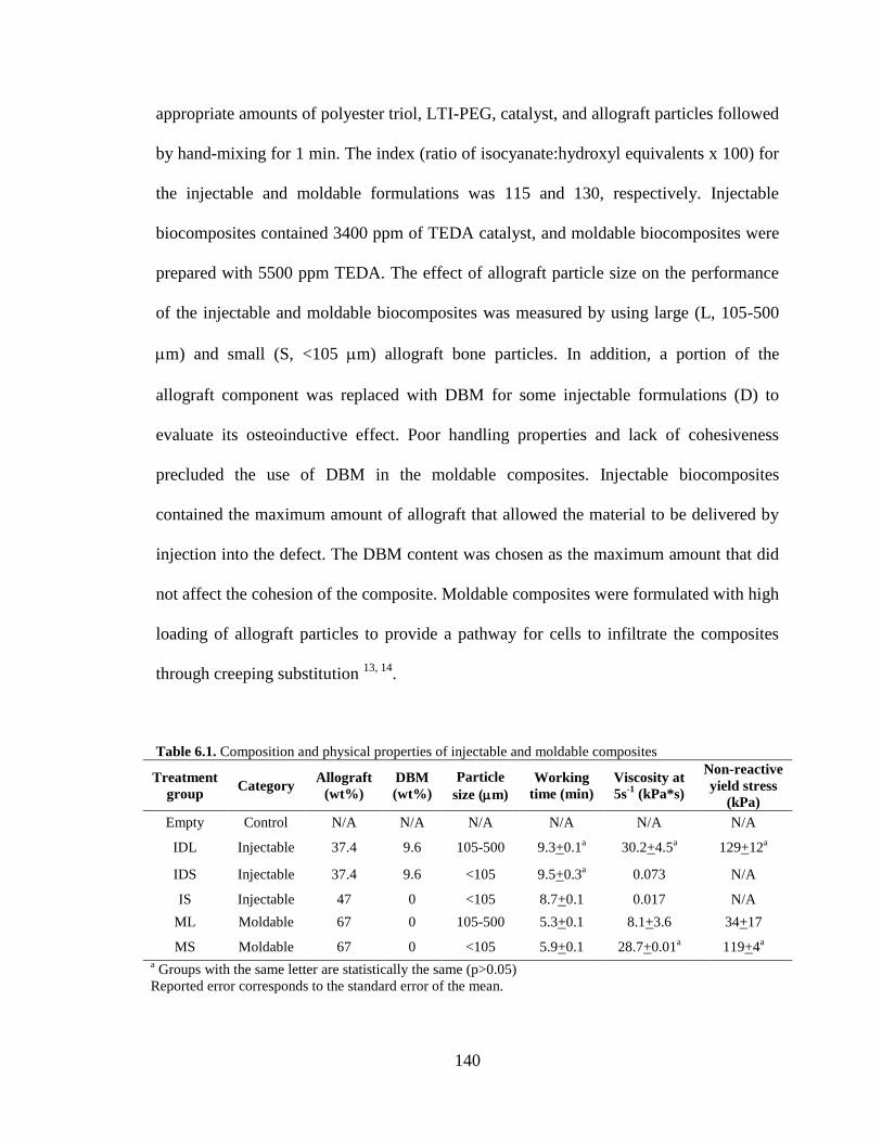
140
appropriate amounts of polyester triol, LTI-PEG, catalyst, and allograft particles followed
by hand-mixing for 1 min. The index (ratio of isocyanate:hydroxyl equivalents x 100) for
the injectable and moldable formulations was 115 and 130, respectively. Injectable
biocomposites contained 3400 ppm of TEDA catalyst, and moldable biocomposites were
prepared with 5500 ppm TEDA. The effect of allograft particle size on the performance
of the injectable and moldable biocomposites was measured by using large (L, 105-500
m) and small (S, <105 m) allograft bone particles. In addition, a portion of the
allograft component was replaced with DBM for some injectable formulations (D) to
evaluate its osteoinductive effect. Poor handling properties and lack of cohesiveness
precluded the use of DBM in the moldable composites. Injectable biocomposites
contained the maximum amount of allograft that allowed the material to be delivered by
injection into the defect. The DBM content was chosen as the maximum amount that did
not affect the cohesion of the composite. Moldable composites were formulated with high
loading of allograft particles to provide a pathway for cells to infiltrate the composites
through creeping substitution 13, 14
.
Table 6.1. Composition and physical properties of injectable and moldable composites
Treatment
group Category
Allograft
(wt%)
DBM
(wt%)
Particle
size (m)
Working
time (min)
Viscosity at
5s-1
(kPa*s)
Non-reactive
yield stress
(kPa)
Empty Control N/A N/A N/A N/A N/A N/A
IDL Injectable 37.4 9.6 105-500 9.3+0.1a 30.2+4.5
a 129+12
a
IDS Injectable 37.4 9.6 <105 9.5+0.3a 0.073 N/A
IS Injectable 47 0 <105 8.7+0.1 0.017 N/A
ML Moldable 67 0 105-500 5.3+0.1 8.1+3.6 34+17
MS Moldable 67 0 <105 5.9+0.1 28.7+0.01a 119+4
a
a Groups with the same letter are statistically the same (p>0.05)
Reported error corresponds to the standard error of the mean.
141
Rheological analysis: initial viscosity and working time
The curing profile of the biocomposites was determined using a TA Instruments
AR2000ex rheometer. Reactive biocomposites were loaded between 25 mm diameter
disposable plates and compressed to a gap of 1.5 mm. Measurements of storage (G’) and
loss (G”) moduli were performed using an oscillatory time sweep method with a
frequency of 1 Hz and amplitude of 1% strain. The working time of the formulations,
defined as the time from the start of mixing until cure, was determined as the G’-G”
cross-over point.
The initial flow characteristics of the biocomposites were analyzed by removing
the catalyst from the formulation (n=3). Non-setting samples were poured between 40
mm cross-hatched parallel plates, compressed to a gap of 1.5 mm, and subjected to a
dynamic frequency sweep (0.1 to 100 rad-sec-1
) at 25C with controlled strain amplitude
of 0.02%. A Cox-Merz transformation was applied to the dynamic data to obtain the
steady state viscosity (, Pa-s) as a function of shear rate (, s-1
). The data were fit to the
Herschel-Bulkley model, which relates the viscosity and shear rate of solid-filled
suspensions with high solids content 15
, to estimate the yield stress (Y, Pa) of the
material:
= Y-1
+ K(n-1) (6.1)
where K is the consistency index of the composite (constant) and n is the power-law
index of the suspending polymer. In this case, the suspending polymer corresponds to the
non-reactive mixture of polyester triol and LTI-PEG prepolymer and has been shown to
exhibit a Newtonian behavior (n=1) 9.
142
In vitro characterization of the biomcomposites
Cylindrical samples of each formulation (n ≥ 3) were prepared for
characterization. To simulate the aqueous environment in the wounds, the formulations
were loaded into 6 mm-diameter cylindrical plastic cups containing 250 l of water and
allowed to set at room temperature (cement formulations set under compression with a 1
lb weight [20.7 psi]). Cylinders were further cured in a vacuum oven at 37oC overnight,
removed from the plastic cups, and cut using a Buehler saw to a length of 12 mm (for
mechanical testing) and ~2 mm (for scanning electron microscopy [SEM] imaging). Thin
sections of cured biocomposite samples were sputter coated with gold and imaged using a
Hitachi S-4200 SEM (Finchampstead, UK). SEM images were analyzed for pore size
using MetaMorph 7.1 image analysis software. The porosity of the mechanical testing
samples was calculated from the composite density which was measured gravimetrically
11. After 24 hours of hydration in phosphate buffered saline (PBS), mechanical properties
were measured using an MTS 898 by preloading the samples to 12 N followed by
compression at a rate of 25 mm/min until failure. The original cross sectional area of the
cylinders was used to calculate compressive stress. The strength reported corresponds to
the point where maximum stress was achieved, and compressive modulus was calculated
as the slope of the linear portion of the stress-strain curve.
Animal Study
Twenty-eight New Zealand White (NZW) rabbits were used in this study. All
surgical and care procedures were carried out at IBEX Preclinical Research, Inc. (Logan,
UT) under aseptic conditions per the approved IACUC protocol. The biocomposite
143
individual components were gamma-irradiated using a dose of 25 kGY. Bilateral critical-
sized defects of 5 mm diameter by 6-8 mm in depth were drilled in the lateral femoral
condyles of each rabbit. Metal wires were placed in the distal-most aspect of the defects
to aid in location. Defects were filled with injectable or moldable formulations (n = 4
defects per formulation) according to a randomized experimental design (different
formulations were placed in contralateral defects). Control defects were left untreated
(Empty). Femoral specimens were harvested after 6 and 12 weeks and placed in 10%
neutral buffered formalin for fixation.
Micro-computed tomography (CT) analysis
A CT80 (SCANCO Medical, Basserdorf, Switzerland) was used to acquire
images of the extracted femurs post-implantation at 6 and 12 weeks. In addition, femurs
from NZW rabbits which were part of other experimental protocols that did not affect the
skeleton (same age, sex, and similar weight to the rabbits used in this study) were
scanned and analyzed as host bone controls. µCT scans were performed in formalin at 70
kVp energy, 114 µA source current, 250 projections per rotation, 400 ms integration time,
and an isotropic voxel size of 36 µm. Axial images were reconstructed using
manufacturer-provided software. Attenuation values were converted to tissue mineral
density (TMD) through calibration with hydroxyapatite (HA) phantoms with densities
between 0 and 780 mg-HA-cm-3
(calibration of the instrument was checked weekly).
Using the cortical borders of the defect for alignment, the reconstructed image stack was
re-oriented such that the depth of the defect was parallel to the z-axis. A radial analysis of
morphometric parameters was conducted from the core of the implant to the interface
144
with host bone. Three concentric annular volumes of interest with thickness of 1 mm and
a cylindrical core with diameter of 1 mm, all with approximate length of 7 mm (from the
outer cortical surface of the femur) were defined for each sample. The 3 inner regions
covered the defect volume, while the outer region provided information about the
interface with host bone. Ossified tissue was differentiated from soft tissue using lower
and upper thresholds of 140 mg-HA-cm-3
and 254 mg-HA-cm-3
respectively, as well as
Gaussian noise filter settings of sigma 0.2 and support 2. Threshold conditions were
chosen visually and kept constant for the analysis of all the samples. An upper threshold
was included to avoid the selection of the metal wires as ossified tissue. Morphometric
parameters within the annular regions were calculated, grouped by treatment and time
point, and plotted versus the mean radial distance (Rm) from the core of the defect
(Rm=(Ro+Ri)/2, where Ro and Ri correspond to the outer and inner radius of each region,
respectively). Bone volume/total volume (BV/TV), trabecular number (Tb.N.), trabecular
thickness (Tb.Th.), and trabecular separation (Tb.Sp.) within the regions of interest were
computed using SCANCO’s Medical microCT systems software.
Histology
After fixation in formalin, femur specimens were trimmed and embedded in
methyl methacrylate. Thin sections were cut in a longitudinal plane through the
approximate center of each defect. Care was taken to ensure that the orientation was as
similar as possible between specimens. Sections of parallel planes were stained with
Goldner’s trichrome stain and Hematoxylin and Eosin (H&E) stain to evaluate the in vivo
145
response of the different formulations. H&E sections were evaluated for inflammation
and the presence of giant cells (GC’s).
Histomorphometry
For quantitative analysis, Goldner’s trichrome stained sections were imaged at
10x magnification with an Olympus camera (DP71) using an Olympus BX60 microscope.
Residual allograft bone particles, newly formed bone, and residual polymer were
quantified in an area of interest 1 mm high 5 mm wide located in the center of the
defect. To analyze the radial remodeling of the scaffolds in time, the rectangular area of
interest was further subdivided into 3 concentric annular regions (each 0.83 mm thick)
representing (1) the area of the defect in contact with the host bone, (2) the mid-region of
the defect, and (3) the core of the defect. Mineralized tissue was stained green, and to
differentiate between the new bone and the residual allograft, the allograft particles were
identified as meeting the following two criteria: (1) acellular, and (2) angular shape. In
the stained sections remaining polymer appeared white, newly deposited osteoid stained
red/fucsia, cellular nuclei stained blue, and red blood cells stained orange/red. For
histomorphometric analysis, Metamorph (Version 7.0.1) was utilized to color threshold
each phase, quantify the number of pixels of each material and compare it to the total
pixels in the area of interest. Similar to the CT data, the content of new bone, allograft
bone, and residual polymer was plotted as a function of the mean radial distance from the
core of the defect (Rm). Goldner’s trichrome stain did not reveal DBM particles, so these
were not included in the histomorphometric analysis. Initial content of allograft bone and
polymer in each formulation was determined from histological sections of scaffolds
146
prepared in the laboratory and processed as described in the previous section. These
values were used to calculate rates of allograft resorption and polymer degradation
throughout remodeling.
Statistical analysis
A one-way analysis of variance (ANOVA) was performed to compare the
rheological and mechanical properties, as well as the pore size of different treatment
groups. Statistical significance was considered for p < 0.05.
Results
Biocomposite characterization
Rheological characterization of the non-reactive and reactive formulations
provided information about their initial handling properties. Moldable biocomposites had
the lowest working times, and addition of DBM slightly delayed the setting of injectable
formulations. Figure 6.1 shows representative curves for moldable and injectable
biocomposites’ initial viscosity as a function of shear rate. Injectable formulations with
small particles exhibited Newtonian behavior at shear rates below ~3 s-1
(initial plateau),
followed by power-law behavior at larger shear rates. While these groups showed
minimal evidence of yield stress (minimal resistance to flow during the rheological
analysis as well as during injection), the Herschel-Bulkley model 15
was used to calculate
the yield stress for the IDL and moldable groups (Table 6.1). The IDL, ML, and MS
groups exhibited power law behavior even at low shear rates. The addition of DBM
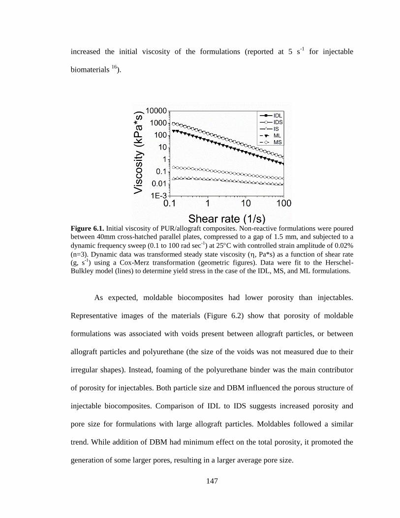
147
increased the initial viscosity of the formulations (reported at 5 s-1
for injectable
biomaterials 16
).
Figure 6.1. Initial viscosity of PUR/allograft composites. Non-reactive formulations were poured
between 40mm cross-hatched parallel plates, compressed to a gap of 1.5 mm, and subjected to a
dynamic frequency sweep (0.1 to 100 rad sec-1
) at 25C with controlled strain amplitude of 0.02%
(n=3). Dynamic data was transformed steady state viscosity (, Pa*s) as a function of shear rate
(g, s-1
) using a Cox-Merz transformation (geometric figures). Data were fit to the Herschel-
Bulkley model (lines) to determine yield stress in the case of the IDL, MS, and ML formulations.
As expected, moldable biocomposites had lower porosity than injectables.
Representative images of the materials (Figure 6.2) show that porosity of moldable
formulations was associated with voids present between allograft particles, or between
allograft particles and polyurethane (the size of the voids was not measured due to their
irregular shapes). Instead, foaming of the polyurethane binder was the main contributor
of porosity for injectables. Both particle size and DBM influenced the porous structure of
injectable biocomposites. Comparison of IDL to IDS suggests increased porosity and
pore size for formulations with large allograft particles. Moldables followed a similar
trend. While addition of DBM had minimum effect on the total porosity, it promoted the
generation of some larger pores, resulting in a larger average pore size.
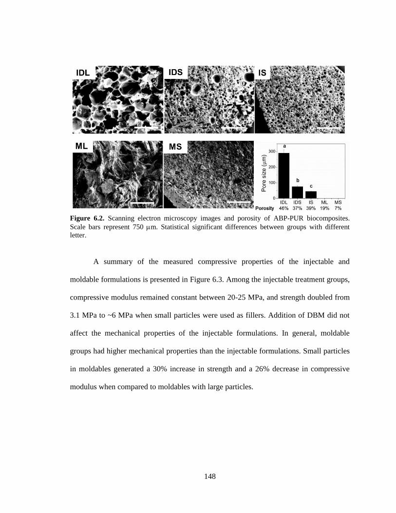
148
Figure 6.2. Scanning electron microscopy images and porosity of ABP-PUR biocomposites.
Scale bars represent 750 m. Statistical significant differences between groups with different
letter.
A summary of the measured compressive properties of the injectable and
moldable formulations is presented in Figure 6.3. Among the injectable treatment groups,
compressive modulus remained constant between 20-25 MPa, and strength doubled from
3.1 MPa to ~6 MPa when small particles were used as fillers. Addition of DBM did not
affect the mechanical properties of the injectable formulations. In general, moldable
groups had higher mechanical properties than the injectable formulations. Small particles
in moldables generated a 30% increase in strength and a 26% decrease in compressive
modulus when compared to moldables with large particles.
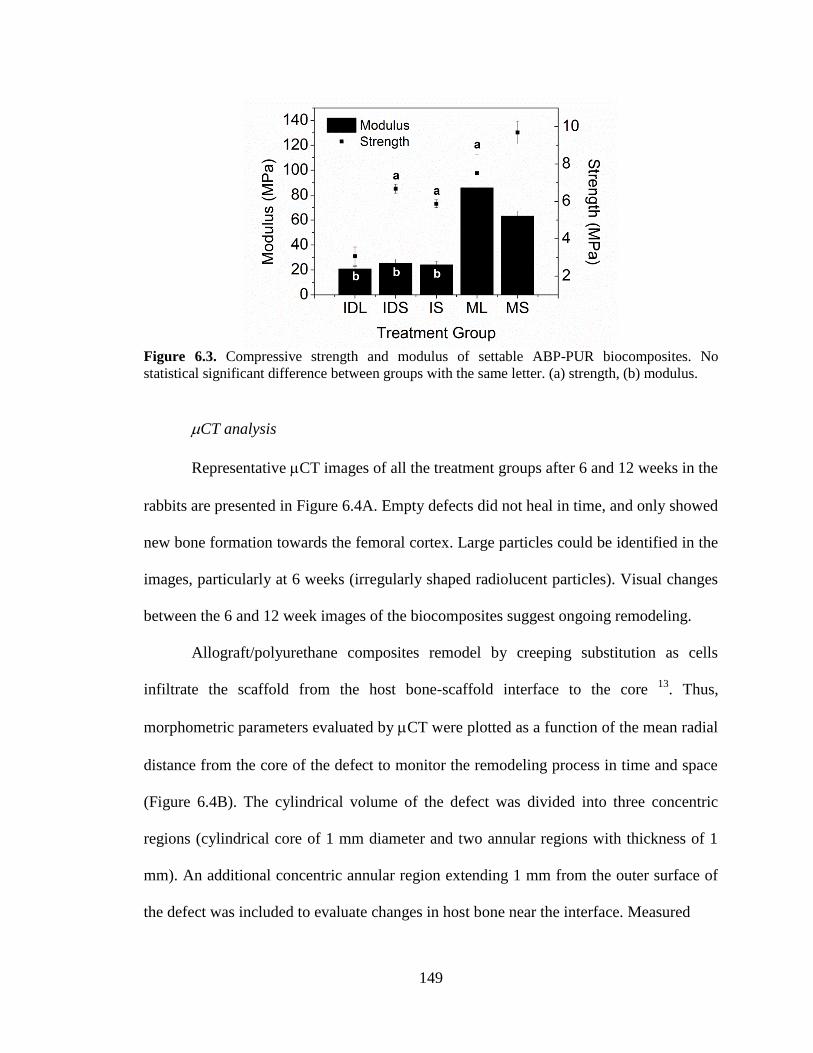
149
Figure 6.3. Compressive strength and modulus of settable ABP-PUR biocomposites. No
statistical significant difference between groups with the same letter. (a) strength, (b) modulus.
CT analysis
Representative CT images of all the treatment groups after 6 and 12 weeks in the
rabbits are presented in Figure 6.4A. Empty defects did not heal in time, and only showed
new bone formation towards the femoral cortex. Large particles could be identified in the
images, particularly at 6 weeks (irregularly shaped radiolucent particles). Visual changes
between the 6 and 12 week images of the biocomposites suggest ongoing remodeling.
Allograft/polyurethane composites remodel by creeping substitution as cells
infiltrate the scaffold from the host bone-scaffold interface to the core 13
. Thus,
morphometric parameters evaluated by CT were plotted as a function of the mean radial
distance from the core of the defect to monitor the remodeling process in time and space
(Figure 6.4B). The cylindrical volume of the defect was divided into three concentric
regions (cylindrical core of 1 mm diameter and two annular regions with thickness of 1
mm). An additional concentric annular region extending 1 mm from the outer surface of
the defect was included to evaluate changes in host bone near the interface. Measured
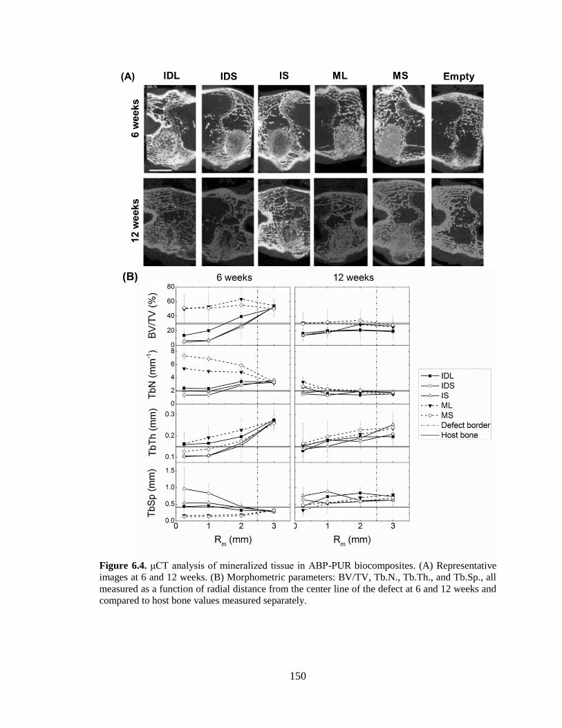
150
Figure 6.4. μCT analysis of mineralized tissue in ABP-PUR biocomposites. (A) Representative
images at 6 and 12 weeks. (B) Morphometric parameters: BV/TV, Tb.N., Tb.Th., and Tb.Sp., all
measured as a function of radial distance from the center line of the defect at 6 and 12 weeks and
compared to host bone values measured separately.
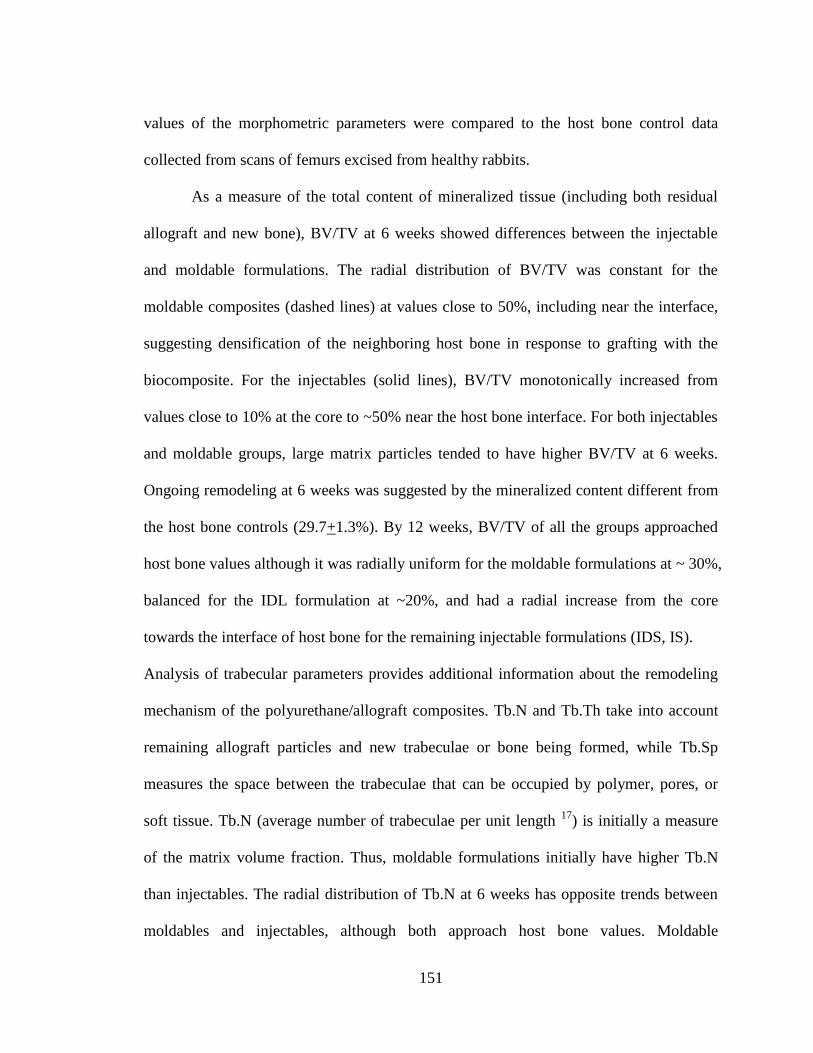
151
values of the morphometric parameters were compared to the host bone control data
collected from scans of femurs excised from healthy rabbits.
As a measure of the total content of mineralized tissue (including both residual
allograft and new bone), BV/TV at 6 weeks showed differences between the injectable
and moldable formulations. The radial distribution of BV/TV was constant for the
moldable composites (dashed lines) at values close to 50%, including near the interface,
suggesting densification of the neighboring host bone in response to grafting with the
biocomposite. For the injectables (solid lines), BV/TV monotonically increased from
values close to 10% at the core to ~50% near the host bone interface. For both injectables
and moldable groups, large matrix particles tended to have higher BV/TV at 6 weeks.
Ongoing remodeling at 6 weeks was suggested by the mineralized content different from
the host bone controls (29.7+1.3%). By 12 weeks, BV/TV of all the groups approached
host bone values although it was radially uniform for the moldable formulations at ~ 30%,
balanced for the IDL formulation at ~20%, and had a radial increase from the core
towards the interface of host bone for the remaining injectable formulations (IDS, IS).
Analysis of trabecular parameters provides additional information about the remodeling
mechanism of the polyurethane/allograft composites. Tb.N and Tb.Th take into account
remaining allograft particles and new trabeculae or bone being formed, while Tb.Sp
measures the space between the trabeculae that can be occupied by polymer, pores, or
soft tissue. Tb.N (average number of trabeculae per unit length 17
) is initially a measure
of the matrix volume fraction. Thus, moldable formulations initially have higher Tb.N
than injectables. The radial distribution of Tb.N at 6 weeks has opposite trends between
moldables and injectables, although both approach host bone values. Moldable
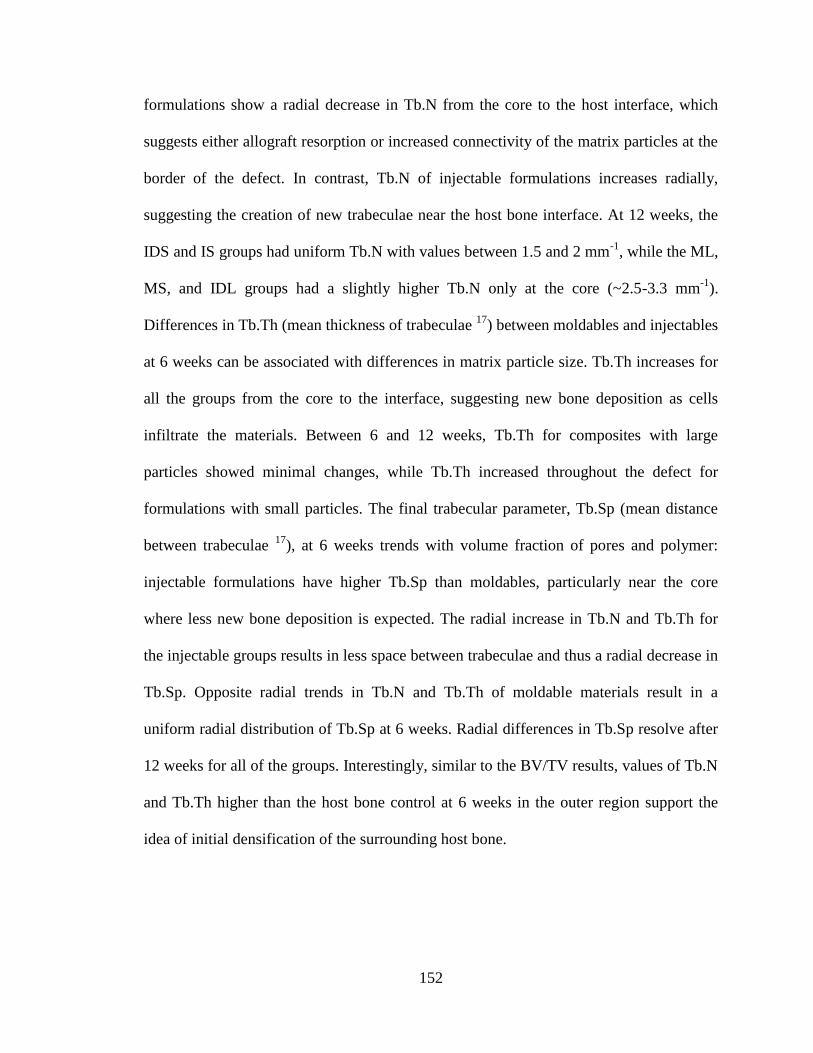
152
formulations show a radial decrease in Tb.N from the core to the host interface, which
suggests either allograft resorption or increased connectivity of the matrix particles at the
border of the defect. In contrast, Tb.N of injectable formulations increases radially,
suggesting the creation of new trabeculae near the host bone interface. At 12 weeks, the
IDS and IS groups had uniform Tb.N with values between 1.5 and 2 mm-1
, while the ML,
MS, and IDL groups had a slightly higher Tb.N only at the core (~2.5-3.3 mm-1
).
Differences in Tb.Th (mean thickness of trabeculae 17
) between moldables and injectables
at 6 weeks can be associated with differences in matrix particle size. Tb.Th increases for
all the groups from the core to the interface, suggesting new bone deposition as cells
infiltrate the materials. Between 6 and 12 weeks, Tb.Th for composites with large
particles showed minimal changes, while Tb.Th increased throughout the defect for
formulations with small particles. The final trabecular parameter, Tb.Sp (mean distance
between trabeculae 17
), at 6 weeks trends with volume fraction of pores and polymer:
injectable formulations have higher Tb.Sp than moldables, particularly near the core
where less new bone deposition is expected. The radial increase in Tb.N and Tb.Th for
the injectable groups results in less space between trabeculae and thus a radial decrease in
Tb.Sp. Opposite radial trends in Tb.N and Tb.Th of moldable materials result in a
uniform radial distribution of Tb.Sp at 6 weeks. Radial differences in Tb.Sp resolve after
12 weeks for all of the groups. Interestingly, similar to the BV/TV results, values of Tb.N
and Tb.Th higher than the host bone control at 6 weeks in the outer region support the
idea of initial densification of the surrounding host bone.
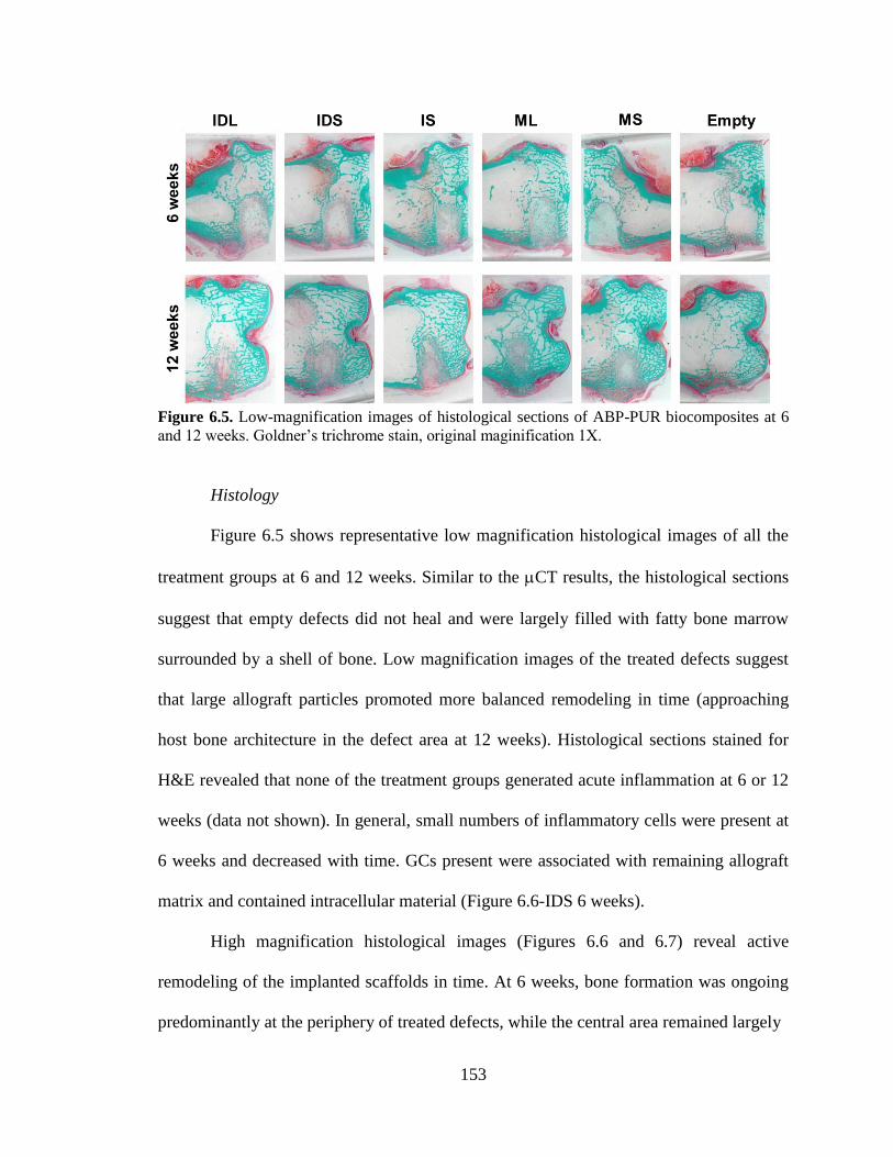
153
Figure 6.5. Low-magnification images of histological sections of ABP-PUR biocomposites at 6
and 12 weeks. Goldner’s trichrome stain, original maginification 1X.
Histology
Figure 6.5 shows representative low magnification histological images of all the
treatment groups at 6 and 12 weeks. Similar to the CT results, the histological sections
suggest that empty defects did not heal and were largely filled with fatty bone marrow
surrounded by a shell of bone. Low magnification images of the treated defects suggest
that large allograft particles promoted more balanced remodeling in time (approaching
host bone architecture in the defect area at 12 weeks). Histological sections stained for
H&E revealed that none of the treatment groups generated acute inflammation at 6 or 12
weeks (data not shown). In general, small numbers of inflammatory cells were present at
6 weeks and decreased with time. GCs present were associated with remaining allograft
matrix and contained intracellular material (Figure 6.6-IDS 6 weeks).
High magnification histological images (Figures 6.6 and 6.7) reveal active
remodeling of the implanted scaffolds in time. At 6 weeks, bone formation was ongoing
predominantly at the periphery of treated defects, while the central area remained largely
154
Figure 6.6. High-magnification images of histological sections of defects treated with injectable
ABP-PUR biocomposites. All of the images show sections stained with Goldner’s trichrome,
except for IDS at 6 weeks, which is stained with H&E. Original magnification 70X. With
Goldner’s trichrome stain: white- remaining polymer; light green particles with angled shapes-
remaining allograft particles; green with cells inside- new mineralized bone with osteocytes; red-
osteoid; orange- red blood cells; purple- nuclei; BM- bone marrow. Arrows in the IDS-6 weeks
section point to giant cells.
Figure 6.7. High-magnification images of histological sections of empty defects and defects
treated with moldable ABP-PUR biocomposites. Goldner’s trichrome stain. Original
magnification reported in each image. With Goldner’s trichrome stain: white- remaining polymer;
light green particles with angled shapes- remaining allograft particles; green with cells inside-
new mineralized bone with osteocytes; red- osteoid; orange- red blood cells; purple- nuclei; BM-
bone marrow.
155
acellular. New bone was deposited appositionally mostly by intramembranous
ossification, except at the periphery of the defects where some endochondral ossification
was present (in particular at 6 weeks). New bone deposited at early time points had
woven structure for all the treatment groups. This resolved for the ML group at 12 weeks
with lamellar bone present in the defect (Figure 6.7-ML 12 weeks). After 12 weeks,
remaining allograft particles were incorporated with new bone and this was more evident
in scaffolds with large particles. An interesting difference between injectable and
moldable materials was that at 6 weeks injectable scaffolds showed evidence of new bone
growing not only on top of allograft particles but also on the surface of the polymer
(center of Figure 6.6-IDL 6 weeks). Most new bone formation in moldable materials
initiated at the surface of allograft particles (center of figure 6.7-ML 6 weeks).
Histomorphometry
Histomorphometric analysis enabled radial quantification of new bone formation,
allograft resorption, and polymer degradation for each treatment group (Figure 6.8).
Differences between the groups at different locations and time points were generally not
statistically significant due to small sample size. As a result, analysis of the
histomorphometry data focused on trends and rates instead of absolute values of each
parameter.
While CT analysis provided information about the mineral phase in the defects,
histology was used to differentiate new bone from residual allograft, as well as to monitor
remaining polymer at each time point and location. The total mineralized content
measured by histomorphometry [NB+A(%) in Figure 6.8] showed similar trends to
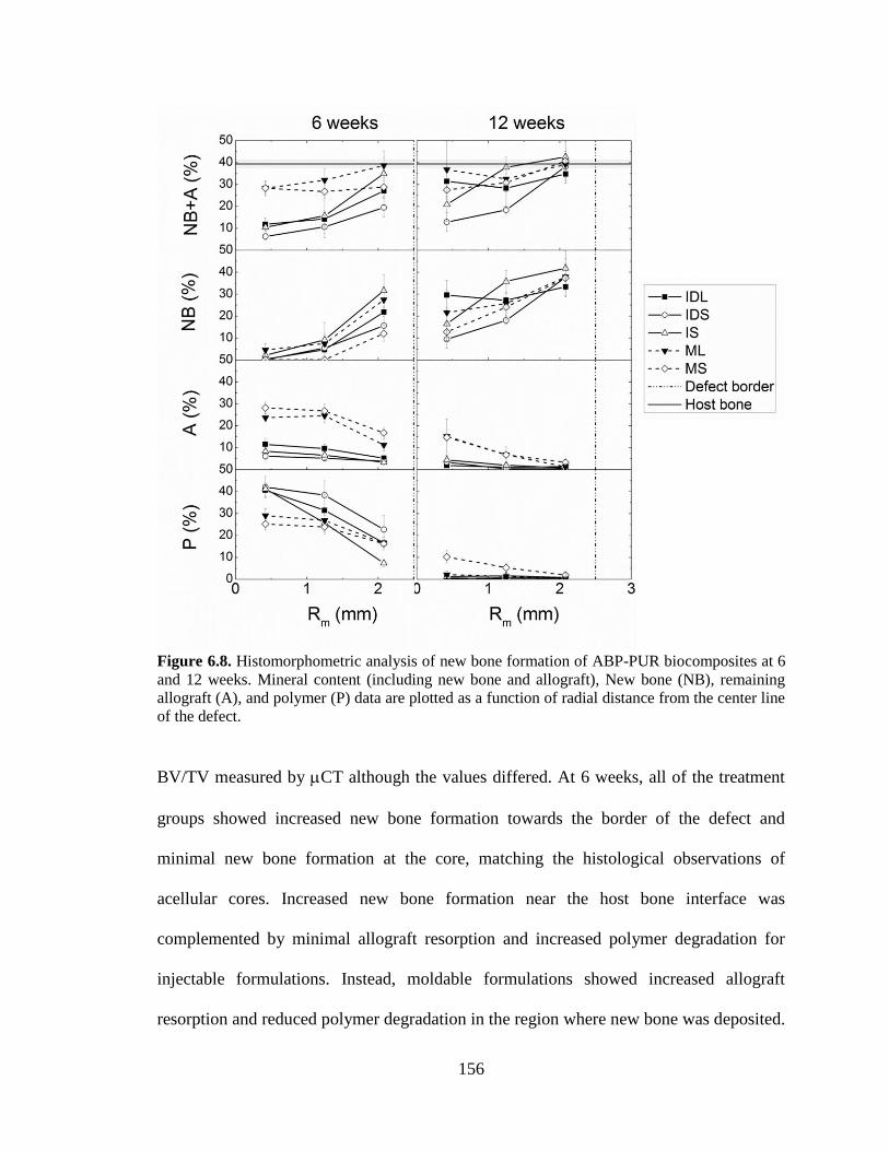
156
Figure 6.8. Histomorphometric analysis of new bone formation of ABP-PUR biocomposites at 6
and 12 weeks. Mineral content (including new bone and allograft), New bone (NB), remaining
allograft (A), and polymer (P) data are plotted as a function of radial distance from the center line
of the defect.
BV/TV measured by CT although the values differed. At 6 weeks, all of the treatment
groups showed increased new bone formation towards the border of the defect and
minimal new bone formation at the core, matching the histological observations of
acellular cores. Increased new bone formation near the host bone interface was
complemented by minimal allograft resorption and increased polymer degradation for
injectable formulations. Instead, moldable formulations showed increased allograft
resorption and reduced polymer degradation in the region where new bone was deposited.
157
At 12 weeks, all of the treatment groups achieved a similar amount of new bone
deposition at the edge of the defect. However, formulations with large particles showed a
reduced radial gradient in new bone deposition, suggesting a more balanced remodeling
throughout the defects. After 12 weeks, remaining polymer throughout the defects was
minimal except at the core of the MS group.
Discussion
The golden standard of care for the treatment of bone defects continues to be the
use of autograft bone. However, unlimited availability and donor site morbidity have
motivated the search for bone substitute materials that also promote healing. As a
biocompatible and osteoconductive alternative for autograft bone, surgeons have been
using allograft bone for more than 20 years 18, 19
. Our group has developed injectable 20, 7,
11, moldable
9, and compression molded
13 composites incorporating allograft bone
particles and biodegradable, lysine-derived polyurethanes. These materials have been
shown to promote cellular infiltration, allograft resorption, and new bone deposition
when implanted in vivo. Recently we reported an enhancement in healing progression of
ABP-PUR composites promoted by balancing the rates of new bone formation and
polymer degradation 9. The aim of the present work was to further improve the
understanding of the remodeling mechanisms of ABP-PUR biocomposites by evaluating
the effects of allograft particle size, loading, and mineral composition on the healing of
femoral plug defects in rabbits.
Characterization of the biocomposites in the lab suggested several effects of the
variables of interest on their handling, structural, and mechanical properties. All the
158
treatment groups in our study contained similar volume fraction of polymer, and were
classified into 2 general categories: injectable and moldable biocomposites. Injectable
formulations were porous and had 47 wt% allograft particles. Instead, moldables had low
porosity and high filler loading (67 wt%) such that the close proximity of the particles
could provide a pathway for cellular infiltration. As their label suggest, injectable
biocomposites could be delivered to the bone defect using a syringe. Interestingly, even
though the IDL and MS groups exhibited the same rheological behavior under non-
reactive conditions, and ML had lower viscosity and yield stress, the moldable
formulations could not be injected. Most likely, the longer working time of IDL allowed
the formulation to be loaded into the syringe and injected before the material completely
transitioned from a viscoelastic to an elastomeric behavior. Addition of DBM increased
the viscosity of injectable formulations. This observation correlates with studies reporting
an increase in the resistance of polymers to flow as a consequence of clustering of
incompatible fillers 21
. Our results align with reports of mechanical properties of
particulate-polymer composites depending on particle loading and size 22, 23
: higher filler
loading reduced the porosity of the biocomposites and resulted in improved compressive
modulus; smaller particle size improved the strength of injectable and moldable
composites. An additional effect of small particles over the structure of the ABP-PUR
biocomposites was the significant reduction in pore size. While this was an unexpected
result, it is possible that the increased number of small versus large allograft particles
generated a more tortuous path for the foaming gas to escape, thus resulting in more and
smaller pores.
159
Contrary to our expectations, the addition of DBM to injectable formulations
hindered remodeling of the rabbit femoral plug defects. DBM has been reported to be
osteoinductive and to stimulate defect healing in dogs 24, 25
, rabbits 26
, and humans 27
.
However, multiple recent studies have reported variability in the osteoinductive character
of DBM 28, 29
, even among lots of the same formulation 30
. Factors that influence the
effectiveness of DBM include: the technique to obtain the bone from human donors,
donor age and gender, differences in preparation and processing methods for bone, and
sterilization protocols among others 29
. Our results further support the idea that not all
DBM is osteoinductive and as a result its use should be highly regulated.
Previous studies have reported a particle size effect on the remodeling of allograft
bone particles in vivo. In combination with autogenous bone marrow, allograft particles
with size in the range of 100-300 m promoted significantly more new bone formation
than larger particles (1000-2000 m) when placed in the femurs of Rhesus monkeys for 8
weeks 31
. Another study in rabbit calvaria reported a greater amount of osteoid tissue and
little fibrous tissue formation when allograft particles with size <450 m were implanted
instead of particles with larger size (up to 1000 m) 32
. Both these studies also reported
enhanced resorption and incorporation of allograft when particles smaller than 450 m
were used. The results of our in vivo study agree with these observations. Independent of
the porosity, ABP-PUR biocomposites with particles in the size range of 105-500 m
achieved improved balanced remodeling throughout the defect after 12 weeks, as well as
deposition of more mature bone. Kluppel et. speculated that the improved osteoinductive
character of the small particles (within the size range studied) could be the result of the
higher surface area exposed for resorption and new bone deposition 32
. While this was
160
our initial expectation when including smaller ABP in the formulations, our results
contradict this hypothesis. Reduction of particle size below 105 m did not favor
improved remodeling. However, the particle size range of 100-500 m seems to be ideal
for new bone deposition.
Combined analysis of our CT and histomorphometry data suggests interesting
differences in the remodeling mechanism of injectable and moldable formulations.
Previously, we and others have demonstrated that bone defects remodel from the
periphery to the interior of the scaffolds as cells infiltrate the materials 9, 33
. Thus, the
remodeling progression of the biocomposites was evaluated by comparing the radial
distribution of morphometric parameters. The decrease of BV/TV and Tb.N between 6
and 12 weeks for the moldable groups suggested active allograft resorption throughout
the scaffold. The radial increase in Tb.Th for this group at 6 weeks suggested new bone
deposition on the surface of allograft particles, which was verified by histological images.
Together, these observations suggest that the low-porosity moldable biocomposites
remodel by creeping substitution: osteoclasts resorb the surface of allograft particles and
are followed by other cells including osteoblasts which deposit new bone. On the other
hand, Tb.N and Tb.Th of injectable formulations increased radially at 6 weeks. This
suggested formation of new trabeculae, and growth of old and new trabeculae from the
host bone interface towards the interior of the scaffold. Careful inspection of histological
images revealed that indeed new bone was deposited on the surface of the PUR phase,
and we believe that this corresponds to the new trabeculae detected by CT. As a result,
our data suggests that injectable composites remodel as cells infiltrate the pores and
deposit new bone on the pore walls as well as on the surface of allograft particles.
161
Interconnected porosity has long been considered a fundamental requirement for
the success of bone scaffolds 34, 4
. However, as discussed above, our results show that
composites with high and low porosity remodel in femoral plug defects, although they
achieve this through different mechanisms. From these observations we propose that a
more important parameter for the successful remodeling of bone scaffolds is the
persistence of a continuous phase. Visual inspection of the histological images obtained
in this work suggests higher continuity in the groups that achieved better remodeling
outcomes (ML, IDL). A method to quantify the continuity or the connectivity of an image
is through the calculation of the Euler number defined as:
(6.2)
where C corresponds to the number of connected objects, and H to the number of holes35
.
As such, E=1 represents an image without holes, E>>1 an image with low connectivity,
and E<<1 a highly connected image with holes. For the remodeling studies, the
connected objects in the defect can be selected as allograft particles, new bone, and
remaining polymer. As a result, connectivity will depend on several parameters such as
filler particle size and loading, scaffold porosity, polymer stability, and filler resorbability.
We hypothesize that a scaffold with increased connectivity (E<<1) will provide a
pathway for cells to infiltrate the scaffold resulting in improved remodeling. Preliminary
Euler number calculations for a single sample of each treatment group (at 6 weeks) are
promising: IDS, IS, and MS had the highest Euler values (2001, 361, and 71 respectively),
while IDL and ML (the formulations that achieved more balanced healing) had the lowest
Euler values representing higher connectivity (-214 and -81 respectively). A recent
publication studying the effect of particle size on the osteointegration of silicate-
162
substituted calcium phosphates (Si-CaP) reported enhanced performance of particles with
size in the range of 250-500 m 36
. Similar to our study, the authors found that smaller
particles (90-125 m) promoted a less favorable healing outcome. However, the
formulation tested was a combination of high loading of particles with slower resorption
rates than allograft, and fast polymer degradation (no evidence of polymer after 4 weeks
in femoral plug defects in sheep). In contrast to the best candidate formulations from this
work (IDL and ML), the Si-CaP will result in high Euler numbers as remodeling
progresses, and as shown by the authors, this state can also promote remodeling of the
defect. Further work is needed to verify these propositions, and this is discussed in the
future work section of this dissertation.
Conclusions
Moldable and injectable ABP-PUR biocomposites supported cellular infiltration,
and remodeling when implanted in rabbit femoral plug defects, although through
different remodeling mechanisms. Moldable biocomposites remodeled by creeping
substitution with new bone being deposited on the surface of allograft particles; cells
infiltrated injectable formulations through the pores and deposited new bone on the
surface of the polymer and allograft particles. Evaluation of the effect of filler mineral
composition on the remodeling of injectable formulations, suggested improved
performance of mineralized allograft particles without the addition of DBM.
Formulations incorporating 105-500 m sized allograft particles promoted deposition of
more mature bone throughout the defects in a radially balanced progression. The results

163
presented point to connectivity and distance between filler particles as possible influential
parameters in the remodeling progression of composite bone scaffolds.
164
References
1 M. A. Woodruff, C. Lange, J. Reichert, A. Berner, F. L. Chen, P. Fratzl, J. T. Schantz, and D. W.
Hutmacher, 'Bone Tissue Engineering: From Bench to Bedside', Materials Today, 15 (2012), 430-
35.
2 K. Rezwan, Q. Z. Chen, J. J. Blaker, and A. R. Boccaccini, 'Biodegradable and Bioactive Porous
Polymer/Inorganic Composite Scaffolds for Bone Tissue Engineering', Biomaterials, 27 (2006),
3413-31.
3 S. Scaglione, E. Lazzarini, C. Ilengo, and R. Quarto, 'A Composite Material Model for Improved
Bone Formation', Journal of Tissue Engineering and Regenerative Medicine, 4 (2010), 505-13.
4 S. Bose, M. Roy, and A. Bandyopadhyay, 'Recent Advances in Bone Tissue Engineering
Scaffolds', Trends in Biotechnology, 30 (2012), 546-54.
5 M. Bohner, and J. Lemaitre, 'Can Bioactivity Be Tested in Vitro with Sbf Solution?', Biomaterials,
30 (2009), 2175-79.
6 J. R. Jones, 'Review of Bioactive Glass: From Hench to Hybrids', Acta Biomater, 9 (2013), 4457-
86.
7 J. M. Page, E. M. Prieto, J. E. Dumas, K. J. Zienkiewicz, J. C. Wenke, P. Brown-Baer, and S. A.
Guelcher, 'Biocompatibility and Chemical Reaction Kinetics of Injectable, Settable
Polyurethane/Allograft Bone Biocomposites', Acta Biomaterialia, 8 (2012), 4405-16.
8 R. Adhikari, P. A. Gunatillake, I. Griffiths, L. Tatai, M. Wickramaratna, S. Houshyar, T. Moore,
R. T. M. Mayadunne, J. Field, M. McGee, and T. Carbone, 'Biodegradable Injectable
Polyurethanes: Synthesis and Evaluation for Orthopaedic Applications', Biomaterials, 29 (2008),
3762-70.
9 J. E. Dumas, E. M. Prieto, K.J. Zienkiewicz, T. Guda, J. C. Wenke, J. Bible, G.E. Holt, and S.A.
Guelcher, 'Balancing the Rates of New Bone Formation and Polymer Degradation Enhances
Healing of Weight-Bearing Allograft/Polyurethane Composites in Rabbit Femoral Defects', Tissue
Eng Part A, Not available - ahead of print (2013).
10 R. Kruger, and J. Groll, 'Fiber Reinforced Calcium Phosphate Cements -- on the Way to
Degradable Load Bearing Bone Substitutes?', Biomaterials, 33 (2012), 5887-900.
11 J. E. Dumas, K. Zienkiewicz, S. A. Tanner, E. M. Prieto, S. Bhattacharyya, and S. A. Guelcher,
'Synthesis and Characterization of an Injectable Allograft Bone/Polymer Composite Bone Void
Filler with Tunable Mechanical Properties', Tissue Engineering Part A, 16 (2010), 2505-18.
12 T. Yoshii, J. E. Dumas, A. Okawa, D. M. Spengler, and S. A. Guelcher, 'Synthesis,
Characterization of Calcium Phosphates/Polyurethane Composites for Weight-Bearing Implants',
Journal of Biomedical Materials Research Part B-Applied Biomaterials, 100B (2012), 32-40.
13 J. E. Dumas, T. Davis, G.E. Holt, T. Yoshii, D. S. Perrien, J. S. Nyman, T. Boyce, and S.A.
Guelcher, 'Synthesis, Characterization, and Remodeling of Weight-Bearing Allograft
Bone/Polyurethane Composites in the Rabbit', Acta Biomaterialia, 6 (2010), 2394-406.
14 Michael J. Eagan, and David R. McAllister, 'Biology of Allograft Incorporation', Clinics in Sports
Medicine, 28 (2009), 203-14.
15 A J Poslinski, Ryan M E, Gupta R K, Seshadri S G, and Frechette F J, 'Rheological Behavior of
Filled Polymeric Systems I. Yield Stress and Shear-Thinning Effects', Journal of Rheology, 32
(1988), 703-35.
165
16 G. Baroud, E. Cayer, and M. Bohner, 'Rheological Characterization of Concentrated Aqueous
Beta-Tricalcium Phosphate Suspensions: The Effect of Liquid-to-Powder Ratio, Milling Time,
and Additives', Acta Biomaterialia, 1 (2005), 357-63.
17 Mary L. Bouxsein, Stephen K. Boyd, Blaine A. Christiansen, Robert E. Guldberg, Karl J. Jepsen,
and Ralph Müller, 'Guidelines for Assessment of Bone Microstructure in Rodents Using Micro–
Computed Tomography', Journal of Bone and Mineral Research, 25 (2010), 1468-86.
18 Carlos E. A. Ferreira, Arthur B. Novaes, Violet I. Haraszthy, Márcio Bittencourt, Carolina B.
Martinelli, and Sonia M. Luczyszyn, 'A Clinical Study of 406 Sinus Augmentations with 100%
Anorganic Bovine Bone', Journal of Periodontology, 80 (2009), 1920-27.
19 Francesco Pieri, Giuseppe Corinaldesi, Milena Fini, Nicolò Nicoli Aldini, Roberto Giardino, and
Claudio Marchetti, 'Alveolar Ridge Augmentation with Titanium Mesh and a Combination of
Autogenous Bone and Anorganic Bovine Bone: A 2-Year Prospective Study', Journal of
Periodontology, 79 (2008), 2093-103.
20 J.E. Dumas, P.B. BrownBaer, E.M. Prieto, T. Guda, R.G. Hale, J.C. Wenke, and S.A. Guelcher,
'Injectable Reactive Biocomposites for Bone Healing in Critical-Size Rabbit Calvarial Defects',
Biomedical Materials, 7 (2012), 024112.
21 Joshua Qingsong Li, and Ronald Salovey, 'Model Filled Polymers: The Effect of Particle Size on
the Rheology of Filled Poly(Methyl Methacrylate) Composites', Polymer Engineering & Science,
44 (2004), 452-62.
22 Shao-Yun Fu, Xi-Qiao Feng, Bernd Lauke, and Yiu-Wing Mai, 'Effects of Particle Size,
Particle/Matrix Interface Adhesion and Particle Loading on Mechanical Properties of Particulate–
Polymer Composites', Composites Part B: Engineering, 39 (2008), 933-61.
23 Kin-tak Lau, Chong Gu, and David Hui, 'A Critical Review on Nanotube and Nanotube/Nanoclay
Related Polymer Composite Materials', Composites Part B: Engineering, 37 (2006), 425-36.
24 JJ Tiedeman, JF Connolly, BS States, and L Lipiello, 'Treatment of Nonunion by Percutaneous
Injection of Bone Marrow and Demineralized Bone Matrix. An Experimental Study in Dogs.',
Clin Orthop Relat Res., 268 (1991), 294-302.
25 SR Frenkel, R Moskovich, J Spivak, ZH Zhang, and AB Prewett, 'Demineralized Bone Matrix.
Enhancement of Spinal Fusion.', Spine (Phila Pa 1976), 18 (1993), 1634-9.
26 CJ Damien, JR Parsons, AB Prewett, F Huismans, E C Shors, and R E Holmes, 'Effect of
Demineralized Bone Matrix on Bone Growth within a Porous Ha Material: A Histologic and
Histometric Study.', J Biomater Appl., 9 (1995), 275-88.
27 M Simion, C Dahlin, P Trisi, and A Piatelli, 'Qualitative and Quantitative Comparative Study on
Different Filling Materials Used in Bone Tissue Regeneration: A Controlled Clinical Study.', Int J
Periodontics Restorative Dent. , 14 (1994), 198-215.
28 Lingfei Wei, Richard J. Miron, Bin Shi, and Yufeng Zhang, 'Osteoinductive and Osteopromotive
Variability among Different Demineralized Bone Allografts', Clinical Implant Dentistry and
Related Research (2013), n/a-n/a.
29 Elliott Gruskin, Bruce A. Doll, F. William Futrell, John P. Schmitz, and Jeffrey O. Hollinger,
'Demineralized Bone Matrix in Bone Repair: History and Use', Advanced Drug Delivery Reviews,
64 (2012), 1063-77.
30 Hyun W. Bae, Li Zhao, Linda E. A. Kanim, Pamela Wong, Rick B. Delamarter, and Edgar G.
Dawson, 'Intervariability and Intravariability of Bone Morphogenetic Proteins in Commercially
166
Available Demineralized Bone Matrix Products', Spine, 31 (2006), 1299-306
10.097/01.brs.0000218581.92992.b7.
31 Cary A. Shapoff, Gerald M. Bowers, Bernard Levy, James T. Mellonig, and Raymond A. Yukna,
'The Effect of Particle Size on the Osteogenic Activity of Composite Grafts of Allogeneic Freeze-
Dried Bone and Autogenous Marrow*†', Journal of Periodontology, 51 (1980), 625-30.
32 Leandro Eduardo Klüppel, Fernando Antonini, Sérgio Olate, Frederico Felipe Nascimento, José
Ricardo Albergaria-Barbosa, and Renato Mazzonetto, 'Bone Repair Is Influenced by Different
Particle Sizes of Anorganic Bovine Bone Matrix: A Histologic and Radiographic Study in Vivo',
Journal of Craniofacial Surgery, 24 (2013), 1074-77 10.97/SCS.0b013e318286a0a3.
33 Megan E. Oest, Kenneth M. Dupont, Hyun-Joon Kong, David J. Mooney, and Robert E.
Guldberg, 'Quantitative Assessment of Scaffold and Growth Factor-Mediated Repair of Critically
Sized Bone Defects', Journal of Orthopaedic Research, 25 (2007), 941-50.
34 A. J. Wagoner Johnson, and B. A. Herschler, 'A Review of the Mechanical Behavior of Cap and
Cap/Polymer Composites for Applications in Bone Replacement and Repair', Acta Biomater, 7
(2011), 16-30.
35 WK Pratt, Digital Imaging Processing (Canada: John Wiley & Sons, 2001).
36 Melanie J. Coathup, Qian Cai, Charlie Campion, Thomas Buckland, and Gordon W. Blunn, 'The
Effect of Particle Size on the Osteointegration of Injectable Silicate-Substituted Calcium
Phosphate Bone Substitute Materials', Journal of Biomedical Materials Research Part B: Applied
Biomaterials, 101B (2013), 902-10.
167
CHAPTER VII
EFFECT OF SUBSTRATE COMPOSITION ON THE IN VITRO DIFFERENTIATION OF
OSTEOCLAST PRECURSORS AND RESORPTIVE ACTIVITY OF OSTEOCLASTS
Introduction
Developing scaffolds that deliver living cells and/or biologically active molecules
to promote the repair and regeneration of injured bone has been the focus of bone tissue
engineering. To this end, organic/inorganic composite formulations that combine the
mechanical and biological advantages of polymers and ceramics have been produced 1, 2
.
Polymeric phases include saturated aliphatic polyesters 3-6
, polypropylene fumarates 7-10
,
and polyurethanes 11-13
, among others. Examples of synthetic ceramic phases are
bioactive glasses 14
, and calcium phosphates 1, while non-synthetic ceramics include
autograft and allograft bone 15
.
The natural process of bone remodeling is a combination of bone resorption by
osteoclasts, followed by new bone deposition by osteoblasts. In order to mimic this
phenomenon in bone defects, effective third generation composite biomaterials (those
that stimulate specific cellular responses at the cellular level) must combine resorbability
and bioactivity 16
. Much attention has been devoted to the bioactivity requirement of
biocomposites, and several formulations have been shown to promote cellular infiltration
and new bone deposition in vivo 17-19, 15, 20, 21
. On the other hand, composite degradation
has been mostly associated with hydrolytic degradation of polymers and the dissolution
of calcium phosphates at rates dependent on their solubility. In order for biocomposites
to be effectively integrated into the remodeling cycle, it has been suggested that they
168
must undergo active resorption at a similar rate of new bone formation 22
. Consequently,
detailed understanding of osteoclasts’ effect on different materials is critical information
for the design of bone-regenerating biocomposites.
In vitro models can be used to characterize the response of different materials to
the action of osteoclasts. Although these models do not take into account all the
interactions present in vivo, they allow the isolation of specific parameters of interest and
serve as a screening tool to identify materials for future animal testing. Previous in vitro
studies of osteoclasts on ceramics have reported the presence of differentiated cells on
bioactive glasses 23-25
, calcium phosphates 26-29
, and dentin (used as an approximation to
bone) 30, 31
. Several of these studies also include evidence of resorbed areas on the surface
of the different matrices. While this confirms that osteoclasts can attach and modify
ceramics, few studies have focused on osteoclastic resorption rates. Furthermore, studies
measuring in vitro resorptive rates of selected calcium phosphates and dentin have been
completed with methods that i) are coupled with the differentiation of precursor cells, ii)
only include one time point, and/or iii) have based the rate calculations on 2D
measurements 32, 33
.
This study’s purpose was to determine the effects of different materials on the
differentiation of osteoclast precursors and the resorptive activity of differentiated
osteoclasts. In order to decouple the processes of precursor differentiation and
osteoclastic resorption, two culture protocols were implemented.
The first protocol was used to study the differentiation progression of osteoclast
precursors. CD11b+ mononuclear monocyte/macrophages (which differentiate into
functional osteoclasts 34, 35
) were isolated from murine bone marrow cells and cultured on
169
the different matrices. Tartrate Resistant Acid Phosphatase (TRAP) is a histochemical
marker of differentiating and active osteoclasts 36, 37
. Additionally, extracellular release of
TRAP by osteoclasts has been found to correlate with resorptive activity both in vitro 38,
39 and in vivo
36, 40. Thus, intracellular and extracellular TRAP levels were monitored in
time to verify the differentiation progression of the seeded precursors on different
matrices. The expression of osteoclastic genes in time was monitored using quantitative
real time PCR to identify different stages of osteoclastic differentiation. Given that TRAP
is expressed both by osteoclast precursors and differentiated cells 41
, it served as an early
marker of differentiation. The vacuolar ATPase subunit d2 (ATP6V0d2) has been
reported to regulate cell fusion during osteoclast differentiation, and to be an essential
component of the proton pump that mediates osteoclastic acidification of the resorption
pit 42
. Thus, ATP6V0d2 (labeled as ATP) was hypothesized to represent a mid- and late-
stage differentiation marker. Cathepsin-K (CTSK) is a lysosomal protease used by
osteoclasts to degrade the organic component of bone 37
. Therefore, expression of CTSK
was expected to increase as osteoclasts became active resorbers.
A second protocol was used to quantify the resorptive activity of osteoclasts
independently from the differentiation process. Bone marrow osteoclast precursors were
first differentiated on tissue culture plastic and then cultured on different matrices.
Scanning electron microscopy and confocal imaging techniques were used to characterize
osteoclast attachment and morphology. Quantification of resorbed volume of dentin at
different time points enabled the calculation of its volumetric resorption rate.
An additional objective of this work was to study the effects of bone
morphogenetic protein 2 (BMP-2) on the differentiation of osteoclast precursors and the
170
resorptive activity of osteoclasts in the presence of mineralized matrices. Our lab has
previously reported that delivery of recombinant human BMP-2 in rabbit femoral plug
defects stimulated new bone formation, allograft resorption and promoted changes in the
degradation mechanism of the polymer 43, 44
. The latter two observations, in combination
with in vitro and pre-clinical studies reporting enhanced osteoclast differentiation and
activity in the presence of BMP-2 45-48
suggested that BMP-2 could be directly
influencing osteoclasts at different stages. The two culture protocols described above
were used to characterize the behavior of precursor and differentiated osteoclasts seeded
on mineralized matrices in the presence of BMP-2.
Experimental
Materials
Dentin (ivory) was kindly provided by Dr. Julie Sterling (Vanderbilt Center for
Bone Biology [VCBB]), and used as a mineralized surface control. Melt-derived 45S5
bioactive glass rods (diameter 10 mm) were purchased from Mo-Sci Health Care (Rolla,
MO). -tricalcium phosphate pellets (48-well compatible) were obtained from 3D Biotek
(Hillsborough,NJ).-minimal essential medium (-MEM), fetal bovine serum (FBS),
Penicillin/Streptomycin solution (P/S), Dulbecco’s phosphate-buffered salt without
calcium and magnesium (PBS), and the Pierce BCA Protein kit were purchased from
Fisher Scientific (Pittsburgh, PA). Osteoclast growth factors (recombinant mouse
macrophage colony stimulating factor (MCSF) and receptor activator of NF-kB Ligand
(RANKL), transforming growth factor beta (TGF-), and interleukin-1 alpha (IL-1)),
were purchased from R&D Systems (Minneapolis, MN). CD11b MicroBeads used in the
171
purification of osteoclast precursors were obtained from Miltenyi Biotec (Auburn, CA).
Chemicals for intracellular (Sigma kit 387A) and extracellular staining of tartrate
resistance acid phosphatase (TRAP), as well as the glutaraldehyde (25% in water) and
osmium tetroxide (4% in water) solutions for electron microscopy, and Triton X-100
were obtained from Sigma-Aldrich (St. Louis, MO). Recombinant human bone
morphogenetic protein-2 (BMP-2) was received from Dr. Josh Wenke (Institute of
Surgical Research, San Antonio, TX). The BMP-2 was provided in a solution with 1:100
BMP-2:sucrose in weight, 2.5% w/v glycine, 0.5% w/v sucrose, 0.1% v/v Tween80, and
0.5 mM glutamic acid (R&D Systems). This solution was aliquoted into small quantities
for cell culture and stored at -80⁰C. Rhodamine phalloidin was purchased from Life
Technologies (Carlsbad, CA) to stain actin filaments of differentiated osteoclasts.
Preparation of matrices for cell culture
Dentin pieces were cut into square chips (~1 mm thick, and cross-sectional area
compatible with a 24-well plate) and bioactive rods were cut into disks (~2 mm thick)
using an IsoMet Low Speed Saw (Buehler). The surface of bioactive glass disks was
polished using wet silicon carbide paper. The resulting material (BG) had a surface
roughness lower than 1m (measured with a Veeco Dektak 150 Stylus Surface
profilometer [Plainview, NY]). After polishing, BG disks were cleaned to ensure
homogeneous surface activity 49
. Disks were sonicated for 5 min in a solution of acetone
and deionized (DI) water (5:95 volume ratio), followed by washing with DI water under
sonication for an additional 5 min. After repeating three washings the clean bioactive
glass (CBG) was either sterilized for cell culture or further modified in the presence of
172
simulated body fluid (SBF, prepared according to published techniques 50
). CBG disks
were submerged in 15 ml of SBF in a conical plastic tube making sure that the polished
surface of the disk was exposed to the solution. The tube was incubated at 37⁰C for 3
days. Modified disks (SBG) were removed from the solution and gently rinsed with DI
water. Polyurethane disks were prepared by mixing polyester polyol (900Da MW), LTI-
PEG prepolymer (formulation index: 130), and 7800 parts per million (ppm)
triethylenediamine catalyst as a 10% solution in dipropylene glycol. All the reagents were
loaded into a 5 ml cup and mixed at low speed using a Hauschild SpeedMixer. The
resulting viscous polymer mixture was loaded into plastic cups (diameter of 14 mm) up to
a height of ~5 mm and allowed to cure overnight. The polyurethane disks (PUR)
removed from the plastic cups had a flat (in contact with the base of the cup) and a
concave surface. Surface characterization and cell seeding was completed on the flat
surface. Mineralized matrices and polyurethane disks were sterilized with ethanol and
conditioned with complete media (-MEM with 10% FBS and 1% P/S) in an incubator at
37⁰C in 5% CO2 overnight. During the entire process, care was taken to ensure that the
working surface of the materials was facing up.
Differentiation of osteoclast precursors on different materials
The differentiation of osteoclast precursors cultured on different materials was
assessed using CD11b+ mouse bone marrow cells. In each experimental run additional
material samples were incubated with complete media (no cells) as controls.
Primary bone marrow cells were extracted from the tibiae and femurs of 4-8 week
old c57bl mice. Mice were sacrificed by cervical dislocation following ethical guidelines.
173
Femurs and tibiae were dissected, followed by complete removal of the surrounding soft
tissue. The bone ends (away from the knee) were cut and the bones were submerged in
PBS and centrifuged to collect the bone marrow. Centrifuged cells were re-suspended in
complete media. Osteoclast precursors were labeled with CD11b microbeads and
separated from the bone marrow population using an AutoMacs magnetic separator
(Miltenyi Biotec) according to manufacturer’s instructions. CD11b+ cells were seeded
onto materials and tissue culture plastic at a density of 1.8x10⁵ cells/ml (per well of a 24-
well plate – density equivalent to 9x10⁴ cells/cm
2). To seed the cells, conditioning media
was removed from all the wells and the samples were air dried inside the sterile hood. A
concentrated drop of complete media (~15 l) containing the cells was located on top of
the materials and cells were allowed to sediment for ~20 min. 1 ml of complete medium
supplemented with RANKL (50 ng/ml) and MCSF (25 ng/ml) was carefully added to
each well and cells were cultured for up to 28 days. Media was replenished every 3-4
days. Previous studies have reported the synergetic effect of BMP-2 with suboptimal
levels of RANKL (<<60 ng/ml) on the in vitro differentiation of osteoclast precursors 46
.
Thus, to study the effect of BMP-2 over the differentiation progression of CD11b+ cells
seeded on different mineralized surfaces, complete media was supplemented with MCSF
(25 ng/ml), RANKL (30 ng/ml), and BMP-2 (30 ng/ml).
Intracellular and extracellular TRAP staining
To verify differentiation of the osteoclast precursors seeded on each material, cells
were fixed and stained for intracellular TRAP activity (Sigma kit 387A). Furthermore,
media removed from the wells during each change was collected and frozen at -80C.
174
Secreted TRAP into the media was quantified using napthol ASBI-phosphate as the
substrate 51
. 50 l of media were mixed with 150 l of reaction buffer (100 mM sodium
acetate, 50 mM sodium tartrate, 2.5 mM Napthol ASBI-Phosphate in 4% 2-
methoxyethanol [ethylene glycol monomethyl ether – EGME]) in a 96-well plate,
followed by incubation at 37C for 35 minutes. The reaction was stopped with the
addition of 0.6 M NaOH solution and fluorescence was read at 405 nm (excitation), 520
nm (emission). Fluorescence from control wells (no cells) was subtracted from
fluorescence values of the corresponding materials with cells. Fluorescence units of
secreted TRAP were further normalized to total protein in the sample.
Quantitative real-time PCR (qPCR) of osteoclast gene markers
Total RNA was isolated from cultured primary cells using the QIAShredder and
RNeasy kits from Qiagen. RNA from minimum 4 samples per treatment group was
pooled together to obtain enough genetic material for an accurate concentration
measurement. cDNA synthesis was performed with the iScript cDNA synthesis kit
(BioRad). Quantitative real time PCR of 9-25 ng cDNA/well was performed using the IQ
Real Time SybrGreen PCR Supermix (Biorad) and 500 nM of primer mix (250 nM
forward, 250 nM reverse). qPCR was completed in a CFX96 system (BioRad) using a 2
step amplification protocol (with annealing temperature of 62⁰C) followed by a melt
curve analysis. Gene expression was normalized to GAPDH. Primer sequences used are
summarized in Table 7.1. Primer optimization was completed by the Molecular Cell
Biology Resource (MCBR) Core at Vanderbilt University.
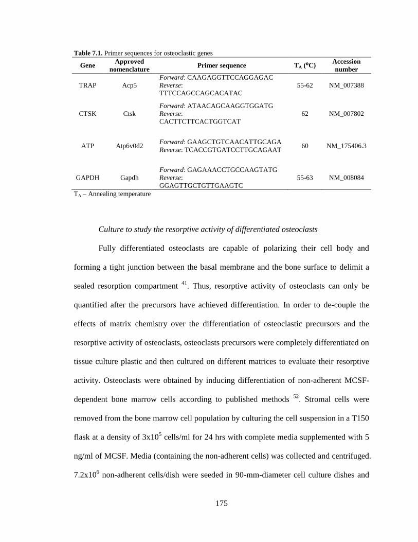
175
Table 7.1. Primer sequences for osteoclastic genes
Gene Approved
nomenclature Primer sequence TA (⁰C)
Accession
number
TRAP Acp5
Forward: CAAGAGGTTCCAGGAGAC
Reverse:
TTTCCAGCCAGCACATAC
55-62 NM_007388
CTSK Ctsk
Forward: ATAACAGCAAGGTGGATG
Reverse:
CACTTCTTCACTGGTCAT
62 NM_007802
ATP Atp6v0d2 Forward: GAAGCTGTCAACATTGCAGA
Reverse: TCACCGTGATCCTTGCAGAAT 60 NM_175406.3
GAPDH Gapdh
Forward: GAGAAACCTGCCAAGTATG
Reverse:
GGAGTTGCTGTTGAAGTC
55-63 NM_008084
TA – Annealing temperature
Culture to study the resorptive activity of differentiated osteoclasts
Fully differentiated osteoclasts are capable of polarizing their cell body and
forming a tight junction between the basal membrane and the bone surface to delimit a
sealed resorption compartment 41
. Thus, resorptive activity of osteoclasts can only be
quantified after the precursors have achieved differentiation. In order to de-couple the
effects of matrix chemistry over the differentiation of osteoclastic precursors and the
resorptive activity of osteoclasts, osteoclasts precursors were completely differentiated on
tissue culture plastic and then cultured on different matrices to evaluate their resorptive
activity. Osteoclasts were obtained by inducing differentiation of non-adherent MCSF-
dependent bone marrow cells according to published methods 52
. Stromal cells were
removed from the bone marrow cell population by culturing the cell suspension in a T150
flask at a density of 3x10⁵ cells/ml for 24 hrs with complete media supplemented with 5
ng/ml of MCSF. Media (containing the non-adherent cells) was collected and centrifuged.
7.2x10⁶ non-adherent cells/dish were seeded in 90-mm-diameter cell culture dishes and
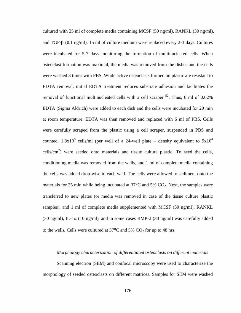
176
cultured with 25 ml of complete media containing MCSF (50 ng/ml), RANKL (30 ng/ml),
and TGF- (0.1 ng/ml). 15 ml of culture medium were replaced every 2-3 days. Cultures
were incubated for 5-7 days monitoring the formation of multinucleated cells. When
osteoclast formation was maximal, the media was removed from the dishes and the cells
were washed 3 times with PBS. While active osteoclasts formed on plastic are resistant to
EDTA removal, initial EDTA treatment reduces substrate adhesion and facilitates the
removal of functional multinucleated cells with a cell scraper 52
. Thus, 6 ml of 0.02%
EDTA (Sigma Aldrich) were added to each dish and the cells were incubated for 20 min
at room temperature. EDTA was then removed and replaced with 6 ml of PBS. Cells
were carefully scraped from the plastic using a cell scraper, suspended in PBS and
counted. 1.8x10⁵ cells/ml (per well of a 24-well plate – density equivalent to 9x10
4
cells/cm2) were seeded onto materials and tissue culture plastic. To seed the cells,
conditioning media was removed from the wells, and 1 ml of complete media containing
the cells was added drop-wise to each well. The cells were allowed to sediment onto the
materials for 25 min while being incubated at 37⁰C and 5% CO2. Next, the samples were
transferred to new plates (or media was removed in case of the tissue culture plastic
samples), and 1 ml of complete media supplemented with MCSF (50 ng/ml), RANKL
(30 ng/ml), IL-110 ng/ml)and in some cases BMP-2 (30 ng/ml) was carefully added
to the wellsCells were cultured at 37⁰C and 5% CO2 for up to 48 hrs.
Morphology characterization of differentiated osteoclasts on different materials
Scanning electron (SEM) and confocal microscopy were used to characterize the
morphology of seeded osteoclasts on different matrices. Samples for SEM were washed
177
with PBS and fixed in a 5% glutaraldehyde solution in DI water for 2 hrs. Samples were
then rinsed with DI water and submerged in 1% osmium tetroxide for 15 min as a
secondary fixation step. Next, the samples were washed 3 times with DI water, and
sequentially dehydrated using increasing concentrations of ethanol. Fixed samples were
dried, sputter-coated with gold and imaged using a Hitachi S-4200 SEM (Finchampstead,
UK). Confocal microscopy was used to verify the presence of actin rings, characteristic
of active osteoclasts, on differentiated cells seeded on dentin. After cell culture, samples
were washed twice with PBS, fixed with 10% formalin for 10 min at room temperature,
and washed again with PBS. Cells were then permeabilized with 0.1% Triton X-100 in
PBS for 5 min, followed by washing with PBS. Cells were incubated with 5 l of
reconstituted methanolic rhodamine phalloidin stock solution per 200 l of PBS (1%
bovine serum album was added to avoid non-specific binding) for 45 min at 37⁰C
protected from light. Samples were washed with PBS, air dried, and cells were imaged
using a Zeiss LSM 510 META inverted microscope using a HeNe1 laser at 543 and 633
nm wavelengths, and 20x magnification.
Quantification of volumetric resorption rates
After 24 or 48 hrs culturing on dentin, active osteoclasts were carefully removed
from the surface using a cell scraper and the samples were air dried overnight. 3D images
of the surface of dentin were obtained using a Zeta instruments Zeta-20 True Color 3D
Optical Profiler (San Jose, CA). Images were collected using a Z-range of 42-48 m and
200 steps resulting in a Z-step size of 0.211-0.239 m. 20x magnification (field of view:
474x356 m2) was used to image at least 3 different areas per sample (total samples: 2).
178
Roughness of the dentin surface with and without cell treatment was measured using the
Zeta 3D imaging software. Zeta Feature Detection Software was used to detect resorption
pits on the surface. A reference surface was established on the non-resorbed areas of
dentin. Deviations from this baseline with depth >1m were selected by the software as
features of interest (resorption pit), and volume of each feature was reported. The total
resorbed volume per image was normalized to the cross sectional area analyzed.
Results
Differentiation of CD11b+ osteoclast precursors mineralized matrices and tissue
culture plastic
Figure 7.1 shows images of CD11b+ osteoclast precursors fed with differentiating
growth factors and cultured on tissue culture plastic and TCP surfaces. After 28 days,
differentiated precursors fused and stained positive for intracellular TRAP (cells cultured
on dentin and BG were also TRAP+). Cells with a wide range of size and number of
nuclei were identified on tissue culture plastic (Figure 7.1A-B). While cells seeded on
mineralized matrices also stained positive for intracellular TRAP, the size of the cells was
significantly smaller (Figure 7.1C). Culture of osteoclast precursors with only MCSF
generated no positive staining for TRAP which confirmed the lack of committed
osteoclasts in the initial cell population (Figure 7.1D).
179
Figure 7.1. Intracellular TRAP staining of osteoclast precursors cultured for 28 days on
A,B,D) on tissue culture plastic, and C) TCP. Cell culture media consisted of complete
media supplemented with MCSF (25ng/ml) and RANKL (50ng/ml) for A-C, or MCSF
(25ng/ml) for D.
Differentiation progression of osteoclast precursors seeded on tissue culture
plastic (well), dentin, and BG was monitored in time by measuring their gene expression
and secretion of TRAP into the media (Figure 7.2). Expression of all the genes was
higher for cells cultured on wells. Cells differentiated on dentin and BG had similar
TRAP, and CTSK gene expression, while ATP expression was more similar between
cells seeded on wells and dentin. However, the measured trends did not match the
expected profile of gene expression in time, and this was true for all the materials.
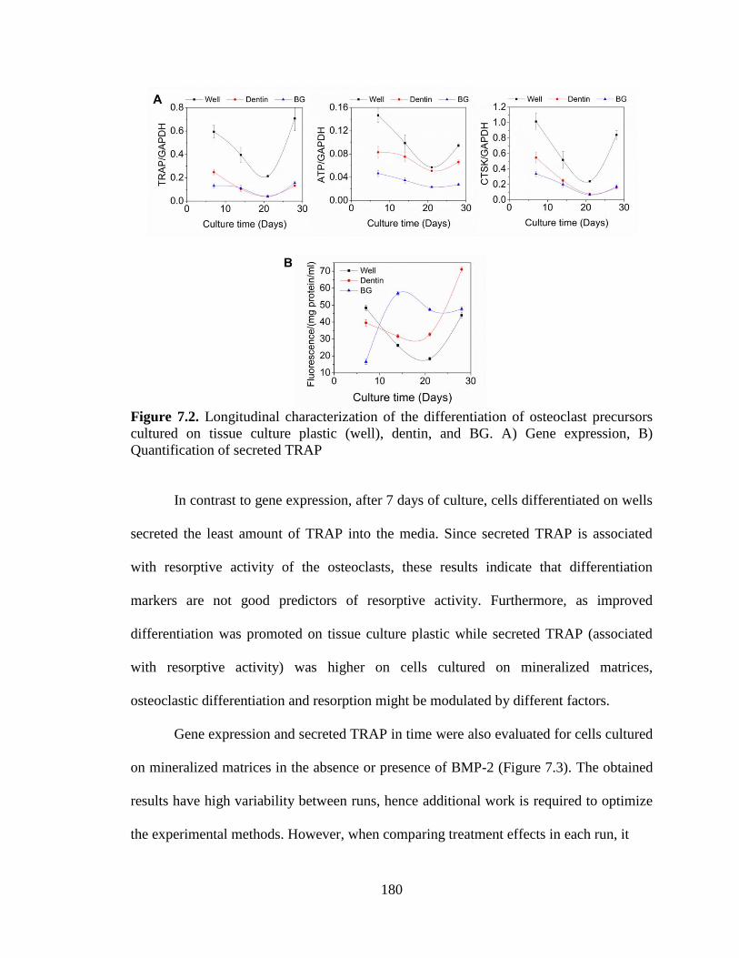
180
Figure 7.2. Longitudinal characterization of the differentiation of osteoclast precursors
cultured on tissue culture plastic (well), dentin, and BG. A) Gene expression, B)
Quantification of secreted TRAP
In contrast to gene expression, after 7 days of culture, cells differentiated on wells
secreted the least amount of TRAP into the media. Since secreted TRAP is associated
with resorptive activity of the osteoclasts, these results indicate that differentiation
markers are not good predictors of resorptive activity. Furthermore, as improved
differentiation was promoted on tissue culture plastic while secreted TRAP (associated
with resorptive activity) was higher on cells cultured on mineralized matrices,
osteoclastic differentiation and resorption might be modulated by different factors.
Gene expression and secreted TRAP in time were also evaluated for cells cultured
on mineralized matrices in the absence or presence of BMP-2 (Figure 7.3). The obtained
results have high variability between runs, hence additional work is required to optimize
the experimental methods. However, when comparing treatment effects in each run, it
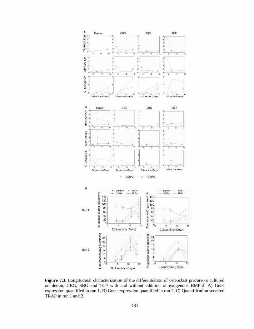
181
Figure 7.3. Longitudinal characterization of the differentiation of osteoclast precursors cultured
on dentin, CBG, SBG and TCP with and without addition of exogenous BMP-2. A) Gene
expression quantified in run 1, B) Gene expression quantified in run 2, C) Quantification secreted
TRAP in run 1 and 2.
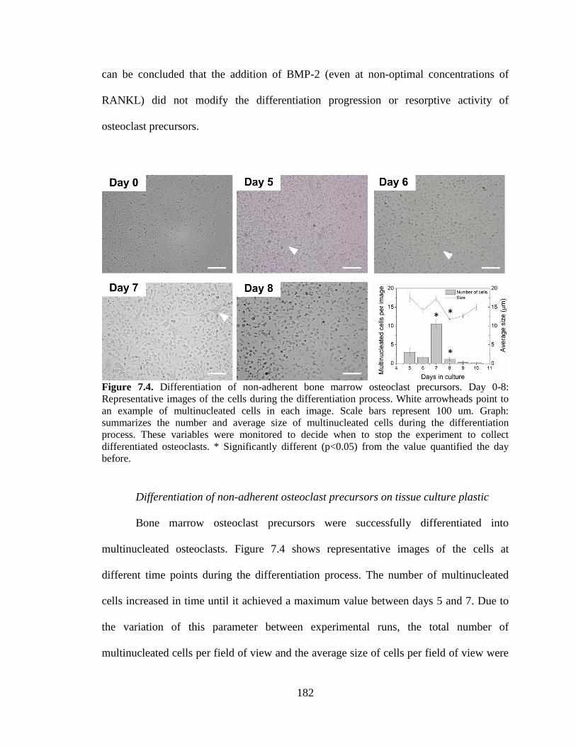
182
can be concluded that the addition of BMP-2 (even at non-optimal concentrations of
RANKL) did not modify the differentiation progression or resorptive activity of
osteoclast precursors.
Figure 7.4. Differentiation of non-adherent bone marrow osteoclast precursors. Day 0-8:
Representative images of the cells during the differentiation process. White arrowheads point to
an example of multinucleated cells in each image. Scale bars represent 100 um. Graph:
summarizes the number and average size of multinucleated cells during the differentiation
process. These variables were monitored to decide when to stop the experiment to collect
differentiated osteoclasts. * Significantly different (p<0.05) from the value quantified the day
before.
Differentiation of non-adherent osteoclast precursors on tissue culture plastic
Bone marrow osteoclast precursors were successfully differentiated into
multinucleated osteoclasts. Figure 7.4 shows representative images of the cells at
different time points during the differentiation process. The number of multinucleated
cells increased in time until it achieved a maximum value between days 5 and 7. Due to
the variation of this parameter between experimental runs, the total number of
multinucleated cells per field of view and the average size of cells per field of view were
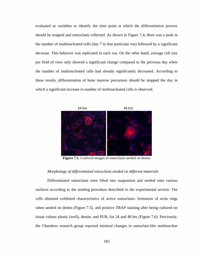
183
evaluated as variables to identify the time point at which the differentiation process
should be stopped and osteoclasts collected. As shown in Figure 7.4, there was a peak in
the number of multinucleated cells (day 7 in that particular run) followed by a significant
decrease. This behavior was replicated in each run. On the other hand, average cell size
per field of view only showed a significant change compared to the previous day when
the number of multinucleated cells had already significantly decreased. According to
these results, differentiation of bone marrow precursors should be stopped the day in
which a significant increase in number of multinucleated cells is observed.
Figure 7.5. Confocal images of osteoclasts seeded on dentin
Morphology of differentiated osteoclasts seeded on different materials
Differentiated osteoclasts were lifted into suspension and seeded onto various
surfaces according to the seeding procedure described in the experimental section. The
cells obtained exhibited characteristics of active osteoclasts: formation of actin rings
when seeded on dentin (Figure 7.5), and positive TRAP staining after being cultured on
tissue culture plastic (well), dentin, and PUR, for 24 and 48 hrs (Figure 7.6). Previously,
the Chambers research group reported minimal changes in osteoclast-like multinuclear
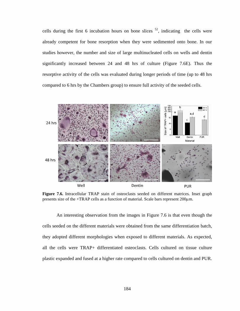
184
cells during the first 6 incubation hours on bone slices 52
, indicating the cells were
already competent for bone resorption when they were sedimented onto bone. In our
studies however, the number and size of large multinucleated cells on wells and dentin
significantly increased between 24 and 48 hrs of culture (Figure 7.6E). Thus the
resorptive activity of the cells was evaluated during longer periods of time (up to 48 hrs
compared to 6 hrs by the Chambers group) to ensure full activity of the seeded cells.
Figure 7.6. Intracellular TRAP stain of osteoclasts seeded on different matrices. Inset graph
presents size of the +TRAP cells as a function of material. Scale bars represent 200m.
An interesting observation from the images in Figure 7.6 is that even though the
cells seeded on the different materials were obtained from the same differentiation batch,
they adopted different morphologies when exposed to different materials. As expected,
all the cells were TRAP+ differentiated osteoclasts. Cells cultured on tissue culture
plastic expanded and fused at a higher rate compared to cells cultured on dentin and PUR.
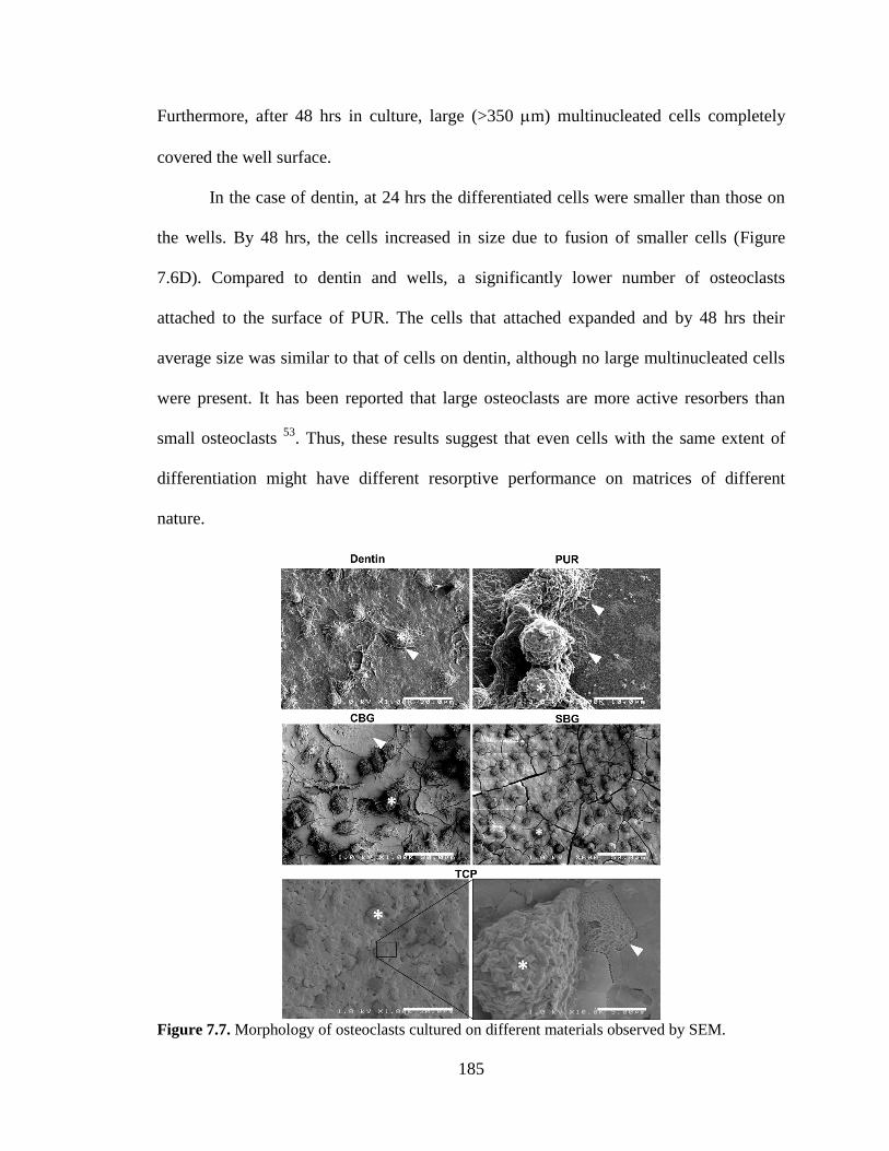
185
Furthermore, after 48 hrs in culture, large (>350 m) multinucleated cells completely
covered the well surface.
In the case of dentin, at 24 hrs the differentiated cells were smaller than those on
the wells. By 48 hrs, the cells increased in size due to fusion of smaller cells (Figure
7.6D). Compared to dentin and wells, a significantly lower number of osteoclasts
attached to the surface of PUR. The cells that attached expanded and by 48 hrs their
average size was similar to that of cells on dentin, although no large multinucleated cells
were present. It has been reported that large osteoclasts are more active resorbers than
small osteoclasts 53
. Thus, these results suggest that even cells with the same extent of
differentiation might have different resorptive performance on matrices of different
nature.
Figure 7.7. Morphology of osteoclasts cultured on different materials observed by SEM.
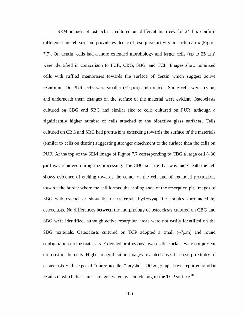
186
SEM images of osteoclasts cultured on different matrices for 24 hrs confirm
differences in cell size and provide evidence of resorptive activity on each matrix (Figure
7.7). On dentin, cells had a more extended morphology and larger cells (up to 25 m)
were identified in comparison to PUR, CBG, SBG, and TCP. Images show polarized
cells with ruffled membranes towards the surface of dentin which suggest active
resorption. On PUR, cells were smaller (~9 m) and rounder. Some cells were fusing,
and underneath them changes on the surface of the material were evident. Osteoclasts
cultured on CBG and SBG had similar size to cells cultured on PUR, although a
significantly higher number of cells attached to the bioactive glass surfaces. Cells
cultured on CBG and SBG had protrusions extending towards the surface of the materials
(similar to cells on dentin) suggesting stronger attachment to the surface than the cells on
PUR. At the top of the SEM image of Figure 7.7 corresponding to CBG a large cell (~30
m) was removed during the processing. The CBG surface that was underneath the cell
shows evidence of etching towards the center of the cell and of extended protrusions
towards the border where the cell formed the sealing zone of the resorption pit. Images of
SBG with osteoclasts show the characteristic hydroxyapatite nodules surrounded by
osteoclasts. No differences between the morphology of osteoclasts cultured on CBG and
SBG were identified, although active resorption areas were not easily identified on the
SBG materials. Osteoclasts cultured on TCP adopted a small (~7m) and round
configuration on the materials. Extended protrusions towards the surface were not present
on most of the cells. Higher magnification images revealed areas in close proximity to
osteoclasts with exposed “micro-needled” crystals. Other groups have reported similar
results in which these areas are generated by acid etching of the TCP surface 30
.
187
Volumetric resorption rate of dentin
The surface topography of dentin was characterized before and after 24 and 48 hr
of osteoclast culture. Cells were removed from the surface and line roughness
measurements were completed. Roughness (Ra) of dentin was initially <1m, and this
parameter significantly increased after osteoclast culture (Figure 7.8A). 3-dimensional
images of the surface suggest that this increase in roughness was associated with the
creation of resorption pits by osteoclasts (Figure 7.8B). Figure 7.8C summarizes the
characteristics of the resorption pits at different time points. Total resorbed volume
normalized to cross-sectional area constantly increased between 0 and 48 hr. Initially
(first 24 hr), this increase was generated by the creation of resorption pits with relatively
constant cross-sectional area. In the second 24 hr period, the cells continued to resorb the
surface by expanding the area of a fraction of the pits (thus increasing the variability of
the pit area), as well as by increasing the excavations’ depths. The dependence of
resorbed volume on time in these first 48 hr of culture can be approximated by a linear
function as shown in Figure 7.8D. According to this, the volumetric resorption rate of
dentin by murine osteoclasts in vitro corresponds to 0.032 m3m
-2hr
-1, or 0.768
m3m
-2day
-1.
Effect of BMP-2 on the volumetric resorption of dentin
In order to evaluate if BMP-2 influenced the resorptive activity of osteoclasts,
differentiated cells were seeded onto dentin slices and 30 ng/ml of BMP-2 were added to
the culture media. After 48 hr of culture, the resorbed volume and area % were measured
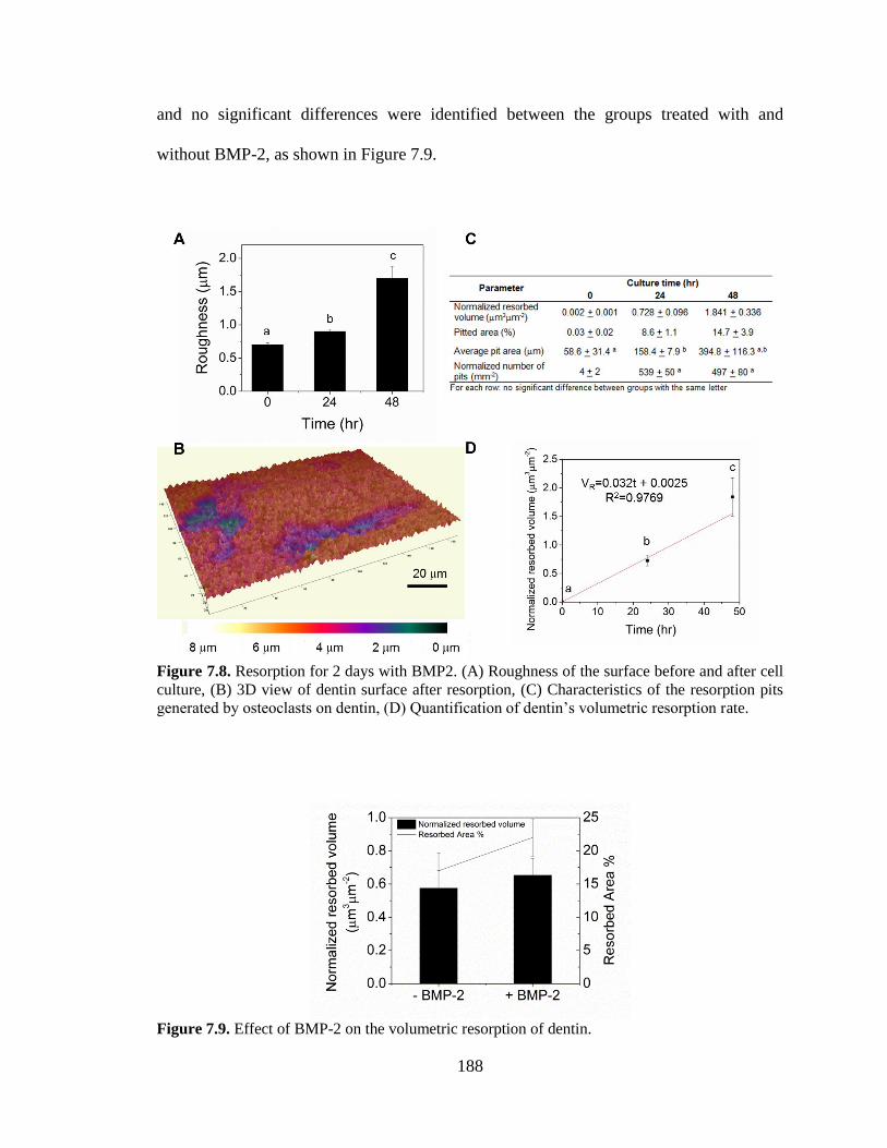
188
and no significant differences were identified between the groups treated with and
without BMP-2, as shown in Figure 7.9.
Figure 7.8. Resorption for 2 days with BMP2. (A) Roughness of the surface before and after cell
culture, (B) 3D view of dentin surface after resorption, (C) Characteristics of the resorption pits
generated by osteoclasts on dentin, (D) Quantification of dentin’s volumetric resorption rate.
Figure 7.9. Effect of BMP-2 on the volumetric resorption of dentin.
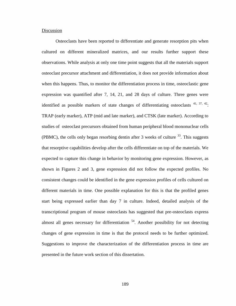
189
Discussion
Osteoclasts have been reported to differentiate and generate resorption pits when
cultured on different mineralized matrices, and our results further support these
observations. While analysis at only one time point suggests that all the materials support
osteoclast precursor attachment and differentiation, it does not provide information about
when this happens. Thus, to monitor the differentiation process in time, osteoclastic gene
expression was quantified after 7, 14, 21, and 28 days of culture. Three genes were
identified as possible markers of state changes of differentiating osteoclasts 41, 37, 42
:
TRAP (early marker), ATP (mid and late marker), and CTSK (late marker). According to
studies of osteoclast precursors obtained from human peripheral blood mononuclear cells
(PBMC), the cells only began resorbing dentin after 3 weeks of culture 33
. This suggests
that resorptive capabilities develop after the cells differentiate on top of the materials. We
expected to capture this change in behavior by monitoring gene expression. However, as
shown in Figures 2 and 3, gene expression did not follow the expected profiles. No
consistent changes could be identified in the gene expression profiles of cells cultured on
different materials in time. One possible explanation for this is that the profiled genes
start being expressed earlier than day 7 in culture. Indeed, detailed analysis of the
transcriptional program of mouse osteoclasts has suggested that pre-osteoclasts express
almost all genes necessary for differentiation 54
. Another possibility for not detecting
changes of gene expression in time is that the protocol needs to be further optimized.
Suggestions to improve the characterization of the differentiation process in time are
presented in the future work section of this dissertation.
190
Osteoclastic differentiation and function are regulated by different humoral agents
52. Maintaining these agents constant in cell culture, our goal was to characterize the
resorptive activity of osteoclasts when exposed to matrices of different nature. Thus, the
differentiation protocol of murine non-adherent bone marrow cells into osteoclasts on
tissue culture plastic was fully characterized. During differentiation, a significant increase
in the number of multinucleated osteoclasts signals that the cells should be collected for
further activity studies. Following the described protocol, functional TRAP+ osteoclasts
that generate characteristic actin rings when seeded on dentin were obtained (Figures 7.4-
7.5).
Microscopy analysis of differentiated osteoclasts cultured on wells, dentin, PUR,
CBG, SBG, and TCP surfaces revealed morphological variances that could be related to
differences in their resorptive activity (Figures 7.5-7.6). Although the seeding procedure
for all the materials was the same, a lower number of cells attached to the PUR disks. The
cells that did attach remained small and round without a polarized cell body characteristic
of actively resorbing osteoclasts (Figure 7.6-PUR). A similar cell body was evident for
the osteoclasts seeded on TCP, although higher number attached to this surface (Figure
7.6-TCP). However, areas with evidence of changes on the substrate surface were present
in close proximity of cells seeded on these materials (PUR and TCP). The changes on the
surface correspond to that reported as resorption of TCP by other authors 27, 28
, and were
shallow and localized. In contrast, osteoclasts cultured on dentin and bioactive glass
surfaces (CBG and SBG) had a polarized structure with extensive protrusions towards the
substrate away from the apical region (Figure 7.6- Dentin, CBG, SBG). The extended
protrusions of cells cultured on dentin, CBG, and SBG suggest tighter attachment to these
191
substrates than to PUR and TCP, and correlate with observations of other groups
reporting different modes of resorption for cells with various sizes 55
or on materials with
different chemistries 32
. Against a widely accepted consensus in the field about
osteoclasts recognizing bone as a substrate for resorption due to the bone mineral content,
it has recently been reported that “bone is not essential for osteoclast activation”, and that
resorptive activity is induced by v3 ligands (vitronectin) which have high affinity for
bone 56
. Since cell culture in this work was completed using 10% FBS in the media, it is
possible that vitronectin present in the serum adhered to the substrates and influenced the
cellular behavior. As suggested in the future work section, it would be worthwhile to
quantify the affinity of vitronectin to the surface of the mineralized matrices and PURs
analyzed in this work to verify if this is the controlling factor that promotes different
resorption rates of materials in vivo.
Measurement of the resorbed volume in time enabled the determination of the in
vitro volumetric resorption rate of dentin as 0.768 m3m
-2day
-1. This value is much
higher than that reported for calcium phosphate ceramics as 0.01 m3m
-2day
-1 32. The
difference between these resorption rates is in line with in vivo observations of calcium
phosphates remaining in the body even after years of implantation 57
. However, the
analysis of calcium phosphates was conducted using human cells (which have lower
metabolic activity than mice cells), as well as a protocol that includes the period of
osteoclast differentiation in the calculation. Thus, it is important to use the same in vitro
protocol described in this work to characterize and compare the resorptive rates of
synthetic matrices. A challenge associated with the measurement of resorption rates of
the synthetic matrices is that these contain pits even before cell culture. We have tested
192
polishing the materials before culture, but due to the porosity of the materials, polishing
exposes pits on the final surface. To overcome this, the pit volume of each sample should
be quantified before and after cell culture, and the resulting difference used to calculate
the corresponding resorption rates.
Addition of BMP-2 to the culture media did not significantly change osteoclastic
differentiation or resorptive activity on mineralized matrices. These results contrast to
previous reports of BMP-2 stimulating the direct differentiation of osteoclasts in vitro.
However, this stimulation took place under very specific culture conditions: on tissue
culture plastic and suboptimal doses of RANKL 46
. It is possible that the addition of a
matrix to the protocol modified the behavior of the cells, and even masked the effect of
BMP-2. Evidence of the impact that the substrate has on the behavior of the cells is
present in Figure 7.5 which shows significant differences in the size of differentiated
osteoclasts cultured on several substrates. An additional observation to keep in mind for
future experiments is that for the experimental runs measuring resorptive activity in the
presence of BMP-2, the cell seeding procedure used was the one described for the
differentiation studies. As a result, the seeded cells were concentrated on a section of the
materials instead of being homogeneously distributed among the whole area.
Consequently, the normalized resorbed volume is lower than that measured for the
resorption rate calculations. While the normalized resorbed volumes should not be
directly compared between these experiments, comparisons between groups in the same
experiment are valid. For future experiments however, the preferred seeding procedure is
the one described for the activity studies since it generates a homogeneous distribution of
the cells on the materials. The preferred seeding procedure removes bias from the results
193
since it allows the experimenter to image random areas of the sample instead of having to
locate the specific area where the cells were initially seeded.
Conclusions
In vitro cell culture methods enabled the characterization of the differentiation
and resorptive activity of osteoclast precursors cultured on different substrates. Pre-
osteoclasts attached and differentiated on mineralized surfaces. BMP-2 did not have a
direct effect on osteoclastic differentiation or activity. When differentiated osteoclasts
where cultured on dentin, CBG, SBG, TCP, and PUR, they exhibited different
morphology. It is expected that differences in morphology will be associated to different
resorption rates, and this remains to be studied in future work. The results from this work
suggest that differentiation of osteoclast precurors is not sufficient for effective resorption.
While this has been reported in the past and associated with lack of expression of specific
genes in the cells 37
, we have shown that even equally differentiated cells behave
differently depending on the substrate for culture. The in vitro cell culture protocol to
study the resorptive activity of murine bone marrow cells on different matrices
introduced in this work was used to determine the volumetric resorptive rate of dentin.
The application of this method for the quantitation and direct comparison between
volumetric resorptive rates of synthetic matrices provides a useful screening tool in the
design of effective biocomposite bone scaffolds.
194
References
1 K. Rezwan, Q. Z. Chen, J. J. Blaker, and A. R. Boccaccini, 'Biodegradable and Bioactive Porous
Polymer/Inorganic Composite Scaffolds for Bone Tissue Engineering', Biomaterials, 27 (2006),
3413-31.
2 A. J. Wagoner Johnson, and B. A. Herschler, 'A Review of the Mechanical Behavior of Cap and
Cap/Polymer Composites for Applications in Bone Replacement and Repair', Acta Biomater, 7
(2011), 16-30.
3 C. Mauli Agrawal, and Robert B. Ray, 'Biodegradable Polymeric Scaffolds for Musculoskeletal
Tissue Engineering', Journal of Biomed Mater Res, 55 (2001), 141-50.
4 Zhen Pan, and Jiandong Ding, 'Poly(Lactide-Co-Glycolide) Porous Scaffolds for Tissue
Engineering and Regenerative Medicine', Interface Focus, 2 (2012), 366-77.
5 Xiaohua Liu, and Peter X. Ma, 'Polymeric Scaffolds for Bone Tissue Engineering', Annals of
Biomedical Engineering, 32 (2004), 477-86.
6 Lakshimi S. Nair, and Cato T. Laurencin, 'Biodegradable Polymers as Biomaterials', Progress in
Polymer Science, 32 (2007), 762-98.
7 Elizabeth M. Christenson, Wafa Soofi, Jennifer Holm, L., Neil Cameron, R., and Antonios G.
Mikos, 'Biodegradable Fumarate-Based Polyhipes as Tissue Engineering Scaffolds',
Biomacromolecules, 8 (2007), 3806-14.
8 S He, MD Timmer, MJ Yaszemski, AW Yasko, PS Engel, and AG Mikos, 'Synthesis of
Biodegradable Poly(Propylene Fumarate) Networks with Poly(Propylene Fumarate)-Diacrylate
Macromers as Crosslinking Agents and Characterization of Their Degradation Products', Polymer,
42 (2001), 1251.
9 R. S. Moglia, J. L. Holm, N. A. Sears, C. J. Wilson, D. M. Harrison, and E. Cosgriff-Hernandez,
'Injectable Polyhipes as High-Porosity Bone Grafts', Biomacromolecules, 12 (2011), 3621-28.
10 X Shi, and AG Mikos, 'Poly(Propylene Fumarate)', in An Introduction to Biomaterials, ed. by SA
Guelcher and JO Hollinger (Boca Raton: CRC Press, 2006), pp. 205-18.
11 R. Adhikari, P. A. Gunatillake, I. Griffiths, L. Tatai, M. Wickramaratna, S. Houshyar, T. Moore,
R. T. M. Mayadunne, J. Field, M. McGee, and T. Carbone, 'Biodegradable Injectable
Polyurethanes: Synthesis and Evaluation for Orthopaedic Applications', Biomaterials, 29 (2008),
3762-70.
12 S Gogolewski, and K Gorna, 'Biodegradable Polyurethane Cancellous Bone Graft Substitutes in
the Treatment of Iliac Crest Defects', J Biomed Mater Res, 80A (2007), 94.
13 SA Guelcher, 'Biodegrabable Polyurethanes: Synthesis and Applications in Regenerative
Medicine', Tissue Engineering: Part B, 14 (2008), 3-17.
14 J. R. Jones, 'Review of Bioactive Glass: From Hench to Hybrids', Acta Biomater, 9 (2013), 4457-
86.
15 J. E. Dumas, T. Davis, G.E. Holt, T. Yoshii, D. S. Perrien, J. S. Nyman, T. Boyce, and S.A.
Guelcher, 'Synthesis, Characterization, and Remodeling of Weight-Bearing Allograft
Bone/Polyurethane Composites in the Rabbit', Acta Biomaterialia, 6 (2010), 2394-406.
16 Larry L. Hench, and Julia M. Polak, 'Third-Generation Biomedical Materials', Science, 295
(2002), 1014-17.
195
17 T. Yoshii, J. E. Dumas, A. Okawa, D. M. Spengler, and S. A. Guelcher, 'Synthesis,
Characterization of Calcium Phosphates/Polyurethane Composites for Weight-Bearing Implants',
Journal of Biomedical Materials Research Part B-Applied Biomaterials, 100B (2012), 32-40.
18 J. Kim, S. McBride, M. Fulmer, R. Harten, Z. Garza, D. D. Dean, V. L. Sylvia, B. Doll, T. L.
Wolfgang, E. Gruskin, and J. O. Hollinger, 'Fiber-Reinforced Calcium Phosphate Cement
Formulations for Cranioplasty Applications: A 52-Week Duration Preclinical Rabbit Calvaria
Study', J Biomed Mater Res B Appl Biomater, 100 (2012), 1170-8.
19 J Kim, MHR Magno, H Waters, B Doll, S McBride, P Alvarez, A Darr, A Vasanji, J Kohn, and
JO Hollinger, 'Bone Regeneration in a Rabbit Critical-Sized Calvarial Model Using Tyrosine-
Derived Polycarbonate Scaffolds', Tissue Engineering: Part A, 18 (2012), 1132-39.
20 JP Fisher, JWM Vehof, D Dean, JPCM van der Waerden, TA Holland, A Mikos, and JA Jansen,
'Soft and Hard Tissue Response to Photocrosslinked Poly(Propylene Fumarate) Scaffolds in a
Rabbit Model', J Biomed Mater Res, 59 (2002), 547-56.
21 S. Scaglione, E. Lazzarini, C. Ilengo, and R. Quarto, 'A Composite Material Model for Improved
Bone Formation', Journal of Tissue Engineering and Regenerative Medicine, 4 (2010), 505-13.
22 ArndtF Schilling, Sandra Filke, Silja Brink, Heike Korbmacher, Michael Amling, and JohannesM
Rueger, 'Osteoclasts and Biomaterials', European Journal of Trauma, 32 (2006), 107-13.
23 Eileen Gentleman, Yann C. Fredholm, Gavin Jell, Nasrin Lotfibakhshaiesh, Matthew D.
O'Donnell, Robert G. Hill, and Molly M. Stevens, 'The Effects of Strontium-Substituted Bioactive
Glasses on Osteoblasts and Osteoclasts in Vitro', Biomaterials, 31 (2010), 3949-56.
24 Y. M. Zhao, Y. M. Zhang, Y. T. Zhao, Y. R. Cai, F. Monchau, A. Lefevre, and H. F. Hildebrand,
'Osteoclast Behaviour on Synthetic Bioglasses with Different Crystallinity in Vitro', in
BIOmaterialien (2005), p. 281.
25 Swati Midha, Wouter van den Bergh, Taek B. Kim, Peter D. Lee, Julian R. Jones, and Christopher
A. Mitchell, 'Bioactive Glass Foam Scaffolds Are Remodelled by Osteoclasts and Support the
Formation of Mineralized Matrix and Vascular Networks in Vitro', Advanced Healthcare
Materials, 2 (2013), 490-99.
26 C. M. Botelho, R. A. Brooks, G. Spence, I. McFarlane, M. A. Lopes, S. M. Best, J. D. Santos, N.
Rushton, and W. Bonfield, 'Differentiation of Mononuclear Precursors into Osteoclasts on the
Surface of Si-Substituted Hydroxyapatite', Journal of Biomedical Materials Research Part A, 78A
(2006), 709-20.
27 Rainer Detsch, Susannne Schaefer, Ulrike Deisinger, Guenter Ziegler, Hermann Seitz, and
Barbara Leukers, 'In Vitro -Osteoclastic Activity Studies on Surfaces of 3d Printed Calcium
Phosphate Scaffolds', Journal of Biomaterials Applications, 26 (2011), 359-80.
28 S. Yamada, D. Heymann, J. M. Bouler, and G. Daculsi, 'Osteoclastic Resorption of Biphasic
Calcium Phosphate Ceramic in Vitro', Journal of Biomedical Materials Research, 37 (1997), 346-
52.
29 S. Yamada, D. Heymann, J.M. Bouler, and G. Daculsi, 'Osteoclastic Resorption of Calcium
Phosphate Ceramics with Different Hydroxyapatite/Β-Tricalcium Phosphate Ratios', Biomaterials,
18 (1997), 1037-41.
30 Zahi Badran, Paul Pilet, Elise Verron, Jean-Michel Bouler, Pierre Weiss, Gaël Grimandi, Jérôme
Guicheux, and Assem Soueidan, 'Assay of in Vitro Osteoclast Activity on Dentine, and Synthetic
Calcium Phosphate Bone Substitutes', Journal of Materials Science: Materials in Medicine, 23
(2012), 797-803.
196
31 Karen Fuller, Jade L. Ross, Kinga A. Szewczyk, Raymond Moss, and Timothy J. Chambers, 'Bone
Is Not Essential for Osteoclas Activation', PLoS one, 5 (2010), 1-14.
32 T. Winkler, E. Hoenig, R. Gildenhaar, G. Berger, D. Fritsch, R. Janssen, M. M. Morlock, and A.
F. Schilling, 'Volumetric Analysis of Osteoclastic Bioresorption of Calcium Phosphate Ceramics
with Different Solubilities', Acta Biomaterialia, 6 (2010), 4127-35.
33 Arndt F. Schilling, Wolfgang Linhart, Sandra Filke, Matthias Gebauer, Thorsten Schinke,
Johannes M. Rueger, and Michael Amling, 'Resorbability of Bone Substitute Biomaterials by
Human Osteoclasts', Biomaterials, 25 (2004), 3963-72.
34 Hidetaka Hayashi, Ken-ichi Nakahama, Takahiro Sato, Takehiko Tuchiya, Yasuyuki Asakawa,
Toshimitu Maemura, Masanobu Tanaka, Mineto Morita, and Ikuo Morita, 'The Role of Mac-1
(Cd11b/Cd18) in Osteoclast Differentiation Induced by Receptor Activator of Nuclear Factor-Κb
Ligand', FEBS Letters, 582 (2008), 3243-48.
35 Kosuke Mizutani, Sudha Sud, and Kenneth J. Pienta, 'Prostate Cancer Promotes Cd11b Positive
Cells to Differentiate into Osteoclasts', Journal of Cellular Biochemistry, 106 (2009), 563-69.
36 GregoryR Mundy, 'Regulatory Mechanisms of Osteoclast Differentiation and Function', Journal of
Bone and Mineral Metabolism, 14 (1996), 59-64.
37 Steven L. Teitelbaum, 'Bone Resorption by Osteoclasts', Science, 289 (2000), 1504-08.
38 Anthony J. Janckila, Ranga N. Parthasarathy, Latha K. Parthasarathy, Ratnam S. Seelan, Yi-
Cheung Hsueh, Jukka Rissanen, Sari L. Alatalo, Jussi M. Halleen, and Lung T. Yam, 'Properties
and Expression of Human Tartrate-Resistant Acid Phosphatase Isoform 5a by Monocyte-Derived
Cells', Journal of Leukocyte Biology, 77 (2005), 209-18.
39 Barrie Kirstein, Timothy J. Chambers, and Karen Fuller, 'Secretion of Tartrate-Resistant Acid
Phosphatase by Osteoclasts Correlates with Resorptive Behavior', Journal of Cellular
Biochemistry, 98 (2006), 1085-94.
40 J. M. Halleen, H. Ylipahkala, S. L. Alatalo, A. J. Janckila, J. E. Heikkinen, H. Suominen, S.
Cheng, and H. K. Väänänen, 'Serum Tartrate-Resistant Acid Phosphatase 5b, but Not 5a,
Correlates with Other Markers of Bone Turnover and Bone Mineral Density', Calcified Tissue
International, 71 (2002), 20-25.
41 William J. Boyle, W. Scott Simonet, and David L. Lacey, 'Osteoclast Differentiation and
Activation', Nature, 423 (2003), 337-42.
42 Haiping Wu, Guoliang Xu, and Yi-Ping Li, 'Atp6v0d2 Is an Essential Component of the
Osteoclast-Specific Proton Pump That Mediates Extracellular Acidification in Bone Resorption',
Journal of Bone and Mineral Research, 24 (2009), 871-85.
43 J. E. Dumas, E. M. Prieto, K.J. Zienkiewicz, T. Guda, J. C. Wenke, J. Bible, G.E. Holt, and S.A.
Guelcher, 'Balancing the Rates of New Bone Formation and Polymer Degradation Enhances
Healing of Weight-Bearing Allograft/Polyurethane Composites in Rabbit Femoral Defects', Tissue
Eng Part A, Not available - ahead of print (2013).
44 J.E. Dumas, P.B. BrownBaer, E.M. Prieto, T. Guda, R.G. Hale, J.C. Wenke, and S.A. Guelcher,
'Injectable Reactive Biocomposites for Bone Healing in Critical-Size Rabbit Calvarial Defects',
Biomedical Materials, 7 (2012), 024112.
45 Jeffrey M. Toth, Scott D. Boden, J Kenneth Burkus, Jeffrey M. Badura, Steven M. Peckham, and
William F. McKay, 'Short-Term Osteoclastic Activity Induced by Locally High Concentrations of
Recombinant Human Bone Morphogenetic Protein–2 in a Cancellous Bone Environment', Spine,
34 (2009), 539-50 10.1097/BRS.0b013e3181952695.
197
46 E. D. Jensen, L. Pham, C. J. Billington, Jr., K. Espe, A. E. Carlson, J. J. Westendorf, A. Petryk, R.
Gopalakrishnan, and K. Mansky, 'Bone Morphogenic Protein 2 Directly Enhances Differentiation
of Murine Osteoclast Precursors', J Cell Biochem, 109 (2010), 672-82.
47 H. Kaneko, T. Arakawa, H. Mano, T. Kaneda, A. Ogasawara, M. Nakagawa, Y. Toyama, Y.
Yabe, M. Kumegawa, and Y. Hakeda, 'Direct Stimulation of Osteoclastic Bone Resorption by
Bone Morphogenetic Protein (Bmp)-2 and Expression of Bmp Receptors in Mature Osteoclasts',
Bone, 27 (2000), 479-86.
48 K. Itoh, N. Udagawa, T. Katagiri, S. Iemura, N. Ueno, H. Yasuda, K. Higashio, J. M. Quinn, M. T.
Gillespie, T. J. Martin, T. Suda, and N. Takahashi, 'Bone Morphogenetic Protein 2 Stimulates
Osteoclast Differentiation and Survival Supported by Receptor Activator of Nuclear Factor-
Kappab Ligand', Endocrinology, 142 (2001), 3656-62.
49 E. Verne, C. Vitale-Brovarone, E. Bui, C. L. Bianchi, and A. R. Boccaccini, 'Surface
Functionalization of Bioactive Glasses', Journal of Biomedical Materials Research Part A, 90A
(2009), 981-92.
50 T. Kokubo, and H. Takadama, 'How Useful Is Sbf in Predicting in Vivo Bone Bioactivity?',
Biomaterials, 27 (2006), 2907-15.
51 Anthony J. Janckila, Karen Takahashi, Susan Z. Sun, and Lung T. Yam, 'Naphthol-Asbi
Phosphate as a Preferred Substrate for Tartrate-Resistant Acid Phosphatase Isoform 5b', Journal of
Bone and Mineral Research, 16 (2001), 788-93.
52 Karen Fuller, Barrie Kirstein, and Timothy J. Chambers, 'Murine Osteoclast Formation and
Function: Differential Regulation by Humoral Agents', Endocrinology, 147 (2006), 1979-85.
53 Morris F. Manolson, Hesheng Yu, Weimin Chen, Yeqi Yao, Keying Li, Rita L. Lees, and Johan
N. M. Heersche, 'The A3 Isoform of the 100-Kda V-Atpase Subunit Is Highly but Differentially
Expressed in Large (≥10 Nuclei) and Small (≤5 Nuclei) Osteoclasts', Journal of Biological
Chemistry, 278 (2003), 49271-78.
54 David Cappellen, Ngoc-Hong Luong-Nguyen, Sandrine Bongiovanni, Olivier Grenet, Christoph
Wanke, and Mira Šuša, 'Transcriptional Program of Mouse Osteoclast Differentiation Governed
by the Macrophage Colony-Stimulating Factor and the Ligand for the Receptor Activator of Nfκb',
Journal of Biological Chemistry, 277 (2002), 21971-82.
55 OkHee Jeon, SuHo Jeong, Yeong-Min Yoo, KyungHwan Kim, DaeSung Yoon, and ChiHyun
Kim, 'Quantification of Temporal Changes in 3d Osteoclastic Resorption Pit Using Confocal Laser
Scanning Microscopy', Tissue Engineering and Regenerative Medicine, 9 (2012), 29-35.
56 Karen Fuller, Jade L. Ross, Kinga A. Szewczyk, Raymond Moss, and Tim J. Chambers, 'Bone Is
Not Essential for Osteoclast Activation', PLoS ONE, 5 (2010), e12837.
57 T. Kraal, M. Mullender, J. H. D. de Bruine, R. Reinhard, A. de Gast, D. J. Kuik, and B. J. van
Royen, 'Resorbability of Rigid Beta-Tricalcium Phosphate Wedges in Open-Wedge High Tibial
Osteotomy: A Retrospective Radiological Study', The Knee, 15 (2008), 201-05.
198
CHAPTER VIII
CONCLUSIONS
The research presented in this dissertation describes the development and
characterization of biodegradable lysine-derived polyurethane (PUR) formulations that
actively participate in the bone healing process. Findings and conclusions presented in
the previous chapters have provided insight into structure-property-performance relations
of PUR foams and composites.
To our knowledge, Chapter III demonstrates the first biofilm-dispersive scaffold
capable of reducing bacterial burden within infected wounds.1 In vitro studies on biofilms
of clinical isolates of Staphylococcus Aureus identified D-Methionine (D-Met), D-
Phenylalanine (D-Phe), D-Proline (D-Pro), and D-Tryptophan (D-Trp) as D-Amino
Acids (D-AA) highly effective at dispersing and preventing biofilm formation.
Incorporation of a free-base D-Met:D-Pro:D-Trp mixture (1:1:1 mass ratio) into a porous
PUR formulation had minimal effects over scaffold porosity and pore size. It did however
reduce the compressive mechanical properties of the materials due to the solid particles
creating defects on the pore walls. The D-AA mixture was released by diffusion
controlled mechanisms for up to 4 weeks, with minimal scaffold degradation. When
implanted into segmental rat defects contaminated with an osteomyelitis bacterial strain
(UAMS-1), PUR foams augmented with ≥5% D-AA mixture significantly reduced
bacterial burden. These results highlight the capability of porous PUR scaffolds to act as
199
delivery vehicles that modify the wound environment and improve the conditions for
future bone remodeling.
PUR composites incorporating -tricalcium phosphate (TCP) particles exhibited
mechanical properties between those of cortical and trabecular bone. Improved interfacial
interactions between the ceramic and PUR phases were promoted by the covalent grafting
of polycaprolactone (PCL) to the surface of TCP particles. As a result, composites
incorporating surface modified particles had reduced water absorption and improved
stability under aqueous conditions as confirmed by the measured mechanical properties.
When tested in vitro, no differences were observed between the differentiation of
osteoblasts and osteoclasts seeded on the composites with modified and unmodified
particles. These results suggest that surface modification of ceramic fillers is a suitable
technique to improve the mechanical performance of settable bone composites. However,
in the case of the TCP particles, a side effect of surface modification was a significant
decrease in particle size. In vitro studies have shown that small particles of
hydroxyapatite (<20 m) generate a strong and cytotoxic inflammatory response.2 To
avoid this response in vivo, surface modification of less brittle and stronger ceramic
fillers should be considered. An example is the use of surface modified bioactive glass
particles, which in turn provide initial weight-bearing capabilities to the PUR scaffolds
(ongoing work in the lab).
Settable ABP-PUR biocomposites exhibited initial mechanical properties
comparable to those of trabecular bone. Independent of allograft particle size, loading,
mineral content, porosity, or BMP-2 dose, all the studied formulations promoted new
bone deposition and progressive healing in time from the host bone interface towards the
200
core of the defects. However, the extent to which healing was achieved varied according
to the parameters listed above.
BMP-2 addition to ABP-PUR biocomposites not only increased the rate of new
bone formation, but also altered the polymer degradation mechanism to approximate a
zero-order process.3 The resulting similar rates of both new bone formation and polymer
degradation resulted in more extensive healing at later time points throughout the graft.
These observations underscore the importance of balancing new bone formation and
polymer degradation rates so as to promote healing of settable weight-bearing bone
composite grafts that maintain bone-like strength while actively remodeling.
Care should be taken with the incorporation of demineralized bone matrix (DBM)
in to bone grafts. Contrary to our initial expectations, when DBM was included in ABP-
PUR composites, healing progression slowed. We associate this with reported variability
of the osteoinductive character of DBM.4-6
This property is affected by several factors
such as donor age and gender, processing, and sterilization protocols.5 However, there are
currently no target properties or standards to describe the osteoinductive capacity of
DBM. Consequently, caution should be exercised by surgeons when incorporating DBM
as part of a treatment, and by scientists when they aim to pose conclusions in studies
utilizing DBM.
In contrast, particle size had a strong effect on the porosity and pore size of ABP-
PUR biocomposites. Both these parameters were reduced upon incorporation of allograft
particles with size <105 m. Furthermore, particle size proved to have an effect on the
healing progression of plug defects. After 12 weeks, particles in the size range of 100-500
m promoted more balanced radial remodeling throughout the defects than particles <105
201
m. Particles in the effective size range also stimulated the deposition of higher quality
bone.
Combined analysis of CT and histomorphometry data suggests interesting
differences in the remodeling mechanism of injectable and moldable ABP-PUR
formulations. Moldable composites (<20% porosity, 67 wt% filler) remodeled by
creeping substitution: osteoclasts resorbed the surface of allograft particles and generated
new pores for cellular inflitration. As part of the infiltrating cells, osteoblasts followed
and deposited new bone on the surface of the remaining allograft particles. In comparison,
injectable composites (37-47% porous, 47 wt% filler) remodeled as cells infiltrated
through the pores and deposited new bone on both the pore walls and the surface of
allograft particles. These observations suggest a fully interconnected osteoconductive
surface is required for bone graft healing. Although porosity might promote faster initial
bone deposition, it also reduces the bone grafts’ mechanical properties. Such reductions
pose significant limitations for weight-bearing applications in which a slower remodeling
progression can favor the appropriate incorporation of the bone graft.
In addition to the in vivo characterization of the various ABP-PUR formulations,
an in vitro cell culture protocol that independently studied the differentiation and
resorptive activity of murine bone marrow cells in the presence of different matrices was
introduced. Pre-osteoclasts attached and differentiated on mineralized matrices. However,
differentiated osteoclasts cultured on dentin, bioactive glass, TCP, and PUR disks
exhibited different morphology. These differences are expected to be associated with
different resorption rates, and this remains to be studied with the implementation of the
developed protocol. Given that osteoclasts were simulateously differentiated and then
202
seeded on the different materials, our observations suggest that osteoclastogenesis
(differentiation) might not be sufficient for effective resorption of matrices. The in vitro
cell culture protocol to study osteoclastic resorptive activity on different matrices was
implemented to determine volumetric resorptive rate of dentin (0.768 m3m
-2day
-1).
Future application of this method to quantify and compare volumetric resorptive rates of
synthetic matrices will provide a useful screening tool to design effective biocomposite
bone scaffolds.
203
References
1 Carlos J. Sanchez Jr, Edna M. Prieto, Chad A. Krueger, Katarzyna J. Zienkiewicz, Desiree R.
Romano, Catherine L. Ward, Kevin S. Akers, Scott A. Guelcher, and Joseph C. Wenke, 'Effects of
Local Delivery of D-Amino Acids from Biofilm-Dispersive Scaffolds on Infection in
Contaminated Rat Segmental Defects', Biomaterials, 34 (2013), 7533-43.
2 JS Sun, HC Liu, WH Chang, J Li, FH Lin, and HC Tai, 'Influence of Hydroxyapatite Particle Size
on Bone Cell Activities: An in Vitro Study', J Biomed Mater Res, 39 (1998), 390-7.
3 J. E. Dumas, E. M. Prieto, K.J. Zienkiewicz, T. Guda, J. C. Wenke, J. Bible, G.E. Holt, and S.A.
Guelcher, 'Balancing the Rates of New Bone Formation and Polymer Degradation Enhances
Healing of Weight-Bearing Allograft/Polyurethane Composites in Rabbit Femoral Defects', Tissue
Eng Part A, Not available - ahead of print (2013).
4 Lingfei Wei, Richard J. Miron, Bin Shi, and Yufeng Zhang, 'Osteoinductive and Osteopromotive
Variability among Different Demineralized Bone Allografts', Clinical Implant Dentistry and
Related Research (2013), n/a-n/a.
5 Elliott Gruskin, Bruce A. Doll, F. William Futrell, John P. Schmitz, and Jeffrey O. Hollinger,
'Demineralized Bone Matrix in Bone Repair: History and Use', Advanced Drug Delivery Reviews,
64 (2012), 1063-77.
6 Hyun W. Bae, Li Zhao, Linda E. A. Kanim, Pamela Wong, Rick B. Delamarter, and Edgar G.
Dawson, 'Intervariability and Intravariability of Bone Morphogenetic Proteins in Commercially
Available Demineralized Bone Matrix Products', Spine, 31 (2006), 1299-306
10.097/01.brs.0000218581.92992.b7.
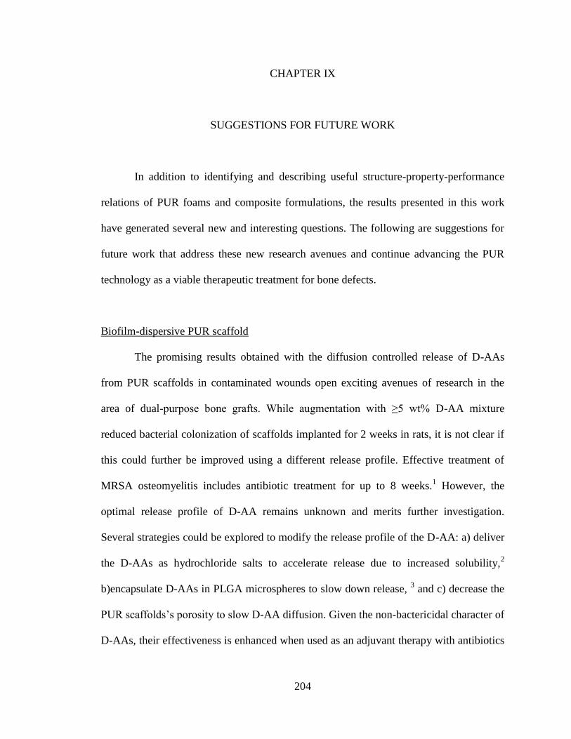
204
CHAPTER IX
SUGGESTIONS FOR FUTURE WORK
In addition to identifying and describing useful structure-property-performance
relations of PUR foams and composite formulations, the results presented in this work
have generated several new and interesting questions. The following are suggestions for
future work that address these new research avenues and continue advancing the PUR
technology as a viable therapeutic treatment for bone defects.
Biofilm-dispersive PUR scaffold
The promising results obtained with the diffusion controlled release of D-AAs
from PUR scaffolds in contaminated wounds open exciting avenues of research in the
area of dual-purpose bone grafts. While augmentation with ≥5 wt% D-AA mixture
reduced bacterial colonization of scaffolds implanted for 2 weeks in rats, it is not clear if
this could further be improved using a different release profile. Effective treatment of
MRSA osteomyelitis includes antibiotic treatment for up to 8 weeks.1 However, the
optimal release profile of D-AA remains unknown and merits further investigation.
Several strategies could be explored to modify the release profile of the D-AA: a) deliver
the D-AAs as hydrochloride salts to accelerate release due to increased solubility,2
b)encapsulate D-AAs in PLGA microspheres to slow down release, 3 and c) decrease the
PUR scaffolds’s porosity to slow D-AA diffusion. Given the non-bactericidal character of
D-AAs, their effectiveness is enhanced when used as an adjuvant therapy with antibiotics
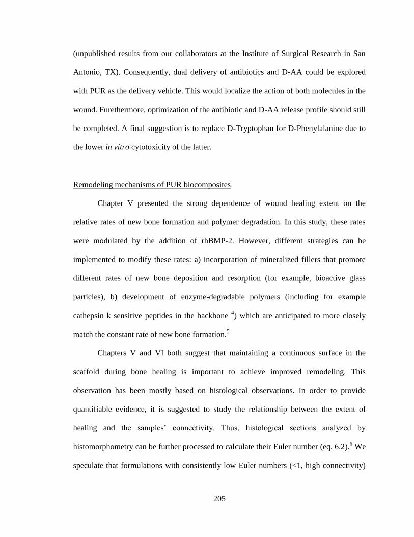
205
(unpublished results from our collaborators at the Institute of Surgical Research in San
Antonio, TX). Consequently, dual delivery of antibiotics and D-AA could be explored
with PUR as the delivery vehicle. This would localize the action of both molecules in the
wound. Furethermore, optimization of the antibiotic and D-AA release profile should still
be completed. A final suggestion is to replace D-Tryptophan for D-Phenylalanine due to
the lower in vitro cytotoxicity of the latter.
Remodeling mechanisms of PUR biocomposites
Chapter V presented the strong dependence of wound healing extent on the
relative rates of new bone formation and polymer degradation. In this study, these rates
were modulated by the addition of rhBMP-2. However, different strategies can be
implemented to modify these rates: a) incorporation of mineralized fillers that promote
different rates of new bone deposition and resorption (for example, bioactive glass
particles), b) development of enzyme-degradable polymers (including for example
cathepsin k sensitive peptides in the backbone 4) which are anticipated to more closely
match the constant rate of new bone formation.5
Chapters V and VI both suggest that maintaining a continuous surface in the
scaffold during bone healing is important to achieve improved remodeling. This
observation has been mostly based on histological observations. In order to provide
quantifiable evidence, it is suggested to study the relationship between the extent of
healing and the samples’ connectivity. Thus, histological sections analyzed by
histomorphometry can be further processed to calculate their Euler number (eq. 6.2).6 We
speculate that formulations with consistently low Euler numbers (<1, high connectivity)
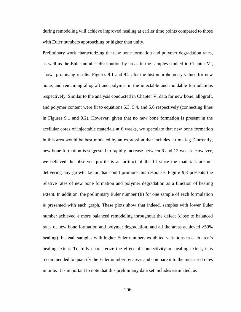
206
during remodeling will achieve improved healing at earlier time points compared to those
with Euler numbers approaching or higher than unity.
Preliminary work characterizing the new bone formation and polymer degradation rates,
as well as the Euler number distribution by areas in the samples studied in Chapter VI,
shows promising results. Figures 9.1 and 9.2 plot the histomorphometry values for new
bone, and remaining allograft and polymer in the injectable and moldable formulations
respectively. Similar to the analysis conducted in Chapter V, data for new bone, allograft,
and polymer content were fit to equations 5.3, 5.4, and 5.6 respectively (connecting lines
in Figures 9.1 and 9.2). However, given that no new bone formation is present in the
acellular cores of injectable materials at 6 weeks, we speculate that new bone formation
in this area would be best modeled by an expression that includes a time lag. Currently,
new bone formation is suggested to rapidly increase between 6 and 12 weeks. However,
we believed the observed profile is an artifact of the fit since the materials are not
delivering any growth factor that could promote this response. Figure 9.3 presents the
relative rates of new bone formation and polymer degradation as a function of healing
extent. In addition, the preliminary Euler number (E) for one sample of each formulation
is presented with each graph. These plots show that indeed, samples with lower Euler
number achieved a more balanced remodeling throughout the defect (close to balanced
rates of new bone formation and polymer degradation, and all the areas achieved >50%
healing). Instead, samples with higher Euler numbers exhibited variations in each area’s
healing extent. To fully characterize the effect of connectivity on healing extent, it is
recommended to quantify the Euler number by areas and compare it to the measured rates
in time. It is important to note that this preliminary data set includes estimated, as
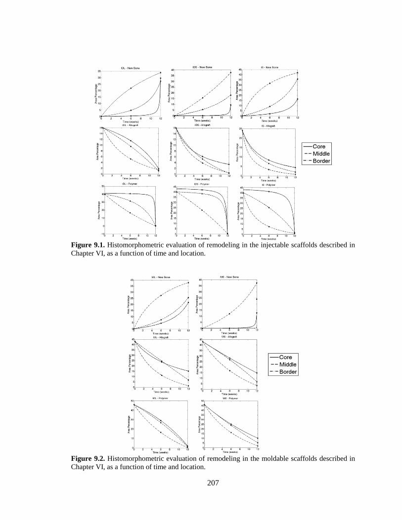
207
Figure 9.1. Histomorphometric evaluation of remodeling in the injectable scaffolds described in
Chapter VI, as a function of time and location.
Figure 9.2. Histomorphometric evaluation of remodeling in the moldable scaffolds described in
Chapter VI, as a function of time and location.
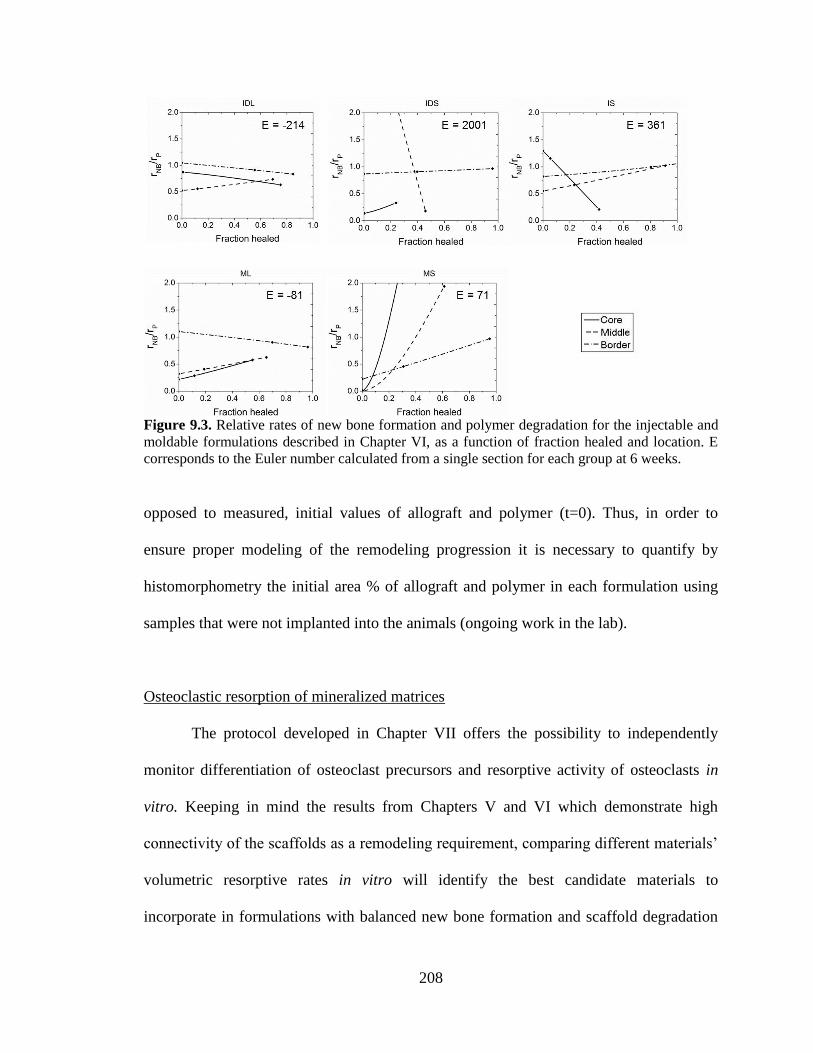
208
Figure 9.3. Relative rates of new bone formation and polymer degradation for the injectable and
moldable formulations described in Chapter VI, as a function of fraction healed and location. E
corresponds to the Euler number calculated from a single section for each group at 6 weeks.
opposed to measured, initial values of allograft and polymer (t=0). Thus, in order to
ensure proper modeling of the remodeling progression it is necessary to quantify by
histomorphometry the initial area % of allograft and polymer in each formulation using
samples that were not implanted into the animals (ongoing work in the lab).
Osteoclastic resorption of mineralized matrices
The protocol developed in Chapter VII offers the possibility to independently
monitor differentiation of osteoclast precursors and resorptive activity of osteoclasts in
vitro. Keeping in mind the results from Chapters V and VI which demonstrate high
connectivity of the scaffolds as a remodeling requirement, comparing different materials’
volumetric resorptive rates in vitro will identify the best candidate materials to
incorporate in formulations with balanced new bone formation and scaffold degradation
209
rates. To start, the protocol should be implemented to quantify and compare the
volumetric resorptive rates of different mineralized matrices used in our lab (TCP,
bioactive glass). While studies have reported osteoclastic effects on the surface of these
materials,8-14
our preliminary studies have shown that these changes are very shallow and
seem to be of significantly smaller volumes than the changes on dentin’s surface. This
also correlates to the observed morphological differences of the cells seeded on each of
these materials. Thus, it is speculated that the volumetric resorption rate of the synthetic
mineralized matrices will be significantly lower than that measured for dentin, and it will
most likely be larger for bioactive glass than for TCP. Rate quantification for these
synthetic matrices poses challenges not present with the dentin samples. In contrast to
dentin, TCP and bioactive glass disks contain pits on the surface even before cells are
cultured on them. To overcome this, the pit volume of each sample should be quantified
before and after cell culture and the resulting difference used to calculate the
corresponding resorption rates. The samples should be very well labeled to avoid
confusion when subtracting initial pitted volumes. Given the location variability of the
initial pits, the subtraction of average initial volume should be avoided to reduce error.
Now that the protocol to quantify volumetric resorption rates is in place, it can be
used to answer questions related to PUR biocomposite formulation for improved healing.
For example, it has recently been reported that the way osteoclasts recognize bone for
resorption is due to its high affinity to vitronectin (an v3 ligand).15
Thus, it would be
interesting to quantify the affinity of vitronectin to the different matrices of interest. A
possible way to do this would be by submerging samples of the same size into a set
protein solution volume. At several time points, the samples could be removed from the
210
solution and incubated in a solution with a set vitronectin antibody set concentration,
followed by incubation with a secondary horseradish peroxidase (HRP)-conjugated
antibody. Further incubation with a solution containing ABTS (an HRP substrate) and
quantification of the optical density at 405 mm would allow determining the total amount
of vitronectin adsorbed on the material’s surface. A follow up question to answer will be:
does the forced adsorption (by drying a known amount of protein on the surface of the
materials) of vitronectin improve the volumetric resorption rates of the matrices? Do
improvements depend on matrix composition? The answers to these questions will aid in
the selection of matrices to further incorporate in PUR biocomposites for in vivo testing.
The polymers used so far in the PUR biocomposite formulation have mostly
undergone hydrolytic degradation in vivo. Results from this work and other research
projects in our lab have shown that a better option would be for the polymers to undergo
cellular degradation. Thus, the protocol developed in Chapter VII can also be
implemented to study the effect that rigidity, as well as oxidative and proteolytic
degradation might have on the volumetric resorptive rates of different polymeric
formulations.
211
References
1 C. Liu, A. Bayer, S. E. Cosgrove, R. S. Daum, S. K. Fridkin, R. J. Gorwitz, S. L. Kaplan, A. W.
Karchmer, D. P. Levine, B. E. Murray, J. Rybak M, D. A. Talan, and H. F. Chambers, 'Clinical
Practice Guidelines by the Infectious Diseases Society of America for the Treatment of
Methicillin-Resistant Staphylococcus Aureus Infections in Adults and Children: Executive
Summary', Clin Infect Dis, 52 (2011), 285-92.
2 B. Li, K. V. Brown, J. C. Wenke, and S. A. Guelcher, 'Sustained Release of Vancomycin from
Polyurethane Scaffolds Inhibits Infection of Bone Wounds in a Rat Femoral Segmental Defect
Model', J Control Release, 145 (2010), 221 - 30.
3 B. Li, T. Yoshii, A. E. Hafeman, J. S. Nyman, J. C. Wenke, and S. A. Guelcher, 'The Effects of
Rhbmp-2 Released from Biodegradable Polyurethane/Microsphere Composite Scaffolds on New
Bone Formation in Rat Femora', Biomaterials, 30 (2009), 6768-79.
4 C. W. Hsu, R. M. Olabisi, E. A. Olmsted-Davis, A. R. Davis, and J. L. West, 'Cathepsin K-
Sensitive Poly(Ethylene Glycol) Hydrogels for Degradation in Response to Bone Resorption',
Journal of Biomedical Materials Research Part A, 98A (2011), 53-62.
5 J. R. Jones, 'Review of Bioactive Glass: From Hench to Hybrids', Acta Biomater, 9 (2013), 4457-
86.
6 WK Pratt, Digital Imaging Processing (Canada: John Wiley & Sons, 2001).
7 Melanie J. Coathup, Qian Cai, Charlie Campion, Thomas Buckland, and Gordon W. Blunn, 'The
Effect of Particle Size on the Osteointegration of Injectable Silicate-Substituted Calcium
Phosphate Bone Substitute Materials', Journal of Biomedical Materials Research Part B: Applied
Biomaterials, 101B (2013), 902-10.
8 Eileen Gentleman, Yann C. Fredholm, Gavin Jell, Nasrin Lotfibakhshaiesh, Matthew D.
O'Donnell, Robert G. Hill, and Molly M. Stevens, 'The Effects of Strontium-Substituted Bioactive
Glasses on Osteoblasts and Osteoclasts in Vitro', Biomaterials, 31 (2010), 3949-56.
9 Y. M. Zhao, Y. M. Zhang, Y. T. Zhao, Y. R. Cai, F. Monchau, A. Lefevre, and H. F. Hildebrand,
'Osteoclast Behaviour on Synthetic Bioglasses with Different Crystallinity in Vitro', in
BIOmaterialien (2005), p. 281.
10 Swati Midha, Wouter van den Bergh, Taek B. Kim, Peter D. Lee, Julian R. Jones, and Christopher
A. Mitchell, 'Bioactive Glass Foam Scaffolds Are Remodelled by Osteoclasts and Support the
Formation of Mineralized Matrix and Vascular Networks in Vitro', Advanced Healthcare
Materials, 2 (2013), 490-99.
11 C. M. Botelho, R. A. Brooks, G. Spence, I. McFarlane, M. A. Lopes, S. M. Best, J. D. Santos, N.
Rushton, and W. Bonfield, 'Differentiation of Mononuclear Precursors into Osteoclasts on the
Surface of Si-Substituted Hydroxyapatite', Journal of Biomedical Materials Research Part A, 78A
(2006), 709-20.
12 Rainer Detsch, Susannne Schaefer, Ulrike Deisinger, Guenter Ziegler, Hermann Seitz, and
Barbara Leukers, 'In Vitro -Osteoclastic Activity Studies on Surfaces of 3d Printed Calcium
Phosphate Scaffolds', Journal of Biomaterials Applications, 26 (2011), 359-80.
13 S. Yamada, D. Heymann, J. M. Bouler, and G. Daculsi, 'Osteoclastic Resorption of Biphasic
Calcium Phosphate Ceramic in Vitro', Journal of Biomedical Materials Research, 37 (1997), 346-
52.
212
14 S. Yamada, D. Heymann, J.M. Bouler, and G. Daculsi, 'Osteoclastic Resorption of Calcium
Phosphate Ceramics with Different Hydroxyapatite/Β-Tricalcium Phosphate Ratios', Biomaterials,
18 (1997), 1037-41.
15 Karen Fuller, Jade L. Ross, Kinga A. Szewczyk, Raymond Moss, and Tim J. Chambers, 'Bone Is
Not Essential for Osteoclast Activation', PLoS ONE, 5 (2010), e12837.

213
APPENDIX A
EXPERIMENTAL PROTOCOLS
Guelcher Lab
Grafting of polycaprolactone to the surface of -TCP particles
214
Method based on the protocol reported in:
C. Kunze, T. Freier, E. Helwig, B. Sandner, D. Reif, A. Wutzler, and H. J. Radusch, 'Surface
Modification of Tricalcium Phosphate for Improvement of the Interfacial Compatibility with
Biodegradable Polymers', Biomaterials, 24 (2003), 967-74.
Materials
-caprolactone (Sigma-Aldrich)
Phosphoric acid (5% in distilled water prepared from concentrated acid bought from
Sigma-Aldrich)
Chloroform (Sigma-Aldrich)
-TriCalcium Phosphate particles (TCP - Berkeley, CA)
Beaker
Magnetic stirrer
Three-necked flask and setup for continuous stirring
Argon
Ceramic funnel
Filter paper
Protonation of TCP particles
1. In a beaker with a magnetic stirrer, add TCP particles and phosphoric acid (5% in
distilled water) with a mass ratio of 1:2
2. Set up the mixing speed such that the TCP particles are well mixed with the acid
3. Mix for 1 hour
4. Stop mixing, allow the protonated TCP particles (p-TCP) to sediment, and remove the
acid solution
5. Wash with distilled water at least three times
6. Filter and dry under vacuum at 40ºC overnight
Grafting reaction
1. Load the three-necked flask with p-TCP particles and -caprolactone using a mass ratio
of 1:4 (molar ratio of 1:10)
2. Setup and start the continuous stirring and flow of argon
3. Heat the reaction flask to 150ºC and allow the reaction to proceed for 4 days
4. To stop the reaction, cool down the reaction flask below 60ºC and continue stirring
Guelcher Lab
Grafting of polycaprolactone to the surface of -TCP particles
215
5. Add chloroform to the reaction flask to solubilize the polymerized polycaprolactone
(PCL)
6. Transfer the content of the reaction flask (PCL and surface modified particles: TCP-PCL)
into 50ml centrifuge tubes
a. TIP: Transfer about 5-10 ml of the reaction content, and 40-45ml of chloroform
to each centrifuge tube
7. Centrifuge for 5 min at 2500 rpm
8. Remove extra chloroform (save this material for further analysis of molecular weight of
the grafted PCL – see note below), add fresh solvent, and mix well
a. NOTE: The molecular weight of the grafted PCL can be measured by GPC. To
do this, collect the chloroform used to wash the reaction flask. Using a rotavapor,
evaporate the chloroform and collect the remaining polymer for further
chromatography analysis
9. Repeat steps 7-8 until the chloroform appears clear after centrifugation.
10. Consolidate all the TCP-PCL powder in one centrifuge tube
11. Dry under vacuum at 40ºC overnight
Guelcher Lab
Measuring the Euler number of a histology section
216
The Euler number is a measure of the connectivity of an image and is calculated by:
E = C – H
Where ‘C’ corresponds to the number of connected objects, and ‘H’ to the number of holes in the
image (Pratt, 2001). As such, E=1 represents an image without holes, E>>1 an image with low
connectivity, and E<<1 a highly connected image with holes.
Materials
Stained histology sections
GIMP software
ImageJ software
MATLAB software
Procedure
1. Image the region of interest of the histology section
2. Using GIMP, manually color over the areas corresponding to new bone, allograft, and
remaining polymer
3. When all the objects have been colored, use GIMP threshold to select the colored areas
a. Colors threshold
b. Use this tool to only select the manually colored areas
c. Export as a TIFF file
4. Convert the TIFF file generated in step 3 into a binary image using ImageJ
a. Open the file using ImageJ
b. Select Process Binary Make binary
c. At this point, the colored areas should be white. If this is not the case, invert
image using: Edit Invert
d. Save and replace the TIFF file
5. Use MATLAB to calculate the Euler number of the image
a. Open MATLAB and import the image
b. Use the function: bweuler()
Guelcher Lab
Preparation of ‘bioactive glass’ and ‘surface modified bioactive glass’ disks for cell culture
217
Materials
Bioglass rods (Mo-Sci Health Care)
IsoMet Low Speed Saw (Buehler)
Silicon carbide paper or sandpaper (800, 1000, 1200 grade)
Deionized Water
X-Acto Knife
Procedure
1. Preparation: Clean saw blade with DI water.
2. Add water to the basin below the saw blade. Secure the bioglass rod and cut one disk at a
speed of 6.5. Discard this disk.
3. Cut pieces of the same thickness. Use the micrometer on the saw to measure the thickness
of each disk. After each cut, remove the disk from the water basin, dry the disk using a
kimwipe, and continue to cut the next disk.
4. After all pieces are cut, polish one side of the disk using the three different sandpapers.
Make sure to add DI water to the sandpaper before polishing
o Polishing times for each roughness:
800: 10 seconds
1000: 10 seconds
1200: 60 seconds
5. Dry the disk after polishing. Make a mark on the non-polished side of each disk using an
x-acto knife
Guelcher Lab
Sample sterilization and conditioning for cell culture
218
Materials
Samples to sterilize (usually as disks)
Sonicator
Beaker
DI water
Sterile DI water (filtered for cell culture)
70% ethanol
-MEM (Fisher scientific)
Fetal bovine serum (FBS – Fisher scientific)
Penicillin/Streptomycin solution (Fisher scientific)
Procedure
1. Load the samples to sterilize into a beaker. If sterilizing several groups, prepare one
beaker per group
2. Sonicate samples in deionized distilled water to remove any loose particles: 3 times, 7.5
min each time
3. Submerge in 70% ethanol for 5 min, 2 times
4. Wash with sterile deionized distilled water for 5 min, 2 times
5. Transfer samples to tissue culture plastic plates
6. Condition the sample in incomplete culture medium (MEM) for 5 min, 2 times
a. NOTE: Make sure that the sample is completely covered by the media – for
example, if using a 24-well plate, add 1 ml of media
7. Incubate the sample in complete medium (MEM containing 10% FBS and 1%
Pen/Strep) overnight in cell culture incubator at 37C
a. NOTE: Make sure that the sample is completely covered by the media – for
example, if using a 24-well plate, add 1 ml of media
8. After the incubation, remove the media and proceed to seed cells according to the
specific experiment
Guelcher Lab
Extraction of murine bone marrow cells
219
Original method received from: Javier Esparza (Vanderbilt Center for Bone Biology)
Materials
1.5 ml centrifuge tubes
0.65ml centrifuge tubes
PBS
70% ethanol
Complete media: MEM + 10% Fetal bovine serum + 1% antibiotics (Pen-Strep)
Dissection instruments: forceps and scissors (Fine Science Tools)
Pipettes (200 l and 1 ml) and sterile pipette tips
6-well plate
Ice box with ice
c57bl male mice 4-8 weeks old
Preparations the day before
1. Using 18G needle, punch 2-3 holes at bottom of the 0.65 ml centrifuge tubes.
2. Introduce a 0.65 ml tube into a 1.5 ml tube. Two of these sets will be needed per mouse.
3. Autoclave the tube sets, as well as the forceps and scissors.
Procedure (work under the hood to maintain sterility)
1. Sedate mice using either isofluorane (sedated for minutes), or ketamine (~30 min
sedated)
a. Isofluorane machine settings: 2.5 Iso, 2.5 Oxygen
b. Ketamine: intramuscular injections completed by personnel from the bone center
2. Sacrifice mice by cervical dislocation and excise femur and tibia keeping the whole
bones
a. TIP: keep the fur wet during all the process (using PBS)
b. If removing bones from several mice, submerge the whole bones in PBS in a 6-
well plate and remove the bones from all the mice. Then continue with the
process below.
3. Remove soft tissue from bone
4. Dunk femur and tibia in 70% ethanol for 10sec.
5. Hold femur and tibia with forceps by the knee and using the scissors cut femur and tibia
ends to expose bone marrow
6. Place bones, with the exposed ends towards the bottom, into the 0.65 ml tube inside the
1.5 ml tube
7. Add 500 l PBS into the 0.65 ml tube to keep marrow from drying up
a. Place samples on ice until centrifugation
8. Spin tubes at 12,000 rpm for 2 min
Guelcher Lab
Extraction of murine bone marrow cells
220
9. Discard 0.65 ml tube. Bone marrow cells should be at the bottom of the 1.5ml tube, in
500 l PBS
10. Remove PBS; add 500 l complete media to wash cells
11. Spin tube at 12,000 rpm for 2 min
12. Remove media and resuspend cells in (1-5 ml) complete media
13. Count cells:
a. Transfer 50 l of cell suspension into 450 l of fresh complete media and mix
well (1:10 dilution)
b. Use the cell counter to count diluted cells
Guelcher Lab
Bone marrow cell purification (CD11b+ selection using microbeads)
221
Method based on: instructions from Dr. James Edwards (Vanderbilt Center for Bone Biology),
and adapted for the AutoMACS sorter in the Flow Cytometry Core at Vanderbilt.
Materials
CD11b MicroBeads (human and mouse – Miltenyi Biotec, Cat. No. 130-049-601)
Separation buffer (also called Running buffer – Miltenyi Biotec, Cat. No. 130-091-221)
Rinsing solution (Miltenyi Biotec, Cat. No. 130-091-222)
MACS separator (in the flow cytometry core – reserve time following the link:
http://www.mc.vanderbilt.edu/root/vumc.php?site=flowcytometry) PBS (Fisher scientific)
Complete media: MEM + 10% Fetal bovine serum, 1% antibiotics (Pen-Strep)
Ice box with ice
15 ml centrifuge tubes
Cell labeling (starting solution: cells in complete media)
1. Determine cell number
2. Centrifuge cells – 5 min / 1700 rpm
3. Remove media and wash with PBS
4. Centrifuge and obtain pellet – 5 min / 1700 rpm
5. Remove PBS
6. Re-suspend pellet in 90 l of MACS Separation Buffer (or Running buffer) per 107 total
cells
7. Add 10 l of CD11b MicroBeads per 107 total cells and mix well by pipetting
8. Incubate for 15 min at 4-8ºC (on ice)
9. Wash cells with 1-2 ml of separation buffer per 107 total cells and centrifuge to obtain
pellet – 10 min, 1200 rpm
10. Remove supernatant
11. Re-suspend up to 108 in 500 l of separation buffer and proceed to magnetic separation
Magnetic separation using the AutoMACS Separator
1. Check the dates on the columns. The columns last ~2weeks. If they need to be
changedlook for a flow cytometry core administrator.
2. Run a CLEANING procedure. The run will end by priming the equipment with running
buffer.
3. Place the magnetically labeled cells in the autoMACS separator, and choose the
separation program: Positive selection – “POSSEL”

Guelcher Lab
Bone marrow cell purification (CD11b+ selection using microbeads)
222
4. Collect positive cell fraction in a 15 ml centrifuge tube (outlet port pos1). This is the
purified CD11b+ cell fraction.
5. When done, run a CLEANING procedure
6. Run the SLEEP program and wait until it is sleeping to remove the bottles
7. Turn off the equipment (button on the lower right)
8. Centrifuge, remove buffer
9. Add 1-5 complete media and count cells
10. Prepare a solution with appropriate cell density for seeding according to each experiment
Guelcher Lab
Preparation of cell culture samples for SEM imaging
223
Materials
PBS (Fisher scientific)
5% glutaraldehyde solution in DI water
DI water
1% Osmium tetroxide solution in DI water (concentrated Osmium tetroxide bought from
Sigma-Aldrich) – TOXIC REAGENT
100%, 95%, 75%, 50%, 25% ethanol in DI water
SEM stubs
Double sided carbon tape
Scanning electron microscope (VINSE)
Cell fixation
1. Wash with PBS
2. Fix in 5% glutaraldehyde solution for ~2 hrs at room temperature, or overnight at 4⁰C
a. NOTE: Make sure the sample is completely covered with the solution
3. Rinse with DI water x3 times at room temperature
4. UNDER THE HOOD: Fix in 1% Osmium tetroxide for 15 min at room temperature
5. Wash with DI water x3 times at room temperature
6. Sequentially dehydrate sample in increasing concentrations of ethanol
a. 25% ethanol – 1 hour
b. 50% ethanol – 1 hour
c. 75% ethanol – 30-45 min
d. 95% ethanol – 1 hour
e. 100% ethanol – 1 hour
f. 100% ethanol – 1 hour
g. 100% ethanol – 1 hour
7. Dry samples either at the critical point in the EM core (preferable) or under vacuum
overnight
Stub preparation for SEM imaging
1. Cover the surface of the SEM stub with double sided carbon tape
2. Locate the sample on top of the carbon tape (making sure the side with the cells is on top)
3. If imaging disks, cut a thin and long strip of carbon tape and put it on top of the sample
making sure it connects the surface of the disk and the metallic SEM stub (this will help
reduce charging)
4. Leave the SEM stubs with samples under vacuum until further processing
5. Sputter-coat with gold (at VINSE)
6. Image
Guelcher Lab
Actin staining of cultured osteoclasts
224
This protocol can be used to stain cells on tissue culture plastic or on disks of different materials.
Materials
PBS (Fisher scientific)
10% formalin
0.1% Triton X-100 in PBS
Rhodamine phalloidin (Life Technologies)
Bovine Serum Albumin (BSA - optional)
Mattek plates (for confocal imaging – Cat. # P50G-0-30-F)
Confocal microscope (Cell Imaging Shared Resource)
Procedure
1. Wash cells 2 times with PBS
2. Fix cells in 10% formalin for 10 min at room temperature
a. Make sure the cells are completely covered with the fixative
3. Wash cells 2 times with PBS
4. Permeabilize cells 0.1% Triton X-100 for 5 min
5. Wash cells 2 times with PBS
6. Incubate cells for 45 min at 37⁰C in 5 l of the reconstituted methanolic stock solution of
rhodamine phalloidin per 200 l of PBS (prepare enough to completely cover the cells)
a. Add 1% BSA to avoid non-specific binding
7. Wash with PBS
8. Air dry samples protected from light
9. Image using confocal microscope. Use Mattek plates (with glass center) to support the
samples during imaging
Guelcher Lab
Differentiation of osteoclast precursors on different matrices
225
This protocol is designed to monitor in time the differentiation process of osteoclast precursors
seeded on matrices with different chemistries. Amounts are based on 24-well plates and disk
samples with diameter of 10-12 mm.
Materials
Matrices with different chemistries (ex. dentin, TCP, bioglass)
Reconstituted MCSF (R&D Systems)
Reconstituted RANKL (R&D Sytems)
BMP-2 (R&D Systems)
Complete media (-MEM + 10% Fetal bovine serum + 1% Pen/Strep)
Forceps
24-well plate
Procedure
1. Sterilize and condition matrix samples (according to protocol)
a. Prepare 3 plates with replicates of the materials. Each plate will be stopped at a
different time point for gene expression analysis
2. Extract bone marrow cells (according to protocol)
3. Select CD11b+ cells from bone marrow population (according to protocol)
4. Remove conditioning media from the sample wells
5. Slowly and drop-wise add 1 ml of complete media containing 1.8x10⁵ CD11b+ cells to
each well
6. Allow the cells to sediment for 25 min in the incubator at 37⁰C
7. Carefully, transfer the samples to a new well plate
8. Using the walls of the wells carefully add 1 ml of complete media augmented with
growth factors:
a. Optimal concentrations: MCSF 25 ng/ml; RANKL 50 ng/ml
b. Concentrations to study the effect of BMP-2: MCSF 25 ng/ml; RANKL 30ng/ml;
BMP-2 30 ng/ml
9. Incubate the samples at 37⁰C for up to 14 days changing the media every 3-4 days.
a. To change the media of the plate that will be cultured for the longest time: collect
media from each well and freeze for future analysis of TRAP activity on culture
supernatant (according to protocol) and replace with fresh complete media
containing growth factors
b. To change the media of the other plates: aspirate the media and replace with
fresh complete media containing growth factors
10. At 3 different time points (for ex. Days 4,7,14 or 1,4,7): stop one of the plates and collect
mRNA for gene expression (according to protocol)
11. After all plates are stopped analyze the TRAP activity on the collected media (protocol)
Guelcher Lab
RNA extraction from cells cultured on different matrices (RNeasy kit)
226
Materials
RNAse Zap wipes
Forceps
24-well plate to transfer samples
1.5 ml RNase free vials
KIMWIPES (these are RNAse free)
RNAse free tips
Pipettes
Centrifuge for 2 ml tubes
70% ethanol
QIAShredder Kit (QIAGEN)
RNeasy mini Kit (QIAGEN)
Sterile Water- RNase free
RNase-Free DNase set – (QIAGEN Cat #79254)
NOTE: Change gloves frequently during the process
Procedure
1. Prepare DNAse digestion mixture (1 column per group of pooled samples)
a. Add 10 l DNAase to 70 l RDD buffer per column (include an extra column in
your calculations to have enough material). Mix by GENTLE inversion.
Reagents sensitive to mechanical force. Keep on ice.
2. COMPLETELY remove media from wells w/ pipette and wash with PBS or water
3. Move samples to empty well
a. NOTE: To achieve enough mRNA you will need to pool samples together. To do
this, either a) combine more than one sample into a clean well and continue with
the steps below, or b) follow step 4 and transfer the RLT buffer to the next well
with a sample; repeat this until the cells from all the wells of interest have been
pooled together into the 700 l of RLT buffer
4. Disrupt cells by adding 700 l RLT Buffer/well (pipette around to mix). All the samples
should be covered in buffer. Incubate at room temperature for about 5 min.
5. Transfer lysate into a QIAshredder spin column (PURPLE) in 2 ml collection tube
(transfer maximum 700 l to each column at a time).
6. Centrifuge at 2 min full speed (13200 rpm) and collect the homogenized lysate.
7. Repeat for remaining lysate
8. To the homogenized lysate, add volume equal to the total amount RLT buffer of 70%
ethanol. Mix by pipetting, do not centrifuge.
9. Add 700 l of lysate with ethanol to pink column in 2 ml collection tube (RNeasy kit)
Guelcher Lab
RNA extraction from cells cultured on different matrices (RNeasy kit)
227
10. Cetrifuge at ≥10,000 rpm for 15 s. DISCARD FLOW THROUGH
11. Repeat with the remaining lysate from the purple shredder columns (each time adding up
to 700 l per column)
12. Add 350 l of wash RW1 to columns and centrifuge for 15 sec at 10,000 rpm. DISCARD
FLOW THROUGH
13. Add 80 l of DNA digestion mix to each column
14. Incubate at room temp for 15 min
15. Add 350 l RW1 again and centrifuge for 15s at 10,000 rpm. Change collection tubes.
16. *****Make sure ethanol has been added to RPE buffer***** Add 500 l Buffer RPE to
the spin column. Centrifuge for 15s at 10,000 rpm. DISCARD FLOW THROUGH
17. Wash again with 500 l RPE. Centrifuge for 2 min at full speed.
18. Centrifuge 1 min at max speed.
19. Transfer column to collection tube with top (1.5 ml)
20. Add 40 l RNase free H20 to column. SOAK 10 min at room temperature
21. Centrifuge 10,000 rpm 1 min
a. RNA will be collected in 40 l water in tube.
22. Remove pink column, close the centrifuge tube and put it on ice.
23. Quantify the concentration of mRNA in each sample using the:
a. Biophotometer and cuvettes (Eppendorf – in the room with the autoclave), or
b. NanoDrop (in Dr. Jamey Young’s lab)
24. Preferably: prepare cDNA immediately. If not possible, store mRNA at -80⁰C
Guelcher Lab
cDNA synthesis and real time qPCR (SYBR green)
228
Materials
RNAse Zap wipes
200 l RNase free vials
KIMWIPES (these are RNAse free)
RNAse free tips
Pipettes
IQ Real Time SybrGreen PCR Supermix (Biorad) – Cat # 1708880
Microseal ‘B’ adhesive tape for PCR plates – Cat #MSB1001
PCR Plates – Cat #MLL9601
Centrifuge for PCR plate
Thermocycler
Biorad PCR machine
iScript cDNA Kit – Cat # 170-8890
Sterile Water- RNase free
IQ Real Time SybrGreen PCR Supermix (Biorad) – Cat # 1708880
Specific primers
cDNA synthesis
Each tube for cDNA synthesis will contain a total of 20 l mixture distributed as:
a) 4l reaction mix
b) 1 l reverse transcriptase
c) X l mRNA
d) Y l nuclease free water
Where X+Y = 15 l, and a,b,d are part of the iScript kit
1. From mRNA concentration measurements, determine the amount of mRNA to reverse-
transcribe into cDNA per treatment group
a. NOTE: the maximum amount possible will be that in 15 l of mRNA solution
2. Prepare enough mixture of reaction mix and reverse transcriptase for the total amount of
cDNA tubes + 1 extra (for ex. If you have 3 treatment groups, pre-mix 16 l of reaction
mix with 4 l of reverse transcriptase)
3. Transfer into each cDNA tube
a. 5 l of reaction mix and reverse transcriptase pre-mix (prepared in step 2)
b. X l of mRNA (determined in step 1)
c. Y l nuclease free water (such that X+Y=15 l)
4. Centrifuge 3,000 rpm for 3 minutes
5. Load into thermal cycler. Select iScript protocol (details below):
a. 5 minutes at 25C
b. 30 minutes at 42C
c. 5 minutes at 85C
d. Hold at 4C
6. Store the cDNA at -20C
Guelcher Lab
cDNA synthesis and real time qPCR (SYBR green)
229
Real time qPCR
This information is to run the real time qPCR in 20 l using a flat surface 96-well PCR plate in a
CFX machine at the Molecular Biology Core at Vanderbilt. Reserve machine ahead of time using
the Core ordering website.
1. Prepare primer mix (2l of primer mix are needed per well)
a. For a final concentration of 500 nM of primer mix in the reaction well: Mix 95 l
water with 2.5 l forward primer and 2.5 l of reverse primer
2. Determine the cDNA template concentration per well
3. Dilute stored cDNA to desired concentration. Keep in mind that only 8 l of cDNA
solution will be transferred to each reaction well
4. Pre-mix supermix and primer mix enough for all the reaction wells and 1-2 extra ones
a. 10 l supermix/well
b. 2 l of primer mix/well (prepared in step 1)
5. While keeping the PCR plate on ice, load into each well:
a. 12 l of the premix prepared in step 4
b. 8 l of cDNA solution prepared in step 3
c. NOTE: when transferring these amounts avoid making bubbles in the solutions
and change tip for each well
6. Place sticker seal on top of plate making sure it is well sealed
7. Centrifuge at 3,000 rpm for 3 min
8. While on ice, take the plate to the core
9. On the reserved machine:
a. Select the lab’s profile User: Guelcher Password: SG3-4977
b. Go to protocol
c. Select existing (unless you have a different protocol to use)
d. Use CFX_2StepAmp+Melt Protocol (found under PCR protocols in sample files
folder)
e. Press: “Edit selected”
f. Enter temperature information according to your experiment, and save the
protocol
g. Under USER PREFERENCES, make sure that your email is included so that
results are sent to you at the end of the run
h. Open machine by pressing OPEN LID (under start run menu)
i. Place plate firmly on metal grid
j. Close lid using the software by pressing CLOSE LID (under start run menu)
k. Click START RUN
Guelcher Lab
Intracellular TRAP staining
230
This method is for staining cells on tissue culture plastic or seeded on disks of materials
Materials
Sigma kit 387A (contains citrate solution, fast Garnet GBC base solution, sodium nitrite
solution, naphthol AS-BI phosphate solution, acetate solution, tartrate solution, and
hematoxylin solution)
Acetone
Formaldehyde, 37%
DI water
Procedure
The amounts below can be modified according to the number of samples as long as the
proportions remain constant.
1. Pre-warm sufficient DI water for day’s use to 37⁰C. Check temperature before use
2. Prepare fixative solution in a glass vial:
a. 25 ml citrate solution + 65 ml acetone + 8 ml of formaldehyde 37%
b. Cap tightly and allow to equilibrate at room temperature
3. In a glass vial prepare diazotized fast Garnet GBC solution:
a. 0.5 ml fast Garnet GBC solution + 0.5 ml sodium nitrite solution
b. Mix by gentle inversion for 30s and let stand 2 min
4. In a clean glass vial prepare staining solution by mixing well the following:
a. 45 ml DI water at 37⁰C
b. 1 ml diazotized fast Garnet GBC solution (from step 3)
c. 0.5 ml Naphthol AS-BI phosphate solution
d. 2 ml acetate solution
e. 1 ml tartrate solution
5. Warm solution to 37⁰C in water bath or incubator
6. Remove media from wells and wash with DI water
7. Add fixative solution to wells (making sure all the cells are covered) and let stand for 30s.
8. Wash with DI water and do not allow cells to dry
9. Add warm staining solution (from step 5) to each well, making sure all the cells are
covered
10. Incubate for 20 min at 37⁰C protected from light
11. Remove the solution and wash thoroughly with DI water
12. Air dry and evaluate microscopically
Guelcher Lab
Napthol AS-BI Analysis of TRAP Activity in Culture Supernatant
231
Secretion of tartrate resistant acid phosphatase (TRAP) 5b into the supernatant has been found to
correlate with bone resorptive activity.
Biochemical measurement of TRAP activity in culture supernatant is typically conducted using
either para-nitrophenylphosphate (pNPP) or naphthol-AS-BI phosphate (N-ASBI-P) as a
substrate. Compared with nPP, N-AS-BI-P is a more specific substrate for TRAP 5b activity
(Janckila et al., 2001). As opposed to nPP, N-ASBI-P is selectively hydrolyzed by the 5b isoform
(Janckila et al., 2001). Vaughan et al. (1971) compared the fluorescent properties of various
naphthol derivatives and concluded that AS-BI phosphate was the most effective substrate for
both acid and alkaline phosphatase. Phosphatase hydrolyzes N-AS-BI-P to yield fluorescent N-
AS-BI.
After quantifying the amount of TRAP in the media, total protein in the sample is used to
normalize the results. Total protein is quantified using the BCA kit.
Materials
Napthol AS-BI phosphate - -20⁰C freezer (in the HPLC lab – Sigma Cat. # N2125)
Sodium Acetate
Sodium Tartrate
EGME (2-Methoxyethanol) (Refrigerator in the mixer lab)
0.6 M NaOH (prepare as needed)
DI water
96-well plates (preferably with clear bottom, but all black also work)
BCA Protein kit (Fisher Scientific)
Procedure
1. Thaw frozen media and vortex to mix
2. Prepare buffer
a. Total = 150 µl * (# samples)
i. Ex. = 0.150* 164= 24.6 ml. Make 27 ml
b. To have a total of 2.5mM Napthol AS-BI Phosphate in 4% EGME in 27 ml of
buffer:
i. (27x10-3
L)*(2.5x10⁻³
mol/L)*(452.19 g/mol) = 0.031 g Napthol ASBI-P
ii. Dissolve in 1 ml EGME (add it and vortex it. If it doesn’t dissolve you
can put it in the 37⁰C incubator for a few minutes and vortex again)
c. To have a total of 100 mM sodium acetate in 27 ml of buffer:
i. (27x10-3
L )*(100x10-3
mol/L)*(82.03 g/mol) = 0.221 g
ii. Add 0.221 g Na Acetate to 13 ml DI water
iii. ml DI water = (Total buffer ml – ml EGME)/2
d. To have a total of 50 mM sodium tartrate in 27 ml of buffer
i. (27x10-3
L)*(50x10-3
mol/L)*(230.08 g/mol) = 0.311 g
ii. Add 0.311g Na Tartrate to 13 mL DI water
iii. ml DI water = (Total buffer ml – ml EGME)/2
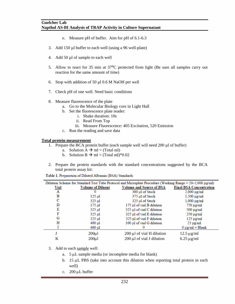
Guelcher Lab
Napthol AS-BI Analysis of TRAP Activity in Culture Supernatant
232
e. Measure pH of buffer. Aim for pH of 6.1-6.3
3. Add 150 µl buffer to each well (using a 96 well-plate)
4. Add 50 µl of sample to each well
5. Allow to react for 35 min at 37⁰C protected from light (Be sure all samples carry out
reaction for the same amount of time)
6. Stop with addition of 50 µl 0.6 M NaOH per well
7. Check pH of one well. Need basic conditions
8. Measure fluorescence of the plate
a. Go to the Molecular Biology core in Light Hall
b. Set the fluorescence plate reader:
i. Shake duration: 10s
ii. Read From Top
iii. Measure Fluorescence: 405 Excitation, 520 Emission
c. Run the reading and save data
Total protein measurement
1. Prepare the BCA protein buffer (each sample well will need 200 µl of buffer)
a. Solution A ml = (Total ml)
b. Solution B ml = (Total ml)*0.02
2. Prepare the protein standards with the standard concentrations suggested by the BCA
total protein assay kit:
J 200l 200 l of vial H dilution 12.5 g/ml
K 200l 200 l of vial J dilution 6.25 g/ml
3. Add to each sample well:
a. 5 L sample media (or incomplete media for blank)
b. 15 L PBS (take into account this dilution when reporting total protein in each
well)
c. 200 L buffer
Guelcher Lab
Napthol AS-BI Analysis of TRAP Activity in Culture Supernatant
233
4. To generate the calibration curve add:
a. 20 l of each standard
b. 200 l of the buffer
5. Incubate at room temperature protected from light for 30 min
6. Measure the absorbance at 562 nm (using the plate reader in the cell culture room)
7. Use the calibration curve to determine the total protein concentration in each well. If the
absorbance reading of the samples is higher than the higher standard, repeat the
experiment using an even more diluted sample.
Guelcher Lab
Measurement of volumetric resorption rates in vitro
234
Differentiation of osteoclast precursors based on published methods from: Karen Fuller,
Barrie Kirstein, and Timothy J. Chambers, 'Murine Osteoclast Formation and Function:
Differential Regulation by Humoral Agents', Endocrinology, 147 (2006), 1979-85.
In this method, osteoclast precursors are first differentiated into active osteoclasts, and then
seeded on different matrices. After culturing the active osteoclasts on the matrices, they are
removed, and the resorbed volume is quantified in time.
Materials
Sterilized and conditioned matrices for analysis (according to protocol)
Complete media: -MEM + 10% Fetal bovine serum + 1% Pen/Strep (all from Fisher
Scientific)
PBS (Fisher Scientific)
Reconstituted MCSF, RANKL, TGF-, IL-1 (R&D Systems)
24-well plate (this can change depending on the size of the samples)
T150 cell culture flasks (Fisher Scientific)
90-mm diameter cell culture dishes
0.02% EDTA solution (Sigma Aldrich)
Cell scraper (Fisher scientific)
Zeta-20 True Color 3D Optical Profiler (Zeta Instruments; located in the National
Research Resource for Imaging Mass Spectrometry – 9th floor in MRB III)
Forceps
Differentiation of non-adherent bone marrow cells
1. Extract murine bone marrow cells from c57bl male mice 4-8 weeks old (according to
protocol)
2. Remove the stromal cells from the bone marrow cell population:
a. Seed the bone marrow cells in a T150 cell culture flask at a density of 3x10⁵
cells/ml
b. Feed with ~30 ml of complete media supplemented with 5 ng/ml MCSF
c. Incubate for 24 hrs at 37⁰C and 5% CO2
d. Aspirate the media and centrifuge (3000 rpm for 3 min)
e. Re-suspend in complete media and count
3. Seed and differentiate the non-adherent cells
a. Seed 7.2x10⁶ cells/dish in 90-mm diameter cell culture dishes
b. Feed with 25 ml of complete media augmented with 50 ng/ml MCSF, 30 ng/ml
RANKL, and 0.1 ng/ml TGF-
c. Every 2-3 change 15 ml of the media per flask
d. Incubate for 5-7 days at 37⁰C and 5% CO2.
i. NOTE: In order to know when to stop the culture, monitor the number of
multinucleated cells each day (for ex. count the number of
multinucleated cells in several regions each day). Stop the culture the day
that the number of multinucleated cells significantly increases compared
to the previous day.
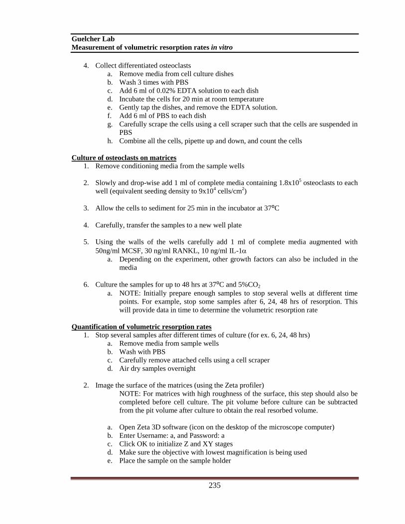
Guelcher Lab
Measurement of volumetric resorption rates in vitro
235
4. Collect differentiated osteoclasts
a. Remove media from cell culture dishes
b. Wash 3 times with PBS
c. Add 6 ml of 0.02% EDTA solution to each dish
d. Incubate the cells for 20 min at room temperature
e. Gently tap the dishes, and remove the EDTA solution.
f. Add 6 ml of PBS to each dish
g. Carefully scrape the cells using a cell scraper such that the cells are suspended in
PBS
h. Combine all the cells, pipette up and down, and count the cells
Culture of osteoclasts on matrices
1. Remove conditioning media from the sample wells
2. Slowly and drop-wise add 1 ml of complete media containing 1.8x10⁵ osteoclasts to each
well (equivalent seeding density to 9x10⁴ cells/cm
2)
3. Allow the cells to sediment for 25 min in the incubator at 37⁰C
4. Carefully, transfer the samples to a new well plate
5. Using the walls of the wells carefully add 1 ml of complete media augmented with
50ng/ml MCSF, 30 ng/ml RANKL, 10 ng/ml IL-1
a. Depending on the experiment, other growth factors can also be included in the
media
6. Culture the samples for up to 48 hrs at 37⁰C and 5%CO2
a. NOTE: Initially prepare enough samples to stop several wells at different time
points. For example, stop some samples after 6, 24, 48 hrs of resorption. This
will provide data in time to determine the volumetric resorption rate
Quantification of volumetric resorption rates
1. Stop several samples after different times of culture (for ex. 6, 24, 48 hrs)
a. Remove media from sample wells
b. Wash with PBS
c. Carefully remove attached cells using a cell scraper
d. Air dry samples overnight
2. Image the surface of the matrices (using the Zeta profiler)
NOTE: For matrices with high roughness of the surface, this step should also be
completed before cell culture. The pit volume before culture can be subtracted
from the pit volume after culture to obtain the real resorbed volume.
a. Open Zeta 3D software (icon on the desktop of the microscope computer)
b. Enter Username: a, and Password: a
c. Click OK to initialize Z and XY stages
d. Make sure the objective with lowest magnification is being used
e. Place the sample on the sample holder
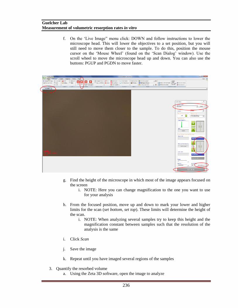
Guelcher Lab
Measurement of volumetric resorption rates in vitro
236
f. On the ‘Live Image” menu click: DOWN and follow instructions to lower the
microscope head. This will lower the objectives to a set position, but you will
still need to move them closer to the sample. To do this, position the mouse
cursor on the ‘Mouse Wheel’ (found on the ‘Scan Dialog’ window). Use the
scroll wheel to move the microscope head up and down. You can also use the
buttons: PGUP and PGDN to move faster.
g. Find the height of the microscope in which most of the image appears focused on
the screen
i. NOTE: Here you can change magnification to the one you want to use
for your analysis
h. From the focused position, move up and down to mark your lower and higher
limits for the scan (set bottom, set top). These limits will determine the height of
the scan.
i. NOTE: When analyzing several samples try to keep this height and the
magnification constant between samples such that the resolution of the
analysis is the same
i. Click Scan
j. Save the image
k. Repeat until you have imaged several regions of the samples
3. Quantify the resorbed volume
a. Using the Zeta 3D software, open the image to analyze
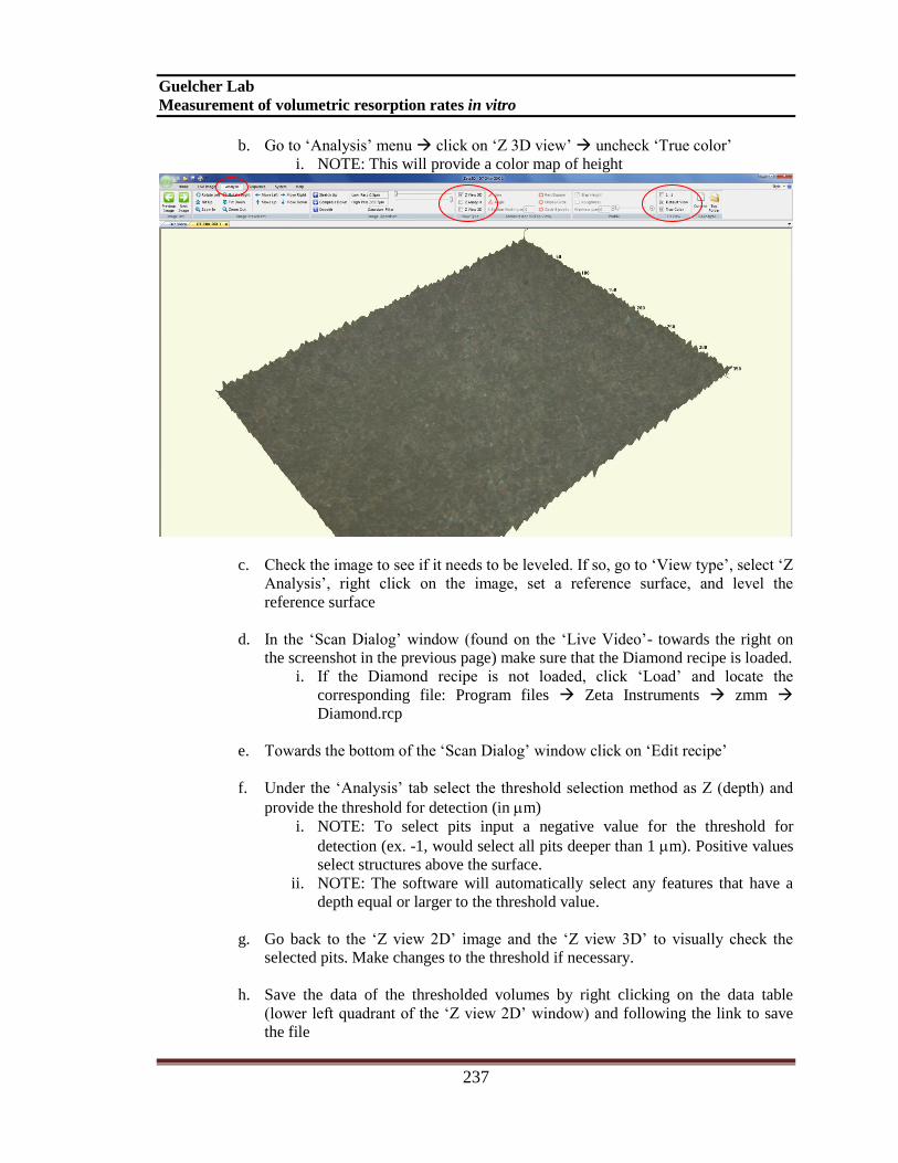
Guelcher Lab
Measurement of volumetric resorption rates in vitro
237
b. Go to ‘Analysis’ menu click on ‘Z 3D view’ uncheck ‘True color’
i. NOTE: This will provide a color map of height
c. Check the image to see if it needs to be leveled. If so, go to ‘View type’, select ‘Z
Analysis’, right click on the image, set a reference surface, and level the
reference surface
d. In the ‘Scan Dialog’ window (found on the ‘Live Video’- towards the right on
the screenshot in the previous page) make sure that the Diamond recipe is loaded.
i. If the Diamond recipe is not loaded, click ‘Load’ and locate the
corresponding file: Program files Zeta Instruments zmm
Diamond.rcp
e. Towards the bottom of the ‘Scan Dialog’ window click on ‘Edit recipe’
f. Under the ‘Analysis’ tab select the threshold selection method as Z (depth) and
provide the threshold for detection (in m)
i. NOTE: To select pits input a negative value for the threshold for
detection (ex. -1, would select all pits deeper than 1 m). Positive values
select structures above the surface.
ii. NOTE: The software will automatically select any features that have a
depth equal or larger to the threshold value.
g. Go back to the ‘Z view 2D’ image and the ‘Z view 3D’ to visually check the
selected pits. Make changes to the threshold if necessary.
h. Save the data of the thresholded volumes by right clicking on the data table
(lower left quadrant of the ‘Z view 2D’ window) and following the link to save
the file