Embed Size (px)
Citation preview
1:15 PM
“Foveal Red Spot,” “Macular Microhole” and “Foveal Photoreceptor Defect” in the Era of High-Resolution Optical Coherence Tomography
• Edward F. Hall, MD • Steven J. Rose, MD • Brian P. Connolly, MD • Ernest G. Guillet, MD • Joseph G Territo • Margaret Powers, BS
OBJECTIVE To suggest anatomically accurate, standardized nomenclature and a
systematic method for categorizing macular microhole, foveal red spot, and related
foveal defects.
PURPOSE "Macular microhole" (MM) and "foveal red spot" (FRS), have been used
interchangeably to describe outer foveal defects. MM has also been used to describe full-thickness foveal defects. We suggest standardized nomenclature and a systematic
method for categorizing these and other related foveal defects based on careful clinical
and high-resolution optical coherence tomography observations.

METHODS Retrospective chart and HR OCT image review of patients presenting with
small (50-150 μm), outer, or full-thickness foveal defects.
RESULTS Thirteen patients presented with central scotoma or blurring in one or both
eyes and a corresponding 50-150 μm red, outer or full-thickness foveal defect detected
by biomicroscopy and HR OCT. Clinical observations and HR OCT image analysis
allowed for classification into three distinct groups of FRS, characterized by an outer
foveal defect, or MM, characterized by a full-thickness foveal defect. Six additional eyes
with an outer foveal defect identical to that observed in our cases of FRS were identified
in patients with a variety of other macular disorders.
CONCLUSION Careful clinical and HR OCT examination allow for the development of
anatomically accurate nomenclature and a systematic method for categorizing patients
with FRS and MM. A characteristic outer foveal defect identical to that noted in patients
with FRS may be observed in other macular disorders and we suggest the term "foveal
photoreceptor defect" to describe this anatomical lesion.
TAKE HOME MESSAGE Clinical and HR OCT exam allow for anatomically accurate
nomenclature and a systematic method for categorizing patients with foveal red spot,
macular microhole, and foveal photoreceptor defect.

1:23 PM
Surgical Outcomes of Macular Holes Associated With Idiopathic Macular Telangiectasia
• Judy E. Kim, MD
OBJECTIVE Macular holes associated with idiopathic macular telangiectasia may have a
poorer surgical outcome and pre-operative OCT findings may guide prognosis.
PURPOSE Idiopathic macular telangiectasia (IMT) Type 2 is an uncommon retinal
condition that may be rarely complicated by development of full thickness macular hole
(FTMH). There are only few reports in the literature regarding management and
outcome of these patients. We describe three patients with type 2 IMT and FTMH who
underwent vitreoretinal surgery for closure of macular holes.
METHODS Retrospective chart review was conducted. These patients were evaluated by
means of optical coherence tomography (OCT), fundoscopy, and fluorescein
angiography (FA) throughout their pre- and post-operative course. Hole closure status
at the last follow-up was evaluated and correlated with pre-operative OCT findings.
Literature review was performed to compare our findings with published reports.
RESULTS All patients showed findings typical for type 2 IMT bilaterally, including
lacunae and retinal thinning seen on OCT and telangiectatic vessels seen on FA.Four
eyes of three patients exhibited FTMH. Pars plana vitrectomy was performed in one eye
of three patients using typical surgical methods for management of FTMH, including
peeling of the internal limiting membrane, C3F8 gas tamponade, and face down
positioning. In one patient, the hole did not close. In the second patient, the hole closed
but reopened. In the third patient, the hole remained closed for the duration of follow-
up. This patient had the smallest intraretinal lacunae prior to FTMH formation and
smallest diameter FTMH prior to surgical repair. Literature review show generally poor
surgical prognosis in these patients.
CONCLUSION Outcomes of vitreoretinal surgery for closure of FTMH in the setting of type
2 IMT may be less successful than typical macular hole surgery without associated IMT.
Pre-operative OCT findings may be helpful in pre-operative discussion of prognosis of
surgical outcome.
TAKE HOME MESSAGE Macular holes associated with IMT may have a poorer surgical
outcome. This may be due to the underlying pathophysiology of IMT and on the extent
of disease state prior to development of FTMH.
1:31 PM
SupraChoroidal Buckling: A Novel Approach in Treating Vitreomacular Interface Disorders in Severe Myopia: Initial 1-Year Data
• Ehab N. El-Rayes, MD, PhD
OBJECTIVE Suprachoridal Buckling approach could be added as a surgical choices for
managing vitreomacular interface disorders in sever myopia.
PURPOSE To evaluate the efficacy of supra choroidal buckling procedure using a supra
choroidal catheter, as a new approach in treating myopic vitreomacular interface
disorders specially in difficult cases of retinoschesis with macular hole retinal
detachment in posterior staphyloma depending on the concept of indenting the choroid
only. One year data study.
METHODS A newly developed supra choroidal catheter was used to deliver cross linked
long acting hyalournic acid( facial filler) in the supra choroidal space in the area of the
staphyloma forming a choroidal indenting effect, prior to the injection, pars plana
vitrectomy was performed without ILM peeling to avoid risk of break the roof of foveal
detachment in case of retinoschesis. This indentation was used to treat 5 patients with
myopic retinoschesis, and 7 patients with myopic macular hole retinal detachment, 4 of
which had failed primary repair by vitrectomy prior to inclusion in this trial. Clinical and
OCT evaluation of these patients were done over a year follow up.
RESULTS Retinal layer restoration was achieved in all 5 eyes with myopic retinoschesis.
This was noticed to be gradual over a period of 8 weeks post operatively. With no
recurrence over the 12 month follow up. Visual acuity improved in 4 ( 80 %) eyes 1 line
or more. 4 of the 7 eyes with macular hole detachment showed closure of the holes in
association with resolution of the detachment. 3 eyes showed resolution of the
detachment and flatting of the edge of the holes but with incomplete closure on OCT. 4
eyes ( 57 %) showed improvement of visual acuity of 1 or more lines with no recurrence
of retinal detachment over the 12 month follow up period. The indentation effect was
sufficient over the 12 month follow up period.
CONCLUSION The indentation effect achieved by supra choroidal approach can be used as
method of managing myopic retinoschesis and myopic macular hole with detachment
even in eye with failed primary vitrectomy, counteracting the role of posterior
staphyloma in the role of the disease. The indentation effect is sufficient for first year of
follow up.
TAKE HOME MESSAGE suprachroidal buckling approach directly counteracts the problem
of retinal stretching in vitreomacular interface problems in sever myopia, adding a new
surgical option.
1:45 PM
Vitrectomy Outcomes for Lamellar Macular Holes Associated with Epiretinal Membranes
• John T. Thompson, MD
OBJECTIVE Objective: To report favorable visual results for the use of vitrectomy in eyes
with lamellar macular holes secondary to epiretinal membranes with reduced visual
acuities.
PURPOSE To evaluate the results and complications of pars plana vitrectomy in eyes with
lamellar macular holes.
METHODS Pars plana vitrectomy was performed in a retrospective, consecutive case
series of 55 eyes with vision loss associated with lamellar macular holes and epiretinal
membranes. Epiretinal membranes were removed in all eyes and internal limiting
membrane was removed using ICG in the last 31 eyes (56.4%) in the series.
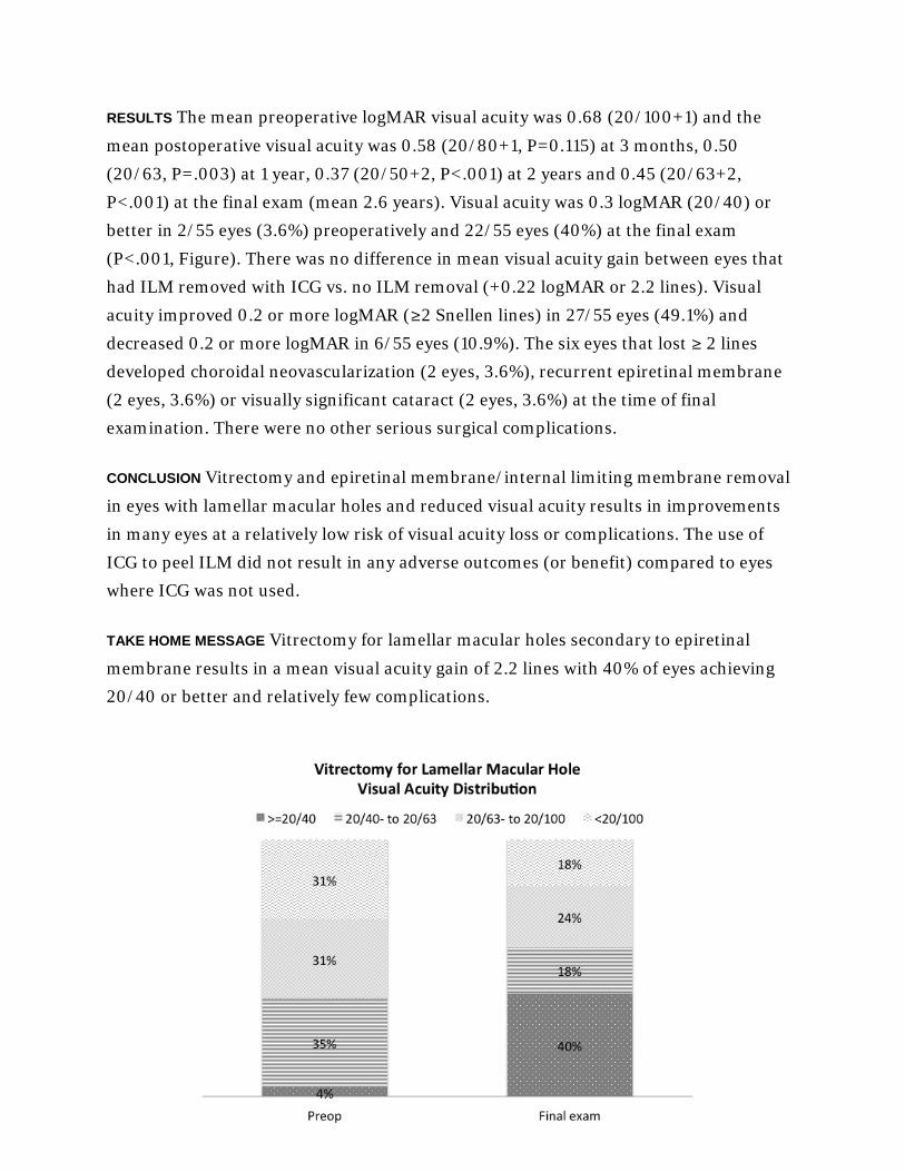
RESULTS The mean preoperative logMAR visual acuity was 0.68 (20/100+1) and the
mean postoperative visual acuity was 0.58 (20/80+1, P=0.115) at 3 months, 0.50
(20/63, P=.003) at 1 year, 0.37 (20/50+2, P<.001) at 2 years and 0.45 (20/63+2,
P<.001) at the final exam (mean 2.6 years). Visual acuity was 0.3 logMAR (20/40) or
better in 2/55 eyes (3.6%) preoperatively and 22/55 eyes (40%) at the final exam
(P<.001, Figure). There was no difference in mean visual acuity gain between eyes that
had ILM removed with ICG vs. no ILM removal (+0.22 logMAR or 2.2 lines). Visual
acuity improved 0.2 or more logMAR (≥2 Snellen lines) in 27/55 eyes (49.1%) and
decreased 0.2 or more logMAR in 6/55 eyes (10.9%). The six eyes that lost ≥ 2 lines
developed choroidal neovascularization (2 eyes, 3.6%), recurrent epiretinal membrane
(2 eyes, 3.6%) or visually significant cataract (2 eyes, 3.6%) at the time of final
examination. There were no other serious surgical complications.
CONCLUSION Vitrectomy and epiretinal membrane/internal limiting membrane removal
in eyes with lamellar macular holes and reduced visual acuity results in improvements
in many eyes at a relatively low risk of visual acuity loss or complications. The use of
ICG to peel ILM did not result in any adverse outcomes (or benefit) compared to eyes
where ICG was not used.
TAKE HOME MESSAGE Vitrectomy for lamellar macular holes secondary to epiretinal
membrane results in a mean visual acuity gain of 2.2 lines with 40% of eyes achieving
20/40 or better and relatively few complications.
1:49 PM
Longitudinal Analysis of the 2008-2010 Medicare Claims Database for Newly Diagnosed Macular Holes
• Peter K. Kaiser, MD • Sunil Srivastava, MD • Pravin U. Dugel, MD
OBJECTIVE To analyze all ophthalmology services and associated costs delivered to
Medicare patients for 12 months following a macular hole diagnosis.
PURPOSE To identify trends in prevalence, diagnostics procedures, interventions, and
related costs associated with Medicare patients with newly diagnosed macular hole.
METHODS Retrospective review of the 2008-2010 Medicare 5% Standard Analytic File for
patients ≥65 years of age; continuously enrolled in Medicare Part A & B from Q1 2008
through Q4 2008; diagnosed with ICD-9 code 362.54 (macular hole) on a HOPD claim,
or a line item diagnosis of 362.54 on a Part B claim in 2009 (defined as the index claim).
A total of 2,033 patients were identified. The study period was defined as one year from
the date of service of the index claim. All claims data were extracted within the study
period. Claims were screened for services that include: ICD-9-CM diagnosis codes with
ophthalmic indications; CPT codes for ophthalmic surgeries and medical services.
RESULTS A majority of the patients in the study were white (88.2%) females (61.2%). The
incidence of macular hole was highest on the west coast with 38.6% of macular hole
claims (fig. 1). The most frequently reported concomitant diagnoses included: 362.56;
366.16; 362.51; 366.9; 365.11; 362.53; 362.83; 366.53; and 362.52. The most common
surgery-related procedures included: 670.42; 669.84; 670.41; 670.28; and 668.21. The
most common medicine-related procedures included: 921.35; 920.15; 922.50; 922.35;
and 922.26. The most common E&M-related procedures included: 920.14; 920.12;
920.04; and 920.02. Additional analyses will be presented at the meeting.
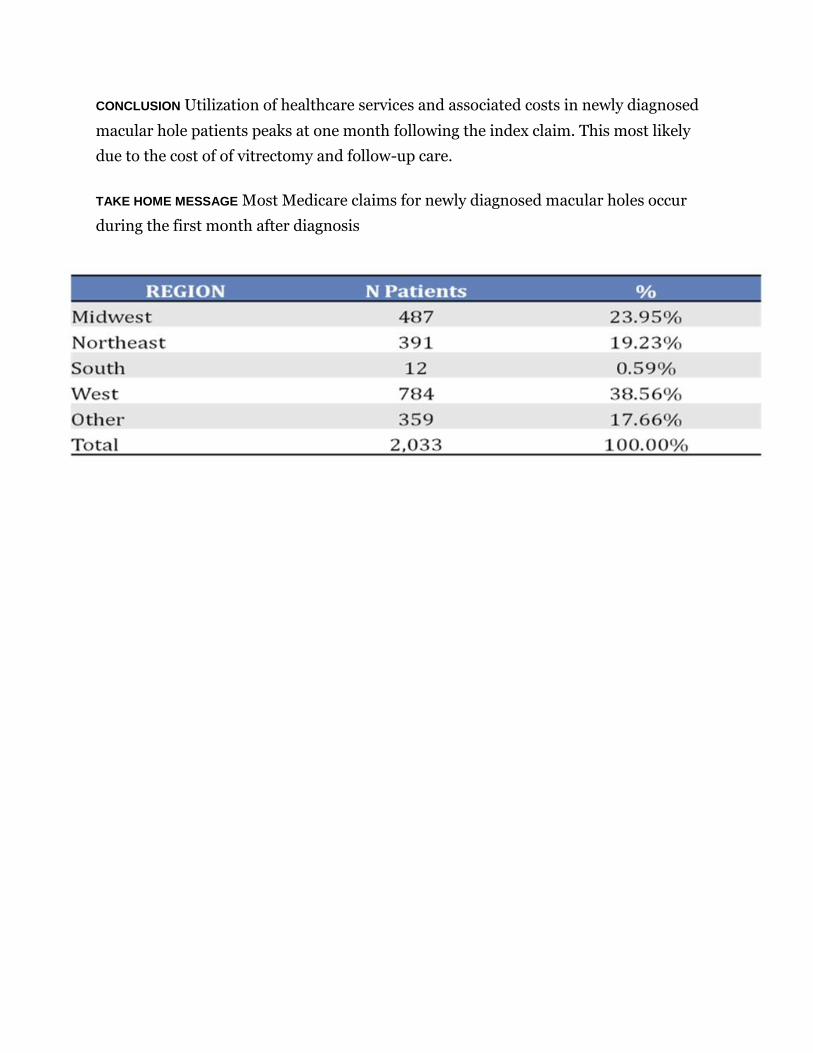
CONCLUSION Utilization of healthcare services and associated costs in newly diagnosed
macular hole patients peaks at one month following the index claim. This most likely
due to the cost of of vitrectomy and follow-up care.
TAKE HOME MESSAGE Most Medicare claims for newly diagnosed macular holes occur
during the first month after diagnosis
1:53 PM
Outcomes and Predictive Factors in Bilateral Macular Holes
• Emmanuel Chang, MD PhD • Antonio Capone, MD • Pooja Garg
OBJECTIVE Evaluate the prevalence, risk factors, outcomes, and visual prognosis in
patients with bilateral macular holes repaired surgically with ICG assisted ILM peeling.
PURPOSE Idiopathic macular holes are a prevalent cause of central vision loss. There are
a limited number of bilateral macular hole outcome reports in literature with various
surgical techniques. Our goal is to evaluate outcomes in patients with bilateral macular
holes characterized using OCT, repaired surgically with ICG assisted ILM peeling, and
report predictive factors and outcomes.
METHODS A retrospective study of all patients who underwent bilateral macular hole
surgery at ARC was performed from 1985 to 2011. Eighty seven cases were identified out
of 4500 patients. Patients with follow up duration less than 3 months or other ocular
conditions limiting visual acuity are excluded. A macular hole surgical repair consisted
of a three port pars plana vitrectomy, ICG assisted ILM peeling, intraocular gas
tamponade, and post operative face down positioning. Our aim is to evaluate potential
predisposing factors on initial presentation as well as family history, age of symptom
onset, time to progression to the second macular hole, and visual and surgical outcomes.
RESULTS Our incidence of bilateral holes was 3.1% in our patient population. We found a
3:1 female to male ratio of patients with bilateral macular holes. The mean time to onset
in the fellow eye is 19.8 months. Approximately one third of patients had asymptomatic
VMT in the opposite eye on initial presentation with macular hole on OCT that
eventually progressed to a macular hole. Surgical closure rate in patients with first
surgery using ICG assisted ILM peeling was 94%. The average length of surgical follow
up for each macular hole is 5.1 years. Fifteen percent of patients developed recurrent
macular holes in one operative eye post-operatively (7% of all surgical eyes). Time to
hole recurrence ranged from 3 months to 8 years with a median of 1.2 years. Strongest
predictor of visual outcome was duration of macular holes less than 6 months yielded
the best results with VA better than 20/40. Stage of hole did not appear to affect visual
outcome contrary to prior reports.
CONCLUSION We report the largest retrospective case series on bilateral macular holes
repaired using a standard ICG assisted ILM peeling surgical technique and discuss the
predictive factors and outcomes in our patient population.
TAKE HOME MESSAGE Outcomes of bilateral macular holes are excellent with ICG-assisted
ILM peeling surgical technique. Prevalence and risk factors are discussed as well as
surgical prognosis.
1:57 PM
25-Gauge Vitrectomy for Macular Holes With and Without Retinal Detachment in Highly Myopic Eyes
• Francesco Boscia, MD
OBJECTIVE Twenty-five-gauge transconjunctival sutureless PPV is suitable for the
management of myopic macular hole, with or without retinal detachment.
PURPOSE To evaluate the surgical outcome of 25-gauge pars plana vitrectomy (PPV) for
macular holes (MH) in highly myopic eyes, with and without retinal detachment (RD).
METHODS Eleven consecutive highly myopic eyes with MH, with and without RD, were
retrospectively studied. Outcome measures were visual acuity (VA), closure of MH,
anatomical retinal reattachment, SD-OCT findings, and complications. Mean patients’
age was 59.2 years (range 40-77). Mean preoperative VA was 0.45±0.68 log MAR. Mean
refractive error was -13.15 (range -6 to -22.0 diopters). Mean axial length was 29.4±2.9
mm. In all cases 25-gauge PPV (Constellation, Alcon) was performed with triamcinolone
visualization. Premacular membrane peeling and ICG-assisted ILM peeling were carried
out. Tamponading agents were in 3 cases SF6 24%, in 5 cases air, and in the others
silicone oil 1000 cs.
RESULTS After a follow-up of 4.14±1 months (range 3-6), VA was preserved (pre-op
0.45±0.68 log MAR, post-op 0.3±0.43 log MAR, p=0.15). Anatomical closure of the
macular hole, confirmed by SD-OCT, and successful retinal reattachment were achieved
in all eyes. IOP was stable during the whole follow-up (pre-op 14.55±3.17 mmHg, post-
op 16±7.22 mmHg, p=0.55).Patients with photoreceptor layer disruption had
significantly worse final best-corrected VA (P = 0.035, 0.005).None of the patients had
hypotony (<5 mmHg), choroidal detachment or endophthalmitis.
CONCLUSION Twenty-five-gauge transconjunctival sutureless PPV showed favorable
results for the management of myopic macular hole with or without retinal detachment.
Photoreceptor layer defects on SD-OCT persist despite surgery, limiting visual outcome.
TAKE HOME MESSAGE Twenty-five-gauge transconjunctival sutureless PPV is suitable for
the management of myopicmacular hole with or without retinal detachment.

2:09 PM
Outcomes of Macular Hole Surgery With Broad ILM Peeling and No Face-Down Positioning
• Raymond Iezzi, MD, MS • Kapil G. Kapoor, MD
OBJECTIVE
To report anatomic and functional outcomes in a single-surgeon retrospective consecutive case series of macular holes (MH) repaired with broad ILM peeling and no post-operative face-down positioning.
PURPOSE
Significant progress has been made in recent years to improve surgical outcomes in MH repair. Despite this, patients are typically instructed to position face down for variable periods of time post-operatively, significantly increasing morbidity. Our purpose is to report surgical methods
associated with a 100% MH closure rate in a large series of patients without any face-down positioning.
METHODS
A retrospective consecutive case-series of all MH surgeries performed at Mayo Clinic by a single surgeon from 3/2009 to 12/2011 was conducted, with IRB approval. After 23ga PPV, ICG dye (.08mg/mL D5W) was applied for 60sec. The ILM was broadly peeled from vascular arcade to arcade, nasally to the disc and temporally for 2 disc diameters using forceps, followed by fluid-air and air-20% SF6 exchanges. Continuous active light management was used to adjust endoillumination for each step. Post-op patients were asked to minimize upgaze and read for the majority of the day for three days. All patients were followed at least 3 months. Wilcoxon signed rank test was used for data analysis.
RESULTS
Forty-six MH repair procedures were performed in 46 eyes of 44 patients. No patients were excluded from the series. Mean age was 68.5 years. Thirty-five patients (76%) were female and 11 (24%) were male. Seventeen patients (37%) were pseudophakic at the time of MH surgery and 29 patients (63%) were phakic. PVD was present pre-operatively in 20 patients (43.4%) and not present in 26 patients (56.6%). Two patients (4.3%) had bilateral MH, and 6 patients (13%) were referred in with recurrent macular holes. Nine (19.6%) MH were stage 2, 27 (58.7%) MH were stage 3, and 10 (21.7%) MH were stage 4. Mean pre-operative BCVA was LogMar 0.60, St. Dev = 0.25 (Snellen 20/81). Mean post-operative BCVA was LogMar 0.20, St. Dev = 0.16 (Snellen 20/32). Post-operative visual acuity was significantly better than pre-operative (p=0.001).There was a 100% MH closure rate, and there were no complications.
CONCLUSION
This is the first series to the authors' knowledge reporting 100% closure rate for MH surgery without face-down positioning. Broad ILM peeling may maximize MH closure. This series further suggests that minimizing upgaze and reading for three-days post-operatively allows adequate MH tamponade in phakic and pseudophakic eyes, obviating the significant burden of face-down positioning in MH repair.
TAKE HOME MESSAGE
Face down positioning is not necessary to achieve 100% macular hole closure. Broad ILM peeling may enhance macular hole closure rates

2:13 PM
OCT Guided Face-Down Positioning for Macular Hole Surgery
• Sumit P. Shah, MD • Varsha Manjunath, MD • Adam H. Rogers, MD • Caroline Baumal, MD • Elias Reichel, MD • Jay S. Duker, MD
OBJECTIVE This retrospective analysis demonstrates the use of SD-OCT to shorten the
duration of face down positioning for most patients while maintaining high macular
hole closure rates.
PURPOSE To utilize spectral-domain optical coherence tomography (SD-OCT) guided
duration of face down positioning in order to study anatomic macular hole closure rates.
METHODS Retrospective review of patients with macular holes undergoing 23g pars plana
vitrectomy and intraocular gas tamponade. SD-OCT imaging was done on post-
operative day one. Patients remained face down for 2 more days if the macular hole was
closed or 6 more days face down if the macular hole was open or indeterminate.
RESULTS Thirty two patients enrolled in the study. The average age was 66 (range, 40 –
90), 20 were females, 12 were males. Eighteen were right eyes and 14 were left eyes.
Nineteen were phakic and 13 were pseudophakic. There were 8 stage two, 12 stage three,
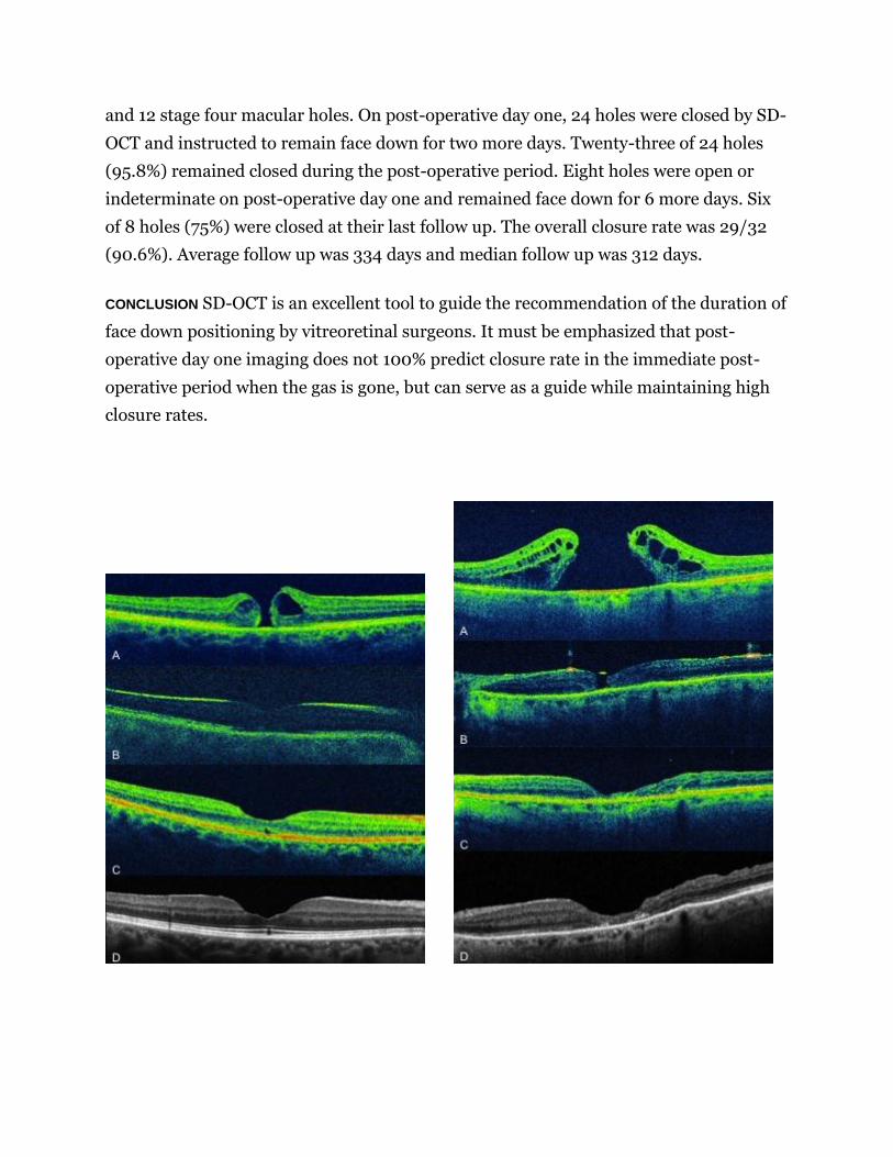
and 12 stage four macular holes. On post-operative day one, 24 holes were closed by SD-
OCT and instructed to remain face down for two more days. Twenty-three of 24 holes
(95.8%) remained closed during the post-operative period. Eight holes were open or
indeterminate on post-operative day one and remained face down for 6 more days. Six
of 8 holes (75%) were closed at their last follow up. The overall closure rate was 29/32
(90.6%). Average follow up was 334 days and median follow up was 312 days.
CONCLUSION SD-OCT is an excellent tool to guide the recommendation of the duration of
face down positioning by vitreoretinal surgeons. It must be emphasized that post-
operative day one imaging does not 100% predict closure rate in the immediate post-
operative period when the gas is gone, but can serve as a guide while maintaining high
closure rates.
2:17 PM
Triamcinolone Acetonide (TA) Assisted
Removal of Internal Limiting Membrane
(ILM)
Homayoun Tabandeh, MD
David S. Boyer, MD
OBJECTIVE To evaluate the status of the perifoveal ILM after TA-assisted stripping of
ILM.
PURPOSE Staining of ILM may be associated with toxicity, increased cost, and increased
surgical time. Recently, there have been reports of infectious endophthalmitis associated
with the use of some of these staining agents. Speckling of ILM by TA assists in
visualization and removal of ILM. The purpose of current study was to evaluate the
status of the perifoveal ILM after TA-assisted stripping of ILM.
METHODS Interventional, non-comparative, clinical case series. Participants: patients
undergoing removal of ILM as part of macular hole or ERM surgery. ILM was visualized
by TA and removed with intraocular ILM forceps. Indocyanine green (ICG) was used to
visualize the status of the remaining ILM. Quality of intraoperative visualization of
retina was graded as good, fair, or poor. The extent of ILM removal was graded: 1)
complete removal of perifoveal ILM, 2) >90% removal of Perifoveal ILM,3) 75-90%
removal of perifoveal ILM, 4) 50-75% removal of perifoveal ILM, 5) < 50% removal of
ILM, and 6) ILM removal with TA visualization was aborted and ILM peeling was
completed by staining with ICG.
RESULTS 21 eyes of 21 patients were included in the study. Intraoperative visualization of
retina was graded as good in 14 eyes, fair in 5 eyes, and poor in 2 eyes. Complete
removal of perifoveal ILM (grade 1) was achieved in 17 (81%) eyes. ILM removal was
grade 2 in 1 eye, grade 3 in 1 eye, and grade 5 in 1 eye. In one eye ICG had to be used for
visualization and subsequent removal of the ILM (grade 6).
CONCLUSION TA is a useful adjunct for intraoperative visualization of ILM. In most cases
ILM can be removed without a need for staining of ILM, reducing risk of toxicity,
infection, and cost. Staining of ILM with dyes such as ICG may be reserved for selected
cases with suboptimal visualization. Techniques for ILM removal will be further
discussed.
TAKE HOME MESSAGE Triamcinolone Acetonideis a useful adjunct for intraoperative
visualization of ILM. In most cases ILM can be removed without a need for staining of
ILM
2:27 PM
Ocriplasmin for the Treatment of Patients
With VMT and Full-Thickness Macular
Hole: Subgroup Responder Analyses from
the Phase III MIVI-TRUST Program
Carl D. Regillo, MD, FACS
OBJECTIVE To evaluate the efficacy of a single intravitreal injection of ocriplasmin as
compared to placebo in the treatment of patients with vitreomacular traction and
macular hole.
PURPOSE The purpose of the MIVI-TRUST Phase III program was to investigate the
effect of a single intravitreal injection of ocriplasmin for the resolution of symptomatic
vitreomacular adhesion (VMA). Subgroup analyses included evaluation of vitreomacular
traction (VMT) release without epiretinal membrane (ERM) and nonsurgical closure of
full-thickness macular hole (FTMH).
METHODS The MIVI-TRUST program was comprised of two phase III, randomized,
double-masked, clinical trials, comparing a single intravitreal injection 125 μg (100 μL)
of ocriplasmin to a single (100 μL) placebo injection, for the treatment of symptomatic
vitreomacular adhesion (VMA). The primary outcome measure was pharmacological
resolution of VMA at Day 28. At baseline, 266 of 652 patients had VMT without ERM
and 153 of a total of 652 treated patients had FTMH. Assessments included best-
corrected visual acuity (VA; ETDRS letters), optical coherence tomography, and adverse
events.
RESULTS At Day 28, 56/188 (29.8%) ocriplasmin patients w/ VMT had resolution of
VMA, compared with 6/78 (7.7%) placebo patients (P≤0.001). The VMA resolution rate
in VMT patients with an adhesion of ≤ 1500 µm in diameter was 38.8% with
ocriplasmin compared to 11.7% with placebo (P≤0.001). Corresponding rates in patients
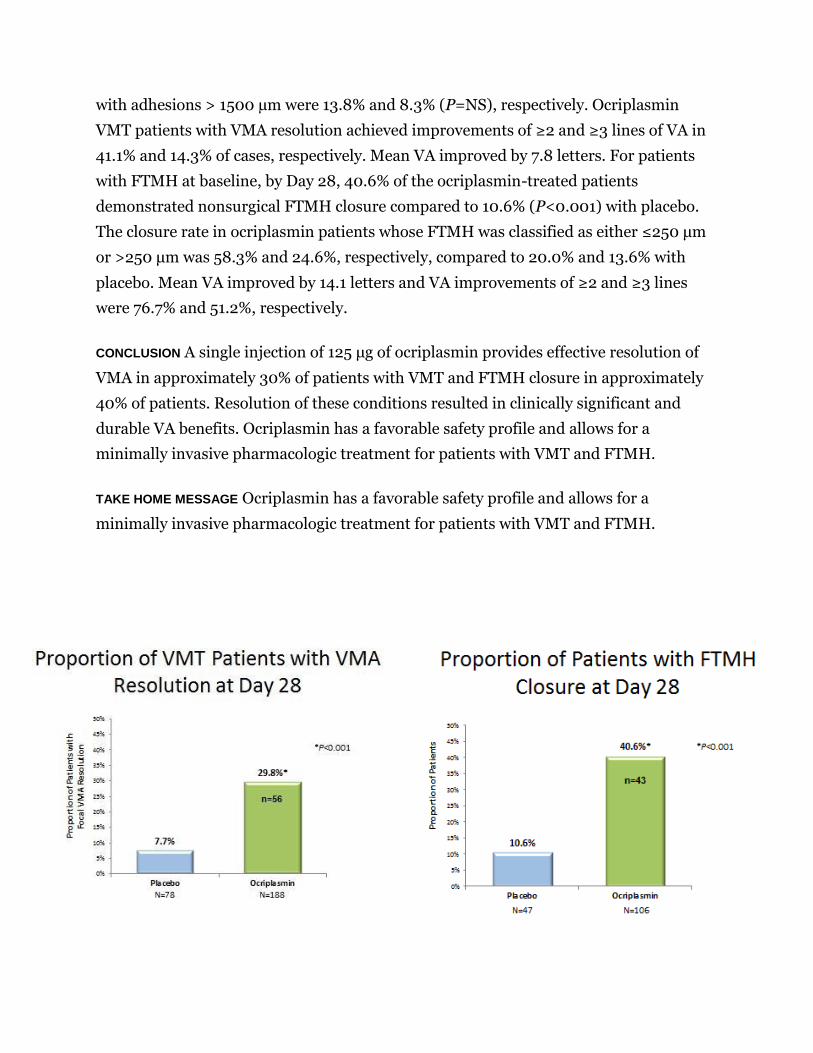
with adhesions > 1500 µm were 13.8% and 8.3% (P=NS), respectively. Ocriplasmin
VMT patients with VMA resolution achieved improvements of ≥2 and ≥3 lines of VA in
41.1% and 14.3% of cases, respectively. Mean VA improved by 7.8 letters. For patients
with FTMH at baseline, by Day 28, 40.6% of the ocriplasmin-treated patients
demonstrated nonsurgical FTMH closure compared to 10.6% (P<0.001) with placebo.
The closure rate in ocriplasmin patients whose FTMH was classified as either ≤250 μm
or >250 μm was 58.3% and 24.6%, respectively, compared to 20.0% and 13.6% with
placebo. Mean VA improved by 14.1 letters and VA improvements of ≥2 and ≥3 lines
were 76.7% and 51.2%, respectively.
CONCLUSION A single injection of 125 μg of ocriplasmin provides effective resolution of
VMA in approximately 30% of patients with VMT and FTMH closure in approximately
40% of patients. Resolution of these conditions resulted in clinically significant and
durable VA benefits. Ocriplasmin has a favorable safety profile and allows for a
minimally invasive pharmacologic treatment for patients with VMT and FTMH.
TAKE HOME MESSAGE Ocriplasmin has a favorable safety profile and allows for a
minimally invasive pharmacologic treatment for patients with VMT and FTMH.
2:31 PM
Ocriplasmin Single Intravitreal Injection for the Resolution of Symptomatic Vitreomacular Adhesion Including Macular Hole: MIVI-TRUST Safety Findings
• Baruch D. Kuppermann, MD, PhD
OBJECTIVE To evaluate the safety of a single intravitreal injection of ocriplasmin
compared to an injection of placebo in the resolution of symptomatic vitreomacular
adhesion including macular hole.
PURPOSE To determine whether ocriplasmin can be used safely as a single intravitreal
injection for the pharmacological resolution of symptomatic vitreomacular adhesion
(VMA) including macular hole.
METHODS Safety was investigated in 2 Phase III clinical trials of a single intravitreal
injection 125 μg (100 μL) of ocriplasmin compared to a single placebo injection (100 μL)
for the pharmacological treatment of symptomatic VMA including macular hole over 6
months. Patients were allowed to have vitrectomy after Day 28 at the investigator's
discretion. Safety assessments included reported adverse events (AEs). Ocular exams
including best-corrected visual acuity (VA), intraocular pressure, slit lamp and dilated
retinal examination, and optical coherence tomography were performed at all study
visits. Fundus photography and fluorescein angiography were done at baseline and
month 6.
RESULTS The most commonly reported ocular AEs deemed possibly study drug-related,
occurring ≥5% incidence and more commonly than placebo included: vitreous floaters,
photopsia, and blurred vision. Median time to onset for most AEs in ocriplasmin-treated
patients was less than 7 days. Incidence of AEs from Day 8 to end of study mirrored
rates in the placebo group. Overall, ocular SAEs in the pivotal studies were reported less
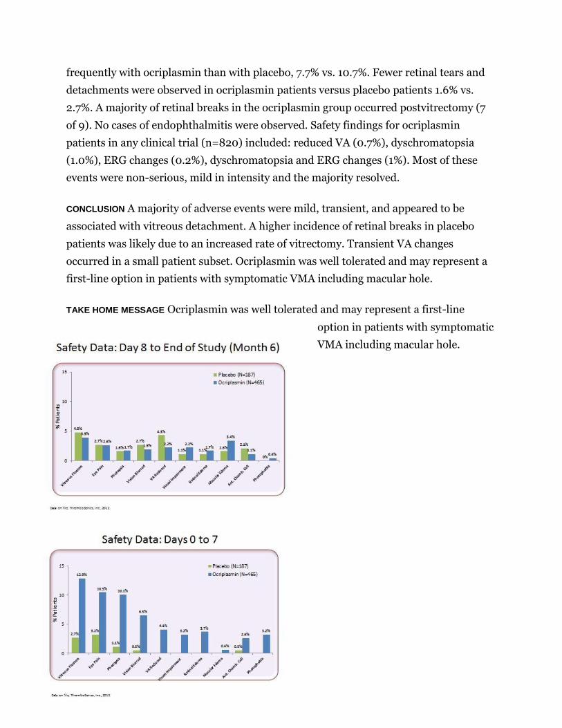
frequently with ocriplasmin than with placebo, 7.7% vs. 10.7%. Fewer retinal tears and
detachments were observed in ocriplasmin patients versus placebo patients 1.6% vs.
2.7%. A majority of retinal breaks in the ocriplasmin group occurred postvitrectomy (7
of 9). No cases of endophthalmitis were observed. Safety findings for ocriplasmin
patients in any clinical trial (n=820) included: reduced VA (0.7%), dyschromatopsia
(1.0%), ERG changes (0.2%), dyschromatopsia and ERG changes (1%). Most of these
events were non-serious, mild in intensity and the majority resolved.
CONCLUSION A majority of adverse events were mild, transient, and appeared to be
associated with vitreous detachment. A higher incidence of retinal breaks in placebo
patients was likely due to an increased rate of vitrectomy. Transient VA changes
occurred in a small patient subset. Ocriplasmin was well tolerated and may represent a
first-line option in patients with symptomatic VMA including macular hole.
TAKE HOME MESSAGE Ocriplasmin was well tolerated and may represent a first-line
option in patients with symptomatic
VMA including macular hole.

![Uveitic macular edema: a stepladder treatment paradigm€¦ · of macular edema [1,3–4], this review will focus on uveitic macular edema specifically. Uveitic macular edema Macular](https://img.pdfslide.net/doc/110x75/5ed770e44d676a3f4a7efe51/uveitic-macular-edema-a-stepladder-treatment-paradigm-of-macular-edema-13a4.jpg)