Embed Size (px)
Citation preview
Fractures and Fractures and dislocationsdislocations
around the elbow in around the elbow in adultsadults
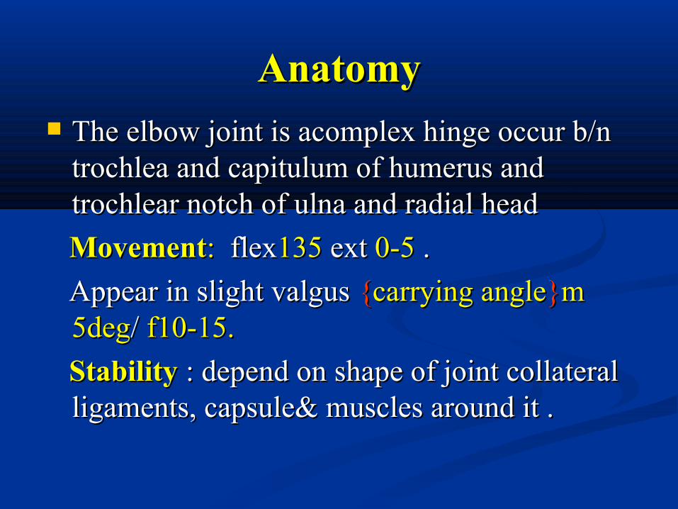
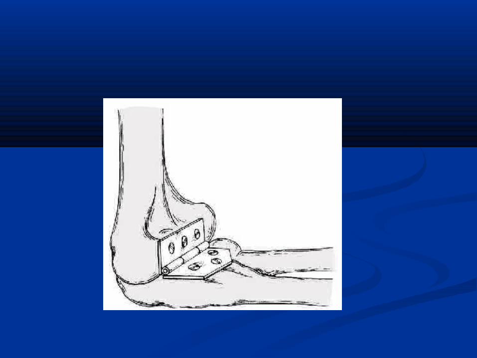
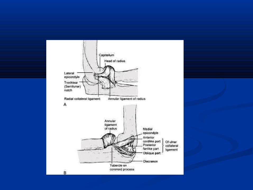
AnatomyAnatomy The elbow joint is acomplex hinge occur b/n The elbow joint is acomplex hinge occur b/n
trochlea and capitulum of humerus and trochlea and capitulum of humerus and trochlear notch of ulna and radial headtrochlear notch of ulna and radial head
MovementMovement:: flex flex135135 ext ext 0-5 0-5 . . Appear in slight valgus Appear in slight valgus {{carrying anglecarrying angle}}m m
5deg5deg/ / f10-15.f10-15. StabilityStability : depend on shape of joint collateral : depend on shape of joint collateral
ligaments, capsule& muscles around it .ligaments, capsule& muscles around it .
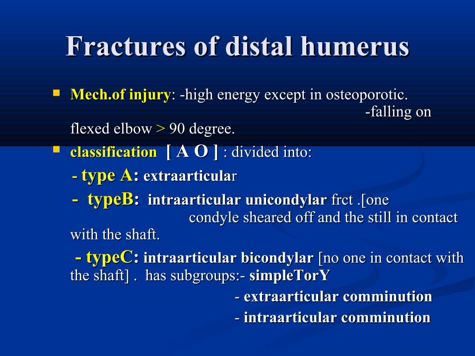
Fractures of distal humerusFractures of distal humerus Mech.of injuryMech.of injury: -high energy except in osteoporotic. : -high energy except in osteoporotic.
-falling on -falling on flexed elbow flexed elbow >> 90 degree. 90 degree.
classificationclassification [ A O ][ A O ] : divided into: : divided into: -- type Atype A:: extraarticulaextraarticularr -- typeBtypeB:: intraarticularintraarticular unicondylarunicondylar frct .[one frct .[one
condyle sheared off and the still in contact condyle sheared off and the still in contact with the shaft.with the shaft.
- typeC- typeC:: intraarticular bicondylarintraarticular bicondylar [no one in contact with [no one in contact with the shaft] . has subgroups:- the shaft] . has subgroups:- simpleTorYsimpleTorY
- - extraarticular comminutionextraarticular comminution - - intraarticular comminutionintraarticular comminution
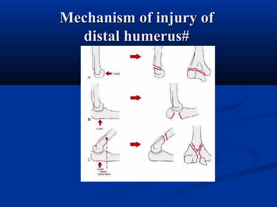
Mechanism of injury of Mechanism of injury of distal humerus# distal humerus#
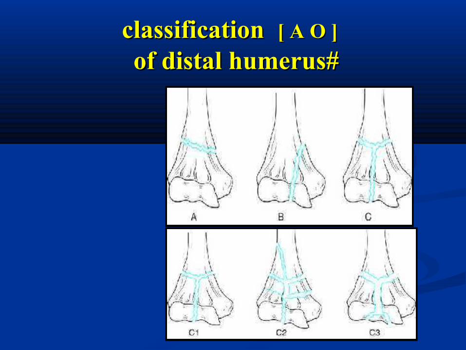
classificationclassification [ A O ][ A O ] of distal humerus# of distal humerus#
other classificationother classification of distal humerus#of distal humerus#
Some classified them intoSome classified them into : : - Supracondylar # . - Supracondylar # . - Intracondylar # . - Intracondylar # . - transcondylar # - transcondylar #
- Chondyles[med.and lat.]#- Chondyles[med.and lat.]# - Articular surface[capitulum and trochlea]#- Articular surface[capitulum and trochlea]# -Epicondyles #-Epicondyles #
Diagnosis Diagnosis of distal humerus#of distal humerus#
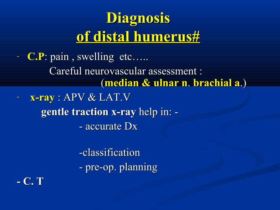
- C.PC.P: pain , swelling etc…..: pain , swelling etc….. Careful neurovascular assessment :Careful neurovascular assessment :
((median & ulnar nmedian & ulnar n. . brachial abrachial a.).)- x-rayx-ray : APV & LAT.V : APV & LAT.V gentle traction x-raygentle traction x-ray help in: - help in: - - accurate Dx - accurate Dx
-classification-classification - pre-op. planning- pre-op. planning- C. T- C. T
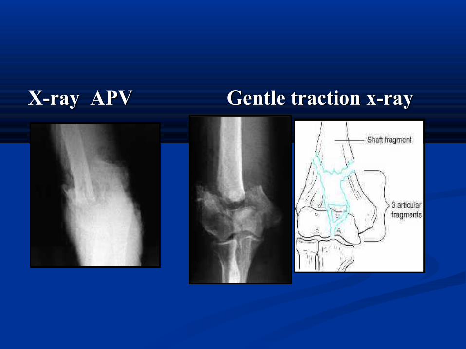
X-ray APVX-ray APV Gentle traction x-rayGentle traction x-ray
Treatment Treatment of distal humerus# of distal humerus#
I. ConservativeI. Conservative:: -(rare) for -(rare) for undisplacedundisplaced #. #. -p.o.p in 90 flexion for -p.o.p in 90 flexion for 6-86-8 w . w .
-weekly x-ray -weekly x-ray II. SurgicalII. Surgical is the treatment of choiceis the treatment of choice.. : :
because fracture because fracture usually unstableusually unstableIII.Alternative:III.Alternative:
Treatment Treatment of distal humerus#of distal humerus#
III.AlternativeIII.Alternative: indicated for:: indicated for:- 1.severely commin.# .1.severely commin.# . -2. severely soft tissue damage.-2. severely soft tissue damage. -3.Patient bad condition. -3.Patient bad condition. - 4.lack of expertise &facilities.- 4.lack of expertise &facilities. - 5. severely osteoporotic(contraversed).- 5. severely osteoporotic(contraversed).
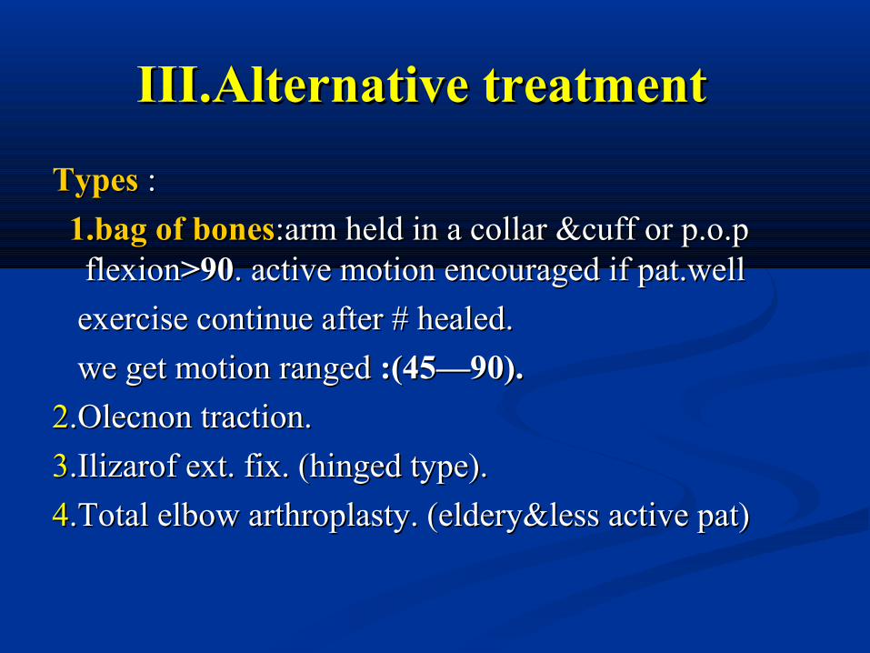
III.Alternative treatmentIII.Alternative treatment
TypesTypes : : 1.bag of1.bag of bonesbones:arm held in a collar &cuff or p.o.p :arm held in a collar &cuff or p.o.p
flexionflexion>90>90. active motion encouraged if pat.well. active motion encouraged if pat.well exercise continue after # healed.exercise continue after # healed. we get motion ranged we get motion ranged :(45—90).:(45—90).22.Olecnon traction..Olecnon traction.33.Ilizarof ext. fix. (hinged type). .Ilizarof ext. fix. (hinged type). 44.Total elbow arthroplasty. (eldery&less active pat).Total elbow arthroplasty. (eldery&less active pat)
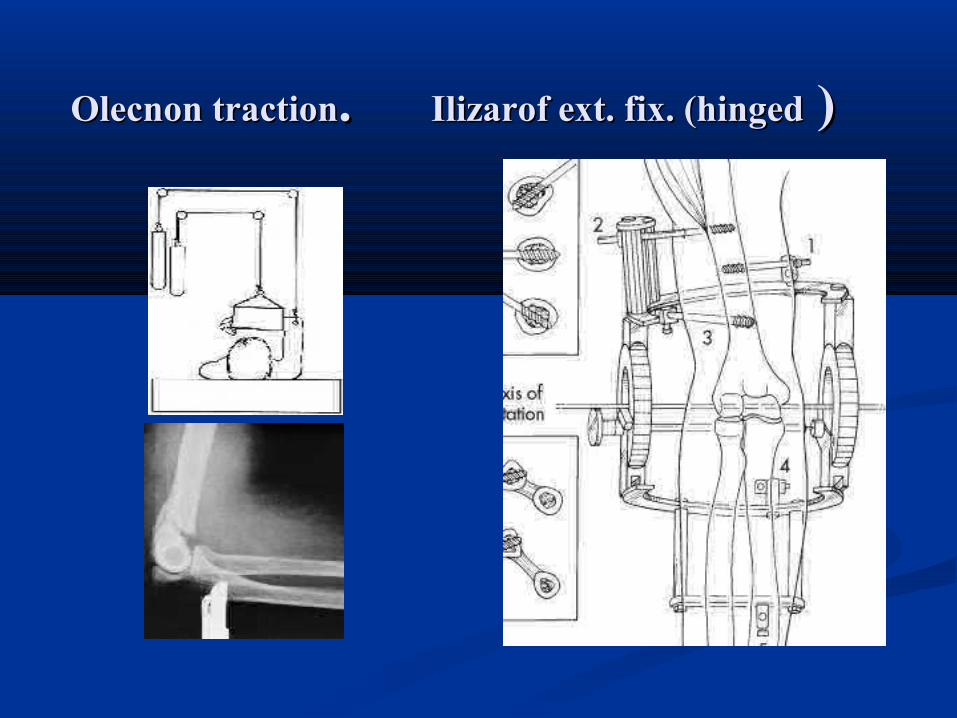
Olecnon tractionOlecnon traction. . Ilizarof ext. fix. (hingedIlizarof ext. fix. (hinged ) )
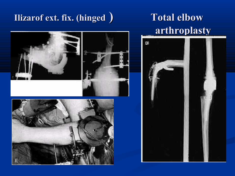
Ilizarof ext. fix. (hingedIlizarof ext. fix. (hinged ) ) Total elbow Total elbow arthroplastyarthroplasty
Surgical treatmentSurgical treatment It include:It include: - pre operative planning- pre operative planning:- :-
- careful reading of x-ray C.T - careful reading of x-ray C.T
- prepare for the worst before op.- prepare for the worst before op. - internal fixation:- internal fixation: ** it should be it should be earlyearly(24-48h)except open #, (24-48h)except open #,
accurate & rigid accurate & rigid to to give good stability& give good stability& permit early motion permit early motion
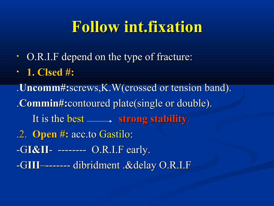
Follow int.fixationFollow int.fixation• O.R.I.F depend on the type of fracture:O.R.I.F depend on the type of fracture:• 1. Clsed #:1. Clsed #: ..Uncomm#:Uncomm#:screws,K.W(crossed or tension band).screws,K.W(crossed or tension band)...Commin#:Commin#:contoured plate(single or double).contoured plate(single or double). It is the It is the bestbest strong stabilitystrong stability...2. .2. Open #:Open #: acc.to acc.to GastiloGastilo::-G-GI&III&II- -------- O.R.I.F early.- -------- O.R.I.F early.-G-GIIIIII–------- dibridment .&delay O.R.I.F–------- dibridment .&delay O.R.I.F
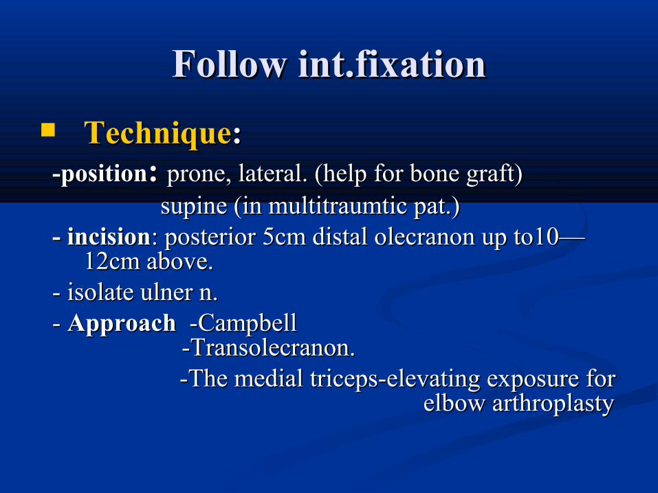
Follow int.fixationFollow int.fixation TechniqueTechnique:: -position-position: : prone, lateral. (help for bone graft) prone, lateral. (help for bone graft) supine (in multitraumtic pat.)supine (in multitraumtic pat.) - incision- incision: posterior 5cm distal olecranon up to10—: posterior 5cm distal olecranon up to10—
12cm above.12cm above. - isolate ulner n.- isolate ulner n. - - ApproachApproach -Campbell -Campbell
-Transolecranon. -Transolecranon. -The medial triceps-elevating exposure for -The medial triceps-elevating exposure for
elbow arthroplasty elbow arthroplasty
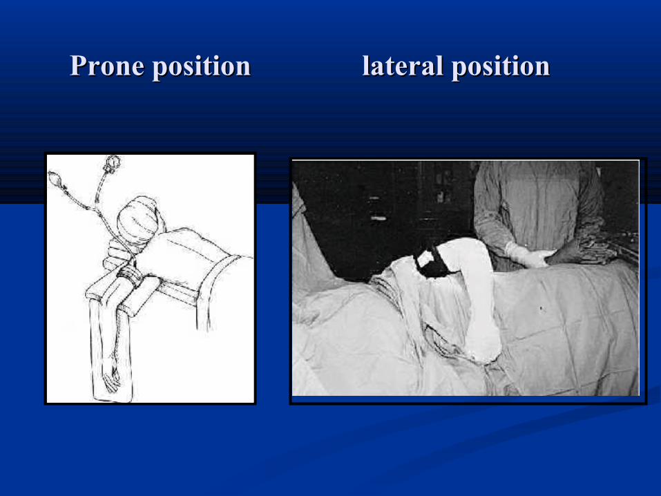
Prone position lateral positionProne position lateral position
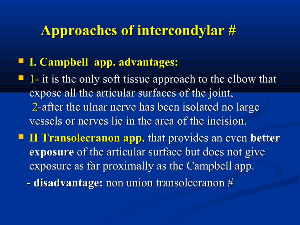
Approaches of intercondylar #Approaches of intercondylar # I. Campbell app. advantages:I. Campbell app. advantages: 1-1- it is the only soft tissue approach to the elbow that it is the only soft tissue approach to the elbow that
expose all the articular surfaces of the joint, expose all the articular surfaces of the joint, 2-2-after the ulnar nerve has been isolated no large after the ulnar nerve has been isolated no large vessels or nerves lie in the area of the incision.vessels or nerves lie in the area of the incision.
IIII Transolecranon app.Transolecranon app. that provides an even that provides an even better better exposureexposure of the articular surface but does not give of the articular surface but does not give exposure as far proximally as the Campbell app. exposure as far proximally as the Campbell app.
- - disadvantage:disadvantage: non union transolecranon # non union transolecranon #
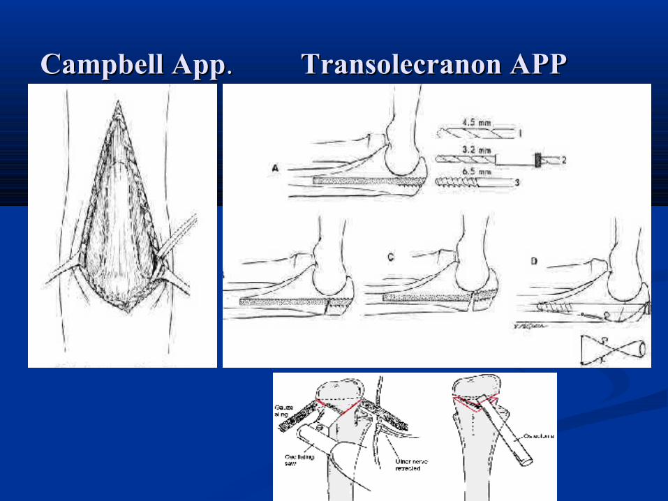
Campbell AppCampbell App. . Transolecranon APPTransolecranon APP
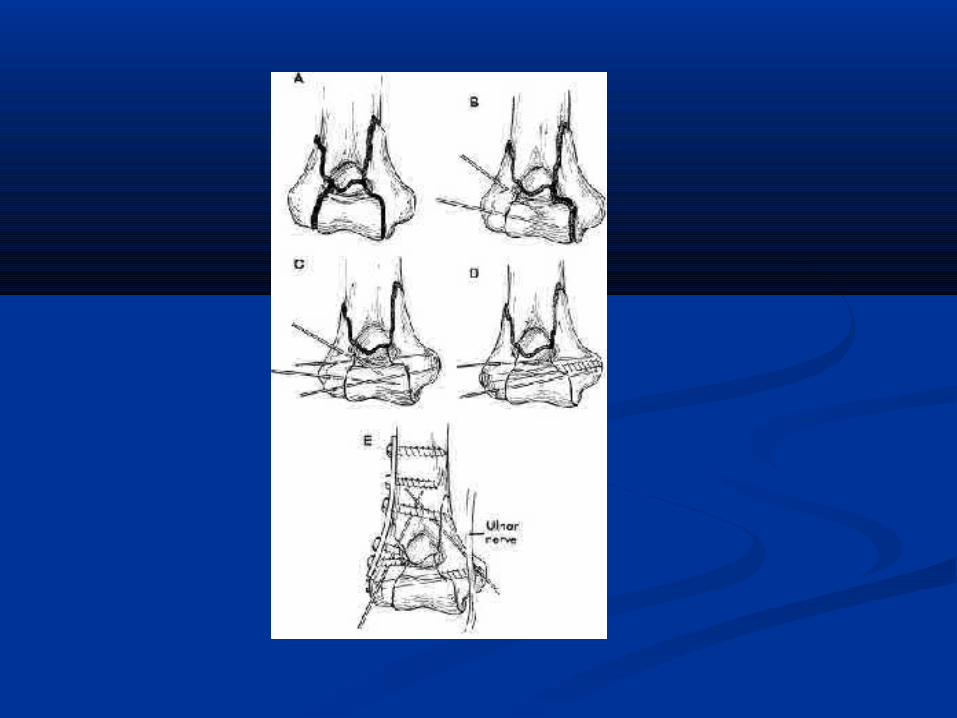
Follow int.fixationFollow int.fixation Steps of reduction of intercondylar #Steps of reduction of intercondylar # :- :-1. 1. Reduction &fix. Of condylesReduction &fix. Of condyles : : 2. 2. Reduction &fix. Of epicondylar ridgeReduction &fix. Of epicondylar ridge : :
to the proximal fragment. (to the proximal fragment. (it form a buttress to it form a buttress to which condyle later attached) which condyle later attached)
3. 3. Reduction &fix. Of reassembled Reduction &fix. Of reassembled condylescondyles: to metaphysis with : screws, K.W : to metaphysis with : screws, K.W or plates.or plates.
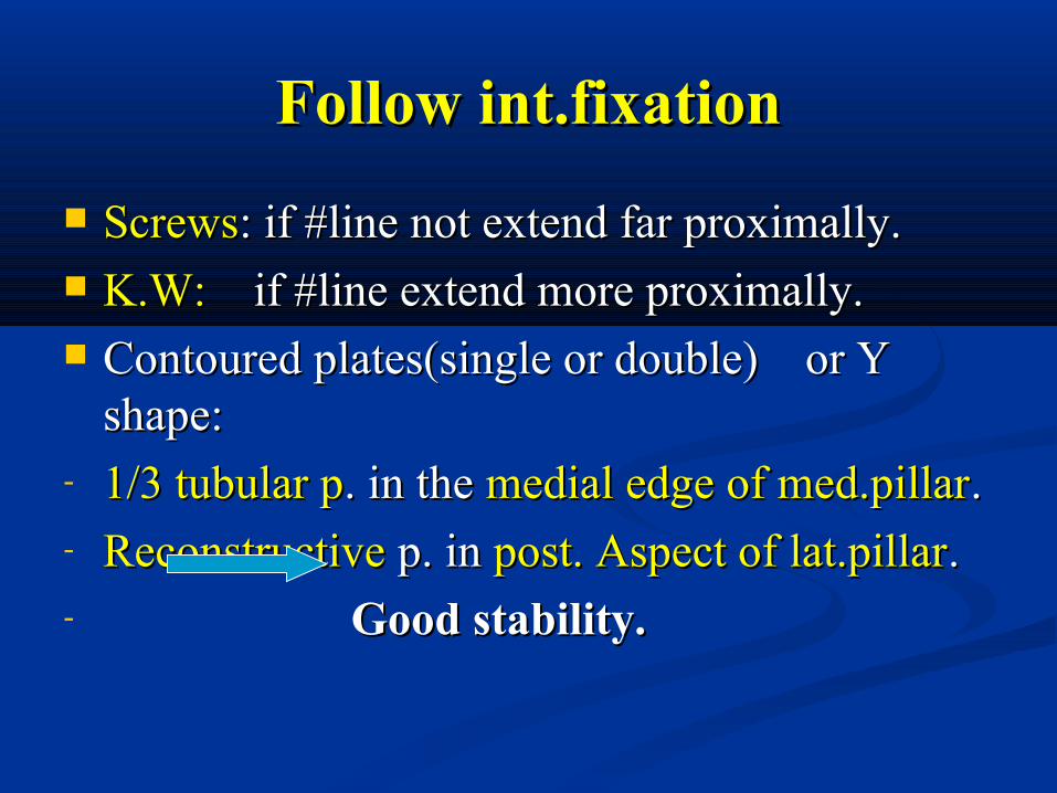
Follow int.fixationFollow int.fixation ScrewsScrews: if #line not extend far proximally.: if #line not extend far proximally. K.W:K.W: if #line extend more proximally. if #line extend more proximally. Contoured plates(single or double) or Y Contoured plates(single or double) or Y
shape:shape:- 1/3 tubular p1/3 tubular p. in the . in the medial edge of med.pillarmedial edge of med.pillar..- ReconstructiveReconstructive p. in p. in post. Aspect of lat.pillarpost. Aspect of lat.pillar. . - Good stability.Good stability.
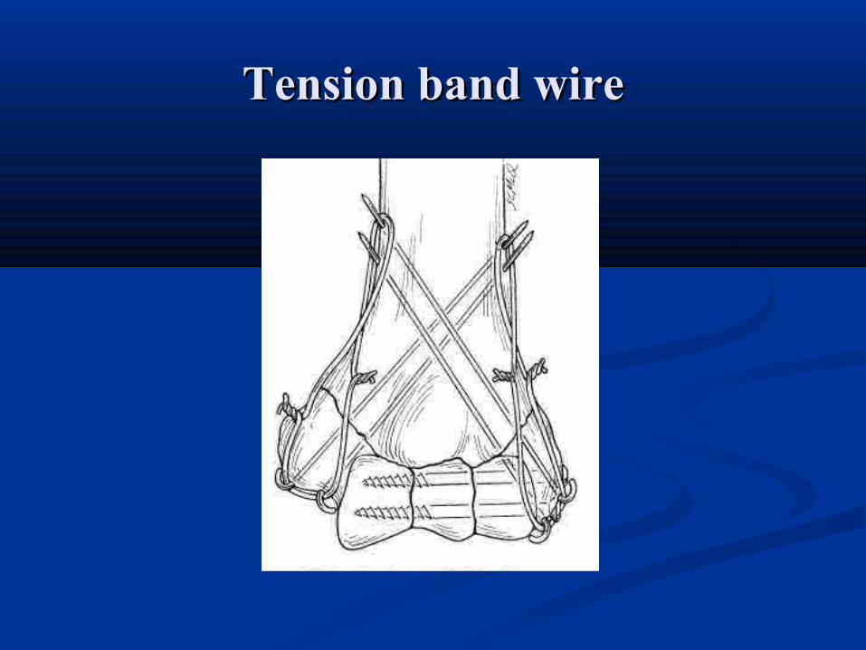
Tension band wireTension band wire
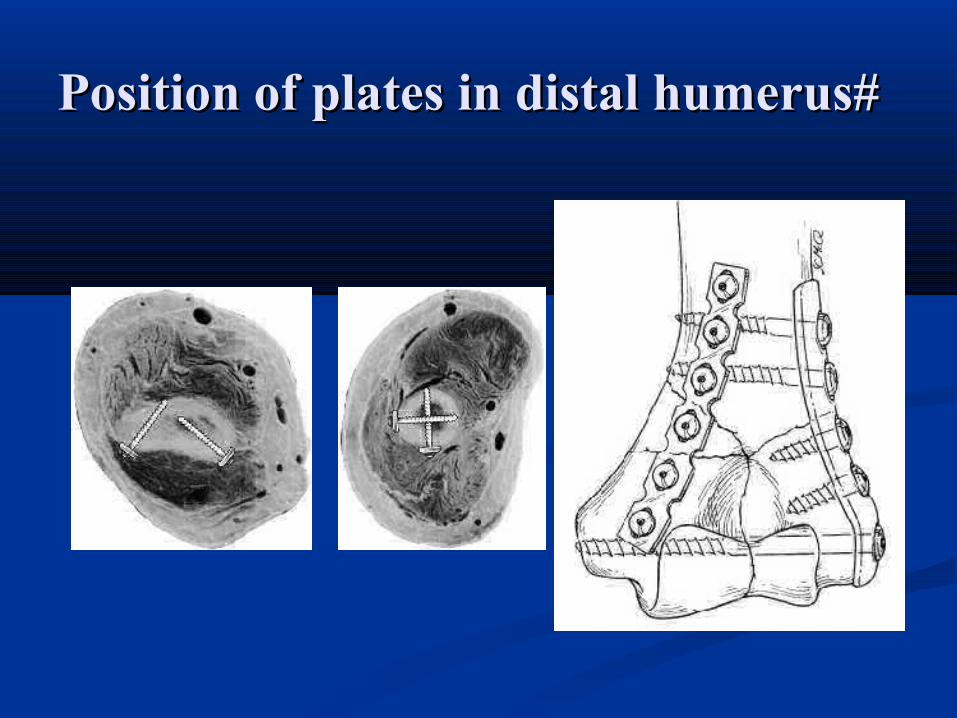
Position of plates in distal humerus#Position of plates in distal humerus#
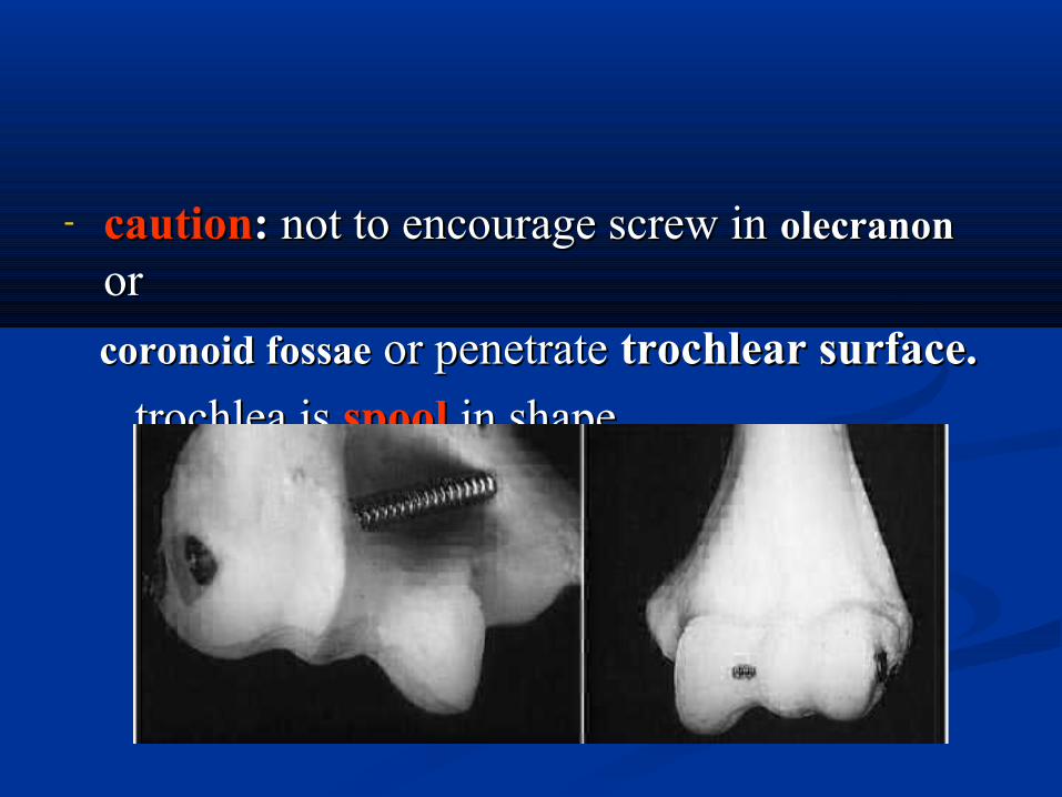
- cautioncaution: : not to encourage screw in not to encourage screw in olecranonolecranon or or
coronoid fossaecoronoid fossae or penetrate or penetrate trochlear surface. trochlear surface. trochlea istrochlea is spoolspool in shapein shape
After treatmentAfter treatment Light post. Splint.Light post. Splint. When wound healing is satisfactoryWhen wound healing is satisfactory7—10d7—10d
Remove p.o.p periodicallyRemove p.o.p periodically & &gentle active gentle active exerciseexercise started. started.
after 3 wafter 3 w p.o.p removed and the arm is p.o.p removed and the arm is supported by a supported by a sling sling with active motion as pain with active motion as pain permit permit
vigorous motionvigorous motion contraindicatedcontraindicated
Transchondylar #Transchondylar #
often is grouped with suprachond#. but often is grouped with suprachond#. but requires requires special considerationsspecial considerations b/s usually b/s usually extends to extends to articular surfacearticular surface . . QuiteQuite unstableunstable . . unite unite slowlyslowly if treated conservatively . if treated conservatively .
-so treated with -so treated with percutaneous pins , lag screwpercutaneous pins , lag screw (through small incision without opening the frac.), or (through small incision without opening the frac.), or canulatedcanulated screw . screw .
if Was intraarticular and not fixed properly can if Was intraarticular and not fixed properly can be complicated by be complicated by avascular necrosisavascular necrosis . .
--Displaced #------Displaced #------ O.R.I.F. O.R.I.F.
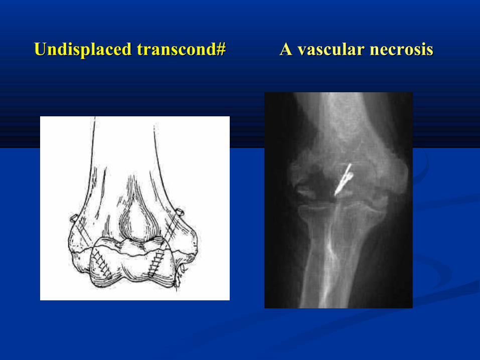
Undisplaced transcond#Undisplaced transcond# A vascular necrosisA vascular necrosis
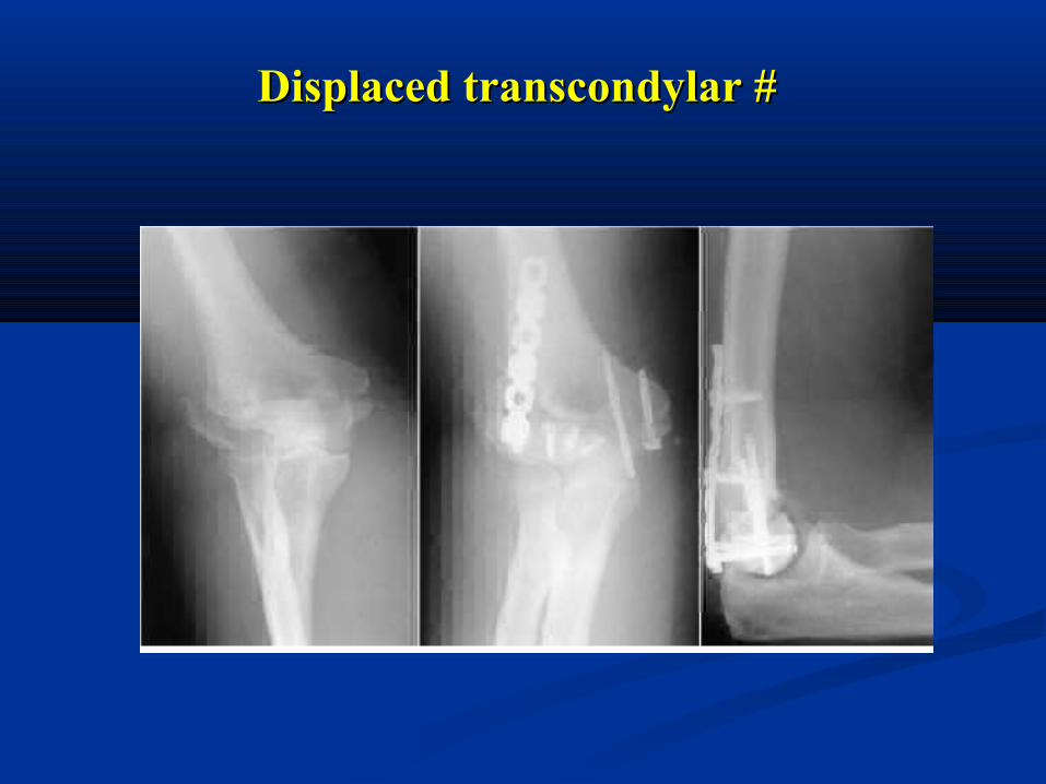
Displaced transcondylar # Displaced transcondylar #
Side swipe fractureSide swipe fracture
-occure in -occure in arm protruded from windowarm protruded from window of car of car and struck with other car . and struck with other car .
- fracture - fracture always openalways open . Vary from . Vary from GI ---- GIIIGI ---- GIII - the - the most combinationmost combination of this fracture consistof this fracture consist of: of: * * open distal 1/3 # of olecranonopen distal 1/3 # of olecranon . . * * anterior dislocation of redial head & distal anterior dislocation of redial head & distal
fragment of ulnafragment of ulna . .
* * comminuted distal humerous fracture .comminuted distal humerous fracture . &other&other
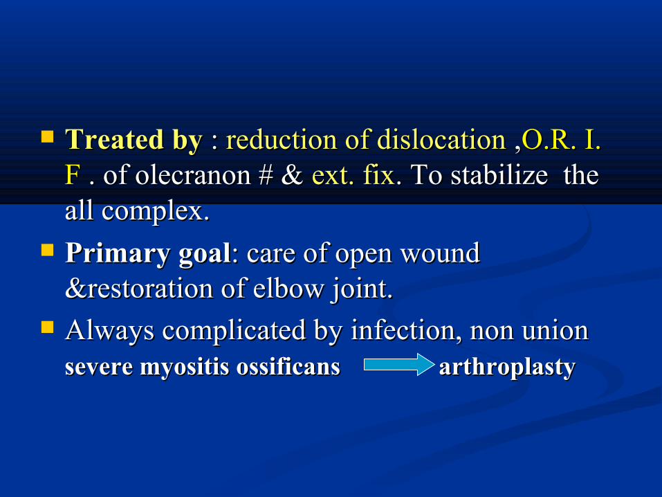
Treated byTreated by : : reduction of dislocationreduction of dislocation , ,O.R. I. O.R. I. FF . of olecranon # & . of olecranon # & ext. fixext. fix. To stabilize the . To stabilize the all complex.all complex.
Primary goalPrimary goal: care of open wound : care of open wound &restoration of elbow joint.&restoration of elbow joint.
Always complicated by infection, non union Always complicated by infection, non union severe myositis ossificanssevere myositis ossificans arthroplastyarthroplasty
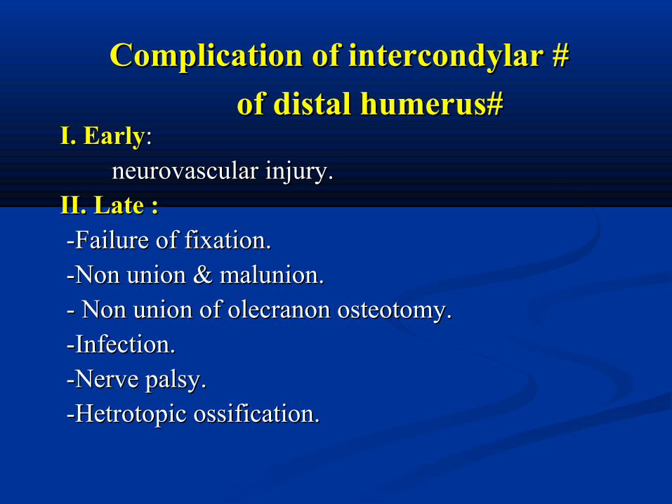
Complication of intercondylar # Complication of intercondylar # of distal humerus#of distal humerus# I. EarlyI. Early:: neurovascular injury.neurovascular injury. II. Late :II. Late : -Failure of fixation. -Failure of fixation. -Non union & malunion.-Non union & malunion. - Non union of olecranon osteotomy.- Non union of olecranon osteotomy. -Infection.-Infection. -Nerve palsy.-Nerve palsy. -Hetrotopic ossification. -Hetrotopic ossification.
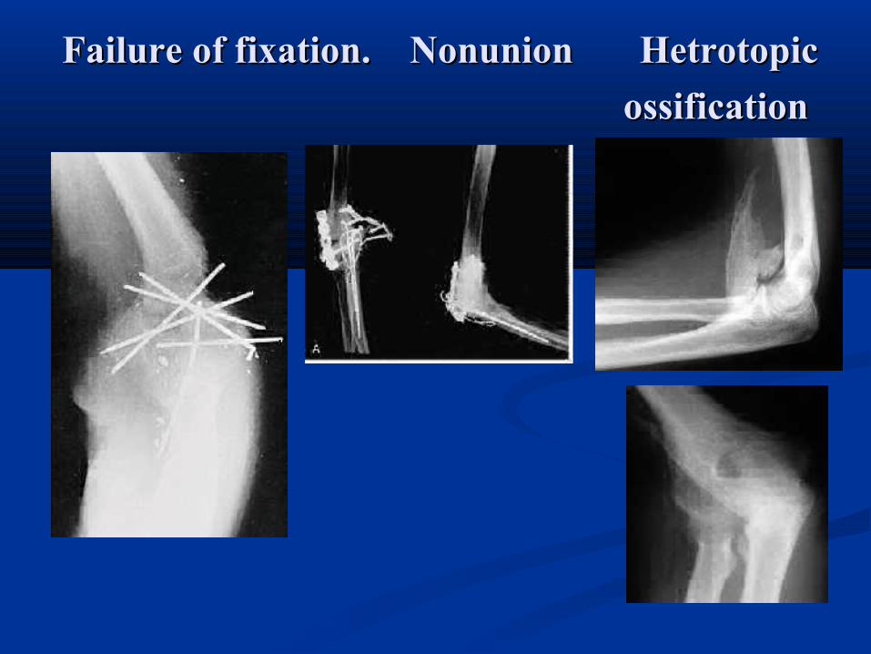
Failure of fixation. NonunionFailure of fixation. Nonunion Hetrotopic Hetrotopic ossificationossification
Fracture of capitulum Fracture of capitulum - - Mech. Of injuiryMech. Of injuiry: F.O.S.H---- head of radius : F.O.S.H---- head of radius
impacted to capitulum ----fracture impacted to capitulum ----fracture classification:classification: - - type Itype I: large fragment of bone and : large fragment of bone and
articular surface (articular surface (involve trochleainvolve trochlea) are ) are fractured.fractured.
--type IItype II: small shell of bone and articular : small shell of bone and articular surface (surface (not involve trochlianot involve trochlia).).
- - type III:type III: comminuted #. comminuted #.
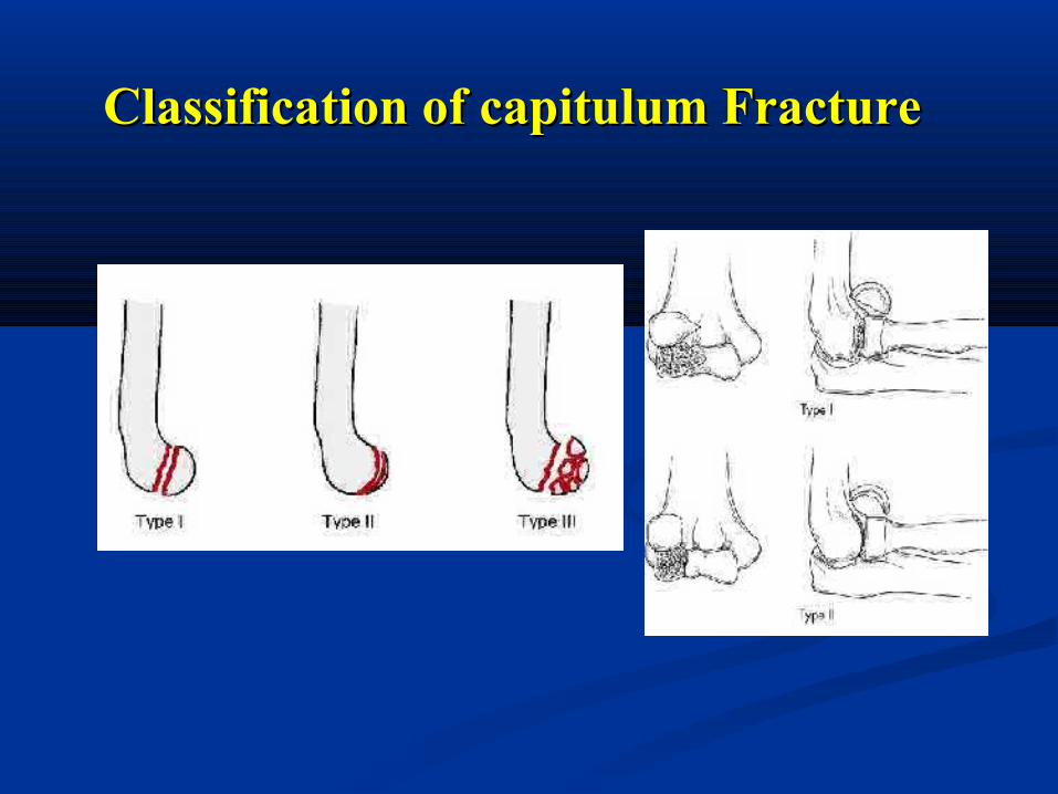
Classification of capitulum FractureClassification of capitulum Fracture
Fracture of capitulum Fracture of capitulum - Diagnosis:-Diagnosis:-- x-ray :x-ray :laterallateral view (diagnostic). & A.P.V view (diagnostic). & A.P.V - Deff. diagDeff. diag.: from # of radial head but the later rarely .: from # of radial head but the later rarely
to displaced anteriorlly to displaced anteriorlly (so any # fragment ant.to lat. (so any # fragment ant.to lat. Condyle isCondyle is capitulum fragment till prove otherwise.)capitulum fragment till prove otherwise.)
- C.T scanC.T scan- TreatmentTreatment: : ( through lat. Approach)( through lat. Approach) Type IType I : : O.R.I. FO.R.I. F with with small AOsmall AO screw or screw or
Herbert's Herbert's screw ( from post. to ant.)screw ( from post. to ant.) Type II&IIIType II&III: : excisionexcision . . --AAfter treatment:fter treatment: like intercondylar # . like intercondylar # .
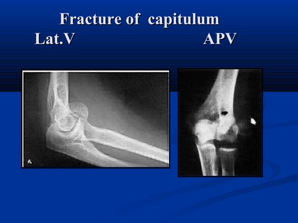
Fracture of capitulumFracture of capitulum Lat.V APV Lat.V APV
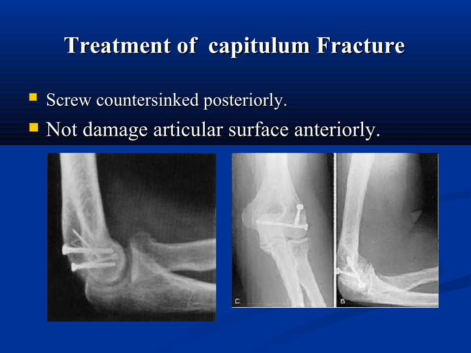
Treatment of capitulum Fracture Treatment of capitulum Fracture
Screw countersinked posteriorly.Screw countersinked posteriorly. Not damage articular surface anteriorly.Not damage articular surface anteriorly.
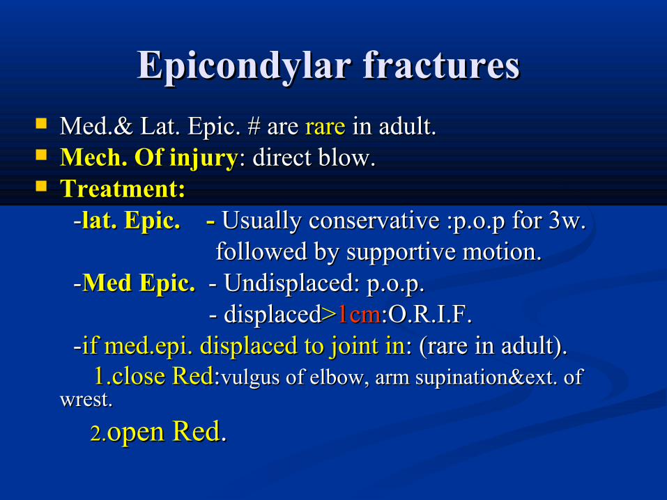
Epicondylar fractures Epicondylar fractures Med.& Lat. Epic. # are Med.& Lat. Epic. # are rarerare in adult. in adult. Mech. Of injuryMech. Of injury: direct blow.: direct blow. Treatment:Treatment: --lat. Epic. - lat. Epic. - Usually conservative :p.o.p for 3w.Usually conservative :p.o.p for 3w. followed by supportive motion. followed by supportive motion. --Med Epic.Med Epic. - Undisplaced: p.o.p. - Undisplaced: p.o.p. - displaced- displaced>>1cm1cm:O.R.I.F.:O.R.I.F. --if med.epi. displaced to joint inif med.epi. displaced to joint in: (rare in adult).: (rare in adult). 1.close1.close RedRed::vulgus of elbow, arm supination&ext. of vulgus of elbow, arm supination&ext. of
wrest.wrest. 2.2.open Redopen Red..
Olecranon fracturesOlecranon fractures Mech. Of injMech. Of inj. : . : directdirect: blow on elbow.: blow on elbow.
indirectindirect:: falling on partially falling on partially flexed flexed elbow with indirect force generated by triceps ___ elbow with indirect force generated by triceps ___ avulsion. avulsion.
classificationclassification:: type Itype I :# of proximal 1/3. :# of proximal 1/3. typetype IIII:# of middle 1/3 .:# of middle 1/3 . type III type III :#of distal 1/3.it may be :#of distal 1/3.it may be
associated with ant. displacement of radius. associated with ant. displacement of radius.
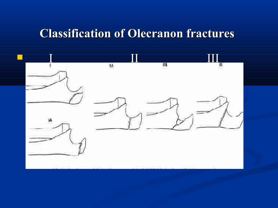
Classification of Olecranon fracturesClassification of Olecranon fractures I II IIII II III
Follow olecranon fractureFollow olecranon fracture Other classification: Other classification: [Colton[Colton]] according to: according to: displacement and the anatomy of the fracture, thus displacement and the anatomy of the fracture, thus
give guidance as to the appropriate type of fixation :give guidance as to the appropriate type of fixation : I.Nondisplaced and stable I.Nondisplaced and stable II.Displaced fracturesII.Displaced fractures - Avulsion fractures - Avulsion fractures - oblique fractures . - oblique fractures . - Transverse fractures- Transverse fractures - Isolated comminuted fractures - Isolated comminuted fractures - Fracture/dislocations - Fracture/dislocations
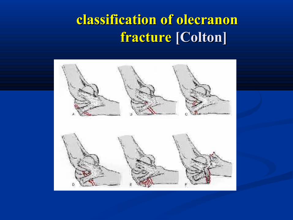
classification of olecranon classification of olecranon fracture fracture [Colton] [Colton]
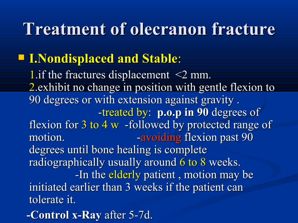
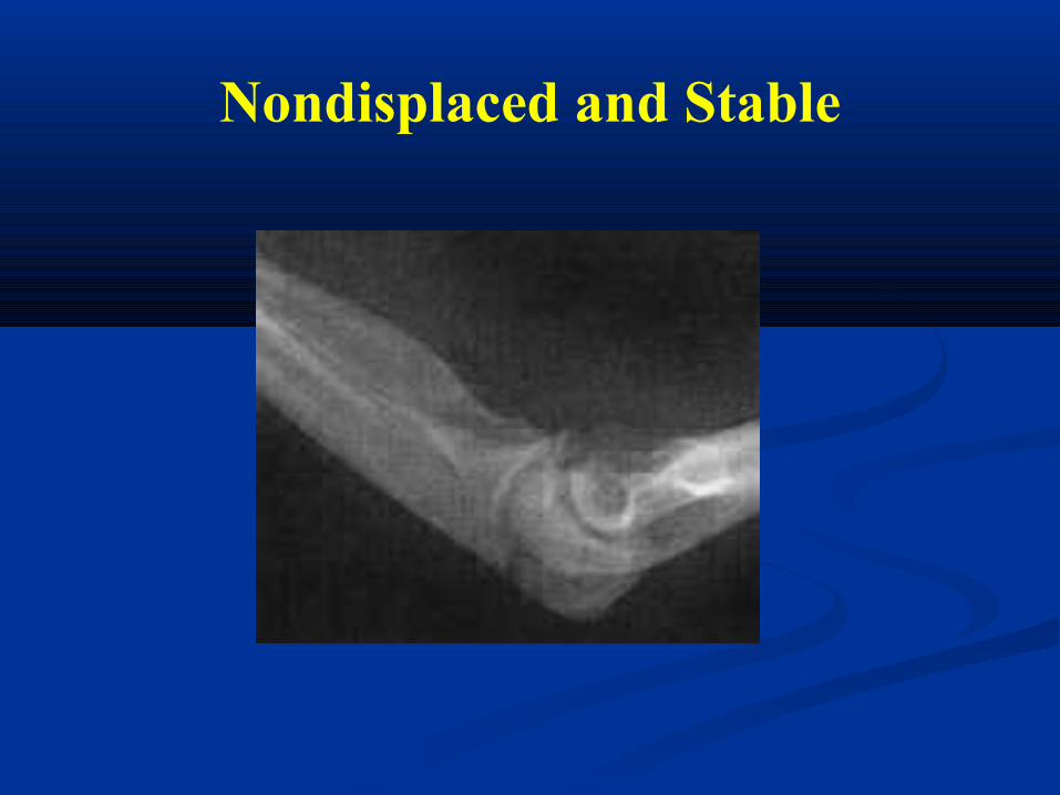
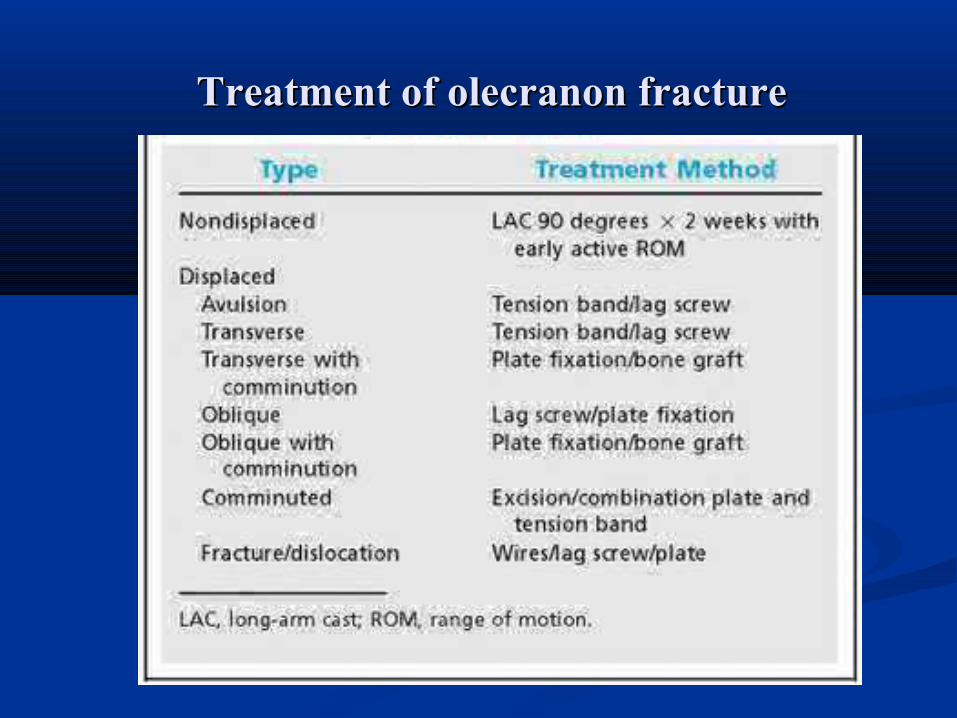
Treatment of olecranon fractureTreatment of olecranon fracture I.Nondisplaced and StableI.Nondisplaced and Stable:: 11.if the fractures displacement <2 mm. .if the fractures displacement <2 mm.
22.exhibit no change in position with gentle flexion to .exhibit no change in position with gentle flexion to 90 degrees or with extension against gravity . 90 degrees or with extension against gravity . - -treated bytreated by: : p.o.p in 90p.o.p in 90 degrees of degrees of flexion for flexion for 3 to 4 w3 to 4 w -followed by protected range of -followed by protected range of motion. -motion. -avoidingavoiding flexion past 90 flexion past 90 degrees until bone healing is complete degrees until bone healing is complete radiographically usually around radiographically usually around 6 to 86 to 8 weeks. weeks. -In the -In the elderlyelderly patient , motion may be patient , motion may be initiated earlier than 3 weeks if the patient can initiated earlier than 3 weeks if the patient can tolerate it.tolerate it.
-Control x-Ray-Control x-Ray after 5-7d. after 5-7d. -P.o.p in full extension-P.o.p in full extension avoided avoided b/s lead to stiffness b/s lead to stiffness
Nondisplaced and Stable
Treatment of olecranon fractureTreatment of olecranon fracture II.Displaced FracturesII.Displaced Fractures:: O.R.I.F is the treatment of choice.O.R.I.F is the treatment of choice. The goals of treatment are:The goals of treatment are: 1.Maintain power of elbow extension. 1.Maintain power of elbow extension. 2.Restore congruity of the articular surface. 2.Restore congruity of the articular surface. 3.Restore stability of the elbow. 3.Restore stability of the elbow. 4.Prevent stiffness of the joint. 4.Prevent stiffness of the joint. 5.Allow the patient to do early motion5.Allow the patient to do early motion
Follow olecranon fractureFollow olecranon fracture
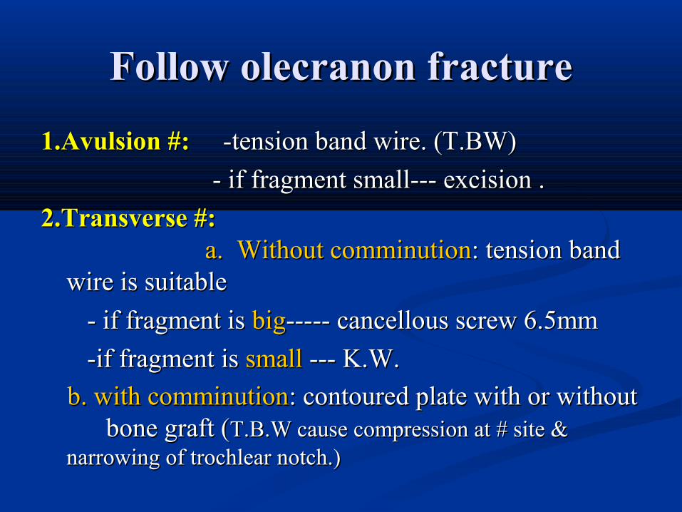
1.Avulsion #:1.Avulsion #: -tension band wire. (T.BW) -tension band wire. (T.BW) - if fragment small--- excision .- if fragment small--- excision .2.Transverse #:2.Transverse #:
a. Without comminutiona. Without comminution: tension band : tension band wire is suitable wire is suitable
- if fragment is - if fragment is bigbig----- cancellous screw 6.5mm ----- cancellous screw 6.5mm -if fragment is -if fragment is smallsmall --- K.W. --- K.W. b. with comminutionb. with comminution: contoured plate with or without : contoured plate with or without
bone graft ( bone graft (T.B.W cause compression at # site & T.B.W cause compression at # site & narrowing of trochlear notch.)narrowing of trochlear notch.)
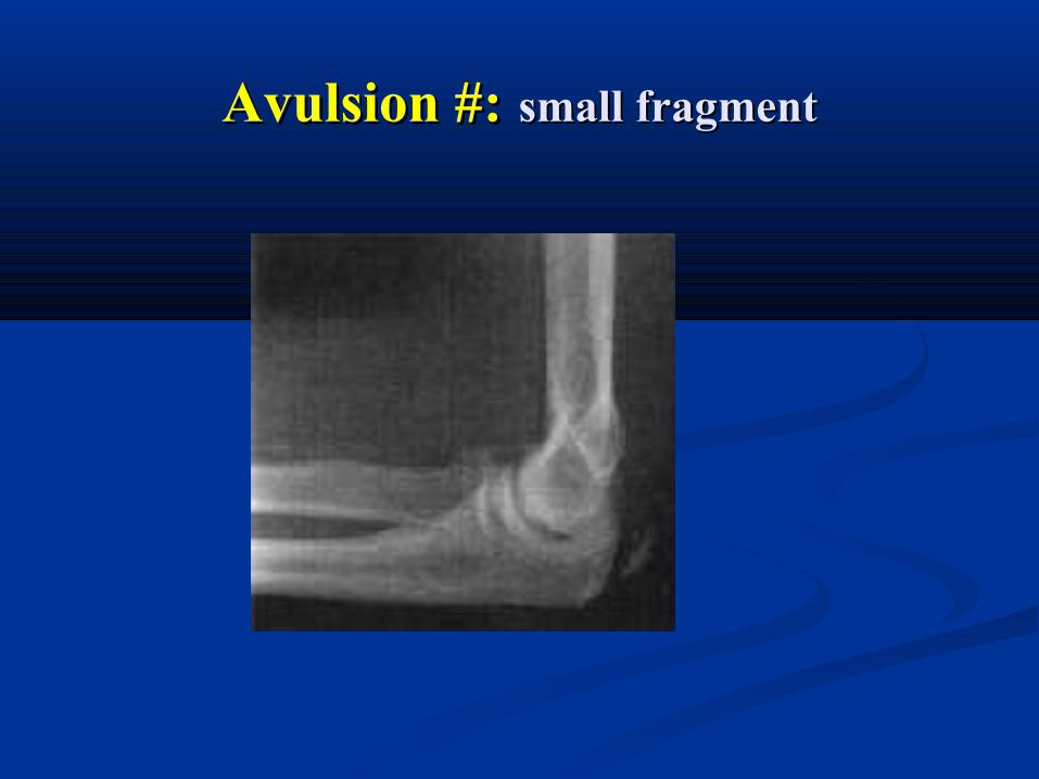
Avulsion #:Avulsion #: small fragmentsmall fragment
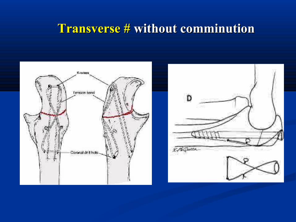
Transverse #Transverse # without comminution without comminution
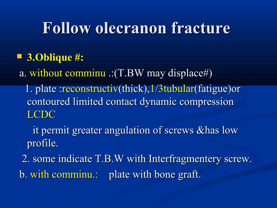
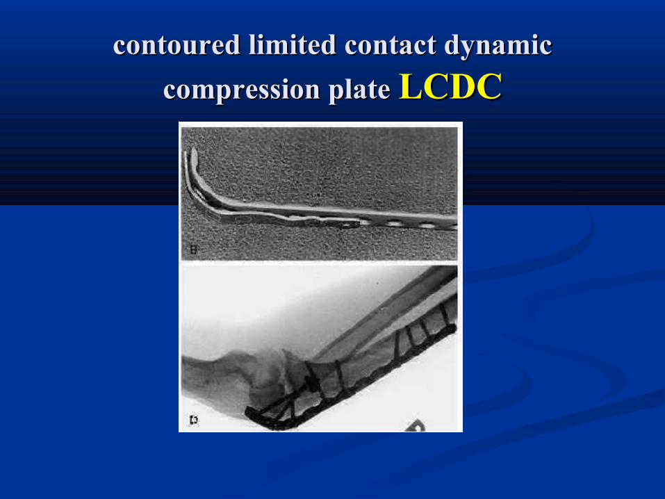
Follow olecranon fractureFollow olecranon fracture 3.Oblique #:3.Oblique #: a. a. without comminu without comminu .:(T.BW may displace#).:(T.BW may displace#) 1. plate :1. plate :reconstructivreconstructiv(thick),(thick),1/3tubular1/3tubular(fatigue)or (fatigue)or
contoured limited contact dynamic compression contoured limited contact dynamic compression LCDCLCDC
it permit greater angulation of screws &has low it permit greater angulation of screws &has low profile. profile.
2. some indicate T.B.W with Interfragmentery screw. 2. some indicate T.B.W with Interfragmentery screw. bb. with comminu.:. with comminu.: plate with bone graft. plate with bone graft.
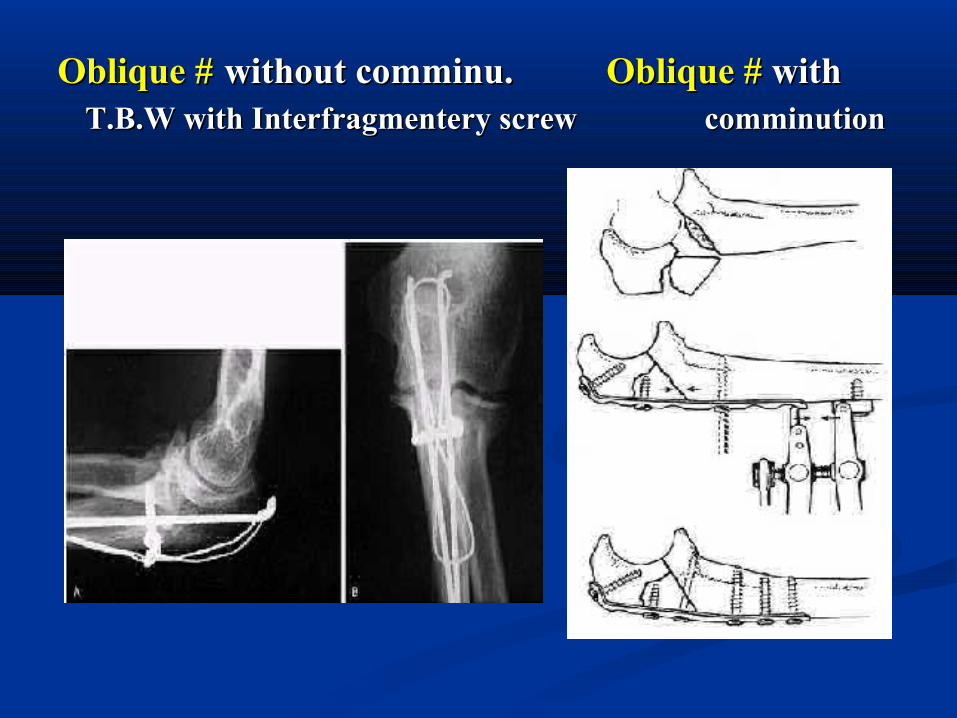
Oblique #Oblique # without comminu. without comminu. Oblique #Oblique # with with T.B.W with Interfragmentery screw comminution T.B.W with Interfragmentery screw comminution
contoured limited contact dynamiccontoured limited contact dynamic compression plate compression plate LCDCLCDC
44.Isolated comminu#.Isolated comminu# results from direct trauma. results from direct trauma. There are multiple fracture planes, &crushing of There are multiple fracture planes, &crushing of many fragments. many fragments. -may be associated with fractures of -may be associated with fractures of the distalthe distal end end of the humerusof the humerus, the , the radial & ulnar shaftsradial & ulnar shafts, and the , and the radial headradial head..
--If no associationIf no association with previous with previous excision.excision. & not in distal 1/3 of olecranon& not in distal 1/3 of olecranon --if associationif association occur (excision unsuitable)--- occur (excision unsuitable)---
combination of plate & tension band wirecombination of plate & tension band wire..
Excision of proximal fragment:Excision of proximal fragment:
-used only if there is proximal# & the remnant distal part form stable base -used only if there is proximal# & the remnant distal part form stable base for trochlea.for trochlea.
AdvantagesAdvantages::1.1. The possibility of non union is eliminated.The possibility of non union is eliminated.2.2. The possibility of traumatic artheritis is menimiz due to irregular articular The possibility of traumatic artheritis is menimiz due to irregular articular
surface.surface.Indication:Indication: severely comminuted fractures in which open reduction and internal severely comminuted fractures in which open reduction and internal
fixation are not Possible. fixation are not Possible. -non articular #. -Non union . -after failed O.R.I.F .-non articular #. -Non union . -after failed O.R.I.F . -when reduction is delayed 10—14d.-when reduction is delayed 10—14d. -in type III open# or if local soft tissue damaged .-in type III open# or if local soft tissue damaged .ContraindicationContraindication:: in distal 1/3 olecranon# joint instbility in distal 1/3 olecranon# joint instbility
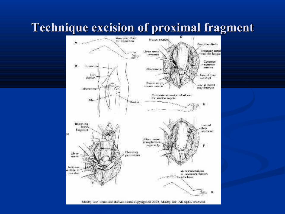
Technique excision of proximal fragmentTechnique excision of proximal fragment
After excision of proximal fragmentAfter excision of proximal fragment
- p.o.p in flexion - p.o.p in flexion 70 deg70 deg. For . For 3w3w.. - gentle motion when wound heal permit 7—- gentle motion when wound heal permit 7—
10d.10d. -avoid-avoid forceful movement (ext. or flex.) for forceful movement (ext. or flex.) for 3 3
month.month.
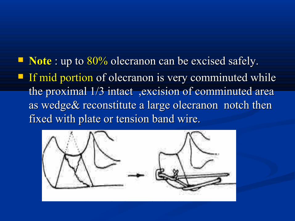
NoteNote : up to : up to 80%80% olecranon can be excised safely. olecranon can be excised safely. If mid portionIf mid portion of olecranon is very comminuted while of olecranon is very comminuted while
the proximal 1/3 intact ,excision of comminuted area the proximal 1/3 intact ,excision of comminuted area as wedge& reconstitute a large olecranon notch then as wedge& reconstitute a large olecranon notch then fixed with plate or tension band wire.fixed with plate or tension band wire.
Follow olecranon fractureFollow olecranon fracture 5.Fracture-Dislocation5.Fracture-Dislocation Fracture-dislocations present a challenging problem Fracture-dislocations present a challenging problem
because of the combination of because of the combination of severe bone and soft severe bone and soft tissue damagetissue damage
. ORIF with . ORIF with restoration of alignment and stability of restoration of alignment and stability of the ulnathe ulna is the goal .This can be achieved by is the goal .This can be achieved by - -intramedullary wiresintramedullary wires or a or a long screwlong screw to to ulnar canal. ulnar canal.
. Often . Often plateplate is required in spite of such soft tissue is required in spite of such soft tissue damage . damage . Primary excisionPrimary excision of the olecranon# of the olecranon# must be must be carefullycarefully consideredconsidered. . ------- ------- joint instabilityjoint instability
Treatment of olecranon fractureTreatment of olecranon fracture
After treatment ofAfter treatment of olecranon fractureolecranon fracture
P.o.p at P.o.p at 90degree90degree for for 3—4w.3—4w. When wound heal permit, (7-10) gentle When wound heal permit, (7-10) gentle
exercise. exercise. Periodic removing of p.o.p.Periodic removing of p.o.p. Maximal function not return before Maximal function not return before 6—126—12m.m.
Complication of olecranon fractureComplication of olecranon fracture
the most common complication are:the most common complication are: -nonunion. -nonunion.
-Limitation of -Limitation of motion (esp. extension). motion (esp. extension). -Subcutaneous -Subcutaneous pain due to fixation devices. pain due to fixation devices.
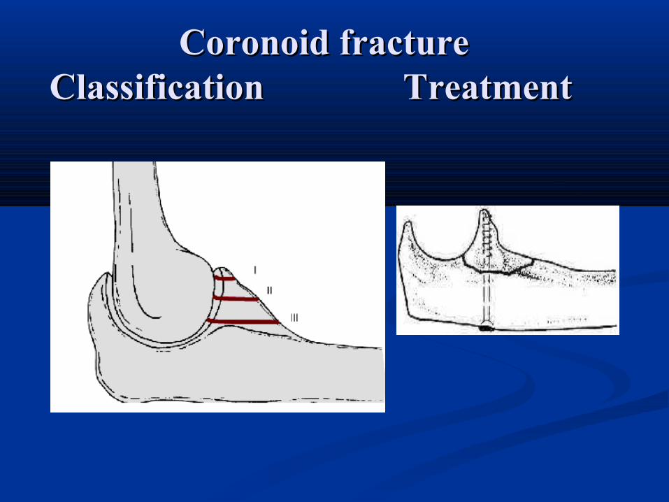
Coronoid fractureCoronoid fractureIt indicate severe trauma to elbow.It indicate severe trauma to elbow.Mech. of inj.Mech. of inj. - Struck of trochlea in coronoid. - Struck of trochlea in coronoid. -avulsion (less common).-avulsion (less common).Classification:Classification: type I: simple avulsion of tip.type I: simple avulsion of tip. type II: involve <50%.type II: involve <50%. type III :involve >50%.type III :involve >50%.TreatmentTreatment: :
typeI&II: heavy suture to the proximal of ulna.typeI&II: heavy suture to the proximal of ulna. typeIII :I.F with screw.typeIII :I.F with screw.
Coronoid fractureCoronoid fracture Classification Treatment Classification Treatment
Fracture of radial head Fracture of radial head
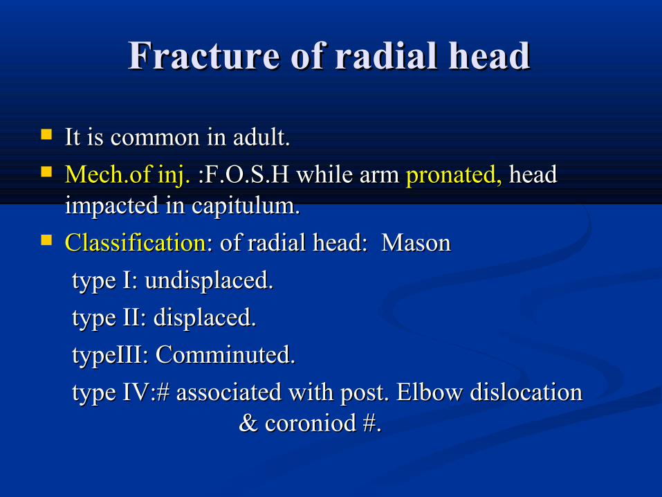
It is common in adult.It is common in adult. Mech.of inj.Mech.of inj. :F.O.S.H while arm :F.O.S.H while arm pronated,pronated, head head
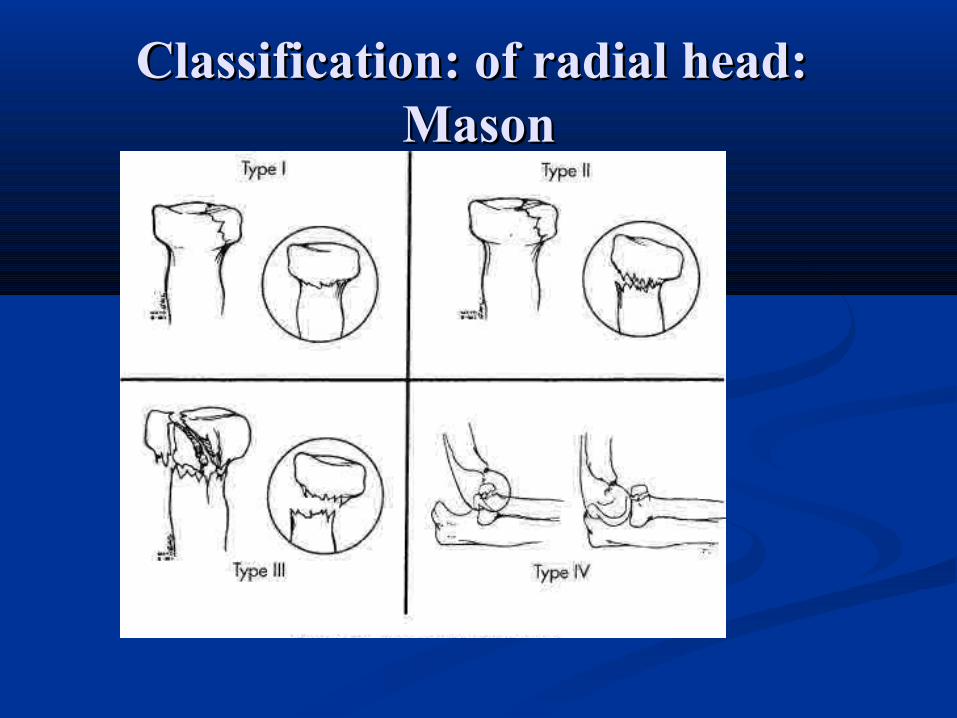
impacted in capitulum.impacted in capitulum. ClassificationClassification: of radial head: Mason: of radial head: Mason type I: undisplaced.type I: undisplaced. type II: displaced.type II: displaced. typeIII: Comminuted. typeIII: Comminuted. type IV:# associated with post. Elbow dislocation type IV:# associated with post. Elbow dislocation
& coroniod #.& coroniod #.
Classification: of radial head: Classification: of radial head: MasonMason
Fracture of radial head Fracture of radial head Treatment:Treatment: I. I. conservativeconservative: for : : for :
-type I. -type I. -type II : - if # in<1/3 of head circum. -type II : - if # in<1/3 of head circum.
-in outer part. -in outer part. - or get 70% of pronation & - or get 70% of pronation &
supination. supination.
Fracture of radial head Fracture of radial head
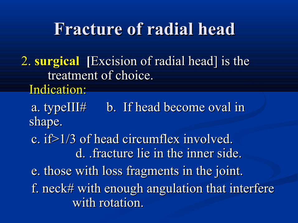
2. 2. surgicalsurgical [ [Excision of radial head] is the Excision of radial head] is the treatment of choice. treatment of choice.
Indication:Indication: a. typeIII# b. If head become oval in a. typeIII# b. If head become oval in
shape.shape. c. if>1/3 of head circumflex involved. c. if>1/3 of head circumflex involved.
d. .fracture lie in the inner side. d. .fracture lie in the inner side. e. those with loss fragments in the joint.e. those with loss fragments in the joint. f. neck# with enough angulation that interfere f. neck# with enough angulation that interfere
with rotation. with rotation.
Excision of radial headExcision of radial head TechniqueTechnique:: -excision should be early 24—48h.-excision should be early 24—48h. -incision:5cm below radial head up to lat. condyle.-incision:5cm below radial head up to lat. condyle. -pass b/n E.C.U&E.D or E.C.U&anconeus. -pass b/n E.C.U&E.D or E.C.U&anconeus. -excision: transverse just proximal biceptal tuber.-excision: transverse just proximal biceptal tuber. -anular lig should be excised. & debris removed-anular lig should be excised. & debris removed After treatmentAfter treatment: p.o.p in90 deg.for : p.o.p in90 deg.for 1w1w then converted then converted
to to sling sling till till 3w3w. Within this interval start gentle active . Within this interval start gentle active motion. motion.
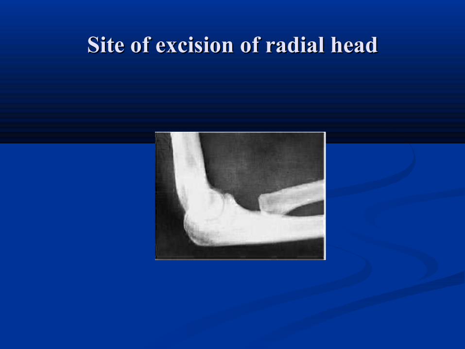
Site of excision of radial headSite of excision of radial head
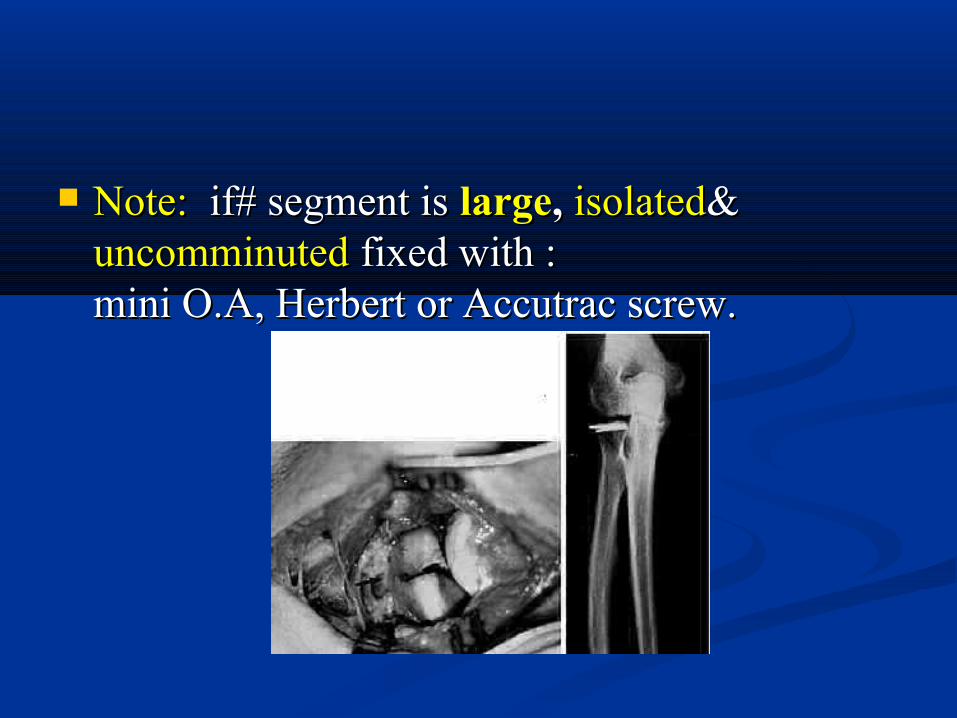
Note:Note: if# segment is if# segment is largelarge,, isolatedisolated& & uncomminuteduncomminuted fixed with : fixed with : mini O.A, Herbert or Accutrac screw. mini O.A, Herbert or Accutrac screw.
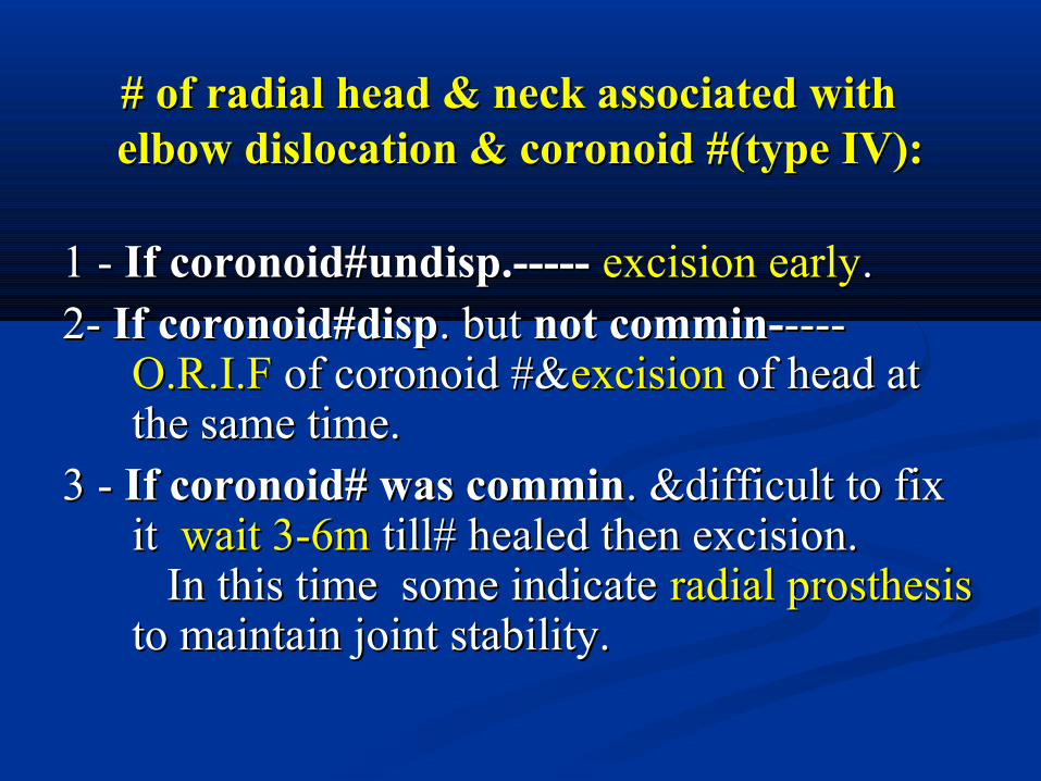
# of radial head & neck associated with # of radial head & neck associated with elbow dislocation & coronoid #(type IV):elbow dislocation & coronoid #(type IV):
1 - 1 - If coronoid#undisp.-----If coronoid#undisp.----- excision earlyexcision early..2- 2- If coronoid#dispIf coronoid#disp. but . but not commin-not commin----- ----
O.R.I.F O.R.I.F of coronoid #&of coronoid #&excisionexcision of head at of head at the same time.the same time.
3 - 3 - If coronoid# was comminIf coronoid# was commin. &difficult to fix . &difficult to fix it it wait 3-6m wait 3-6m till# healed then excision. till# healed then excision. In this time some indicate In this time some indicate radial prosthesisradial prosthesis to maintain joint stability.to maintain joint stability.
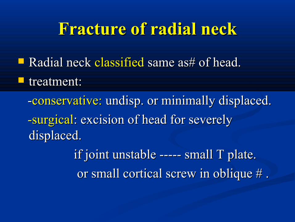
Fracture of radial neckFracture of radial neck Radial neck Radial neck classifiedclassified same as# of head. same as# of head. treatment:treatment: --conservative:conservative: undisp. or minimally displaced. undisp. or minimally displaced. -surgical-surgical: excision of head for severely : excision of head for severely
displaced.displaced. if joint unstable ----- small T plate.if joint unstable ----- small T plate. or small cortical screw in oblique # . or small cortical screw in oblique # .
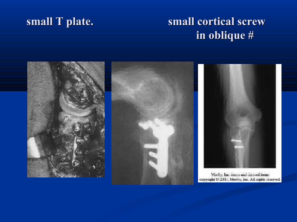
small T plate. small cortical screw small T plate. small cortical screw in oblique # in oblique #
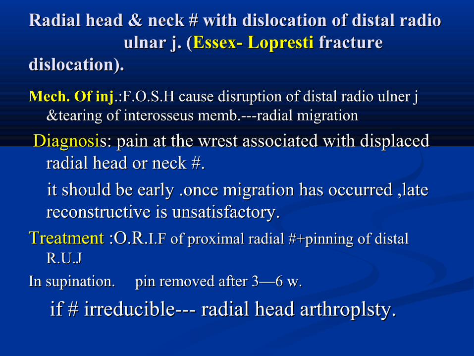
Radial head & neck # with dislocation of distal radio Radial head & neck # with dislocation of distal radio ulnar j. (ulnar j. (Essex- LoprestiEssex- Lopresti fracture fracture dislocation).dislocation).
Mech. Of injMech. Of inj.:F.O.S.H cause disruption of distal radio ulner j .:F.O.S.H cause disruption of distal radio ulner j &tearing of interosseus memb.---radial migration&tearing of interosseus memb.---radial migration
DiagnosiDiagnosis: pain at the wrest associated with displaced s: pain at the wrest associated with displaced radial head or neck #.radial head or neck #.
it should be early .once migration has occurred ,late it should be early .once migration has occurred ,late reconstructive is unsatisfactory. reconstructive is unsatisfactory.
TreatmentTreatment :O.R. :O.R.I.F of proximal radial #+pinning of distal I.F of proximal radial #+pinning of distal R.U.J R.U.J
In supination. pin removed after 3—6 w.In supination. pin removed after 3—6 w.
if # irreducible--- radial head arthroplsty.if # irreducible--- radial head arthroplsty.
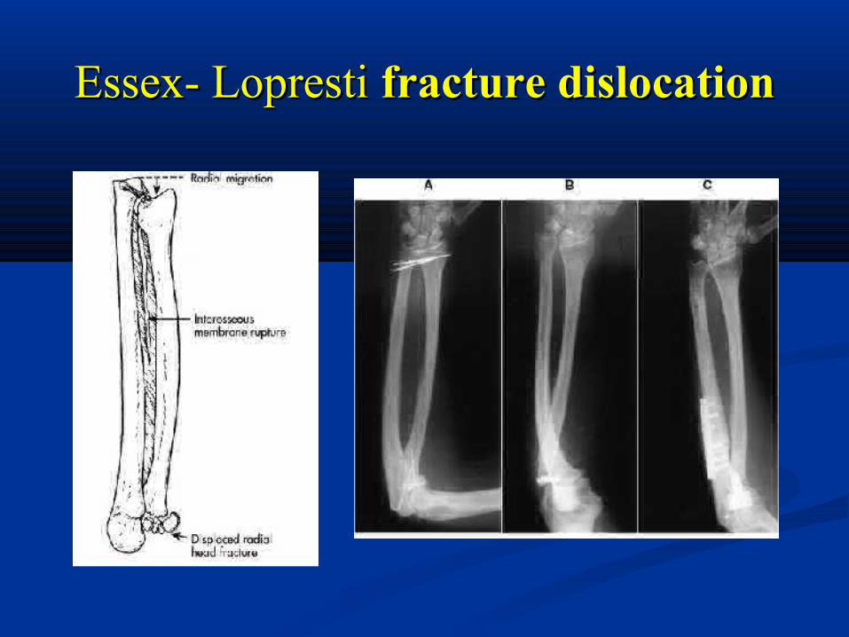
Essex- LoprestiEssex- Lopresti fracture dislocation fracture dislocation
Dislocation of elbow joint Dislocation of elbow joint Form 20% of joint dislocation (Form 20% of joint dislocation (after shoulder&after shoulder&
finger)finger) classification: classification: posterior [most common 80%]posterior [most common 80%] -ant. - med. - lat. - divergent. [rare].-ant. - med. - lat. - divergent. [rare]. posterior or post.lat. dislocationposterior or post.lat. dislocation : : mech of injmech of inj. :FOSH while elbow extended.. :FOSH while elbow extended.Diagnosis:Diagnosis: -C.P it may ass. with neurovascular inj -C.P it may ass. with neurovascular inj (median & ulnar n. &brachial a.) (median & ulnar n. &brachial a.) - X—Ray.- X—Ray.
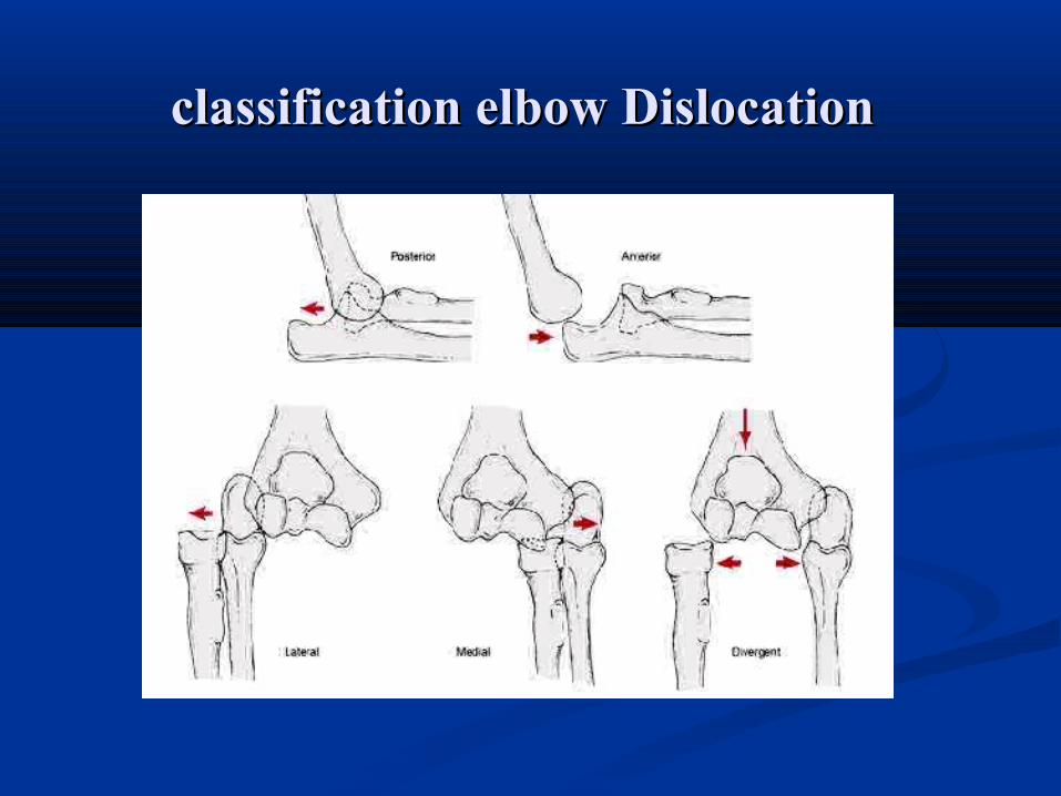
classification elbow Dislocationclassification elbow Dislocation
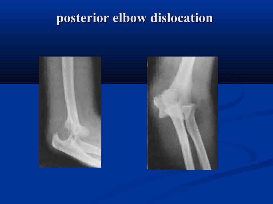
posterior elbow dislocationposterior elbow dislocation
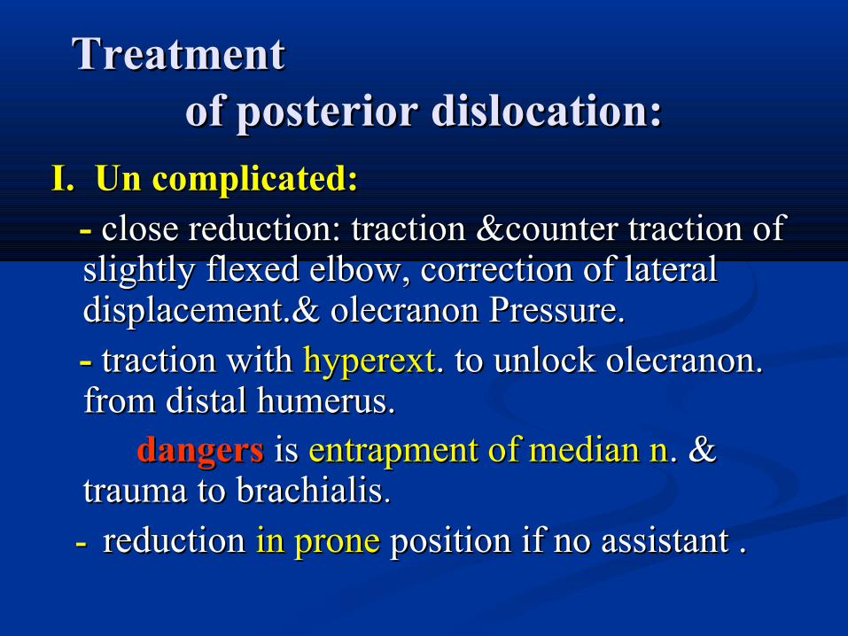
Treatment Treatment of posterior dislocation:of posterior dislocation:
I. Un complicated:I. Un complicated: -- close reduction: traction &counter traction of close reduction: traction &counter traction of
slightly flexed elbow, correction of lateral slightly flexed elbow, correction of lateral displacement.& olecranon Pressure. displacement.& olecranon Pressure.
-- traction with traction with hyperexthyperext. to unlock olecranon. . to unlock olecranon. from distal humerus.from distal humerus.
dangersdangers is is entrapment of median nentrapment of median n. & . & trauma to brachialistrauma to brachialis..
-- reduction reduction in pronein prone position if no assistant . position if no assistant .
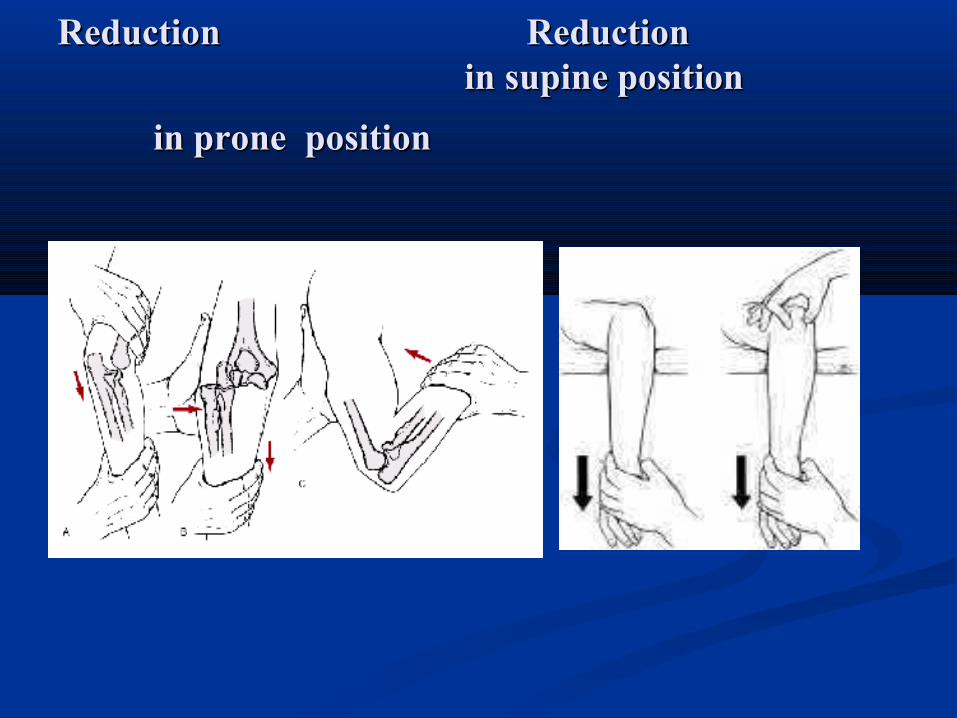
Reduction Reduction Reduction Reduction in supine position in supine position
in prone positionin prone position
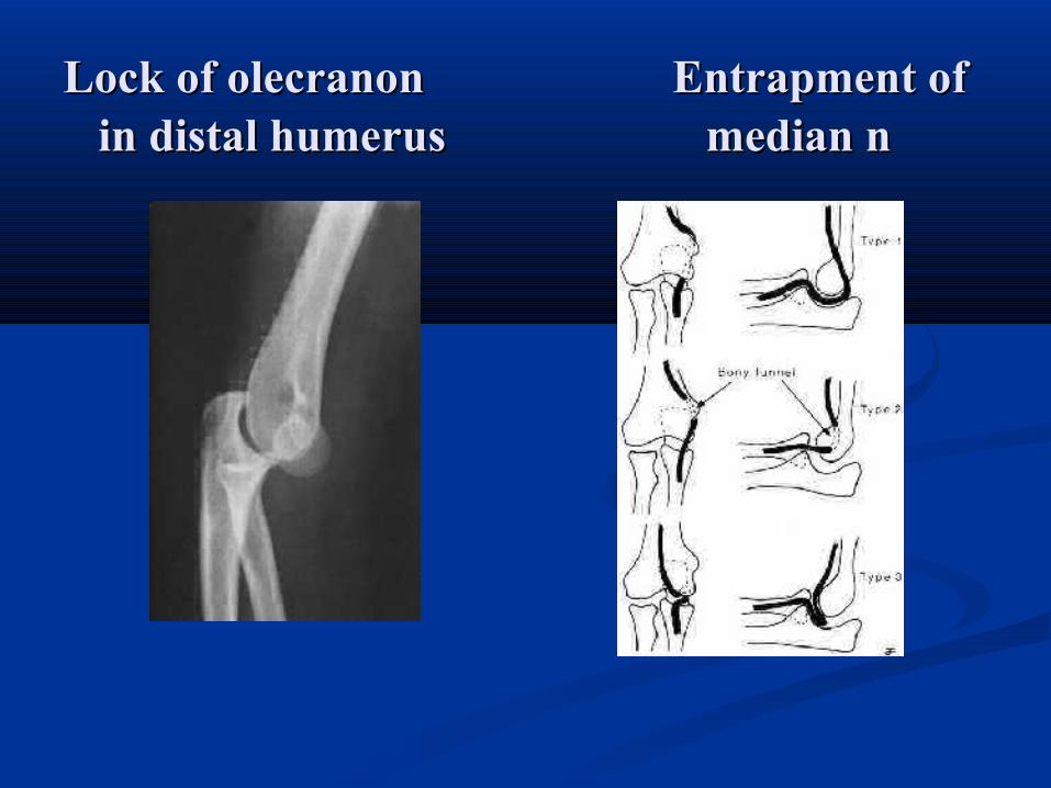
Lock of olecranonLock of olecranon Entrapment of Entrapment of in distal humerus in distal humerus median n median n
Treatment Treatment of posterior dislocation:of posterior dislocation:
II. Complicated:II. Complicated: associated with : associated with : 1 -coronoid# 2 - radial head #.1 -coronoid# 2 - radial head #. 3 -olecranon # 4 -medial epicondylar# . 3 -olecranon # 4 -medial epicondylar# .
1.Dislocation with coronoid #:treated as before.1.Dislocation with coronoid #:treated as before. 2. Dislocation with radial head #:2. Dislocation with radial head #: --a.a. we try to preserve radial head especially if we try to preserve radial head especially if
associated with coronoid# or medial lig.- by O.R. I.F. associated with coronoid# or medial lig.- by O.R. I.F. -b.-b. if# irreducible:- if# irreducible:-**stitch of med. lig.& pronater stitch of med. lig.& pronater
mass& p.o.p for 3-4w then excisionmass& p.o.p for 3-4w then excision.. *-*- or early excision &immobilization for 3-4w. or early excision &immobilization for 3-4w.
but if the joint unstable --- but if the joint unstable --- temporal arthroplasty . temporal arthroplasty .
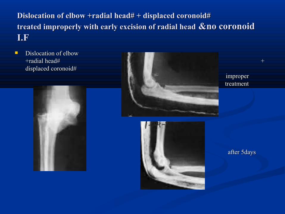
Dislocation of elbow +radial head# + displaced coronoid#Dislocation of elbow +radial head# + displaced coronoid#treated improperly with early excision of radial headtreated improperly with early excision of radial head &no coronoid &no coronoid I.FI.F
Dislocation of elbow Dislocation of elbow +radial head# + +radial head# + displaced coronoid# displaced coronoid#
improper improper treatment treatment
after 5days after 5days
Complication of Dislocation Complication of Dislocation of elbow jointof elbow joint
1. Stiffness& post traumatic arthritis1. Stiffness& post traumatic arthritis . .2. 2. Neurovascular injury.Neurovascular injury.3. 3. Hetrotopic calcificationHetrotopic calcification .(severe inj. long .(severe inj. long
immobilization, aggressive passive motion) .immobilization, aggressive passive motion) . treatmenttreatment:- NSAID& Radiotherapy but ineffective.:- NSAID& Radiotherapy but ineffective. -Resection of calcification but delayed till -Resection of calcification but delayed till
12 month. 12 month. -Early resection is contra indicated.-Early resection is contra indicated. -passive motion also avoided.-passive motion also avoided.
4.Recurrent instability due to: 4.Recurrent instability due to: a .weak collat. Lig. a .weak collat. Lig. b. residual articcular defect b. residual articcular defect in trochlea or trochlearin trochlea or trochlear notchnotch
c. c. ununited coronoid# ununited coronoid# d. unhealed ant. Capsule.d. unhealed ant. Capsule.
Treatment:Treatment: b/s the cause not clear, so b/s the cause not clear, so number of surgical procedures was tried :number of surgical procedures was tried :
1. Block of tibial bone put on coronoid. 1. Block of tibial bone put on coronoid. 2.Transfere of biceps tendon to coronoid.2.Transfere of biceps tendon to coronoid.3.Creation of cruciating lig. from triceps 3.Creation of cruciating lig. from triceps &bifceps .&bifceps .4. Collateral lig. Reconstruction.4. Collateral lig. Reconstruction.
THE END THE END
Dr. Jamal MaqramDr. Jamal Maqram
MoKazem.com
هذه المحاضرة هي من سلسلة محاضرات تم إعدادها و تقديمها من قبل الطباء المقيمين •في شعبة الجراحة العظمية في مشفى دمشق, تحت إشراف د. بشار ميرعلي.
الموقع غير مسؤول عن الخطاء الواردة في هذه المحاضرة. •
•This lecture is one of a series of lectures were prepared and presented by residents in the department of orthopedics in Damascus hospital, under the supervision of Dr. Bashar Mirali.
•This site is not responsible of any mistake may exist in this lecture.
Dr. Muayad Kadhimد. مؤيد كاظم