Embed Size (px)
Citation preview
Frank Viborg MortensenOverlæge, dr.med. Kirurgisk Afdeling L
Århus Universitetshospital
� R0 resection� 25 % residual liver volume� No extrahepatic disease (Relative)
� Performance� CT-sc. of the thorax and abdomen� LAP/LUS� Laparotomi� IOUS
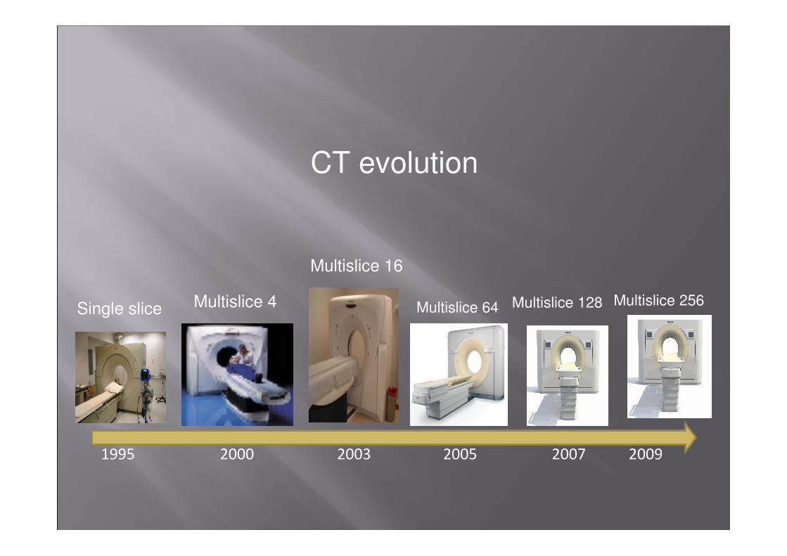
1995 2000 2003 2005 2007 2009
Single slice Multislice 4
Multislice 16
Multislice 64 Multislice 128 Multislice 256
CT evolution
� Retrospective analysis� 45 konsecutive pt. with CRC liver metastases resectable after MDCT of the thorax og abdomen.
� LAP/LUS in GA� IOUS � 62 yr ( ±10.6)� Number of metastases 2.1 ± 1.0
Mortensen FV et al. Scandinavian Journal of Surgery 2006;95:172-6
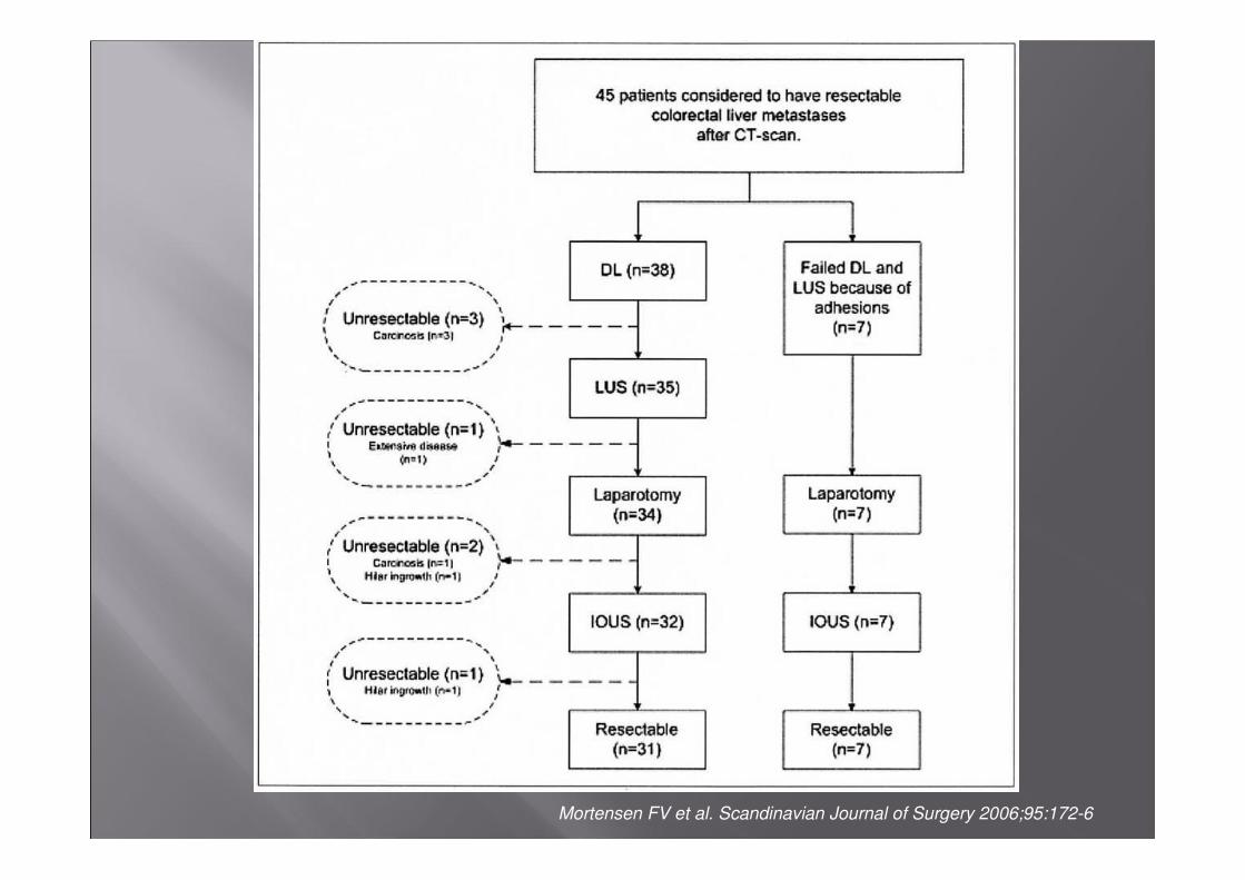
Mortensen FV et al. Scandinavian Journal of Surgery 2006;95:172-6
� Retrospective analysis� 70 consecutive patients with CRC liver metastases
� Inclusion period 1. January 2008 untill 30. June 2009.
� IOUS� 66.6 yr (32.6-88.5)� Median number of metastases 2 (0-9)� Syncrone/metacrone 38/32
Andersen KJ et al.
Experimental and Clinical Hepatology 2001;7:34-8
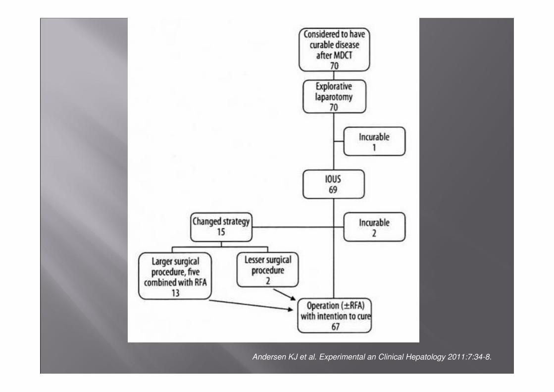
Andersen KJ et al. Experimental an Clinical Hepatology 2011:7:34-8.
� Average price for LAP/LUS ≈ 18.000 kr� Average price for avoiding unnessesary laparotomy ≈ 450.000 kr
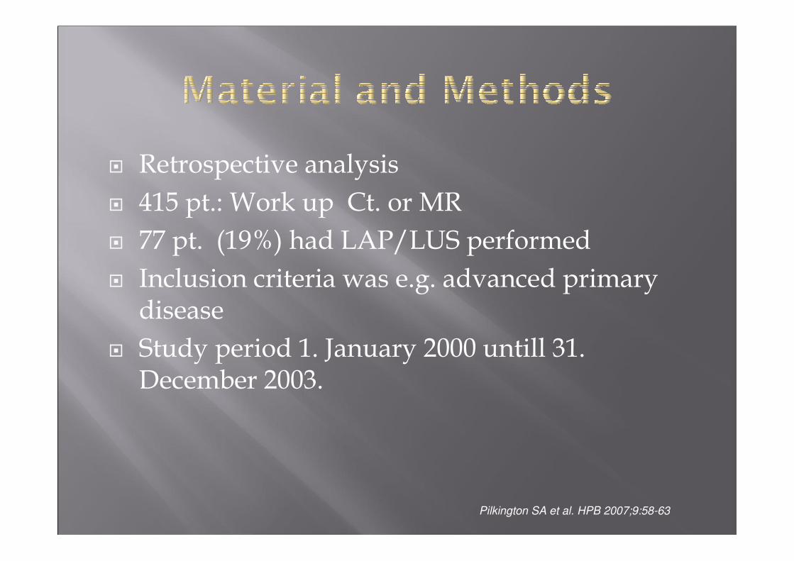
� Retrospective analysis� 415 pt.: Work up Ct. or MR� 77 pt. (19%) had LAP/LUS performed� Inclusion criteria was e.g. advanced primary disease
� Study period 1. January 2000 untill 31. December 2003.
Pilkington SA et al. HPB 2007;9:58-63
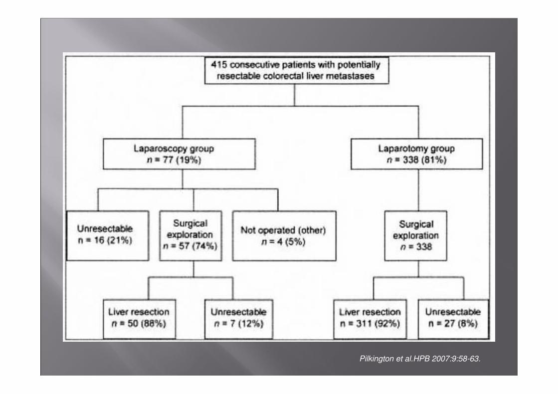
Pilkington et al.HPB 2007:9:58-63.
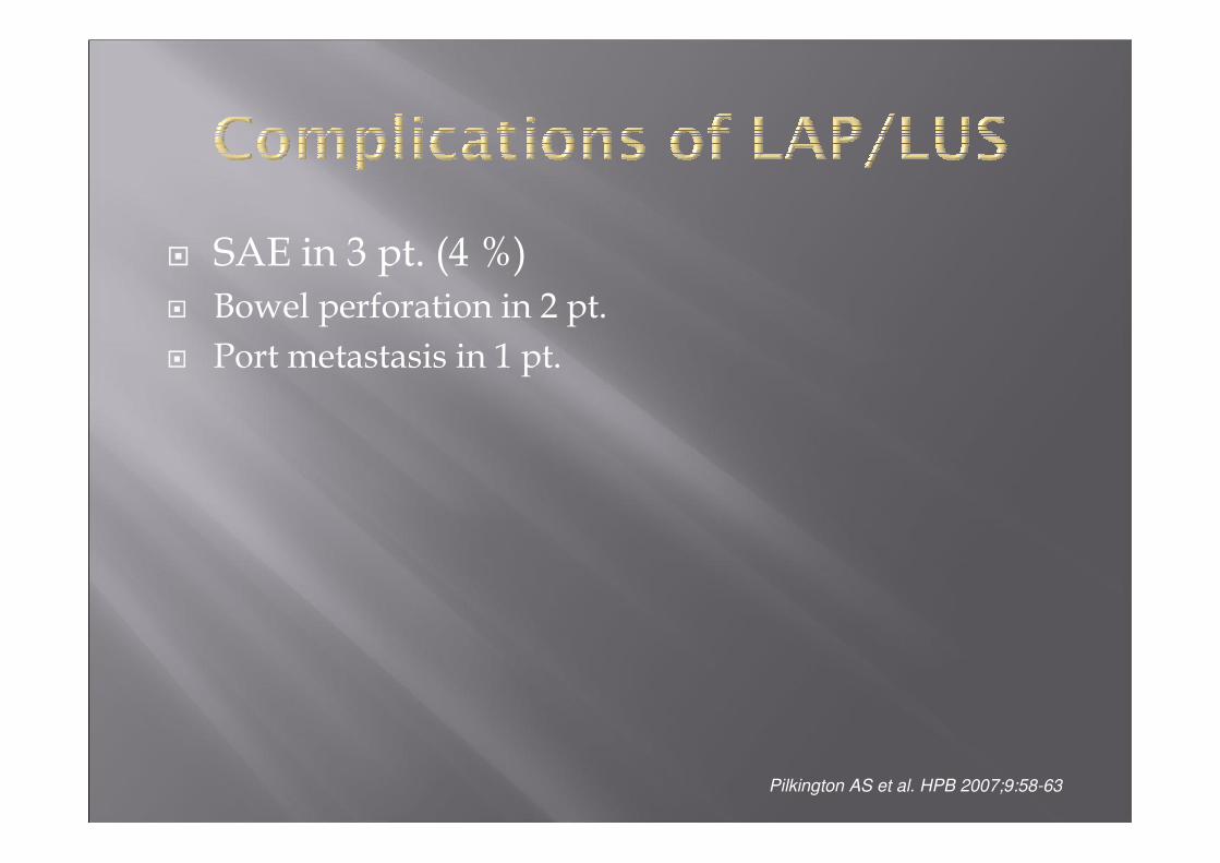
� SAE in 3 pt. (4 %)� Bowel perforation in 2 pt.� Port metastasis in 1 pt.
Pilkington AS et al. HPB 2007;9:58-63
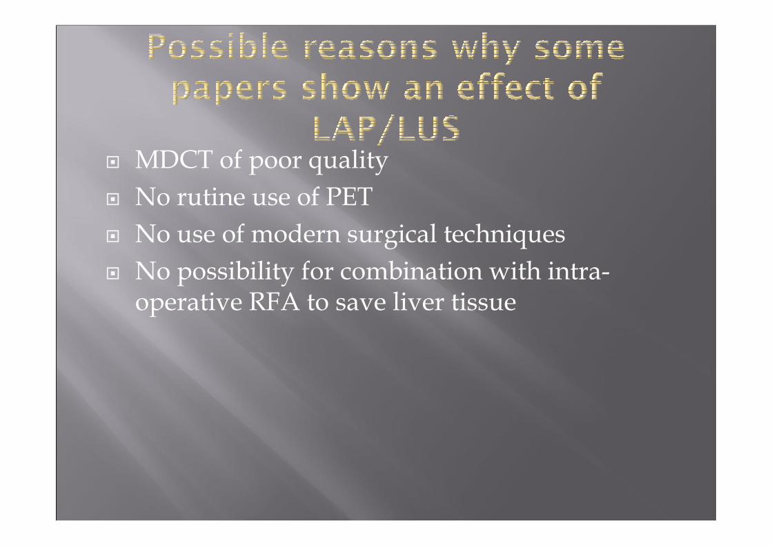
� MDCT of poor quality� No rutine use of PET� No use of modern surgical techniques� No possibility for combination with intra-operative RFA to save liver tissue
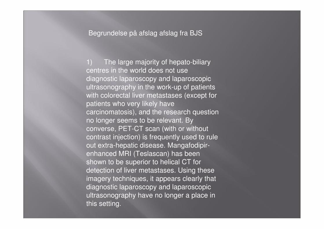
1) The large majority of hepato-biliary centres in the world does not use diagnostic laparoscopy and laparoscopic ultrasonography in the work-up of patients with colorectal liver metastases (except for patients who very likely have carcinomatosis), and the research question no longer seems to be relevant. By converse, PET-CT scan (with or without contrast injection) is frequently used to rule out extra-hepatic disease. Mangafodipir-enhanced MRI (Teslascan) has been shown to be superior to helical CT for detection of liver metastases. Using these imagery techniques, it appears clearly that diagnostic laparoscopy and laparoscopic ultrasonography have no longer a place in this setting.
Begrundelse på afslag afslag fra BJS