Embed Size (px)
Citation preview
cancers
Review
Nobiletin and Derivatives: Functional Compoundsfrom Citrus Fruit Peel for ColonCancer Chemoprevention
Joanna Xuan Hui Goh 1, Loh Teng-Hern Tan 2,3, Joo Kheng Goh 4, Kok Gan Chan 5,6,* ,Priyia Pusparajah 7, Learn-Han Lee 2,8 and Bey-Hing Goh 1,8,*
1 Biofunctional Molecule Exploratory (BMEX) Research Group, School of Pharmacy, Monash UniversityMalaysia, Bandar Sunway 47500, Selangor Darul Ehsan, Malaysia; [email protected]
2 Novel Bacteria and Drug Discovery (NBDD) Research Group, Microbiome and Bioresource ResearchStrength, Jeffrey Cheah School of Medicine and Health Sciences, Monash University Malaysia,Bandar Sunway 47500, Selangor Darul Ehsan, Malaysia; [email protected] (L.T.-H.T.);[email protected] (L.-H.L.)
3 Institute of Biomedical and Pharmaceutical Sciences, Guangdong University of Technology,Guangzhou 510006, China
4 School of Science, Monash University Malaysia, Jalan Lagoon Selatan, Bandar Sunway 47500,Selangor Darul Ehsan, Malaysia; [email protected]
5 Division of Genetics and Molecular Biology, Institute of Biological Sciences, Faculty of Science,University of Malaya, Kuala Lumpur 50603, Malaysia
6 International Genome Centre, Jiangsu University, Zhenjiang 212013, China7 Medical Health and Translational Research Group, Jeffrey Cheah School of Medicine and Health Sciences,
Monash University Malaysia, Bandar Sunway 47500, Selangor, Malaysia; [email protected] Asian Centre for Evidence Synthesis in Population, Implementation and Clinical Outcomes (PICO),
Health and Well-being Cluster, Global Asia in the 21st Century (GA21) Platform, Monash UniversityMalaysia, Bandar Sunway, 47500, Malaysia
* Correspondence: [email protected] (K.G.C.); [email protected] (B.-H.G.)
Received: 28 May 2019; Accepted: 19 June 2019; Published: 21 June 2019�����������������
Abstract: The search for effective methods of cancer treatment and prevention has been a continuouseffort since the disease was discovered. Recently, there has been increasing interest in exploringplants and fruits for molecules that may have potential as either adjuvants or as chemopreventiveagents against cancer. One of the promising compounds under extensive research is nobiletin (NOB),a polymethoxyflavone (PMF) extracted exclusively from citrus peel. Not only does nobiletin itselfexhibit anti-cancer properties, but its derivatives are also promising chemopreventive agents; examplesof derivatives with anti-cancer activity include 3′-demethylnobiletin (3′-DMN), 4′-demethylnobiletin(4′-DMN), 3′,4′-didemethylnobiletin (3′,4′-DMN) and 5-demethylnobiletin (5-DMN). In vitro studieshave demonstrated differential efficacies and mechanisms of NOB and its derivatives in inhibiting andkilling of colon cancer cells. The chemopreventive potential of NOB has also been well demonstratedin several in vivo colon carcinogenesis animal models. NOB and its derivatives target multiplepathways in cancer progression and inhibit several of the hallmark features of colorectal cancer(CRC) pathophysiology, including arresting the cell cycle, inhibiting cell proliferation, inducingapoptosis, preventing tumour formation, reducing inflammatory effects and limiting angiogenesis.However, these substances have low oral bioavailability that limits their clinical utility, hencethere have been numerous efforts exploring better drug delivery strategies for NOB and theseare part of this review. We also reviewed data related to patents involving NOB to illustrate theextensiveness of each research area and its direction of commercialisation. Furthermore, this reviewalso provides suggested directions for future research to advance NOB as the next promising candidatein CRC chemoprevention.
Cancers 2019, 11, 867; doi:10.3390/cancers11060867 www.mdpi.com/journal/cancers
Cancers 2019, 11, 867 2 of 34
Keywords: nobiletin; colorectal cancer; chemoprevention; bioactivities
1. Introduction
Colorectal cancer (CRC) is the third most prevalent cancer reported in both men and women,ranking just after prostate or breast cancer and lung cancer [1]. Although in many cases there is noreadily apparent cause of CRC, a number of factors have been found to be closely associated with thismalignancy including gender, age, genetic predisposition, lifestyle, diet or as a complication from otherdiseases such as inflammatory bowel disease (IBD). Statistics showed that the death rate from CRC is40% higher in males as compared to females, with the prevalence increasing with age, especially above50 years old; however, there is a new and worrying trend of increasing incidence of colorectal cancer inthe age group younger than 50, which, while slight, is still worrying [2,3].
The incidence of CRC has reduced as modern screening strategies have enabled much earlierdetection of potentially malignant lesions, allowing for early intervention such as surgical excision ofadenoma before it undergoes malignant transformation [4,5]. Although there has been a reduction, thehigh number of cases remains a major concern and the search for new and better treatments for CRChas been a key focus in pharmacological research. Standard therapy for cancer typically involves thetriple regimen of surgery, chemotherapy, and radiation treatment. Efforts in exploring and developingnew treatments are very much needed due to the limitations of the current treatment regimen—rangingfrom side effects, to complications and the development of drug resistance.
Researchers are attempting to explore multiple avenues for novel leads as anti-cancer agents withan increasing trend to focus on natural sources like plants and fruits [6–8]. However, while it is key tofind new treatments to existing cancers, a crucial aspect that is also being explored is prevention ofcancerous growths; in particular, this would be of benefit for those at risk due to the various factorsoutlined earlier. One of the effective strategies to control cancer is chemoprevention, which is definedas the use of a natural or synthetic agent to reverse, inhibit, or prevent the progression of cancer [9].
Plants and fruits are often part of a diet recommended to prevent various illnesses includingcancer [10]. These beneficial properties may be from the chemicals they contain as well as theirmetabolites which enter our alimentary canal and eventually end up in our colon and rectum. If thecompounds responsible can be isolated and purified for use as a treatment, this may be a milestone innew cancer therapies and prophylaxis. While an extensive review of polyphenols like apigenin andluteolin on anti-colorectal cancer effect can readily be found [11], this study highlights the potentialchemopreventive effect on CRC of another flavonoid, namely nobiletin (NOB).
NOB, a polymethoxyflavone (PMF), is likely named after Citrus nobilis. This compound is oneof the most ubiquitous flavones that can be isolated exclusively from the peel of citrus fruits [12].Besides CRC, there is concurrently ongoing research looking into the effect of NOB on other types ofcancers such as breast cancer [13,14], ovarian cancer [15], gastric cancer [16,17], lung cancer [18,19],liver cancer [20] and bone cancer [21]. There are also recent studies attesting to the benefits ofNOB in anti-neurodegeneration [22,23], anti-diabetes [24], anti-obesity [25–27], antimicrobial [28],anti-allergy [29] and anti-inflammatory effects [30,31]. There are also a number of articles that supportclaims purporting to the role of NOB in reducing the risk of cardiovascular diseases [32,33] andosteoporosis [34,35].
Interestingly, this compound can be metabolised into a number of metabolites which also showsignificant anti-cancer effects. There are several recent reviews on the bioactivities of these citrusPMF [36] as well as the potential chemopreventive abilities of these PMFs toward cancers in general [37].This review paper aims to gather the results of the in vivo and in vitro studies done in recent years andcompile various molecular pathways by which the compound NOB and its derivatives act in CRCprevention which will in turn help to facilitate future research that targets these specific mechanisms.
Cancers 2019, 11, 867 3 of 34
2. Research Methodology
The main focus was to search for all relevant primary research papers published which lookedinto the use of NOB and its derivatives as a chemopreventive agent for CRC. A systematic searchwas performed to identify published literature on the chemopreventive potentials of NOB againstCRC using Google Scholar. For studies published in foreign languages like Chinese, Japaneseor Korean, an attempt was made to locate the translated version. The search strategy wasperformed using keywords ‘nobiletin’ and ‘colorectal cancer’ to locate relevant papers. This wasalso supplemented with keyword search of the terms ‘cancer statistics’, ‘colorectal cancer’, ‘coloncancer’, ‘metabolites’, ‘synergism’, ‘biotransformation’, ‘mechanism’, ‘apoptosis’, ‘anti-inflammatory’,‘inflammation’, ‘cell proliferation’, ‘cell cycle arrest’, ‘metastasis’, ‘tumour’, ‘angiogenesis’, ‘absorption’,‘metabolism’, ‘toxicity’, ‘distribution’, ‘elimination’, ‘solubility’ and ‘delivery’ combined using Booleanoperators with ‘nobiletin’. PubChem and EMBASE were used as alternatives to ensure inclusionof all relevant papers while SciFinder database was mainly used to locate patents related to NOB.The reference lists of relevant articles collected were also screened for additional studies to be includedin the review.
3. Nobiletin and Its Derivatives
The compound nobiletin (NOB) can be extracted exclusively from citrus fruits, namely mandarinoranges (Citrus reticulate), sweet oranges or Valencia oranges (Citrus sinesis), Miaray mandarins (Citrusmiaray) [38], flat lemons or Hayata (Citrus depressa) [39,40], tangerines (Citrus tangerine), bitter oranges(Citrus aurantium) [12], Unshu Mikans or Satsuma mandarins (Citrus Unshiu arnicia indica) [41,42],Cleopatra mandarins (Citrus reshni) [43], mandarin oranges (Citrus tachibana), Koji Oranges (Citrusleiocarpa), Natsu Mikans (Citrus tardiva), Jimikan (Citrus succosa), Kinokuni Mandarins (Citrus Kinokuni),Fukushu (Citrus erythrosa), Sunkat (Citrus sunki) and hybrids of the mandarin orange with pomelo(Citrus deliciosa) [44]. Citrus tangerine was reported to contain the highest content of NOB, approximatelyfive times of that in Citrus sinesis [45].
PMF can be isolated from orange peel through different types of chemical extraction processes,for example, the supercritical fluid extraction, microwave assisted extraction [46] and Soxhlet method,which is capable of extracting large sample volumes [43]. Through the supercritical fluid extractionprocess, the supercritical fluid extractor is used to process the orange peel grinds that have beenfreeze-dried. Then, the extract is further treated with carbon dioxide and ethanol to concentrate thebioactive compound [47]. A special method to improve NOB yield through the supercritical fluidextraction method is currently patented in Korea [48]. It was found that the maximal yield of NOBoccurs at a temperature of 80 ◦C and pressure of 30 MPa with an optimum sample particle size of375 µm [40].
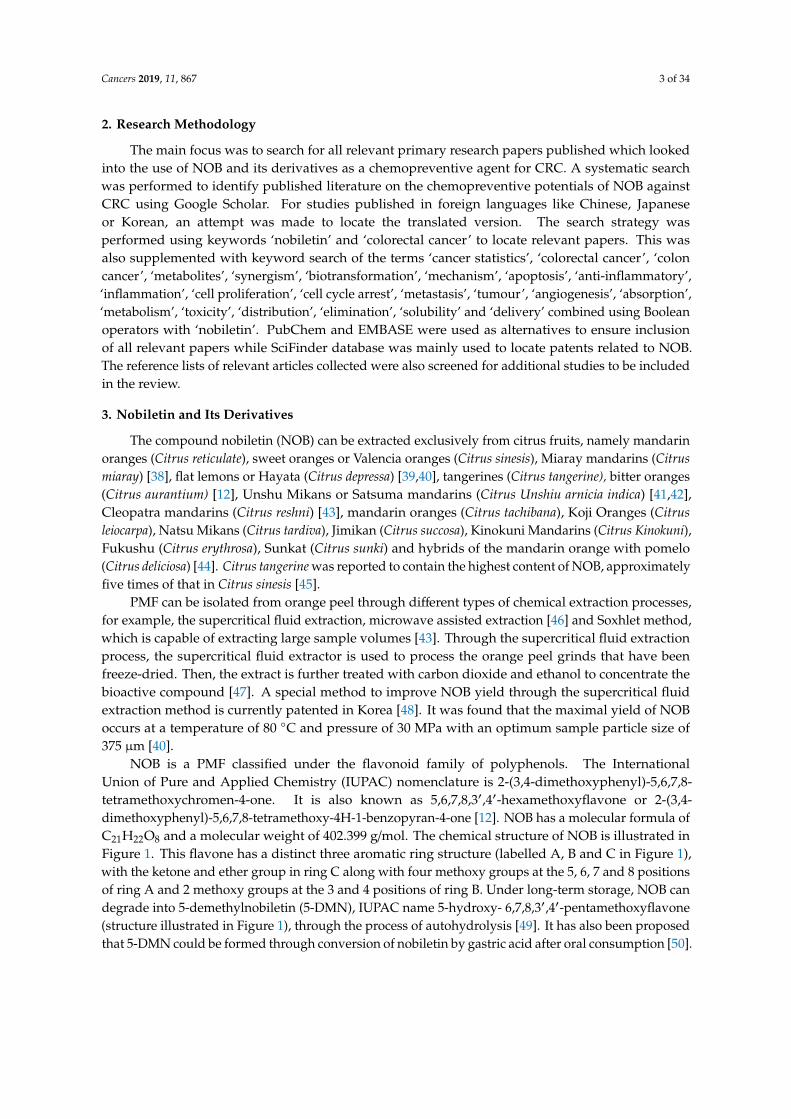
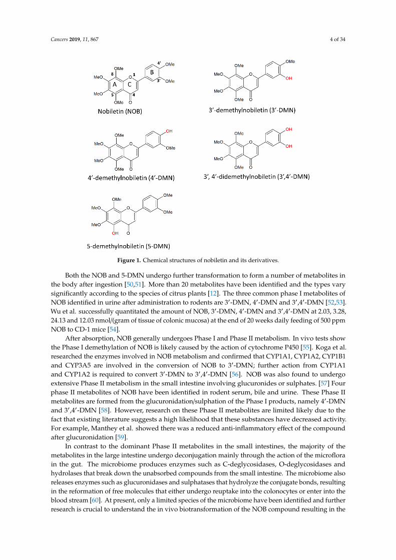
NOB is a PMF classified under the flavonoid family of polyphenols. The InternationalUnion of Pure and Applied Chemistry (IUPAC) nomenclature is 2-(3,4-dimethoxyphenyl)-5,6,7,8-tetramethoxychromen-4-one. It is also known as 5,6,7,8,3′,4′-hexamethoxyflavone or 2-(3,4-dimethoxyphenyl)-5,6,7,8-tetramethoxy-4H-1-benzopyran-4-one [12]. NOB has a molecular formula ofC21H22O8 and a molecular weight of 402.399 g/mol. The chemical structure of NOB is illustrated inFigure 1. This flavone has a distinct three aromatic ring structure (labelled A, B and C in Figure 1),with the ketone and ether group in ring C along with four methoxy groups at the 5, 6, 7 and 8 positionsof ring A and 2 methoxy groups at the 3 and 4 positions of ring B. Under long-term storage, NOB candegrade into 5-demethylnobiletin (5-DMN), IUPAC name 5-hydroxy- 6,7,8,3′,4′-pentamethoxyflavone(structure illustrated in Figure 1), through the process of autohydrolysis [49]. It has also been proposedthat 5-DMN could be formed through conversion of nobiletin by gastric acid after oral consumption [50].
Cancers 2019, 11, 867 4 of 34Cancers 2019, 11, x 4 of 34
Figure 1. Chemical structures of nobiletin and its derivatives.
Both the NOB and 5-DMN undergo further transformation to form a number of metabolites in the body after ingestion [50,51]. More than 20 metabolites have been identified and the types vary significantly according to the species of citrus plants [12]. The three common phase I metabolites of NOB identified in urine after administration to rodents are 3′-DMN, 4′-DMN and 3′,4′-DMN [52,53]. Wu et al. successfully quantitated the amount of NOB, 3′-DMN, 4′-DMN and 3′,4′-DMN at 2.03, 3.28, 24.13 and 12.03 nmol/(gram of tissue of colonic mucosa) at the end of 20 weeks daily feeding of 500 ppm NOB to CD-1 mice [54].
After absorption, NOB generally undergoes Phase I and Phase II metabolism. In vivo tests show the Phase I demethylation of NOB is likely caused by the action of cytochrome P450 [55]. Koga et al. researched the enzymes involved in NOB metabolism and confirmed that CYP1A1, CYP1A2, CYP1B1 and CYP3A5 are involved in the conversion of NOB to 3′-DMN; further action from CYP1A1 and CYP1A2 is required to convert 3′-DMN to 3′,4′-DMN [56]. NOB was also found to undergo extensive Phase II metabolism in the small intestine involving glucuronides or sulphates. [57] Four phase II metabolites of NOB have been identified in rodent serum, bile and urine. These Phase II metabolites are formed from the glucuronidation/sulphation of the Phase I products, namely 4′-DMN and 3′,4′-DMN [58]. However, research on these Phase II metabolites are limited likely due to the fact that existing literature suggests a high likelihood that these substances have decreased activity. For example, Manthey et al. showed there was a reduced anti-inflammatory effect of the compound after glucuronidation [59].
In contrast to the dominant Phase II metabolites in the small intestines, the majority of the metabolites in the large intestine undergo deconjugation mainly through the action of the microflora in the gut. The microbiome produces enzymes such as C-deglycosidases, O-deglycosidases and hydrolases that break down the unabsorbed compounds from the small intestine. The microbiome also releases enzymes such as glucuronidases and sulphatases that hydrolyze the conjugate bonds, resulting in the reformation of free molecules that either undergo reuptake into the colonocytes or enter into the blood stream [60]. At present, only a limited species of the microbiome have been identified and further research is crucial to understand the in vivo biotransformation of the NOB compound resulting in the generation of multiple metabolites with different activities [61]. It is likely that the subtle variances in the gut microbiome in different individuals may result in different pharmacodynamic effects after administration of NOB. For instance, 4′-DMN and 3′,4′-DMN have
Figure 1. Chemical structures of nobiletin and its derivatives.
Both the NOB and 5-DMN undergo further transformation to form a number of metabolites inthe body after ingestion [50,51]. More than 20 metabolites have been identified and the types varysignificantly according to the species of citrus plants [12]. The three common phase I metabolites ofNOB identified in urine after administration to rodents are 3′-DMN, 4′-DMN and 3′,4′-DMN [52,53].Wu et al. successfully quantitated the amount of NOB, 3′-DMN, 4′-DMN and 3′,4′-DMN at 2.03, 3.28,24.13 and 12.03 nmol/(gram of tissue of colonic mucosa) at the end of 20 weeks daily feeding of 500 ppmNOB to CD-1 mice [54].
After absorption, NOB generally undergoes Phase I and Phase II metabolism. In vivo tests showthe Phase I demethylation of NOB is likely caused by the action of cytochrome P450 [55]. Koga et al.researched the enzymes involved in NOB metabolism and confirmed that CYP1A1, CYP1A2, CYP1B1and CYP3A5 are involved in the conversion of NOB to 3′-DMN; further action from CYP1A1and CYP1A2 is required to convert 3′-DMN to 3′,4′-DMN [56]. NOB was also found to undergoextensive Phase II metabolism in the small intestine involving glucuronides or sulphates. [57] Fourphase II metabolites of NOB have been identified in rodent serum, bile and urine. These Phase IImetabolites are formed from the glucuronidation/sulphation of the Phase I products, namely 4′-DMNand 3′,4′-DMN [58]. However, research on these Phase II metabolites are limited likely due to thefact that existing literature suggests a high likelihood that these substances have decreased activity.For example, Manthey et al. showed there was a reduced anti-inflammatory effect of the compoundafter glucuronidation [59].
In contrast to the dominant Phase II metabolites in the small intestines, the majority of themetabolites in the large intestine undergo deconjugation mainly through the action of the microflorain the gut. The microbiome produces enzymes such as C-deglycosidases, O-deglycosidases andhydrolases that break down the unabsorbed compounds from the small intestine. The microbiome alsoreleases enzymes such as glucuronidases and sulphatases that hydrolyze the conjugate bonds, resultingin the reformation of free molecules that either undergo reuptake into the colonocytes or enter into theblood stream [60]. At present, only a limited species of the microbiome have been identified and furtherresearch is crucial to understand the in vivo biotransformation of the NOB compound resulting in the
Cancers 2019, 11, 867 5 of 34
generation of multiple metabolites with different activities [61]. It is likely that the subtle variancesin the gut microbiome in different individuals may result in different pharmacodynamic effects afteradministration of NOB. For instance, 4′-DMN and 3′,4′-DMN have been shown to exhibit higheranti-cancer and anti-inflammatory effects than NOB itself, but the rate of conversion from NOB to thesemetabolites may vary from one person to another [54,62]. The mechanisms of NOB in chemopreventionare elaborated under ‘Section 5—Chemopreventive effects of NOB, 5-DMN and NOB-metabolites’.
Early in vitro studies using rat liver S9 extracts reveals 3′-DMN as the main metabolite of NOBafter 24 h of treatment [42]. However, further High-Performance Liquid Chromatography (HPLC)analysis on in vivo experiments showed that the concentration of nobiletin and its metabolites differ inthe colonic mucosa—the concentration of 3′-DMN is almost equal to NOB, while 3′,4′-DMN is about5.9-fold more than NOB, and 4′-DMN being the most concentrated, at 11.9 times the concentration ofNOB. Integrating these values, the concentration of NOB is actually 20 times significantly lower in thecolon when compared to the total concentration of its metabolites [54]. Convincing evidence has shownthat these metabolites generated in vivo following oral administration of NOB result in significantaccumulation in colonic tissues which is associated with the chemopreventive effect for CRC.
Interestingly, growing evidence suggests that the metabolites have more potent anti-cancer activitythan their parent compounds, and the high concentration of the metabolites of NOB found in the colonmay indicate that anti-cancer effect of NOB is largely conferred by its metabolites. This is consistentwith the findings of Wu et al. who discovered that by treating HCT116 cell lines with NOB and itsmetabolites results in a 3.3 to 7.6-fold increase in apoptotic cells [54]. A recent study by Chiou et al. alsoshows that the hydroxylated PMF, 5-DMN is more potent than NOB in terms of its chemopreventiveeffect on colon malignancy for both in vivo studies using xenograft mice and in vivo studies usingthree different colon cell lines. Chiou and colleagues reported that 5-DMN shows different levels ofinhibition in different types of cell lines, with the highest efficacies in COLO205 cell lines, followedby HCT116 and HT-29 [49]. This is consistent with the findings of Qiu et al. stating that the halfmaximal inhibitory concentration (IC50) required for 5-DMN to exert an inhibitory effect on the growthof HCT116 cells is 8.4 µM as compared to the notably higher value of 37 µM for NOB. Similarly,the IC50 required for 5-DMN against HT-29 cells is 22 µM as compared to the higher IC50 of 46.2 µM forNOB [63]. This may suggest that the hydroxyl group at the 5th position on the A ring is an importantfunctional group involved in the molecular interactions [49].
4. Pathogenesis of Colorectal Cancer
The mechanisms leading to CRC development are part of a rather complex process. The pathogenesisof CRC is arbitrarily subdivided into three stages here: initiation, progression and metastasis. Eachpathway is known to be regulated by chemical signals, called cytokines, which allow the progressionfrom one stage to the next, whilst inflammation is the underlying result of each stage [11].
Cancer generally starts with a mutated cell which deviates from the normal cell growth cycleand progresses through the cell cycle rapidly with no differentiation of structure or function. It canbe attributed to the down regulation of the regulatory genes or up-regulation of oncogenes. Thisgradually leads to the formation of a mass of undifferentiated cells called an adenoma. This lumpof cells does not perform any specific function but competes with the surrounding normal cells fornutrients. In more than 60% of colorectal adenomas, the dysregulation of adenomatous polyposis coligene resulting from the Wnt/β-catenin pathway is the major culprit in triggering this process [64].
The initial adenoma progresses on to an intermediate adenoma when the epidermal growth factorreceptor (EGFR) is activated, which in turn triggers the phosphatidylinositol-3-kinase pathway andresults in tumour formation [65]. Also, the inactivation of transforming growth factor-β (TGF-β) andthe loss of function of p53 further aggravates tumour growth by preventing apoptosis [66,67].
Cancers 2019, 11, 867 6 of 34
Eventually, a tumour becomes malignant when angiogenesis occurs, and the cancer cells arereleased into the bloodstream and spread to other parts of the body through a process known asmetastasis. Intercellular adhesion molecule-1 (ICAM-1) and matrix metalloproteinase (MMPs) areclosely associated with the promotion of angiogenesis and metastasis. To illustrate, the MMP disruptsthe integrity of the basal membrane allowing the cancer cells to enter the surrounding blood vesselsand thus the blood stream through a process known as intravasation [68,69].
5. Chemopreventive Effects of Nobiletin, 5-DMN and NOB-Metabolites
In one of earliest in vitro studies, the antiproliferative effect of NOB was evaluated against HT-29colon cancer cells [70]. The study determined that the IC50 and IC90 of NOB against HT-29 cellwere 4.7 µM and 13.9 µM, respectively, via the 3H-thymidine uptake assay [70]. As a product ofautohydrolysis of NOB, 5-DMN was also evaluated for its antiproliferative effect against colon cancercells. In the H-thymidine uptake assay, the IC50 and IC90 of 5-DMN against HT-29 was reported to be8.5 µM and 171 µM, respectively [70]. In the following years, NOB and 5-DMN were also reported tobe cytotoxic towards different colon cancer cell lines, including HCT116, HT-29, SW489, COLO320,COLO205 and Caco-2 (Table 1). Despite the stronger anti-proliferative effect of NOB observed inthe earlier study [70], recent studies increasingly showed that 5-DMN exhibits stronger cytotoxiceffects against different colon cancer cells as compared to NOB [49,63]. These contradictory resultsare potentially due to the different aspects of cancer focused in each study. Based on these in vitrostudies, NOB and 5-DMN were shown to exhibit their cytotoxic effects towards colon cancer cells,predominantly via cell cycle arrest and induction of apoptosis (Table 1).
Multiple in vivo studies demonstrated that NOB offers a protective effect against severalcarcinogens, such as the azoxymethane (AOM) and the 2-amino-1-methyl-6-phenylimidazo[4,5-b]pyridine (PhIP) (Table 2). AOM/DSS has been used to induce colitis in mice for the purpose ofcreating mouse models that replicate colitis induced CRC in humans [71]; however, PhIP, a heterocyclicamine, is a food-derived carcinogen that is abundantly released in the process of cooking fish andmeat [72,73]. Administration of 0.01% wt of NOB to mice for five weeks in their diet resulted inthe reduction of abnormal growths induced by colonic carcinogen AOM in the colons of the mice;there was a 50% reduction as compared to the controls [41]. Another similar study to determine theanti-adenocarcinoma effects of NOB also showed positive results but with lower efficacies, whereby34 weeks administration of 0.01% or 0.05% wt. of NOB reduced the frequency of adenocarcinomaby 12% and 32%, respectively [74]. In addition to that, Wu et al. demonstrated that NOB treatmentsuccessfully reduced the rate of cell proliferation by 69%, tumour incidence by 40%, tumour multiplicityby 71%, and downregulated TNF-α, IL-1β and IL-6 by 65%, 69% and 45% respectively in AOM/DSStreated mice [54]. Consistent with the inhibitory effect against AOM induced colon carcinogenesis,NOB also showed significant reduction in the high density of colonic aberrant crypt foci (ACF) locatedin the transverse colon in PhlP-induced F344 rats [75]. This shows that NOB is effective in preventingCRC triggered by different types of carcinogens.
Further support for NOB as a prospective candidate for chemoprevention is that NOB is knownto inhibit different pathways leading to cancer via a number of different mechanisms which includesinhibiting cell cycle progression [54,76], limiting inflammation [76], inducing apoptosis [54], preventingangiogenesis [77] and reducing tumour formation [49,54,78]. This subsection will describe themechanism of action of NOB, its autohydrolysis product, 5-DMN and its three common metabolites,namely 3′-DMN, 4′-DMN and 3′,4′-DMN, in chemoprevention of CRC in detail.
Cancers 2019, 11, 867 7 of 34
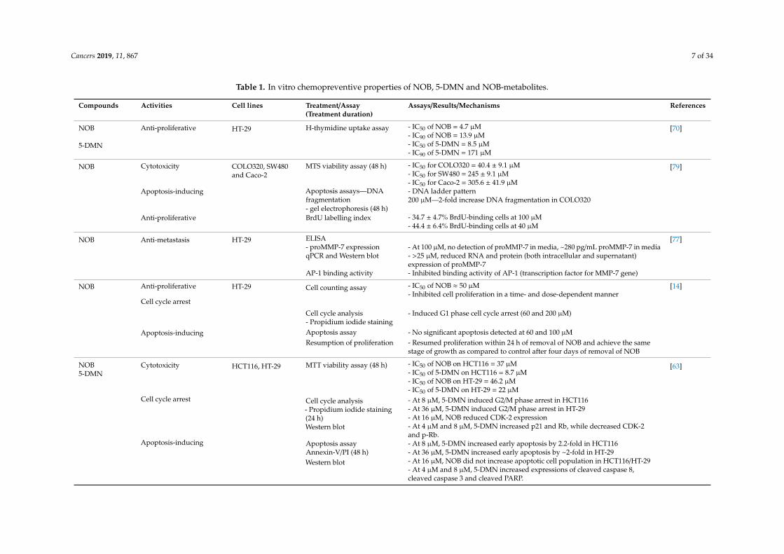
Table 1. In vitro chemopreventive properties of NOB, 5-DMN and NOB-metabolites.
Compounds Activities Cell lines Treatment/Assay(Treatment duration)
Assays/Results/Mechanisms References
NOB Anti-proliferative HT-29 H-thymidine uptake assay - IC50 of NOB = 4.7 µM [70]- IC90 of NOB = 13.9 µM
5-DMN - IC50 of 5-DMN = 8.5 µM- IC90 of 5-DMN = 171 µM
NOB Cytotoxicity COLO320, SW480and Caco-2
MTS viability assay (48 h) - IC50 for COLO320 = 40.4 ± 9.1 µM [79]- IC50 for SW480 = 245 ± 9.1 µM- IC50 for Caco-2 = 305.6 ± 41.9 µM
Apoptosis-inducing Apoptosis assays—DNAfragmentation
- DNA ladder pattern200 µM—2-fold increase DNA fragmentation in COLO320
- gel electrophoresis (48 h)Anti-proliferative BrdU labelling index - 34.7 ± 4.7% BrdU-binding cells at 100 µM
- 44.4 ± 6.4% BrdU-binding cells at 40 µM
NOB Anti-metastasis HT-29 ELISA [77]- proMMP-7 expression - At 100 µM, no detection of proMMP-7 in media, ~280 pg/mL proMMP-7 in mediaqPCR and Western blot - >25 µM, reduced RNA and protein (both intracellular and supernatant)
expression of proMMP-7AP-1 binding activity - Inhibited binding activity of AP-1 (transcription factor for MMP-7 gene)
NOB Anti-proliferative HT-29 Cell counting assay - IC50 of NOB ≈ 50 µM [14]- Inhibited cell proliferation in a time- and dose-dependent manner
Cell cycle arrest
Cell cycle analysis - Induced G1 phase cell cycle arrest (60 and 200 µM)- Propidium iodide staining
Apoptosis-inducing Apoptosis assay - No significant apoptosis detected at 60 and 100 µMResumption of proliferation - Resumed proliferation within 24 h of removal of NOB and achieve the same
stage of growth as compared to control after four days of removal of NOB
NOB5-DMN
Cytotoxicity HCT116, HT-29 MTT viability assay (48 h) - IC50 of NOB on HCT116 = 37 µM [63]- IC50 of 5-DMN on HCT116 = 8.7 µM- IC50 of NOB on HT-29 = 46.2 µM- IC50 of 5-DMN on HT-29 = 22 µM
Cell cycle arrest Cell cycle analysis- Propidium iodide staining(24 h)Western blot
- At 8 µM, 5-DMN induced G2/M phase arrest in HCT116- At 36 µM, 5-DMN induced G2/M phase arrest in HT-29- At 16 µM, NOB reduced CDK-2 expression- At 4 µM and 8 µM, 5-DMN increased p21 and Rb, while decreased CDK-2and p-Rb.
Apoptosis-inducing Apoptosis assay - At 8 µM, 5-DMN increased early apoptosis by 2.2-fold in HCT116Annexin-V/PI (48 h) - At 36 µM, 5-DMN increased early apoptosis by ~2-fold in HT-29Western blot - At 16 µM, NOB did not increase apoptotic cell population in HCT116/HT-29
- At 4 µM and 8 µM, 5-DMN increased expressions of cleaved caspase 8,cleaved caspase 3 and cleaved PARP.
Cancers 2019, 11, 867 8 of 34
Table 1. Cont.
Compounds Activities Cell lines Treatment/Assay(Treatment Duration)
Assays/Results/Mechanisms References
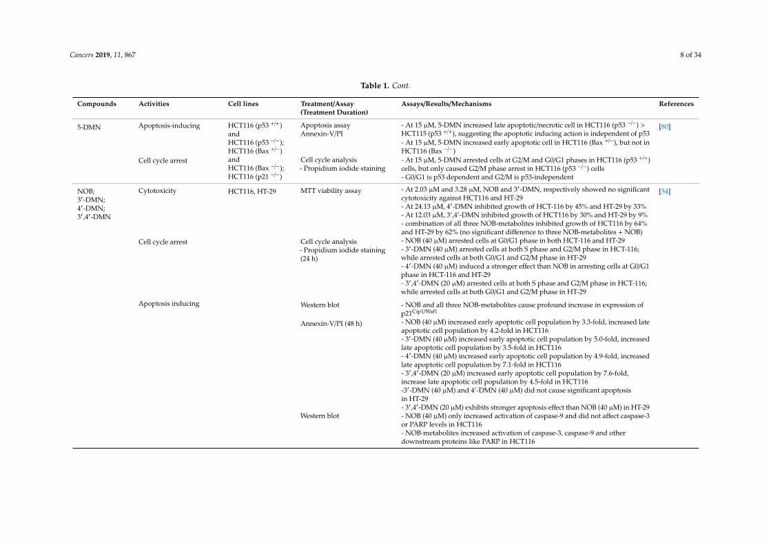
5-DMN Apoptosis-inducing HCT116 (p53 +/+)andHCT116 (p53 −/−);HCT116 (Bax +/−)andHCT116 (Bax −/−);HCT116 (p21 −/−)
Apoptosis assayAnnexin-V/PI
- At 15 µM, 5-DMN increased late apoptotic/necrotic cell in HCT116 (p53 −/−) >HCT115 (p53 +/+), suggesting the apoptotic inducing action is independent of p53
[80]
- At 15 µM, 5-DMN increased early apoptotic cell in HCT116 (Bax +/−), but not inHCT116 (Bax −/−)
Cell cycle arrest Cell cycle analysis- Propidium iodide staining
- At 15 µM, 5-DMN arrested cells at G2/M and G0/G1 phases in HCT116 (p53 +/+)cells, but only caused G2/M phase arrest in HCT116 (p53 −/−) cells- G0/G1 is p53 dependent and G2/M is p53-independent
NOB;3′-DMN;4′-DMN;3′,4′-DMN
Cytotoxicity HCT116, HT-29 MTT viability assay - At 2.03 µM and 3.28 µM, NOB and 3′-DMN, respectively showed no significantcytotoxicity against HCT116 and HT-29
[54]
- At 24.13 µM, 4′-DMN inhibited growth of HCT-116 by 45% and HT-29 by 33%- At 12.03 µM, 3’,4’-DMN inhibited growth of HCT116 by 30% and HT-29 by 9%- combination of all three NOB-metabolites inhibited growth of HCT116 by 64%and HT-29 by 62% (no significant difference to three NOB-metabolites + NOB)
Cell cycle arrest Cell cycle analysis- Propidium iodide staining(24 h)
- NOB (40 µM) arrested cells at G0/G1 phase in both HCT-116 and HT-29- 3′-DMN (40 µM) arrested cells at both S phase and G2/M phase in HCT-116;while arrested cells at both G0/G1 and G2/M phase in HT-29- 4′-DMN (40 µM) induced a stronger effect than NOB in arresting cells at G0/G1phase in HCT-116 and HT-29- 3′,4′-DMN (20 µM) arrested cells at both S phase and G2/M phase in HCT-116;while arrested cells at both G0/G1 and G2/M phase in HT-29
Apoptosis inducing Western blot - NOB and all three NOB-metabolites cause profound increase in expression ofp21Cip1/Waf1
Annexin-V/PI (48 h) - NOB (40 µM) increased early apoptotic cell population by 3.3-fold, increased lateapoptotic cell population by 4.2-fold in HCT116- 3′-DMN (40 µM) increased early apoptotic cell population by 5.0-fold, increasedlate apoptotic cell population by 3.5-fold in HCT116- 4′-DMN (40 µM) increased early apoptotic cell population by 4.9-fold, increasedlate apoptotic cell population by 7.1-fold in HCT116- 3′,4′-DMN (20 µM) increased early apoptotic cell population by 7.6-fold,increase late apoptotic cell population by 4.5-fold in HCT116-3′-DMN (40 µM) and 4’-DMN (40 µM) did not cause significant apoptosisin HT-29- 3′,4′-DMN (20 µM) exhibits stronger apoptosis effect than NOB (40 µM) in HT-29
Western blot - NOB (40 µM) only increased activation of caspase-9 and did not affect caspase-3or PARP levels in HCT116- NOB-metabolites increased activation of caspase-3, caspase-9 and otherdownstream proteins like PARP in HCT116
Cancers 2019, 11, 867 9 of 34
Table 1. Cont.
Compounds Activities Cell lines Treatment/Assay(Treatment duration)
Assays/Results/Mechanisms References
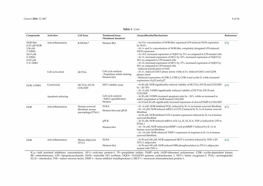
NOB-Met(2.03 µM NOB:3.28 µM3′-DMN:24.13 µM4′-DMN:12.03 µM3′,4′-DMN
Anti-inflammatory RAW264.7 Western Blot - At 0.5× concentration of NOB-Met, supressed LPS-induced iNOS expressionby 56.4%
[76]
- At 1× and 2× concentration of NOB-Met, completely abrogated LPS-inducediNOS expression- At ×0.5, increased expression of NQO1 by 21% as compared to LPS-treated cells- At ×1, increased expression of HO-1 by 10%, increased expression of NQO1 by34% as compared to LPS-treated cells- At ×2, increased expression of HO-1 by 37%, increased expression of NQO1 by50% as compared to LPS-treated cells- Induced translocation of Nrf2
Cell cycle arrest HCT116 Cell cycle analysis- Propidium iodide stainingWestern blot
- At 1×, induced G0/G1 phase arrest; while at 2×, induced G0/G1 and G2/Mphases arrest- Reduced expressions of CDK-2, CDK-4, CDK-6 and cyclin D, while increasedexpressions of p53 and p27
NOB, 5-DMN Cytotoxicity HCT116, HT-29,COLO205
MTT viability assay - At 40 µM, NOB significantly reduced viability of HCT116, HT-29 and COLO205by ~20–30%
[49]
- At >5 µM, 5-DMN significantly reduced viability of HCT116, HT-29 andCOLO205
Apoptosis inducing Cell cycle analysis- SubG1 quantificationWestern
- At 20 µM, 5-DMN increased apoptosis ratio by ~26%, while no increased insubG1 population in NOB-treated COLO205- At 10 and 20 µM, significantly increased expression of cleaved PARP in COLO205
NOB Anti-inflammatory Human synovialfibroblast, mousemacrophage J774A.1
ELISA - At >4 µM, NOB inhibited PGE2 induced by IL-1α in human synovial fibroblast [81]
Western blot and qPCR - At >16 µM, NOB reduced mRNA of COX-2 induced by IL-1α in human synovialfibroblast- At 64 µM, NOB inhibited COX-2 protein expression induced by IL-1α in humansynovial fibroblast
qPCR - At 32 µM, NOB reduced mRNA of IL-1α, IL-1β, IL-6, TNF-α induced by LPS inJ774A.1
Western blot - At >16 µM, NOB reduced proMMP-1 and proMMP-3 induced by IL-1α inhuman synovial fibroblast- At >16 µM, NOB enhanced TIMP-1 expression in response to IL-1α in humansynovial fibroblast
NOB Anti-inflammatory Mouse adipocyte3T3-L1
ELISA - At 50 and 100 µM, NOB suppressed MCP-1 secretion induced by TNF-α IN3T3-L1 adipocytes
[82]
Western blot - At 50 and 100 µM, NOB reduced ERK phosphorylation in 3T3-L1 adipocytestreated with TNF-α
IC50—half maximal inhibitory concentration; AP-1—activator protein-1; PI—propidium iodide; PARP—poly (ADP-ribosome) polymerase; CDK—cyclin-dependent kinase;Rb—retinoblastoma; LPS—lipopolysaccharide; iNOS—inducible NO synthase; NQO1—NAD(P)H quinone oxidoreductase 1; HO-1—heme oxygenase-1; PGE2—prostaglandinE2; IL—interleukin; TNF—tumor necrosis factor; TIMP-1—tissue inhibitor metalloprotease-1; MCP-1—monocyte chemoattractant protein-1.
Cancers 2019, 11, 867 10 of 34
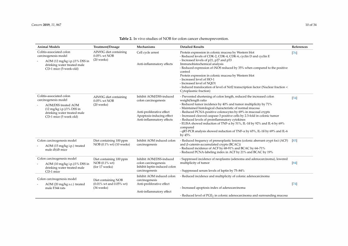
Table 2. In vivo studies of NOB for colon cancer chemoprevention.
Animal Models Treatment/Dosage Mechanisms Detailed Results References
Colitis-associated coloncarcinogenesis model
- AOM (12 mg/kg i.p.)/1% DSS indrinking water treated maleCD-1 mice (5-week-old)
AIN93G diet containing0.05% wt NOB(20 weeks)
Cell cycle arrest Protein expression in colonic mucosa by Western blot- Reduced levels of CDK-2, CDK-4, CDK-6, cyclin D and cyclin E- Increased levels of p21, p27 and p53
[76]
Anti-inflammatory effects Immunohistochemical analysis- Reduced expression of iNOS reduced by 35% when compared to the positivecontrolProtein expression in colonic mucosa by Western blot- Increased level of HO-1- Increased level of NQO1- Induced translocation of level of Nrf2 transcription factor (Nuclear fraction <Cytoplasmic fraction)
Colitis-associated coloncarcinogenesis model
- AOM/DSS treated AOM(12 mg/kg i.p.)/1% DSS indrinking water treated maleCD-1 mice (5 week old)
AIN93G diet containing0.05% wt NOB(20 weeks)
Inhibit AOM/DSS-inducedcolon carcinogenesis
- Prevented shortening of colon length, reduced the increased colonweight/length ratio- Reduced tumor incidence by 40% and tumor multiplicity by 71%- Maintained histological characteristic of normal mucosa
[54]
Anti-proliferative effect - Reduced PCNA-positive colonocytes by 69% in mucosal cryptsApoptosis-inducing effect - Increased cleaved caspase-3 positive cells by 2.3-fold in colonic tumorAnti-inflammatory effects - Reduced levels of proinflammatory cytokines
- ELISA showed reduction of TNF-α by 51%, IL-1ß by 92% and IL-6 by 69%compared- qRT-PCR analysis showed reduction of TNF-α by 65%, IL-1ß by 69% and IL-6by 45%
Colon carcinogenesis model
- AOM (15 mg/kg i.p.) treatedmale db/db mice
Diet containing 100 ppmNOB (0.1% wt) (10 weeks)
Inhibit AOM induced coloncarcinogenesis
- Reduced frequency of preneoplastic lesions (colonic aberrant crypt foci (ACF)and β-catenin-accumulated crypts (BCAC))- Reduced incidence of ACF by 68-91% and BCAC by 64–71%- Reduced PCNA-labeling index in ACF by 21% and BCAC by 19%
[83]
Colon carcinogenesis model
- AOM (10 mg/kg i.p.)/1% DSS indrinking water treated maleCD-1 mice
Diet containing 100 ppmNOB (0.1% wt)(for 17 weeks)
Inhibit AOM/DSS-inducedcolon carcinogenesis
- Suppressed incidence of neoplasms (adenoma and adenocarcinoma), loweredmultiplicity of tumor [84]
Inhibit leptin-induced coloncarcinogenesis - Suppressed serum levels of leptin by 75–84%
Colon carcinogenesis model
- AOM (20 mg/kg s.c.) treatedmale F344 rats
Diet containing NOB(0.01% wt and 0.05% wt)(34 weeks)
Inhibit AOM induced coloncarcinogenesis
- Reduced incidence and multiplicity of colonic adenocarcinoma
[74]Anti-proliferative effect- Increased apoptosis index of adenocarcinoma
Anti-inflammatory effect- Reduced level of PGE2 in colonic adenocarcinoma and surrounding mucosa
Cancers 2019, 11, 867 11 of 34
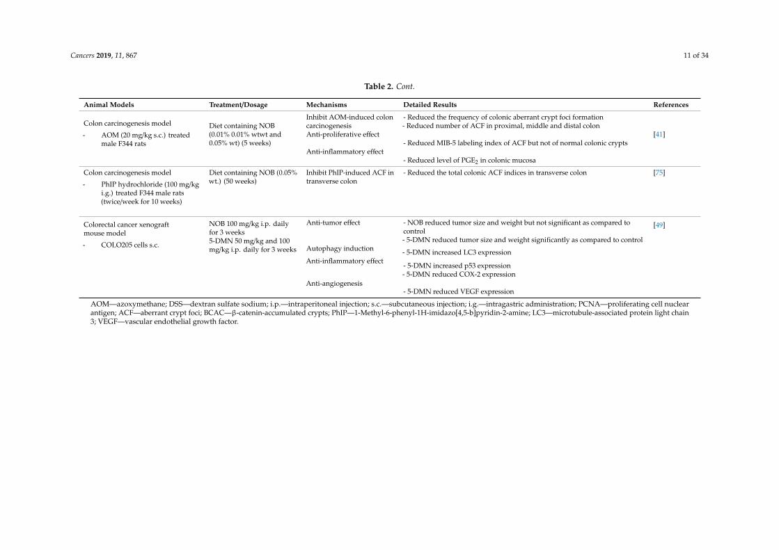
Table 2. Cont.
Animal Models Treatment/Dosage Mechanisms Detailed Results References
Colon carcinogenesis model
- AOM (20 mg/kg s.c.) treatedmale F344 rats
Diet containing NOB(0.01% 0.01% wtwt and0.05% wt) (5 weeks)
Inhibit AOM-induced coloncarcinogenesis
- Reduced the frequency of colonic aberrant crypt foci formation- Reduced number of ACF in proximal, middle and distal colon
[41]Anti-proliferative effect- Reduced MIB-5 labeling index of ACF but not of normal colonic crypts
Anti-inflammatory effect- Reduced level of PGE2 in colonic mucosa
Colon carcinogenesis model
- PhIP hydrochloride (100 mg/kgi.g.) treated F344 male rats(twice/week for 10 weeks)
Diet containing NOB (0.05%wt.) (50 weeks)
Inhibit PhIP-induced ACF intransverse colon
- Reduced the total colonic ACF indices in transverse colon [75]
Colorectal cancer xenograftmouse model
- COLO205 cells s.c.
NOB 100 mg/kg i.p. dailyfor 3 weeks5-DMN 50 mg/kg and 100mg/kg i.p. daily for 3 weeks
Anti-tumor effect - NOB reduced tumor size and weight but not significant as compared tocontrol- 5-DMN reduced tumor size and weight significantly as compared to control
[49]
Autophagy induction - 5-DMN increased LC3 expressionAnti-inflammatory effect
- 5-DMN increased p53 expression- 5-DMN reduced COX-2 expression
Anti-angiogenesis- 5-DMN reduced VEGF expression
AOM—azoxymethane; DSS—dextran sulfate sodium; i.p.—intraperitoneal injection; s.c.—subcutaneous injection; i.g.—intragastric administration; PCNA—proliferating cell nuclearantigen; ACF—aberrant crypt foci; BCAC—β-catenin-accumulated crypts; PhIP—1-Methyl-6-phenyl-1H-imidazo[4,5-b]pyridin-2-amine; LC3—microtubule-associated protein light chain3; VEGF—vascular endothelial growth factor.
Cancers 2019, 11, 867 12 of 34
5.1. Cell Cycle Arrest
Uncontrolled cell growth that arises from genomic instability is known to contribute totumorigenesis [85]. One way to counteract CRC is to halt its cell cycle progression. The cellcycle is akin to a biological growth clock that tightly regulates each stage of cell growth, where anymutated or abnormal cells will be arrested at either the G1 or G2 checkpoints; however, this mechanismis disrupted in cancerous conditions [86]. To progress through the stages, the regulatory protein cyclinacts like a key, as it needs to phosphorylate the cyclin-dependent kinase (CDK) complexes to allowprogression to the next stage [87].
5.1.1. Action of NOB and Its Metabolites Inducing Cell Arrest
Notably, different metabolites of NOB work by different mechanisms against different cells.The flow cytometry test showed NOB and 4′-DMN arrest cells at G0/G1 phase in both HCT116 andHT-29 cell lines, despite the inhibitory effect of 4′-DMN being higher than that of NOB. Both 3′-DMNand 3′,4′-DMN arrest cells at both S phase and G2/M phase in HCT116 cell lines but arrest cells atboth G0/G1 and G2/M phase in HT-29 cells. The inhibitory effect of 3′,4′-DMN is higher than that of3′-DMN as only half the concentration is needed to induce a similar end result [54]. The fact that themetabolites 3′-DMN and 3′,4′-DMN exhibit more potent anti-cancer effects than NOB may suggestthat demethylation at the 3′ and 4′-position significantly enhances its inhibitory effect [51].
In vitro tests using HCT116 cells reveal NOB and all three of the common metabolites increase theexpression of CDK inhibitor, p21Cip1/Waf1 [54]. p21Cip1/Waf1, also known as p21 or P21/CDKN1A is anegative regulator for progression of the cell cycle that is responsible for the hypo-phosphorylation ofretinoblastoma (Rb) proteins, leading to cell cycle arrest at the G1/S transition [86,88,89]. Although p21is usually associated with the degradation of cyclin D1 [86], it is interesting to note that only 4′-DMNbut not other metabolites nor the NOB itself causes significant reduction in cyclin D1 level. This maypartly explain the strongest cell cycle arresting effect of 4′-DMN at the G0/G1 phase as compared to theother compounds aforementioned [54].
Proliferating cell nuclear antigen (PCNA) acts as a cofactor for DNA polymerase δ. It is animportant marker commonly used to detect cell proliferation due to its increased expression throughthe G1 phase and S phase transition of cells [90,91]. Analysis from immunohistochemical tests recorded69% reduction of cells with PCNA compared with the untreated controls [54]. Interestingly, evidencealso reveals that p21 potentially suppresses action of PCNA. Interaction with the carboxy terminal ofp21 inhibits PCNA from activating DNA polymerase δ, thus blocking DNA synthesis and preventingcell proliferation [92,93]. In this light, specific research of NOB on p21 and PCNA may be required toelucidate the pathways in further details.
Wu et al. studied the combinatory effect of NOB and its metabolites at different concentrations onHCT116 cells. At half the original concentration present in the colon, there is a decreasing trend ofcells in S phase and G2/M phase but an increasing trend was noted in the G0/G1 phase. The cell cyclearrest effect seems to be dose dependent as flow cytometry recorded the population of cells arrestedat the G0/G1 phase to be 57.8% higher than the untreated cells and significantly increased to 91.0%when the concentration of NOB and metabolites was doubled. To validate the findings, the levels ofkey signalling proteins were measured. Results showed that treatment with NOB and its metaboliteslowered the levels of CDK-2, CDK-4, CDK-6 and cyclin D, raised the level of p52 and p27, but did notalter levels of p21 and cyclin E. In contrast, in vivo tests in AOM/DSS induced mice solely treated withNOB did document decreased expression of cyclin E and increased expression of CDK inhibitor p21.This difference is hypothesised to be due to the cell type specific response towards NOB, which is yet tobe confirmed by further research [76]. The cell cycle is tightly regulated by key signalling proteins suchas cyclins and cyclin dependent kinases (CDKs). To illustrate, complexes such as cyclin D-CDK-4/6and cyclin E-CDK-2 facilitates the transition from G1 phase to S phase, while cell transition from G1phase to S phase can be inhibited by tumour suppressor p53 and CDK inhibitors p21 and p27 [89,91].
Cancers 2019, 11, 867 13 of 34
It is also worth mentioning here that, at the effective concentration that arrests cell cycle, NOBproduces a cytostatic effect, meaning it arrests growth without killing the cell [14]. As compared toother flavonoids like tangeretin (IC50 = 1.6 µM) and quercetin (IC50 = 0.84 µM), a slightly higherIC50 of 4.7 µM is required for the cell proliferation inhibition action by NOB in HT-29 cell lines andIC50 of 8.4 µM for 5-DMN [70]. However, the inhibitory effect of NOB may only be temporary. It isdemonstrated that, with the removal of NOB, the treated cells resume cell proliferation within 24 hand regain similar growth status comparable to the control within 96 h [14]. This may also imply that,in order to sustain the inhibitory effect of NOB, the treatment with NOB has to be long-term to ensurecontinuous cell proliferation inhibition. This is possible as NOB is considered a natural compoundand has no effect on healthy cells [83]. One might argue that the effect of NOB may be problematicfor naturally fast-proliferating cells like healthy non-adenomatous intestinal lining cells. However,there is reassurance based on previous research that showed NOB is 10 times more selective towardstransformed cancerous cells as compared to normal healthy cells [79].
5.1.2. Action of 5-DMN Inducing Cell Cycle Arrest
Treatment with 5-DMN also shows a similar increase of Rb in a dose dependent manner. Notably,5-DMN does not affect the level of CDK-4, but there is a significant reduction of CDK-2 levels [63],hence indicating a reduced possibility of complex formation with cyclin A or cyclin E [94]. p21 isknown to play a key role in arresting the cells at the cells at the G2/M phase through the inhibition ofCDK-2/Cyclin E complex formation [95,96], and 5-DMN has been found to be able to arrest cell cyclesat both the G0/G1 phase and G2/M phase in HCT116 (p53 +/+), but is only able to accumulate cells at theG2/M phase in HCT116 (p53 −/−). This suggests that G0/G1 arrest is dependent on p53 while G2/M isindependent of p53 [80]. Using HT-29 cell lines, Qiu et al. reported that 5-DMN effectively causes cellcycle arrest at the G2/M phase [63]. This effect possibly arises from the downregulation of cdc25 proteinexpression, which is important to activate the cyc2/cyclin B1 through the process of dephosphorylatingthe inactive tyrosine residues Thr-14 and Thr-15 located in cdc2 ATP binding domain [97,98].
To sum up, different derivatives of NOB potentially arrest the cell at different stages of the cellcycle, mainly through downregulating the expression of proteins or kinases such as CDKs involvedin the cell proliferation pathways and preventing the formation of cyclin complexes that allow cellcycle progression.
5.2. Programmed Cell Death
As growth of a cell is tightly regulated by the cell cycle, death of a damaged or aged cell alsoneeds to be programmed to maintain homeostasis in our body. There are three models of programmedcell death (PCD), namely apoptosis, autophagy and necrosis [99]. A tumour mass of cancerous cells isformed when the cancerous cells develop the ability to evade cell death. Not responding to the deathsignal, the cells continue to grow and proliferate, leading to progression of cancer. Thus, NOB, being anagent that targets the key signalling pathways of programmed cell death, may help in chemopreventionof CRC. Apoptosis will be the main focus in this subsection, while autophagy will only be discussedbriefly. Necrosis, the most abrupt death of all three, will not be discussed in this section as there areno data in this area. Although necrotic death is usually associated with inflammation, this does notexclude its possibility to be exploited as a means to eliminate cancerous cells [100].
Apoptosis can be induced by two core mechanisms, namely the extrinsic and the intrinsic pathway.The cell death signals in the extrinsic pathway come from external sources such as the Fas ligandsor tumour necrosis factors [101]. The intrinsic pathway generally arises from the mitochondrialintracellular protein of the Bcl-2 family. Bcl-2 is an important regulator for apoptosis, which plays arole in mitochondrial disruption that activates the caspases [102]. High levels of Bcl-2 are expressed invarious types of cancer and is associated with chemoresistance. Levels of Bcl-2 need to be lowered topromote apoptosis [103,104]. As a result of reduced Bcl-2 levels, a cascade of activity is activated in thecell leading to apoptosis with caspase-9 acting as the initiator caspase in the intrinsic pathway [105]
Cancers 2019, 11, 867 14 of 34
and caspase-8 in the extrinsic pathway. It may also be crucial to mention here that the procaspase-8forms a complex called Death Inducing Signalling Complex (DISC) before it is activated to caspase-8.The downstream effect would be the activation of the executioner caspase-3, and other caspases suchas caspase-1, caspase-6 and caspase-7 [106,107] which then cleaves Poly (ADP-ribosome) polymerase(PARP) [108,109]. Soon after, the cell starts to bleb and shrink while its nucleus is condensedand fragmentised, proteolysis happens and the cell loses adhesion to the extracellular matrix andneighbouring cells [110,111]. Once the cell undergoes apoptosis, its contents are taken up by the bodyand recycled for new cell synthesis.
Action of NOB and Metabolites Inducing Programmed Cell Death
In vitro tests using different colon cell lines such as HCT116 and HT-29 reveals that the actionof NOB and its various metabolites vary in different cell types. NOB was only shown to induceapoptosis of colon cancer cells when tested at high concentration. Zheng et al. [79] demonstratedthat NOB increased DNA fragmentation in COLO302 only at 200 µM. Treatment with NOB, 3′-DMN,4′-DMN and 3′,4′-DMN in HCT116 cell lines raise the early apoptotic cell population by 3.3-fold,5.0-fold, 4.9-fold and 7.6-fold, respectively, while also resulting in 4.2-fold, 3.5-fold 7.1-fold and 4.5-foldincrements in the late apoptotic cell population, respectively. In contrast, 3′-DMN and 4′-DMN did notcause any significant changes in apoptotic cell population in HT-29 cell lines, but the pro-apoptoticeffect of 3′,4′-DMN was observed to be higher than that of NOB. An in vitro test using HCT116 showsthat all three metabolites of NOB are able to induce the activation of caspase-3, caspase-9 and PARP,while NOB can only induce activation of caspase-9 but not that of caspase-3 or PARP. A negativeresult was also reported on the apoptosis inducing effect of NOB, whereby no apoptosis was detectedwhen NOB was tested at concentrations of up to 100 µM in HT-29 [14]. In contrast, an in vivo testin AOM/DSS treated mice revealed a 2.3-fold increase of caspase-3 levels with NOB treatment [54].Nevertheless, we can be certain that the metabolites of NOB render a higher proapoptotic effect ascompared to their parent compound NOB.
Several previous works have demonstrated that NOB and its derivatives exhibit pro-apoptosisproperties. However, this effect is known to be tissue specific. To illustrate, apoptosis is observed incolon cell lines but not the HL-60, promyelocytic leukaemia cell lines [79,112]. Interestingly, apoptosiscould also be related to MMP-7. It has been discovered that cells that express MMP-7 are less sensitiveto the Fas ligand-induced apoptosis as MMP-7 has a higher tendency to produce the non-apoptoticform of soluble Fas ligand by releasing the ligands in the cell membranes [77]. MMP will be furtherdiscussed in Section 5.4.
In vivo tests using xenograft mice show that 5-DMN triggers apoptosis at a concentration of40 µM [49]. 5-DMN has been reported to increase levels of caspase-8, caspase-3 and PARP in adose dependent manner, and results in 2.2-fold increase in the population of early apoptotic cells ascompared to the control. In contrast, there is no apparent increase in the apoptotic effect even afterdoubling the concentration of NOB, suggesting NOB is required at a significantly higher concentrationto induce a pro-apoptotic effect [14,63]. However, the effect of 5-DMN was notably distinct for differenttypes of cancer cell lines. To illustrate, 5-DMN can induce early apoptosis in HCT116 colon cancer celllines at a concentration as low as 8 µM while 5-DMN only slightly raises the apoptotic activities at aconcentration as high as 36 µM in colon HT-29 cancer cell lines [63].
Annexin-V/PI analysis reveals that 5-DMN significantly increased the Annexin-V positive cells,especially in the late apoptotic or necrotic cell population among the HCT116 (p53 −/−) cells, suggestingthat the action of 5-DMN may be independent of p53 [80]. The fact that 5-DMN induces early apoptosisin HCT116 (Bax +/+) cells but not in HCT116 (Bax −/−) may suggest that Bax (Bcl-2 associated Xprotein) is important for apoptosis to occur. In other words, absence of Bax confers resistance toapoptosis [80,113,114]. A recent study by Chiou et al. proved that 5-DMN increases the expressionof p53 proteins, which not only induces apoptosis, but also triggers cell death by autophagy thatcontributes to the prevention of tumour growth [49]. However, the detailed mechanism of how NOB
Cancers 2019, 11, 867 15 of 34
prevents CRC development through the autophagy process is yet to be elucidated. From the currentstage of knowledge, autophagy is known to exhibit both the pro-tumour and anti-tumour formationeffects. In response to stress, autophagy acts as a protective mechanism for cell survival, which hasalready been elaborated in previous literature [115]. It is likely that overactivation of autophagycontributes to suppressing tumour formation by inhibiting the anti-autophagic-related genes (ATGs)in oncogenesis and activating the pro-ATG [116]. The effect of autophagy and apoptosis may besynergistic in chemoprevention of cancer [117,118].
To conclude, NOB was shown to be less effective in inducing apoptosis of colon cancer cells.Instead, the metabolites of NOB, 3′-DMN, 4′-DMN and 3′,4′-DMN were suggested to be responsiblefor the induction of apoptosis of colon and modulation of cancer cell growth in the colon carcinogenesisanimal model [54] On top of that, the autohydrolysis product of NOB, 5-DMN could induce apoptosisin colon cancer cells at a lower concentration as compared to NOB.
5.3. Anti-Inflammation
Inflammation is a natural physiological response of our body, characterised by five main signs,namely loss of function, redness, pain, heat and swelling. Inflammation plays a significant role at timesof infection and injury. It triggers the immune system to function and helps to protect our body throughthe release of chemical molecules called pro-inflammatory signals. However, too much inflammationis also not a good sign. There is increasing evidence narrating the interrelation between tumorigenesisand inflammation [119]. Whilst chronic inflammation is a hallmark of cancer, the inflammatorycytokines aggravate cancer progression by preventing differentiation of cells and promoting tumourformation [120]. The inflammatory cells release ROS after being activated, leading to the oxidativedamage of DNA and p53 mutation [121–123]. The mechanism that triggers inflammation is a rathercomplex pathway and has been covered by previous literature [123,124]; only the anti-inflammatoryeffects mediated by NOB are discussed in this review.
Increasing evidence shows that progression of CRC can be accelerated by the upregulation ofpro-inflammatory cytokines expressions—for example, TNF-α, IL-1α, IL-1β and IL-6 [81,119,125].These proinflammatory cytokines enhance the secretion of inflammatory mediator PGE2. Song et al.reported that treatment with NOB results in a noteworthy reduction of tumour size and frequencycorrelated with the significant lowering of IL-1, IL-6, iNOS and COX-2 levels [78]. ELISA test quantifiedthe reduction of TNF-α, IL-1β and IL-6 at 51%, 92% and 69%, respectively, in the NOB treated groupwhile real-time qRT-PCR quantified the reduction of the above pro-inflammatory cytokines at 65%,69% and 45%, respectively, when compared to the control mice [54].
Anti-Inflammation Effect of NOB and Its Metabolites
Besides NOB, multiple studies have shown that its metabolites, especially, 4′-DMN and 3′,4′-DMN,also exhibit significant inhibitory effects towards nitric oxide production, iNOS and cyclooxygenase(COX) expressions in both in vivo and in vitro conditions [30,31,62,76,81,126]. However, the combinedeffect of NOB and its metabolites warrants further investigation [76]. Notably, NOB selectively inhibitsCOX-2 and did not affect COX-1 [81]. COX-2 is normally absent in healthy cells, but its release istriggered when the environment is inflammatory or hypoxic [127]. COX-2 is known to enhance CRCcarcinogenesis, and inhibiting COX-2 also limits the production of PGE2 [128], which may be associatedwith the inhibition of cell proliferation in colonic mucosa [41]. iNOS speeds up the conversion ofL-arginine to NO through the process of oxidative deamination. NO is a potent inflammatory mediatorthat activates signalling molecules that trigger the process of inflammation and mutagenesis [129].By inhibiting the iNOS and its downstream products, NOB helps in reducing the inflammationobserved in chronic diseases like ulcerative colitis and CRC [130]. Introduction of NOB to colonictissues harvested from mice treated with AOM/DSS results in a 35% reduction of cells expressing iNOScompared to the untreated tissue. This is consistent with the in vitro test. By administering NOB andits metabolites at a concentration equivalent to that found in the colons to the LPS-induced RAW 246.7
Cancers 2019, 11, 867 16 of 34
macrophages is shown to effectively and completely inhibits expression of iNOS, while, at half theconcentration, the expression of iNOS was lowered by 56.4% compared to the untreated LPS-inducedmacrophages [76]. This shows that a similar process is likely to happen in the human body and NOB isindeed a promising anti-inflammatory agent.
Additionally, NOB also increases the release of the Nrf2-dependent enzymes which regulatePhase II enzyme production, such as heme oxygenase-1 (HO-1) and NQO1. HO-1 is an anti-oxidativeenzyme that exhibits its anti-inflammatory effect by producing anti-oxidants like carbon monoxideand bilirubin. It is also important to note that HO-1 is not solely controlled by Nrf-2 [76]. NQO1upregulation counteracts the increased expression of IL-1β and TNF-α induced by LPS [131]. This isconsistent with the findings of Khor et al. reporting that a lower Nrf-2 expression greatly increasessusceptibility of mice models to AOM-induced colitis [132]. The colonic mucosa of AOM/DSS-inducedmice orally administrated with NOB was found to have increment of nuclear Nrf2 by 1.94-fold andreduction of cytoplasmic Nrf2 by 36% when compared to the AOM/DSS-treated mice. The upregulationand translocation of Nrf2 transcription factor is thought to be the cause for subsequent increment ofHO-1 and NQO1 by 2.78-fold and 2.59-fold, respectively. This is consistent with the results from thecombinatory effect of NOB and metabolites treatment on macrophages cell lines, which induces a10% increase in the level of HO-1 and a 34% increase in the level of NQO1 when the concentrationratio of NOB and metabolites is equivalent to that in the colon [76]. In short, Nrf-2, which neutralisescarcinogens and reactive oxygen species (ROS), is identified as a key signalling pathway to target inthe effort to halt CRC progression [133,134].
5.4. Anti-Angiogenesis
Angiogenesis refers to the process by which new blood vessels are formed. This process isan important pathway that results in the progression of all types of cancer. This is because, whenthe tumour mass grows, it naturally needs more nutrients and nourishment to support its growth.To achieve this necessity, new blood vessels have to be formed surrounding the tumour mass to ensurea continuous supply of oxygen and glucose to support the growing cell mass [135,136].
Anti-Angiogenesis Effect of NOB
It is postulated that NOB prevents metastasis by inhibiting the activity of activator protein-1(AP-1), a dimeric protein, thus preventing DNA binding [77]. Another hypothesis suggests that NOBacts via the Nuclear Factor-kappa B (NF-κB) pathway, altering the gene expression by modulating thepromoter regions [126,137].
For angiogenesis to occur, the vascular endothelial growth factor (VEGF) plays a key role. VEGFnot only acts as a signal to induce new blood vessel growth, but also inhibits the apoptosis induction.VEGF works by activating the mitogen activated protein kinase (MAPK). This kinase triggers the signaltransduction and allows the endothelial cells to proliferate in order to form new blood vessels [138–140].To elaborate on VEGF, it is necessary to mention leptin and insulin-like growth factor 1 (IGF-1) here.There is evidence suggesting that bidirectional cross talk exists between the leptin protein and IGF-1,a serum growth factor. Acting together, they not only catalyse the cell proliferation process, but alsotransactivate the epidermal growth factor receptor (EGFR), which enhances the migration and invasionpower of the cancer malignancy [141].
Miyamoto et al. discovered that leptin, a protein that regulates energy balance and body masshas a positive correlation with CRC, where leptin is thought to be a mitogenic factor that leads tothe development of colon cancer [84]. It induces cell proliferation by activating the nuclear factorκB, p38 MAPK and p42/44 MAPK [142]. Previous evidence demonstrates that introduction of 0.1 to10 nM of leptin enhances the proliferation rate of HT-29 cells by 1.3 to 1.6 times [84] through c-JunNH2 terminal kinase and extracellular regulated kinase (ERK) 1/2 activation [143,144]. In otherwords, the chances of developing CRC can be reduced if leptin concentration is regulated. Treatmentwith NOB suppresses cell proliferation induced by leptin through inhibition of mitogen-activated
Cancers 2019, 11, 867 17 of 34
protein/extracellular signal-regulated kinase (MEK) 1/2 [145]. Consistent with the in vitro findings,a reduction of 75% of leptin concentration by NOB, partly through the inactivation of the insulinsignalling pathway, was reported at the end of the 17-week study in an in vivo model using Institutefor Cancer Research (ICR) mice [83,84]. In this light, Miyamoto et al. conducted a study aiming todetermine the prognosis of cancer in obese rats with flavonoid intervention. They reported that theflavonoids significantly reduced the incidence of β-catenin accumulated crypt (BCAC) by 64% to 71%and aberrant crypt foci (ACF) by 68% to 91% and proposed that this arises from the effect of NOB indownregulating the secretion of IGF-1 [83].
Metalloproteinase (MMP) plays a fundamental role in angiogenesis. MMP induces the proteinthat breaks down the extracellular matrix (ECM), thus making the blood vessel more permeable andallowing the cancerous cells to detach from the lump to flow, extravasate or invade the other partsof the body, causing the spread of tumour to other vital organs. This is a process called metastasis,leading to malignancy and cancer aggravation. The mortality rate greatly increases when cancerouscells metastasise to vital organs like the liver [146]. Similar to how a dexamethasone steroid acts, NOBis proven to be able to increase the expression of tissue inhibitor metalloprotease-1 (TIMP-1) in humansynovial cells [81]. However, the benefit of upregulating TIMP-1 in CRC is debatable due to its bilateralrole in cancer progression. Although TIMP-1 upregulation contributes to the anti-oncogenic effect,enhanced expression of it may lead to early phase tumour development via the pathways independentof MMPs. With this understanding, TIMP-1 glycosylation can function as a biomarker to aid in CRCstaging [147].
There are more than 20 types of MMP involved in metastasis [148], with each one of them playinga distinct role [149]. However, whether MMP is produced by cancer cells or their surrounding stromalcells is still an ongoing debate [150]. Abnormally high levels of MMP-1, MMP-2, MMP-3, MMP-7,MMP-9 and MMP-13 have been implicated in CRC [150]. Treatment with NOB significantly inhibitsrelease of pro-MMPs especially pro-MMP-7 (also known as metrilysin) mRNA in HT-29 cell lines.To illustrate, NOB at a concentration range of 25 µM to 100 µM, the proMMP-7 levels in the mediadiminishes significantly by 35% to 47% [77]. The maximal expression of MMP-7 arises from theβ-catenin/TCF complex transcription factors formed in the presence of mutated APC genes [150–152].Apart from MMP-7, the action of NOB on other MMPs in CRC is yet to be investigated. MMP-9, whichis mainly secreted by inflammatory cells, is correlated with the transition phase from adenoma toadenocarcinoma, while the upregulation of MMP-3 usually suggests poor prognosis as it has a positivecorrelation with low microsatellite stability. On the other hand, high levels of MMP-12 reduces CRCmortality as it can potentially inhibit angiogenesis [150] by secreting angiostatin, a chemical that haltstumour progression and inhibits tumour neovascularisation [153–155]. As mentioned in the previoussection, NOB suppresses MEK. This suppression of MEK then further diminishes the expressionof pro-MMPs, which results in the reduction of MMP and subsequently confers anti-angiogenesiseffect [144,145]. Briefly, NOB prevents angiogenesis and metastasis in CRC mainly via the inhibition ofMMP, EGFR and VEGF through the regulation of leptin and IGF-1.
6. Pharmacokinetics, Bioavailability and Delivery Systems of NOB
The pharmacokinetic properties of NOB represent a key factor to be considered in an attempt toformulate it into a therapeutic product. Understanding the interactions between the compound andour body opens ways to creative strategies in solving the problem which require novel formulation indelivering NOB for chemoprevention purpose. For oral delivery, an important consideration is thebioavailability of the active compound. However, the bioavailability studies on NOB are limited [156].Therefore, understanding the pharmacokinetic profile of NOB becomes even more crucial to assistingin the prediction of bioefficacy as the absorption, metabolism and elimination pattern indirectly affectits bioavailability.
Cancers 2019, 11, 867 18 of 34
There are many factors that affect the absorption of a compound; one important consideration isthe molecular structure [157]. The proper absorption of any compound is depicted by its solubility andpermeability across physiological barriers of which both properties are directly related to its molecularstructure. Attributed to its unique chemical structure with multiple methoxy groups, NOB is lipophilicin nature and can easily pass through the cell membrane. Murakami et al. successfully demonstratedthe relatively high permeation of NOB across differentiated Caco-2 cells which mimics the epithelialcells lining the small intestine. A significantly high 48.1% of NOB has been found to permeate throughthe basolateral compartment while another 39.3% remains on the apical compartment four hours afterintroduction of NOB in a Caco-2 monolayer trans-well permeability assay [158]. Parallel artificialmembrane permeation assay (PAMPA) deciphered the permeability of NOB, 4′-DMN and 3′-DMNat 1.38 × 10⁻⁶ cm/s, 1.14 × 10⁻⁶ cm/s and 1.05 × 10⁻⁶ cm/s, respectively [159]. It was discovered thatthe methoxylated flavonoids show five to eight-fold higher permeability in the intestinal wall than itsunmethoxylated counterparts [160]. The drawback is that PMF in general has limited solubility. Resultsfrom the high-throughput lyophilisation solubility assay (LYSA) reveal that NOB has a low solubilityat 12 µg/mL, while its metabolites, 4′-DMN, 3′-DMN and 5-DMN exhibited two to three-fold highersolubilities of 22 µg/mL, 29 µg/mL and 32 µg/mL, respectively [156,161]. In general, the solubilityincreased with the number of hydroxyl group of the compound. This may also partly explain thehigher activity of the derivatives of NOB as compared to its parent compound, which has a highernumber of methoxy groups.
After absorption, NOB is found to be widely distributed throughout the body, as a significantamount of NOB could be detected in organs such as the stomach, small intestine, large intestine,brain, liver and kidney within four hours of single dose administration [162,163]. Interestingly, NOBwas suggested to be absorbed through the muscularis layer of the gastrointestinal tract, especiallythe stomach tissue into the blood circulation given the distinctly higher concentration of NOB in themuscularis (390 ± 120 nmol/g) as compared to other organs [162]. Furthermore, it is also noteworthythat NOB was found to be distributed in the mucous membrane and muscularis from the large intestine(cecum, colon and rectum) of a rat at 4.3 ± 1.6 nmol/g after one hour of oral administration by gastricintubation [162]. More recent evidence demonstrated that the levels of NOB in the colonic mucosa ofmice were 2.03 nmol/g of tissue after long-term oral administration of NOB (0.05 wt%) containing diet.Furthermore, Wu et al. [164] also suggested that the dose of NOB used (0.05 wt%) could be equivalentto approximately 100 mg/day for human oral consumption, which is achievable in humans.
After oral administration of NOB to rats, the mean plasma concentration of NOB was quantifiedin several pharmacokinetic studies. Wang et al. [163] reported that the plasma levels of total NOBand its metabolites could reach as high as 10 µg/mL (25 µM). Using a highly sensitive LiquidChromatography-Mass Spectrometry/Mass Spectrometry-Electrospray ionisation (LC-MS/MS-ESI)method, the maximum concentration (Cmax) of NOB in rat plasma was determined at 0.4 µg/mL (1 µM)after oral administration of 5 mg/kg NOB [165]. Meanwhile, a maximum concentration of 1.78 µg/mL(4.4 µM) was measured by a validated HPLC method in rat plasma after oral administration of 50 mg/kgNOB [166]. In addition, another study by Manthey et al. [167] reported that a peak of NOB serum levelof 9.03 µg/mL (22.4 µM) was detected by HPLC-ESI-MS in rats after oral gavage of 50 mg/kg NOB.Nevertheless, these studies demonstrated a relatively early peak time (Tmax) of 0.25 to one hour afteroral administration of NOB in rats. This high rate of cellular uptake may be attributed to the highlyhydrophobic nature of the compound rendered by the presence of six methoxy groups [162].
NOB undergoes extensive metabolism after being taken orally. As detailed in Section 3, NOBundergoes Phase I and Phase II metabolism after being absorbed in the small intestine where it maybe conjugated to sulphate and glucuronide, and then again deconjugated by the microflora in thecolon [60]. The three common phase I metabolites of NOB have been identified as 3′-DMN, 4′-DMNand 3′,4′-DMN [52,53]. Wang et al. found evidence of transformation of 3′-DMN and 4′-DMN into3′,4′-DMN in the colon [163]. The liver is another important organ involved in metabolising NOB.Koga et al. identified three metabolites, demethylated at the 4, 6 or 7 positions respectively under the
Cancers 2019, 11, 867 19 of 34
action of human liver microsomes when incubated aerobically with NADPH [56]. The metabolitesexhibit distinct activity and distribution pattern in different areas of the body. It was found that 4’-DMNis the major metabolite present in the small intestine and liver while 3′,4′-DMN was predominantlypresent in the colon and spleen [163]. An in vitro test on NOB using rat liver S-9 extract shows thatonly 7% of NOB metabolites were detected towards the end of a 24 hour treatment, while 72.6% ofNOB remains unchanged towards the end of the experiment [158]. This may be attributed to the slowrate of demethylation of NOB showed by Murakami et al. [162].
The elimination half-life of NOB from the blood plasma of a rat was reported as 1.8 h via a validatedHPLC test [166] while Kumar et al. reported a terminal half-life of NOB at 4.75 ± 0.57 h followingoral administration and a terminal half-life of 1.51 ± 0.61 h following parenteral administration usingthe LC-MS/MS-ESI method [165]. Despite the wide distribution throughout the body, concentrationof NOB quickly diminishes with time and becomes undetectable in the serum, stomach, intestines,liver and kidney. Aside from the parent compound, mono-demethylated metabolites and conjugatedNOB are detected in the urine, with the concentration of conjugated NOB revealing a time-dependentincrement over a period of 24 h [162]. Since NOB is rapidly eliminated from the body, significantadverse effects reported after administration of NOB are rare.
Although the in vitro results were promising, most of the reported concentrations of NOBevaluated (>20 µM) were not achievable in physiological conditions as demonstrated by in vivopharmacokinetic studies of NOB. Comparing the high experimental levels used against the relativelylow peak plasma concentration—a mere 1.78 µg/mL (4.4 µM)—after one hour of oral administration of50 mg/kg NOB [166] and the rapid elimination from the body [162] points out a limitation to utilizingNOB in its unaltered natural form as a clinical drug. In fact, the levels of NOB detected in the colonicmucosa ranged between 2 to 4 µM using the assumption that one gram of tissue is equivalent of 1 mL ofvolume [54,162]. However, there was a study demonstrating that NOB at lower concentration (≤5 µM)exhibited antiproliferative effects against colon cancer cells [70], perhaps indicating true promise forclinical use after all.
To further substantiate the notion of NOB for colon cancer chemoprevention, multiple in vivostudies have demonstrated that dietary treatment with NOB could inhibit colon carcinogenesis inrats [54,76]. As mentioned earlier, this may be related to the fact that, while bioavailability of NOBitself is low, much of its anti-CRC effect may be via its metabolites. Wu et al. [54] indicated that theNOB level in the colonic mucosa only accounted for <5% of the total levels of NOB and its metabolitesafter oral administration of NOB. The study further suggested that the NOB metabolites, which wereformed as a result of phase I and II metabolism and biotransformation by gut microbiome, play animportant role in colon carcinogenesis inhibition [54]. Although there is some suggestion that lowerdoses can have an effect on cancer, clearly, enhancement of NOB bioavailability is necessary and alsorepresents a major challenge that needs to be addressed to achieve the desired therapeutic effect.
Given the importance of actually delivering adequate amounts of NOB to the target site toachieve chemopreventive activity, we also reviewed the delivery systems aiming to enhance thebioavailability of NOB in the gut. For chemoprevention of CRC, oral delivery represents the preferredroute. There is a growing interest to formulate lipophilic natural compounds such as NOB intoemulsion, as these systems not only improve the bioavailability of the active compound, but alsoreduce the rate of degradation during storage [168]. Yang et al. attempted to enhance the solubilityof NOB by encapsulating NOB with citrus oil-based emulsion. The team discovered that dissolvingNOB at a higher temperature and in an oil with log P close to NOB, such as bergamot oil, helps toincrease solubility of the compound [169]. Yao et al. also experimented with the possibility of usingself-microemulsifying drug delivery systems (SMEDDS) to improve the permeability of NOB in therat intestines and reported that SMEDDS resulted in similar efficacies to micelles, but showed betterabsorption profile when compared to sub-microemulsions [170]. Self-assembled NOB proliposomeswere also reported to improve the absorptive rate and confer longer mean residence time as comparedto NOB suspension in rats [171].
Cancers 2019, 11, 867 20 of 34
Furthermore, Chen and colleagues demonstrated that, through the addition of hydroxypropylmethylcellulose (HPMC), the retention of NOB in nanoemulsion is increased by 25% [172]. Eventhough the fabrication of supersaturating nanoemulsion with the addition of HPMC aimed to improvethe physical stability of NOB and prevent precipitation of NOB in the emulsion, the fabrication did notperform as expected at high NOB concentration where precipitation still occurred during storage anddigestion process in the gut [172]. To address the issue of component precipitation in the emulsionsystem, a recent intervention of nanoemulsion-filled hydrogel matrix has been developed to stabilizeNOB and prevent precipitation during delivery along the GI tract [173]. Interestingly, the hydrogelscould provide a controlled release of NOB along the GI tract, thereby the hydrogel shrank at acidiccondition pH 1.2 but swelled and burst at pH 7.4. Due to the lower bioaccessibility of NOB in hydrogelas compared to nanoemulsion during digestion, the nanoemulsion-filled hydrogel matrix could confera sustainable absorption of NOB through a controlled release in the intestinal tract [173].
Aside from the liquid formulations, Onoue and colleagues proposed a solid formulation of NOBwith the intention to further enhance the bioavailability in addition to solving the stability issues whichshowed a remarkable 13-fold increment in bioavailability compared to the nanosized NOB amorphoussolid dispersion [174]. However, the results only quantitate the brain permeability, but the data forcolon effect is still lacking. Further research is needed to establish the practicability and feasibilityof each delivery method to address the bioavailability challenges before NOB can be used in aidingpatients at high risk of CRC.
7. Toxicity
Although NOB is derived from a natural source, excessive intake of any substance might lead tosome changes in the body. To illustrate, there are several case studies reporting that ingestion of productscontaining bitter orange causes adverse effects such as tachycardia and ventricular fibrillation [175].To address this concern, a number of studies have been carried out to further evaluate this problem.
Body weight is commonly used as a surrogate marker for toxicity, whereby the weight is expectedto reduce significantly if toxicity occurs. Wu et al. concluded there was no significant change in bodyweight, weight of liver, spleen appearance and behaviour throughout the length of their research,suggesting that the oral intake of NOB at the effective concentration 40 µM or 0.05% for a period of3 to 20 weeks does not lead to adverse side effects [41,49,54]. This is consistent with the findings ofMurakami et al. suggesting that NOB leads to no cytotoxic effects [42]. The action of NOB is morelikely of a cytostatic nature rather than cytocidal as, at the concentration that inhibits cell proliferation,it does not trigger apoptosis. Furthermore, the treated cells continue to grow normally once the effectof NOB diminishes [14]. This shows that NOB could be a safer option for CRC treatment as it is lesscytotoxic as compared to the available chemotherapy agents.
8. Commercial Uses
A search on Google Scholar using the keywords “nobiletin patents” gives about 1160 relevantresults. This initial search was refined through the advanced search function for patents using theSciFinder database which helps to narrow down the number of patents directly and indirectly relatedto NOB to 300.
After a close analysis of the patents, we found that, among the 300 patents related to the concept ofNOB, the largest portion of the total patents involves the usage of NOB in the medical, pharmaceutical andnutraceutical fields. The patents include both the application in traditional treatments and also westernmedications, where there are about 20 patents related to cardiovascular diseases [176–178], hypertensionand hypercholesterolemia [179], roughly 10 patents targeting the central nervous system [180,181] orneurodegenerative disorders [182], diabetes [183,184] and obesity [185] each, and about five patentsconcerning body metabolism and hormonal functions, bone-related disorders [186], oral issues suchas ulcer and halitosis [187,188], liver-related problems like hepatitis [189] and anti-infectives such asanti-bacterial, anti-viral [190] and vaccines, respectively. The patents also include a small number of
Cancers 2019, 11, 867 21 of 34
NOB usage in diseases like prostate disease, asthma [191,192], allergy [193], eye relief, prevention andimprovements of conditions like hair fall [194], dysuria [195] and muscular atrophy [196].
A large proportion of the patents are related to anti-cancer treatments, which account for almost13% of the total patents. The types of cancers covered are broad, ranging from the more prevalentones like lung cancer [197] and breast cancer [198] to those lower down the prevalence indices likeuterine, liver cancer, oral [199], and skin cancer [19]. Application wise, some major areas that involvethe usage of NOB compounds include the synergistic effect of NOB with existing chemotherapeuticagents targeting the multidrug resistance cancer [200] which aim to increase therapeutic efficacy aswell as aiming to address the side effects from conventional chemotherapeutic treatments, especiallydiarrhoea [201]. Some common cancer inhibition pathways leading to cancer that are targeted by thecompound include anti-angiogenesis [202], anti-proliferation and anti-tumour or anti-neoplastic effects.
The usage of NOB in nutraceutical industries is also extensive; there are up to 20 patents of beveragesthat contain NOB, with some of the fortified drinks claiming to have pharmacological effects [203].Next in the line, comprising one-fifth of the total patents, are the methods of extraction [204–206],purification [207,208], preparation [209,210], manufacturing [44,211,212] analysis [213], drug deliveryand pharmacokinetics information such as ways to improve absorption, solubility and bioavailability.
Apart from that, the use of NOB in fields other than medicine is also very broad, which includesabout 20 patents in cosmetics [214–216], another 20 patents in the food industry, which include its useas preservatives [217], flavourings or food additives [218], and four patents in the agricultural industry,of which mostly it is used as pesticide controls. Last but not least, there are also patents of NOB usagein stem cell technology and genetic analysis.
9. Future Directions
While NOB and its metabolites seem to have tremendous potential as chemopreventive agents,at this juncture of time, more intensive research is needed to resolve the challenges that arise fromthe limitations of this compound. As mentioned earlier, NOB showed dose dependent anti-cancereffects, but the challenge is to increase its bioavailability to enhance the chemopreventive effect. This isimportant as oral administration seems to be a more promising route of administration at the momentas intraperitoneal injection has been associated with severe side effects such as ischemic stroke [36].In addition, given that the chemopreventive metabolites appear to be formed by via metabolism withinthe gut, the oral route seems to be a promising way of delivering drugs to the target site.
Although several effective delivery systems were developed to enhance the bioavailability ofNOB, studies on targeted-delivery of NOB to the colon are still limited. Despite having deliverysystems that enhance aqueous solubility and bioavailability, a colon-specific drug delivery system ishighly desirable for efficient drug delivery of NOB to the colon or where the colorectal cancer reside.In 2012, a folate-modified self-microemulsifying drug delivery system (FSMEDDS) was developedwith the aim to improve solubility of curcumin and specifically target colorectal cancer cells mediatedby the binding of folate receptors in facilitating the endocytosis of the formulation [219]. Given theprevious evidence of the successful preparation of NOB in SMEDDS [170], this intervention couldbe possible to be implemented to facilitate specific uptake of NOB into colorectal cancer cells via theFSMEDDS and further coated by Eudragit®S 100 (Evonik Industries AG, Essen, Germany), whichprevent dissolution of the formulation under the condition of pH < 7.5 [219,220]. Recently, a dualstimuli-responsive Pickering emulsion (pH and magnetic- responsive) reported may hold immensepotential for the biomedical field, particularly in the treatment of colorectal cancer [221,222]; to achievean active targeting of specific sites, an external magnetic field could be utilized to direct the movementand accumulation of the drug carrier at the targeted sites to exert their therapeutic effects [223].
In addition to that, biotechnology can be applied to biosynthetically produce NOB in larger yieldsas citrus peel may only contain a limited amount of this bioactive product. In this regard, Itoh et al.successfully isolated five genes from C. depressa which encode the flavonoids by O-methyltransferases(FOMT), a precursor for a number of flavonoids. Quercetin has been synthesised via this method
Cancers 2019, 11, 867 22 of 34
and it is highly likely that the same enzyme is also involved in the biosynthesis of NOB, suggestingthat it may also be possible for NOB to be synthesised using this strategy [39]. However, the data onthe effectiveness in this application is still lacking as there is limited research that uses this methodto synthesise NOB. Apart from biotechnology, the introduction of reliable, efficient and economicalvalidation methods such as ultraperformance liquid chromatography coupled with quadrupoletime-of-flight mass spectrometry (UPLC-Q-TOP-MS) which allows high rate of separation of PMFcompounds within 12 min also opens up more possibilities for NOB to be marketed [224].
In addition, another major challenge of chemotherapy that we are facing today is the developmentof drug resistance in cancer treatment. One possible cause that results in chemoresistance may beattributed to the cancer stem cell (CSC). CSCs are known to play a crucial role in tumour formationas they possess unique characteristics including unlimited cell renewal capacity and the ability toevade drug penetration [225,226]. Seeing the limitation of the single cell in vitro model [227], Silva et al.came up with a brilliant method of culturing cells into a three-dimensional block, which they nameda 3D spheroid. At day seven, the 3D spheroids mimic the tumour lump, with the undifferentiatedcells in the outer region surrounding the hypoxic inner core. Experiments showed that 2.9-fold higherconcentration is needed to exhibit the same effect reported in the two-dimensional cell model [47].
Interestingly, the concomitant exposure of NOB and its metabolites gives rise to synergistic effectsthat are distinct from the response caused by NOB alone [228,229]. Therefore, the combinatory effectof NOB and its various metabolites should be explored in order to establish a solid foundation ofunderstanding of the synergistic effect of NOB and its natural metabolites generated through thebiotransformation process. Apart from that, compelling evidence showed that NOB produces asynergistic effect in tumour growth inhibition when co-administered with atorvastatin. When usedtogether, only half the minimal effective concentration of each drug is required to achieve the targetedtherapeutic outcome. Wu and co-authors reported a series of mechanisms by which this combinationworks, namely through altering important cellular signals that triggers inflammation, inhibiting cellcycle progression, inducing apoptosis and preventing angiogenesis and metastasis [164,230]. In thislight, the drugs already in the market can be combined with NOB and tested for their synergistic effectsin inhibiting CRC. In addition, the combinatory effect of NOB and its metabolites needs to be furtherelucidated to achieve a precisely targeted biological action in CRC chemoprevention. More clinicaltrials in human subjects with due ethical considerations are warranted as disparity will certainly existif the data is solely extracted from in vitro or animal tests.
10. Conclusions
While there is a significant research focus on cancer, science is still at an early stage in understandingthis noxious condition affecting people from every segment of society, but answers are critical as cancer’sprevalence and variance are continuously on the rise. The current clinical practice in cancer treatment,which largely consists of the three broad fields, namely surgery, chemotherapy and radiotherapy,may be helpful to patients to some extent but more intensive and in-depth ongoing studies are neededin the quest for a panacea for cancer given the high mortality rates of this malady. Many more patientswill be relieved from pain and suffering if scientific research can shine a light on the root causes ofcancer and focus on its prevention so as to nip the problem in the bud before the need to treat it arises.
The advancement in science has allowed the discovery of numerous beneficial compounds offeredby nature. It is reassuring to learn that NOB, a compound that is extracted from the ubiquitous citrusspecies confers a wide range of beneficial biological effects that includes cancer prevention. On top ofthat, the autohydrolysis product, 5-DMN and several metabolites of NOB such as 3′-DMN, 4′-DMNand 3′,4′-DMN, demonstrate more potent effects as compared to their parent compound NOB. It isapparent that NOB is indeed a prospective compound that exhibits a promising chemopreventive effecton CRC, especially for the types which are induced by carcinogens or associated with diseases such ascolitis. In addition to that, this review also focuses on the underlying molecular mechanism of whichNOB acts in CRC. The plus point is that NOB and its products target a number of different hallmarks
Cancers 2019, 11, 867 23 of 34
of cancer. To illustrate, NOB is endowed with anti-proliferative, pro-apoptotic, anti-inflammatory andanti-angiogenesis effects, which renders it the potential to counteract the pathology of CRC in patientsat various stages of cancer progression.
Besides NOB, many compounds under the polymethoxyflavones family are currently promisingcandidates in the field of cancer research, yet it is too early for science to conclude a best compound toformulate as the elixir. More studies, be it in vitro, in vivo or clinical studies, are needed to unravelthe full potential of each possible compound. Furthermore, it would be worthwhile to explore thesynergistic effect or possible interactions between NOB and well-known anti-cancer drugs by bothexperimental and clinical studies. The vast number of existing patents of NOB across various industriesmay suggest that this compound does have commercial value besides its noteworthy pharmacologicalbenefits. Further research work needs to be intensified to overcome the current gap and limitation informulation, for instance to increase the bioavailability and to enhance the efficacies of NOB in CRCchemoprevention. Although significant advances have been made, there is still a long way to go beforeNOB could truly become part of the arsenal of CRC chemoprevention.
Author Contributions: The literature searches and data collection was performed by J.X.H.G. & L.T.-H.T.The manuscript was written by J.X.H.G. and L.T.-H.T. The manuscript was critically reviewed and edited byJ.X.H.G., L.T.-H.T., P.P., L.-H.L., J.K.G., K.G.C. and B.-H.G., B.-H.G. provided vital guidance and insight to thework. The project was conceptualized by B.-H.G.
Funding: This research was funded by a MOSTI eScience Fund (02-02-10-SF0215), University grants from theUniversity of Malaya (PG136-2016A and PG135-2016A) and External Industry Grant from Biotek Abadi Sdn. Bhd.(vote no. GBA-81811A).
Acknowledgments: This work was inspired by the Monash Pharmacy Degree Course, Unit PAC3512 entitled“Current aspects of pharmaceutical research”.
Conflicts of Interest: The authors declare no conflict of interest.
References
1. Siegel, R.L.; Miller, K.D.; Jemal, A. Cancer statistics, 2017. CA Cancer J. Clin. 2017, 67, 7–30. [CrossRef][PubMed]
2. Bernstein, C.N.; Blanchard, J.F.; Kliewer, E.; Wajda, A. Cancer risk in patients with inflammatory boweldisease: A population-based study. Cancer 2001, 91, 854–862. [CrossRef]
3. Rubin, D.C.; Shaker, A.; Levin, M.S. Chronic intestinal inflammation: Inflammatory bowel disease andcolitis-associated colon cancer. Front. Immunol. 2012, 3, 107. [CrossRef] [PubMed]
4. Siegel, R.L.; Miller, K.D.; Fedewa, S.A.; Ahnen, D.J.; Meester, R.G.; Barzi, A.; Jemal, A. Colorectal cancerstatistics, 2017. CA Cancer J. Clin. 2017, 67, 177–193. [CrossRef] [PubMed]
5. Simon, K. Colorectal cancer development and advances in screening. Clin. Interv. Aging 2016, 11, 967–976.[PubMed]
6. Tan, L.T.H.; Lee, L.H.; Yin, W.F.; Chan, C.K.; Abdul Kadir, H.; Chan, K.G.; Goh, B.H. Traditional Uses,Phytochemistry, and Bioactivities of Cananga odorata (Ylang-Ylang). Evid. Based Complement. Altern. Med.2015, 2015, 30. [CrossRef]
7. Chan, W.-K.; Tan, L.T.-H.; Chan, K.-G.; Lee, L.-H.; Goh, B.-H. Nerolidol: A Sesquiterpene Alcohol withMulti-Faceted Pharmacological and Biological Activities. Molecules 2016, 21, 529. [CrossRef]
8. Tan, H.-L.; Chan, K.-G.; Pusparajah, P.; Saokaew, S.; Duangjai, A.; Lee, L.-H.; Goh, B.-H. Anti-CancerProperties of the Naturally Occurring Aphrodisiacs: Icariin and Its Derivatives. Front. Pharmacol. 2016, 7.[CrossRef]
9. Steward, W.P.; Brown, K. Cancer chemoprevention: A rapidly evolving field. Br. J. Cancer 2013, 109, 1.[CrossRef]
10. Tang, C.; Hoo, P.C.-X.; Tan, L.T.-H.; Pusparajah, P.; Khan, T.M.; Lee, L.-H.; Goh, B.-H.; Chan, K.-G. GoldenNeedle Mushroom: A Culinary Medicine with Evidenced-Based Biological Activities and Health PromotingProperties. Front. Pharmacol. 2016, 7. [CrossRef]
11. Alam, M.N.; Almoyad, M.; Huq, F. Polyphenols in Colorectal Cancer: Current State of Knowledge includingClinical Trials and Molecular Mechanism of Action. BioMed Res. Int. 2018, 2018. [CrossRef] [PubMed]
Cancers 2019, 11, 867 24 of 34
12. Chen, S.; Cai, D.; Pearce, K.; Sun, P.Y.; Roberts, A.C.; Glanzman, D.L. Reinstatement of long-term memoryfollowing erasure of its behavioral and synaptic expression in Aplysia. eLife 2014, 3. [CrossRef] [PubMed]
13. Surichan, S.; Arroo, R.R.; Ruparelia, K.; Tsatsakis, A.M.; Androutsopoulos, V.P. Nobiletin bioactivation inMDA-MB-468 breast cancer cells by cytochrome P450 CYP1 enzymes. Food Chem. Toxicol. 2018, 113, 228–235.[CrossRef] [PubMed]
14. Morley, K.L.; Ferguson, P.J.; Koropatnick, J. Tangeretin and nobiletin induce G1 cell cycle arrest but notapoptosis in human breast and colon cancer cells. Cancer Lett. 2007, 251, 168–178. [CrossRef] [PubMed]
15. Jiang, Y.-P.; Guo, H.; Wang, X.-B. Nobiletin (NOB) suppresses autophagic degradation via over-expressingAKT pathway and enhances apoptosis in multidrug-resistant SKOV3/TAX ovarian cancer cells.Biomed. Pharmacother. 2018, 103, 29–37. [CrossRef] [PubMed]
16. Moon, J.Y.; Cho, M.; Ahn, K.S.; Cho, S.K. Nobiletin induces apoptosis and potentiates the effects of theanticancer drug 5-fluorouracil in p53-mutated SNU-16 human gastric cancer cells. Nutr. Cancer 2013,65, 286–295. [CrossRef] [PubMed]
17. Moon, J.Y.; Cho, S.K. Nobiletin induces protective autophagy accompanied by ER-stress mediated apoptosisin human gastric cancer SNU-16 cells. Molecules 2016, 21, 914. [CrossRef] [PubMed]
18. Uesato, S.; Yamashita, H.; Maeda, R.; Hirata, Y.; Yamamoto, M.; Matsue, S.; Nagaoka, Y.; Shibano, M.;Taniguchi, M.; Baba, K. Synergistic antitumor effect of a combination of paclitaxel and carboplatin withnobiletin from Citrus depressa on non-small-cell lung cancer cell lines. Planta Med. 2014, 80, 452–457.[CrossRef]
19. Song, M.; Wu, X.; Charoensinphon, N.; Wang, M.; Zheng, J.; Gao, Z.; Xu, F.; Li, Z.; Li, F.; Zhou, J. Dietary5-demethylnobiletin inhibits cigarette carcinogen NNK-induced lung tumorigenesis in mice. Food Funct.2017, 8, 954–963. [CrossRef]
20. Ma, X.; Jin, S.; Zhang, Y.; Wan, L.; Zhao, Y.; Zhou, L. Inhibitory effects of nobiletin on hepatocellular carcinomain vitro and in vivo. Phytother. Res. 2014, 28, 560–567. [CrossRef]
21. Cheng, H.-L.; Hsieh, M.-J.; Yang, J.-S.; Lin, C.-W.; Lue, K.-H.; Lu, K.-H.; Yang, S.-F. Nobiletin inhibits humanosteosarcoma cells metastasis by blocking ERK and JNK-mediated MMPs expression. Oncotarget 2016,7, 35208–35223. [CrossRef] [PubMed]
22. Braidy, N.; Behzad, S.; Habtemariam, S.; Ahmed, T.; Daglia, M.; Mohammad Nabavi, S.; Sobarzo-Sanchez, E.;Fazel Nabavi, S. Neuroprotective effects of citrus fruit-derived flavonoids, nobiletin and tangeretin inAlzheimer’s and Parkinson’s disease. CNS Neurol. Disord. Drug Targets (Former. Curr. Drug Targets CNSNeurol. Disord.) 2017, 16, 387–397. [CrossRef] [PubMed]
23. Qi, G.; Guo, R.; Tian, H.; Li, L.; Liu, H.; Mi, Y.; Liu, X. Nobiletin protects against insulin resistance anddisorders of lipid metabolism by reprogramming of circadian clock in hepatocytes. Biochim. Biophys. ActaMol. Cell Biolo. Lipids 2018, 1863, 549–562. [CrossRef] [PubMed]
24. Kumar, V.; Veeranjaneyulu, A. Complications of Diabetes and Role of a Citrus Flavonoid Nobiletin in ItsTreatment. In Herbs for Diabetes and Neurological Disease Management; Apple Academic Press: Oakville, ON,Canada, 2018; pp. 197–224.
25. Morrow, N.M.; Telford, D.E.; Sutherland, B.G.; Edwards, J.Y.; Huff, M.W. Nobiletin Corrects Intestinal LipidMetabolism in Ldlr-/-Mice Fed a High-Fat Diet. Atheroscler. Suppl. 2018, 32, 28. [CrossRef]
26. Yuk, T.; Kim, Y.; Yang, J.; Sung, J.; Jeong, H.S.; Lee, J. Nobiletin Inhibits Hepatic Lipogenesis via Activation ofAMP-Activated Protein Kinase. Evid. Based Complement. Altern. Med. 2018, 2018. [CrossRef] [PubMed]
27. Tung, Y.-C.; Li, S.; Huang, Q.; Hung, W.-L.; Ho, C.-T.; Wei, G.-J.; Pan, M.-H. 5-Demethylnobiletin and5-Acetoxy-6, 7, 8, 3′, 4′-pentamethoxyflavone Suppress Lipid Accumulation by Activating the LKB1-AMPKPathway in 3T3-L1 Preadipocytes and High Fat Diet-Fed C57BL/6 Mice. J. Agric. Food Chem. 2016,64, 3196–3205. [CrossRef] [PubMed]
28. Yao, X.; Zhu, X.; Pan, S.; Fang, Y.; Jiang, F.; Phillips, G.O.; Xu, X. Antimicrobial activity of nobiletin andtangeretin against Pseudomonas. Food Chem. 2012, 132, 1883–1890. [CrossRef]
29. Onishi, S.; Nishi, K.; Yasunaga, S.; Muranaka, A.; Maeyama, K.; Kadota, A.; Sugahara, T. Nobiletin,a polymethoxy flavonoid, exerts anti-allergic effect by suppressing activation of phosphoinositide 3-kinase.J. Funct. Foods 2014, 6, 606–614. [CrossRef]
30. Lai, C.-S.; Li, S.; Chai, C.-Y.; Lo, C.-Y.; Dushenkov, S.; Ho, C.-T.; Pan, M.-H.; Wang, Y.-J. Anti-inflammatoryand antitumor promotional effects of a novel urinary metabolite, 3′, 4′-didemethylnobiletin, derived fromnobiletin. Carcinogenesis 2008, 29, 2415–2424. [CrossRef]
Cancers 2019, 11, 867 25 of 34
31. Narayana, J.L.; Huang, H.-N.; Wu, C.-J.; Chen, J.-Y. Epinecidin-1 antimicrobial activity: In vitro membranelysis and In vivo efficacy against Helicobacter pylori infection in a mouse model. Biomaterials 2015, 61, 41–51.[CrossRef]
32. Eguchi, A.; Murakami, A.; Li, S.; Ho, C.T.; Ohigashi, H. Suppressive effects of demethylated metabolites ofnobiletin on phorbol ester-induced expression of scavenger receptor genes in THP-1 human monocytic cells.Biofactors 2007, 31, 107–116. [CrossRef] [PubMed]
33. Eguchi, A.; Murakami, A.; Ohigashi, H. Nobiletin, a citrus flavonoid, suppresses phorbol ester-inducedexpression of multiple scavenger receptor genes in THP-1 human monocytic cells. FEBS Lett. 2006,580, 3321–3328. [CrossRef] [PubMed]
34. Lee, Y.-S.; Asai, M.; Choi, S.-S.; Yonezawa, T.; Teruya, T.; Nagai, K.; Woo, J.-T.; Cha, B.-Y. Nobiletin preventsbody weight gain and bone loss in ovariectomized C57BL/6J mice. Pharmaco. Pharm. 2014, 5, 959–965.[CrossRef]
35. Tominari, T.; Hirata, M.; Matsumoto, C.; Inada, M.; Miyaura, C. Polymethoxy flavonoids, nobiletin andtangeretin, prevent lipopolysaccharide-induced inflammatory bone loss in an experimental model forperiodontitis. J. Pharmacol. Sci. 2012, 119, 390–394. [CrossRef] [PubMed]
36. Gao, Z.; Gao, W.; Zeng, S.-L.; Li, P.; Liu, E.-H. Chemical structures, bioactivities and molecular mechanismsof citrus polymethoxyflavones. J. Funct. Foods 2018, 40, 498–509. [CrossRef]
37. Tung, Y.-C.; Chou, Y.-C.; Hung, W.-L.; Cheng, A.-C.; Yu, R.-C.; Ho, C.-T.; Pan, M.-H. Polymethoxyflavones:Chemistry and Molecular Mechanisms for Cancer Prevention and Treatment. Curr. Pharmacol. Rep. 2019,5, 98–113. [CrossRef]
38. Uckoo, R.M.; Jayaprakasha, G.; Vikram, A.; Patil, B.S. Polymethoxyflavones isolated from the peel of MiarayMandarin (Citrus miaray) have biofilm inhibitory activity in Vibrio harveyi. J. Agric. Food Chem. 2015,63, 7180–7189. [CrossRef] [PubMed]
39. Itoh, N.; Iwata, C.; Toda, H. Molecular cloning and characterization of a flavonoid-O-methyltransferasewith broad substrate specificity and regioselectivity from Citrus depressa. BMC Plant Biol. 2016, 16, 180.[CrossRef]
40. Lee, Y.-H.; Charles, A.L.; Kung, H.-F.; Ho, C.-T.; Huang, T.-C. Extraction of nobiletin and tangeretin fromCitrus depressa Hayata by supercritical carbon dioxide with ethanol as modifier. Ind. Crops Prod. 2010,31, 59–64. [CrossRef]
41. Kohno, H.; Yoshitani, S.-I.; Tsukio, Y.; Murakami, A.; Koshimizu, K.; Yano, M.; Tokuda, H.; Nishino, H.;Ohigashi, H.; Tanaka, T. Dietary administration of citrus nobiletin inhibits azoxymethane-induced colonicaberrant crypt foci in rats. Life Sci. 2001, 69, 901–913. [CrossRef]
42. Murakami, A.; Nakamura, Y.; Torikai, K.; Tanaka, T.; Koshiba, T.; Koshimizu, K.; Kuwahara, S.; Takahashi, Y.;Ogawa, K.; Yano, M. Inhibitory effect of citrus nobiletin on phorbol ester-induced skin inflammation,oxidative stress, and tumor promotion in mice. Cancer Res. 2000, 60, 5059–5066. [PubMed]
43. Uckoo, R.M.; Jayaprakasha, G.K.; Patil, B.S. Rapid separation method of polymethoxyflavones from citrususing flash chromatography. Sep. Purif. Technol. 2011, 81, 151–158. [CrossRef]
44. Teruya, T.; Teruya, Y.; Sueyoshi, K.; Yamano, A.; Jitai, Y. Manufacturing method of fermentation treatedproducts containing high-content nobiletin and tangeretin. Patent JP 2015202065, 16 November 2015.
45. Kawaii, S.; Tomono, Y.; Katase, E.; Ogawa, K.; Yano, M. HL-60 differentiating activity and flavonoid content ofthe readily extractable fraction prepared from Citrus juices. J. Agric. Food Chem. 1999, 47, 128–135. [CrossRef]
46. Tsukayama, M.; Ichikawa, R.; Yamamoto, K.; Sasaki, T.; Kawamura, Y. Microwave-assisted rapid extractionof polymethoxyflavones from dried peels of Citrus yuko Hort. ex Tanaka. J. Jpn. Soc. Food Sci. Technol. 2009,56, 359–362. [CrossRef]
47. Silva, I.; Estrada, M.F.; V. Pereira, C.; da Silva, A.B.; Bronze, M.R.; Alves, P.M.; Duarte, C.M.; Brito, C.;Serra, A.T. Polymethoxylated Flavones from Orange Peels Inhibit Cell Proliferation in a 3D Cell Model ofHuman Colorectal Cancer. Nutr. Cancer 2018, 70, 257–266. [CrossRef] [PubMed]
48. Seo, J.W.; Jang, D.R.; Kim, Y.U. Preparation Method of Citrus Peel Extract with Increased PolymethoxyflavoneContent by Supercritical Fluid Extraction. Patent KR 1838266, 14 March 2018.
49. Chiou, Y.-S.; Zheng, Y.-N.; Tsai, M.-L.; Lai, C.-S.; Ho, C.-T.; Pan, M.-H. 5-Demethylnobiletin more potentlyinhibits colon cancer cell growth than nobiletin in vitro and in vivo. J. Food Bioact. 2018, 2, 91–97. [CrossRef]
Cancers 2019, 11, 867 26 of 34
50. Zheng, J.; Bi, J.; Johnson, D.; Sun, Y.; Song, M.; Qiu, P.; Dong, P.; Decker, E.; Xiao, H. Analysis of 10 metabolitesof polymethoxyflavones with high sensitivity by electrochemical detection in high-performance liquidchromatography. J. Agric. Food Chem. 2015, 63, 509–516. [CrossRef]
51. Zheng, J.; Song, M.; Dong, P.; Qiu, P.; Guo, S.; Zhong, Z.; Li, S.; Ho, C.T.; Xiao, H. Identification of novelbioactive metabolites of 5-demethylnobiletin in mice. Mol. Nutr. Food Res. 2013, 57, 1999–2007. [CrossRef]
52. Li, S.; Wang, Z.; Sang, S.; Huang, M.T.; Ho, C.T. Identification of nobiletin metabolites in mouse urine.Mol. Nutr. Food Res. 2006, 50, 291–299. [CrossRef]
53. Yasuda, T.; Yoshimura, Y.; Yabuki, H.; Nakazawa, T.; Ohsawa, K.; Mimaki, Y.; Sashida, Y. Urinary metabolitesof nobiletin orally administered to rats. Chem. Pharm. Bull. 2003, 51, 1426–1428. [CrossRef]
54. Wu, X.; Song, M.; Wang, M.; Zheng, J.; Gao, Z.; Xu, F.; Zhang, G.; Xiao, H. Chemopreventive effects ofnobiletin and its colonic metabolites on colon carcinogenesis. Mol. Nutr. Food Res. 2015, 59, 2383–2394.[CrossRef] [PubMed]
55. Wang, Z.; Li, S.; Jonca, M.; Lambros, T.; Ferguson, S.; Goodnow, R.; Ho, C.T. Comparison of supercriticalfluid chromatography and liquid chromatography for the separation of urinary metabolites of nobiletin withchiral and non-chiral stationary phases. Biomed. Chromatogr. 2006, 20, 1206–1215. [CrossRef] [PubMed]
56. Koga, N.; Ohta, C.; Kato, Y.; Haraguchi, K.; Endo, T.; Ogawa, K.; Ohta, H.; Yano, M. In vitro metabolism ofnobiletin, a polymethoxy-flavonoid, by human liver microsomes and cytochrome P450. Xenobiotica 2011,41, 927–933. [CrossRef] [PubMed]
57. Wang, M. Biotransformation of Polymethoxyflavones and Its Implication on Biological Activities. Ph.D.Thesis, University of Massachusetts, Amherst, MA, USA, 2017.
58. Xu, L.; He, Y.; Guo, X.; Lu, Y.; Wang, C.; Wang, Z. Identification of metabolites of nobiletin in rats usingultra-performance liquid chromatography coupled with triple-quadrupole mass spectrometry. Yao Xue XueBao (Acta Pharm. Sin.) 2011, 46, 1483–1487.
59. Manthey, J.A.; Bendele, P. Anti-inflammatory activity of an orange peel polymethoxylated flavone, 3′, 4′, 3, 5,6, 7, 8-heptamethoxyflavone, in the rat carrageenan/paw edema and mouse lipopolysaccharide-challengeassays. J. Agric. Food Chem. 2008, 56, 9399–9403. [CrossRef] [PubMed]
60. Kemperman, R.A.; Bolca, S.; Roger, L.C.; Vaughan, E.E. Novel approaches for analysing gut microbes anddietary polyphenols: Challenges and opportunities. Microbiology 2010, 156, 3224–3231. [CrossRef] [PubMed]
61. Ma, C. Biotransformation of Polymethoxyflavones by Gut Microbiome and Molecular Characterization ofPolymethoxyflavones by Surface Enhanced Raman Spectroscopy. Ph.D. Thesis, University of Massachusetts,Amherst, MA, USA, 2015.
62. Li, S.; Sang, S.; Pan, M.-H.; Lai, C.-S.; Lo, C.-Y.; Yang, C.S.; Ho, C.-T. Anti-inflammatory property of theurinary metabolites of nobiletin in mouse. Bioorg. Med. Chem. Lett. 2007, 17, 5177–5181. [CrossRef] [PubMed]
63. Qiu, P.; Dong, P.; Guan, H.; Li, S.; Ho, C.T.; Pan, M.H.; McClements, D.J.; Xiao, H. Inhibitory effects of5-hydroxy polymethoxyflavones on colon cancer cells. Mol. Nutr. Food Res. 2010, 54, S244–S252. [CrossRef][PubMed]
64. Fearon, E.R.; Vogelstein, B. A genetic model for colorectal tumorigenesis. Cell 1990, 61, 759–767. [CrossRef]65. Ciardiello, F.; Tortora, G. EGFR antagonists in cancer treatment. N. Engl. J. Med. 2008, 358, 1160–1174.
[CrossRef] [PubMed]66. Shi, Y.; Massagué, J. Mechanisms of TGF-β signaling from cell membrane to the nucleus. Cell 2003,
113, 685–700. [CrossRef]67. Neuzillet, C.; Tijeras-Raballand, A.; Cohen, R.; Cros, J.; Faivre, S.; Raymond, E.; de Gramont, A. Targeting the
TGFβ pathway for cancer therapy. Pharmacol. Ther. 2015, 147, 22–31. [CrossRef] [PubMed]68. Said, A.H.; Raufman, J.-P.; Xie, G. The role of matrix metalloproteinases in colorectal cancer. Cancers 2014,
6, 366–375. [CrossRef] [PubMed]69. Maeda, K.; Kang, S.-M.; Sawada, T.; Nishiguchi, Y.; Yashiro, M.; Ogawa, Y.; Ohira, M.; Ishikawa, T.;
Hirakawa-YS Chung, K. Expression of intercellular adhesion molecule-1 and prognosis in colorectal cancer.Oncol. Rep. 2002, 9, 511–514. [CrossRef] [PubMed]
70. Manthey, J.A.; Guthrie, N. Antiproliferative activities of citrus flavonoids against six human cancer cell lines.J. Agric. Food Chem. 2002, 50, 5837–5843. [CrossRef] [PubMed]
Cancers 2019, 11, 867 27 of 34
71. Parang, B.; Barrett, C.W.; Williams, C.S. AOM/DSS Model of Colitis-Associated Cancer. In GastrointestinalPhysiology and Diseases; Springer: Berlin/Heidelberg, Germany, 2016; pp. 297–307.
72. Ito, N.; Hasegawa, R.; Sano, M.; Tamano, S.; Esumi, H.; Takayama, S.; Sugimura, T. A new colon and mammarycarcinogen in cooked food, 2-amino-1-methyl-6-phenylimidazo [4, 5-b] pyridine (PhIP). Carcinogenesis 1991,12, 1503–1506. [CrossRef] [PubMed]
73. Nakagama, H.; Nakanishi, M.; Ochiai, M. Modeling human colon cancer in rodents using a food-bornecarcinogen, PhIP. Cancer Sci. 2005, 96, 627–636. [CrossRef]
74. Suzuki, R.; Kohno, H.; Murakami, A.; Koshimizu, K.; Ohigashi, H.; Yano, M.; Tokuda, H.; Nishino, H.;Tanaka, T. Citrus nobiletin inhibits azoxymethane-induced large bowel carcinogenesis in rats. Biofactors 2004,21, 111–114. [CrossRef]
75. Tang, M.X.; Ogawa, K.; Asamoto, M.; Chewonarin, T.; Suzuki, S.; Tanaka, T.; Shirai, T. Effects of nobiletin onPhIP-induced prostate and colon carcinogenesis in F344 rats. Nutr. Cancer 2011, 63, 227–233. [CrossRef]
76. Wu, X.; Song, M.; Gao, Z.; Sun, Y.; Wang, M.; Li, F.; Zheng, J.; Xiao, H. Nobiletin and its colonic metabolitessuppress colitis-associated colon carcinogenesis by down-regulating iNOS, inducing antioxidative enzymesand arresting cell cycle progression. J. Nutr. Biochem. 2017, 42, 17–25. [CrossRef]
77. Kawabata, K.; Murakami, A.; Ohigashi, H. Nobiletin, a citrus flavonoid, down-regulates matrixmetalloproteinase-7 (matrilysin) expression in HT-29 human colorectal cancer cells. Biosci Biotechnol.Biochem. 2005, 69, 307–314. [CrossRef] [PubMed]
78. Song, M.; Wu, X.; Zheng, J.; Xiao, H. 5-Demethylnobiletin inhibits colon carcinogenesis in azoxymethane/dextransulfate sodium-treated mice (123.3). FASEB J. 2014, 28, 123.3.
79. Zheng, Q.; Hirose, Y.; Yoshimi, N.; Murakami, A.; Koshimizu, K.; Ohigashi, H.; Sakata, K.; Matsumoto, Y.;Sayama, Y.; Mori, H. Further investigation of the modifying effect of various chemopreventive agents onapoptosis and cell proliferation in human colon cancer cells. J. Cancer Res. Clin. Oncol. 2002, 128, 539–546.[CrossRef] [PubMed]
80. Qiu, P.; Guan, H.; Dong, P.; Li, S.; Ho, C.T.; Pan, M.H.; McClements, D.J.; Xiao, H. The p53-, Bax-andp21-dependent inhibition of colon cancer cell growth by 5-hydroxy polymethoxyflavones. Mol. Nutr. FoodRes. 2011, 55, 613–622. [CrossRef] [PubMed]
81. Lin, N.; Sato, T.; Takayama, Y.; Mimaki, Y.; Sashida, Y.; Yano, M.; Ito, A. Novel anti-inflammatory actionsof nobiletin, a citrus polymethoxy flavonoid, on human synovial fibroblasts and mouse macrophages.Biochem. Pharmacol. 2003, 65, 2065–2071. [CrossRef]
82. Yasunaga, S.; Domen, M.; Nishi, K.; Kadota, A.; Sugahara, T. Nobiletin suppresses monocyte chemoattractantprotein-1 (MCP-1) expression by regulating MAPK signaling in 3T3-L1 cells. J. Funct. Foods 2016, 27, 406–415.[CrossRef]
83. Miyamoto, S.; Yasui, Y.; Ohigashi, H.; Tanaka, T.; Murakami, A. Dietary flavonoids suppressazoxymethane-induced colonic preneoplastic lesions in male C57BL/KsJ-db/db mice. Chem. Biol. Interact.2010, 183, 276–283. [CrossRef]
84. Miyamoto, S.; Yasui, Y.; Tanaka, T.; Ohigashi, H.; Murakami, A. Suppressive effects of nobiletin onhyperleptinemia and colitis-related colon carcinogenesis in male ICR mice. Carcinogenesis 2008, 29, 1057–1063.[CrossRef]
85. Armaghany, T.; Wilson, J.D.; Chu, Q.; Mills, G. Genetic alterations in colorectal cancer. Gastrointest. Cancer Res.2012, 5, 19.
86. Owa, T.; Yoshino, H.; Yoshimatsu, K.; Nagasu, T. Cell cycle regulation in the G1 phase: A promising targetfor the development of new chemotherapeutic anticancer agents. Curr. Med. Chem. 2001, 8, 1487–1503.[CrossRef]
87. Johnson, D.; Walker, C. Cyclins and cell cycle checkpoints. Ann. Rev. Pharmacol. Toxicol. 1999, 39, 295–312.[CrossRef] [PubMed]
88. Sherr, C.J. Cancer cell cycles. Science 1996, 274, 1672–1677. [CrossRef] [PubMed]89. Malumbres, M.; Barbacid, M. Cell cycle, CDKs and cancer: A changing paradigm. Nat. Rev. Cancer 2009,
9, 153. [CrossRef] [PubMed]90. Kurki, P.; Vanderlaan, M.; Dolbeare, F.; Gray, J.; Tan, E. Expression of proliferating cell nuclear antigen
(PCNA)/cyclin during the cell cycle. Exp. Cell Res. 1986, 166, 209–219. [CrossRef]
Cancers 2019, 11, 867 28 of 34
91. McKay, J.A.; Douglas, J.J.; Ross, V.G.; Curran, S.; Loane, J.F.; Ahmed, F.Y.; Cassidy, J.; McLeod, H.L.;Murray, G.I. Analysis of key cell-cycle checkpoint proteins in colorectal tumours. J. Pathol. J. Pathol. Soc.Great Br. Irel. 2002, 196, 386–393. [CrossRef]
92. Kroker, A.J.; Bruning, J.B. p21 exploits residue Tyr151 as a tether for high-affinity PCNA binding. Biochemistry2015, 54, 3483–3493. [CrossRef] [PubMed]
93. Soria, G.; Gottifredi, V. PCNA-coupled p21 degradation after DNA damage: The exception that confirms therule? DNA Repair 2010, 9, 358–364. [CrossRef]
94. Morgan, D.O. Cyclin-dependent kinases: Engines, clocks, and microprocessors. Annu. Rev. Cell Dev. Biol.1997, 13, 261–291. [CrossRef]
95. Karimian, A.; Ahmadi, Y.; Yousefi, B. Multiple functions of p21 in cell cycle, apoptosis and transcriptionalregulation after DNA damage. DNA Repair 2016, 42, 63–71. [CrossRef]
96. Bertoli, C.; Skotheim, J.M.; De Bruin, R.A. Control of cell cycle transcription during G1 and S phases. Nat. Rev.Mol. Cell Biol. 2013, 14, 518–528. [CrossRef]
97. Taylor, W.R.; Stark, G.R. Regulation of the G2/M transition by p53. Oncogene 2001, 20, 1803–1815. [CrossRef][PubMed]
98. Borgne, A.; Meijer, L. Sequential dephosphorylation of p34cdc2 on Thr-14 and Tyr-15 at theprophase/metaphase transition. J. Biol. Chem. 1996, 271, 27847–27854. [CrossRef] [PubMed]
99. Ouyang, L.; Shi, Z.; Zhao, S.; Wang, F.T.; Zhou, T.T.; Liu, B.; Bao, J.K. Programmed cell death pathways incancer: A review of apoptosis, autophagy and programmed necrosis. Cell Prolif. 2012, 45, 487–498. [CrossRef][PubMed]
100. Zong, W.-X.; Ditsworth, D.; Bauer, D.E.; Wang, Z.-Q.; Thompson, C.B. Alkylating DNA damage stimulates aregulated form of necrotic cell death. Genes Dev. 2004, 18, 1272–1282. [CrossRef] [PubMed]
101. Sayers, T.J. Targeting the extrinsic apoptosis signaling pathway for cancer therapy. Cancer Immunol. Immunother2011, 60, 1173–1180. [CrossRef]
102. Wang, X. The expanding role of mitochondria in apoptosis. Genes Dev. 2001, 15, 2922–2933. [PubMed]103. Llambi, F.; Green, D.R. Apoptosis and oncogenesis: Give and take in the BCL-2 family. Curr. Opin. Genet. Dev.
2011, 21, 12–20. [CrossRef]104. Engel, T.; Henshall, D.C. Apoptosis, Bcl-2 family proteins and caspases: The ABCs of seizure-damage and
epileptogenesis? Int. J. Physiol. Pathophysiol. Pharmacol. 2009, 1, 97–115.105. Chan, C.K.; Supriady, H.; Goh, B.H.; Kadir, H.A. Elephantopus scaber induces apoptosis through
ROS-dependent mitochondrial signaling pathway in HCT116 human colorectal carcinoma cells.J. Ethnopharmacol. 2015, 168, 291–304. [CrossRef]
106. Nuñez, G.; Benedict, M.A.; Hu, Y.; Inohara, N. Caspases: The proteases of the apoptotic pathway. Oncogene1998, 17, 3237–3245. [CrossRef]
107. Fernandes-Alnemri, T.; Litwack, G.; Alnemri, E.S. CPP32, a novel human apoptotic protein with homologyto Caenorhabditis elegans cell death protein Ced-3 and mammalian interleukin-1 beta-converting enzyme.J. Biol. Chem. 1994, 269, 30761–30764. [PubMed]
108. Kerr, J.F.; Wyllie, A.H.; Currie, A.R. Apoptosis: A basic biological phenomenon with wideranging implicationsin tissue kinetics. Br. J. Cancer 1972, 26, 239. [CrossRef] [PubMed]
109. Oliver, F.J.; de la Rubia, G.; Rolli, V.; Ruiz-Ruiz, M.C.; de Murcia, G.; Ménissier-de Murcia, J. Importance ofpoly (ADP-ribose) polymerase and its cleavage in apoptosis Lesson from an uncleavable mutant. J. Biol. Chem.1998, 273, 33533–33539. [CrossRef] [PubMed]
110. Saraste, A.; Pulkki, K. Morphologic and biochemical hallmarks of apoptosis. Cardiovasc. Res. 2000, 45, 528–537.[CrossRef]
111. Martin, S.J.; Green, D.R. Protease activation during apoptosis: Death by a thousand cuts? Cell 1995,82, 349–352. [CrossRef]
112. Lee, W.-R.; Shen, S.-C.; Lin, H.-Y.; Hou, W.-C.; Yang, L.-L.; Chen, Y.-C. Wogonin and fisetin induce apoptosisin human promyeloleukemic cells, accompanied by a decrease of reactive oxygen species, and activation ofcaspase 3 and Ca2+-dependent endonuclease. Biochem. Pharmacol. 2002, 63, 225–236. [CrossRef]
113. Fletcher, J.I.; Huang, D.C. Controlling the cell death mediators Bax and Bak: Puzzles and conundrums.Cell Cycle 2008, 7, 39–44. [CrossRef]
Cancers 2019, 11, 867 29 of 34
114. Wei, M.C.; Zong, W.-X.; Cheng, E.H.-Y.; Lindsten, T.; Panoutsakopoulou, V.; Ross, A.J.; Roth, K.A.;MacGregor, G.R.; Thompson, C.B.; Korsmeyer, S.J. Proapoptotic BAX and BAK: A requisite gatewayto mitochondrial dysfunction and death. Science 2001, 292, 727–730. [CrossRef]
115. Wang, S.-Y.; Yu, Q.-J.; Zhang, R.-D.; Liu, B. Core signaling pathways of survival/death in autophagy-relatedcancer networks. Int. J. Biochem. Cell Biol. 2011, 43, 1263–1266. [CrossRef]
116. Kundu, M.; Thompson, C.B. Autophagy: Basic principles and relevance to disease. Annu. Rev. PathmechdisMech. Dis. 2008, 3, 427–455. [CrossRef]
117. Eum, K.-H.; Lee, M. Crosstalk between autophagy and apoptosis in the regulation of paclitaxel-induced celldeath in v-Ha-ras-transformed fibroblasts. Mol. Cell. Biochem. 2011, 348, 61–68. [CrossRef] [PubMed]
118. Amelio, I.; Melino, G.; Knight, R.A. Cell death pathology: Cross-talk with autophagy and its clinicalimplications. Biochem. Biophys. Res. Commun. 2011, 414, 277–281. [CrossRef] [PubMed]
119. Terzic, J.; Grivennikov, S.; Karin, E.; Karin, M. Inflammation and colon cancer. Gastroenterology 2010,138, 2101–2114.e5. [CrossRef] [PubMed]
120. Klampfer, L. Cytokines, inflammation and colon cancer. Curr. Cancer Drug Targets 2011, 11, 451–464.[CrossRef] [PubMed]
121. Meira, L.B.; Bugni, J.M.; Green, S.L.; Lee, C.-W.; Pang, B.; Borenshtein, D.; Rickman, B.H.; Rogers, A.B.;Moroski-Erkul, C.A.; McFaline, J.L. DNA damage induced by chronic inflammation contributes to coloncarcinogenesis in mice. J. Clin. Investig. 2008, 118, 2516–2525. [CrossRef] [PubMed]
122. Westbrook, A.M.; Wei, B.; Braun, J.; Schiestl, R.H. Intestinal mucosal inflammation leads to systemicgenotoxicity in mice. Cancer Res. 2009, 69, 4827–4834. [CrossRef]
123. Kraus, S.; Arber, N. Inflammation and colorectal cancer. Curr. Opin. Pharmacol. 2009, 9, 405–410. [CrossRef]124. Itzkowitz, S.H.; Yio, X. Inflammation and cancer IV. Colorectal cancer in inflammatory bowel disease: The role
of inflammation. Am. J. Physiol. Gastrointest. Liver Physiol. 2004, 287, G7–G17. [CrossRef]125. Takahashi, M.; Wakabayashi, K. Gene mutations and altered gene expression in azoxymethane-induced
colon carcinogenesis in rodents. Cancer Sci. 2004, 95, 475–480. [CrossRef]126. Xiong, Y.; Chen, D.; Yu, C.; Lv, B.; Peng, J.; Wang, J.; Lin, Y. Citrus nobiletin ameliorates experimental colitis
by reducing inflammation and restoring impaired intestinal barrier function. Mol. Nutr. Food Res. 2015,59, 829–842. [CrossRef]
127. Kaidi, A.; Qualtrough, D.; Williams, A.C.; Paraskeva, C. Direct transcriptional up-regulation ofcyclooxygenase-2 by hypoxia-inducible factor (HIF)-1 promotes colorectal tumor cell survival and enhancesHIF-1 transcriptional activity during hypoxia. Cancer Res. 2006, 66, 6683–6691. [CrossRef] [PubMed]
128. Goodwin, J. Are prostaglandins proinflammatory, antiinflammatory, both or neither? J. Rheumatol. Suppl.1991, 28, 26–29. [PubMed]
129. Surh, Y.-J.; Chun, K.-S.; Cha, H.-H.; Han, S.S.; Keum, Y.-S.; Park, K.-K.; Lee, S.S. Molecular mechanismsunderlying chemopreventive activities of anti-inflammatory phytochemicals: Down-regulation of COX-2and iNOS through suppression of NF-κB activation. Mutat. Res./Fundam. Mol. Mech. Mutagen. 2001,480, 243–268. [CrossRef]
130. Rao, C.V. Nitric oxide signaling in colon cancer chemoprevention. Mutat. Res./Fundam. Mol. Mech. Mutagen.2004, 555, 107–119. [CrossRef] [PubMed]
131. Rushworth, S.A.; MacEwan, D.J.; O’Connell, M.A. Lipopolysaccharide-induced expression of NAD (P) H:Quinone oxidoreductase 1 and heme oxygenase-1 protects against excessive inflammatory responses inhuman monocytes. J. Immunol. 2008, 181, 6730–6737. [CrossRef]
132. Khor, T.O.; Huang, M.-T.; Kwon, K.H.; Chan, J.Y.; Reddy, B.S.; Kong, A.-N. Nrf2-deficient mice have anincreased susceptibility to dextran sulfate sodium–induced colitis. Cancer Res. 2006, 66, 11580–11584.[CrossRef]
133. Klaunig, J.E.; Kamendulis, L.M.; Hocevar, B.A. Oxidative stress and oxidative damage in carcinogenesis.Toxicol. Pathol. 2010, 38, 96–109. [CrossRef]
134. Kwak, M.-K.; Kensler, T.W. Targeting NRF2 signaling for cancer chemoprevention. Toxicol. Appl. Pharmacol.2010, 244, 66–76. [CrossRef]
135. Nishida, N.; Yano, H.; Nishida, T.; Kamura, T.; Kojiro, M. Angiogenesis in cancer. Vasc. Health Risk Manag.2006, 2, 213–219. [CrossRef]
Cancers 2019, 11, 867 30 of 34
136. Prager, G.; Poettler, M. Angiogenesis in cancer. Hämostaseologie 2012, 32, 105–114.137. Park, M.H.; Hong, J.T. Roles of NF-κB in cancer and inflammatory diseases and their therapeutic approaches.
Cells 2016, 5, 15. [CrossRef]138. Ferrara, N.; Gerber, H.-P.; LeCouter, J. The biology of VEGF and its receptors. Nat. Med. 2003, 9, 669.
[CrossRef] [PubMed]139. Carmeliet, P. VEGF as a key mediator of angiogenesis in cancer. Oncology 2005, 69, 4–10. [CrossRef] [PubMed]140. Berra, E.; Pagès, G.; Pouysségur, J. MAP kinases and hypoxia in the control of VEGF expression.
Cancer Metastasis Rev. 2000, 19, 139–145. [CrossRef] [PubMed]141. Saxena, N.K.; Taliaferro-Smith, L.; Knight, B.B.; Merlin, D.; Anania, F.A.; O’Regan, R.M.; Sharma, D.
Bidirectional crosstalk between leptin and insulin-like growth factor-I signaling promotes invasion andmigration of breast cancer cells via transactivation of epidermal growth factor receptor. Cancer Res. 2008,68, 9712–9722. [CrossRef] [PubMed]
142. Fenton, J.I.; Hord, N.G.; Lavigne, J.A.; Perkins, S.N.; Hursting, S.D. Leptin, insulin-like growth factor-1,and insulin-like growth factor-2 are mitogens in ApcMin/+ but not Apc+/+ colonic epithelial cell lines.Cancer Epidemiol. Prev. Biomark. 2005, 14, 1646–1652. [CrossRef] [PubMed]
143. Rouet-Benzineb, P.; Aparicio, T.; Guilmeau, S.; Pouzet, C.; Descatoire, V.; Buyse, M.; Bado, A. Leptincounteracts sodium butyrate-induced apoptosis in human colon cancer HT-29 cells via NF-κB signaling.J. Biol. Chem. 2004, 279, 16495–16502. [CrossRef] [PubMed]
144. Miyata, Y.; Sato, T.; Yano, M.; Ito, A. Activation of protein kinase C βII/ε-c-Jun NH2-terminal kinase pathwayand inhibition of mitogen-activated protein/extracellular signal-regulated kinase 1/2 phosphorylation inantitumor invasive activity induced by the polymethoxy flavonoid, nobiletin. Mol. Cancer Ther. 2004,3, 839–847. [PubMed]
145. Miyata, Y.; Sato, T.; Imada, K.; Dobashi, A.; Yano, M.; Ito, A. A citrus polymethoxyflavonoid, nobiletin, is anovel MEK inhibitor that exhibits antitumor metastasis in human fibrosarcoma HT-1080 cells. Biochem. Biophys.Res. Commun. 2008, 366, 168–173. [CrossRef]
146. Fong, Y. Surgical therapy of hepatic colorectal metastasis. CA Cancer J. Clin. 1999, 49, 231–255. [CrossRef]147. Kim, Y.-S.; Kim, S.-H.; Kang, J.-G.; Ko, J.-H. Expression level and glycan dynamics determine the net effects
of TIMP-1 on cancer progression. BMB Rep. 2012, 45, 623–628. [CrossRef] [PubMed]148. Chambers, A.F.; Matrisian, L.M. Changing views of the role of matrix metalloproteinases in metastasis.
J. Natl. Cancer Inst. 1997, 89, 1260–1270. [CrossRef] [PubMed]149. Waas, E.; Wobbes, T.; Lomme, R.; DeGroot, J.; Ruers, T.; Hendriks, T. Matrix metalloproteinase 2 and 9 activity
in patients with colorectal cancer liver metastasis. Br. J. Surg. 2003, 90, 1556–1564. [CrossRef] [PubMed]150. Zucker, S.; Vacirca, J. Role of matrix metalloproteinases (MMPs) in colorectal cancer. Cancer Metastasis Rev.
2004, 23, 101–117. [CrossRef] [PubMed]151. Brabletz, T.; Jung, A.; Dag, S.; Hlubek, F.; Kirchner, T. β-Catenin regulates the expression of the matrix
metalloproteinase-7 in human colorectal cancer. Am. J. Pathol. 1999, 155, 1033–1038. [CrossRef]152. Crawford, H.C.; Fingleton, B.M.; Rudolph-Owen, L.A.; Goss, K.J.H.; Rubinfeld, B.; Polakis, P.; Matrisian, L.M.
The metalloproteinase matrilysin is a target of β-catenin transactivation in intestinal tumors. Oncogene 1999,18, 2883–2891. [CrossRef]
153. Egeblad, M.; Werb, Z. New functions for the matrix metalloproteinases in cancer progression. Nat. Rev. Cancer2002, 2, 161–174. [CrossRef]
154. Bingle, á.; Brown, N.; Lewis, C. The role of tumour-associated macrophages in tumour progression:Implications for new anticancer therapies. J. Pathol. J. Pathol. Soc. Great Br. Irel. 2002, 196, 254–265. [CrossRef]
155. Kerkelä, E.; Ala-aho, R.; Klemi, P.; Grénman, S.; Shapiro, S.D.; Kähäri, V.M.; Saarialho-Kere, U. Metalloelastase(MMP-12) expression by tumour cells in squamous cell carcinoma of the vulva correlates with invasiveness,while that by macrophages predicts better outcome. J. Pathol. 2002, 198, 258–269. [CrossRef]
156. Li, S.; Pan, M.-H.; Lo, C.-Y.; Tan, D.; Wang, Y.; Shahidi, F.; Ho, C.-T. Chemistry and health effects ofpolymethoxyflavones and hydroxylated polymethoxyflavones. J. Funct. Foods 2009, 1, 2–12. [CrossRef]
157. Scholz; Williamson. Interactions affecting the bioavailability of dietary polyphenols in vivo. Int. J. Vitam.Nutr. Res. 2007, 77, 224–235. [CrossRef] [PubMed]
158. Murakami, A.; Kuwahara, S.; Takahashi, Y.; Ito, C.; Furukawa, H.; Ju-ichi, M.; Koshimizu, K.; OHIGASHI, H.In vitro absorption and metabolism of nobiletin, a chemopreventive polymethoxyflavonoid in citrus fruits.Biosci. Biotechnol. Biochem. 2001, 65, 194–197. [CrossRef] [PubMed]
Cancers 2019, 11, 867 31 of 34
159. Kansy, M.; Senner, F.; Gubernator, K. Physicochemical high throughput screening: Parallel artificial membranepermeation assay in the description of passive absorption processes. J. Med. Chem. 1998, 41, 1007–1010.[CrossRef] [PubMed]
160. Wen, X.; Walle, T. Methylated flavonoids have greatly improved intestinal absorption and metabolic stability.Drug Metab. Dispos. 2006, 34, 1786–1792. [CrossRef] [PubMed]
161. Van de Waterbeemd, H. Physico-Chemical Approaches to Drug Absorption. In Drug Bioavailability: Estimationof Solubility, Permeability, Absorption and Bioavailability; Wiley: Hoboken, NJ, USA, 2003; pp. 3–20.
162. Murakami, A.; Koshimizu, K.; Ohigashi, H.; Kuwahara, S.; Kuki, W.; Takahashi, Y.; Hosotani, K.; Kawahara, S.;Matsuoka, Y. Characteristic rat tissue accumulation of nobiletin, a chemopreventive polymethoxyflavonoid,in comparison with luteolin. Biofactors 2002, 16, 73–82. [CrossRef] [PubMed]
163. Wang, M.; Zheng, J.; Zhong, Z.; Song, M.; Wu, X. Tissue Distribution of Nobiletin and Its Metabolites in Mice afterOral Administration of Nobiletin; Federation of American Societies for Experimental Biology: Bethesda, MD,USA, 2013.
164. Wu, X.; Song, M.; Qiu, P.; Rakariyatham, K.; Li, F.; Gao, Z.; Cai, X.; Wang, M.; Xu, F.; Zheng, J. Synergisticchemopreventive effects of nobiletin and atorvastatin on colon carcinogenesis. Carcinogenesis 2017, 38, 455–464.[CrossRef] [PubMed]
165. Kumar, A.; Devaraj, V.; Giri, K.C.; Giri, S.; Rajagopal, S.; Mullangi, R. Development and validation of ahighly sensitive LC-MS/MS-ESI method for the determination of nobiletin in rat plasma: Application to apharmacokinetic study. Biomed. Chromatogr. 2012, 26, 1464–1471. [CrossRef] [PubMed]
166. Singh, S.P.; Tewari, D.; Patel, K.; Jain, G.K. Permeability determination and pharmacokinetic study ofnobiletin in rat plasma and brain by validated high-performance liquid chromatography method. Fitoterapia2011, 82, 1206–1214. [CrossRef] [PubMed]
167. Manthey, J.A.; Cesar, T.B.; Jackson, E.; Mertens-Talcott, S. Pharmacokinetic Study of Nobiletin and Tangeretinin Rat Serum by High-Performance Liquid Chromatography—Electrospray Ionization—Mass Spectrometry.J. Agric. Food Chem. 2011, 59, 145–151. [CrossRef]
168. McClements, D.J. Emulsion design to improve the delivery of functional lipophilic components. Annu. Rev.Food Sci. Technol. 2010, 1, 241–269. [CrossRef] [PubMed]
169. Yang, Y.; Zhao, C.; Chen, J.; Tian, G.; McClements, D.J.; Xiao, H.; Zheng, J. Encapsulation of polymethoxyflavonesin citrus oil emulsion-based delivery systems. J. Agric. Food Chem. 2017, 65, 1732–1739. [CrossRef] [PubMed]
170. Yao, J.; Lu, Y.; Zhou, J.P. Preparation of nobiletin in self-microemulsifying systems and its intestinalpermeability in rats. J. Pharm. Pharm. Sci. 2008, 11, 22–29. [CrossRef] [PubMed]
171. Lin, W.; Yao, J.; Zhou, J. Preparation of self-assemble nobiletin proliposomes and its pharmacokinetics in rats.Yao Xue Xue Bao (Acta Pharm. Sin.) 2009, 44, 192–196.
172. Chen, H.; An, Y.; Yan, X.; McClements, D.J.; Li, B.; Li, Y. Designing self-nanoemulsifying delivery systems toenhance bioaccessibility of hydrophobic bioactives (nobiletin): Influence of hydroxypropyl methylcelluloseand thermal processing. Food Hydrocoll. 2015, 51, 395–404. [CrossRef]
173. Lei, L.; Zhang, Y.; He, L.; Wu, S.; Li, B.; Li, Y. Fabrication of nanoemulsion-filled alginate hydrogel to controlthe digestion behavior of hydrophobic nobiletin. LWT Food Sci. Technol. 2017, 82, 260–267. [CrossRef]
174. Onoue, S.; Uchida, A.; Takahashi, H.; Seto, Y.; Kawabata, Y.; Ogawa, K.; Yuminoki, K.; Hashimoto, N.;Yamada, S. Development of high-energy amorphous solid dispersion of nanosized nobiletin, a citruspolymethoxylated flavone, with improved oral bioavailability. J. Pharm. Sci. 2011, 100, 3793–3801. [CrossRef][PubMed]
175. Jordan, S.; Murty, M.; Pilon, K. Products containing bitter orange or synephrine: Suspected cardiovascularadverse reactions. Can. Med. Assoc. J. 2004, 171, 993–994.
176. Yang, G.; Li, S.; Long, T.; Yang, Y.; Li, Y. Application of Polymethoxylflavone in Preparation of PreventionDrug for Cardiovascular Inflammation. Patent CN 107281179, 24 October 2017.
177. Wu, X.; Zheng, D.; Qin, Y.; Liu, Z.; Zhu, X. Application of Nobiletin in Medicine for Preventing or TreatingHeart Failure. Patent CN 106924241, 7 July 2017.
178. Morimoto, T.; Hasegawa, K.; Murakami, A.; Fukuda, H.; Takahashi, K. Cardiac Disease Treatment AgentsContaining Nobiletin. Patent JP 2011037798, 24 February 2011.
179. Caramelli, G. Product with Blood Lipid-Lowering Activity. Patent IT 2008RM0232, 2 August 2008.
Cancers 2019, 11, 867 32 of 34
180. Ohizumi, Y.; Kajima, K.; Maruyama, K.; Ishibashi, M. Pharmaceutical Composition and Food ContainingCitrus Butanol Extract for Preventing and/or Treating Central Nervous System Disease. Patent WO2017208869, 7 December 2017.
181. Ohizumi, Y.; Kajima, K.; Maruyama, K. Pharmaceutical and food composition containing Anredera cordifoliaand nobiletin. Patent JP 6238089, 29 November 2017.
182. Jeon, M.R.; L, S.A.; Yoon, G.J.; Park, J.H. Composition for Preventing or Treating Neurodegenerative DiseaseComprising Nobiletin as Active Ingredient. Patent KR 2017090073, 25 September 2017.
183. Wu, X.; Mei, Z.; Zheng, D.; Liu, Z.; Zhu, X.; Zhou, Y.; Zeng, L.; Liang, Z. Application of Nobiletin inPreparation or Screening of Diabetic Cardiomyopathy Drug. Patent CN 108403684, 17 August 2018.
184. Guthrie, N. Compositions Comprising at Least one Polymethoxyflavone, Flavonoid, Liminoid, and/orTocotrienol Useful in Combination Therapies for Treating Diabetes. Patent WO 2014203059, 24 December 2014.
185. Kim, T.J.; Kim, H.G.; Kwon, Y.I.; Lee, J.U. Obesity inhibiting Composition Comprising Powder of CitrusGrandis Cultivated by Eco Friendly Method as Active Ingredient. Patent KR 2016111554, 27 September 2016.
186. Miyaura, C.; Inada, M. Preventive or Therapeutic Compositions Containing Heptamethoxyflavone for BoneDiseases. Patent JP 2012232916, 22 October 2012.
187. Liao, X. Manufacture Method of Chinese Medicine Composition for Treatment of Halitosis. PatentCN 105434729, 30 March 2016.
188. Wang, L.; Tian, A.; Li, S.; Chen, J.; Li, B. Mouth Smell-Improving Agent and Its Preparation Method. PatentCN 103893334, 2 July 2014.
189. Huang, R.L.; Hsu, S.W. Polymethoxylated Flavone for Manufacturing Drugs Against Hepatitis-B with DrugResistance. Patent TW I535439, 1 June 2016.
190. Kim, D.H.; Han, M.J.; Cho, E.H.; Kim, Y.R. Natural Products for Treating Cancer and HIV-Related diseases.Patent KR 2012011169, 7 February 2012.
191. Zhang, T.; Liao, M.; Gong, S.; Xie, X.; Sun, W.; Wang, L.; Zheng, Y. Application of Total Flavonoid Extract fromCitrus Aurantium in Manufacturing Medicines for Treating Asthma. Patent CN 102935131, 20 February 2013.
192. Li, K. Application of Nobiletin in Medicine for Treating Allergic Asthma. Patent CN 102552242, 11 July 2012.193. Sugawara, T.; Kadota, A.; Kikuchi, T. Antiallergic Oral Composition Containing β-Lactoglobulin and
Nobiletin. Patent JP 2015036369, 23 February 2015.194. Seo, J.W.; Choi, B.G.; Cheng, J.H.; Cho, M.J. Citrus Pericarp Extracts for Preventing Hair Loss and Promoting
Hair Growth. Patent KR 1651833, 19 September 2016.195. Ito, Y.; Hikiyama, E.; Yamada, S.; Woo, J.-T.; Teruya, Y.; Sugaya, K.; Nishijima, S.; Wakuda, H.; Shinozuka, K.
Medicinal Composition for Preventing or Improving Dysuria, Antagonist Against Dysuria-Related Receptor,and Method for Preventing or Improving Dysuria Using Medicinal Composition or Antagonist. PatentWO 2016075960, 19 May 2016.
196. Sakata, Y.; Nakamura, H.; Oshio, K. Muscular Atrophy Preventing Agent Containing Citrus Depressa Extract.Patent WO 2013099982, 4 July 2013.
197. Li, S.; Yang, G.; Long, T. Application of (demethyl) polymethoxyflavone and taxol medicine in producing themedicine for treating non-small cell lung cancer. Patent CN 106562954, 19 April 2017.
198. Nakano, S.; Ono, M.; Hayashi, C. Agent and Method for Inhibiting Breast Cancer Cell Proliferation ComprisingNobiletin. Patent JP 2016017042, 4 February 2016.
199. Chen, G.; Wang, H. Application of Nobiletin in the Preparation of Health Products or Medicines for Preventingand/or Treating Oral Cancer. Patent CN 105030559, 11 November 2015.
200. Ma, W.-Z.; Feng, S.-L.; Yao, X.-J.; Yuan, Z.-W.; Liu, L.; Xie, Y. Use of Nobiletin in Cancer Treatment. PatentAU 2015101287, 22 October 2015.
201. Zhang, Z. Chinese Medicinal Composition Containing Extracts from Citrus and Scutellaria for TreatingCancer Chemotherapy Related Diarrhea. Patent CN 103655835, 26 March 2014.
202. Li, M.; Jin, H.; Yang, Z.; Xu, G.; Lin, Y.; Lin, Q.; Zhang, Z. Medical Application of Flavonoids of CitrusReticulata Pericarp as Angiogenesis Inhibitor. Patent CN 101947215, 19 January 2011.
203. Zhou, H.; Xie, B.; Zang, X.; Cheng, L.; Liang, G. A Multiple Index Component content Determination,Fingerprint Construction and Preparation Method for Liver-Tonifying Eyesight-Improving Oral Liquid[Machine Translation]. Patent CN 105510452, 20 April 2016.
204. Guo, J.; Liang, L.; Song, J.; Li, H.; Yang, J.; Chen, B.; Wang, S. Method for Extracting Nobiletin and Hesperetinfrom Citrus. Patent CN 106632196, 10 May 2017.
Cancers 2019, 11, 867 33 of 34
205. Cao, J.; Hu, S.; Liu, X.; Cao, W.; Pang, X.; Dai, H.; Da, J. A method of Extracting Flavonoids active Ingredientsin Citrus Reticulata Pericarp. Patent CN 104297026, 21 January 2015.
206. Yamaguchi, K.; Mogami, K.; Yamaguchi, Y.; Hitomi, N.; Murata, K.; Tani, Y. Manufacture of Nobiletin bySolvent Extraction and Nobiletin-Containing Extract. Patent JP 2012056938, 22 March 2012.
207. Sun, C.; Wang, Y.; Chen, K.; Li, X.; Cao, J. Process Forextn. And Purifn. of Polymethoxylated FlavonoidsCompound from Fruit of Citrus Reticulate. Patent CN 107011308, 4 August 2017.
208. Li, X.; Zhang, J.; Sun, C.; Chen, K. Method for Isolating and Purifying Seven Flavonoids from Citrus Tangerinaoil Cell Layer. Patent CN 103610800, 5 March 2014.
209. Liang, H.; Wu, D.; Li, B.; Li, Y.; Li, J. Stable Nobiletin liquid Preparation and Preparation Method Thereof.Patent CN 107998073, 8 May 2018.
210. Yang, W.; Song, Y.; Chen, H.; Luo, X.; Yuan, J. A Technique Based on Multi-Solvents for Preparing Nobiletin.Patent CN 105669626, 15 June 2016.
211. Iwashita, M.; Umehara, M.; Onishi, S.; Yamamoto, M.; Yamagami, K.; Ishigami, T. Method for ManufacturingNobiletin-Containing Solid Dispersion. Patent WO 2018025871, 8 February 2018.
212. Woo, J.T.; Komaki, M. Polymethoxyflavonoid Dissolved Composition and its Manufacturing Method. PatentJP 2015221761, 10 December 2015.
213. Chen, Y.; Yu, Y.; Yang, D.; Wei, W.; He, Z.; Lin, X.; Xie, H. Measurement Method for Seventeen Kinds ofPhenol Substances in Grape and Citrus Fruit Using High Performance Liquid Chromatography (HPLC).Patent CN 102706980, 3 October 2012.
214. Kusano, S.; Tamasu, S. Composition Containing 4’-Demethylnobiletin for skin Whitening Cosmetics,Medicines, Foods and Drinks. Patent JP 2017226612, 28 December 2017.
215. Choi, B.G.; Lee, D.R. Skin Moisturizers Containing Citrus Peel Extracts. Patent KR 2017000068, 6 January 2017.216. Karabey, F. Nobiletin Molecules in Cosmetic Preparationsuse. Patent TR 2014000324, 2015.217. Zhang, X.; Chen, S.; Wang, X.; Xie, F.; Liu, X.; Wang, J.; Yan, A.; Gao, N.; Li, F. A Snap Bean Preservative
[Machine Translation]. Patent CN 106172719, 7 December 2016.218. Krohn, M.; Seibert, S.; Kleber, A.; Wonschik, J. Sweetener and/or Sweetness Enhancer, Sweetener Composition,
Methods of Making the Same and Consumables Containing the Same. Patent WO 2012107203, 16 August 2012.219. Zhang, L.; Zhu, W.; Yang, C.; Guo, H.; Yu, A.; Ji, J.; Gao, Y.; Sun, M.; Zhai, G. A novel folate-modified
self-microemulsifying drug delivery system of curcumin for colon targeting. Int. J. Nanomed. 2012, 7, 151–162.[CrossRef]
220. Bansode, S.T.; Kshirsagar, S.J.; Madgulkar, A.R.; Bhalekar, M.R.; Bandivadekar, M.M. Design and developmentof SMEDDS for colon-specific drug delivery. Drug Dev. Ind. Pharm. 2016, 42, 611–623. [CrossRef]
221. Low, L.E.; Tan, L.T.-H.; Goh, B.-H.; Tey, B.T.; Ong, B.H.; Tang, S.Y. Magnetic cellulose nanocrystal stabilizedPickering emulsions for enhanced bioactive release and human colon cancer therapy. Int. J. Biol. Macromol.2019, 127, 76–84. [CrossRef] [PubMed]
222. Low, L.E.; Tey, B.T.; Ong, B.H.; Chan, E.S.; Tang, S.Y. Palm olein-in-water Pickering emulsion stabilized byFe3O4-cellulose nanocrystal nanocomposites and their responses to pH. Carbohydr. Polym. 2017, 155, 391–399.[CrossRef] [PubMed]
223. Vangijzegem, T.; Stanicki, D.; Laurent, S. Magnetic iron oxide nanoparticles for drug delivery: Applicationsand characteristics. Exp. Opin. Drug Deliv. 2019, 16, 69–78. [CrossRef] [PubMed]
224. Xing, T.T.; Zhao, X.J.; Zhang, Y.D.; Li, Y.F. Fast separation and sensitive quantitation of polymethoxylatedflavonoids in the peels of citrus using UPLC-Q-TOF-MS. J. Agric. Food Chem. 2017, 65, 2615–2627. [CrossRef][PubMed]
225. Yang, T.; Rycaj, K.; Liu, Z.-M.; Tang, D.G. Cancer Stem Cells: Constantly Evolving and Functionally HeterogeneousTherapeutic Targets; AACR: Philadelphia, PA, USA, 2014.
226. Chen, K.; Huang, Y.-H.; Chen, J.-L. Understanding and targeting cancer stem cells: Therapeutic implicationsand challenges. Acta Pharmacol. Sin. 2013, 34, 732–740. [CrossRef] [PubMed]
227. Tan, L.T.H.; Low, L.E.; Tang, S.Y.; Yap, W.H.; Chuah, L.H.; Chan, C.K.; Lee, L.H.; Goh, B.H. A reliable andaffordable 3D tumor spheroid model for natural product drug discovery: A case study of curcumin. Prog.Drug Discov. Biomed. Sci. 2019, 2, 1–5.
228. DiMarco-Crook, C.; Xiao, H. Diet-based strategies for cancer chemoprevention: The role of combinationregimens using dietary bioactive components. Annu. Rev. Food Sci. Technol. 2015, 6, 505–526. [CrossRef]

Cancers 2019, 11, 867 34 of 34
229. Funaro, A.; Wu, X.; Song, M.; Zheng, J.; Guo, S.; Rakariyatham, K.; Rodriguez-Estrada, M.T.; Xiao, H.Enhanced Anti-Inflammatory Activities by the Combination of Luteolin and Tangeretin. J. Food Sci. 2016,81, H1320–H1327. [CrossRef]
230. Wu, X.; Song, M.; Qiu, P.; Li, F.; Wang, M.; Zheng, J.; Wang, Q.; Xu, F.; Xiao, H. A metabolite of nobiletin,4′-demethylnobiletin and atorvastatin synergistically inhibits human colon cancer cell growth by inducingG0/G1 cell cycle arrest and apoptosis. Food Funct. 2018, 9, 87–95. [CrossRef]
© 2019 by the authors. Licensee MDPI, Basel, Switzerland. This article is an open accessarticle distributed under the terms and conditions of the Creative Commons Attribution(CC BY) license (http://creativecommons.org/licenses/by/4.0/).