Embed Size (px)
Citation preview
Functional Analogues of Cytochrome c Oxidase, Myoglobin, and Hemoglobin
James P. Collman,* Roman Boulatov, Christopher J. Sunderland, and Lei Fu
Department of Chemistry, Stanford University, Stanford, California 94305
Received April 22, 2003
Contents1. Introduction 5612. Biomimetic Analogues of Hemoglobin and
Myoglobin563
2.1. The Proteins 5632.2. Synthetic Analogues of Mb 565
2.2.1. The Molecular Origin of CO vs O2Discrimination by Mb and Hb
565
2.2.2. Electrostatic and H-Bonding Effects onHeme’s Affinity for Small Molecules
570
2.2.3. Reversible Oxygen Carriers in ProticMedia
572
2.3. Reversible Cooperative O2 Carriers:Biomimetic Analogues of Hb
573
3. Functional Analogues of the Heme/CuB Site ofCytochrome c Oxidase
574
3.1. The Enzyme 5743.2. Methodology of Electrocatalytic Studies of
Heme/Cu Analogues576
3.3. General Considerations for the Design ofBiomimetic Heme/Cu Analogues forElectrocatalytic Studies
577
3.4. Electrocatalytic O2 Reduction by Simple FePorphyrins
578
3.5. Biomimetic Electrocatalytic Studies Prior to2000
582
3.6. Role(s) of CuB Based on BiomimeticElectrocatalytic Studies
583
4. Conclusions 5855. Acknowledgments 5866. Supporting Information Available 5867. References 586
1. IntroductionThe majority of modern organisms, including many
prokaryotes, are aerobes;1 that is, they use molecularoxygen as the terminal electron acceptor for energygeneration. Although nearly every redox gradient innature appears to be utilized by one organism oranother,2-4 aerobic metabolism predominates, inlarge part due to the highly exergonic nature of thefour-electron, four-proton (4e/4H+) reduction of O2 toH2O (reaction 1). A multicellular aerobe requires an
efficient means not only of catalyzing the fast reduc-tion of O2 to H2O without generating toxic partially
reduced oxygen species, but also of delivering O2 to,and storing O2 in, various cells. In humans, O2reduction catalysis, dioxygen transport, and storageare performed by cytochrome c oxidase (CcO),5-14
hemoglobin (Hb),15-18 and myoglobin (Mb),15,19-21
respectively.The functional and structural complexity of these
components of aerobic metabolism increases frommyoglobin to cytochrome c oxidase, and correspond-ingly our understanding of them decreases in thesame order. Myoglobin is a monomeric protein con-taining a single five-coordinate heme whose functionis to reversibly form a dioxygen adduct while avoidingirreversible heme autoxidation. Our current under-standing of the stereoelectronic requirements for suchreactivity derives in large part from studies ofsynthetic heme-dioxygen complexes.22 Picket-fenceporphyrins (see section 2.2) afforded the first unam-biguous implementation of the idea that reversibleoxygenation of myoglobin and hemoglobin is possiblebecause the five-coordinate ferrous heme is im-mobilized in a sterically hindered hydrophobic ma-trix. Subsequent studies of dioxygen reactivity ofsuperstructured porphyrins have contributed to ourunderstanding of related physiologically importantproblems, for example, the origin of O2 vs CO dis-crimination by Mb and Hb.23-25 However, despite anenormous amount of biochemical and biomimeticdata, we still lack adequate knowledge of such issuesas a quantitative correlation between the propertiesof the O2-binding pocket (such as polarity and thenumber and position of hydrogen bonds) and the O2affinity of the heme, and the mechanism(s) of theautoxidation of O2 adducts.26 Indeed, while numerousface-protected porphyrins bind O2 reversibly in non-protic media, few synthetic systems form O2 adductsthat do not undergo rapid autoxidation in an aqueoussolution.27-30
In contrast to myoglobin, O2-transporting humanhemoglobin is a tetramer,31,32 which cooperativelybinds four O2 molecules. Whereas our understandingof the basis of cooperativity in hemoglobin is rathersophisticated,17 the biologically relevant mechanismof homoallosteric O2 binding is yet to be reproducedoutside the protein matrix. Only a few notableattempts have been made. This topic is worth pursu-ing vigorously because a biologically relevant modelof cooperative O2 binding should reveal the minimumstructural requirements for cooperativity and canhelp better understand the energetics of allostericcontrol in Hb (e.g., the relative contribution of proteinsolvation).
O2 + 4H+ + 4e- h 2H2O∆E°(pH 7) ) 0.8 V; ∆G° ) 80 kcal/mol (1)
561Chem. Rev. 2004, 104, 561−588
10.1021/cr0206059 CCC: $48.50 © 2004 American Chemical SocietyPublished on Web 12/16/2003
Cytochrome c oxidase (CcO) contains several redoxcofactors in addition to the bimetallic, heme/Cu,catalytic site at which O2 is reduced. The enzymeutilizes a large fraction of the free energy releasedin O2 reduction to translocate protons from themitochondrial matrix to the intermembrane space,working against the electrochemical proton gradi-ent.12,13,33,34 In 1995, nearly simultaneous publicationof single-crystal X-ray diffraction structures of bovineheart CcO by Yoshikawa35 and of bacterial CcO byIwata36 enormously facilitated both biochemical andbiomimetic studies of cytochrome c oxidase. Numer-ous details regarding the O2 reduction mechanismof, and proton pumping by, CcO have been uncovered,but many important questions remain unanswered.
This review is limited to functional syntheticanalogues of these three heme proteins: myoglobin,hemoglobin, and cytochrome c oxidase. For thepurpose of this review, we define functional ana-logues of myoglobin as synthetic Fe porphyrins whoseO2 adducts are formed reversibly in solution at closeto ambient temperature and are stable enough to bestudied by conventional spectroscopic techniques(τ1/2 > 10 s). Because cooperativity is essential for thebiological function of Hb, in this review we consideras functional hemoglobin analogues only those mol-ecules that bind O2 reversibly and cooperatively.Similarly, since the physiological function of cyto-chrome c oxidase is to catalyze reduction of O2 to H2O,we limit our discussion to systems that both repro-duce the structural motif of the heme/Cu site andcatalyze 4e-/4H+ reduction of O2 under physiologi-cally relevant conditions. This excludes cofacialdiporphyrins,37,38 which are efficient O2 reductioncatalysts but whose structure is more relevant to the
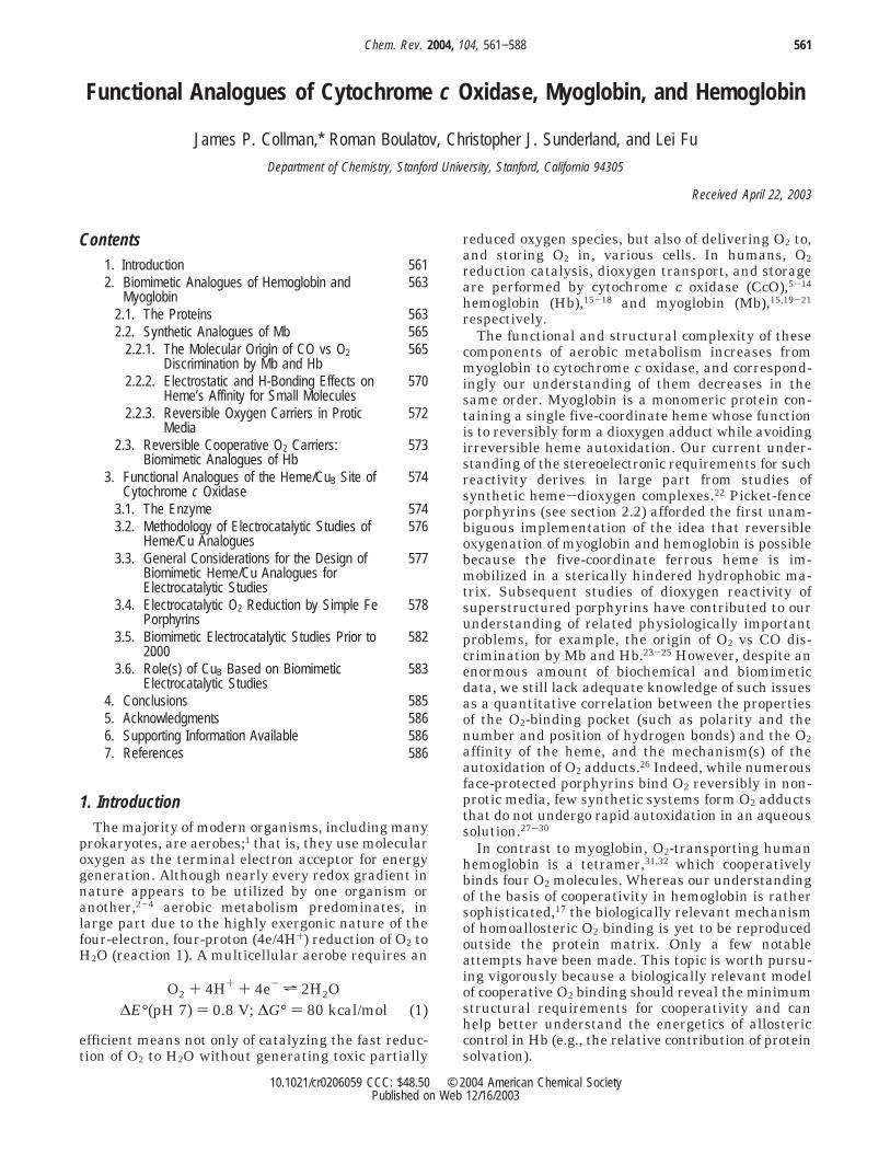
James P. Collman was born in 1932 and received B.S. and M.S. degreesfrom the University of Nebraska in 1954 and 1956, and a Ph.D. from theUniversity of Illinois in 1958 under the supervision of R. C. Fuson. In1958−1967 he was on the faculty of the University of North Carolina atChapel Hill; he moved to Stanford in 1967, where he is Daubert Professorof Chemistry. His research interests are very broad, extending acrossinorganic and organic chemistry, and also include superconductivity. Hisprincipal research is directed toward the development of structural andfunctional analogues of the active sites in heme proteins, particularlycytochrome oxidase. His work has been recognized by many awards; heis a member of the National Academy of Sciences.
Roman Boulatov received his Ph.D. in chemistry from Stanford in 2002,working under the guidance of Prof. James P. Collman on biomimeticstudies of cytochrome c oxidase, utilization of metalloporphyrins forcatalysis of multielectron reductions, and creation of new bonds betweentransition metals stabilized by porphyrins. Currently, he is a postdoc inthe laboratories of Professor George M. Whitesides at Harvard, workingon several problems in nanoscience. Boulatov’s scientific accolades includethe IUPAC Prize for young chemists (2002), a Stanford GraduateFellowship (1998−2002), and a Presidential Fellowship (Russia, 1994−1996).
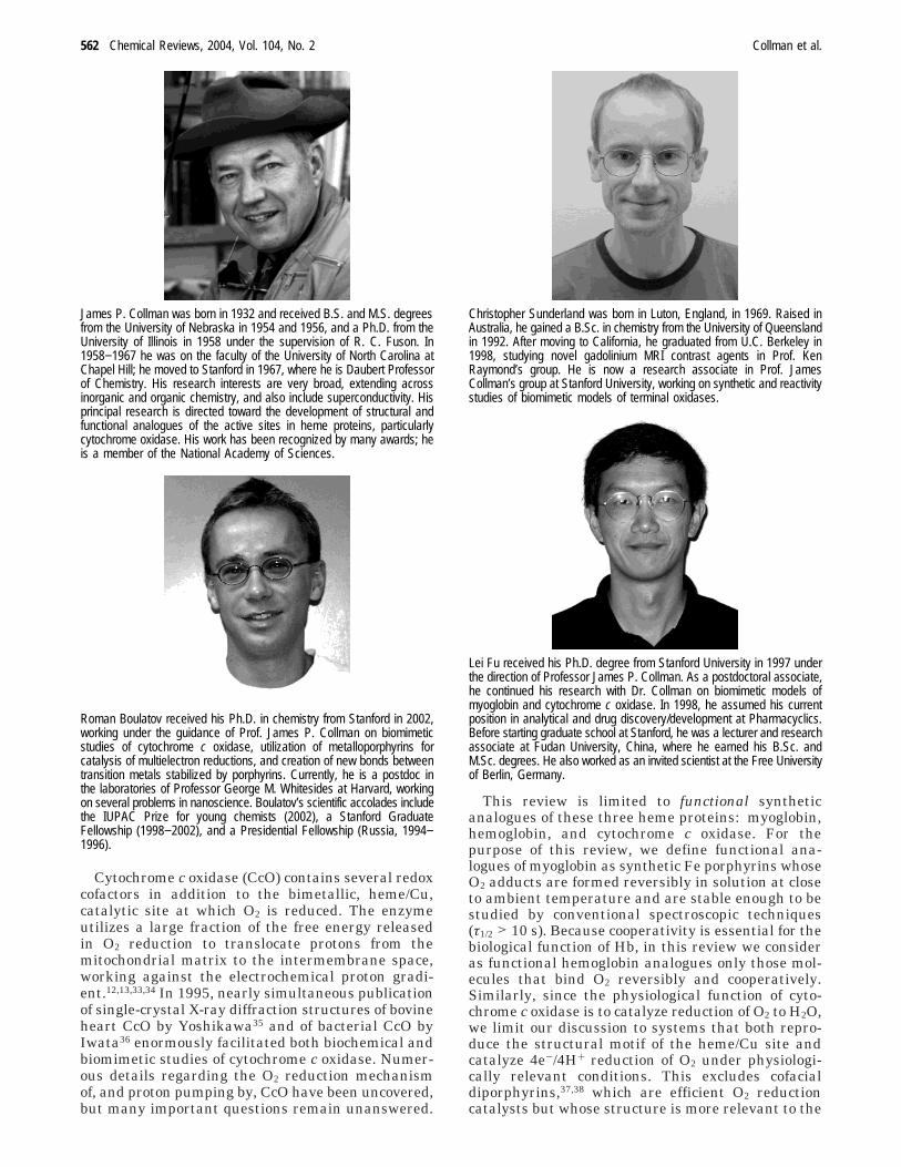
Christopher Sunderland was born in Luton, England, in 1969. Raised inAustralia, he gained a B.Sc. in chemistry from the University of Queenslandin 1992. After moving to California, he graduated from U.C. Berkeley in1998, studying novel gadolinium MRI contrast agents in Prof. KenRaymond’s group. He is now a research associate in Prof. JamesCollman’s group at Stanford University, working on synthetic and reactivitystudies of biomimetic models of terminal oxidases.
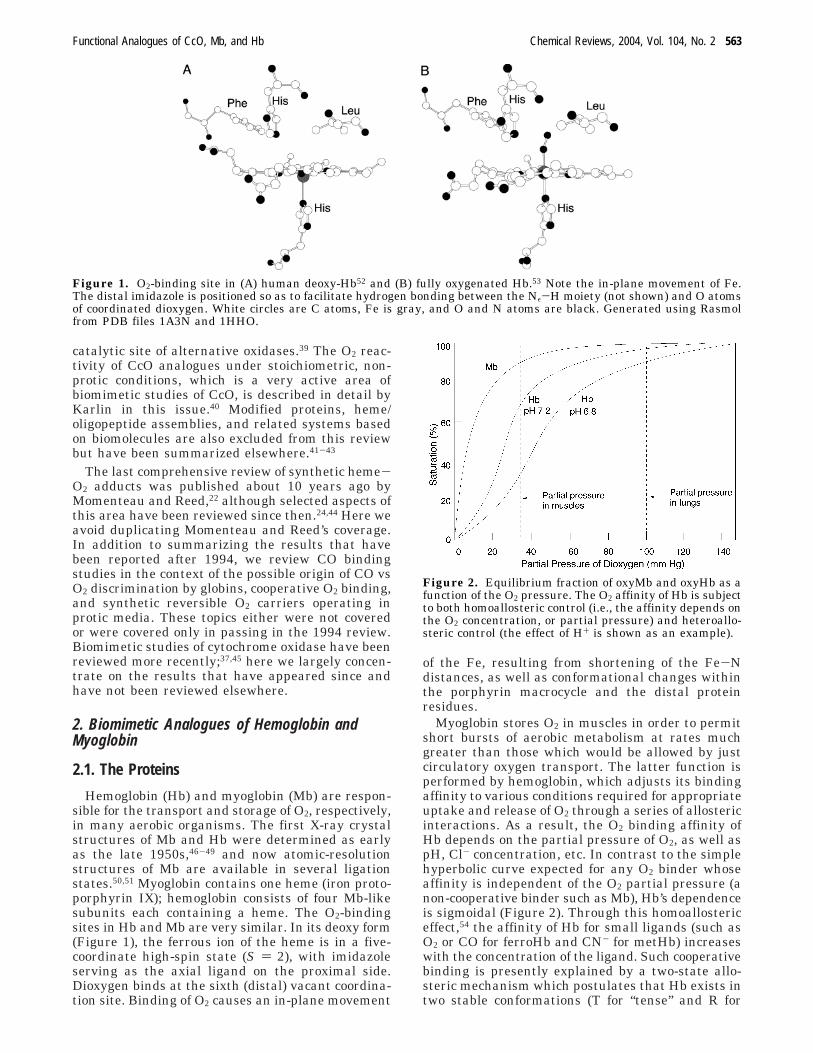
Lei Fu received his Ph.D. degree from Stanford University in 1997 underthe direction of Professor James P. Collman. As a postdoctoral associate,he continued his research with Dr. Collman on biomimetic models ofmyoglobin and cytochrome c oxidase. In 1998, he assumed his currentposition in analytical and drug discovery/development at Pharmacyclics.Before starting graduate school at Stanford, he was a lecturer and researchassociate at Fudan University, China, where he earned his B.Sc. andM.Sc. degrees. He also worked as an invited scientist at the Free Universityof Berlin, Germany.
562 Chemical Reviews, 2004, Vol. 104, No. 2 Collman et al.
catalytic site of alternative oxidases.39 The O2 reac-tivity of CcO analogues under stoichiometric, non-protic conditions, which is a very active area ofbiomimetic studies of CcO, is described in detail byKarlin in this issue.40 Modified proteins, heme/oligopeptide assemblies, and related systems basedon biomolecules are also excluded from this reviewbut have been summarized elsewhere.41-43
The last comprehensive review of synthetic heme-O2 adducts was published about 10 years ago byMomenteau and Reed,22 although selected aspects ofthis area have been reviewed since then.24,44 Here weavoid duplicating Momenteau and Reed’s coverage.In addition to summarizing the results that havebeen reported after 1994, we review CO bindingstudies in the context of the possible origin of CO vsO2 discrimination by globins, cooperative O2 binding,and synthetic reversible O2 carriers operating inprotic media. These topics either were not coveredor were covered only in passing in the 1994 review.Biomimetic studies of cytochrome oxidase have beenreviewed more recently;37,45 here we largely concen-trate on the results that have appeared since andhave not been reviewed elsewhere.
2. Biomimetic Analogues of Hemoglobin andMyoglobin
2.1. The Proteins
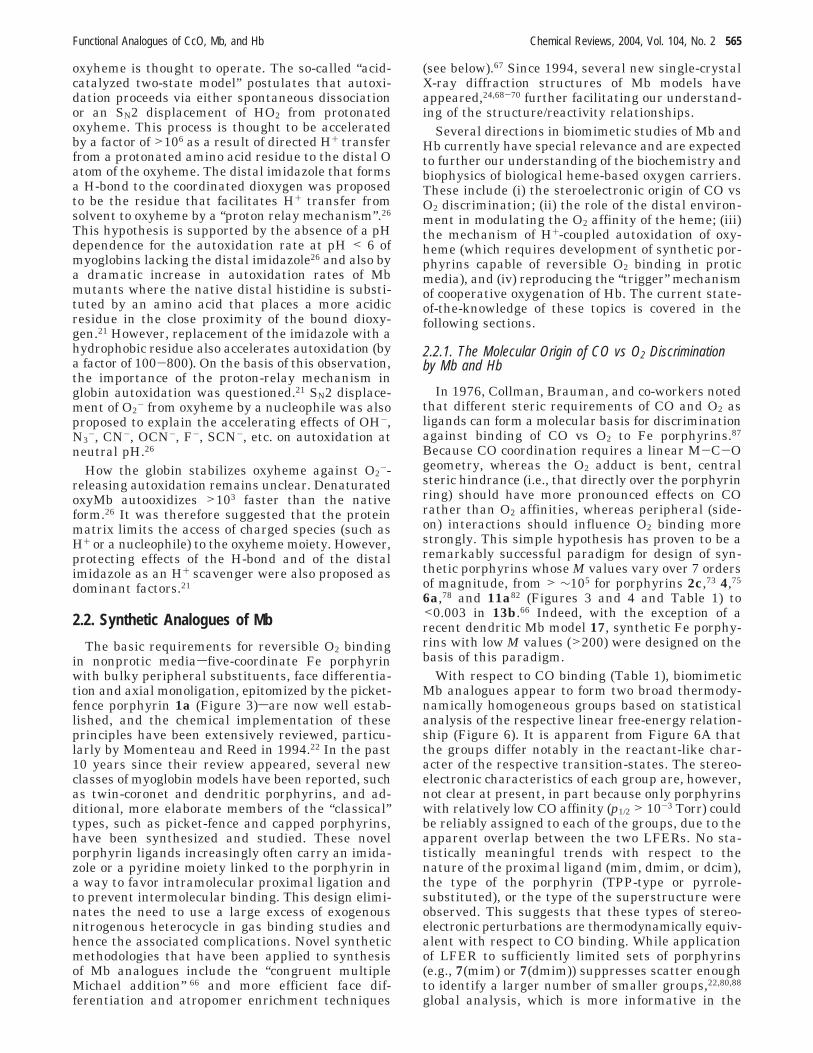
Hemoglobin (Hb) and myoglobin (Mb) are respon-sible for the transport and storage of O2, respectively,in many aerobic organisms. The first X-ray crystalstructures of Mb and Hb were determined as earlyas the late 1950s,46-49 and now atomic-resolutionstructures of Mb are available in several ligationstates.50,51 Myoglobin contains one heme (iron proto-porphyrin IX); hemoglobin consists of four Mb-likesubunits each containing a heme. The O2-bindingsites in Hb and Mb are very similar. In its deoxy form(Figure 1), the ferrous ion of the heme is in a five-coordinate high-spin state (S ) 2), with imidazoleserving as the axial ligand on the proximal side.Dioxygen binds at the sixth (distal) vacant coordina-tion site. Binding of O2 causes an in-plane movement
of the Fe, resulting from shortening of the Fe-Ndistances, as well as conformational changes withinthe porphyrin macrocycle and the distal proteinresidues.
Myoglobin stores O2 in muscles in order to permitshort bursts of aerobic metabolism at rates muchgreater than those which would be allowed by justcirculatory oxygen transport. The latter function isperformed by hemoglobin, which adjusts its bindingaffinity to various conditions required for appropriateuptake and release of O2 through a series of allostericinteractions. As a result, the O2 binding affinity ofHb depends on the partial pressure of O2, as well aspH, Cl- concentration, etc. In contrast to the simplehyperbolic curve expected for any O2 binder whoseaffinity is independent of the O2 partial pressure (anon-cooperative binder such as Mb), Hb’s dependenceis sigmoidal (Figure 2). Through this homoallostericeffect,54 the affinity of Hb for small ligands (such asO2 or CO for ferroHb and CN- for metHb) increaseswith the concentration of the ligand. Such cooperativebinding is presently explained by a two-state allo-steric mechanism which postulates that Hb exists intwo stable conformations (T for “tense” and R for
Figure 1. O2-binding site in (A) human deoxy-Hb52 and (B) fully oxygenated Hb.53 Note the in-plane movement of Fe.The distal imidazole is positioned so as to facilitate hydrogen bonding between the Nε-H moiety (not shown) and O atomsof coordinated dioxygen. White circles are C atoms, Fe is gray, and O and N atoms are black. Generated using Rasmolfrom PDB files 1A3N and 1HHO.
Figure 2. Equilibrium fraction of oxyMb and oxyHb as afunction of the O2 pressure. The O2 affinity of Hb is subjectto both homoallosteric control (i.e., the affinity depends onthe O2 concentration, or partial pressure) and heteroallo-steric control (the effect of H+ is shown as an example).
Functional Analogues of CcO, Mb, and Hb Chemical Reviews, 2004, Vol. 104, No. 2 563

“relaxed”) having different ligand affinities. Thetrigger mechanism for the T-to-R conversion, at leastin ferroHb, is an in-plane movement of the ferrousion, which converts from the high- to low-spin stateupon coordination of the exogenous ligand. Thismovement is transmitted through conformationalchanges within the protein matrix to the subunitinterface, where it leads to the disruption of saltbridges. The endergonicity of the latter transforma-tion accounts for the lower ligand affinity of theT-state Hb.
Heme ligand affinities are also regulated by polar,hydrophobic, and/or steric interactions between heme-bound exogenous ligands and the distal amino acidresidues. The most significant distal effect invokedin the stabilization of O2 in oxy-Mb and oxy-Hb is ahydrogen-bonding interaction between bound O2 andthe distal histidine (Figure 2). Initially proposed byPauling,55 the existence of the H-bond was at firstshown by EPR and resonance Raman studies ofcobalt-substituted oxyhemoglobin.56 Subsequent X-ray structure analysis53,57 and neutron diffractionstudies58 on oxy-Mb and oxy-Hb provided directevidence for such hydrogen bonding. Appropriatelypositioned hydrogen bonds can dramatically increasethe O2 affinity: for example, the 104-fold higher O2affinity of a unique hemoglobin from the bloodwormAscaris relative to human Hb is thought to resultfrom multiple H-bonds between coordinated dioxygenand distal Tyr and Glu residues.59
In addition to O2, CO is a biologically significantligand for Mb and Hb. The in vivo degradation ofheme generates one molecule of CO per heme catabo-lized, resulting in a partial pressure of CO on theorder of 1 × 10⁻³ Torr at the cellular level.60 BecauseCO has a high affinity to ferrohemes, it is a poten-tially powerful endogenous inhibitor of certain he-moproteins. Outside the protein matrix, imidazole-ligated heme has a CO affinity much greater thanthat of Mb or Hb, indicating that the globin disfavorsCO binding.61 It is generally accepted now that distalamino acids stabilize the dioxygen adduct whiledestabilizing the CO analogue. The atomic mecha-nism of the globin’s discrimination against CO hasbeen extensively studied both biochemically andbiomimetically, but it remains one of the morecontroversial issues in the biochemistry of Mb andHb.
The ratio of the half-saturation pressures of O2 andCO (p1/2
O2/p1/2CO), referred to as the M value, is a
useful measure of a complex’s susceptibility to COinhibition. Hb and Mb are clearly designed by natureto transport and store O2 in the presence of theendogenous poison, CO. Typical M values for Mb andHb are ∼102, whereas the intrinsic affinity of theimidazole-ligated heme for CO is >104 times thanthat for O2. Steric, electrostatic, and H-bondingproperties of the distal pocket appear to contributeto discrimination of CO vs O2, which have differentproperties as ligands. Whereas CO prefers to bindnormal to the heme plane, the O2 adduct is intrinsi-cally bent (Figure 1B). Likewise, substantial localiza-tion of the negative charge at the distal O of oxyheme(which is often viewed as a ferriheme-superoxide
adduct) contrasts with little charge separation in theheme-CO adduct. Distal groups have been shown toaffect Mb’s M values.62 The consensus opinion in thefield appears to be that spectroscopic data collectedon Mb-CO in the past 25 years indicate a slightlybent Fe-C-O unit (5-10°), which is also tilted by5-10° from the axis normal to the average 24-atomplane of the porphyrin.25 Indeed, even in synthetichemes with sterically-induced very low affinity to CO,no significant distortion of the Fe-C-O unit isobserved.24 Analysis of the kinetic and equilibriumdata for various Mb mutants suggests that the stericstrain, accommodated by a distortion of the Fe-C-Ounit and of the globin, accounts for <20% of thedifference in CO affinities of the heme vs Mb. It wasargued that electrostatic and H-bonding effects aremainly responsible for CO vs O2 discrimination byMb and Hb.25,63 However, the quantitative contribu-tions of each of these factors remain a contentiousissue. For example, Jameson analyzed the O2 and CObinding affinities of a series of Mb mutants andconcluded that factors stabilizing O2 binding anddestabilizing CO binding correlate only weakly.64
Confusion about the stereoelectronic effects of thedistal environment on the affinities of gaseous mol-ecules to globins has been compounded by a tacitassumption by many workers in the field that kineti-cally determined equilibrium constants (k+/k-) for COand O2 binding are equivalent to those measuredthermodynamically. However, thermodynamicallyand kinetically derived equilibrium constants maydiffer by as much as a factor of 5.65 This happenswhen O2 diffusion through the protein matrix to theO2-binding site has activation barrier(s) comparableto the activation barriers of steps involved in theFe-O bond homolysis/formation. The latter reactionis traditionally studied in kinetic photolysis experi-ments. As a result, the kinetic experiments quantifya process that is but one step of a more complexreaction studied in equilibrium measurements. Thedifference between the kinetically and thermody-namically determined equilibrium constants is ex-pected to be less significant, if at all, in small-molecule Mb analogues, wherein Fe is generallyeasily accessible.
In addition to preventing irreversible aerobic oxi-dation of ferroheme through the intermediacy ofperoxo-bridged hemes, the globins stabilize oxyhemeagainst heterolytic cleavage of the Fe-O bond, whichresults in the release of superoxide (autoxidation).Prevention of such a process is important bothbecause superoxide is toxic and because the resultingferriheme cannot serve as an oxygen carrier. Eventhough autoxidation of human Mb and Hb underphysiological conditions is relatively slow (the ap-parent rate constant <0.01 h-1, τ1/2 > 60 h), about1-2% of all Hb in a human is in the ferric (met) format any given time.26 The autoxidation is slowest atpH 7.5-9 (depending on the protein) and is acceler-ated by orders of magnitude both in more and in lessacidic solutions; the rate also depends on the partialpressure of O2. The mechanism of autoxidationremains uncertain. Neither outer-sphere electrontransfer nor spontaneous dissociation of O2
- from
564 Chemical Reviews, 2004, Vol. 104, No. 2 Collman et al.
oxyheme is thought to operate. The so-called “acid-catalyzed two-state model” postulates that autoxi-dation proceeds via either spontaneous dissociationor an SN2 displacement of HO2 from protonatedoxyheme. This process is thought to be acceleratedby a factor of >106 as a result of directed H+ transferfrom a protonated amino acid residue to the distal Oatom of the oxyheme. The distal imidazole that formsa H-bond to the coordinated dioxygen was proposedto be the residue that facilitates H+ transfer fromsolvent to oxyheme by a “proton relay mechanism”.26
This hypothesis is supported by the absence of a pHdependence for the autoxidation rate at pH < 6 ofmyoglobins lacking the distal imidazole26 and also bya dramatic increase in autoxidation rates of Mbmutants where the native distal histidine is substi-tuted by an amino acid that places a more acidicresidue in the close proximity of the bound dioxy-gen.21 However, replacement of the imidazole with ahydrophobic residue also accelerates autoxidation (bya factor of 100-800). On the basis of this observation,the importance of the proton-relay mechanism inglobin autoxidation was questioned.21 SN2 displace-ment of O2
- from oxyheme by a nucleophile was alsoproposed to explain the accelerating effects of OH-,N3
-, CN-, OCN-, F-, SCN-, etc. on autoxidation atneutral pH.26
How the globin stabilizes oxyheme against O2--
releasing autoxidation remains unclear. DenaturatedoxyMb autooxidizes >103 faster than the nativeform.26 It was therefore suggested that the proteinmatrix limits the access of charged species (such asH+ or a nucleophile) to the oxyheme moiety. However,protecting effects of the H-bond and of the distalimidazole as an H+ scavenger were also proposed asdominant factors.21
2.2. Synthetic Analogues of Mb
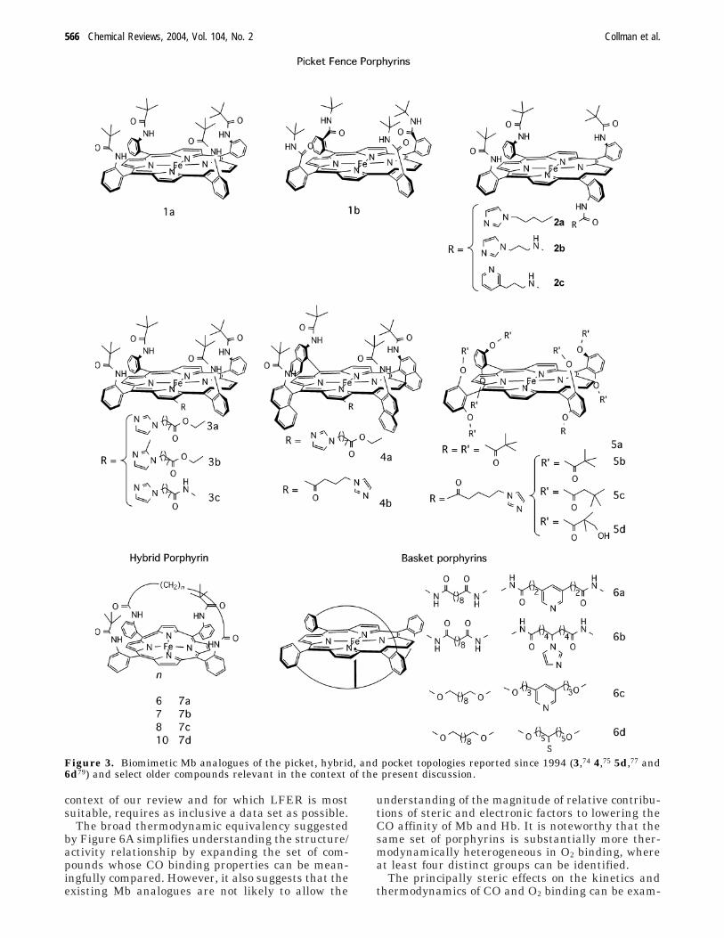
The basic requirements for reversible O2 bindingin nonprotic mediasfive-coordinate Fe porphyrinwith bulky peripheral substituents, face differentia-tion and axial monoligation, epitomized by the picket-fence porphyrin 1a (Figure 3)sare now well estab-lished, and the chemical implementation of theseprinciples have been extensively reviewed, particu-larly by Momenteau and Reed in 1994.22 In the past10 years since their review appeared, several newclasses of myoglobin models have been reported, suchas twin-coronet and dendritic porphyrins, and ad-ditional, more elaborate members of the “classical”types, such as picket-fence and capped porphyrins,have been synthesized and studied. These novelporphyrin ligands increasingly often carry an imida-zole or a pyridine moiety linked to the porphyrin ina way to favor intramolecular proximal ligation andto prevent intermolecular binding. This design elimi-nates the need to use a large excess of exogenousnitrogenous heterocycle in gas binding studies andhence the associated complications. Novel syntheticmethodologies that have been applied to synthesisof Mb analogues include the “congruent multipleMichael addition” 66 and more efficient face dif-ferentiation and atropomer enrichment techniques
(see below).67 Since 1994, several new single-crystalX-ray diffraction structures of Mb models haveappeared,24,68-70 further facilitating our understand-ing of the structure/reactivity relationships.
Several directions in biomimetic studies of Mb andHb currently have special relevance and are expectedto further our understanding of the biochemistry andbiophysics of biological heme-based oxygen carriers.These include (i) the steroelectronic origin of CO vsO2 discrimination; (ii) the role of the distal environ-ment in modulating the O2 affinity of the heme; (iii)the mechanism of H+-coupled autoxidation of oxy-heme (which requires development of synthetic por-phyrins capable of reversible O2 binding in proticmedia), and (iv) reproducing the “trigger” mechanismof cooperative oxygenation of Hb. The current state-of-the-knowledge of these topics is covered in thefollowing sections.
2.2.1. The Molecular Origin of CO vs O2 Discriminationby Mb and Hb
In 1976, Collman, Brauman, and co-workers notedthat different steric requirements of CO and O2 asligands can form a molecular basis for discriminationagainst binding of CO vs O2 to Fe porphyrins.87
Because CO coordination requires a linear M-C-Ogeometry, whereas the O2 adduct is bent, centralsteric hindrance (i.e., that directly over the porphyrinring) should have more pronounced effects on COrather than O2 affinities, whereas peripheral (side-on) interactions should influence O2 binding morestrongly. This simple hypothesis has proven to be aremarkably successful paradigm for design of syn-thetic porphyrins whose M values vary over 7 ordersof magnitude, from > ∼105 for porphyrins 2c,73 4,75
6a,78 and 11a82 (Figures 3 and 4 and Table 1) to<0.003 in 13b.66 Indeed, with the exception of arecent dendritic Mb model 17, synthetic Fe porphy-rins with low M values (>200) were designed on thebasis of this paradigm.
With respect to CO binding (Table 1), biomimeticMb analogues appear to form two broad thermody-namically homogeneous groups based on statisticalanalysis of the respective linear free-energy relation-ship (Figure 6). It is apparent from Figure 6A thatthe groups differ notably in the reactant-like char-acter of the respective transition-states. The stereo-electronic characteristics of each group are, however,not clear at present, in part because only porphyrinswith relatively low CO affinity (p1/2 > 10-3 Torr) couldbe reliably assigned to each of the groups, due to theapparent overlap between the two LFERs. No sta-tistically meaningful trends with respect to thenature of the proximal ligand (mim, dmim, or dcim),the type of the porphyrin (TPP-type or pyrrole-substituted), or the type of the superstructure wereobserved. This suggests that these types of stereo-electronic perturbations are thermodynamically equiv-alent with respect to CO binding. While applicationof LFER to sufficiently limited sets of porphyrins(e.g., 7(mim) or 7(dmim)) suppresses scatter enoughto identify a larger number of smaller groups,22,80,88
global analysis, which is more informative in the
Functional Analogues of CcO, Mb, and Hb Chemical Reviews, 2004, Vol. 104, No. 2 565
context of our review and for which LFER is mostsuitable, requires as inclusive a data set as possible.
The broad thermodynamic equivalency suggestedby Figure 6A simplifies understanding the structure/activity relationship by expanding the set of com-pounds whose CO binding properties can be mean-ingfully compared. However, it also suggests that theexisting Mb analogues are not likely to allow the
understanding of the magnitude of relative contribu-tions of steric and electronic factors to lowering theCO affinity of Mb and Hb. It is noteworthy that thesame set of porphyrins is substantially more ther-modynamically heterogeneous in O2 binding, whereat least four distinct groups can be identified.
The principally steric effects on the kinetics andthermodynamics of CO and O2 binding can be exam-
Figure 3. Biomimetic Mb analogues of the picket, hybrid, and pocket topologies reported since 1994 (3,74 4,75 5d,77 and6d79) and select older compounds relevant in the context of the present discussion.
566 Chemical Reviews, 2004, Vol. 104, No. 2 Collman et al.
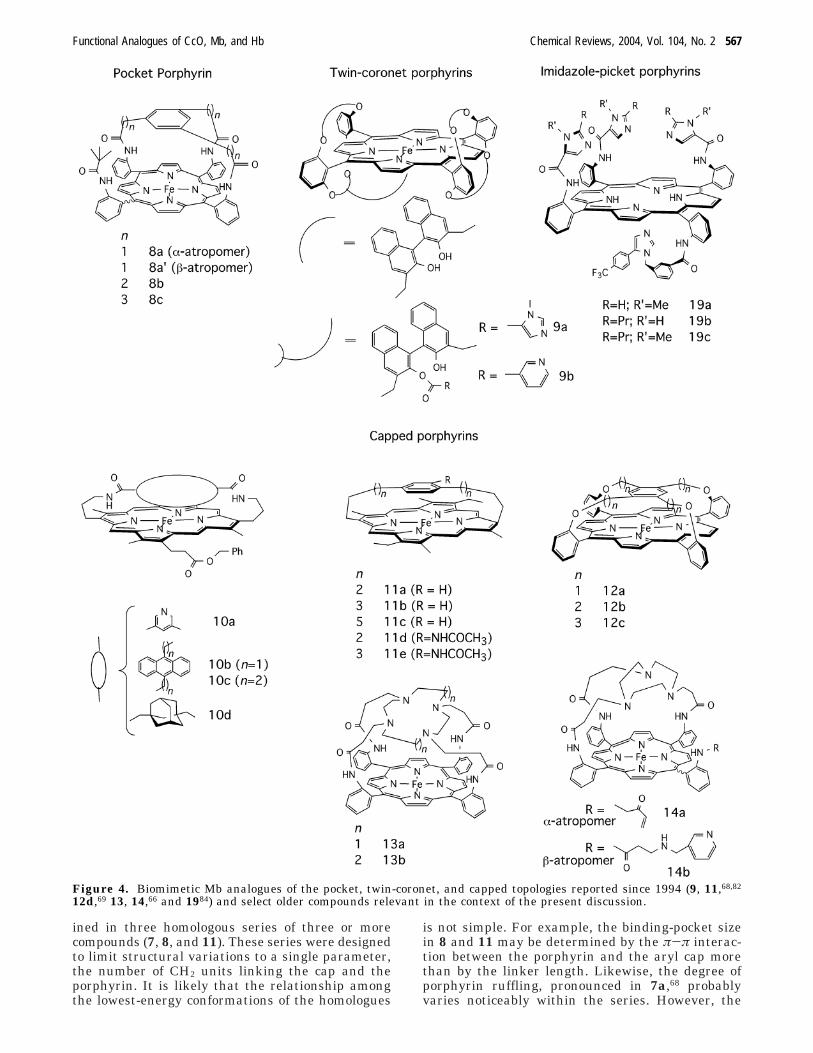
ined in three homologous series of three or morecompounds (7, 8, and 11). These series were designedto limit structural variations to a single parameter,the number of CH2 units linking the cap and theporphyrin. It is likely that the relationship amongthe lowest-energy conformations of the homologues
is not simple. For example, the binding-pocket sizein 8 and 11 may be determined by the π-π interac-tion between the porphyrin and the aryl cap morethan by the linker length. Likewise, the degree ofporphyrin ruffling, pronounced in 7a,68 probablyvaries noticeably within the series. However, the
Figure 4. Biomimetic Mb analogues of the pocket, twin-coronet, and capped topologies reported since 1994 (9, 11,68,82
12d,69 13, 14,66 and 1984) and select older compounds relevant in the context of the present discussion.
Functional Analogues of CcO, Mb, and Hb Chemical Reviews, 2004, Vol. 104, No. 2 567
broad capacity of the binding pocket to accommodatethe steric demands of a sixth ligand on Fe probablycorrelates positively with the linker lengths.
A strong positive correlation between the M valueand the length of the cap/porphyrin linkers is clearlyobserved for all series (Table 2). This correlationfurther confirms the validity of the paradigm thatpurely steric discrimination against CO, based ondifferent steric properties of the Fe-C-O and Fe-O-O moieties, is possible. However, while the sterichindrance in the binding pocket has a predictable and
uniform effect on M values in these three series ofMb analogues, it has a variable influence on thekinetic parameters, particularly those of O2 binding.Thus, the effect of sterics in CO vs O2 discriminationappears to be more complex than a decrease ink+(CO).
Single-crystal X-ray diffraction studies of CO-ligated sterically encumbered Mb models also clearlyindicate that the expansion of the distal superstruc-ture, sometimes accompanied by increased porphyrinnonplanarity, and not tilting or bending of CO, is the
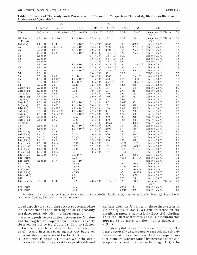
Table 1. Kinetic and Thermodynamic Parameters of CO, and for Comparison Those of O2, Binding to BiomimeticAnalogues of Myoglobina
CO O2
k+, M-1 s-1 k-, s-1 p1/2, Torr k+, M-1 s-1 k-, s-1 p1/2, Torr M conditions ref
Mb 3-5 × 10⁵ 1.5-40 × 10-3 0.014-0.025 1-2 × 10⁷ 10-30 0.37-1 20-40 phosphate pH 7 buffer, 7125 °C
Hb, human, 4.6 × 10⁶ 9 × 10⁻³ 1.4 × 10⁻³ 3.3 × 10⁷ 13.1 0.22 150 phosphate pH 7 buffer, 71R state 25 °C
1(dmim) 1.4 × 10⁶ 0.14 8.9 × 10⁻³ 1.1 × 10⁸ 4600 38 4280 toluene, 25 °C 722a 3.6 × 10⁷ 7.8 × 10⁻³ 2.2 × 10⁻⁵ 4.3 × 10⁸ 2900 0.58 2.7 × 10⁴ toluene, 25 °C 722b 2.9 × 10⁷ 0.014 4.9 × 10⁻⁵ 2.6 × 10⁸ 3900 1.26 2.6 × 10⁴ toluene, 25 °C 732c 4.8 × 10⁷ 0.33 6.4 × 10⁻⁴ 3.0 × 10⁸ 1.9 × 10⁵ 52.2 7.6 × 10⁴ toluene, 25 °C 733a 3.1 × 10⁷ 6.4 × 10⁸ 1.4 × 10³ 0.29 toluene, 25 °C 743b 2.9 × 10⁶ 1.6 × 10⁸ 4.6 × 10⁴ 38 toluene, 25 °C 743c 1.1 × 10⁷ 3.7 × 10⁸ 1.3 × 10⁴ 4.6 toluene, 25 °C 744 3.3 × 10⁶ 1.5 × 10⁻⁴ 2.4 × 10⁷ 5.0 × 10³ 18 1.2 × 10⁵ toluene, 25 °C 755b 1.8 × 10⁶ 4.9 × 10⁻⁴ 2.7 × 10⁷ 2.4 × 10³ 13 3 × 10⁴ toluene, 25 °C 765c 1.3 × 10⁷ 7.2 × 10⁻⁵ 6.3 × 10⁷ 1.2 × 10³ 2.5 3 × 10⁴ toluene, 25 °C 765d 2.0 × 10⁵ 2.6 × 10⁴ 55 33.5 CH2Cl2, 25 °C 776a 3.5 × 10⁷ 0.03 9 × 10⁻⁵ 3.6 × 10⁸ 5000 2 2 × 10⁴ toluene, 20 °C 786b 4.0 × 10⁷ 0.0067 1.7 × 10⁻⁵ 3.1 × 10⁸ 620 0.29 17000 toluene, 20 °C 786c 6.8 × 10⁷ 0.069 1 × 10⁻⁴ 3.0 × 10⁸ 4 × 10⁴ 18 1.8 × 10⁵ toluene, 20 °C 786d 8.8 × 10⁴ 28 0.026 5.2 × 10⁶ 800 12.4 480 toluene, 20 °C 797a(dmim) 2.0 × 10⁴ 0.08 0.50 5.4 × 10⁵ 15 2.7 5.4 toluene, 20 °C 807b(dmim) 1.0 × 10⁵ 0.031 0.03 1.0 × 10⁷ 67 0.67 21 toluene, 20 °C 807c(dmim) 1.7 × 10⁵ 0.050 0.03 1.5 × 10⁷ 540 3.6 121 toluene, 20 °C 807d(dmim) 4.7 × 10⁶ 0.11 0.002 3.9 × 10⁸ 7500 1.9 827 toluene, 20 °C 807a(mim) 8.0 × 10⁴ 0.0082 0.01 2.2 × 10⁶ 2.0 0.07 7 toluene, 20 °C 807b(mim) 1.0 × 10⁶ 0.0026 2.6 × 10⁻⁴ 2.1 × 10⁷ 5.0 0.023 88 toluene, 20 °C 807c(mim) 1.8 × 10⁶ 0.002 1.1 × 10⁻⁴ 3.0 × 10⁷ 27 0.091 818 toluene, 20 °C 807d(mim) 6.3 × 10⁷ 0.0027 4.2 × 10⁻⁶ 6.2 × 10⁸ 130 0.02 5000 toluene, 20 °C 808a(mim) 5.8 × 10⁵ 8.6 × 10⁻³ 1.5 × 10⁻³ 2.2 × 10⁶ 9 0.36 270 toluene, 25 °C 728b(mim) 1.5 × 10⁶ 9.4 × 10⁻³ 6.5 × 10⁻⁴ 1.7 × 10⁷ 71 0.36 550 toluene, 25 °C 728a(dmim) 9.8 × 10⁴ 0.055 0.067 1.9 × 10⁶ 280 12.6 216 toluene, 25 °C 728b(dmim) 2.1 × 10⁵ 0.053 0.026 5.2 × 10⁶ 800 12.4 480 toluene, 25 °C 728c(dmim) 0.001 7.4 × 10⁸ 34500 4 3500 toluene, 25 °C 729a 2.1 × 10⁷ 0.23 1.1 × 10⁻³ 4.0 × 10⁷ 2000 1.3 1180 toluene, 25 °C 719b 1.6 × 10⁷ 3.2 1.7 × 10⁻² 2.7 × 10⁷ 2500 9.4 550 toluene, 25 °C 7110a(dcim) 6 × 10² 0.24 37 1.1 × 10⁴ 68 540 14 toluene, 20 °C 8110b(dcim) 3 × 10⁴ 0.05 0.17 1.0 × 10⁵ 800 700 4100 toluene, 20 °C 8110c(dcim) 6 × 10⁶ 0.05 0.0009 6.5 × 10⁷ 1000 1.4 1500 toluene, 20 °C 8110d(dcim) 1 × 10⁴ 0.05 0.57 1.5 × 10⁵ 690 300 530 toluene, 20 °C 8111a(mim) 4.9 × 10⁶ 0.014 0.0015 1.0 × 10⁷ 104 ∼100 ∼105 toluene, 20 °C 8211a(dcim) 3.0 × 10⁶ 0.7 0.023 8.2 × 10⁷ 105 ∼150 ∼6000 toluene, 20 °C 8211b(dcim) 1.1 × 10⁷ 0.13 0.0012 1.0 × 10⁸ 105 ∼70 7 × 10⁴ toluene, 20 °C 8211c(dcim) 1.0 × 10⁶ 0.014 0.015 1.0 × 10⁷ 104 ∼150 ∼105 toluene, 20 °C 8212a(mim) 1.0 × 10⁶ 0.05 5 × 10⁻³ 23 4300 toluene, 25 °C 7212a(dmim) 0.20 4000 2 × 10⁴ toluene, 25 °C 7212b(dcim) 4.1 × 10⁶ 0.17 4.1 × 10⁻³ toluene, 25 °C 7213a(dmim) >3500 760 <0.22 toluene, 20 °C 6613a(dcim) >3500 ∼760 <0.22 toluene, 20 °C 6613b(dmim) >3500 22 <0.006 toluene, 4 °C 6613b(dcim) >3500 ∼3 <0.003 toluene, 20 °C 6614a(dmim) 2.9 2.3 0.79 toluene, 20 °C 6614b 1.5 3.7 2.5 toluene, 20 °C 6616a(C12mim) 3.9 × 10⁶ 0.14 0.028 2.8 × 10⁸ 1.5 × 10⁴ 32 1100 phosphate pH 7 buffer, 29
25 °C17a(dmim) 0.35 0.035 0.1 toluene, 25 °C 8317b(dmim) 0.19 0.016 0.08 toluene, 25 °C 83
a For chemical structures see Figures 3-5. dmim, 1,2-dimethylimidazole; mim, 1-methylimidazole; dcim, 1,5-dicyclohexyl-imidazole; C12mim, 5-dodecyl-1-methylimidazole.
568 Chemical Reviews, 2004, Vol. 104, No. 2 Collman et al.

predominant form of steric distortions (Figure 7).Minimal distortion of the Fe-CO unit in modelcompounds is also supported by extensive spectro-scopic evidence.89
A less-recognized corollary of this observation isthat the linear Fe-C-O moiety in CO adducts of Mband Hb does not by itself rule out sterics as the majormolecular mechanism of the discrimination. Spirohas discussed this issue in detail and ruled out stericeffects.25 However, in our opinion, this conclusion isbased too heavily on computational results, as thenumber of mutants studied is still too small toreasonably claim that all possible ways to induce and/
or distribute strain upon CO binding have beenprobed.
The effects of polarity (electrostatic potential) andof H-bonding within the binding pocket, while studiedextensively in Mb and Hb, and currently thought tobe at least as important as sterics in determining theM values of globins,21,23,25 have been relatively littleexamined in Mb models. Kinetic and thermodynamicdata on CO and O2 binding exist for three pairs ofsynthetic porphyrins (10a vs 11c; 14b vs 8b(mim);9a vs 4) with pockets of comparable sizes but ofsignificantly different polarities. The latter correlateswith the C-O and Fe-C stretching frequencies and13C NMR chemical shifts of the CO adducts.83 In themost dramatic example (10a, M ) 14 vs 11c, M )105), the amide and pyridinyl moieties of the bindingpocket in 10a appear to cause >104-fold reduction inthe M value. Relative to 11c, the k+(CO), k+(O2), andk-(O2) values in 10a are 102-103 times lower, whereask-(CO) is 20-fold higher. An increase in the k- valueis often interpreted as an indicator of electronicdestabilization of the bound ligand. However, theorigin of lower k+(CO), k+(O2), and k-(O2) is notknown, as such a relative decrease cannot be at-tributed to steric factors. Substantially higher selec-tivity toward O2 binding is observed in 14b, whose
Figure 5. Dendritic, lipid-derivatized, and supramolecular analogues of Mb.29,30,83,85,86
Table 2. Correlation Coefficients for HomologousSeries 7 (Four Homologues), 8 (Three Homologues),and 11 (Three Homologues)a
CO O2
series k+ k- p1/2 k+ k- p1/2 M
7(mim) 0.89 -0.68 -0.70 0.90 0.94 -0.40 0.937(dmim) 0.89 0.55 -0.72 0.89 -0.53 -0.01 0.938(dmim) no data no data -0.99 0.87 0.87 -0.88 0.9011(dcim) -0.37 -0.85 -0.18 -0.87 -0.94 0.19 0.92
a A positive value corresponds to an increase in the param-eter with increasing length of cap/porphyrin linkers. Statisti-cally meaningful coefficients are in italics.
Functional Analogues of CcO, Mb, and Hb Chemical Reviews, 2004, Vol. 104, No. 2 569

tacn-defined binding pocket is somewhat larger butmore polar than that in 8b(mim) (Figure 8) andwhose M value (2.5) is >200 times less (M8b(mim) )550). Hydrogen bonding to bound O2 was invoked torationalize the lower M values of 9a relative to thoseof the sterically comparable 4.71
In contrast to the stereoelectronic properties of thebinding pocket, the nature of the proximal ligandsimidazole vs pyridine (6a vs 6b; 9a vs 9b; and 2b vs2c), or 1-MeIm vs 1,2-Me2Im (7, 8 and 12a)sappearsto have a limited effect on the M values. However,in a single example, thiolate was found to increasethe selectivity by a factor of 400 (6d vs 6c, Table 1).
By the LFER criterion, 6d is an outlier among theporphyrins in Table 1.
Recently reported dendritic porphyrins 17a and17b bind O2 in preference of CO by a factor of >10,even though they lack rigid, centrally positionedsuperstuctures.83 Both the unusually high O2 affinity,presumably assisted by a H-bond between an amidegroup and bound dioxygen (see below), and therelatively low CO affinities contribute to the very lowM values of these porphyrins.
2.2.2. Electrostatic and H-Bonding Effects on Heme’sAffinity for Small Molecules
Dipolar interactions with distal amino acids, par-ticularly the H-bond between the imidazole residueof distal histidine and oxygen atoms of oxyheme, areestimated to enhance O2 binding to Mb and Hb by afactor of 103 (which is partially compensated byendergonic displacement of H2O required for O2binding).21 The hydrogen bond not only favors O2binding but also is thought to stabilize oxyhemeagainst superoxide-releasing autoxidation and con-tribute to CO vs O2 discrimination. However, theelectronic effects within the binding pocket of globinson heme’s ligand affinities remain poorly understood,in large part because it is impossible to replace asingle amino acid and leave the rest of the proteinunchanged and because of the dearth of high-resolu-tion structural data on such mutants. Hence, biomi-metic analogues, which are more amenable to precisestructural modification and spectroscopic character-ization, could conceivably be particularly useful inprobing these issues. Yet, it has been difficult todesign reversibly O2-binding porphyrins with suitablypositioned H-bond donors. The existence of relativelyweak H-bonds, especially between anilidine groupsof the porphyrin ligand and coordinated O2, is nowestablished.22 Five pairs of structurally similar por-phyrins are available wherein only one member canform a H-bond to coordinated O2 (Table 3). With oneexception, the compound with an accessible H-bonddonor has O2 affinities 10-30 times higher than thoseof the analogue lacking an H-bond donor. In 6a, theexistence of a H-bond between the amide group andcoordinated O2 was demonstrated by low-tempera-ture proton and 17O NMR, IR, and Raman spec-troscopies.78 The higher O2 affinity of 6a vs 6c ismainly due to the lower dissociation rate constantin 6a, which was interpreted as additional evidence
Figure 6. Linear free-energy relationship (LFER) for CO(A) and O2 (B) binding to biomimetic Mb analogues. Signswith the gray background, k+; signs with the whitebackground, k-. (A) triangles, group 1; squares, group 2.(B) triangles, group 1; squares, groups 2 and 3; diamonds,group 4. The least-squares fits (broken lines, group 1; solidlines, group 2) were performed to the following LFERequations: log k+ ) k0 - R log p1/2 - log H and log k- )k0 - (R - 1) ln p1/2, where H is Henry’s constant (0.96 ×10⁻⁵ Torr-1 M for CO and 0.72 × 10⁻⁵ Torr-1 M for O2 intoluene at 25 °C). Data reported at other temperatureswere adjusted to 25 °C. The parameter R (the slope of thelog k vs log K graphs) is the contribution of the free energyof the reactants to the free energy of the transition state.Assignment of porphyrins into different groups was per-formed by maximizing ∑
i(Flogk+,logK
i + Flogk-,logKi ) for both
groups, where FX,Y are the correlation coefficients and i isthe group number. Very low slopes of the log kon(O2) vs logK(O2) dependence preclude statistics-based grouping. Dataused to create the figures are available as SupportingInformation.
Table 3. O2 Affinity of Select Mb Models Capable ofForming an H-Bond to Bound O2 and TheirAnalogues Lacking Accessible H-Bond Donorsa
H-bond effectaccessibleH-bonddonor
p1/2,Torr ref
analoguewithoutH-donor
p1/2′,Torr ref
p1/2/p1/2′
∆∆G,kcal/mol
2a 0.58 72 5b 13 76 30 2.03a 0.29 746a 2 78 6c 18 78 9 1.311a 9.5 82 11d 55.7 82 6 1.111b 2.8 82 11e 22.7 82 8 1.25d 33.5 77 5c 2.5 76 0.07 -1.6
a The same conditions as those cited in Table 1, except forcompounds 11 (toluene, -45 °C).68
570 Chemical Reviews, 2004, Vol. 104, No. 2 Collman et al.
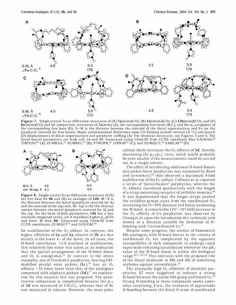
for stabilization of the O2 adduct. In contrast, thehigher affinities of 2a and 3a relative to 5b are duemainly to the lower k+ of the latter. In all cases, theH-bond contributes <2.0 kcal/mol of stabilization;this relatively low value was taken as an indicationthat the spatial arrangement of the H-bond donorand O2 is nonoptimal.22 In contrast to the aboveexamples, one of Tsuchida’s porphyrins, bearing OH-modified pivalyl ester pickets (5c),76 has an O2affinity >10 times lower than that of the analogouscompound with aliphatic pickets (5d);77 no explana-tion for this unusual fact was proposed. The quan-titative comparison is complicated because the p1/2of 5d was measured in CH2Cl2, whereas that of 5cwas measured in toluene. However, the more polar
solvent likely increases the O2 affinity of 5d, therebydecreasing the p1/2/p1/2′ ratio, which would probablybe even smaller if the measurements could be carriedout in a single solvent.
The effect of introducing additional H-bond donorsinto picket-fence porphyrins was examined by Reedand co-workers;95 they observed a maximum 9-foldstabilization of the O2 adduct. Collman et al. reporteda series of “picnic-basket” porphyrins, wherein theO2 affinity correlated qualitatively with the lengthof a strap connecting two pairs of anilidine moieties.96
It was hypothesized that the longer straps pushedthe anilidine groups away from the coordinated O2,increasing the O‚‚‚HN distance and hence weakeningthe H-bond. A remarkable (102-104-fold) increase inthe O2 affinity of Co porphyrins was observed byChang et al. upon the introduction of a carboxylic acidmoiety at a distance suitable for formation of H-bonding with Co-coordinated O2.97
Despite some progress, the studies of biomimeticMb analogues with H-bond donors in the vicinity ofcoordinated O2 are complicated by the apparentsusceptibility of such compounds to undergo rapidsuperoxide-releasing autoxidation whenever the pKavalue of the H-bond donor is within the biologicalrange.68,77,81,95 This contrasts with the proposed roleof the distal imidazole in Mb and Hb of stabilizingoxyheme against autoxidation.26
The unusually high O2 affinities of dendritic por-phyrins 17 were suggested to indicate a strongH-bond between the amide NH group and bound O2.83
Strong H-bonding in these compounds seems some-what surprising. First, the existence of appreciableH-bonding between the distal O atom of coordinated
Figure 7. Single-crystal X-ray diffraction structures of (A) 7a(mim)(CO), (B) 12c(mim)(CO), (C) 12b(mim)(CO), and (D)8a′(mim)(CO) and for comparison, structures of 7a(mim) (A), the corresponding free bases (B,C), and the R4 atropomer ofthe corresponding free base (D). X-M is the distance between the centroid of the distal superstucture and Fe (or theporphyrin centroid for free bases). Major conformational distortions upon CO binding include vertical (A-C) and lateral(D) displacements of distal superstucture and porphyrin ruffling (A). For chemical structures, see Figures 3 and 4. Thelisted metric parameters are from refs 24 and 80. Generated using Chem3D from CCDS coordinate files GAJHAA,90
YIMYOU80 (A); ZUMRAA,70 ZUMREC70 (B); PYROPR,91 JIXNAF92 (C); and ZUMQUT,70 SAHCAF93 (D).
Figure 8. Single-crystal X-ray diffraction structures of (A)the free base for 8b and (B) an analogue of 14b. M-X isthe distance between the metal (porphyrin centroid for A)and the centroid of the cap; min. M-cap is the the shortestcontact between the metal (porphyrin centroid for A) andthe cap. On the basis of both parameters, 14b has a lesssterically congested cavity, yet it manifests higher p1/2(CO)and lower M than 8b. Generated using Chem3D fromCCDS coordinates ZUMQUT70 and GIXJOM.94
Functional Analogues of CcO, Mb, and Hb Chemical Reviews, 2004, Vol. 104, No. 2 571
O2 and the anilidine group of tetraaminophenylpor-phyrin (TAPP, compounds 1a, 2, 3, 6a) and theapparent lack of such stabilizing polar interactionsin “inverted” picket-fence porphyrins (1b) define therange of Fe-Namide distances that may be optimal forH-bonding to bound O2 in TAPP-based Mb analogues.In 17, the amide NH groups are separated from thephenyl groups of the TAPP platform by four ad-ditional carbon atoms. While the flexible linker couldconceivably adopt a conformation that would bringthe NH groups closer to coordinated O2, such confor-mation is not sterically favorable, since the NHmoieties are directly connected to a tertiary center.Second, adoption of such a conformation is likelyassociated with a significant entropic penalty. Third,H-bonds between the NH groups and the O atoms atthe base of the first dedritic shell create thermody-namically favorable five-membered rings and hencewould effectively compete with H-bonding to coordi-nated O2 with lower entropic and steric penalties. Ittherefore appears that understanding the fascinatinghigh O2 affinity of dendritic porphyrins 17 requiresfurther study.
Interesting albeit qualitative data were recentlyreported by Collman et al. on O2 affinities of imida-zole-derivatized Co tetraarylporphyrins, 19 (Figure4).98 The Co rather than the Fe derivatives were usedbecause the higher stability of Co-O2 adducts andtheir S ) 1/2 spin state make oxycobalt porphyrinsamenable to magnetic studies. Most studies werecarried out on 19cCo derivatives, but 19aCo ana-logues behave similarly.
As expected of an imidazole-ligated five-coordinateporphyrin, 19cCo reversibly forms an O2 adduct,19cCoO2, best described as a Co-superoxide com-plex. Oxygenation was followed by visible absorptionand EPR spectroscopies, and the product was furthercharacterized by oxygen-isotope-sensitive bands inthe resonance Raman spectra. In CH2Cl2, 19cComanifests a visible spectrum characteristic of a five-coordinate CoII porphyrin (the Soret band at λmax 416nm which moves to 443 nm upon exposure of thesolution to 1 atm O2). The EPR spectrum of 19cCo isconsistent with identification of this species as a Co-(por) with an axial nitrogen ligand. Upon exposureto dioxygen, a new EPR signal is observed corre-sponding to the expected metalloporphyrin super-oxide, 19cCoO2; this spectrum is very similar to thatof CoMbO2 and other oxygen-binding cobaltohemes.A band at 1148/1077 cm-1 (16O2/18O2) in the resonanceRaman spectrum of 19cCoO2 corresponds to an O-Obond stretch of a Co(por)-superoxide complex.
Oxygenation of the bimetallic 19cCo/M complexes(M ) AgI, CuI, ZnII) generates a superoxide deriva-tive, based on its EPR and oxygen-isotope-sensitiveresonance Raman (νO-O, 1143/1070 cm-1,16O2/18O2)spectroscopies. Despite the apparent lack of anysteric impediment for formation of a CoIII-O-O-CuII
bridged peroxo species, such intramolecular electrontransfer does not occur, and the adduct exists as anO2
-/CuI redox isomer. While the presence of the distalmetal apparently has little effect on the amount ofelectron density on bound O2 (as judged by the O-Ofrequency), it decreases k-(O2) so significantly that
the O2 adducts do not deoxygenate, even uponprolonged exposure to dynamic vacuum (<5 mTorr).The EPR spectra of 19cCoO2/M (M ) AgI, CuI, ZnII)have significantly more pronounced hyperfine struc-tures than that of 19cCoO2, suggesting that O2 in19cCoO2/M has less Co-O2 rotational freedom thanin 19cCoO2. The hyperfine structure of the superox-ide signal of 19cCoO2/CuI sharpens upon deuterationof the amide NH groups, demonstrating that thereare discernible interactions occurring between thebound oxygen and amido-NH groups. In 19cCo, nomeasurable difference in the hyperfine structure isobserved upon deuteration of the amido NH groups.It was hypothesized that the greater dioxygen affinityof 19cCo/M arises at least in part from structuralchanges in the ligand scaffold, induced by coordina-tion of the distal metal ion, which stabilize theamideNH‚‚‚O2Co(por) interaction. In effect, a distalmetal ion preorganizes the metalloporphyrin struc-ture into a high-affinity state, which may not beenergetically accessible in the monometallic ana-logue. The positively charged distal environment mayfurther stabilize the CoO2 adduct through electro-static interactions with the partial negative chargeof the bound oxygen. This work clearly demonstratesthe importance of electrostatic effects in the bindingpocket on the affinity of small molecules to a metal-loporphyrin. More recently, the dioxygen adducts of19cFe and 19cFe/Cu have been shown to be ferri-porphyrin-superoxide complexes in solution.99 Thedioxygen affinity and stability against irreversibleoxidation are greatly increased by CuI.
2.2.3. Reversible Oxygen Carriers in Protic Media
The steric bulk of the globin moiety effectivelyprevents the bimolecular reaction between a ferro-heme and an oxyheme, which is the dominant mech-anism of irreversible aerobic oxidation of simple FeII
porphyrins in solution. The protein also efficientlysuppresses Fe-O bond heterolysis in oxyheme, aprocess that generates ferriheme with the release ofsuperoxide, O2
- (or hydroperoxyl radical, HO2, if theoxyheme is protonated). The protective mechanismof the globin in the latter reaction is not wellunderstood, but the hydrophobic, water-poor environ-ment of the O2-binding pocket is thought to beimportant. Such a property was built into all but onereported-to-date synthetic porphyrin-based systemsthat bind O2 reversibly in the presence of water.
The earliest example is that of Tsuchida based onpicket-fence-type porphyrins embedded in phospho-lipid vesicles76 or lipid-derivatized tetrakis(o-ami-nophenyl)porphyrins, 16 (Figure 5).28,29,85,100 In aque-ous media, this so-called lipidporphyrin assemblesinto unilamellar vesicles of ∼100 nm diameter. In thepresence of a slight molar excess of dodecylimidazole(C12im) or 5-dodecyl-1-methylimidazole (C12mim), anO2Fe(porphyrin) adduct is formed reversibly. Thelipid environment significantly retards the autoxi-dation rate (Table 4) but apparently has little effecton the kinetics of O2 and CO binding, suggesting thelack of barriers for O2 or CO diffusion through thelipid layer. Faster autoxidation in the presence of C12-im, which favors formation of the bis-imidazole
572 Chemical Reviews, 2004, Vol. 104, No. 2 Collman et al.
adduct, may be due to imidazole-assisted displace-ment of O2
-. Although the assemblies manifest anexponential (non-cooperative) oxygenation curve, asexpected, they were reported to have O2-transportingefficiencies (defined as the difference in the fractionof oxygenated and deoxygenated complex under theO2 tensions in lungs and muscles) comparable to thatof human erythrocytes (20% vs 22%).28
Aida’s group reported a family of dendritic Feporphyrins (15, Figure 5).30 Every member binds O2reversibly in anhydrous toluene; in toluene/H2Omixtures (1000 equiv of H2O relative to the porphy-rin) the stability toward autoxidation correlates withthe size of the dendritic shell. The highest-generationmember was claimed to be incredibly resistant toautoxidation (kapp ≈ 3 × 10⁻⁹ h-1!). Unlike the lipidsystem, the dendritic shell significantly slows diffu-sion of O2 and CO. For example, under 1 atm CO,the dioxygenated porphyrin converts into the COadduct with a half-life of 50 h.
Recently, Tsuchida and co-workers reported verypreliminary results on reversible dioxygenation of aninteresting supramolecular system comprised of phen-ethylimidazole-ligated tetrakis(N-methylpyridinium)-porphyrinatoiron(II) and cyclodextrin (18, Figure 5).27
Irreversible oxidation of FeII was remarkably slow(τ1/2 ) 40 min at 5 °C) and followed first-orderkinetics, suggesting superoxide-releasing autoxida-tion as the dominant mechanism. It was hypothesizedthat the 4+ charge of the porphyrin prevents forma-tion of peroxo-bridged porphyrin dimers and alsosuppresses the protonation of the distal O atom inthe O2 adduct, thereby slowing autoxidation.
2.3. Reversible Cooperative O2 Carriers:Biomimetic Analogues of Hb
Cooperative ligand binding to polytopic receptorshas been implemented in several synthetic sys-tems.101 Certain aspects of the molecular “trigger” ofcooperative Hb oxygenation have also been demon-strated biomimetically, but the overall activatingmechanism is yet to be reproduced outside theprotein matrix. As a result, biomimetic chemistry hasnot contributed to understanding the mechanism ofcooperativity in Hb, nor have the general principlesthat underlie cooperativity in Hb been utilized as acontrol mechanism in artificial systems.
Tsuchida et al. developed the first synthetic systemto manifest cooperative O2 binding using solutions
of poly-L-lysine/heme assembly in aprotic solvents.102
In the absence of O2, the ferrous centers are six-coordinate, being ligated intramolecularly by twoprimary amines of the polylysine chain. Binding ofO2 replaces one of the coordinated amines, inducinga conformational change in the polymer backbone(which is manifested in a decrease in the helixcontent of the polymer). This is thought to stericallydestabilize binding of the second amine ligand toother Fe porphyrins, inducing a switch of hemesthroughout the polymer from the six-coordinate to thefive-coordinate state, which has higher O2 affinity.Oxygenation was claimed to be fully reversible, butthe system required dithionate, which is a commonlyused reductant for converting ferric to ferrous hemes.Autoxidation of oxyheme is probably facilitated bydisplacement of O2
- with amine. Cooperative bindingof CO and CN- to these heme-modified polymers wasalso observed.
Soon thereafter, O2 uptake by polycrystalline ad-ducts of ferrous picket-fence porphyrin 1a (Figure 3)and 2-methylimidazole or 1,2-dimethylimidazole(dmim) was found to be cooperative, with Hill coef-ficients of 2.6 and 3, respectively.103 The system isrobust, retaining cooperativity even after 50 oxygen-ation/deoxygenation cycles. The mechanism of coop-erativity remains, however, unknown. The differentO2 affinity of the material at low and high O2 tensionswas ascribed to two distinct polymorphs. However,in the absence of any structural data on thesepolymorphs, it is impossible to know whether suchpresumed polymorphism is also associated withstructural changes within individual molecules. Nev-ertheless, the speculation that the phase transitionis triggered by a 5% decrease in a linear dimensionof the ferroheme/imidazole adduct upon O2 bindingis plausible. However, the possibility that otherconformational changes induce the phase transitioncannot be ruled out. The cooperative O2 uptake bythese solids was likened to spin-crossover systems,wherein a sharp cooperative transition from one spinstate to another occurs upon temperature change.104
Such a cooperative spin transition depends on crystalsize, counterion, solvate molecules, and defect con-tent; nucleation and domain growth are thought todetermine the kinetics of the transition. Indeed,crystals of 1a(dmim) suitable for X-ray diffractionstudies could only be grown as an ethanol solvate,which uptakes O2 non-cooperatively. Removal of
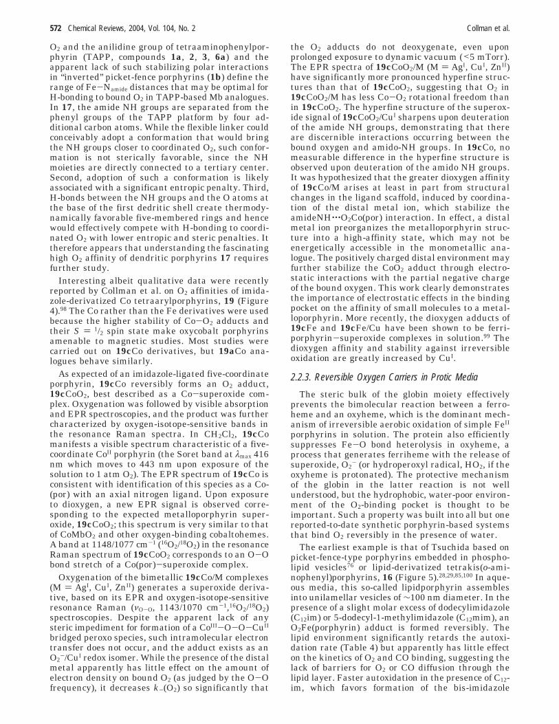
Table 4. Reversible O2 Carriers under Protic Conditionsa
solvent p1/2(O2), Torr τ1/2, h ref
Hb, human, R chain H2O pH 7 0.22 >60 715b/dpmc (vesicles)b H2O pH 7 37 >36 7615a(mim) toluene/H2O 1.5 3015b(mim) toluene/H2O 6 3015c(mim) toluene/H2O 3 × 10⁸ 3016a(C12mim) (liposome) H2O pH 7.4 32 50 2916a(C12im) (liposome) H2O pH 7.4 30 (at 37 °C) 17 28, 2916b (as “fibers”) H2O pH 7 25 4 8517b(mim) toluene/H2O 0.016 (in dry toluene) 2 8318 DMF/H2O 0.7 (5 °C) 27
a dpmc, 1,2-bis(myristoyl)-sn-glycerophosphocholine; mim, 1-methylimidazole; dmim, 1,2-dimethylimidazole; C12im, 5-dodec-ylimidazole; C12mim, 5-dodecyl-1-methylimidazole. b 5b:dmpc ratio 1:1000.
Functional Analogues of CcO, Mb, and Hb Chemical Reviews, 2004, Vol. 104, No. 2 573
EtOH under vacuum regenerates the microcrystal-line solid, manifesting the cooperativity.104
Bayer claimed cooperative O2 binding to a heme-containing polymer,105 but these results are nowconsidered erroneous, mainly because the reportedUV-vis spectra of what was thought to be dioxygenadducts do not correspond to spectra of any knowndioxygenated Fe porphyrins.22,103
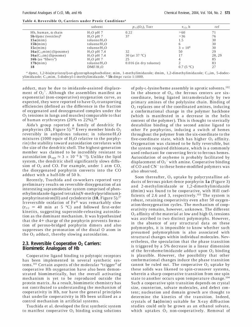
Cooperative binding of O2 to the Co and of CO tothe Fe derivatives of the so-called “gable” porphyrinwas reported by Tabushi and co-workers.106-108 Co-operativity appears to arise from perturbation, by O2or CO (X), of an equilibrium between metalloporphy-rins containing one and two bisimidazolylmethane(bim) ligands (Figure 9). In 20(bim), bim constrainsthe metal ions into the out-of-plane position, whichdisfavors the formation of six-coordinate sites. Thisdecreases the binding affinity of X (K1), because thein-plane movement of the metal necessary for theformation of a six-coordinate site introduces strain(estimated to be ∼1.2 kcal/mol for the Co/O2 system)into the molecule. This additional strain is relaxedin equilibrium 4, resulting in K3 < K4. As a result ofthis difference, a range of bim concentrations existswhere compounds 20(bim) and 20(bim)2X are favoredover 20(bim)2 and 20(bim)X in equilibria 3 and 4,respectively. Because K1 < K2′, under these conditionsformation of 20(bim)2X2 from 20(bim) is cooperative.At sufficiently low concentrations of bim, equilibrium2 remains more favorable relative to the sequence ofequilibria 2′ and 4, despite K2′ > K2, and formationof 20(bim)2X2 is anti-cooperative. Likewise, at highbim concentrations, equilibrium 3 becomes impor-tant, so that 20(bim)2 is substantially populated, and20(bim)2X2 is formed from 20(bim) non-cooperatively,primarily via equilibria 3, 1′, and 2′. Such concentra-tion dependence was indeed observed experimentally.Depending on the values of K3 and K4, as much as100-fold excess of bim is required to observe coopera-
tive binding. Thus, even though cooperativity in gableporphyrins partially mimics the “trigger” mechanismof cooperativity in Hb, it is not an adequate biomi-metic analogue of such a mechanism because of itsbasis in additional ligation equilibria.
3. Functional Analogues of the Heme/CuB Site ofCytochrome c Oxidase
3.1. The EnzymeCytochrome c and ubiquinol oxidases belong to a
superfamily of heme/Cu terminal oxidases,14 enzymesresponsible for coupling the oxidation of ferrocyto-chrome c or ubiquinol, respectively, to the 4e-/4H+
reduction of molecular oxygen. This is the final stepin respiration, ferrocytochrome c and ubiquinol beingthe most oxidizing electron carriers of the respiratorychains in eukaryotes and certain prokaryotes, re-spectively. In addition to clearing the respiratorychain of low-energy electrons, heme/Cu terminaloxidases also contribute to maintaining the trans-membrane electrochemical proton gradient by ac-tively translocating protons. No attempts have beenmade to mimic the “proton pumping” capacity ofheme/Cu terminal oxidases, and this aspect of theenzymatic reactivity will not be discussed in thisreview.
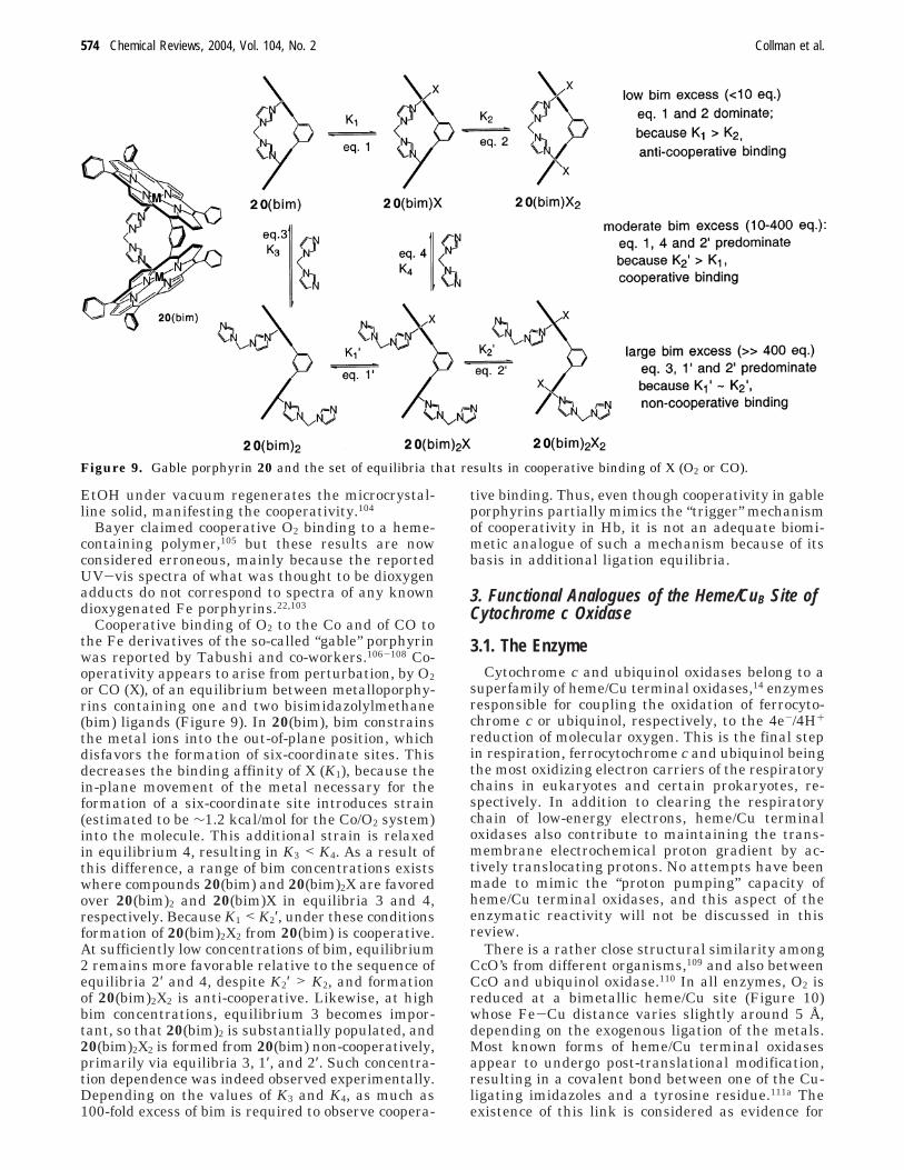
There is a rather close structural similarity amongCcO’s from different organisms,109 and also betweenCcO and ubiquinol oxidase.110 In all enzymes, O2 isreduced at a bimetallic heme/Cu site (Figure 10)whose Fe-Cu distance varies slightly around 5 Å,depending on the exogenous ligation of the metals.Most known forms of heme/Cu terminal oxidasesappear to undergo post-translational modification,resulting in a covalent bond between one of the Cu-ligating imidazoles and a tyrosine residue.111a Theexistence of this link is considered as evidence for
Figure 9. Gable porphyrin 20 and the set of equilibria that results in cooperative binding of X (O2 or CO).
574 Chemical Reviews, 2004, Vol. 104, No. 2 Collman et al.
the formation of the tyrosine radical during catalyticturnover.111b In addition to the bimetallic site, heme/Cu terminal oxidases contain between one and threeother redox cofactors, such as bimetallic CuA andheme(s), whose functions are electron storage andelectron transport from the external reductant to thecatalytic site.
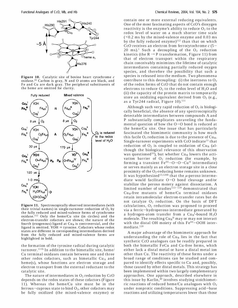
The nature of intermediates in O2 reduction by CcOdepends on the redox states of these cofactors (Figure11). Whereas the heme/Cu site must be in theferrous-cuprous state to bind O2, other cofactors maybe fully oxidized (the mixed-valence enzyme) or
contain one or more external reducing equivalents.One of the most fascinating aspects of CcO’s dioxygenreactivity is the enzyme’s ability to reduce O2 to theredox level of water on a much shorter time scale(<0.2 ms by the mixed-valence enzyme and 0.03 msby the fully reduced enzyme)112 than that on whichCcO receives an electron from ferrocytochrome c (5-20 ms).1 Such a decoupling of the O2 reductionkinetics (the R f P transformation, Figure 11) fromthat of electron transport within the respiratorychain conceivably minimizes the lifetime of catalyticintermediates containing partially reduced oxygenspecies, and therefore the possibility that such aspecies is released into the medium. Two phenomenacontribute to this decoupling: (i) the inertness to O2of the redox forms of CcO that do not contain enoughelectrons to reduce O2 to the redox level of H2O and(ii) the capacity of the protein matrix to temporarilystore an oxidizing equivalent derived from O2 (e.g.,as a Tyr244 radical, Figure 10).111
Although such very rapid reduction of O2 is biologi-cally beneficial, the absence of any spectroscopicallydetectable intermediates between compounds A andP substantially complicates unraveling the funda-mental question of how the O-O bond is reduced atthe heme/Cu site. One issue that has particularlyfascinated the biomimetic community is how muchthis facile O2 reduction is due to the presence of CuB.Single-turnover experiments with CcO indicate11 thatreduction of O2 is coupled to oxidation of CuB (al-though the biological relevance of this observationwas questioned33), but whether CuB lowers the acti-vation barrier of O2 reduction (for example, byforming a transient FeIII-O-O-CuII intermediate)or serves mainly as an electron storage site in a closeproximity of the O2-reducing heme remains unknown.It was hypothesized13,126b that the µ-peroxo interme-diate would facilitate O-O bond cleavage and/orstabilize the peroxo moiety against dissociation. Alimited number of studies113-119 demonstrated thatCuB-free mutants of heme/Cu terminal oxidasesretain intramolecular electron-transfer rates but donot catalyze O2 reduction. On the basis of DFTcalculations, O2 reduction was proposed to proceedvia a ferric-hydroperoxo intermediate generated bya hydrogen-atom transfer from a CuB
I-bound H2Omolecule. The resulting CuB
II may or may not interactwith the distal OH moiety of this FeIII-OOH inter-mediate.120
A major advantage of the biomimetic approach forunderstanding the role of CuB lies in the fact thatsynthetic CcO analogues can be readily prepared inboth the bimetallic FeCu and Cu-free forms, whicheither lack a distal metal or have a distal metal ionother than Cu. The reactivity of these forms under abroad range of conditions can be studied and com-pared to identify effects specific to Cu and, possibly,those caused by other distal metals. This strategy hasbeen implemented within two largely complementaryapproaches. One approach, described elsewhere inthis issue by Karlin,40 involves studying stoichiomet-ric reactions of reduced heme/Cu analogues with O2under nonprotic conditions. Suppressing acid-basereactions and utilizing temperatures lower than those
Figure 10. Catalytic site of bovine heart cytochrome coxidase.35 Carbon is gray, N and O atoms are black, andFe and Cu are dark gray. The peripheral substituents ofthe heme are omitted for clarity.
Figure 11. Spectroscopically observed intermediates (withtheir trivial names) in single-turnover reduction of O2 bythe fully reduced and mixed-valence forms of cytochromeoxidase.112 Only the heme/Cu site (in circles) and theelectron-transfer cofactors are shown; the nature of thefourth (exogenous) ligand at CuB is controversial, and theligand is omitted. YOH ) tyrosine. Cofactors whose redoxstates are different in corresponding intermediates derivedfrom the fully reduced and mixed-valence forms arehighlighted in bold.
Functional Analogues of CcO, Mb, and Hb Chemical Reviews, 2004, Vol. 104, No. 2 575

available in aqueous solutions often allows detectionand/or isolation of intermediates that are too short-lived to be observed when the enzyme reacts with O2.This impetus is reminiscent of one of the objectivesof enzymology in organic solvents.121,122 The majorlimitation of this approach as applied to biomimeticstudies is that the availability of protons in aqueousmedia may fundamentally change the dioxygen re-activity of the FeCu core. For example, a negativelycharged distal oxygen in a putative ferric-peroxointermediate can be stabilized by protonation (form-ing the ferric-hydroperoxo species, which is a pro-posed intermediate in the CcO catalytic cycle) ratherthan coordination to Cu (yielding a bridged peroxoderivative, which is commonly observed in biomi-metic studies).
The alternative approach, reviewed in detail here,has been to study electrochemical O2 reductioncatalyzed by synthetic heme/Cu analogues. In suchstudies, biologically relevant conditions of pH, elec-trochemical potential, and electron flux can be re-produced, and the reactivity can be studied understeady-state turnover. Unfortunately, this methodmakes the use of spectroscopic techniques to char-acterize the system during catalysis extremely chal-lenging; so far, electrocatalytic studies have been akinetic method.
3.2. Methodology of Electrocatalytic Studies ofHeme/Cu Analogues
Electrochemistry is particularly fitting for studyingcatalysis of a redox reaction such as reduction of O2.Several electrochemical methods are used in bio-chemistry to assay activity of CcO preparations.123
The technique that is especially suitable for studyingcatalytic behavior of biomimetic heme/Cu analoguesis rotating ring-disk votammetry,124 wherein theelectrode serves both as a source of electrons and asa catalyst support.37 A water-insoluble catalyst isdeposited, either alone or as a mixture with a matrixcompound, on the disk (Figure 12), and the assembly
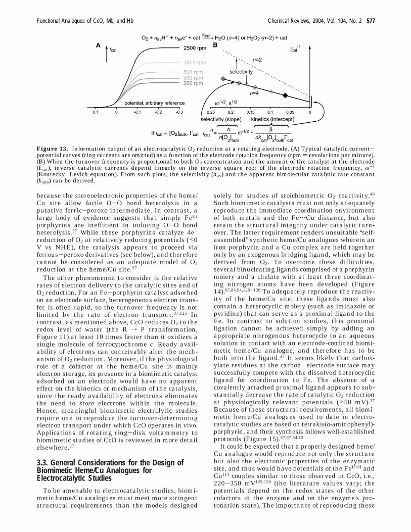
is brought into contact with an aqueous bufferedsolution. Rotation of the electrode generates a flowof the solution to the electrode, delivering the reac-tants to the catalyst. The products of the catalysisdiffuse (or are hydrodynamically transported) to thering electrode, set at a potential where H2O2 israpidly oxidized to O2 (usually between 0.8 and 1 Vvs NHE). If the catalyst is less than 100% selectivetoward 4e- O2 reduction, i.e., if it reduces a fractionof O2 only to H2O2, this fraction can be quantified bycomparing the disk and ring currents. Analysis of thecatalytic currents as a function of the electroderotation frequency (Figure 13) and the substrateconcentration allows quantification of both catalytickinetics and catalytic selectivity (often presented asthe average number of electrons by which one O2molecule is reduced, nav). In the regime wherecatalytic currents are directly proportional to boththe substrate concentration and the amount of thecatalyst at the electrode, the Koutecky-Levich equa-tion provides an adequate general description of theelectrode kinetics (Figure 13).37,125 The slope of a plotof the inverse catalytic currents vs the inversesquare-root of the electrode rotation frequency yieldsnav, and its intercept is proportional to the apparentbimolecular catalytic rate constant.124 Importantmechanistic information is available from an analysisof catalytic currents as a function of the pH.
To maximize the biological relevance of the datacollected in such biomimetic electrocatalytic studies,two phenomena must be kept in mind. One is the factthat the kinetics, selectivity, and mechanism of O2reduction often depend strongly on the electrochemi-cal potential, with the 4e- pathway becoming in-creasing more facile at more reducing potentials. Atpotentials more reducing than those at which heme/Cu terminal oxidases operate in vivo (>50 mV vsNHE), biomimetic catalysis may proceed via inter-mediates that are not biologically relevant.37 Forexample, CcO can couple oxidation of ferrocytochromec (E°(FeIII/II) ≈ 250 mV) to reduction of O2, in part
Figure 12. Rotating ring-disk electrode (RRDE). (A) A typical commercially available RRDE. (B) Schematic representationof operating RRDE: rotation of the electrode (block arrow) generates a flow of electrolyte containing the reactants (solidarrows) to the catalyst and sweeps the products of catalysis by the ring electrode. If the catalyst produces H2O2 (or O2
-),a portion of it gets oxidized at the ring, producing current (broken arrow).
576 Chemical Reviews, 2004, Vol. 104, No. 2 Collman et al.
because the stereoelectronic properties of the heme/Cu site allow facile O-O bond heterolysis in aputative ferric-peroxo intermediate. In contrast, alarge body of evidence suggests that simple FeIII
porphyrins are inefficient in inducing O-O bondheterolysis.37 While these porphyrins catalyze 4e-
reduction of O2 at relatively reducing potentials (<0V vs NHE), the catalysis appears to proceed viaferrous-peroxo derivatives (see below), and thereforecannot be considered as an adequate model of O2reduction at the heme/Cu site.37
The other phenomenon to consider is the relativerates of electron delivery to the catalytic sites and ofO2 reduction. For an Fe-porphyrin catalyst adsorbedon an electrode surface, heterogeneous electron trans-fer is often rapid, so the turnover frequency is notlimited by the rate of electron transport.37,125 Incontrast, as mentioned above, CcO reduces O2 to theredox level of water (the R f P transformation,Figure 11) at least 10 times faster than it oxidizes asingle molecule of ferrocytochrome c. Ready avail-ability of electrons can conceivably alter the mech-anism of O2 reduction. Moreover, if the physiologicalrole of a cofactor at the heme/Cu site is mainlyelectron storage, its presence in a biomimetic catalystadsorbed on an electrode would have no apparenteffect on the kinetics or mechanism of the catalysis,since the ready availability of electrons eliminatesthe need to store electrons within the molecule.Hence, meaningful biomimetic electrolytic studiesrequire one to reproduce the turnover-determiningelectron transport under which CcO operates in vivo.Applications of rotating ring-disk voltammetry tobiomimetic studies of CcO is reviewed in more detailelsewhere.37
3.3. General Considerations for the Design ofBiomimetic Heme/Cu Analogues forElectrocatalytic Studies
To be amenable to electrocatalytic studies, biomi-metic heme/Cu analogues must meet more stringentstructural requirements than the models designed
solely for studies of stoichiometric O2 reactivity.40
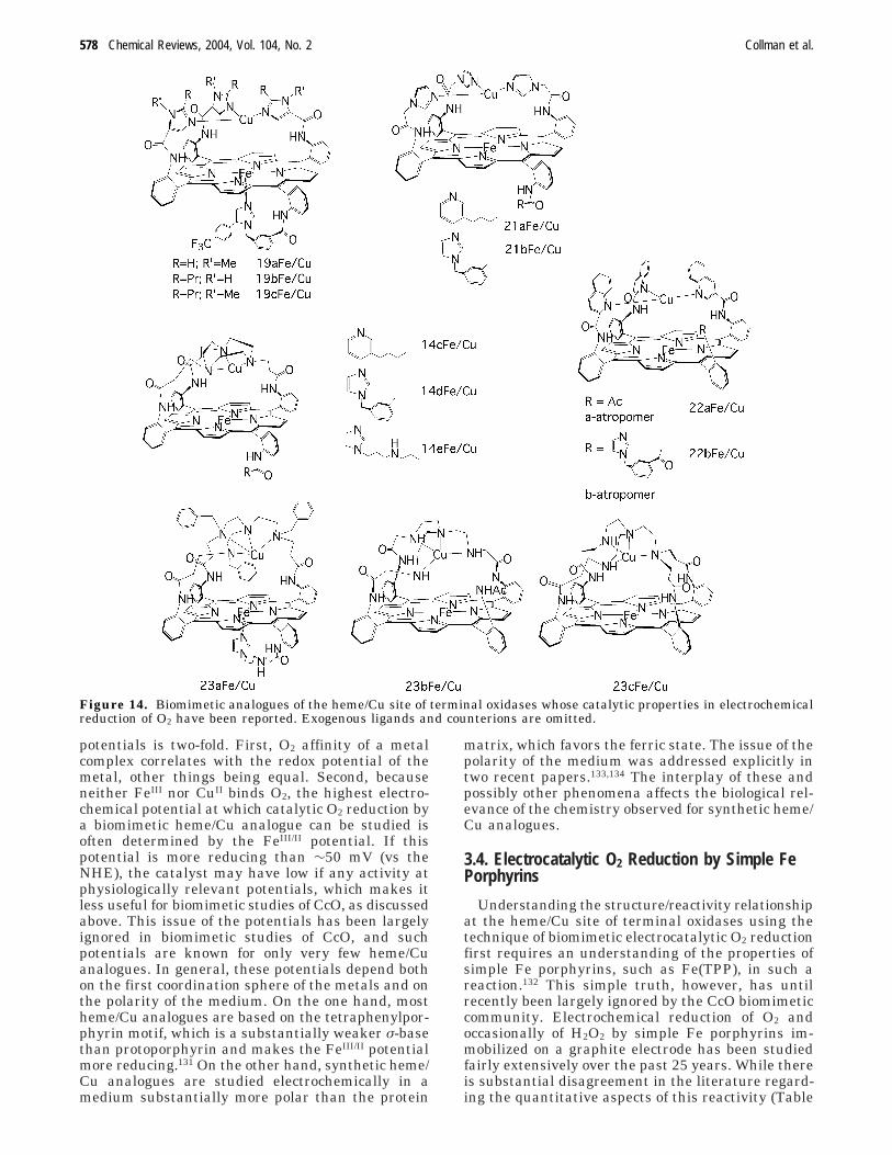
Such biomimetic catalysts must not only adequatelyreproduce the immediate coordination environmentof both metals and the Fe‚‚‚Cu distance, but alsoretain the structural integrity under catalytic turn-over. The latter requirement renders unsuitable “self-assembled” synthetic heme/Cu analogues wherein aniron porphyrin and a Cu complex are held togetheronly by an exogenous bridging ligand, which may bederived from O2. To overcome these difficulties,several binucleating ligands comprised of a porphyrinmoiety and a chelate with at least three coordinat-ing nitrogen atoms have been developed (Figure14).67,84,94,126-128 To adequately reproduce the reactiv-ity of the heme/Cu site, these ligands must alsocontain a heterocyclic moiety (such as imidazole orpyridine) that can serve as a proximal ligand to theFe. In contrast to solution studies, this proximalligation cannot be achieved simply by adding anappropriate nitrogenous heterocycle to an aqueoussolution in contact with an electrode-confined biomi-metic heme/Cu analogue, and therefore has to bebuilt into the ligand.37 It seems likely that carbox-ylate residues at the carbon-electrode surface maysuccessfully compete with the dissolved heterocyclicligand for coordination to Fe. The absence of acovalently attached proximal ligand appears to sub-stantially decrease the rate of catalytic O2 reductionat physiologically relevant potentials (>50 mV).37
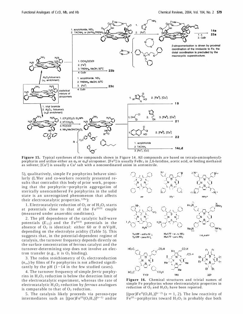
Because of these structural requirements, all biomi-metic heme/Cu analogues used to date in electro-catalytic studies are based on tetrakis(o-aminophenyl)-porphyrin, and their synthesis follows well-establishedprotocols (Figure 15).37,67,84,12
It could be expected that a properly designed heme/Cu analogue would reproduce not only the structurebut also the electronic properties of the enzymaticsite, and thus would have potentials of the FeIII/II andCuII/I couples similar to those observed in CcO, i.e.,220-350 mV129,130 (the literature values vary; thepotentials depend on the redox states of the othercofactors in the enzyme and on the enzyme’s pro-tonation state). The importance of reproducing these
Figure 13. Information output of an electrocatalytic O2 reduction at a rotating electrode. (A) Typical catalytic current-potential curves (ring currents are omitted) as a function of the electrode rotation frequency (rpm ) revolutions per minute).(B) When the turnover frequency is proportional to both O2 concentration and the amount of the catalyst at the electrode(Γcat), inverse catalytic currents depend linearly on the inverse square root of the electrode rotation frequency, ω-1
(Koutecky-Levich equation). From such plots, the selectivity (nav) and the apparent bimolecular catalytic rate constant(kapp) can be derived.
Functional Analogues of CcO, Mb, and Hb Chemical Reviews, 2004, Vol. 104, No. 2 577
potentials is two-fold. First, O2 affinity of a metalcomplex correlates with the redox potential of themetal, other things being equal. Second, becauseneither FeIII nor CuII binds O2, the highest electro-chemical potential at which catalytic O2 reduction bya biomimetic heme/Cu analogue can be studied isoften determined by the FeIII/II potential. If thispotential is more reducing than ∼50 mV (vs theNHE), the catalyst may have low if any activity atphysiologically relevant potentials, which makes itless useful for biomimetic studies of CcO, as discussedabove. This issue of the potentials has been largelyignored in biomimetic studies of CcO, and suchpotentials are known for only very few heme/Cuanalogues. In general, these potentials depend bothon the first coordination sphere of the metals and onthe polarity of the medium. On the one hand, mostheme/Cu analogues are based on the tetraphenylpor-phyrin motif, which is a substantially weaker σ-basethan protoporphyrin and makes the FeIII/II potentialmore reducing.131 On the other hand, synthetic heme/Cu analogues are studied electrochemically in amedium substantially more polar than the protein
matrix, which favors the ferric state. The issue of thepolarity of the medium was addressed explicitly intwo recent papers.133,134 The interplay of these andpossibly other phenomena affects the biological rel-evance of the chemistry observed for synthetic heme/Cu analogues.
3.4. Electrocatalytic O2 Reduction by Simple FePorphyrins
Understanding the structure/reactivity relationshipat the heme/Cu site of terminal oxidases using thetechnique of biomimetic electrocatalytic O2 reductionfirst requires an understanding of the properties ofsimple Fe porphyrins, such as Fe(TPP), in such areaction.132 This simple truth, however, has untilrecently been largely ignored by the CcO biomimeticcommunity. Electrochemical reduction of O2 andoccasionally of H2O2 by simple Fe porphyrins im-mobilized on a graphite electrode has been studiedfairly extensively over the past 25 years. While thereis substantial disagreement in the literature regard-ing the quantitative aspects of this reactivity (Table
Figure 14. Biomimetic analogues of the heme/Cu site of terminal oxidases whose catalytic properties in electrochemicalreduction of O2 have been reported. Exogenous ligands and counterions are omitted.
578 Chemical Reviews, 2004, Vol. 104, No. 2 Collman et al.
5), qualitatively, simple Fe porphyrins behave simi-larly (L’Her and co-workers recently presented re-sults that contradict this body of prior work, propos-ing that the porphyrin-porphyrin aggregation ofsterically unencumbered Fe porphyrins in the solidstate is an unrecognized phenomenon that affectstheir electrocatalytic properties.128a):
1. Electrocatalytic reduction of O2 or of H2O2 startsat potentials close to that of the FeIII/II couple(measured under anaerobic conditions).
2. The pH dependence of the catalytic half-wavepotentials (E1/2) and the FeIII/II potentials in theabsence of O2 is identical: either 60 or 0 mV/pH,depending on the electrolyte acidity (Table 5). Thissuggests that, in the potential-dependent regime ofcatalysis, the turnover frequency depends directly onthe surface concentration of ferrous catalyst and theturnover-determining step does not involve an elec-tron transfer (e.g., it is O2 binding).
3. The redox stoichiometry of O2 electroreduction(nav) by films of Fe porphyrins is not affected signifi-cantly by the pH (1-14 in the few studied cases).
4. The turnover frequency of simple ferric porphy-rins in H2O2 reduction is below the detection limit ofthe electrocatalytic experiment, whereas the rate ofelectrocatalytic H2O2 reduction by ferrous analoguesis comparable to that of O2 reduction.
5. The catalysis likely proceeds via peroxo-typeintermediates such as [(por)FeIII(O2Hx)](x-1) and/or
[(por)FeII(O2Hx)](x-2) (x ) 1, 2). The low reactivity ofFeIII-porphyrins toward H2O2 is probably due both
Figure 15. Typical syntheses of the compounds shown in Figure 14. All compounds are based on tetra(o-aminophenyl)-porphyrin and utilize either an R4 or R3â atropomer. [FeII] is usually FeBr2 in 2,6-lutidine, acetic acid, or boiling methanolas solvent; [CuI] is usually a CuI salt with a noncoordinated anion in acetonitrile.
Figure 16. Chemical structures and trivial names ofsimple Fe porphyrins whose electrocatalytic properties inreduction of O2 and H2O2 have been reported.
Functional Analogues of CcO, Mb, and Hb Chemical Reviews, 2004, Vol. 104, No. 2 579
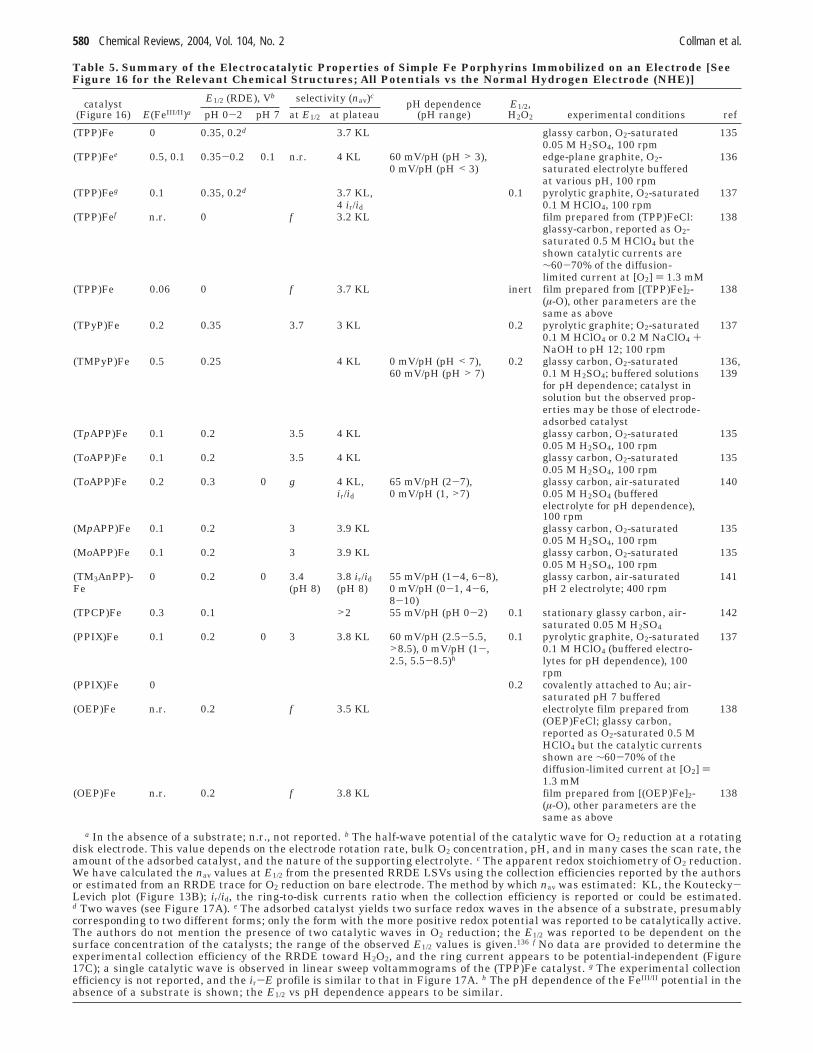
Table 5. Summary of the Electrocatalytic Properties of Simple Fe Porphyrins Immobilized on an Electrode [SeeFigure 16 for the Relevant Chemical Structures; All Potentials vs the Normal Hydrogen Electrode (NHE)]
E1/2 (RDE), Vb selectivity (nav)ccatalyst
(Figure 16) E(FeIII/II)a pH 0-2 pH 7 at E1/2 at plateaupH dependence
(pH range)E1/2,H2O2 experimental conditions ref
(TPP)Fe 0 0.35, 0.2d 3.7 KL glassy carbon, O2-saturated 1350.05 M H2SO4, 100 rpm
(TPP)Fee 0.5, 0.1 0.35-0.2 0.1 n.r. 4 KL 60 mV/pH (pH > 3), edge-plane graphite, O2- 1360 mV/pH (pH < 3) saturated electrolyte buffered
at various pH, 100 rpm(TPP)Feg 0.1 0.35, 0.2d 3.7 KL, 0.1 pyrolytic graphite, O2-saturated 137
4 ir/id 0.1 M HClO4, 100 rpm(TPP)Fef n.r. 0 f 3.2 KL film prepared from (TPP)FeCl: 138
glassy-carbon, reported as O2-saturated 0.5 M HClO4 but theshown catalytic currents are∼60-70% of the diffusion-limited current at [O2] ) 1.3 mM
(TPP)Fe 0.06 0 f 3.7 KL inert film prepared from [(TPP)Fe]2- 138(µ-O), other parameters are thesame as above
(TPyP)Fe 0.2 0.35 3.7 3 KL 0.2 pyrolytic graphite; O2-saturated 1370.1 M HClO4 or 0.2 M NaClO4 +NaOH to pH 12; 100 rpm
(TMPyP)Fe 0.5 0.25 4 KL 0 mV/pH (pH < 7), 0.2 glassy carbon, O2-saturated 136,60 mV/pH (pH > 7) 0.1 M H2SO4; buffered solutions 139
for pH dependence; catalyst insolution but the observed prop-erties may be those of electrode-adsorbed catalyst
(TpAPP)Fe 0.1 0.2 3.5 4 KL glassy carbon, O2-saturated 1350.05 M H2SO4, 100 rpm
(ToAPP)Fe 0.1 0.2 3.5 4 KL glassy carbon, O2-saturated 1350.05 M H2SO4, 100 rpm
(ToAPP)Fe 0.2 0.3 0 g 4 KL, 65 mV/pH (2-7), glassy carbon, air-saturated 140ir/id 0 mV/pH (1, >7) 0.05 M H2SO4 (buffered
electrolyte for pH dependence),100 rpm
(MpAPP)Fe 0.1 0.2 3 3.9 KL glassy carbon, O2-saturated 1350.05 M H2SO4, 100 rpm
(MoAPP)Fe 0.1 0.2 3 3.9 KL glassy carbon, O2-saturated 1350.05 M H2SO4, 100 rpm
(TM3AnPP)- 0 0.2 0 3.4 3.8 ir/id 55 mV/pH (1-4, 6-8), glassy carbon, air-saturated 141Fe (pH 8) (pH 8) 0 mV/pH (0-1, 4-6, pH 2 electrolyte; 400 rpm
8-10)(TPCP)Fe 0.3 0.1 >2 55 mV/pH (pH 0-2) 0.1 stationary glassy carbon, air- 142
saturated 0.05 M H2SO4(PPIX)Fe 0.1 0.2 0 3 3.8 KL 60 mV/pH (2.5-5.5, 0.1 pyrolytic graphite, O2-saturated 137
>8.5), 0 mV/pH (1-, 0.1 M HClO4 (buffered electro-2.5, 5.5-8.5)h lytes for pH dependence), 100
rpm(PPIX)Fe 0 0.2 covalently attached to Au; air-
saturated pH 7 buffered(OEP)Fe n.r. 0.2 f 3.5 KL electrolyte film prepared from 138
(OEP)FeCl; glassy carbon,reported as O2-saturated 0.5 MHClO4 but the catalytic currentsshown are ∼60-70% of thediffusion-limited current at [O2] )1.3 mM
(OEP)Fe n.r. 0.2 f 3.8 KL film prepared from [(OEP)Fe]2- 138(µ-O), other parameters are thesame as above
a In the absence of a substrate; n.r., not reported. b The half-wave potential of the catalytic wave for O2 reduction at a rotatingdisk electrode. This value depends on the electrode rotation rate, bulk O2 concentration, pH, and in many cases the scan rate, theamount of the adsorbed catalyst, and the nature of the supporting electrolyte. c The apparent redox stoichiometry of O2 reduction.We have calculated the nav values at E1/2 from the presented RRDE LSVs using the collection efficiencies reported by the authorsor estimated from an RRDE trace for O2 reduction on bare electrode. The method by which nav was estimated: KL, the Koutecky-Levich plot (Figure 13B); ir/id, the ring-to-disk currents ratio when the collection efficiency is reported or could be estimated.d Two waves (see Figure 17A). e The adsorbed catalyst yields two surface redox waves in the absence of a substrate, presumablycorresponding to two different forms; only the form with the more positive redox potential was reported to be catalytically active.The authors do not mention the presence of two catalytic waves in O2 reduction; the E1/2 was reported to be dependent on thesurface concentration of the catalysts; the range of the observed E1/2 values is given.136 f No data are provided to determine theexperimental collection efficiency of the RRDE toward H2O2, and the ring current appears to be potential-independent (Figure17C); a single catalytic wave is observed in linear sweep voltammograms of the (TPP)Fe catalyst. g The experimental collectionefficiency is not reported, and the ir-E profile is similar to that in Figure 17A. h The pH dependence of the FeIII/II potential in theabsence of a substrate is shown; the E1/2 vs pH dependence appears to be similar.
580 Chemical Reviews, 2004, Vol. 104, No. 2 Collman et al.
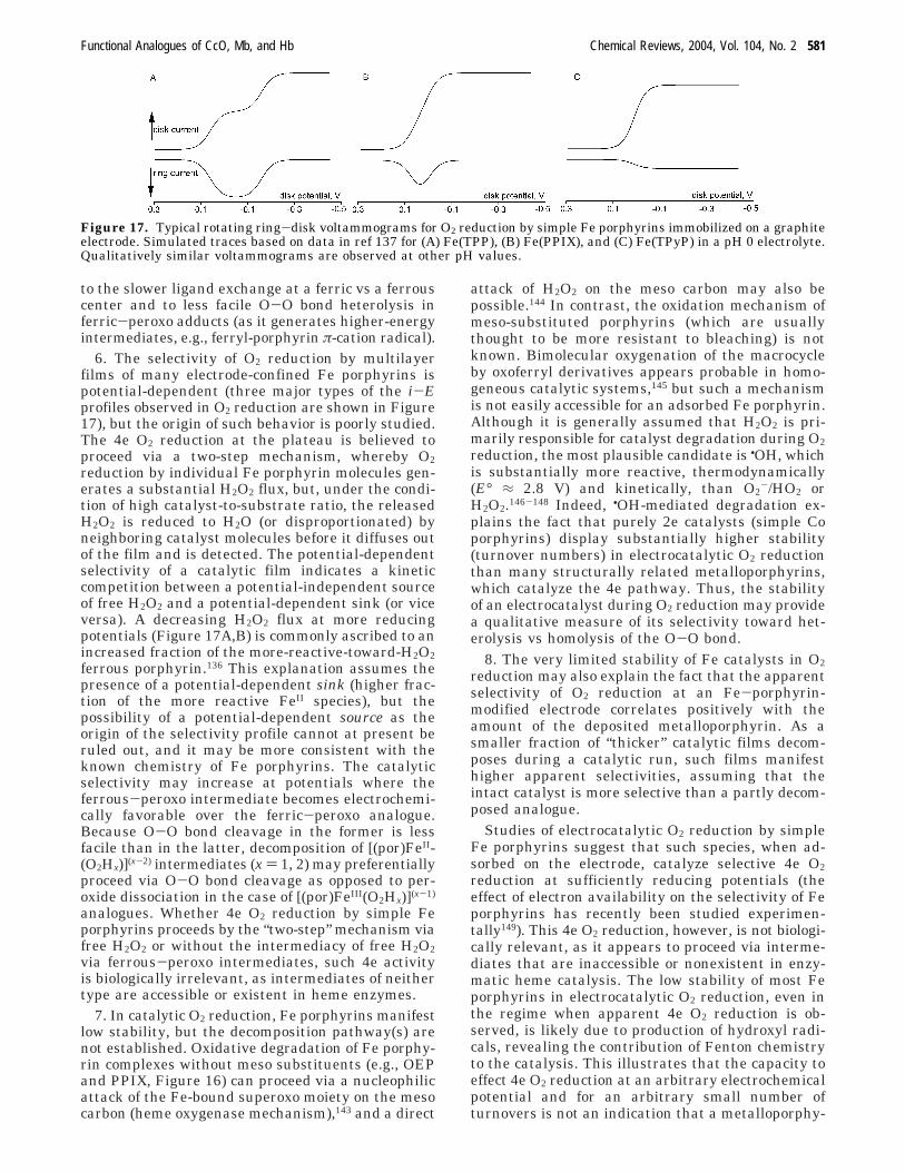
to the slower ligand exchange at a ferric vs a ferrouscenter and to less facile O-O bond heterolysis inferric-peroxo adducts (as it generates higher-energyintermediates, e.g., ferryl-porphyrin π-cation radical).
6. The selectivity of O2 reduction by multilayerfilms of many electrode-confined Fe porphyrins ispotential-dependent (three major types of the i-Eprofiles observed in O2 reduction are shown in Figure17), but the origin of such behavior is poorly studied.The 4e O2 reduction at the plateau is believed toproceed via a two-step mechanism, whereby O2reduction by individual Fe porphyrin molecules gen-erates a substantial H2O2 flux, but, under the condi-tion of high catalyst-to-substrate ratio, the releasedH2O2 is reduced to H2O (or disproportionated) byneighboring catalyst molecules before it diffuses outof the film and is detected. The potential-dependentselectivity of a catalytic film indicates a kineticcompetition between a potential-independent sourceof free H2O2 and a potential-dependent sink (or viceversa). A decreasing H2O2 flux at more reducingpotentials (Figure 17A,B) is commonly ascribed to anincreased fraction of the more-reactive-toward-H2O2ferrous porphyrin.136 This explanation assumes thepresence of a potential-dependent sink (higher frac-tion of the more reactive FeII species), but thepossibility of a potential-dependent source as theorigin of the selectivity profile cannot at present beruled out, and it may be more consistent with theknown chemistry of Fe porphyrins. The catalyticselectivity may increase at potentials where theferrous-peroxo intermediate becomes electrochemi-cally favorable over the ferric-peroxo analogue.Because O-O bond cleavage in the former is lessfacile than in the latter, decomposition of [(por)FeII-(O2Hx)](x-2) intermediates (x ) 1, 2) may preferentiallyproceed via O-O bond cleavage as opposed to per-oxide dissociation in the case of [(por)FeIII(O2Hx)](x-1)
analogues. Whether 4e O2 reduction by simple Feporphyrins proceeds by the “two-step” mechanism viafree H2O2 or without the intermediacy of free H2O2via ferrous-peroxo intermediates, such 4e activityis biologically irrelevant, as intermediates of neithertype are accessible or existent in heme enzymes.
7. In catalytic O2 reduction, Fe porphyrins manifestlow stability, but the decomposition pathway(s) arenot established. Oxidative degradation of Fe porphy-rin complexes without meso substituents (e.g., OEPand PPIX, Figure 16) can proceed via a nucleophilicattack of the Fe-bound superoxo moiety on the mesocarbon (heme oxygenase mechanism),143 and a direct
attack of H2O2 on the meso carbon may also bepossible.144 In contrast, the oxidation mechanism ofmeso-substituted porphyrins (which are usuallythought to be more resistant to bleaching) is notknown. Bimolecular oxygenation of the macrocycleby oxoferryl derivatives appears probable in homo-geneous catalytic systems,145 but such a mechanismis not easily accessible for an adsorbed Fe porphyrin.Although it is generally assumed that H2O2 is pri-marily responsible for catalyst degradation during O2
reduction, the most plausible candidate is •OH, whichis substantially more reactive, thermodynamically(E° ≈ 2.8 V) and kinetically, than O2
-/HO2 orH2O2.146-148 Indeed, •OH-mediated degradation ex-plains the fact that purely 2e catalysts (simple Coporphyrins) display substantially higher stability(turnover numbers) in electrocatalytic O2 reductionthan many structurally related metalloporphyrins,which catalyze the 4e pathway. Thus, the stabilityof an electrocatalyst during O2 reduction may providea qualitative measure of its selectivity toward het-erolysis vs homolysis of the O-O bond.
8. The very limited stability of Fe catalysts in O2
reduction may also explain the fact that the apparentselectivity of O2 reduction at an Fe-porphyrin-modified electrode correlates positively with theamount of the deposited metalloporphyrin. As asmaller fraction of “thicker” catalytic films decom-poses during a catalytic run, such films manifesthigher apparent selectivities, assuming that theintact catalyst is more selective than a partly decom-posed analogue.
Studies of electrocatalytic O2 reduction by simpleFe porphyrins suggest that such species, when ad-sorbed on the electrode, catalyze selective 4e O2
reduction at sufficiently reducing potentials (theeffect of electron availability on the selectivity of Feporphyrins has recently been studied experimen-tally149). This 4e O2 reduction, however, is not biologi-cally relevant, as it appears to proceed via interme-diates that are inaccessible or nonexistent in enzy-matic heme catalysis. The low stability of most Feporphyrins in electrocatalytic O2 reduction, even inthe regime when apparent 4e O2 reduction is ob-served, is likely due to production of hydroxyl radi-cals, revealing the contribution of Fenton chemistryto the catalysis. This illustrates that the capacity toeffect 4e O2 reduction at an arbitrary electrochemicalpotential and for an arbitrary small number ofturnovers is not an indication that a metalloporphy-
Figure 17. Typical rotating ring-disk voltammograms for O2 reduction by simple Fe porphyrins immobilized on a graphiteelectrode. Simulated traces based on data in ref 137 for (A) Fe(TPP), (B) Fe(PPIX), and (C) Fe(TPyP) in a pH 0 electrolyte.Qualitatively similar voltammograms are observed at other pH values.
Functional Analogues of CcO, Mb, and Hb Chemical Reviews, 2004, Vol. 104, No. 2 581
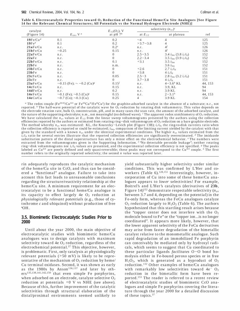
rin adequately reproduces the catalytic mechanismof the heme/Cu site in CcO, and thus can be consid-ered a “functional” analogue. Failure to take intoaccount this fact leads to unreasonable conclusionsregarding the structure/reactivity relationship at theheme/Cu site. A minimum requirement for an elec-trocatalyst to be a functional heme/Cu analogue isits capacity to effect largely 4e O2 reduction atphysiologically relevant potentials (e.g., those of cy-tochrome c and ubiquinol) without production of free•OH.
3.5. Biomimetic Electrocatalytic Studies Prior to2000
Until about the year 2000, the main objective ofelectrocatalytic studies with biomimetic heme/Cuanalogues was to design catalysts with maximumselectivity toward 4e O2 reduction, regardless of theelectrochemical potential.37 This objective, however,is problematic. First, only catalysis at physiologicallyrelevant potentials (>50 mV) is likely to be repre-sentative of the mechanism of O2 reduction by heme/Cu terminal oxidases. Second, it was shown as earlyas the 1980s by Anson136,137 and later by oth-ers135,138,141,142,150 that even simple Fe porphyrins,when adsorbed on an electrode, catalyze selective O2
reduction at potentials <0 V vs NHE (see above).Because of this, further improvement of the catalyticselectivities through structural elaboration of thedistal/proximal environments seemed unlikely to
yield substantially higher selectivity under similarconditions. This was confirmed by L’Her and co-workers (Table 6).128,151 Interestingly, however, in-corporation of Cu into some of these heme/Cu ana-logues appears to lower selectivities! For example,Boitrel’s and L’Her’s catalysts (derivatives of 23b,Figure 14)152 demonstrate respectable selectivity (nav
between 3.7 and 4, depending on the potential) in theFe-only form, whereas the FeCu analogues catalyzeO2 reduction largely to H2O2 (Table 6). The authorshypothesized that this startling result arises becausethe “copper center does not interfere with the O2
molecule bound to Fe” or the “copper ion...is no longercoordinated”. It appears more likely, however, thatthe lower apparent selectivity of the FeCu derivativesmay arise from faster degradation of the bimetalliccatalyst relative to the monometallic analogue. Suchrapid degradation of an immobilized Fe porphyrincan conceivably be mediated only by hydroxyl radi-cals, which seems to suggest that Cu coordinated tothese particular ligands facilitates O-O bond ho-molysis either in Fe-bound peroxo species or in freeH2O2, which is generated as a byproduct of O2
reduction.132 Other examples of heme/Cu analogueswith remarkably low selectivities toward 4e- O2
reduction in the bimetallic form have been re-ported.153 The reader is referred to a recent reviewof electrocatalytic studies of biomimetic CcO ana-logues and simple Fe porphyrins covering the litera-ture through the year 2000 for a detailed discussionof these topics.37
Table 6. Electrocatalytic Properties toward O2 Reduction of the Functional Heme/Cu Site Analogues [See Figure14 for the Relevant Chemical Structures; All Potentials vs the Normal Hydrogen Electrode (NHE)]
selectivity (nav)ccatalyst
(Figure 14) Ecat, V aE1/2(O2), Vat pH 7b at E1/2 at plateau ref
19Fe/Cud 0.1 0.2 ∼4 4 12519Fed 0.1 0.2 >3.7-3.8 4 12521aFe/Cu n.r. 0.2e n.r. 4f 12621bFe/Cu ∼0.25 0.15 n.r. 4f 12622aFe/Cu n.r. 0 2.5-3.5 3.4 ilim (3.8 ir/id) 15222aFe n.r. 0 2.2-3.4 3.5 ilim (3.9 ir/id) 15222bFe/Cu n.r. 0.1 <3.1 3.5 ilim 15222bFe n.r. n.m. 3.6 ilim 15223bFe/Cu n.r. 0 <2.6 2.7 ir/id 15123bFe n.r. 0 <3.8 4 ir/id 15123cFe/Cu n.r. 0.05 2.5-3 2.8 ilim (3.2 ir/id) 15123cFe n.r. 0 3.7-3.8 4 ir/id 15123aFe/Cu ∼0.15 (Fe); ∼ -0.2 (Cu)g 0.15f n.r. 4-3.6h KL 44, 15314cFe/Cu n.r. 0.15 n.r. 3.9, KL 9414dFe/Cu n.r. 0.2 n.r. 3.9 KL 9414eFe/Cu ∼0.1 (Fe); ∼0.5 (Cu)g 0.15 n.r. 2-3.1h 44, 15314eCo/Cu ∼0.7 (Co); ∼0.3 (Cu) 0.25 4 3.9 KL 154
a The redox couple (FeIII/II/CuII/I or FeIIICuII/FeIICuI) for the graphite-adsorbed catalyst in the absence of a substrate; n.r., notreported. b The half-wave potential of the catalytic wave for O2 reduction by rotating disk voltammetry. This value depends onthe electrode rotation rate, bulk O2 concentration, pH, and in many cases the scan rate, the amount of the adsorbed catalyst, andthe nature of the supporting electrolyte; n.m., not meaningful (no defined wave). c The apparent redox stoichiometry of O2 reduction.We have calculated the nav values at E1/2 from the linear sweep voltammograms presented by the authors using the collectionefficiencies reported by the authors or estimated from rotating ring-disk voltammogram of O2 reduction on a bare graphite electrode.The method whereby nav was estimated: KL, the Koutecky-Levich plot (Figure 13B); ir/id, the ring-to-disk currents ratio whenthe collection efficiency is reported or could be estimated; ilim, comparison of the limiting current yielded by the catalyst with thatgiven by the standard with a known nav under the identical experimental conditions. The higher nav values estimated from their/id ratio for several entries illustrate that the reported collection efficiencies are significantly overestimated. d The imidazolesubstitution pattern of the distal superstructure has only a minor effect on the electrochemical behavior. e The numbers wereextracted from the voltammograms given in the Supporting Information. f “No detectable peroxide leakage”; neither rotatingring-disk voltammograms nor ir/id values are presented, and the experimental collection efficiency is not specified. g The peakslabeled as CuII/I are poorly defined and only quasi-reversible; these peaks may not correspond to the CuII/I couple. h The firstnumber refers to the originally reported selectivity; the second n value was reported later.
582 Chemical Reviews, 2004, Vol. 104, No. 2 Collman et al.
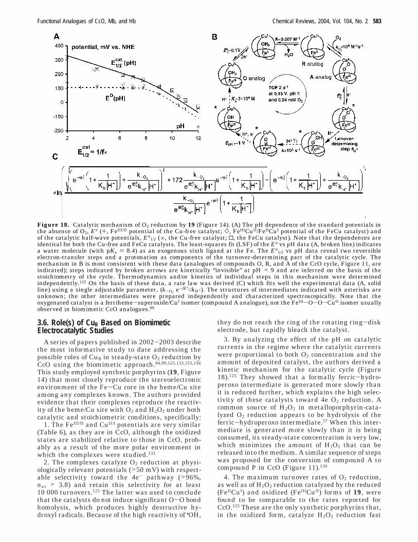
3.6. Role(s) of CuB Based on BiomimeticElectrocatalytic Studies
A series of papers published in 2002-2003 describethe most informative study to date addressing thepossible roles of CuB in steady-state O2 reduction byCcO using the biomimetic approach.84,99,125,133,155,156
This study employed synthetic porphyrins (19, Figure14) that most closely reproduce the stereoelectronicenvironment of the Fe-Cu core in the heme/Cu siteamong any complexes known. The authors providedevidence that their complexes reproduce the reactiv-ity of the heme/Cu site with O2 and H2O2 under bothcatalytic and stoichiometric conditions, specifically:
1. The FeIII/II and CuII/I potentials are very similar(Table 6), as they are in CcO, although the oxidizedstates are stabilized relative to those in CcO, prob-ably as a result of the more polar environment inwhich the complexes were studied.133
2. The complexes catalyze O2 reduction at physi-ologically relevant potentials (>50 mV) with respect-able selectivity toward the 4e- pathway (>96%,nav > 3.8) and retain this selectivity for at least10 000 turnovers.125 The latter was used to concludethat the catalysts do not induce significant O-O bondhomolysis, which produces highly destructive hy-droxyl radicals. Because of the high reactivity of •OH,
they do not reach the ring of the rotating ring-diskelectrode, but rapidly bleach the catalyst.
3. By analyzing the effect of the pH on catalyticcurrents in the regime where the catalytic currentswere proportional to both O2 concentration and theamount of deposited catalyst, the authors derived akinetic mechanism for the catalytic cycle (Figure18).125 They showed that a formally ferric-hydro-peroxo intermediate is generated more slowly thanit is reduced further, which explains the high selec-tivity of these catalysts toward 4e O2 reduction. Acommon source of H2O2 in metalloporphyrin-cata-lyzed O2 reduction appears to be hydrolysis of theferric-hydroperoxo intermediate.37 When this inter-mediate is generated more slowly than it is beingconsumed, its steady-state concentration is very low,which minimizes the amount of H2O2 that can bereleased into the medium. A similar sequence of stepswas proposed for the conversion of compound A tocompound P in CcO (Figure 11).120
4. The maximum turnover rates of O2 reduction,as well as of H2O2 reduction catalyzed by the reduced(FeIICuI) and oxidized (FeIIICuII) forms of 19, werefound to be comparable to the rates reported forCcO.125 These are the only synthetic porphyrins that,in the oxidized form, catalyze H2O2 reduction fast
Figure 18. Catalytic mechanism of O2 reduction by 19 (Figure 14). (A) The pH dependence of the standard potentials inthe absence of O2, E° (+, FeIII/II potential of the Cu-free catalyst; ], FeIIICuII/FeIICuI potential of the FeCu catalyst) andof the catalytic half-wave potentials, E°1/2 (×, the Cu-free catalyst; 0, the FeCu catalyst). Note that the dependences areidentical for both the Cu-free and FeCu catalysts. The least-squares fit (LSF) of the E° vs pH data (A, broken line) indicatesa water molecule (with pKa ) 8.4) as an exogenous sixth ligand at the Fe. The E°1/2 vs pH data reveal two reversibleelectron-transfer steps and a protonation as components of the turnover-determining part of the catalytic cycle. Themechanism in B is most consistent with these data (analogues of compounds O, R, and A of the CcO cycle, Figure 11, areindicated); steps indicated by broken arrows are kinetically “invisible” at pH < 9 and are inferred on the basis of thestoichiometry of the cycle. Thermodynamics and/or kinetics of individual steps in this mechanism were determinedindependently.125 On the basis of these data, a rate law was derived (C) which fits well the experimental data (A, solidline) using a single adjustable parameter, (k-O2 e-fE°2/kH+). The structures of intermediates indicated with asterisks areunknown; the other intermediates were prepared independently and characterized spectroscopically. Note that theoxygenated catalyst is a ferriheme-superoxide/CuI isomer (compound A analogue), not the FeIII-O-O-CuII isomer usuallyobserved in biomimetic CcO analogues.99
Functional Analogues of CcO, Mb, and Hb Chemical Reviews, 2004, Vol. 104, No. 2 583
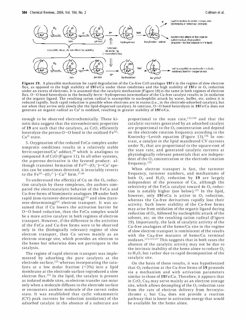
enough to be observed electrochemically. These ki-netic data suggest that the stereoelectronic propertiesof 19 are such that the catalysts, as CcO, efficientlyheterolyze the peroxo O-O bond in the oxidized FeIII-CuII state.
5. Oxygenation of the reduced FeCu complex undernonprotic conditions results in a relatively stableferric-superoxo/CuI adduct,99 which is analogous tocompound A of CcO (Figure 11). In all other systems,the µ-peroxo derivative is the favored product: al-though transient formation of FeIII-(O2
-)‚‚‚CuI spe-cies can be sometimes detected, it invariably revertsto the FeIII-(O2
2-)-CuII form.37,40
To understand the effect(s) of Cu on the O2 reduc-tion catalysis by these complexes, the authors com-pared the electrocatalytic behavior of the FeCu andCu-free forms of biomimetic analogues 19 under bothrapid (non-turnover-determining)125 and slow (turn-over-determining)155 electron transport. It was as-sumed that if Cu lowers the activation barrier forO-O bond reduction, then the FeCu complex wouldbe a more active catalyst in both regimes of electrontransport. However, if the difference in the activitiesof the FeCu and Cu-free forms were to be observedonly in the (biologically relevant) regime of slowelectron transport, then Cu serves mainly as anelectron storage site, which provides an electron tothe heme but otherwise does not participate in thecatalysis.
The regime of rapid electron transport was imple-mented by adsorbing the pure catalysts at theelectrode surface,125 whereas incorporating the cata-lysts at a low molar fraction (<3%) into a lipidmembrane at the electrode surface reproduced a slowelectron flux.155 In the lipid, the catalyst is presentas isolated mobile sites, so electron transfer can occuronly when a molecule diffuses to the electrode surfaceor encounters another molecule of the correct redoxstate. It was established that cyclic voltammetric(CV) peak currents for reduction (oxidation) of theadsorbed catalyst in the absence of a substrate are
proportional to the scan rate,133,156 and that thecatalytic currents generated by an adsorbed catalystare proportional to the O2 concentration and dependon the electrode rotation frequency according to theKoutecky-Levich equation (Figure 13).156 In con-trast, a catalyst in the lipid manifested CV currentsunder N2 that are proportional to the square-root ofthe scan rate, and generated catalytic currents atphysiologically relevant potentials that are indepen-dent of the O2 concentration or the electrode rotationfrequency.155
When electron transport is fast, the turnoverfrequency, turnover numbers, and mechanisms ofboth O2 and H2O2 reduction by 19 are largelyindependent of the presence of Cu, although theselectivity of the FeCu catalyst toward 4e O2 reduc-tion is notably higher (see below).125 In the lipid,however, only 19FeCu is active in O2 reduction,whereas the Cu-free derivatives rapidly lose theiractivity. Such lower stability of the Cu-free formsmay arise from oxidation of the organic ligand duringreduction of O2, followed by nucleophilic attack of thesolvent, etc. on the resulting cation radical (Figure19). Importantly, the lack of catalytic activity of theCu-free analogues of the heme/Cu site in the regimeof slow electron transport is reminiscent of the resultswith the CuB-free mutants of heme/Cu terminaloxidases.113,115,157 This suggests that in both cases theabsence of the catalytic activity may not be due tothe intrinsic inability of the heme alone to reduce O2to H2O, but rather due to rapid decomposition of thecatalytic site.
On the basis of these results, it was hypothesizedthat O2 reduction at the Cu-free forms of 19 proceedsvia a mechanism and with activation parameterssimilar to those of 19Fe/Cu. Therefore, it appears thatin CcO, CuB may serve mainly as an electron storagesite, which allows decoupling of the O2 reduction ratefrom the rate of electron delivery from ferrocyto-chrome c, but CuB does not provide a reactionpathway that is lower in activation energy that wouldbe available for the heme alone.
Figure 19. A plausible mechanism for rapid degradation of the Cu-free CcO analogue 19Fe in the regime of slow electronflux, as opposed to the high stability of 19Fe/Cu under these conditions and the high stability of 19Fe in O2 reductionunder an excess of electrons. It is assumed that the catalytic mechanism (Figure 18) is the same in both regimes of electronflux. O-O bond heterolysis in the formally ferric-hydroperoxo intermediate of the Cu-free catalyst results in 1e oxidationof the organic ligand. The resulting cation radical is susceptible to nucleophilic attack by water, buffer, etc. unless it isreduced rapidly. Such rapid reduction is possible when electrons are in excess (i.e., in the electrode-adsorbed catalyst), butnot when they arrive only slowly (for the lipid-dispersed catalyst). In contrast, O-O bond heterolysis in 19Fe/Cu does notgenerate an organic radical as CuI is oxidized, resulting in greater stability of 19Fe/Cu.
584 Chemical Reviews, 2004, Vol. 104, No. 2 Collman et al.

The role of CuB as a “conduit” of electrons in CcOwas suggested from studies using a myoglobin con-taining an engineered heme/CuB site.43
To understand why 19Fe/Cu is significantly moreselective toward 4e O2 reduction relative to theCu-free analogues (Figure 20) in the regime whereboth forms of the catalyst have similar catalytickinetics and mechanism, catalytic O2 reduction wasexamined in the presence of selective superoxidereductants (e.g., trolox and N-(mercaptopropionyl)-glycine, MPG).125 The use of superoxide dismutase,however, was precluded by the experimental setup.In rotating ring-disk voltammetry, the ring currentby itself does not provide information about thecontributions of O2
- and H2O2 to the partially reducedoxygen flux generated by the catalyst. However, inthe presence of superoxide reductants, free O2
- israpidly reduced to H2O2. This reduction increases thering current (because it takes twice as much currentto oxidize H2O2 to O2 than O2
-), without contributingto the disk current (because the chosen superoxidereductants are not electroactive at physiologicallyrelevant potentials). Hence, an apparent increase inthe ring-to-disk current ratio when the catalysis iscarried out in the presence of trolox or MPG wouldindicate generation of free O2
- by the catalyst.Indeed, addition of these reductants to a film of theCu-free catalyst resulted in higher ring currents, themagnitude of the increase correlating with the rela-tive rate constants for O2
- reduction by the scaven-gers used. In contrast, no statistically meaningfuleffect was observed in analogous experiments withthe FeCu catalyst. It was concluded that the distalCu ion prevents O2
- releasing autoxidation of oxy-genated catalyst via an unknown mechanism.125
Faster degradation of oxygenated Cu-free complexwas also observed in noncatalytic experiments undernonprotic conditions. If such an effect is present inmitochondria, it would decrease the flux of O2
- from3 to 4% to the actually observed value: 1-2% of theconsumed O2.146
The effect of the distal Cu ion on the susceptibilityof biomimetic catalysts 19 to inhibition by CN- andCO was also studied.156 The cytotoxicity of CN- is dueto formation of a stable oxidized CcO-CN adduct,whose reduction by ferrocytochrome c is unfavorable,resulting in respiratory shutdown.158 Because hu-
mans have an active CN- detoxification pathway,158
tolerance to CN- is determined in large part by thekinetic and thermodynamic affinity of oxidized CcOto CN-. The relative affinities of biomimetic com-plexes 19 to CN-, N3
-, and CO were comparable tothose reported for CcO, although the absolute affini-ties were lower for the oxidized and higher for thereduced catalysts. This was attributed to the moreefficient stabilization of charged species in the morepolar environment experienced by the catalysts incontact with aqueous electrolyte relative to theprotein matrix around the heme/Cu site in CcO. Theauthors compared156 the catalytic properties of 19FeCuand the Cu-free analogues for O2 reduction in thepresence of CN- at pH 7 and non-turnover-limitingelectron flux (to eliminate any effect of Cu on thecatalytic reactivity per se). The amount of CN- thatcauses a 2-fold decrease in the catalytic activity of19aFeCu at physiologically relevant potentials is upto 5 times higher than that of the Cu-free analogue.The higher tolerance of 19aFeCu catalyst towardCN- was due to the 20 mV more positive FeIII/II
potentials and the 2.5 lower affinity of both theoxidized FeIIICuII and reduced FeIICuI forms. Thelatter was unexpected, because the affinity does notappear to correlate with the overall positive chargeon the complex (which is higher for 19aFeCu thanfor 19aFe).156 While both electrochemical and spec-troscopic evidence suggests that CuI and CuII bindCN- as a fourth ligand, calculations suggest thatsuch binding would not introduce enough stericcongestion in the heme pocket to significantly affectthe affinity of Fe. Qualitatively similar results wereobserved with CO, but the difference in susceptibili-ties of the FeCu and Cu-free derivatives of 19 wasonly about 2-fold. These results contrast with findingsthat Cu-less cytochrome bo oxidase from Escherichiacoli binds CN-, N3
-, F-, and formate with signifi-cantly lower affinities than does the wild type.157
Conceivably, charge compensation, which is relativelymore important in the low-polarity protein environ-ment around the heme/Cu site, obscures more subtlesteric or electronic effects that disfavor CN- or N3
-
binding to 19Fe/Cu in direct contact with an aqueouselectrolyte.
4. ConclusionsA great deal of creative effort has been expended
in designing, synthesizing, and studying functionalmodels of heme enzymes involved in O2 transport,storage, and utilization. Such work has provided asignificant body of new information relevant tounderstanding the chemistry of, and the structure/reactivity relationship within, the binding pockets ofMb and Hb and at the catalytic site of heme/Cuterminal oxidases.
Biomimetic studies of Mb and Hb have clearlyidentified the minimum stereoelectronic require-ments for reversible oxygenation of ferroheme, atleast in nonprotic environments. Important initialexperiments have demonstrated that embedding fer-roheme in a sufficiently large hydrophobic envelopeallows reversible O2 binding, even in protic media.This work is in large part motivated by the desire to
Figure 20. Fractions of catalytic currents consumed by19Fe and 19FeCu that are released as partially reducedoxygen species (PROS, H2O2 or O2
-) under rapid electronflux as a function of the electrochemical potential.125
Functional Analogues of CcO, Mb, and Hb Chemical Reviews, 2004, Vol. 104, No. 2 585
develop “blood substitutes”.159,160 A synthetic Fe por-phyrin with an M value in a certain range can nowbe rationally designed on the basis of the paradigmthat steric strain localized directly over the Fe atomdecreases CO binding more strongly than that of O2.A more polar binding pocket appears to enhance theeffect of sterics in CO vs O2 discrimination. However,utilization of polarity alone has not yet produced Feporphyrins with biologically relevant M values. It hasbeen speculated in reference to Mb and Hb that “itis easier to place H-bond donors at strategic positionsin the protein that it is to engineer a protein scaffoldnear the iron atom that is rigid enough to exploitsmall geometry difference between the bound lig-ands.” 25 The reverse has so far been true in biomi-metic chemistry. As biomimetic chemists, we wouldremain skeptical of the “polarity” hypothesis in Mbuntil a polar mechanism of discrimination is at leastreproduced outside the protein matrix, much less canbe used to rationally design O2 carriers.
The biomimetic studies of cytochrome c oxidasehave concentrated on understanding the role of CuB
in O2 reduction. Electrochemistry constitutes the onlytechnique developed so far to study the catalyticproperties of synthetic heme/Cu analogues understeady-state turnover and physiologically relevantconditions of pH, electrochemical potential, andelectron flux. It has been shown that the distal Cu isobligatory for O2 reduction activity under biologicallyrelevant turnover-determining electron flux.155 Incontrast, when electrons are in excess, distal Cu hasa limited effect on the mechanism and kinetics of O2
reduction at steady state or the stability of thecatalyst.125 On the basis of these observations, it washypothesized that, during O2 reduction by CcO, CuB
may serve largely as an electron storage (“preload-ing”) site that does not, however, provide an O2
reduction pathway with activation energy signifi-cantly lower than that available for the heme aloneexposed to excess of electrons. On the basis of thesedata, it was speculated that CuB is essential toachieve rapid 4e reduction of O2 using electrons fromslowly diffusing ferrocytochrome c.
In closing, we cannot help but to point out direc-tions in biomimetic studies of hemoproteins involvedin O2 transport, storage, and reduction that, in ouropinion, are most promising with respect to under-standing not only the biochemical issues, but also thefundamental problem of transition-metal catalyzedactivation of small molecules.
The design of biomimetic analogues of ever-closerstructural similarity to the active sites of Mb, Hb,and CcO is the requirement for continuing progressin the area. In the Mb and Hb arena, design ofsuperstructures with biologically relevant H-bonddonors is critical to probe the effect of polarity on O2
and CO binding kinetics and thermodynamics. Novelsolutions for obtaining reversible O2 binding in proticmedia are necessary. No developments in syntheticmodels of Hb cooperativity have been made in thepast 15 years. It can be expected, however, that anincreasing interest in “molecular machines” andmeans of molecular control will stimulate attempts
to realize biologically relevant allosteric mechanismsin synthetic molecular and supramolecular systems.
In the arena of CcO’s heme/Cu analogues, of noteis the absence of studies with bimetallic complexesmimicking the histidyl-tyrosine linkage (Figure 10).Inclusion of such a redox-active, protic group willcertainly add complexity to the chemical behavior ofthese Fe/Cu models. Development of methodologiesto better mimic the conditions of steady-state O2reduction at the heme/Cu site of CcO is probably themost interesting and most challenging area of bio-mimetic studies of the enzyme. An ambitious butpotentially very promising solution lies in incorporat-ing biomimetic catalysts into self-assembled mono-layers (SAMs) on electrodes. This would provide abetter-defined chemical environment relative to thataccessible in current methods based on films ofundefined morphology at the electrode surface. Inaddition, it would allow control over the rate ofelectron transfer from the electrode to the catalyticsite, thereby mimicking temporal control of electrondelivery to the heme/Cu site as a result of diffusionalreaction between cytochrome oxidase and ferrocyto-chrome c in vivo. However, novel cytochrome oxidasemodel compounds functionalized for incorporationinto SAMs are required. Finally, a true functionalanalogue of the heme/Cu oxidases must act as aproton pump. Since the process is based on spatialseparation of protons, mimicking it requires well-defined three-dimensional assemblies on the nano-or microscale. Due to the challenges in creating suchassemblies and a poorly understood molecular basisfor directional movement of protons against anelectrochemical gradient, modeling the proton pump-ing capacity of cytochrome oxidase with a syntheticsystem is probably a longer-term target.
5. AcknowledgmentsJ.P.C. thanks all co-workers who appear as coau-
thors in cited references for their invaluable contri-butions to biomimetic studies of Mb, Hb, and cyto-chrome c oxidase in his laboratories. Long-standingfinancial support from the NIH and NSF for thisbiomimetic work in the J.P.C. labs is gratefullyacknowledged.
6. Supporting Information AvailableTabulated data used to create Figure 6. This
material is available free of charge via the Internetat http://www.pubs.acs.org.
7. References(1) Alberts, B.; Johnson, A.; Lewis, J.; Raff, M.; Walter, P. Molecular
Biology of the Cell; Garland Science: New York, 2002.(2) Moodie, A. D.; Ingledew, W. J. Adv. Microb. Physiol. 1990, 31,
225.(3) Gunsalus, R. P. In Encyclopedia of Microbiology, 2nd ed.;
Lederberg, J., Ed.; Academic Press: San Diego, 2000; Vol. 1.(4) Coates, J. D.; Mechaelidou, U.; Bruce, R. A.; O’Connor, S. M.;
Crespi, J. N.; Achenbach, L. A. Appl. Environ. Microbiol. 1999,65, 5234.
(5) Barrientos, A.; Barros, M. H.; Valnot, I.; Rotig, A.; Rustin, P.;Tzagoloff, A. Gene 2002, 286, 53.
(6) Kitagawa, T. J. Inorg. Biochem. 2000, 82, 9.(7) Yoshikawa, S.; Shinzawa-Itoh, K.; Tsukihara, T. J. Inorg.
Biochem. 2000, 82, 1.
586 Chemical Reviews, 2004, Vol. 104, No. 2 Collman et al.
(8) Wikstrom, M. Biochim. Biophys. Acta 2000, 1458, 188.(9) Mills, D. A.; Florens, L.; Hiser, C.; Qian, J.; Ferguson-Miller, S.
Biochim. Biophys. Acta 2000, 1458, 180.(10) Babcock, G. T. Proc. Natl. Acad. Sci. U.S.A. 1999, 96, 12971.(11) Michel, H.; Behr, J.; Harrenga, A.; Kannt, A. Annu. Rev. Biophys.
Biomol. Struct. 1998, 27, 329.(12) (a) Michel, H. Biochemistry 1999, 38, 15129. (b) Michel, H.
Nature 1999, 402, 602.(13) Babcock, G. T.; Wikstrom, M. Nature 1992, 356, 301.(14) Pereira, M. M.; Santana, M.; Teixeira, M. Biochim. Biophys. Acta
2001, 1505, 185 and references therein.(15) Riggs, A. F. Curr. Opin. Struct. Biol. 1991, 1, 915.(16) Disorders of Hemoglobin: Genetics, Pathophysiology, and Clini-
cal Management; Steinberg, M. H., Forget, B. G., Higgs, D. R.,Nagel, R. L., Eds.; Cambridge University Press: Cambridge,2001.
(17) Eaton, W. A.; Henry, E. R.; Hofrichter, J.; Mozzarelli, A. Nat.Struct. Biol. 1999, 6, 351.
(18) Perutz, M. F. In Molecular Basis of Blood Diseases; Stamatoy-annopoulos, G., Ed.; Saunders: Philadelphia, 1987.
(19) Flogel, U.; Merx, M. W.; Godecke, A.; Decking, U. K. M.;Schrader, J. Proc. Natl. Acad. Sci. U.S.A. 2001, 98, 735.
(20) Moller, J. K. S.; Skibsted, L. H. Chem. Rev. 2002, 102, 1167.(21) Springer, B. A.; Sligar, S. G.; Olson, J. S.; Phillips, G. N., Jr.
Chem. Rev. 1994, 94, 699.(22) Momenteau, M.; Reed, C. A. Chem. Rev. 1994, 94, 659.(23) Spiro, T. G.; Zgierski, M. Z.; Kozlowski, P. M. Coord. Chem. Rev.
2001, 219, 923.(24) Slebodnick, C.; Ibers, J. A. JBIC, J. Biol. Inorg. Chem. 1997, 2,
521.(25) Spiro, T. G.; Kozlowski, P. M. Acc. Chem. Res. 2001, 34, 137 and
references therein.(26) Shikama, K. Chem. Rev. 1998, 98, 1357.(27) Lomatsu, T.; Hayakawa, S.; Tsuchida, E.; Nishide, H. Chem.
Commun. 2003, 50.(28) Komatsu, T.; Moritake, M.; Nakagawa, A.; Tsuchida, E. Chem.
Eur. J. 2002, 8, 5469.(29) Tsuchida, E.; Komatsu, T.; Arai, K.; Yamada, K.; Nishide, H.;
Fuhrhop, J. Langmuir 1995, 11, 1877 and references therein.(30) Jiang, D.; Aida, T. Chem. Commun. 1996, 1523.(31) Trent, J. T.; Hargrove, M. S. J. Biol. Chem. 2002, 277, 19538.(32) Burmester, T.; Weich, B.; Reinhardt, S.; Hankein, T. Nature
2000, 407, 520.(33) Zaslavsky, D.; Gennis, R. B. Biochim. Biophys. Acta 2000, 1458,
164.(34) Rogers, M. S.; Dooley, D. M. Adv. Protein Chem. 2001, 58, 387.(35) Tsukihara, T.; Aoyama, H.; Yamashita, E.; Tomizaki, T.; Yamagu-
chi, H.; ShinzawaItoh, K.; Nakashima, R.; Yaono, R.; Yoshikawa,S. Science 1996, 272, 1136.
(36) Iwata, S.; Ostermeier, C.; Ludwig, B.; Michel, H. Nature 1995,376, 660.
(37) Collman, J. P.; Boulatov, R.; Sunderland, C. J. In The PorphyrinHandbook; Kadish, K. M., Smith, K. M., Guilard, R., Eds.;Academic Press: Boston, 2003; Vol. 11, p 1.
(38) Collman, J. P.; Wagenknecht, P. S.; Hutchison, J. E. Angew.Chem. 1994, 106, 1620.
(39) Junemann, S. Biochim. Biophys. Acta 1997, 1321, 107.(40) Kim, E.; Chufan, E. E.; Kamaraj, K.; Karlin, K. D. Chem. Rev.
2004, 104, 1077 (in this issue).(41) Raven, E. L.; Mauk, A. G. Adv. Inorg. Chem. 2001, 51, 1.(42) (a) Ozaki, S. i.; Matsui, T.; Roach, M. P.; Watanabe, Y. Coord.
Chem. Rev. 2000, 198, 39. (b) Lombardi, A.; Nastri, F.; Pavone,V. Chem. Rev. 2001, 101, 3162.
(43) Sigman, J. A.; Kim, H. K.; Zhao, X.; Carey, J. R.; Lu, Y. Proc.Natl. Acad. Sci. 2003, 100, 3629.
(44) Collman, J. P.; Fu, L. Acc. Chem. Res. 1999, 32, 455.(45) Naruta, Y.; Sasaki, T.; Tani, F.; Tachi, Y.; Kawato, N.; Naka-
mura, N. J. Inorg. Biochem. 2001, 83, 239.(46) Kendrew, J. C.; Bodo, G.; Dintzis, H. M.; Parrish, R. G.; Wyckoff,
H. W.; Phillips, D. C. Nature 1958, 181, 662.(47) Kendrew, J. C.; Dickerson, R. E.; Strandberg, B. E.; Hart, R.
G.; Davies, D. R.; Phillips, D. C.; Shore, V. C. Nature 1960, 185,422.
(48) Perutz, M. F.; Rossmann, M. G.; Cullis, A. F.; Muirhead, H.; Will,G.; North, A. C. T. Nature 1960, 185, 416.
(49) Cullis, A. F.; Muirhead, H.; Perutz, M. F.; Rossmann, M. G.;North, A. C. T. Proc. R. Soc. A 1962, 265, 161.
(50) Ostermann, A.; Tanaka, I.; Engler, N.; Niimura, N.; Parak, F.G. Biophys. Chem. 2002, 95, 183.
(51) Vojtechovsky, J.; Chu, K.; Berendzen, J.; Sweet, R. M.;Schlichting, I. Biophys. J. 1999, 77, 2153.
(52) Tame, J. R. H.; Vallone, B. Acta Crystallogr. 2000, D56, 805.(53) Shaanan, B. Nature 1982, 296, 682.(54) Imai, K. Allosteric Effects in Haemoglobin; Cambridge University
Press: Cambridge, 1982.(55) Pauling, L.; Weiss, J. J. Nature 1964, 203, 182.(56) Kitagawa, T.; Ohfrias, M. R.; Rousseau, D. L.; Ikeda-Saito, M.;
Yonetani, T. Nature 1982, 298, 869.(57) Phillips, S. E. V. Mol. Biol. 1980, 142, 531.
(58) Phillips, S. E. V.; Schoenborn, B. P. Nature 1981, 292, 81.(59) Goldberg, D. E. Chem. Rev. 1999, 99, 3371.(60) Metz, G.; Sjostrand, T. Acta Physiol. Scand. 1954, 31, 334.(61) Wang, J. H.; Nakahara, A.; Fleischer, E. E. J. Am. Chem. Soc.
1958, 80, 1109.(62) Balasubramanian, S.; Lambright, D. G.; Simmons, J. H.; Gill,
S. J.; Boxer, S. G. Biochemistry 1994, 33, 8355.(63) Sigfridsson, E.; Ryde, U. J. Biol. Inorg. Chem. 1999, 4, 99 and
references therein.(64) Jameson, G. B. In Metal-Containing Polymeric Materials;
Carraher, C., Zeldin, M., Sheats, J., Culbertson, B., Pittman, C.U., Jr., Eds.; Plenum: New York, 1996.
(65) Huang, X.; Boxer, S. G. Nat. Struct. Biol. 1994, 1, 226.(66) Collman, J. P.; Herrmann, P. C.; Fu, L.; Eberspacher, T. A.;
Eubanks, M.; Boitrel, B.; Hayoz, P.; Zhang, X. M.; Brauman, J.I.; Day, V. W. J. Am. Chem. Soc. 1997, 119, 3481.
(67) Collman, J. P.; Broring, M.; Fu, L.; Rapta, M.; Schwenninger,R.; Straumanis, A. J. Org. Chem. 1998, 63, 8082.
(68) Tang, H.; Dolphin, D. Inorg. Chem. 1996, 35, 6539.(69) Johnson, M. R.; Seok, W. K.; Ma, W. P.; Slebodnick, C.; Wilcoxen,
K. M.; Ibers, J. A. J. Org. Chem. 1996, 61, 3298.(70) Slebodnick, C.; Fettinger, J. C.; Peterson, H. B.; Ibers, J. A. J.
Am. Chem. Soc. 1996, 118, 3216.(71) Tani, F.; Matsu-ura, M.; Ariyama, K.; Setoyama, T.; Shimada,
T.; Kobayashi, S.; Hayashi, T.; Matsuo, T.; Hisaeda, Y.; Naruta,Y. Chem. Eur. J. 2003, 9, 865.
(72) Collman, J. P.; Brauman, J. I.; Iverson, B. L.; Sessler, J. L.;Morris, R. M.; Gibson, Q. H. J. Am. Chem. Soc. 1983, 105, 3052.
(73) Collman, J. P.; Brauman, J. I.; Doxsee, K. M.; Sessler, J. L.;Morris, R. M.; Gibson, Q. H. Inorg. Chem. 1983, 22, 1427.
(74) Tsuchida, E.; Komatsu, T.; Kumamoto, S.; Ando, K.; Nishide,H. J. Chem. Soc., Perkin Trans. 2 1995, 747.
(75) Komatsu, T.; Sano, K.; Tsuchida, E. Chem. Commun. 1998, 977.(76) Tsuchida, E.; Komatsu, T.; Arai, K.; Nishide, H. J. Chem. Soc.,
Dalton Trans. 1993, 2465.(77) Komatsu, T.; Okada, T.; Moritake, M.; Tsuchida, E. Bull. Chem.
Soc. Jpn. 2001, 74, 1695.(78) (a) Momenteau, M.; Loock, B.; Lavalette, D.; Tetreau, C.;
Mispelter, J. J. Chem. Soc., Chem. Commun. 1983, 962. (b)Gerothannasis, I. P.; Momenteau, M.; Loock, B. J. Am. Chem.Soc. 1989, 111, 7006.
(79) El-Kasmi, D.; Tetreau, C.; Lavalette, D.; Momenteau, M. J. Am.Chem. Soc. 1995, 117, 6041.
(80) Tetreau, C.; Lavalette, D.; Momenteau, M.; Fischer, J.; Weiss,R. J. Am. Chem. Soc. 1994, 116, 11840.
(81) Traylor, T. G.; Koga, N.; Deardurff, L. A. J. Am. Chem. Soc. 1985,107, 6504.
(82) David, S.; James, B. R.; Dolphin, D.; Traylor, T. G.; Lopez, M.A. J. Am. Chem. Soc. 1994, 116, 6.
(83) Zingg, A.; Felber, B.; Gramlich, V.; Fu, L.; Collman, J. P.;Diederich, F. Helv. Chim. Acta 2002, 85, 333.
(84) Collman, J. P.; Sunderland, C. J.; Boulatov, R. Inorg. Chem.2002, 41, 2282.
(85) Tsuchida, E.; Komatsu, T.; Yanagimoto, T. J. PorphyrinsPhthalocyanines 2000, 4, 81.
(86) Weyermann, P.; Diederich, F. J. Chem. Soc., Perkin Trans. 2000,4231.
(87) Collman, J. P.; Brauman, J. I.; Halbert, T. R.; Suslick, K. S. Proc.Natl. Acad. Sci. U.S.A. 1976, 73, 3333.
(88) Tetreau, C.; Leondiadis, L.; Lavalette, D.; Momenteau, M. J.Chem. Soc., Perkin Trans. 1992, 73.
(89) Ray, G. B.; Li, X. Y.; Ibers, J. A.; Sessler, J. L.; Spiro, T. G. J.Am. Chem. Soc. 1994, 116, 162.
(90) Momenteau, M.; Scheidt, W. R.; Eigenbrot, C. W.; Reed, C. A.J. Am. Chem. Soc. 1988, 110, 1207.
(91) Jameson, G. B.; Ibers, J. A. J. Am. Chem. Soc. 1980, 102, 2823.(92) Kim, K.; Ibers, J. A. J. Am. Chem. Soc. 1991, 113, 6077.(93) Kim, K.; Fettinger, J.; Sessler, J. L.; Cyr, M.; Hugdahl, J.;
Collman, J. P.; Ibers, J. A. J. Am. Chem. Soc. 1989, 111, 403.(94) Collman, J. P.; Schwenninger, R.; Rapta, M.; Broring, M.; Fu,
L. Chem. Commun. 1999, 137.(95) Wuenschell, G. E.; Tetreau, C.; Lavalette, D.; Reed, C. A. J. Am.
Chem. Soc. 1992, 114, 3346.(96) Collman, J. P.; Zhang, X. M.; Wong, K.; Brauman, J. I. J. Am.
Chem. Soc. 1994, 116, 6245.(97) Chang, C. K.; Liang, Y.; Aviles, G. J. Am. Chem. Soc. 1995, 117,
4191.(98) Collman, J. P.; Berg, K. E.; Sunderland, C. J.; Aukauloo, A.;
Vance, M. A.; Solomon, E. I. Inorg. Chem. 2002, 16, 6583.(99) Collman, J. P.; Sunderland, C. J.; Berg, K. E.; Vance, M. A.;
Solomon, E. I. J. Am. Chem. Soc. 2003, 125, 6648.(100) Tsuchida, E.; Nishide, H.; Yuasa, M.; Sekine, M. Chem. Lett.
1983, 473.(101) See for example: Shinkai, S.; Ikeda, M.; Sugasaki, A.; Takeuchi,
M. Acc. Chem. Res. 2001, 34, 494.(102) Tsuchida, E.; Hasegawa, E.; Honda, K. Biochim. Biophys. Acta
1976, 427, 520.(103) Collman, J. P.; Brauman, J. I.; Rose, E.; Suslick, K. S. Proc. Natl.
Acad. Sci. U.S.A. 1978, 75, 1052.
Functional Analogues of CcO, Mb, and Hb Chemical Reviews, 2004, Vol. 104, No. 2 587
(104) Jameson, G. B.; Molinaro, F. S.; Ibers, J. A.; Collman, J. P.;Brauman, J. I.; Rose, E.; Suslick, K. S. J. Am. Chem. Soc. 1980,102, 3224.
(105) Bayer, E.; Holzbach, G. Angew. Chem., Int. Ed. Engl. 1977, 16,117.
(106) Tabushi, I.; Sasaki, T. J. Am. Chem. Soc. 1983, 105, 2901.(107) Tabushi, I.; Kugimiya, S.; Sasaki, T. J. Am. Chem. Soc. 1985,
107, 5159.(108) Tabushi, I.; Kugimiya, S. J. Am. Chem. Soc. 1986, 108, 6926.(109) Abramson, J.; Svensson-Ek, M.; Byrne, B.; Iwata, S. Biochim.
Biophys. Acta 2001, 1544, 1.(110) Abramson, J.; Riistama, S.; Larsson, G.; Jasaitis, A.; Svensson-
Ek, M.; Laakkonen, L.; Puustinen, A.; Iwata, S.; Wikstrom, M.Nat. Struct. Biol. 2000, 7, 910.
(111) (a) Pinakoulaki, E.; Pfitzner, U.; Ludwig, B.; Constantinos, V.C. J. Biol. Chem. 2002, 277, 13563. (b) Proshlyakov, D. A.;Pressler, M. A.; DeMaso, C.; Leykam, J. F.; DeWitt, D. L.;Babcock, G. T. Science 2000, 290, 1588.
(112) Morgan, J. E.; Verkhovsky, M. I.; Palmer, G.; Wikstroem, M.Biochemistry 2001, 40, 6882.
(113) Mogi, T.; Hirano, T.; Nakamura, H.; Anraku, Y.; Orii, Y. FEBSLett. 1995, 370, 259.
(114) Brown, S.; Rumbley, J. N.; Moody, A. J.; Thomas, J. W.; Gennis,R. B.; Rich, P. R. Biochim. Biophys. Acta 1994, 1183, 521.
(115) Calhoun, M. W.; Hill, J. J.; Lemieux, L. J.; Ingledew, W. J.;Alben, J. O.; Gennis, R. B. Biochemistry 1993, 32, 11524.
(116) Lemon, D. D.; Calhoun, M. W.; Gennis, R. B.; Woodruff, W. H.Biochemistry 1993, 32, 11953.
(117) Hosler, J. P.; Ferguson-Miller, S.; Calhoun, M. W.; Thomas, J.W.; Hill, J.; Lemieus, L.; Ma, J.; Georgiou, C.; Fetter, J.;Shapleigh, J. P. J. Bioenerg. Biomembr. 1993, 25, 121.
(118) Hunter, D. J. B.; Moody, A. J.; Rich, P. R.; Ingledew, W. J. FEBSLett. 1997, 412, 43.
(119) Shapleigh, J. P.; Hosler, J. P.; Tecklenburg, M. M. J.; Kim, Y.;Babcock, G. T.; Gennis, R. B.; Ferguson-Miller, S. Proc. Natl.Acad. Sci. U.S.A. 1992, 89, 4786.
(120) (a) Blomberg, M. R. A.; Siegbahn, P. E. M.; Babcock, G. T.;Wikstrom, M. J. Am. Chem. Soc. 2000, 122, 12848. (b) Blomberg,M. R. A.; Siegbahn, P. E. M.; Wikstrom, M. Inorg. Chem. 2003,42, 5231.
(121) Klibanov, A. M. Nature 2001, 409, 241.(122) Enzymatic Reactions in Organic Media; Koskinen, A. M., Kli-
banov, A. M., Eds.; Blackie-Pergamon: London, 1996.(123) Weitzman, P. D. J.; Watkins, P. J. In Enzyme Assays: A Practical
Approach; Eisenthal, R., Danson, M. J., Eds.; Oxford UniversityPress: Oxford, 1992.
(124) Bard, A. J.; Faulkner, L. R. Electrochemical Methods; Wiley:New York, 2001.
(125) Boulatov, R.; Collman, J. P.; Shiryaeva, I. M.; Sunderland, C. J.J. Am. Chem. Soc. 2002, 124, 11923.
(126) (a) Collman, J. P.; Rapta, M.; Broring, M.; Raptova, L.; Schwen-ninger, R.; Boitrel, B.; Fu, L.; L’Her, M. J. Am. Chem. Soc. 1999,121, 1387. (b) Collman, J. P.; Fu, L.; Herrmann, P. C.; Zhang,X. M. Science 1997, 275, 949.
(127) Collman, J. P.; Broring, M.; Fu, L.; Rapta, M.; Schwenninger,R. J. Org. Chem. 1998, 63, 8084.
(128) (a) Ricard, D.; L’Her, M.; Richard, P.; Boitrel, B. Chem. Eur. J.2001, 7, 3291. (b) Didier, A.; L’Her, M.; Boitrel, B. Org. Biomol.Chem. 2003, 1, 1274.
(129) Verkhovsky, M. I.; Morhan, J. E.; Wikstrom, M. Biochemistry1995, 34, 7483.
(130) Rich, P. R.; Moody, A. J. In Bioenergetics; Graber, P., Milazzo,G., Eds.; Birkhauser: Basel, 1997; Vol. 4.
(131) The FeIII/II potential of the synthetic heme/Cu analogues inFigure 14 is ∼0.15 V (vs NHE);37 for heme, this potential is∼ -0.1 V under comparable experimental conditions (Pilloud,D. L.; Chen, X.; Dutton, P. L.; Moser, C. C. J. Phys. Chem. B2000, 104, 2868).
(132) Likewise, knowledge of the electrochemical behavior of Cucenters alone under conditions similar to those at which elec-
trocatalysis is observed is equally important for gaining acomplete understanding of the electrocatalytic properties ofheme/Cu analogues. Unfortunately, little is known about O2reactivity of CuI or of CuI complexes in aqueous media underelectrochemical conditions. Simple CuI salts generate H2O2 andsubsequently hydroxyl radicals via Fenton chemistry (see forexample: Tomat, R.; Salmaso, R.; Zecchin, S. Electrochim. Acta1994, 39, 2475). A porphyrinatozinc derivative, 19ZnCu, is notelectrocatalytically active when adsorbed on a graphite electrodeat pH 7,125 which is not unexpected as 19ZnCuI is aerobicallystable. However, electron transfer in films of 19ZnCu appearsto be significantly retarded, which may affect the observedchemistry. There is some indication that 21ZnCu behavessimilarly.126 Little else is known on this subject.
(133) Shiryaeva, I. M.; Collman, J. P.; Boulatov, R.; Sunderland, C. J.Anal. Chem. 2003, 79, 494.
(134) Collman, J. P.; Fudickar, W.; Shiryaeva, I. M. Inorg. Chem. 2003,42, 3384.
(135) Kobayashi, N.; Nishiyama, Y. J. Electroanal. Chem. 1984, 181,107.
(136) Shi, C.; Anson, F. C. Inorg. Chem. 1990, 29, 4298.(137) Shigehara, K.; Anson, F. C. J. Phys. Chem. 1982, 86, 2776.(138) Oyaizu, K.; Haryono, A.; Natori, J.; Shinoda, H.; Tsuchida, E.
Bull. Chem. Soc. Jpn. 2000, 73, 1153.(139) Forshey, P. A.; Kuwana, T. Inorg. Chem. 1983, 22, 699.(140) Su, Y. O.; Kuwana, T.; Chen, S. M. J. Electroanal. Chem. 1990,
288, 177.(141) Bettelheim, A.; Parash, R.; Ozer, D. J. Electrochem. Soc. 1982,
129, 2247.(142) Kobayashi, N.; Osa, T. J. Electroanal. Chem. 1983, 157, 269.(143) Sono, M.; Roach, M. P.; Coulter, E. D.; Dawson, J. H. Chem. Rev.
1996, 96, 2841.(144) Kalish, H. R.; Latos-Grazynski, L.; Balch, A. L. J. Am. Chem.
Soc. 2000, 122, 12478.(145) Cunningham, I. D.; Danks, T. N.; O’Connel, K. T. A.; Scott, P.
W. J. Chem. Soc., Perkin Trans. 2 1999, 2133.(146) Huie, R. E.; Neta, P. In Reactive Oxygen Species in Biological
Systems; Gilbert, D. L., Colton, C. A., Eds.; Kluwer: New York,1999.
(147) Cabelli, D. E. In Peroxyl Radicals; Alfassi, Z. B., Ed.; Wiley: NewYork, 1997.
(148) Active Oxygen in Chemistry; Foote, C. S., Valentine, J. S.,Greenberg, A., Liebman, J. E., Eds.; Chapman: New York, 1995.
(149) Collman, J. P.; Shiryaeva, I. M.; Boulatov, R. Inorg. Chem. 2003,42, 4807.
(150) Loetzbeyer, T.; Schuhmann, W.; Schmidt, H.-L. J. Electroanal.Chem. 1995, 395, 339.
(151) Ricard, D.; Andrioletti, B.; Boitrel, B.; L’Her, M. Chem. Commun.1999, 1523.
(152) Ricard, D.; Didier, A.; L’Her, M.; Boitrel, B. ChemBioChem 2001,2, 144.
(153) Collman, J. P.; Fu, L.; Herrmann, P. C.; Wang, Z.; Rapta, M.;Broring, M.; Schwenninger, R.; Boitrel, B. Angew. Chem., Int.Ed. 1998, 37, 3397.
(154) Collman, J. P.; Fu, L.; Herrmann, P. C.; Zhang, X. M. Science1997, 275, 949.
(155) Collman, J. P.; Boulatov, R. Angew. Chem., Int. Ed. 2002, 41,3487.
(156) Collman, J. P.; Boulatov, R.; Shiryaeva, I. M.; Sunderland, C. J.Angew. Chem., Int. Ed. 2002, 41, 4139.
(157) Moody, A. J.; Mitchell, R.; Jeal, A. E.; Rich, P. R. Biochem. J.1997, 324, 743.
(158) Clinical and Experimental Toxicology of Cyanides; Ballantyne,B., Marrs, T. C., Eds.; Wright: Bristol, UK, 1987.
(159) Squires, J. E. Science 2002, 295, 1002.(160) Riess, J. G. Chem. Rev. 2001, 101, 2797.
CR0206059
588 Chemical Reviews, 2004, Vol. 104, No. 2 Collman et al.