Embed Size (px)
Citation preview

Functional Analysis of the Epidermal-Specific MYB GenesCAPRICE and WEREWOLF in Arabidopsis W
Rumi Tominaga,a Mineko Iwata,a Kiyotaka Okada,a,b and Takuji Wadaa,1
a Plant Science Center, RIKEN, Tsurumi-ku, Yokohama, Kanagawa 230-0045, Japanb Department of Botany, Graduate School of Science, Kyoto University, Kitashirakawa Oiwake-cho, Sakyo-ku,
Kyoto 606-8502, Japan
Epidermis cell differentiation in Arabidopsis thaliana is a model system for understanding the developmental end state of
plant cells. Two types of MYB transcription factors, R2R3-MYB and R3-MYB, are involved in cell fate determination. To
examine the molecular basis of this process, we analyzed the functional relationship of the R2R3-type MYB gene
WEREWOLF (WER) and the R3-type MYB gene CAPRICE (CPC). Chimeric constructs made from the R3 MYB regions of WER
and CPC used in reciprocal complementation experiments showed that the CPC R3 region cannot functionally substitute for
the WER R3 region in the differentiation of hairless cells. However, WER R3 can substantially substitute for CPC R3. There
are no differences in yeast interaction assays of WER or WER chimera proteins with GLABRA3 (GL3) or ENHANCER OF
GLABRA3 (EGL3). CPC and CPC chimera proteins also have similar activity in preventing GL3 WER and EGL3 WER
interactions. Furthermore, we showed by gel mobility shift assays that WER chimera proteins do not bind to the GL2
promoter region. However, a CPC chimera protein, which harbors the WER R3 motif, still binds to the GL2 promoter region.
INTRODUCTION
Cell fate determination is a critical step in the developmental
processes of plants and involves the participation of a large
number of transcription factors. The MYB family is one of the
largest groups of transcription factors in the Arabidopsis thaliana
genome, with >125 members (Kranz et al., 1998; Stracke et al.,
2001). Members of this gene family encode proteins character-
ized by two 50- to 52-residue imperfect repeats (R2 and R3 MYB
domains). Each of these MYB repeats contains three a-helices,
with the second and third helices forming a helix-turn-helix struc-
ture when bound to DNA (Ogata et al., 1992). It has been pro-
posed that plant R2R3 MYB genes originated from an ancestral
gene encoding a three-MYB repeat protein (R1, R2, and R3) that
survives in animals today as c-MYB and related genes (Lipsick,
1996) and in plants as the small pc-MYB gene family (Braun and
Grotewold, 1999). After the loss of the R1 repeat, a rapid ampli-
fication of the R2R3 MYB gene family apparently occurred 250 to
400 million years ago in plants (Rabinowicz et al., 1999).
Because of its well-characterized genetics and ease of obser-
vation, the epidermis of Arabidopsis has been used as a model
for understanding cell fate determination. Root epidermal cells
are generated at the root apical meristem and differentiate into
either of two cell types (hair cells or hairless cells) in a cell position–
dependent manner (Dolan et al., 1994). Epidermal cells in contact
with two cortical cells differentiate into hair cells, whereas cells
touching only one cortical cell develop into hairless cells. Wild-
type Arabidopsis has eight hair cell files aligned longitudinally
along the root.
From previous work it is clear that two types of MYB-related
transcription factors (R2R3 and R3) are involved in epidermis dif-
ferentiation. The closely related WEREWOLF (WER), GLABRA1
(GL1), and MYB23 genes encode R2R3-type MYB genes. WER
promotes differentiation to the non-hair cell fate, so that WER
mutant root epidermal cells mostly differentiate into hair cells
(Lee and Schiefelbein, 1999). In contrast with the R2R3-type
MYB genes, the CAPRICE (CPC) gene encodes a single MYB
repeat protein that lacks any discernable transcriptional activa-
tion domain. Mutation of this gene results in a reduced number of
root hairs, implying that it is critical in the induction of hair cell fate
(Wada et al., 1997, 2002). Koshino-Kimura et al. (2005) reported
that the transcription of GL2, which encodes a homeodomain-
leucine zipper protein and is thought to act farthest downstream
in the root hair regulatory pathway (Lee and Schiefelbein, 1999;
Galway et al., 1994; Rerie et al., 1994; Wada et al., 1997;
Bernhardt et al., 2005), is controlled by a protein complex that
includes WER, GL3, ENHANCER OF GLABRA3 (EGL3), and
TRANSPARENT TESTA GLABRA1 (TTG1) proteins. GL3 and
EGL3 encode basic helix-loop-helix (bHLH) proteins and affect
hairless cell differentiation in a redundant manner (Bernhardt
et al., 2003; Zhang et al., 2003). Most hairless cells are converted
into root hair cells in the gl3 egl3 double mutant, whereas gl3 and
egl3 single mutants have only slightly increased numbers of hair
cells (Bernhardt et al., 2003). Like WER, these bHLH proteins also
regulate GL2 expression in hairless cells (Bernhardt et al., 2003;
Zhang et al., 2003). Using the yeast two-hybrid system, GL3 and
EGL3 were shown to interact with WER (Bernhardt et al., 2003)
and with a WD40 protein (TTG1) (Payne et al., 2000; Esch et al.,
2003; Zhang et al., 2003). The CPC protein was proposed to
1 Address correspondence to [email protected] author responsible for distribution of materials integral to thefindings presented in this article in accordance with the policy describedin the Instructions for Authors (www.plantcell.org) is: Takuji Wada([email protected]).W Online version contains Web-only data.www.plantcell.org/cgi/doi/10.1105/tpc.106.045732
The Plant Cell, Vol. 19: 2264–2277, July 2007, www.plantcell.org ª 2007 American Society of Plant Biologists

disrupt this protein complex by competitive binding with WER,
leading to repression of GL2 expression (Wada et al., 2002;
Koshino-Kimura et al., 2005).
In this study, the differences between WER and CPC R3 MYB
functions were examined. Chimeras made from the R3 regions of
WER and CPC were designed to identify which residues specify
their functional identity because it would have been thought that
WER and CPC had opposite functions for root hair formation in
Arabidopsis. Either the entire WER R3 domain or portions of it
were exchanged with the corresponding regions of CPC to form
a series of chimeric constructs. We then transformed wer mutant
plants with these chimeras and scored any resulting comple-
mentation by counting root hairs. The R3 domains of CPC and
WER were also exchanged and used to complement cpc mu-
tant plants. We found that the WER R3 domain can function-
ally replace CPC R3, but CPC R3 cannot functionally replace
WER R3. All of the WER and CPC chimera proteins interact with
GL3/EGL3. These results substantially support the competition
model for CPC and WER in determining root hair cell fate. In ad-
dition, we suggest that CPC evolved from WER as a result of trun-
cation of the activation domain and loss of DNA binding ability.
RESULTS
Evolutionary Relationship between WER and CPC
The WER gene encodes a MYB protein containing R2 and R3
repeats. The CPC gene encodes a single R3 MYB repeat protein
that shares 54% sequence identity with the R3 motif of WER
(Figure 1A). The R3 motif, like c-MYB, is composed of three
helices (h1, h2, and h3) (Figures 1A and 1B; Ogata et al., 1992). A
comparison of each helix of WER R3 and CPC R3 indicated that
h1 and h2 are similar, but h3 is considerably different. WER R3 h3
is mainly composed of neutral polar residues, whereas the CPC
Figure 1. WER (R2R3 MYB) and CPC (R3 MYB) Genes in Arabidopsis.
(A) Sequence alignment of the WER and CPC proteins. Shaded letters indicate identical residues. The MYB DNA binding domains present in each of
these proteins are indicated. Although WER has two MYB domains (R2 and R3), CPC has only R3. The positions of the three helices (h) forming R3 MYB
are shown with green lines.
(B) Helical diagrams of helix 1, helix 2, and helix 3 in WER R3 and CPC R3 with nonpolar residues in yellow, polar uncharged residues in green, acidic
residues in red, and basic residues in blue.
Functional Analysis of CPC and WER 2265

R3 h3 is mainly composed of nonpolar and acidic residues
(Figure 1B).
To provide a framework for examining WER and CPC evolu-
tion, we estimated the phylogeny of R2R3- and R3-type Myb
proteins based on R3 amino acid sequences (Figure 2B). MYB36,
MYB37, MYB38, MYB68, MYB84, and MYB87 belong to another
R2R3-type Myb clade (Kranz et al., 1998). PAP1 and PAP2 form a
side branch of the CPC/WER cluster and are known to interact
with bHLH proteins (Zimmermann et al., 2004). The neighbor-
joining tree of just the R3 Myb regions (Figure 2A) yields a similar
clustering to a tree constructed from the complete sequences
(Kranz et al., 1998; Stracke et al., 2001). This neighbor-joining
tree of R3 Myb sequences has four distinct branches consisting
of the MYB68 (MYB36, MYB37, MYB38, MYB68, MYB84, and
MYB87), WER (GL1, WER, and MYB23), PAP (PAP1 and PAP2),
and CPC (CPC, TRY, ETC1, ETC2, and At4g10160) subgroups
(Figure 2A). The WER subgroup and CPC subgroup branches are
statistically supported by having bootstrap values in excess of
90%. Branching of the WER and CPC clusters from a common
trunk suggests that the evolution of R3 in WER and CPC began
with duplication of a single common ancestor (Figure 2A).
WER R3 Cannot Be Replaced by the MYB Domain of CPC
In the wer mutant, most of the root epidermal cells differentiate
into root hairs (Lee and Schiefelbein, 1999). By contrast, the cpc
mutant has a reduced number of root hairs (Wada et al., 1997). To
investigate the functional differences between the R3 motifs
of CPC and WER, seven WER:WER-CPC chimera constructs un-
der control of the WER promoter (WER:WC1-WER:WC7) were
Figure 2. Phylogenetic Tree Displaying the Relationship among R3 Myb Regions.
(A) A neighbor-joining phylogenetic tree of the amino acid sequences of R3 Myb regions (CPC, TRY, ETC1, ETC2, At4g01060, WER, MYB23, GL1,
MYB36, MYB37, MYB38, MYB68, MYB84, MYB87, PAP1, and PAP2). Distances are shown as the p-distance multiplied by 103. Branches with
bootstraps of 90% or greater are in bold. Branches with bootstraps between 70 and 90% are marked with a circle. Branches with bootstraps below
70% are unmarked.
(B) Amino acid sequences of Myb R3 motifs of CPC, TRY, ETC1, ETC2, At4g01060 WER, GL1, MYB23, MYB36, MYB37, MYB38, MYB68, MYB84,
MYB87, PAP1, and PAP2. Boxes outlined in red indicate identical amino acids.
2266 The Plant Cell

Figure 3. Complementation of the wer Mutant Phenotype by WER:WER-CPC Chimera Constructs.
(A) Schematic representation of chimera WER R3 (yellow) and CPC R3 (red) constructs. Complementation results are on the right. Numbers indicate the
amino acids removed from WER regions as indicated in (B). Only WER:WER and WER:WC2 could complement the wer mutant phenotype.
(B) Alignment of the MYB R3 regions of WER and CPC. Shaded letters indicate identical residues. The positions of the three helices forming R3 MYB are
indicated with green lines.
(C) Phenotypes of Col-0, wer, and wer transformants. Transformants with WER:WER and WER:WC2 had a decreased number of root hairs compared
with wer. Bar ¼ 100 mm.
Functional Analysis of CPC and WER 2267

introduced into the wer mutant to test their ability to complement
the wer root hair phenotype (Figure 3). At least four individual
homozygous T3 lines were analyzed for each construct (see
Supplemental Figure 3B online), among which three typical lines
were chosen for root hair assays (Table 1). Only two transgenic
lines, WER:WER and WER:WC2, complemented the wer mutant
phenotype (Figures 3A and 3C, Table 1), indicating that only the
WC2 chimera protein retains the biochemical activity of WER
protein. The WER:WC7 construct, which harbors S26E and
K30G substitutions in the WER R3 region, does not rescue the
wer mutant phenotype (Figures 3A and 3C, Table 1), demon-
strating that as few as two amino acid changes are sufficient to
disrupt WER function (Figure 3A).
GL2 Expression Is Positively Regulated Only by
WER and WC2
To determine whether wer complementation (Figures 3A and 3C,
Table 1) was due to the epistatic effects of the WER chimeras on
GL2 promoter activity, we introduced GL2:b-glucuronidase
(GUS) into these transgenic lines (Figure 4). In a wild-type back-
ground, the GL2 promoter drove GUS expression within dif-
ferentiating root epidermal cells located in the hairless cell file
position (Figure 4A; Masucci et al., 1996). This position-dependent
GL2:GUS expression was abolished in the wer mutant (Figure 4B;
Lee and Schiefelbein, 1999). GL2:GUS was expressed in WER:
WER-complemented wer mutant lines about the same as the
wild type but was somewhat higher in WER:WC2-complemented
wer lines under these experimental conditions (Figures 4A, 4C,
and 4E). By contrast, GL2:GUS was not expressed in WER:WC1-,
WER:WC3-, WER:WC4-, WER:WC5-, WER:WC6-, or WER:
WC7-complemented wer background lines, which also did not
complement the wer mutant phenotype (Figures 4B, 4D, and
4F to 4J). These results indicate that positive regulation of
GL2 in hairless cells is limited to WER:WER- and WER:WC2-
complemented wer lines.
The MYB Domain of CPC Can Be Replaced by the
WER R3 Domain
To determine whether WER R3 could substitute for CPC R3, we
exchanged CPC:CPC with the corresponding WER R3 regions
(CPC:CW1-CPC:CW5) (Figure 5A). These chimeric constructs
were introduced into a cpc-2 mutant under the control of the
CPC promoter. For each construct, at least five individual T2
lines were analyzed (see Supplemental Figure 4 online). Typical
lines were chosen and root hairs assayed (Table 2). CPC:CPC
complemented the cpc-2 mutant (Columbia-0 [Col-0] back-
ground) phenotype (Figures 5A and 5C, Table 2) just as in
cpc-1 (Wassilewskija background) (Wada et al., 1997). All of the
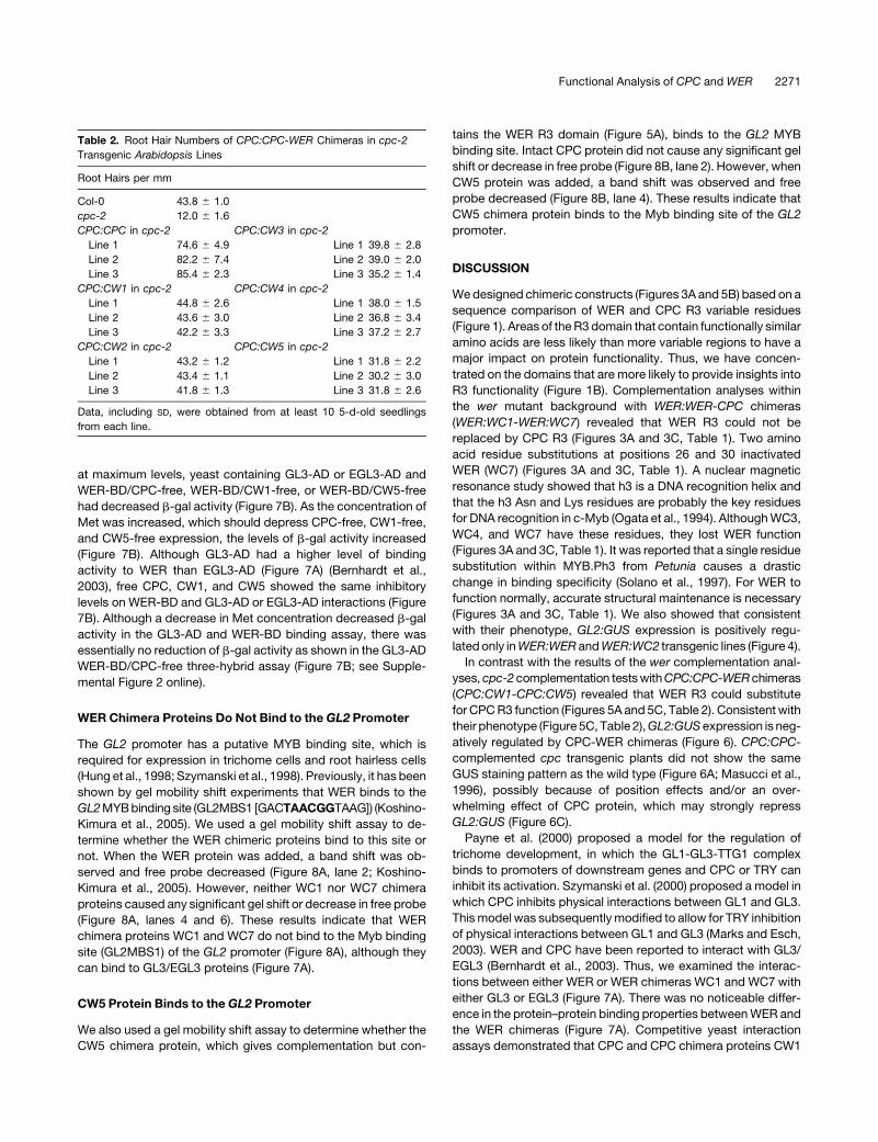
chimeric constructs rescued the reduced-hair cpc-2 phenotype,
though the degree of rescue differed. CPC:CPC in the cpc
background had an increased number of root hairs compared
with the wild-type background. CPC:CW1 and CPC:CW2 in cpc
restored the number of root hairs to wild-type levels. CPC:CW3
and CPC:CW4 in cpc transformants had somewhat fewer than
the wild type. The number of root hairs in the CPC:CW5 cpc
transformant was the least (Figure 5C, Table 2). These results
suggest that although the entire R3 structure is required for
strong function, CPC can act without strict structural conserva-
tion of the R3 domain (Figures 5A and 5C, Table 2).
GL2 Expression Is Repressed by CPC and
CPC Chimera Proteins
GL2:GUS expression is normally limited to hairless cell files
(Figure 6A; Masucci et al., 1996). The GL2:GUS gene is ex-
pressed in almost all epidermal cells of the cpc mutant but was
not expressed in 35S:CPC transgenic plants (Figure 6B; Lee and
Schiefelbein, 2002; Wada et al., 2002). To define the effect of
CPC:CPC-WER chimeras on GL2 expression, we introduced
GL2:GUS into CPC:CPC-WER chimera transgenic lines with a
cpc background (Figures 6C to 6H). As described above, all five
chimeric constructs (CPC:CW1-CPC:CW5) rescued the cpc
mutant phenotype (Figures 5A and 5C, Table 2). Like 35S:CPC-
complemented cpc transformants, no appreciable GL2:GUS
expression was observed in any of these transgenic lines when
roots were incubated in X-Gluc solution at 378C for 3.5 h (see
Supplemental Figure 1 online). With the exception of the
CPC:CPC-complemented cpc transgenic line, which had no
discernable expression (Figure 6C), after incubation overnight
they exhibited weak wild-type-like GUS activity in the hairless
cell file position (Figures 6D to 6H). The strength of GL2:GUS
activity in these transgenic lines was directly correlated with the
degree of complementation provided by the chimera construct.
The CPC:CPC construct in the cpc background produces a
greater number of root hairs than the wild type. CPC:CW1- and
CPC:CW2-complemented cpc transgenic lines have almost the
same number of root hairs as the wild type, and CPC:CW3-,
CPC:CW4-, and CPC:CW5-complemented cpc transgenic lines
produce lower numbers of root hairs than the wild type (Figures
Table 1. Root Hair Numbers of WER:WER-CPC Chimeras in wer
Transgenic Arabidopsis Lines
Root Hairs per mm
Col-0 43.8 6 1.0
wer 72.0 6 1.8
WER:WER in wer WER:WC4 in wer
Line 1 36.8 6 2.6 Line 1 76.2 6 2.6
Line 2 15.9 6 2.4 Line 2 74.4 6 2.0
Line 3 26.0 6 2.9 Line 3 82.0 6 2.5
WER:WC1 in wer WER:WC5 in wer
Line 1 85.3 6 1.5 Line 1 77.0 6 2.6
Line 2 80.5 6 4.5 Line 2 66.4 6 2.6
Line 3 81.1 6 3.2 Line 3 70.1 6 2.5
WER:WC2 in wer WER:WC6 in wer
Line 1 45.4 6 1.3 Line 1 91.0 6 2.6
Line 2 46.4 6 1.5 Line 2 86.6 6 2.5
Line 3 45.8 6 2.0 Line 3 73.5 6 2.5
WER:WC3 in wer WER:WC7 in wer
Line 1 83.8 6 2.8 Line 1 66.5 6 4.3
Line 2 89.2 6 2.9 Line 2 66.1 6 2.7
Line 3 87.2 6 2.9 Line 3 66.8 6 2.5
Data, including SD, were obtained from at least 10 5-d-old seedlings
from each line.
2268 The Plant Cell

5A, 5C, and 6C to 6H, Table 2). These results strongly suggest
that each of the CPC chimera proteins has the ability to inhibit
GL2 expression.
WER and WER Chimera Proteins Interact Equally with
GL3 or EGL3
WER protein physically interacts with either of the GL3 or EGL3
proteins in yeast cells (Bernhardt et al., 2003). To examine the
possibility that the WER chimera proteins physically associate
with GL3 or EGL3, we employed the yeast two-hybrid assay
(Fields and Sternglanz, 1994). The Myb regions of the WER,
WC1, and WC7 constructs were fused to the binding domain
(BD) of GAL4, and GL3 or EGL3 was fused to the activation
domain (AD) of GAL4. Yeast containing either empty pBridge
(BD) or pGAD424 vectors in conjunction with any of the corre-
sponding protein fusions did not exhibit significant b-gal activity,
whereas yeast containing WER-BD, WC1-BD or WC7-BD, and
GL3-AD or EGL3-AD exhibited b-gal activity (Figure 7A). There
were no significant differences in b-gal activity among yeast
isolates containing WER-BD GL3-AD, WC1-BD GL3-AD, or
WC7-BD GL3-AD (Figure 7A). Although EGL3-AD exhibited
one-sixth the level of binding activity as GL3-AD (Figure 7A;
Bernhardt et al., 2003), yeast isolates containing each of the
EGL3 fusions had approximately equal b-gal activities (Figure
7A). Thus, WER-BD, WC1-BD, and WC7-BD interacted equally
well with either GL3-AD or EGL3-AD (Figure 7A).
CPC and CPC Chimera Proteins Compete with WER for
Binding Sites on GL3 or EGL3
As presented in a previous model, TRY and GL1 compete for a
GL3 protein binding site to form different types of complexes that
are involved in Arabidopsis trichome development (Marks and
Esch, 2003). TRY also prevents the interaction between GL1 and
GL3 (Esch et al., 2003). CPC protein has also been found to
physically interact with both GL3 and EGL3 in yeast cells
(Bernhardt et al., 2003), suggesting a competition model for
CPC and WER (Lee and Schiefelbein, 1999; Bernhardt et al.,
2003). To determine whether CPC and the CPC chimera proteins
compete with WER for a binding site on GL3 or EGL3 equally,
CPC, CW1, and CW5 were cloned into the free site of the
WER-BD constructs to form the WER-BD/CPC-free, WER-BD/
CW1-free, and WER-BD/CW5-free constructs for the yeast three-
hybrid assay. Under conditions of low Met concentrations, in
which CPC-free, CW1-free, and CW5-free expression would be
Figure 4. Regulation of GL2:GUS in the wer Mutant Background.
Expression of the GL2:GUS reporter in the developing root epidermis of 5-d-old seedlings in Col-0, wer, and wer transformants. GL2 promoter activity is
reduced in the epidermis of the wer line. Transformants WER:WER and WER:WC2 had increased GL2 promoter activity compared with wer. Bar ¼100 mm.
Functional Analysis of CPC and WER 2269

Figure 5. Complementation of the cpc Mutant by CPC:CPC-WER Chimera Constructs.
(A) Schematic representation of chimera CPC R3 (red) and WER R3 (yellow) constructs. Complementation results are on the right. Each of the
constructs complemented the cpc mutant phenotype. Numbers indicate replaced WER regions as shown in (B).
(B) Alignment of the MYB R3 regions of WER and CPC. Shaded letters indicate identical residues. The positions of the three helices forming R3 MYB are
shown with green lines.
(C) Phenotypes of Col-0, cpc, and cpc transformants. All transgenic plant lines had an increased number of root hairs compared with cpc. Bar¼ 100 mm.
2270 The Plant Cell
at maximum levels, yeast containing GL3-AD or EGL3-AD and
WER-BD/CPC-free, WER-BD/CW1-free, or WER-BD/CW5-free
had decreased b-gal activity (Figure 7B). As the concentration of
Met was increased, which should depress CPC-free, CW1-free,
and CW5-free expression, the levels of b-gal activity increased
(Figure 7B). Although GL3-AD had a higher level of binding
activity to WER than EGL3-AD (Figure 7A) (Bernhardt et al.,
2003), free CPC, CW1, and CW5 showed the same inhibitory
levels on WER-BD and GL3-AD or EGL3-AD interactions (Figure
7B). Although a decrease in Met concentration decreased b-gal
activity in the GL3-AD and WER-BD binding assay, there was
essentially no reduction of b-gal activity as shown in the GL3-AD
WER-BD/CPC-free three-hybrid assay (Figure 7B; see Supple-
mental Figure 2 online).
WER Chimera Proteins Do Not Bind to the GL2 Promoter
The GL2 promoter has a putative MYB binding site, which is
required for expression in trichome cells and root hairless cells
(Hung et al., 1998; Szymanski et al., 1998). Previously, it has been
shown by gel mobility shift experiments that WER binds to the
GL2 MYB binding site (GL2MBS1 [GACTAACGGTAAG]) (Koshino-
Kimura et al., 2005). We used a gel mobility shift assay to de-
termine whether the WER chimeric proteins bind to this site or
not. When the WER protein was added, a band shift was ob-
served and free probe decreased (Figure 8A, lane 2; Koshino-
Kimura et al., 2005). However, neither WC1 nor WC7 chimera
proteins caused any significant gel shift or decrease in free probe
(Figure 8A, lanes 4 and 6). These results indicate that WER
chimera proteins WC1 and WC7 do not bind to the Myb binding
site (GL2MBS1) of the GL2 promoter (Figure 8A), although they
can bind to GL3/EGL3 proteins (Figure 7A).
CW5 Protein Binds to the GL2 Promoter
We also used a gel mobility shift assay to determine whether the
CW5 chimera protein, which gives complementation but con-
tains the WER R3 domain (Figure 5A), binds to the GL2 MYB
binding site. Intact CPC protein did not cause any significant gel
shift or decrease in free probe (Figure 8B, lane 2). However, when
CW5 protein was added, a band shift was observed and free
probe decreased (Figure 8B, lane 4). These results indicate that
CW5 chimera protein binds to the Myb binding site of the GL2
promoter.
DISCUSSION
We designed chimeric constructs (Figures 3A and 5B) based on a
sequence comparison of WER and CPC R3 variable residues
(Figure 1). Areas of the R3 domain that contain functionally similar
amino acids are less likely than more variable regions to have a
major impact on protein functionality. Thus, we have concen-
trated on the domains that are more likely to provide insights into
R3 functionality (Figure 1B). Complementation analyses within
the wer mutant background with WER:WER-CPC chimeras
(WER:WC1-WER:WC7) revealed that WER R3 could not be
replaced by CPC R3 (Figures 3A and 3C, Table 1). Two amino
acid residue substitutions at positions 26 and 30 inactivated
WER (WC7) (Figures 3A and 3C, Table 1). A nuclear magnetic
resonance study showed that h3 is a DNA recognition helix and
that the h3 Asn and Lys residues are probably the key residues
for DNA recognition in c-Myb (Ogata et al., 1994). Although WC3,
WC4, and WC7 have these residues, they lost WER function
(Figures 3A and 3C, Table 1). It was reported that a single residue
substitution within MYB.Ph3 from Petunia causes a drastic
change in binding specificity (Solano et al., 1997). For WER to
function normally, accurate structural maintenance is necessary
(Figures 3A and 3C, Table 1). We also showed that consistent
with their phenotype, GL2:GUS expression is positively regu-
lated only in WER:WER and WER:WC2 transgenic lines (Figure 4).
In contrast with the results of the wer complementation anal-
yses, cpc-2 complementation tests with CPC:CPC-WER chimeras
(CPC:CW1-CPC:CW5) revealed that WER R3 could substitute
for CPC R3 function (Figures 5A and 5C, Table 2). Consistent with
their phenotype (Figure 5C, Table 2), GL2:GUS expression is neg-
atively regulated by CPC-WER chimeras (Figure 6). CPC:CPC-
complemented cpc transgenic plants did not show the same
GUS staining pattern as the wild type (Figure 6A; Masucci et al.,
1996), possibly because of position effects and/or an over-
whelming effect of CPC protein, which may strongly repress
GL2:GUS (Figure 6C).
Payne et al. (2000) proposed a model for the regulation of
trichome development, in which the GL1-GL3-TTG1 complex
binds to promoters of downstream genes and CPC or TRY can
inhibit its activation. Szymanski et al. (2000) proposed a model in
which CPC inhibits physical interactions between GL1 and GL3.
This model was subsequently modified to allow for TRY inhibition
of physical interactions between GL1 and GL3 (Marks and Esch,
2003). WER and CPC have been reported to interact with GL3/
EGL3 (Bernhardt et al., 2003). Thus, we examined the interac-
tions between either WER or WER chimeras WC1 and WC7 with
either GL3 or EGL3 (Figure 7A). There was no noticeable differ-
ence in the protein–protein binding properties between WER and
the WER chimeras (Figure 7A). Competitive yeast interaction
assays demonstrated that CPC and CPC chimera proteins CW1
Table 2. Root Hair Numbers of CPC:CPC-WER Chimeras in cpc-2
Transgenic Arabidopsis Lines
Root Hairs per mm
Col-0 43.8 6 1.0
cpc-2 12.0 6 1.6
CPC:CPC in cpc-2 CPC:CW3 in cpc-2
Line 1 74.6 6 4.9 Line 1 39.8 6 2.8
Line 2 82.2 6 7.4 Line 2 39.0 6 2.0
Line 3 85.4 6 2.3 Line 3 35.2 6 1.4
CPC:CW1 in cpc-2 CPC:CW4 in cpc-2
Line 1 44.8 6 2.6 Line 1 38.0 6 1.5
Line 2 43.6 6 3.0 Line 2 36.8 6 3.4
Line 3 42.2 6 3.3 Line 3 37.2 6 2.7
CPC:CW2 in cpc-2 CPC:CW5 in cpc-2
Line 1 43.2 6 1.2 Line 1 31.8 6 2.2
Line 2 43.4 6 1.1 Line 2 30.2 6 3.0
Line 3 41.8 6 1.3 Line 3 31.8 6 2.6
Data, including SD, were obtained from at least 10 5-d-old seedlings
from each line.
Functional Analysis of CPC and WER 2271

and CW5 similarly prevent WER–GL3/EGL3 interactions (Figure
7B). Thus, it is apparent that all of the WER and CPC chimera
proteins have the ability to interact with GL3/EGL3. Because
the R3 domains of both WER and CPC contain the con-
served [DE]Lx2[RK]x3Lx6Lx3R predicted bHLH interaction motif
(Zimmermann et al., 2004), any point of exchange might be
possible. We were also able to demonstrate that WER chimeras
WC1 and WC7 lose their DNA binding ability to GL2MBS1 in the
GL2 promoter (Figure 8A), although a faint band, which was not
retarded to the same degree as with intact WER, was observed in
WC1 (Figure 8A, lane 4). Because high protein concentrations
can alter target recognition (Andersson et al., 1999), the high
purity of WC1 protein (60% purity for WER, 82% for WC1, and
53% for WC7) could have increased the actual concentration to
the point where nonspecific DNA binding occurred at a low level
(Figure 8A, lane 4). These data together suggest a regulatory
cascade model in which WER and CPC competitively bind to
GL3/EGL3 (Figure 9A). Once the WER-GL3/EGL3-TTG1 com-
plex is formed, it binds to the GL2 promoter and promotes
expression of GL2 protein, which leads to the hairless cell fate
(Figure 9B; Koshino-Kimura et al., 2005). On the other hand, WER
chimera proteins lost their ability to bind to the GL2 promoter;
thus, chimera proteins do not result in a hairless cell fate (Figure
9B). CPC and the CPC chimeras bind to the TTG1-GL3/EGL3
complex to prevent activation of the GL2 promoter, eventually
resulting in root hair formation (Figure 9C).
We also show that the CW5 chimera protein, which has WER
R3 motif sequences, retains its DNA binding ability (Figure 8B).
These results suggest a couple of possible evolutionary scenar-
ios. The CPC and CPC-like R3-type MYB genes could have
arisen from the R2R3-type MYB gene family, including WER
(Figure 2A), with the functional novelty of CPC and CPC-like MYB
Figure 6. Regulation of GL2:GUS in the cpc Mutant Background.
Expression of the GL2:GUS reporter in the developing root epidermis of 5-d-old seedlings in Col-0, cpc, and cpc transformants. GL2 promoter activity is
increased in the epidermis of the cpc line. All transformant lines had reduced GL2 promoter activity compared with cpc with overnight incubation in
X-Gluc solution. Bar ¼ 100 mm.
2272 The Plant Cell

proteins arising from loss of the acidic region activation domain
and/or loss of DNA binding though residue change. In the former
case (Figure 10B), the proto-CPC chimeras may be analogous to
C1-I in maize (Zea mays), in which C1-I protein lacking the
activator domain acts as an inhibitor (Paz-Ares et al., 1990).
Because this initial truncation would have maintained its DNA
binding activity, it would act as a dominant repressor in compe-
tition with WER (Figure 10B). Because repression is the only
function subject to selection, any biochemical properties that
didn’t contribute to repression activity would be free to drift.
Thus, DNA binding ability could be lost or modified without
consequence. All that would remain is a conversion to bHLH
binding at the same spot as WER (Figure 10C).
Alternatively, loss of DNA binding activity in a WER-like dupli-
cation due to amino acid residue changes would release the
proto-CPC molecule from having regulatory consequences (Fig-
ure 10D), thus freeing it for whole-scale truncations (Figure 10C).
In addition, there are incomplete repetitions in the WER genome
sequence presumed to cause a simple loop crossing-over event
that would cause deletion of N-terminal region and/or C-terminal
activation domain of WER (see Supplemental Figure 5 online).
These provide the mechanical possibilities for truncations to
make proto-CPC. Newly evolved CPC would have thus lost the
ability to bind DNA and activate transcription (Figure 10C), al-
lowing it the freedom to evolve a specialized competitive inhib-
itory function through the selective loss of other functions
important for gene activation.
Figure 7. Protein Interactions with Native Myb versus Chimeric Myb
Proteins.
(A) Comparison of protein interactions using a yeast two-hybrid assay
between WER, WC1, and WC7 with GL3 and EGL3. The WER, WC1, and
WC7 proteins were compared as GAL4 binding domain (BD) fusions,
whereas GL3 and EGL3 were expressed as GAL4 activation domain (AD)
fusions.
(B) CPC, CW1, and CW5 competition for the WER binding sites of GL3
and EGL3. Using the pBridge vector (Clontech), a third protein (CPC,
CW1, or CW5) under the control of a Met-repressible promoter was
expressed in a yeast interaction assay at varying Met concentrations (0,
15, 30, and 125 mM). CPC, CW1 or CW5 were expressed as a free protein
(no AD or BD domains). Samples were normalized by OD550. The
strength of the interaction was determined by b-gal activity.
Figure 8. The DNA Binding Properties of WER and CPC Chimera
Proteins.
(A) WER, WC1, or WC7 protein was added with or without a 200-fold
excess of competitor.
(B) CPC or CW5 protein was added with or without a 200-fold excess of
competitor.
Arrows indicate shifted bands, and arrowheads indicate free probe.
Digoxigenin-labeled DNA of GL2MBS1 was used as the probe.
Functional Analysis of CPC and WER 2273

METHODS
Plant Materials and Growth Conditions
Arabidopsis thaliana Col-0 ecotype and cognate cpc-2 and wer-1 mutant
plants were used. Seeds were sterilized with 10% (v/v) bleach with 0.02%
(v/v) Triton X-100 for 5 min. After seeds were rinsed five times in sterile
water, seeds were germinated and grown on square Petri dishes
containing half-strength Okada and Shimura medium [2.5 mM KNO3,
1 mM MgSO4, 1 mM Ca(NO3)2, 25 mM Fe-EDTA, 1.25 mM K-PO4, pH 5.5,
35 mM H3BO3, 7 mM MnCl2, 0.25 mM CuSO4, 0.5 mM ZnSO4, 0.1 mM
NaMo4, 5 mM NaCl, and 0.05 mM CoCl2] (Okada and Shimura, 1990) with
1.5% (w/v) agar. After sowing, dishes were wrapped with Micropore
surgical tape (3M Healthcare) to prevent desiccation. The dishes were
then kept in darkness at 48C for 2 d and then transferred to a growth
chamber at 228C under constant white light (white fluorescent lamp
model FL20S-EXNH; Toshiba).
Gene Constructs
Primers
All primer sequences used in this article are listed in Supplemental Table
1 online.
WER:WER-CPC Chimeric Constructs
To define the relationship between the R3 motifs of WER and CPC, based
on published data (Lee and Schiefelbein, 2001), we first tested a construct
harboring WER. This construct (designated WER:WER; Figure 3A, Table
1) was introduced into the wer mutant background. To make the
WER:WER chimera constructs, we used a 6.1-kb PCR-amplified WER:
WER genome fragment that includes the 4.0-kb 59 region, the 1.0-kb
coding region, and the 1.1-kb 39 region as well as pBS-gCPC, including
the 1.3-kb 59 region, the 0.9-kb coding region, and the 0.45-kb 39 region
(Wada et al., 1997) as amplification templates. The WER:WER region,
amplified using primers RT11/RT12, and the CPC region, amplified using
primers TW1149/TW1150, were ligated (WER:WC1). The WER:WER
region was amplified using primers RT32/RT34, the WER:WC1 fragment
was amplified using primers RT37/RT11, and the products were ligated to
form WER:WC2. The WER:WER region, amplified with primers RT33/
RT35, and the WER:WC1 fragment, amplified with primers RT36/RT12,
were ligated to make WER:WC3. The WER:WER fragment was amplified
with primers RT101/RT102 and was self-ligated to form WER:WC4. The
WER:WER region, amplified with primers RT103/RT12, and the CPC
region, amplified by primers RT104/TW1150, were ligated to make
WER:WC5. The WER:WER fragment amplified with primers WERCPC6-1/
WERCPC6-2 was also self-ligated to form WER:WC6. To create WER:
WC7, PCR-mediated mutagenesis was performed on WER:WER using
the QuickChange site-directed mutagenesis kit (Stratagene), with the
primers WERCPC7-1/WERCPC7-2. PCR-generated constructs were
completely sequenced following isolation of the clones to check for
amplification-induced errors. Finally, the amplified and ligated constructs
were cloned into the transformation vector pJHA212K (Yoo et al., 2005).
CPC:CPC-WER Chimera Constructs
To create CPC:CPC-WER chimera constructs (Figure 5A), we used pBS-
gCPC and pBS-WER as templates. The CPC:CPC region, amplified by
primers CPCWER-a-1/CPCWER-a-2, and the WER region, amplified by
primer pair CPCWER-a-3/CPCWER-a-4, were ligated to form CPC:CW1.
The CPC:CW1 fragment, amplified by primers CPCWER-b-1/CPCWER-
b-2, was self-ligated to make CPC:CW2, which was then amplified using
primers CPCWER-c-1/CPCWER-c-2 and self-ligated to form CPC:CW3.
The CPC:CW3 fragment, amplified with primers CPCWER-d-1/
CPCWER-d-2, was self-ligated to create CPC:CW4. The WER region,
amplified with primer pair RT32/RT35, and the CPC:CPC region, ampli-
fied with the primer pair NEKO16/RT86, were ligated to make CPC:CW5.
PCR-generated constructs were completely sequenced following isola-
tion of the clones to check for amplification-induced errors. These
amplified and ligated constructs were also cloned into the transformation
vector pJHA212K (Yoo et al., 2005).
Transgenic Plants
Plant transformation was performed by a vacuum transformation proce-
dure (Bechtold et al., 1993) or floral dip method (Clough and Bent, 1998),
and transformants were selected on a 0.53 Murashige and Skoog agar
plate containing 50 mg�1 kanamycin. Homozygous transgenic lines were
selected by kanamycin resistance. For CPC:CPC-WER and WER:WER-
CPC chimera constructs, we isolated at least 24 T1 lines for each
construct and selected at least five T2 and T3 lines on the basis of their
segregation ratios for kanamycin resistance (see Supplemental Figures 4
and 5 online). For each transgenic line, at least 10 individual 5-d-old
seedlings were assayed for root hair numbers. Some outliers were elim-
inated from the data because of the possibility that positional or other
aberrant effects would distort the data. However, outliers may be exam-
ined in the future and may provide additional clarification of the data.
Figure 9. Regulatory Cascade Models for WER and CPC Chimeras.
(A) WER and CPC proteins competitively bind to the GL3/EGL3-TTG1
complex.
(B) The WER-GL3/EGL3-TTG1 complex can bind the GL2 promoter to
promote GL2 expression, which leads to the hairless cell fate, whereas
the WER chimera-GL3/EGL3-TTG1 complex cannot bind to the GL2
promoter, thus preventing GL2 expression.
(C) Both CPC-GL3/EGL3-TTG1 and CPC chimera-GL3/EGL3-TTG1
complexes prevent expression of GL2. The absence of GL2 results in
root hair formation.
2274 The Plant Cell

A GL2:GUS construct (Wada et al., 2002) was introduced into trans-
genic lines by crossing plants and analyzing F2 seedlings for GL2:GUS by
PCR (GUSþ00þ/GUSþ09-). For each transgenic line, at least ten indi-
vidual seedlings were assayed for GUS activity.
Histology
Primary roots of 5-d-old transgenic seedlings were excised and im-
mersed in X-Gluc solution containing 1.0 mM X-Gluc (5-bromo-4-chloro-
3-indolyl-b-glucuronide), 1.0 mM K3Fe(CN)6, 1.0 mM K4Fe(CN)6, 100 mM
NaPi, pH 7.0, 100 mM EDTA, and 0.1% Triton X-100. Excised primary
roots were incubated at 378C for 3.5 h or overnight.
Microscopy
For observation of the root hairs, root images were obtained with a three-
dimensional digital fine microscope (VC4500-PC; Omron) or with a digital
microscope (VH-8000; Keyence). For each transgenic line, at least 10
individual 5-d-old seedlings were analyzed for root hair number and root
GUS activity.
Construction, Transformation, and Analysis of Yeast Constructs
pGL3-AD and pEGL3-AD were constructed by cloning GL3 and EGL3
coding regions from Col-0 cDNA into pGAD424 (Clontech). pWER-BD,
pWC1-BD, and pWC7-BD were constructed by cloning WER, WC1, and
WC7 coding regions from Col-0, WER:WC1 transformants, or WER:WC7
transformants, respectively, into pBridge MCSI (Clontech). The con-
structs used for WER-CPC, -CW1, or -CW5 competition assays, pWER-
BD/CPC-free, pWER-BD/CW1-free, or pWER-BD/CW5-free, were
generated by cloning CPC, CW1, or CW5 coding regions into pBridge
MCSII (Clontech). The appropriate pGAD424- and pBridge-based con-
structs were transformed into the yeast strain Y187 using the Yeastmaker
2 transformation system (Clontech). Cells were selected on plates
containing SD synthetic medium (2% glucose and 13 yeast nitrogen
base) lacking Leu and Trp. Liquid cultures of SD synthetic medium lacking
Leu and Trp were used to measure b-galactosidase (b-gal) activity
(Ausubel et al., 1995). Cells were grown to an OD600 of 0.7 to 1.0, pelleted
by centrifugation, and suspended in z-buffer (60 mM Na2HPO4, 40 mM
NaH2PO4, 10 mM KCl, 1 mM MgSO4, and 50 mM b-mercaptoethanol, pH
7.0). Cells were permeabilized by adding a final concentration of 0.005%
SDS and 3.5% (v/v) chloroform. o-nitrophenyl-D-galactopyranoside
(Sigma-Aldrich) was added as a substrate. After incubation at 308C, the
reaction was stopped with sodium carbonate and measured for activity at
OD420. b-gal activity was determined using the equation U ¼ 1000 3
[OD420]/time (in seconds) 3 volume (in mL) 3 [OD600]. For each compar-
ison, three independent yeast isolates were tested three times.
Bacterial Expression of Proteins and Purification of His-Tagged
Recombinant Proteins
The WER coding sequence was amplified from pGEX_WER (Koshino-
Kimura et al., 2005) as a template. The WC1 and WC7 coding sequences
were amplified from total root cDNA of WER:WC1 transformants and
WER:WC7 transformants, as described above. The CPC and CW5 coding
sequences were amplified from pWER-BD/CPC-free and pWER-BD/
CW5-free as templates. The fragments were cloned into expression
vector pColdTF (TaKaRa Bio). DNA sequences were checked, and the
constructs were transformed into Escherichia coli strain Rosetta 2 (DE3)
(Novagen). These transformed bacteria were used for purifying the
recombinant proteins (custom-made by TaKaRa Bio).
Gel Mobility Shift Assay
Oligonucleotides for gel mobility shift assays were labeled with a Roche
DIG gel shift kit (2nd Generation; Roche). The sequence of GL2MBS1 is
the same as reported previously (Koshino-Kimura et al., 2005). DNA–
protein binding reactions were basically performed by incubating 32 fmol
of digoxigenin-labeled oligonucleotide with 100 ng of each protein in 20
mL of binding buffer [10 mM Tris-HCl, pH 7.5, 50 mM NaCl, 1 mM EDTA,
10 mM DTT, 5% 80 mg�1 poly(dI-dC), and 100 mg�1 BSA] at 228C for 15
min, and then free and bound complexes were resolved by electropho-
resis through 1-mm 5% native polyacrylamide gels (Real Gel Plate; BIO
CRAFT) in 0.53 TBE buffer at 8 V cm�1 for 60 min.
Accession Numbers
Sequence data from this article can be found in the GenBank/EMBL data
libraries under the following accession numbers: WER (AL391149), GL1
(AF495524), At MYB23 (Z95747), CPC (AB004871), TRY (AY519523),
Figure 10. Evolutionary Models of CPC and WER.
(A) WER binds the GL2 promoter to promote GL2 expression.
(B) Proto-CPC protein derived from WER (yellow) truncation can bind the promoter but prevents expression of GL2.
(C) CPC cannot bind to the GL2 promoter, strongly preventing GL2 expression.
(D) Proto-CPC protein derived from WER amino acid substitution cannot bind to the GL2 promoter.
Functional Analysis of CPC and WER 2275

ETC1 (AY519518), ETC2 (AY234411), At4g01060 (AY519522), MYB36
(AF062878), MYB37 (AF062879), MYB38 (AF062880), MYB68 (AF062901),
MYB84 (Y14209), MYB87 (AF062914), PAP1 (AF325123), PAP2
(AF087936), GL2 (AB117767), GL3 (AF246291), and EGL3 (AF027732).
Supplemental Data
The following materials are available in the online version of this article.
Supplemental Figure 1. Regulation of the GL2:GUS Expression
Pattern in Col-0, cpc, or cpc Transformants.
Supplemental Figure 2. Protein Interactions between WER-BD and
GL3-AD or EGL3-AD.
Supplemental Figure 3. Complementation of the wer Mutant by
WER:WER-CPC Chimera Constructs.
Supplemental Figure 4. Complementation of the cpc Mutant by
CPC:CPC-WER Chimera Constructs.
Supplemental Figure 5. Homologous Recombination in the WER
Genome Sequence.
Supplemental Table 1. Primer Sequences Used in This Study.
ACKNOWLEDGMENTS
We thank Y. Koshino-Kimura and T. Mayama for technical advice,
H. Oka for technical assistance, and T. Ishida, T. Kurata, R. Sano, and
T. Gohara for useful advice.
Received July 11, 2006; revised July 2, 2007; accepted July 6, 2007;
published July 20, 2007.
REFERENCES
Andersson, K.B., Berge, T., Matre, V., and Gabrielsen, O.S. (1999).
Sequence selectivity of c-Myb in vivo. Resolution of a DNA target
specificity paradox. J. Biol. Chem. 274: 21986–21994.
Ausubel, F., Brent, R., Kingston, R.E., Moore, D.D., Seidman, J.G.,
Smith, J.A., and Struhl, K. (1995). Current Protocols in Molecular
Biology. (New York: John Wiley & Sons).
Bechtold, N., Ellis, J., and Pelletier, G. (1993). In planta Agrobacterium-
mediated gene transfer by infiltration of adult Arabidopsis thaliana
plants. C. R. Acad. Sci. Paris 316: 1194–1199.
Bernhardt, C., Lee, M.M., Gonzalez, A., Zhang, F., Lloyd, A., and
Schiefelbein, J. (2003). The bHLH genes GLABRA3 (GL3) and EN-
HANCER OF GLABRA3 (EGL3) specify epidermal cell fate in the
Arabidopsis root. Development 130: 6431–6439.
Bernhardt, C., Zhao, M., Gonzalez, A., Lloyd, A., and Schiefelbein, J.
(2005). The bHLH genes GL3 and EGL3 participate in an intercellular
regulatory circuit that controls cell patterning in the Arabidopsis root
epidermis. Development 132: 291–298.
Braun, E.L., and Grotewold, E. (1999). Newly discovered plant c-myb-
like genes rewrite the evolution of the plant myb gene family. Plant
Physiol. 121: 21–24.
Clough, S.J., and Bent, A.F. (1998). Floral dip: A simplified method for
Agrobacterium-mediated transformation of Arabidodpsis thaliana.
Plant J. 16: 735–743.
Dolan, L., Duckett, M.D., Grierson, C., Linstead, P., Schneider, K.,
Lawson, E., Dean, C., Poethig, S., and Roberts, K. (1994). Clonal
relationships and cell patterning in the root epidermis of Arabidopsis.
Development 120: 2465–2474.
Esch, J.J., Chen, M., Sanders, M., Hillestad, M., Ndkium, S.,
Idelkope, B., Neizer, J., and Marks, M.D. (2003). A contradictory
GLABRA3 allele helps define gene interactions controlling trichome
development in Arabidopsis. Development 130: 5885–5894.
Fields, S., and Sternglanz, R. (1994). The two-hybrid system: An assay
for protein-protein interactions. Trends Genet. 10: 286–292.
Galway, M.E., Masucci, J.D., Lloyd, A.M., Walbot, V., Davis, R.W.,
and Schiefelbein, J.W. (1994). The TTG gene is required to specify
epidermal cell fate and cell patterning in the Arabidopsis root. Dev.
Biol. 166: 740–754.
Hung, C.Y., Lin, Y., Zhang, M., Pollock, S., Marks, M.D., and
Schiefelbein, J. (1998). A common position-dependent mechanism
controls cell-type patterning and GLABRA2 regulation in the root and
hypocotyl epidermis of Arabidopsis. Plant Physiol. 117: 73–84.
Koshino-Kimura, Y., Wada, T., Tachibana, T., Tsugeki, R., Ishiguro,
S., and Okada, K. (2005). Regulation of CAPRICE transcription by
MYB proteins for root epidermis differentiation in Arabidopsis. Plant
Cell Physiol. 46: 817–826.
Kranz, H.D., et al. (1998). Towards functional characterisation of the
members of the R2R3-MYB gene family from Arabidopsis thaliana.
Plant J. 16: 263–276.
Lee, M.M., and Schiefelbein, J. (1999). WEREWOLF, a MYB-related
protein in Arabidopsis, is a position-dependent regulator of epidermal
cell patterning. Cell 99: 473–483.
Lee, M.M., and Schiefelbein, J. (2001). Developmentally distinct MYB
genes encode functionally equivalent proteins in Arabidopsis. Devel-
opment 128: 1539–1546.
Lee, M.M., and Schiefelbein, J. (2002). Cell pattern in the Arabidopsis
root epidermis determined by lateral inhibition with feedback. Plant
Cell 14: 611–618.
Lipsick, J.S. (1996). One billion years of Myb. Oncogene 13: 223–235.
Marks, M.D., and Esch, J.J. (2003). Initiating inhibition. Control of
epidermal cell patterning in plants. EMBO Rep. 4: 24–25.
Masucci, J.D., Rerie, W.G., Foreman, D.R., Zhang, M., Galway, M.E.,
Marks, M.D., and Schiefelbein, J.W. (1996). The homeobox gene
GLABRA2 is required for position-dependent cell differentiation in
the root epidermis of Arabidopsis thaliana. Development 122: 1253–
1260.
Ogata, K., Hojo, H., Aimoto, S., Nakai, T., Nakamura, H., Sarai, A., Ishii, S.,
and Nishimura, Y. (1992). Solution structure of a DNA-binding unit of
Myb: A helix-turn-helix-related motif with conserved tryptophans form-
ing a hydrophobic core. Proc. Natl. Acad. Sci. USA 89: 6428–6432.
Ogata, K., Morikawa, S., Nakamura, H., Sekikawa, A., Inoue, T.,
Kanai, H., Sarai, A., Ishii, S., and Nishimura, Y. (1994). Solution
structure of a specific DNA complex of the Myb DNA-binding domain
with cooperative recognition helices. Cell 79: 639–648.
Okada, K., and Shimura, Y. (1990). Reversible root tip rotation in
Arabidopsis seedlings induced by obstacle-touching stimulus. Sci-
ence 250: 274–276.
Payne, C.T., Zhang, F., and Lloyd, A.M. (2000). GL3 encodes a bHLH
protein that regulates trichome development in Arabidopsis through
interaction with GL1 and TTG1. Genetics 156: 1349–1362.
Paz-Ares, J., Ghosal, D., and Saedler, H. (1990). Molecular analysis of
the C1-I allele from Zea mays: A dominant mutant of the regulatory C1
locus. EMBO J. 9: 315–321.
Rabinowicz, P.D., Braun, E.L., Wolfe, A.D., Bowen, B., and Grotewold,
E. (1999). Maize R2R3 Myb genes: Sequence analysis reveals ampli-
fication in the higher plants. Genetics 153: 427–444.
Rerie, W.G., Feldmann, K.A., and Marks, M.D. (1994). The GLABRA2
gene encodes a homeo domain protein required for normal trichome
development in Arabidopsis. Genes Dev. 8: 1388–1399.
Solano, R., Fuertes, A., Sanchez-Pulido, L., Valencia, A., and Paz-
Ares, J. (1997). A single residue substitution causes a switch from the
2276 The Plant Cell

dual DNA binding specificity of plant transcription factor MYB.Ph3 to
the animal c-MYB specificity. J. Biol. Chem. 272: 2889–2895.
Stracke, R., Werber, M., and Weisshaar, B. (2001). The R2R3-MYB
gene family in Arabidopsis thaliana. Curr. Opin. Plant Biol. 4: 447–456.
Szymanski, D.B., Jilk, R.A., Pollock, S.M., and Marks, M.D. (1998).
Control of GL2 expression in Arabidopsis leaves and trichomes.
Development 125: 1161–1171.
Szymanski, D.B., Lloyd, A.M., and Marks, M.D. (2000). Progress in the
molecular genetic analysis of trichome initiation and morphogenesis in
Arabidopsis. Trends Plant Sci. 5: 214–219.
Wada, T., Kurata, T., Tominaga, R., Koshino-Kimura, Y., Tachibana,
T., Goto, K., Marks, M.D., Shimura, Y., and Okada, K. (2002). Role of
a positive regulator of root-hair development, CAPRICE, in Arabidopsis
root epidermal cell differentiation. Development 129: 5409–5419.
Wada, T., Tachibana, T., Shimura, Y., and Okada, K. (1997). Epider-
mal cell differentiation in Arabidopsis determined by a Myb homolog,
CPC. Science 277: 1113–1116.
Yoo, S.Y., Bomblies, K., Yoo, S.K., Yang, J.W., Choi, M.S., Lee, J.S.,
Weigel, D., and Ahn, J.H. (2005). The 35S promoter used in a se-
lectable marker gene of a plant transformation vector affects the
expression of the transgene. Planta 221: 523–530.
Zhang, F., Gonzalez, A., Zhao, M., Payne, C.T., and Lloyd, A. (2003).
A network of redundant bHLH proteins functions in all TTG1-dependent
pathways of Arabidopsis. Development 130: 4859–4869.
Zimmermann, I.M., Heim, M.A., Weisshaar, B., and Uhrig, J.F. (2004).
Comprehensive identification of Arabidopsis thaliana MYB transcrip-
tion factors interacting with R/B-like BHLH proteins. Plant J. 40:
22–34.
Functional Analysis of CPC and WER 2277

DOI 10.1105/tpc.106.045732; originally published online July 20, 2007; 2007;19;2264-2277Plant Cell
Rumi Tominaga, Mineko Iwata, Kiyotaka Okada and Takuji WadaArabidopsis
in WEREWOLF and CAPRICEFunctional Analysis of the Epidermal-Specific MYB Genes
This information is current as of March 22, 2020
Supplemental Data /content/suppl/2007/09/25/tpc.106.045732.DC1.html
References /content/19/7/2264.full.html#ref-list-1
This article cites 35 articles, 21 of which can be accessed free at:
Permissions https://www.copyright.com/ccc/openurl.do?sid=pd_hw1532298X&issn=1532298X&WT.mc_id=pd_hw1532298X
eTOCs http://www.plantcell.org/cgi/alerts/ctmain
Sign up for eTOCs at:
CiteTrack Alerts http://www.plantcell.org/cgi/alerts/ctmain
Sign up for CiteTrack Alerts at:
Subscription Information http://www.aspb.org/publications/subscriptions.cfm
is available at:Plant Physiology and The Plant CellSubscription Information for
ADVANCING THE SCIENCE OF PLANT BIOLOGY © American Society of Plant Biologists