Embed Size (px)
Citation preview
Biochemical Pharmacology, Vol. 54, pp. 551-562, 1997. 0 1997 Elsevier Science Inc. All rights reserved.
ISSN 0006s2952/97/$17.00 + 0.00 PII SOOOS-2952(97)00205-O
ELSEVIER
Functionally Nonequivalent Interactions of Guanosine 5’-Triphosphate, Inosine 5’0Triphosphate, and Xanthosine 5’-Triphosphate with the Retinal G-Protein, Transducin, and with Gi-Proteins
in HL-60 Leukemia Cell Membranes Jan F. Klinker” and Roland Seifert j-f‘
*INSTITLJT Fii~ NEUROPSYCHOPHARMAK~L~~~E, FREIE UNIVERSIT~T BERLIN, ULMENALLEE 30, D-14050 BERLIN, GERMANY, ANL) ~INSTITUT FUR PHARMAKOLOGIE, FREIE UNIVERSITAT BERLIN, THIELALLEE 69/73, D-14195
BERLIN, GERMANY
ABSTRACT. G-proteins mediate signal transfer from receptors to effector systems. In their guanosine 5’.triphosphate (GTP)-bound form, G-protein ol-subunits activate effector systems. Termination of G-protein activation is achieved by the high-affinity GTPase [E.C. 3.6.1.-j of their a-subunits. Like GTP, inosine 5’-triphosphate (ITP) and xanthosine 5’etriphosphate (XTP) can support effector system activation. We studied the interactions of GTP, ITP, and XTP with the retinal G-protein, transducin (TD), and with G-proteins in HL-60 leukemia cell membranes. TD hydrolyzed nucleoside 5’triphosphates (NTPs) in the order of efficacy GTP > ITP > XTP. NTPs eluted TD from rod outer segment disk membranes in the same order of efficacy. ITP and XTP competitively inhibited TD-catalyzed GTP hydrolysis. In HL-60 membranes, the chemoattractants N-formyl-t_-methionyl-L-leucyl-L-phenylalanine (fMLP) and leukotriene B, (LTB,) effectively activated GTP and ITP hydrolysis by G,-proteins. fIvILP and LTB, were at least lo-fold more potent activators of ITPase than of GTPase. Complement C5a effectively activated the GTPase of G,-proteins but was only a weak stimulator of ITPase. The potency of C5a to activate GTP and ITP hydrolysis was similar. The fMLPstimulated GTPase had a lower K, value than the fMLP-stimulated ITPase, whereas the opposite was true for the V,,,,, values. fMLP, C5a, and LTB, did not stimulate XTP hydrolysis. Collectively, our data show that GTP, ITP, and XTP hind to G-proteins with different affinities, that G-proteins hydrolyze NTPs with different efficacies, and that chemoattractants stimulate GTP and ITP hydrolysis by G,-proteins in a receptor-specific manner. On the basis of our results and the data in the literature, we put forward the hypothesis that GTP, ITP, and XTP act as differential signal amplifiers and signal sorters at the G-protein level. BIOCHEM PHARMACOL 54;5:551-562, 1997. 0 1997 Elsevier Science Inc.
KEY WORDS. GTPase; ITPase; XTPase; transducin; G,-proteins; chemoattractants
G-proteins are membrane-attached proteins consisting of
an a-subunit and a py-complex, and mediate coupling of
agonist-occupied heptahelical receptors to cellular effector
systems. In their resting state, cw-subunits are GDP liganded.
Interaction of an activated receptor with G-proteins leads
to the exchange of GDP for guanosine 5’-triphosphate
(GTP).” GDP release is the rate-limiting step of G-protein
i Corresponding author: Dr. Roland Seifert, Howard Hughes Medical Institute, Beckman Center, B-l 59, Stanford University Medical Center, Stanford, CA 94305, USA; TEL.: +l-415-7257754; FAX: +I-415- 7258021; e-mail: [email protected].
P Abbreuianom: tMLP, N-formyl-L-methionyl-L_leucyl-L-phenylala; GppNHp, guanosine 5’-[P,y-imidoltriphosphate; GTP, guanosine 5’-triphos- phate; GTPyS, guanosine 5’-0-(3-thiutriphosphate); ITP, inosine 5’.triphos- phate; LTB,, leukorriene B4; NDPK, nucleoside diphosphate kinase; NTP, nucleoside 5’-triphosphate; PTX, pertussis toxin; ROS, retinal rod outer segment; TD, transducin; TD,, cu-subumt of transducin; TD,, P-subunit of transducin; XppNHp, xanthosine 5’-[P,y-imidoltriphosphate; XTP, xantho- sine 5’-triphosphate; XTPyS, xanthosine 5’-O-(3-thiotriphosphate).
Received 21 August 1996; accepted 11 March 1997.
activation. Once they have bound GTP, G-protein (Y-
subunits activate or inhibit effector systems (for review, see
[l, 21). Examples of G-protein-regulated effecters are ad-
enylyl cyclase [E.C. 4.6.1.1.1 (activated by G, via numerous
receptors), the retinal cGMP-degrading phosphodiesterase
[E.C. 3.1.4.35.1 (activated by transducin (TD) via rhodop- sin) and phospholipase C-p [E.C. 3.1.4.10.1 (activated by G,-proteins via chemoattractant receptors in neutrophilic cells) (for review, see [l-4]). Termination of G-protein activation is achieved by the high-affinity GTPase [E.C. 3.6.1. - ] of o-subunits that cleaves GTP to GDP and P, (for review, see [l-4]).
Even in the early days of G-protein research, it was recognized that inosine 5’-triphosphate (ITP) can substi- tute for GTP in G-protein activation [5-IO]. The chemical difference between the bases of GTP and ITP concerns substitution of C, of the purine ring. In guanine, C2 is substituted with an amino group, whereas hypoxanthine,
552 Klinker and Seifert
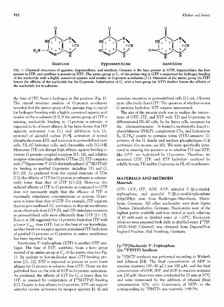
Guanine Hypoxanthine Xanthine
FIG. 1. Chemical structures of guanine, hypoxanthine, and xanthine. Guanine is the base present in GTP, hypoxanthine the base present in ITP, and xanthine is present in XTP. The amino group at C, of the purine ring in GTP is important for hydrogen bonding of the nucleotide with a highly conserved aspartic acid residue in G-protein a-subunits [ 111. 0 mission of the amino group (in ITP) lowers the affinity of the nucleotide for the G-nrotein. Substitution of C, with a keto group (in XTP) further lowers the affinity of the nucleotide for aesubunits.
the base of ITP, bears a hydrogen at this position (Fig. 1). The crystal structure analysis of G-protein or-subunits revealed that the amino group of the guanine ring is crucial for hydrogen bonding with a highly conserved aspartic acid residue of the a-subunits [l 11. If the amino group of GTP is missing, nucleotide binding to G-protein a-subunits is expected to be of lower affinity. It has been shown that ITP supports activation (via G,) and inhibition (via G,- proteins) of adenylyl cyclase [S-9], activation of retinal phosphodiesterase [lo], and secretion in permeabilized mast cells, HL-60 leukemia cells, and chromaffin cells [12-141. Moreover, ITP can disrupt high-affinity agonist binding to various G-protein-coupled receptors [15, 161, and inhibits receptor-stimulated high-affinity GTPase [7]. ITP competes with [35S]guanosine 5’-O-(3-thiotriphosphate) ([“S]GTPyS) for binding to purified G-proteins, including TD and G, [17-201. As predicted from the crystal structure of TDa [l 11, the affinity of ITP for G-protein a-subunits is substan- tially lower than that of GTP [17-201. However, the reduced affinity of ITP to G-proteins as compared to GTP does not necessarily imply that the efficacy of ITP at maximally stimulatory concentrations to activate G-pro- teins is lower than that of GTP. For example, ITP supports thyrotropin-mediated AC activation in thyroid membranes more effectively than GTP [5], and ITP stimulates secretion in permeabilized cells more efficiently than GTP [13-151. Ross et al. [8] suggested that G-proteins hydrolyze ITP with a lower V,,, than GTP, but to the best of our knowledge, neither basal nor receptor agonist-stimulated ITP hydrolysis of purified G-proteins or G-proteins in native membranes has been reported so far.
Xanthosine 5’-triphosphate (XTP) is another GTP ana- logue. The base of XTP, xanthine, bears a keto group instead of an amino group at C2 of the purine ring (see Fig. 1). By analogy to low-molecular mass GTP-binding pro- teins [21, 221, XTP is expected to possess an even lower affinity for G-protein cr-subunits than GTP. There are few published data on the role of XTP in G-protein activation. As predicted, the affinity of XTP for G, is lower than for ITP, as assessed by competition of [35S]GTPyS binding [17]. Despite its low affinity to G-proteins, XTP can support adenylyl cyclase activation by receptor agonists [5, 61 and
stimulate secretion in permeabilized cells [12-14, 231 even more effectively than GTP. The question of whether or not G-proteins hydrolyze XTP remains unanswered.
The aim of the present study was to analyze the interac- tions of GTP, ITP, and XTP with TD and G-proteins in differentiated HL-60 cells. In the latter cells, receptors for the chemoattractants N-formyl-L-methionyl-L-leucyl+ phenylalanine (FMLP), complement C5a, and leukotriene B, (LTB,) couple to pertussis toxin (PTX)-sensitive G- proteins of the G, family and mediate phospholipase C-p activation (for review, see [4]). We were specifically inter- ested in treating the question as to whether ITP and XTP, like GTP, are hydrolyzed by G-proteins. Therefore, we measured GTP, ITP, and XTP hydrolysis catalyzed by soluble bovine TD and by G-proteins in HL-60 membranes.
MATERIALS AND METHODS Materials
GTP, GDP, ITP, ADP, ATP, adenylyl 5’.[p,y-imido]- triphosphate, and guanylyl 5’-[P,y-imidoltriphosphate (GppNHp), were from Boehringer-Mannheim, Mann- heim, Germany. All other nucleotides were from Sigma Chemie, Deisenhofen, Germany. Nucleotides were of the highest purity available and were stored as stock solutions of 10 mM each in distilled water at -20°C. Nucleotide dilutions were prepared fresh daily in distilled water. [32P]Pi (8500 -9 100 Ci/mmol) was obtained from DuPont/New England Nuclear, Bad Homburg, Germany.
[y-32P/Nucleoside 5’-Triphosphate ([r-3ZP]NTP) Synthesis
[T-~~P]NTP synthesis was performed according to Walseth and Johnson [24]. The final concentration of ADP in reaction mixtures (50-80 FL) was 100 FM, and the final concentration of GDP, IDP, and XDP in reaction mixtures was 250 PM. Reactions were conducted for 15 min at 37°C and were terminated by the addition of ethanol (final concentration 50%, v/v). Conversion of NDPs to the corresponding [y-32P]NTPs was routinely >99.5%.
Interactions of GTP, ITP and XTP with G-proteins 553
TD Preparation and TD Elution from Rod Outer Segment (ROS) Disk Membranes
Bovine ROS disk membranes were prepared according to
Papermaster and Dreyer [25]. The membranes were washed
six times with isotonic buffer (5 mM MgCl,, 0.1 mM
EDTA, 1 mM dithiothreitol, 0.1 M NaCl, and 10 mM
Tris/HCl, pH 7.5) and six times with hypotonic buffer (0.1
mM EDTA, 1 mM dithiothreitol, and 10 mM Tris/HCl, pH
7.5) [26]. TD was eluted from ROS disk membranes in
hypotonic buffer substituted with 100 FM GTP. Mem-
branes were centrifuged for 40 min at 4°C and 226,000 X
g. The TD-containing supernatant fluid was carefully re-
moved and concentrated in an Amicon concentration
chamber (PM 10 membrane) (Amicon, Wit-ten, Germany).
Thereafter, TD was diluted with GTP-free hypotonic buffer
[26]. This concentration/dilution procedure was repeated
three times to remove free GTP. TD (50-60 FM) was
stored in 50-100 FL aliquots at -80°C. Analysis of TD by
SDS polyacrylamide gel electrophoresis and subsequent
silver staining revealed a purity of >98% (data not shown).
To study TD elution with various NTPs, ROS disk
membranes were subjected to six isotonic and six hypotonic
washes as described above. ROS disk membranes were
suspended at 3.0-3.7 mg of protein/ml in hypotonic buffer.
Two hundred microliters of this suspension were placed
into ultracentrifuge tubes containing stock solutions of
NTPs to give the desired final concentrations. The con-
tents of tubes were mixed, immediately placed into a
Beckman TLA 100.1 rotor (Beckman, Munich, Germany)
and centrifuged for 15 min at 365,000 X g in a Beckman
TL-100 ultracentrifuge. All procedures were performed at
4°C. Thereafter, 100 I_LL of the supernatant fluid were
removed, diluted with 200 PL of hypotonic buffer, and
stored at -20°C until further analysis by SDS polyacryl-
amide electrophoresis and silver staining.
ATPase, STPase, ITPase, and XTPase Assays
To determine the ATPase, GTPase, ITPase, and XTPase
activity of TD, reaction mixtures (100 I.LL) contained TD
(200-300 nM), unlabeled NTPs at various concentrations,
the corresponding [y-32P]NTPs (0.5-l .5 $Zi/tube), and a
buffer consisting of 5 mM EDTA, 1 mM dithiothreitol,
0.2% (w/v) bovine serum albumin, and 50 mM triethanol-
amine/HCl, pH 7.4. To obtain blank values, tubes contain-
ing all components described above and hypotonic buffer
instead of TD were processed in parallel with the TD-
containing tubes. Reactions were conducted for 30-45 mm
at 25°C and were terminated by the addition of 900 ~.LL of
a stirred suspension consisting of 5% (w/v) activated
charcoal and 50 mM NaH,PO+ pH 2.0. Under these
conditions, (32P]Pi release was linear. Reaction mixtures
were centrifuged for 40 min at 4°C at 2,500 X g. Seven
hundred microliters of the supernatant fluid of reaction
mixtures were removed, and Cerenkov radiation of [32PJP,
was determined.
To determine the GTPase, ITPase, and XTPase activity
in HL-60 membranes, membranes (800-1200 I_L~ of pro-
tein) were suspended in 1.5 mL of 10 mM triethanolaminel
HCl, pH 7.4, and centrifuged for 10 min at 30,000 X g at
4°C to remove endogenous nucleotides as far as possible.
Thereafter, membranes were suspended at 500-1000 kg of
protein/ml in 10 mM triethanolamine/HCl, pH 7.4, and
immediately used for experiments. Reaction mixtures (100
yL) contained washed HL-60 membranes (5-10 Fg of
protein/tube), 0.5 p,M [y-32P]NTPs (0.2-0.3 l_&i/tube), 0.5
mM M&l,, 0.1 mM EGTA, 0.1 mM ATP, 1 mM adenylyl
5’-[l&y-imidoltriphosphate, 5 mM creatine phosphate, 40
kg of creatine kinase, 1 mM dithiothreitol, and 0.2% (w/v)
bovine serum albumin in 50 mM triethanolamine/HCl, pH
7.4. In some experiments, EDTA (5 mM) was included into
reaction mixtures instead of MgClJEGTA. Reactions were
conducted for 15-20 min at 25’C. Under these conditions,
[32P]P, release was linear. Stopping of reactions and extrac-
tion of [‘2P]P, was performed as described above. To study
the substrate concentration dependency of NTP hydrolysis,
reaction mixtures contained unlabeled NTPs at various
concentrations and the corresponding [y~72P]NTPs (0.5
pCi/tube).
Miscellaneous
Protein was determined according to Peterson [27]. SDS
polyacrylamide gel electrophoresis and silver staining of
gels were performed as described [28, 291. Immunoblotting
with G-protein (Y. common- and B COlll”lllll -peptide antisera was
performed as described [30]. HL-60 cells were grown in
suspension culture and differentiated with dibutyryl CAMP
(0.2 mM) for 48 hr [31]. Treatment of cells with PTX was
carried out as described [31]. HL-60 membranes were
prepared as described (321. Concentration-response curves
were analyzed by nonlinear regression, using the Prism
program (GraphPad, Prism, San Diego, CA).
RESULTS
To study GTP, ITP, and XTP hydrolysis by TD, we
designed experimental conditions that could exclude a
contribution of nucleoside diphosphate kinase [E.C.
2.6.4.6.1 (NDPK) on NTP hydrolysis. Certain types of
NDPKs can be closely associated with G-proteins and
catalyze the formation of GDP to GTP by NTP in a strictly
Mg ‘+-dependent m anner [33, 341. Therefore, in the pres
ence of Mg’+, coupled NDPK/GTPase reactions may resulr
in apparent NTPase activity ]35]. To avoid this potential
complication in the interpretation of the NTP hydrolysis
studies, we assessed NTP hydrolysis by soluble TD in the
presence of EDTA (see Materials and Methods). EDTA
inhibits NDPK-catalyzed transphosphorylation [33,34], but
binding of guanine nucleotides to the o-subunit of TD
(TD,) is not influenced by Mg2+ [36]. In the presence of
EDTA, TD catalyzed the hydrolysis of GTP, ITP, and XTP
in a substrate concentration-dependent and saturable man-
554 Klinker and Seifert
3.5
0
A
/1 0
+
A , I I
0 i0 2.0 30 s i NTP (W) NTP (-log M)
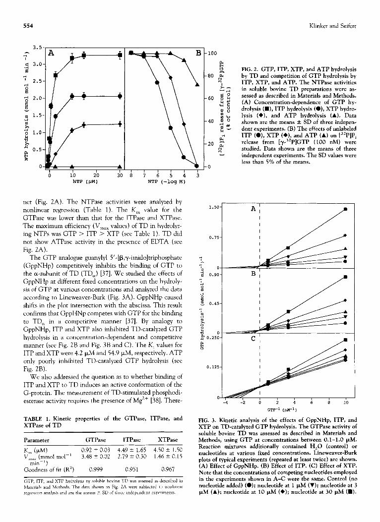
ner (Fig. 2A). The NTP ase activities were analyzed by nonlinear regression (Table 1). The K, value for the GTPase was lower than that for the ITPase and XTPase.
The maximum efficiency (V,,, values) of TD in hydrolyz- ing NTPs was GTP > ITP > XTP (see Table 1). TD did
not show ATPase activity in the presence of EDTA (see Fig. 2A).
The GTP analogue guanylyl 5’-[B,y-imidoltriphosphate (GppNHp) competitively inhibits the binding of GTP to the a-subunit of TD (TD,) [37]. We studied the effects of GppNHp at different fixed concentrations on the hydroly- sis of GTP at various concentrations and analyzed the data
according to Lineweaver-Burk (Fig. 3A). GppNHp caused shifts in the plot intersection with the abscissa. This result
confirms that GppHNp competes with GTP for the binding to TD, in a competitive manner [37]. By analogy to GppNHp, ITP and XTP also inhibited TD-catalyzed GTP
hydrolysis in a concentration-dependent and competitive manner (see Fig. 2B and Fig. 3B and C). The K, values for ITP and XTP were 4.2 p,M and 54.9 FM, respectively. ATP only poorly inhibited TD-catalyzed GTP hydrolysis (see
Fig. 2B). We also addressed the question as to whether binding of
ITP and XTP to TD induces an active conformation of the G-protein. The measurement of TD-stimulated phosphodi-
esterase activity requires the presence of Mg2+ [38]. There-
TABLE 1. Kinetic properties of the GTPase, ITPase, and XTF’ase of TD
Parameter GTPase ITPase XTPase
K, (cLM) 0.92 + 0.03 4.49 2 1.65 4.50 + 1.50 V,,, (mm01 mol-’ 3.48 2 0.02 2.79 + 0.30 1.46 + 0.15
min-‘) Goodness of fit (R’) 0.999 0.95 1 0.967
GTE’, ITI?, and XTP hydrolysis by soluble bovine TD was assessed as described in
Materials and Methods. The data shown m Fig. 2A were subjected to nonhnear
regresamn analysis and are the means + SD of three independent experments.
FIG. 2. GTP, ITP, XTP, and ATP hydrolysis by TD and competition of GTP hydrolysis by ITP, XTP, and ATP. The NTPase activities in soluble bovine TD preparations were as- sessed as described in Materials and Methods. (A) Concentration-dependence of GTP hy- drolysis (W), ITP hydrolysis (O), XTP hydrae lysis (+), and ATP hydrolysis (A). Data shown are the means f SD of three indepen- dent experiments. (B) The effects of unlabeled ITP (O), XTP (*), and ATP (A) on [32P]Pi release from [y-32P]GTP (100 nM) were studied. Data shown are the means of three independent experiments. The SD values were less than 5% of the means.
0.125
0 -4 -2 0 2 4 6 8 10
GTP-1 (PM-~)
FIG. 3. Kinetic analysis of the effects of GppNHp, ITP, and XTP on TD-catalyzed GTP hydrolysis. The GTPase activity of soluble bovine TD was assessed as described in Materials and Methods, using GTP at concentrations between 0.1-1.0 PM. Reaction mixtures additionally contained Hz0 (control) or nucleotides at various fixed concentrations. Lineweaver-Burk plots of typical experiments (repeated at least twice) are shown. (A) Effect of GppNHp. (B) Effect of ITP. (C) Effect of XTP. Note that the concentrations of competing nucleotides employed in the experiments shown in A-C were the same. Control (no nucleotide added) (0); nucleotide at 1 PM (V); nucleotide at 3 PM (A); nucleotide at 10 PM (+); nucleotide at 30 PM (H).
Hz0 GTP ITP
XTP ATP Hz0 GTP ITP ITP ITP
1IllM 1mM 1mM 1mM
GTP GTP GTP H20 ITP
1OOnM 1pM 1O~MlOO~M 1pM 10pM lOO/.iM 1mM
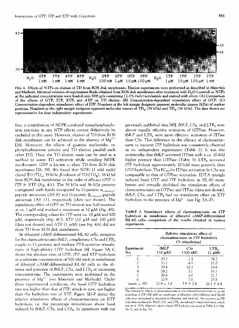
FIG. 4. Effects of NTPs on elution of TD from ROS disk membranes. Elution experiments were performed as described in Materials and Methods. Identical volumes of supematant fluids obtained from ROS disk membranes after treatment with H,O (control) or NTPs at the indicated concentrations were loaded onto SDS gels containing 12.5% (w/v) acrylamide and stained with silver. (A) Comparison of the effects of GTP, ITP, XTP, and ATP on TD elution. (B) Concentration-dependent stimulatory effect of GTP. (C) Concentration-dependent stimulatory effect of ITP. Numbers at the left margin designate apparent molecular masses (kDa) of marker proteins. Numbers at the right margin designate apparent molecular masses of TD, (39 kDa) and TD, (36 kDa). The data shown are representative for four independent experiments.
fore, a contribution of NDPK-catalyzed transphosphoryla- tion reactions in any NTP effects cannot definitively be excluded in this assay. However, elution of TD from ROS disk membranes can be achieved in the absence of MgZf [26]. Moreover, the effects of guanine nucleotides on phosphodiesterase activity and TD elution parallel each other [37]. Thus, the TD elution assay can be used as a method to assess TD activation while avoiding NDPK
involvement. GTP is known to elute TD from ROS disk membranes [26, 391. We found that NTPs (1 mM each)
eluted TD (TD,, 39 kDa; B-subunit of TD (TDa), 36 kDa) from ROS disk membranes in the order of efficacy GTP > ITP + XTP (Fig. 4A). The 39 kDa and 36 kDa proteins comigrated with bands recognized by G-protein (Y common-
peptide antiserum (AS 8) and G-protein B c”mm”” -peptide antiserum (AS ll), respectively (data not shown). The stimulatory effect of GTP on TD elution was half-maximal at ca. 1 PM and reached a maximum at 10 PM (Fig. 4B). The corresponding values for ITP were ca. 10 PM and 100 I.LM, respectively (Fig. 4C). XTP (10 I.LM and 100 p,M) (data not shown) and ATP (1 mM) (see Fig. 4A) did not elute TD from ROS disk membranes.
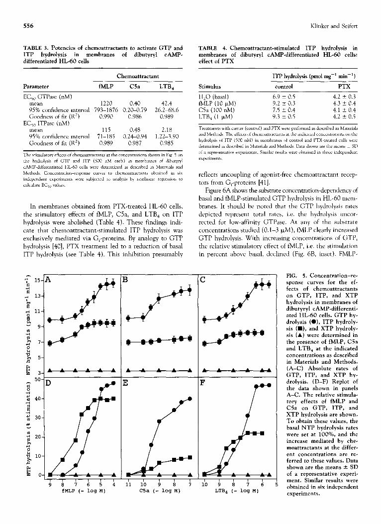
In dibutyryl CAMP-differentiated HL-60 cells, receptors
for the chemoattractants fMLP, complement C5a and LTB, couple to G,-proteins and mediate PTX-sensitive stimula- tions of high-affinity GTP hydrolysis [4]. Figure SA-C shows the absolute rates of GTP, ITP, and XTP hydrolysis at a substrate concentration of 500 nM each in membranes of dibutyryl CAMP-differentiated HL-60 cells in the ab- sence and presence of fMLP, C5a, and LTB, at increasing concentrations. The experiments were performed in the presence of MgZf (see Materials and Methods). Under these experimental conditions, the basal GTP hydrolysis rate was higher than that of ITP, which in turn, was higher than the hydrolysis rate of XTP. Figure 5D-F depict the relative stimulatory effects of chemoattractants on NTP hydrolysis, i.e. the percentage stimulations above basal induced by fMLP, C5a, and LTB,. In agreement with our
previously published data [40], fMLP, C5a, and LTB, were almost equally effective activators of GTPase. However, fMLP and LTB, were more effective activators of ITPase than C5a. This difference in the efficacy of chemoattrac-
tants to increase ITP hydrolysis was consistently observed in six independent experiments (Table 2). It was also noteworthy that fMLP activated ITPase with a ca. lo-fold
higher potency than GTPase (Table 3). LTB, activated ITP hydrolysis approximately 20-fold more potently than GTP hydrolysis. The EC,, for ITPase activation by C5a was
comparable to that of GTPase activation. EDTA strongly reduced basal GTP and ITP hydrolysis in HL-60 mem- branes and virtually abolished the stimulatory effects of chemoattractants on GTPase and ITPase (data not shown). fMLP, C5a, and LTB, had no stimulatory effect on XTP
hydrolysis in the presence of MgZf (see Fig. 5A-F).
TABLE 2. Stimulatory effects of chemoattractants on 1TP hydrolysis in membranes of dibutyryl CAMP-differentiated HL-60 cells: comparison of the results obtained in various experiments
Relative stimulatory effect of chemoattractants on ITP hydrolysis
(% stimulation)
Experiment fMLP LTB, No. (10 PM) (lO::M) (1 PM)
1 33.3 2.4 34.7 2 33.2 4.9 21.0 3 39.4 10.3 19.8 4 282 5.1 33.1 5 30.4 4.8 22.2 6 32.9 7.8 25.8
mean 2 SD 32.9 2 3.8 5.9 -+ 2.8 26.1 t 6.4
The stlmulatory effects of chemoattractants at the mdicated concentrations on the
hydrolysis of ITP (500 nM) m membranes of dtbutytyl CAMP-differentiated HL-60
cells were determined as described in Materials and Methods. The increasea m ITP
hydrolysis medlated by NLP, C5a, and LTB, are referred TV basal hydrolysis. which was set at 100%. Absolute values of basal ITP hydrolysis are given m Table 4, m Figs.
5A-C, and m Fig. 7A.
556 Klinker and Seifert
TABLE 3. Potencies of chemoattractants to activate GTP and ITP hydrolysis in membranes of dibutyryl CAMP- differentiated HL-60 cells
TABLE 4. Chemoattractant-stimulated ITP hydrolysis in membranes of dibutyryl CAMP-differentiated HL-60 cells: effect of PTX
Chemoattractant ITP hydrolysis (pm01 mg-’ min-‘)
Parameter fMLP C5a LTB, Stimulus control PTX
EC,, GTPase (nM) mean 1220 0.40 42.4 95% confidence interval 793-1876 0.20-0.79 26.2-68.6 Goodness of fit (R*) 0.990 0.986 0.989
EC,, ITPase (nM) mean 115 0.48 2.18 95% confidence interval 71-185 0.24-0.94 1.22-3.90 Goodness of fit (R’) 0.989 0.987 0.985
The stimulatory effects of chemoattractants at the concentratmns shown in Fig. 5 on
the hydrolysis of GTP and ITP (500 nM each) in membranes of dihutyryl
CAMP-differennated HL-60 cells were determined as described m Mater& and
Methods. Concentration-response cuwes to chemoattractants ohtained in six
independent experiments were subjected to analysis by nonlinear regression to
calculate ECSo values.
In membranes obtained from PTX-treated HL-60 cells,
the stimulatory effects of fMLP, C5a, and LTB, on ITP
hydrolysis were abolished (Table 4). These findings indi- cate that chemoattractant-stimulated ITP hydrolysis was exclusively mediated via G,-proteins. By analogy to GTP hydrolysis [40], PTX treatment led to a reduction of basal ITP hydrolysis (see Table 4). This inhibition presumably
4
=:$:
fMLP (- log M)
T
3
A % : : A
I I I I 1
11 10 9 8 7 C5a (- .og M)
H,O (basal) fMLP (10 pM) C5a (100 nM) LTB, (1 CLM)
6.9 ? 0.5 4.2 -t 0.3 9.2 -t 0.3 4.3 * 0.4 7.5 ? 0.4 4.1 2 0.4 9.3 +- 0.5 4.2 ? 0.5
Treatments wth carrier (control) and PTX were performed as described m Materials
and Methods. The effects of chemoattracrants at the mdicated concentrations on the
hydrolysis of ITP (500 nM) in membranes of control and PTX-treated cells were
determined as described in Materials and Methods. Data shown are the meanz t- SD
of a representawe experiment. Similar results were obtained m three independent
experiments.
reflects uncoupling of agonist-free chemoattractant recep- tors from G,-proteins [41].
Figure 6A shows the substrate concentration-dependency of basal and fMLP-stimulated GTP hydrolysis in HL-60 mem- branes. It should be noted that the GTP hydrolysis rates depicted represent total rates, i.e. the hydrolysis uncor- rected for low-affinity GTPase. At any of the substrate concentrations studied (0.1-3 FM), fMLP clearly increased GTP hydrolysis. With increasing concentrations of GTP, the relative stimulatory effect of fMLP, i.e. the stimulation in percent above basal, declined (Fig. 6B, inset). FMLP-
A : : A :A
7 r L 10 9 8 7 6
LTBI (- log M)
FIG. 5. Concentration-re- sponse curves for the ef- fects of chemoattractants on GTP, ITP, and XTP hydrolysis in membranes of dibutyryl CAMP-differenti- ated HL-60 cells. GTP hy- drolysis (O), ITP hydroly- sis (W), and XTP hydroly- sis (A) were determined in the presence of fMLP, C5a and LTB, at the indicated concentrations as described in Materials and Methods. (A-C) Absolute rates of GTP, ITP, and XTP hy- drolysis. (D-F) Replot of the data shown in panels A-C. The relative stimula- tory effects of fMLP and C5a on GTP, ITP, and XTP hydrolysis are shown. To obtain these values, the basal NTP hydrolysis rates were set at lOO%, and the increase mediated by che- moattractants at the differ- ent concentrations are re- ferred to these values. Data shown are the means f SD of a representative experi- ment. Similar results were obtained in six independent experiments.
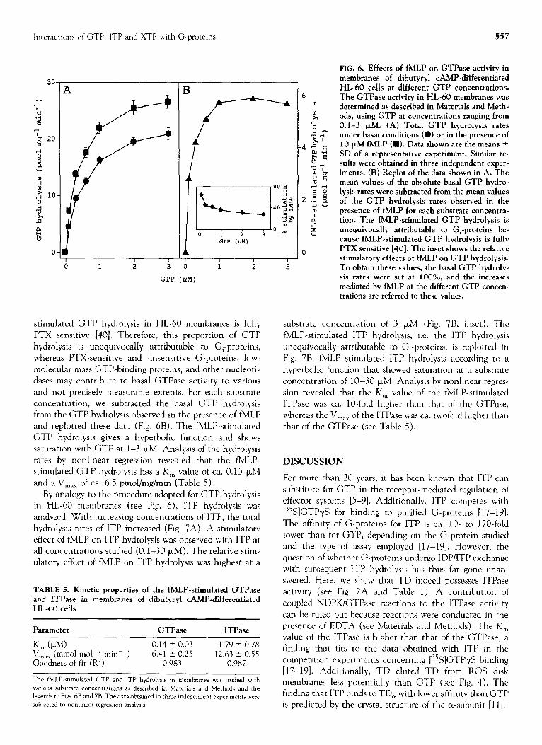
Interactions of GTP, ITP and XTP wtth G-proteins 557
GTP (PM)
1 I I I
0 1 2 3 6 i i
GTP (FM)
3
stimulated GTP hydrolysis in HL-60 membranes is fully P’TX sensitive [40]. Therefore, this proportion of GTP hydrolysis is unequivocally attributable to Gi-proteins, whereas PTX-sensitive and -insensitive G-proteins, low- molecular mass GTP-binding proteins, and other nucleoti- dases may contribute to basal GTPase activity to various and not precisely measurable extents. For each substrate concentration, we subtracted the basal GTP hydrolysis from the GTP hydrolysis observed in the presence of fMLP and replotted these data (Fig. 6B). The fMLP-stimulated GTP hydrolysis gives a hyperbolic function and shows saturation with GTP at l-3 PM. Analysis of the hydrolysis rates by nonlinear regression revealed that the fMLP- stimulated GTP hydrolysis has a K,, value of ca. 0.15 PM and a V,,, of ca. 6.5 pmol/mg/min (Table 5).
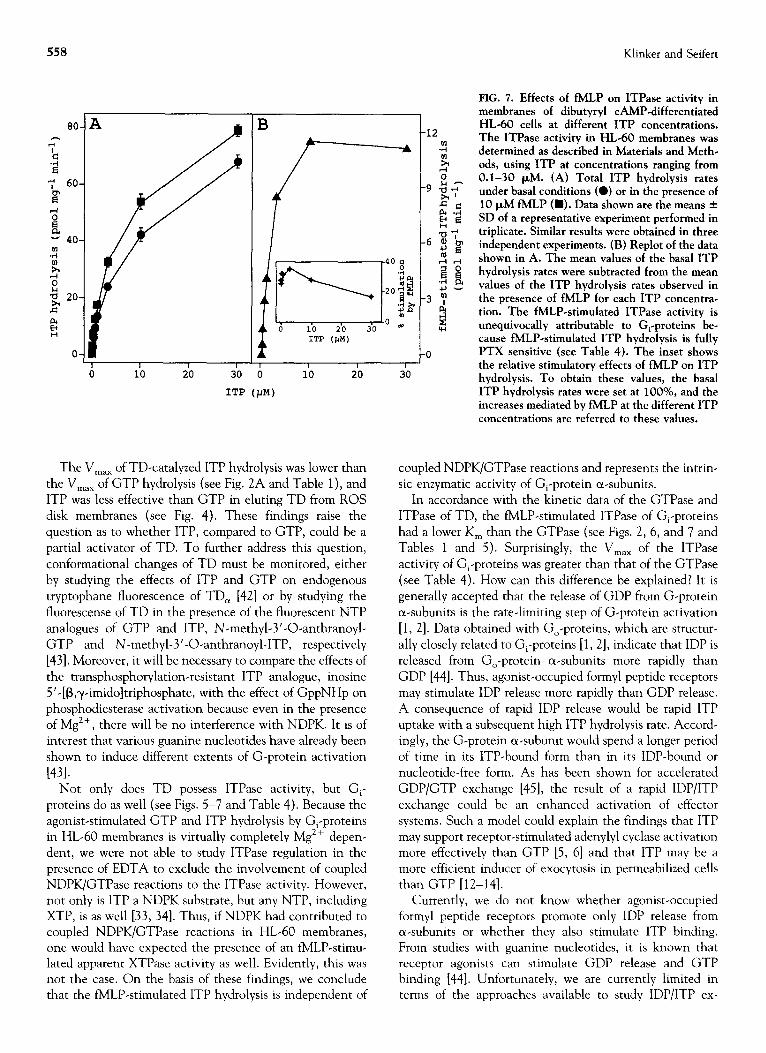
By analogy to the procedure adopted for GTP hydrolysis in HL-60 membranes (see Fig. 6), ITP hydrolysis was analyzed. With increasing concentrations of ITP, the total hydrolysis rates of ITP increased (Fig. 7A). A stimulatory effect of fMLP on ITP hydrolysis was observed with ITP at all concentrations studied (0.1-30 FM). The relative stim- ulatory effect of fMLP on ITP hydrolysis was highest at a
TABLE 5. Kinetic properties of the fMLP-stimulated GTPase and ITPase in membranes of dibutyryl CAMP-differentiated HL-60 cells
Parameter GTPase ITPase
K,, (PM) 0.14 2 0.03 1.79 + 0.28 V ,n,lX (mm01 rnol- ’ min- ’ ) 6.41 0.25 -c 12.63 0.55 -+ Goodness of fit (R’) 0.983 0.987
The tMLI’-stlmulared GTP and ITP hydrolysis in membranes was studied with
YXIOUS substrate concentrations as described in Materials and Methods and the
legends 10 Flpr. 6B and 7R. The data obtamed in three independent experimenta were
subjected to nonlinear regression analysis.
FIG. 6. Effects of fMLP on GTPase activity in membranes of dibutyryl CAMP-differentiated HL-60 cells at different GTP concentrations. The GTPase activity in HL-60 membranes was determined as described in Materials and Meth- ods, using GTP at concentrations ranging from 0.1-3 FM. (A) Total GTP hydrolysis rates under basal conditions (0) or in the presence of 10 PM fMLP (a). Data shown are the means + SD of a representative experiment. Similar re- sults were obtained in three independent exper- iments. (B) Replot of the data shown in A. The mean values of the absolute basal GTP hydro- lysis rates were subtracted from the mean values of the GTP hydrolysis rates observed in the presence of fMLP for each substrate concentra- tion. The fMLP-stimulated GTP hydrolysis is unequivocally attributable to G,-proteins be- cause fMLP-stimulated GTP hydrolysis is fully PTX sensitive [40]. The inset shows the relative stimulatory effects of fMLP on GTP hydrolysis. To obtain these values, the basal GTP hydroly- sis rates were set at 1000/o, and the increases mediated by fMLP at the different GTP concen- trations are referred to these values.
substrate concentration of 3 PM (Fig. 7B, inset). The fMLP-stimulated ITP hydrolysis, i.e. the ITP hydrolysis unequivocally attributable to G,-proteins, is replotted in Fig. 7B. fMLP stimulated ITP hydrolysis according to a hyperbolic function that showed saturation at a substrate concentration of lo-30 FM. Analysis by nonlinear regres- sion revealed that the K, value of the fMLP-stimulated ITPase was ca. IO-fold higher than that of the GTPase, whereas the V,,, of the ITPase was ca. twofold higher than that of the GTPase (see Table 5).
DISCUSSION
For more than 20 years, it has been known that ITP can substitute for GTP in the receptor-mediated regulation of effector systems [5-91. Additionally, ITP competes with [35S]GTPyS for binding to purified G-proteins [17-191. The affinity of G-proteins for ITP is ca. IO- to 170-fold lower than for GTP, depending on the G-protein studied and the type of assay employed [17-191. However, the question of whether G-proteins undergo IDP/ITP exchange with subsequent ITP hydrolysis has thus far gone unan- swered. Here, we show that TD indeed possesses ITPase activity (see Fig. 2A and Table 1). A contribution of coupled NDPK/GTPase reactions to the ITPase activity can be ruled out because reactions were conducted in the presence of EDTA (see Materials and Methods). The K, value of the ITPase is higher than that of the GTPase, a finding that fits co the data obtained with ITP in the competition experiments concerning [35S]GTPyS binding [17-191. Additionally, TD eluted TD from ROS disk membranes less potentially than GTP (see Fig. 4). The finding that ITP binds to TD, with lower affinity than GTP is predicted by the crystal structure of the au-subunit [I I].
558 Klinker and Seifert
10 20
1'~ (JIM)
-0 T I I I ..^
30 0 io 2.0
ITP (PM)
The Max of TD-catalyzed ITP hydrolysis was lower than
the V,,, of GTP hydrolysis (see Fig. 2A and Table 1 ), and ITP was less effective than GTP in eluting TD from ROS disk membranes (see Fig. 4). These findings raise the question as to whether ITP, compared to GTP, could be a partial activator of TD. To further address this question, conformational changes of TD must be monitored, either by studying the effects of ITP and GTP on endogenous tryptophane fluorescence of TD, [42] or by studying the fluorescense of TD in the presence of the fluorescent NTP analogues of GTP and ITP, N-methyl-3’-O-anthranoyl- GTP and N-methyl-3’-0-anthranoyl-ITP, respectively [43]. Moreover, it will be necessary to compare the effects of the transphosphorylation-resistant ITP analogue, inosine 5’-[B,y-imidoltriphosphate, with the effect of GppNHp on phosphodiesterase activation because even in the presence of Mg *+, there will be no interference with NDPK. It is of interest that various guanine nucleotides have already been shown to induce different extents of G-protein activation
]431. Not only does TD possess ITPase activity, but G,-
proteins do as well (see Figs. 5-7 and Table 4). Because the agonist-stimulated GTP and ITP hydrolysis by G,-proteins in HL-60 membranes is virtually completely Mg2+ depen- dent, we were not able to study ITPase regulation in the presence of EDTA to exclude the involvement of coupled NDPK/GTPase reactions to the ITPase activity. However, not only is ITP a NDPK substrate, but any NTP, including XTP, is as well [33, 341. Thus, if NDPK had contributed to coupled NDPK/GTPase reactions in HL-60 membranes, one would have expected the presence of an fMLP-stimu- lated apparent XTPase activity as well. Evidently, this was not the case. On the basis of these findings, we conclude that the fMLP-stimulated ITP hydrolysis is independent of
FIG. 7. Effects of fMLP on ITPase activity in membranes of dibutyryl CAMP-differentiated HL-60 cells at different ITP concentrations. The ITPase activity in HL-60 membranes was determined as described in Materials and Meth- ods, using ITP at concentrations ranging from 0.1-30 PM. (A) Total ITP hydrolysis rates under basal conditions (a) or in the presence of 10 PM fMLP (m). Data shown are the means + SD of a representative experiment performed in triplicate. Similar results were obtained in three independent experiments. (B) Replot of the data shown in A. The mean values of the basal ITP hydrolysis rates were subtracted from the mean values of the ITP hydrolysis rates observed in the presence of fMLP for each ITP concentra- tion. The fMLP-stimulated ITPase activity is unequivocally attributable to G,-proteins be- cause fMLP-stimulated ITP hydrolysis is fully PTX sensitive (see Table 4). The inset shows the relative stimulatory effects of fMLP on ITP
JU hydrolysis. To obtain these values, the basal ITP hydrolysis rates were set at lOO%, and the increases mediated by fMLP at the different ITP concentrations are referred to these values.
coupled NDPK/GTPase reactions and represents the intrin- sic enzymatic activity of G,-protein a-subunits.
In accordance with the kinetic data of the GTPase and ITPase of TD, the fMLP-stimulated ITPase of G,-proteins had a lower K, than the GTPase (see Figs. 2, 6, and 7 and Tables 1 and 5). Surprisingly, the V,,, of the ITPase activity of G,-proteins was greater than that of the GTPase (see Table 4). How can this difference be explained? It is generally accepted that the release of GDP from G-protein a-subunits is the rate-limiting step of G-protein activation [l, 21. Data obtained with Go-proteins, which are structur- ally closely related to G,-proteins [l, 21, indicate that IDP is released from Go-protein a-subunits more rapidly than GDP [44]. Thus, agonist-occupied formyl peptide receptors may stimulate IDP release more rapidly than GDP release. A consequence of rapid IDP release would be rapid ITP uptake with a subsequent high ITP hydrolysis rate. Accord- ingly, the G-protein a-subunit would spend a longer period of time in its ITP-bound form than in its IDP-bound or nucleotide-free form. As has been shown for accelerated GDP/GTP exchange [45], the result of a rapid IDP/ITP exchange could be an enhanced activation of effector systems. Such a model could explain the findings that ITP may support receptor-stimulated adenylyl cyclase activation more effectively than GTP [5, 61 and that ITP may be a more efficient inducer of exocytosis in permeabilized cells than GTP [12-141.
Currently, we do not know whether agonist-occupied formyl peptide receptors promote only IDP release from a-subunits or whether they also stimulate ITP binding. From studies with guanine nucleotides, it is known that receptor agonists can stimulate GDP release and GTP binding [44]. Unfortunately, we are currently limited in terms of the approaches available to study IDP/ITP ex-
Interactions of GTP, ITP and XTP with G-proteins 559
change on G,-proteins and effector system activation in HL-60 membranes. First, in view of the already rapid spontaneous GDP release from G,-proteins in HL-60 mem- branes [46], it is highly unlikely that receptor-stimulated IDP release can be detected in this system. Second, radio- labeled ITP analogues such as [35S]inosine 5’-O-(3-thio- triphosphate) or [3H]inosine 5’-[B,y-imidoltriphosphate are not generally available, and it is doubtful whether the affinity of these radioligands for G-protein c-w-subunits is high enough for binding experiments. Third, the effects of ITP on fMLP-stimulated phospholipase C activation in HL-60 membranes cannot be interpreted unequivocally because ITP is also an effective purinoceptor agonist and because purinoceptors activate phospholipase C via G,- proteins as do formyl peptide receptors [47, 481. These limitations also apply for XTP (see Discussion below).
We should like to emphasize that the observed relative differences in ITP hydrolysis rates between TD and Gi- proteins in HL-60 membranes (see Tables 1 and 5) do not necessarily reflect G-protein subtype-specific differences in NTP hydrolysis. The differences could equally well be attributed to differences in the experimental conditions. Specifically, NTP hydrolysis by TD was studied under basal conditions, i.e. in the absence of exogenously added rho- dopsin and Mg*+, whereas NTP hydrolysis by G,-proteins in HL-60 membranes was assessed under maximal receptor agonist stimulation and in the presence of Mg*+.
fMLP and LTB, stimulated ITP hydrolysis more potently than GTP hydrolysis (see Fig. 5D and 5F and Table 3). This could indicate that formyl peptide and LTB, receptors couple to G,-proteins with higher affinity in the presence of IDP/ITP than in the presence of GDP/GTP. In other words, GTP, at the substrate concentration studied (500 nM), could uncouple formyl peptide- and LTB4-receptors from G,-proteins more efficiently than ITP (500 nM) because GTP has a higher affinity for G-proteins than ITP (see Figs. 2, 6, and 7 and Tables 1 and 5) [17-201. However, such a model cannot explain why there is no such difference in potency concerning GTPase- and ITPase activation by C5a receptors and why C5a is such a weak activator of ITP hydrolysis relative to GTP hydrolysis (see Fig. 5 and Table 2).
Intriguingly, differential effects of GTP vs ITP on recep- tor/G-protein interaction have previously been reported. Specifically, ITP supports thyrotropin-stimulated adenylyl cyclase activation in bovine thyroid membranes more efficiently than GTP [5]. In contrast, no such difference between GTP and ITP was found for prostaglandin E, in this system [5]. In turkey erythrocyte membranes, ITP gives rise to a more effective adenylyl cyclase stimulation by B-adrenoceptor agonists than GTP [6]. Collectively, our present data and previously published results clearly show that GDP/GTP and IDP/ITP differentially affect the inter- action of various receptors that are linked to the same class of G-proteins. A possible explanation for these observa- tions could be that GDP- and IDP-liganded G-protein or-subunits adopt different conformations that fit differen-
tially to various receptors. As has already been discussed above, biophysical studies would be required to substantiate the hypothesis that qualitative and/or quantitative confor- mational differences between guanine- and hypoxanthine nucleotide-liganded G-protein ol-subunits exist.
Compared to ITP, considerably less is known about the function and role of XTP in G-protein activation. Previous studies had shown that XTP can substitute for GTP in supporting receptor-mediated adenylyl cyclase activation [6]. Here, we show that XTP, like GppNHp and ITP, competitively inhibits the binding of GTP to the guanine nucleotide-binding site of TD, (see Figs. 2A and 3C). Moreover, TD shows measurable XTPase activity (see Fig. 2A and Table 1 ), and XTP elutes TD from ROS disk membranes (see Fig. 4A). Compared to ITP, XTP was much less potent than ITP in inhibiting GTP hydrolysis and in eluting TD from ROS disk membranes. This finding is not surprising, because hydrogen bonding of the keto group-substituted C, of the purine ring (see Fig. 1) with the highly conserved aspartic acid residue-268 in TD, cannot occur [l I]. Analogous observations as with TD have been made for the binding of GTP, ITP, and XTP to bacterial GTPases [49, 501, elongation factor-Tu [51] and low- molecular mass GTP-binding proteins [21, 221.
TD hydrolyzes XTP at a lower maximal rate than GTP and ITP (see Fig. 2A and Table 1 ), and XTP elutes TD from ROS disk membranes much less efficiently than GTP and ITP (see Fig. 4A). On the basis of these findings, XTP could be considered as a weak partial activator of TD. If this were true, then XTP would be predicted to induce smaller maximal changes in endogenous tryptophane fluorescence of TD than GTP and ITP, and N-methyl-3’-O-anthranoyl- XTP would be expected to induce smaller fluorescence signals upon binding to TD than the corresponding GTP and ITP analogues [43].
However, with regard to other G-proteins than TD, XTP may be a more effective activator than GTP. Specifically, XTP is more effective than GTP in supporting B-adreno- ceptor-mediated adenylyl cyclase activation in turkey erythrocyte membranes [6], and XTP stimulates exocytosis in permeabilized cells more effectively than GTP or even GTPase-resistant GTP analogues [12-14, 231. It is possible that XTP, once it has bound to certain G-protein (Y- subunits, stabilizes a conformation of the G-protein that shows high biological activity. By analogy to observations made with the GTPase-resistant GTP analogues GTPyS and GppNHp [l, 21, th e activity of an XTP-liganded G-protein a-subunit would be expected to he increased if the XTP hydrolysis rate were low. In fact, in HL-60 membranes, we could not detect receptor agonist-stimu- lated XTPase activity although chemoattractant-stimulated GTP- and ITPase activity was readily observed (see Fig. 5). These findings indicate that G,-proteins in HL-60 mem- branes do not possess XTPase activity or that their XTPase activity is below the detection limit of our assay.
To further analyze the mechanism of interaction of XTP with G-proteins, studies with XTP analogues such as
560 Klinker and Seifert
xanthosine 5’-0-(3-thiotriphosphate) (XTPyS) and xan- thosine 5’-[P,y-imidoltriphosphate (XppNHp) must be performed. However, we anticipate that the affinity of these nucleotides to G-protein a-subunits will still be low. Thus, it will be necessary to mutate the highly conserved aspartic acid residue involved in hydrogen bonding with the amino group of C, of guanine (see Fig. 1) [ 111 into an asparagine residue. The consequence of analogous mutations in baca- terial GTPases, elongation factor Tu, and low molecular mass GTP-binding proteins is a dramatic increase in affinity of the mutated proteins for xanthine nucleotides with a parallel decrease in affinity for guanine nucleotides [Zl, 22, 49-511. Only with xanthine nucleotide-preferring G- protein a-subunits can release experiments with [3H]XDP or [or-32P]XDP, and binding studies with [%]XTPyS and [3H]XppNHp be performed.
G,-proteins in a receptor-specific manner. These findings suggest that GTP, ITP, and XTP are differential signal sorters and signal amplifiers at the G-protein level.
Could the effects of ITP and XTP on G-proteins be of physiological relevance? The bulk intracellular GTP con- centration is estimated to be ca. 50 PM (for review, see [52]). Thus, G-proteins would be expected to always be saturated with GTP. However, GTP cannot be assumed to have free access to the G-proteins. Rather, NDPK- generated GTP may be preferred over exogenous GTP for G-protein activation [52-541. Little is known about the physiological intracellular concentrations of IDP/ITP and XDP/XTP, but we assume that the bulk intracellular con- centrations of these nucleotides are far lower than those of GDP/GTP. This does, however, not exclude the possibility that under certain physiological and/or pathological condi- tions, sufficient XTP and ITP is available to bind to G-protein a-subunits. ITP and XTP could reach the CI- subunits either via diffusion from the cytosol or could locally be generated by NDPK from IDP and XDP, respec- tively, because NDPK is not base specific. The observed chemoattractant-specific differences in GTPase and ITPase activation of G,-proteins in HL-60 membranes could pro- vide, at least in part, the molecular basis for the quite different effects of fMLP, C5a, and LTB, in intact HL-60 cells [4].
Future research on the roles of GTP, ITP, and XTP in G-protein activation will follow various directions. First, biophysical studies with purified G-proteins using nonfluo- rescent and fluorescent nucleotides must be performed. Second, the effects of transphosphorylation-resistant ITP and XTP analogues on activation of effector systems must be studied. Third, the molecular basis of the receptor- specific effects of GTP and ITP on G-protein-mediated signalling must be explored, because this may be a novel target for receptor-specific pharmacological manipulation of signalling processes at a postreceptor level. Fourth, the intracellular GTP, ITP, and XTP concentrations under basal and receptor-stimulated conditions, in pathological settings such as trauma and hypoxia, and under GTP- depleting conditions have to be carefully compared. Finally, studies with xanthosine nucleotide-preferring mutants of G-protein a-subunits are expected to provide substantial insight into the mechanisms underlying the interactions of XDP/XTP and their analogues with G-proteins.
The authors are grateful to Mrs. Eoelyn Gk$ and Mr. Henning
Damm for expert technical assistance and to Drs. Karsten Spicher and
Klaus-Dieter Hinsch for providing antisera. We also acknowledge the most constructive criticisms of the reviewers of this paper. This work
was supported in part by grants from the Deutsche Forschungsgemein-
s&aft.
References
XTP and ITP may also be of pharmacological importance for G-protein activation. Specifically, depletion of intracel- lular guanine nucleotide pools in HL-60 cells by inosine monophosphate dehydrogenase [E.C. 1.1.1.205.] inhibitors such as tiazofurin results in differentiation and changes in G-protein function [55-571. It will be interesting to treat the question as to whether tiazofurin leads to increases in the cellular concentrations of ITP and XTP with conse- quent differential G-protein activation by these NTPs compared to GTP. Intriguingly, GTP depletion in SH- SYSY neuroblastoma cells differentially affects the potency of agonists for various receptors to inhibit adenylyl cyclase
1581.
5.
6.
7.
8.
In conclusion, GTP, ITP, and XTP bind to G-protein a-subunits with different affinities. G-proteins hydrolyze GTP, ITP, and XTP to different maximal extents and may lead to the formation of differently activated c-r-subunits. Chemoattractants stimulate the ITPase and GTPase of
9.
10.
Gilman AG, G-proteins: Transducers of receptor-generated
signals. Annu Rev Biochem 56: 615-649, 1987.
Neer EJ, Heterotrimeric G-proteins: Organizers of transmem-
brane signals. Cell 80: 249-257, 1995.
Stryer L, Visual excitation and recovery. J Biol Chem 266:
10711-10714, 1991.
Klinker JF, Wenzel-Seifert K and Seifert R, G-protein-
coupled receptors in HL-60 human leukemia cells. Gen
Phurnu~ol 27: 33-54, 1996.
Wolff J and Cook H, Activation of thyroid membrane
adenylate cyclase by purine nucleotides. J Biol Chem 248:
350-355, 1973.
Bilezikian JP and Aurbach GD, The effects of nucleotides on
the expression of P-adrenergic adenylate cyclase activity in
membranes from turkey erythrocytes. J Biol Chem 249:
157-161, 1973.
Cassel D and Selinger Z, Catecholamine-stimulated GTPase
activity in turkey erythrocyte membranes. Biochim Biophys
Acta 452: 538-551, 1976.
Ross EM, Maguire ME, Sturgill TW, Biltonen RL and Gilman
AG, Relationship between the P-adrenergic receptor and
adenylate cyclase. J Biol Chem 252: 5761-5775, 1977.
Jakobs KH, Saur W and Schultz G, Inhibition of platelet
adenylate cyclase by epinephrine requires GTP. FEBS Lett
85: 167-170, 1978.
Miki N, Keirns JJ, Marcus FR, Freeman J and Bitensky MW,
Regulation of cyclic nucleotide concentrations in photorecep-
tors: An ATP-dependent stimulation of cyclic nucleotide
phosphodiesterase by light. Proc Natl Acad Sci USA 70: 3820-3824, 1973.
Interactions of GTP, ITP and XTP with G-proteins 561
11.
12.
13
14.
15.
16.
17.
18.
19
20.
21.
22
23.
24.
25.
26.
27.
28.
29.
30.
Noel JP, Hamm HE and Sigler PB, The 2.2 A crystal structure
of transducin-cx complexed with GTl’yS. Nature 366: 654-
663, 1993.
Howell TW, Cockcroft S and Gomperts BD, Essential synergy
hetween Ca” and guanine nucleotides in exocytotic secre-
tion from permeabilized rat mast cells. .I Cell Biol 105:
191-197, 1987.
Stutchfield J and Cockcroft S, Guanine nucleotides stimulate
pc’lyphosyhvinosiride phosphodiesterase and exocytotic se-
cretion from HL60 cells permeahilized with streptolysin 0.
Biochem 1 250: 375-382, 1988.
Morgan A and Burgoyne RD, Stimulation of Ca’+-independent
catecholamine secretion from digitonin-permeabilized bovine
adrenal chrolnaffin cells hy guanine nucleotide analogues.
RelationshIp to arachidonate release. Biochem J 269: 521-
526, 1990.
Blume AJ, interaction of ligands with the opiate receptors of
brain membranes: Regulation by ions and nucleotides. Proc
Nat1 Acad Sci USA 75: 1713-1717, 1978.
Cascieri MA and Liang T, Characterization of the substance
I’ receptor m rat brain cortex membranes and the inhibition
of radioligand binding by guanine nucleotides. J Biol Chem
258: 5158-5164, 1983.
Northup JK, Smigel MD and Gilman AG, The guanine
nucleotide activating site of the regulatory component of
adenylatc c&se. J Biol Chem 257: 11416-11423, 1982.
Higashquna T, Ferguson KM and Sternweis PC, Regulation of
hc,rmt)ne-sensitive GTP-dependent regulatory proteins by
chloride. J Bid Chem 262: 3597-3602, 1987.
Kellrhcr DJ, Dudycz LW, Wright GE and Johnson CL,
Ahlltty ofguaninc nucleotide derivatives to hind and activate
bovine transducin. Mel Phurmacol 30: 603-608, 1986.
Casey PJ, Fong HKW, Simon MI and Gilman AC, Gz, a
guanme nucleotide-binding protein with unique biochemical
properties. _/ Biol Chem 265: 2383-2390, 1990.
Hoffenberg S, Nikolova L, Pan JY, Daniel DS, Wessling-
Rcsnick M, Knoll BJ and Dickey RF, Functional and struc-
tural interactions of the rah5 D136N mutant with xanthine
nucleotidcs. Biochem Biophys Kes Commun 215: 241-249,
1995.
31.
32.
33.
Heterogeneity of three electrophoretlcally distinct G,, (Y-
subunits in mammalian brain. FEBS Lett 307: 215-218, 1992.
Seifert R, H-18er A, Offermanns S, Buschauer A and Schunack
W, Histamine increases cytosolic Ca2+ in dibutyryl-CAMP-
differentiated HL-60 cells via H,-receptors and is an incom-
plete secretagogue. Mel Pharmncol 42: 227-234, 1992.
Seifert R and Schultz G, Reversible activation of NADPH
oxidase in membranes of HL-6L7 human leukemic cells.
Biochem Biophys Res Commun 146: 1296-1302, 1987.
Seifcrt R, Rosenthal W, Schultz G, Wieland T, Gierschik P
and Jakohs KH, The role of nucleosidc diphosphate kinase
reactions in G protein activation of NADPH oxidase b\
guaninr and adenine nucleotides. Eur 1 Rivchem 175: 5 l-55,
1988.
34.
35.
36.
37.
38.
39.
40.
Schmidt G, Lenzen C, Simon I, Deuter R, Cool RH, Goody
RS and Wittinghofer A, Biochemical and biological conse-
quences of changing the specificity of ~21’“” from guanosine
to xanthosme nucleotides. Oncogene 12: 87-96, 1996.
Burgoyne RD and Handel SE, Activation of exocytosis by
GTP analogues in adrenal chromaffin cells revealed by
patch-clamp capacitance measurement. FEBS Lect 344: 139-
142. 1994.
Walseth TF and Johnson RA, The enzymatic preparation of
[a-‘lP]nucleoside triphosphates, cyclic [“PIAMP, and cyclic
[‘zP]GMP. Biochim Biophys Acta 526: 11-31, 1979.
Papermaster DS and Llreyer WJ, Rhodopsin content in the
outer segment of bovine and frog retinal rods. Biochemistry 13:
2438-2444, 1974.
Kroll S, Phillips WJ and Cerione RA, The regulation of the
cyclic GMP phosphodiesterase by the GDP-bound form of the
cu-subunit of transducin. J Biol Chem 264: 4490-4497, 1989.
Peterson GL, L)etermination of total protein. Methods Enzy-
mol91: 95-119, 1983.
Laemmli UK, Cleavage of structural proteins during the
,Issemhly of the head of bacteriophage T4. Nature 227:
680-685, 1970.
Jungblut PR and Seifert R, Analysis by high-resolution
two-dimensIona electrophoresis of differentiation-dependent
alteraciona m cytosolic protein pattern of HL-60 leukemic
cells. I B&hem B~ophys Methods 21: 47-58, 1990.
41.
42.
43.
44.
45.
46.
47.
Kowluru A and Metz SA, Characterization of nucleoside
diphosphokinase activity in human and rodent pancreatic
p-cells: Evidence for its role in the formation of guanosine
triphosphate, a permissive factor for nutrient-induced insulin
secretion. Biochemistry 33: 12495-l 2503, 1994.
Kikkawa S, Takahashi K, Takahashi K, Shimada N, UI M,
Kimura N and Katada T, Conversion of GDP into GTP hy
nucleoside diphosphate kinase on the GTP-hinding proteins.
J Biol Chem 265: 21536-21540, 1990.
Wessling-Resnick M and Johnson GL, Kinetic and hydrody-
namic properties of transducin: Comparison of physical and
structural parameters for GTP-binding proteins. Biochemistry
26: 4316-4323, 1987.
Yamanaka G, Eckstcin F and Stryer L, Interaction of retinal
transducin with guanosine triphosphate analogues: Specificity
of the y-phosphate binding region. Hiochemistr~ 25: 6149-
6153, 1986.
Bitensky MW, Miki N, Krirna JJ, Keirns M, Baraban JM,
Freeman J, Wheeler MA, Lacy J and Marcus FR, Activation
of photoreceptor disk memhrane phosphodiesterase hy light
and ATP. Adv Cyclic Nucleotide Res 5: 213-240, 1975.
Bigay J and Chabre M, Purlflcation of tranducin. Methods
Enzrmol 237: 139-146, 1994.
Klinker JF, Schwaner I, Offermanns S, Hageltiken A and
Seifert R, Llifferential activation of dihutyryl CAMP-differen-
tiated HL-60 human leukemia cells by chemoattractants.
Biochem Pharmacol 48: 1857-1864, 1994.
Gierschik I’, Sidiropoulos D, Steisslinger M and Jakobs KH,
Na ’ regulation of formyl peptide receptor-mediated signal
transduction in HL 60 cells. Evidence that the cation pre-
vents activation of the G-protein by unoccupied receptors.
Eur J Phannacol 172: 481-492, 1989.
Phillips WJ and Cerione RA, The intrinsic fluorescence of
the 01 subunit of transducin. Measurement of receptor-depen-
dent guanine nucleotide exchange. j Rid Chem 263: 15498-
15505, 1988.
Remmers AE and Neuhig RR, Partial G-protein activation by
fluorescent guanine nucleotide analogs. J Biol Chem 271:
4791-4797, 1996.
Florio VA and Sternweis PC, Mechanisms of muscarinic
receptor action on G,, in reconstituted phospholipid vesicles.
J Bid Chem 264: 3909-3915, 1989.
Iiri T, Herzmark P, Nakamoto JM, Van L)op C and Bourne
HR, Rapid GDP release from G,,, in patients with gain and
loss of endocrine function. Nature 371: 164-168, 1994.
Wieland T, Kreias J, Gierschik P and Jakohs KH, Role of GDP
in formyl-peptide-receptor-induced activation of guanine-
nucleotide-hinding proteins in membranes of HL 60 cells. Eur
1 Bbchem 205: 1201-1205, 1992.
Seifert R, Burdr R and Schultz G, Activarion of NADPH
oxidase by purine and pyrimidine nucleotides involves G
proteins and is potentiated by chemotactic peptides. Biochem J 259: 813-819, 1989.
Spicher K, Niirnherg B, J@er B, Rosenthal W and Schultz G, 48. Cowen L>, Baker B and Dubyak GR, Pertussis toxin produces
562 Klinker and Seifert
49.
50.
51.
52.
53.
differential inhibitory effects on basal, Pz-purinergic, and chemotactic peptide-stimulated inositol phospholipid break- down in HL-60 cells and HL-60 membranes. J Biol Chem 265: 16181-16189, 1990. Powers T and Walter P, Reciprocal stimulation of GTP hydrolysis by two directly interacting GTPases. Science 269: 1422-1424, 1995. Maier T, Lottspeich F and Bock A, GTP hydrolysis by HypB is essential for nickel insertion into hydrogenases of Esche- rich coli. Em J Biochem 230: 133-138, 1995. Weijland A and Parmeggiani A, Toward a model for the interaction between elongation factor Tu and the ribosome. Science 259: 1311-1314, 1993. Otero A, Transphosphorylation and G protein activation. Biochem Phannucol 39: 1399-1404, 1990. Klinker JF, Laugwitz K-L, Hageltiken A and Seifert R, Mastoparan activates GTP formation and high-affinity GTP hydrolysis in various cell membranes: G-protein activation wia nucleoside diphosphate kinase may be a general mechanism of mastoparan action. Biochem Pharmacol 5 1: 217-223, 1996.
54.
55.
56.
57.
58.
Wieland T and Jakobs KH, Evidence for nucleoside diphos- phokinase-dependent channeling of guanosine 5’-(y-thio)- triphosphate to guanine nucleotide-binding proteins. Mel Pharmucol42: 731-735, 1992. Rizzo MT, Tricot G, Hoffman R, Jayaram HN, Weber G, Garcia JGN and English D, Inosine monophosphate dehydro- genase inhibitors. Probes for investigations of the functions of guanine nucleotide binding proteins in intact cells. Cell Signal
2: 509-519, 1990. Kharbanda SM, Sherman ML and Kufe DW, Effects of tiazofurin on guanine nucleotide binding regulatory proteins in HL-60 cells. Blood 75: 583-588, 1990. Goldstein BM, Leary JF, Farley BA, Marquez VE, Levy PC and Rowley PT, Induction of HL60 cell differentiation by tiazofurin and its analogues: Characterization and efficacy. Blood 78: 5933598, 1991. Yabaluri N and Medzihradsky F, Reversible modulation of opioid receptor binding in intact neural cells by endogenous guanosine triphosphate. Mel Pharmacol 48: 690-695, 1995.