Fungi of Ophthalmic Importance - Home - NC State ...Systemic Mycoses Host tissues can be damaged...
128
Fungi of Ophthalmic Importance - Home - NC State ...Systemic Mycoses Host tissues can be damaged directly by inflammatory processes or by degradative enzymes produced by the fungi
Diane Hendrix, DVM, DACVOProfessor of Ophthalmology
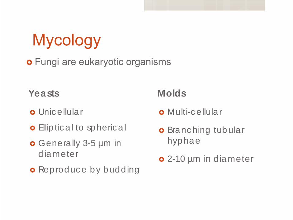
Mycology Fungi are eukaryotic organisms
Yeasts
Unicellular
Elliptical to spherical
Generally 3-5 µm in diameter
Reproduce by budding
Molds
Multi-cellular
Branching tubular hyphae
2-10 µm in diameter
Dimorphic
Fungi that can grow as either yeasts or molds
Polymorphic
Fungi that exhibit multiple forms simultaneously
Environmental temperature, nutrient factors and kinetic factors determine the type of growth observed.
Mycology
Presenter
Presentation Notes
Di blasto histo coccidiodes Poly candida
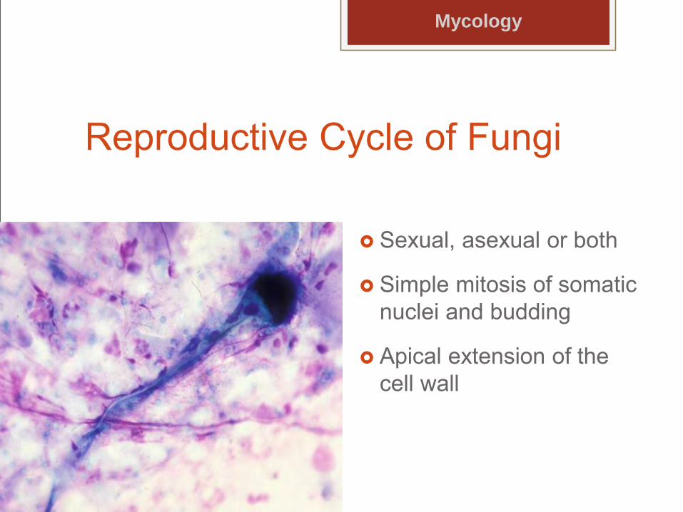
Reproductive Cycle of Fungi
Sexual, asexual or both
Simple mitosis of somatic nuclei and budding
Apical extension of the cell wall
Mycology
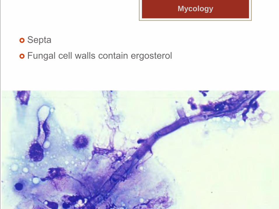
Septa
Fungal cell walls contain ergosterol
Mycology
Fungi and the eye Not permanent floral residents
Transient colonizers of the external eye
Predominant fungal isolates from normal equine eyes Aspergillus
Alternaria
Penicillium
Fusarium
Cladosporium
Absidia
Presenter
Presentation Notes
horses
Ocular defenses An intact corneal epithelium provides resistance to
fungal penetration and infection.
Unfavorable environment for the growth of any opportunistic fungi due to:
Normal ocular surface flora
Normal lacrimal flow
Mechanical movements of eyelids and nictitans
Presenter
Presentation Notes
Local defenses to fungi are generally quite effective because ocular infection is not common unless anatomic barriers are compromised.
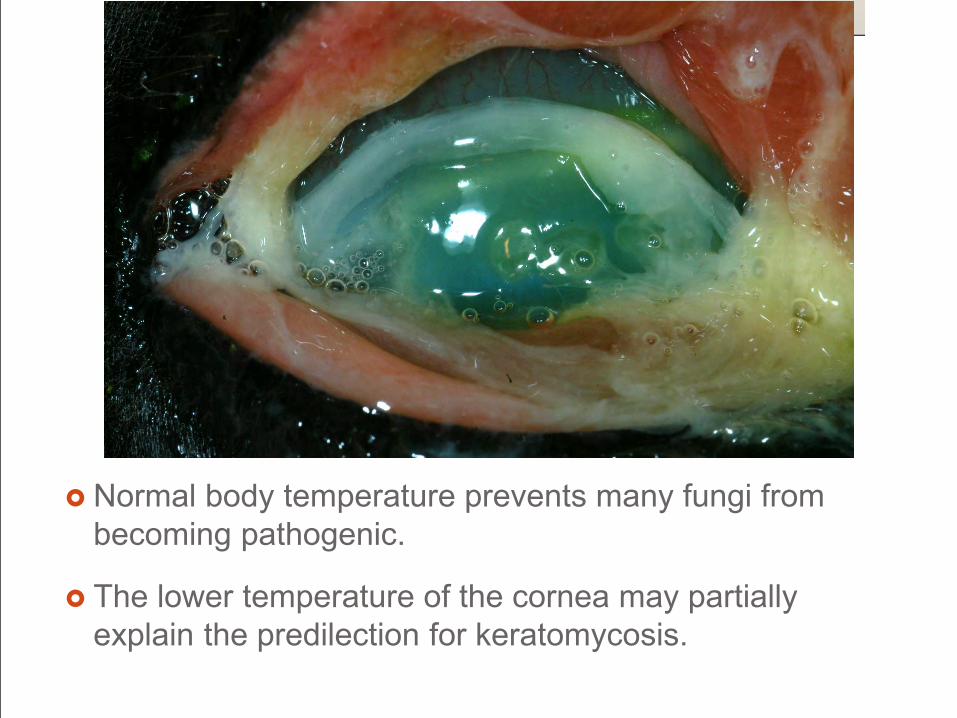
Normal body temperature prevents many fungi from becoming pathogenic.
The lower temperature of the cornea may partially explain the predilection for keratomycosis.
Immunity Primarily cell mediated
Local immunosuppression by corticosteroids may predispose an animal to a fungal infection
Systemic or topical antibacterial agents
alter normal flora
decrease natural microbial barriers
encourage colonization and growth of fungi
Presenter
Presentation Notes
by decreasing the cellular immune mechanisms.
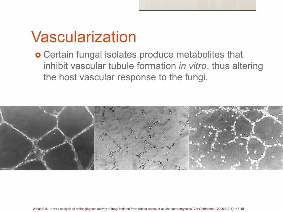
Vascularization Certain fungal isolates produce metabolites that
inhibit vascular tubule formation in vitro, thus altering the host vascular response to the fungi.
Welch PM, .In vitro analysis of antiangiogenic activity of fungi isolated from clinical cases of equine keratomycosis. Vet Ophthalmol. 2000;3(2-3):145-151.
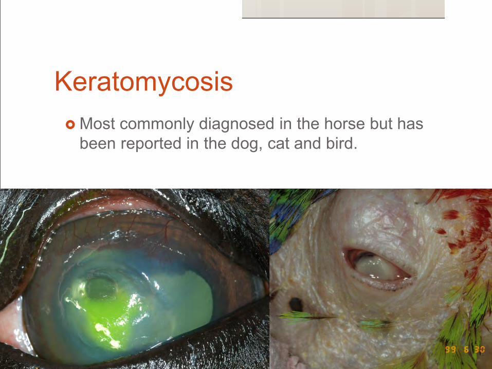
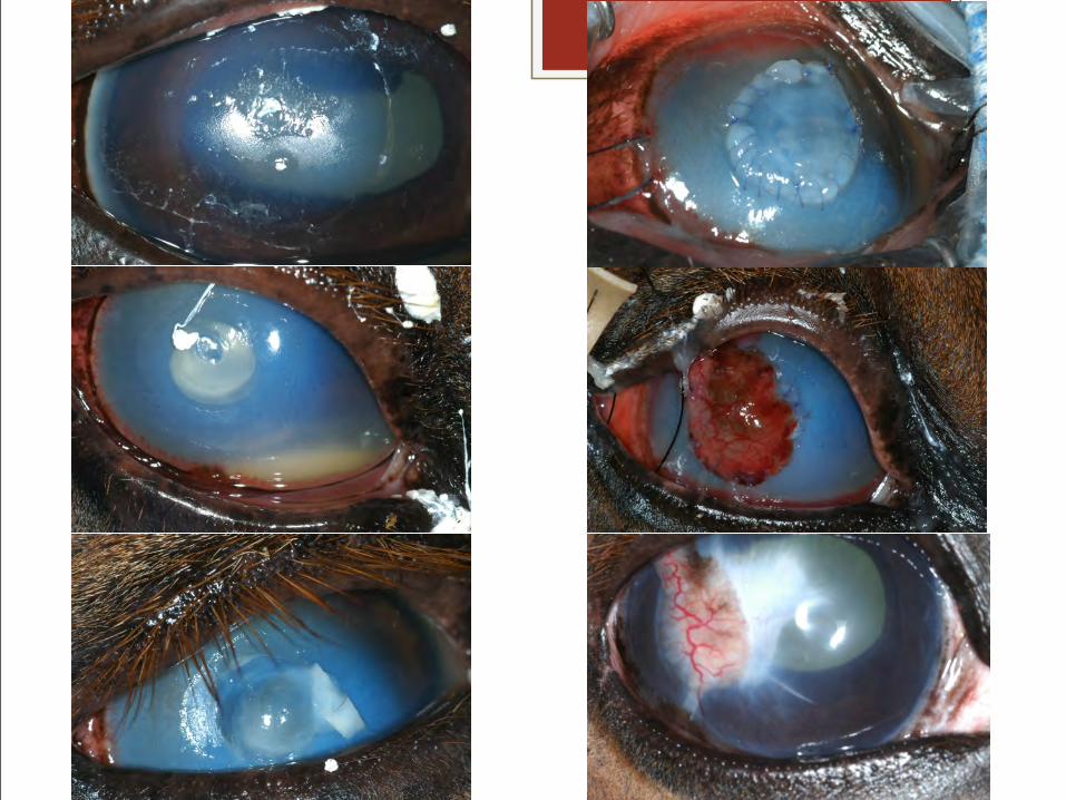
Keratomycosis Most commonly diagnosed in the horse but has
been reported in the dog, cat and bird.
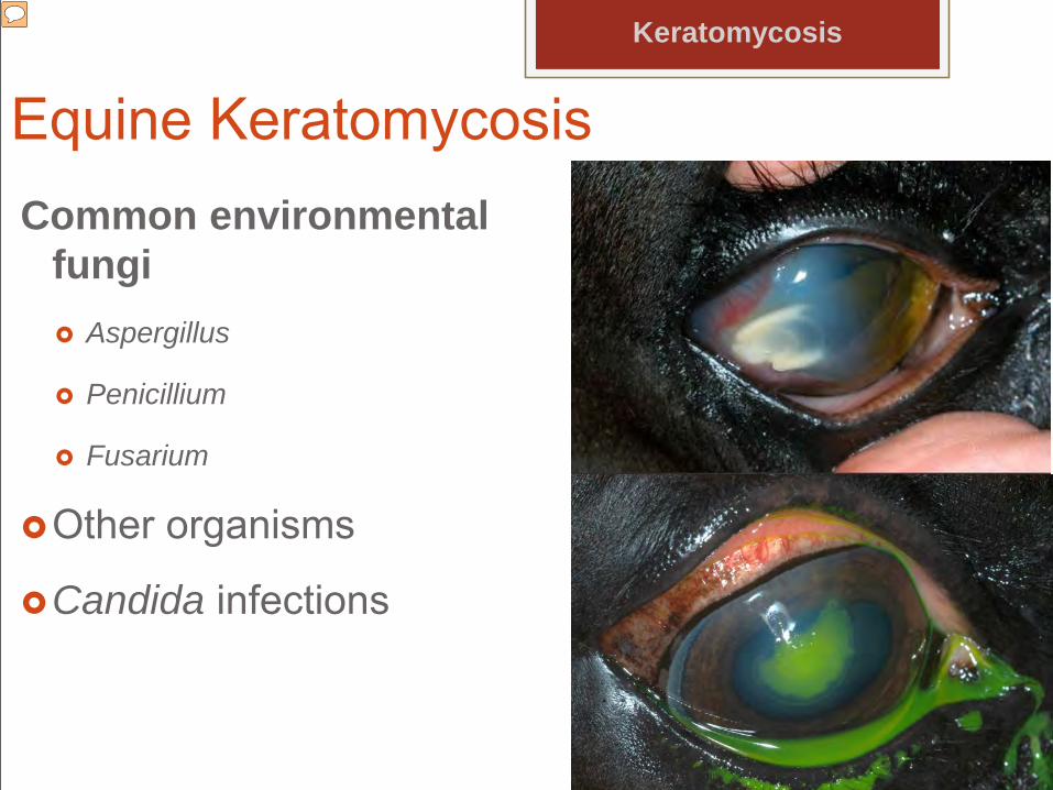
Equine KeratomycosisCommon environmental
fungi Aspergillus
Penicillium
Fusarium
Other organisms
Candida infections
Keratomycosis
Presenter
Presentation Notes
such as Cylindrocarpon destructans that have not been cultured from normal eyes occasionally cause disease. Candida can occur on the conjunctiva and cornea. It seems to be more of an opportunistic infection.
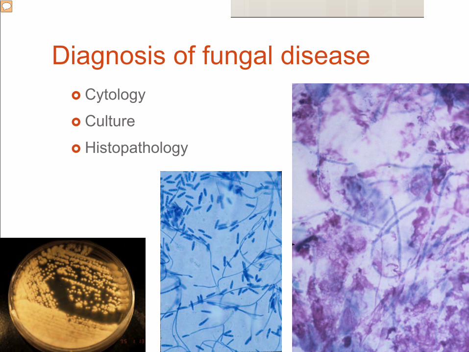
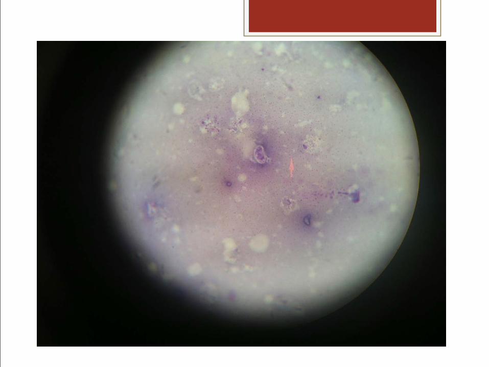
Cytology
Culture
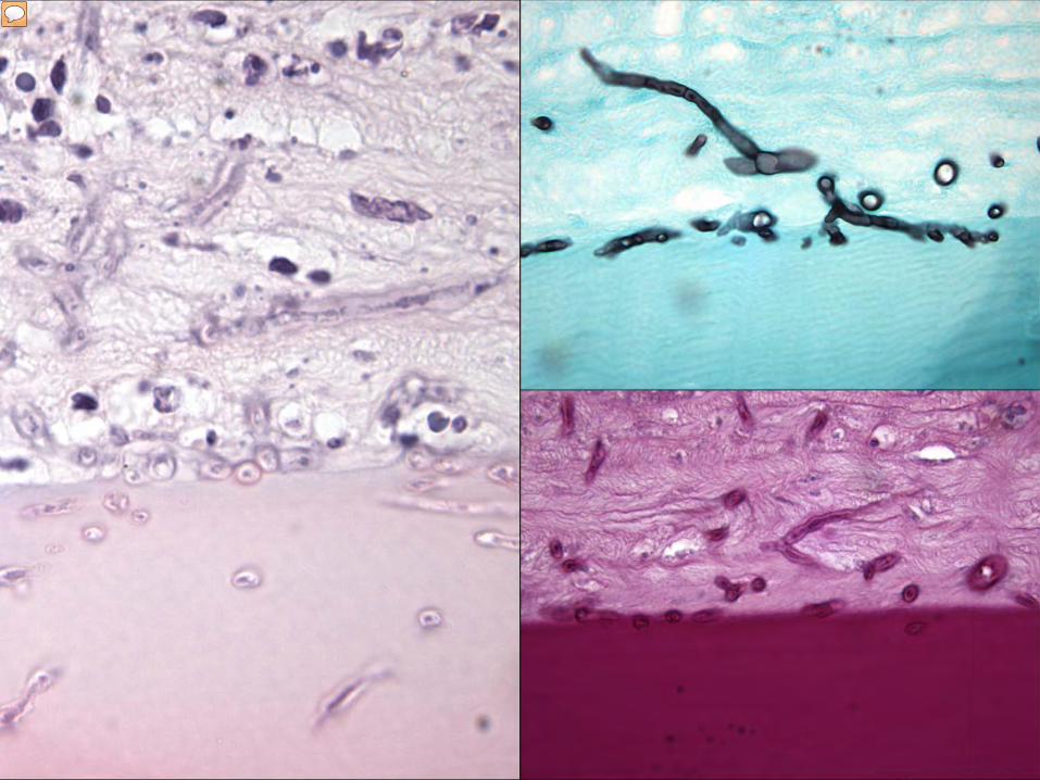
Histopathology
Diagnosis of fungal disease
Presenter
Presentation Notes
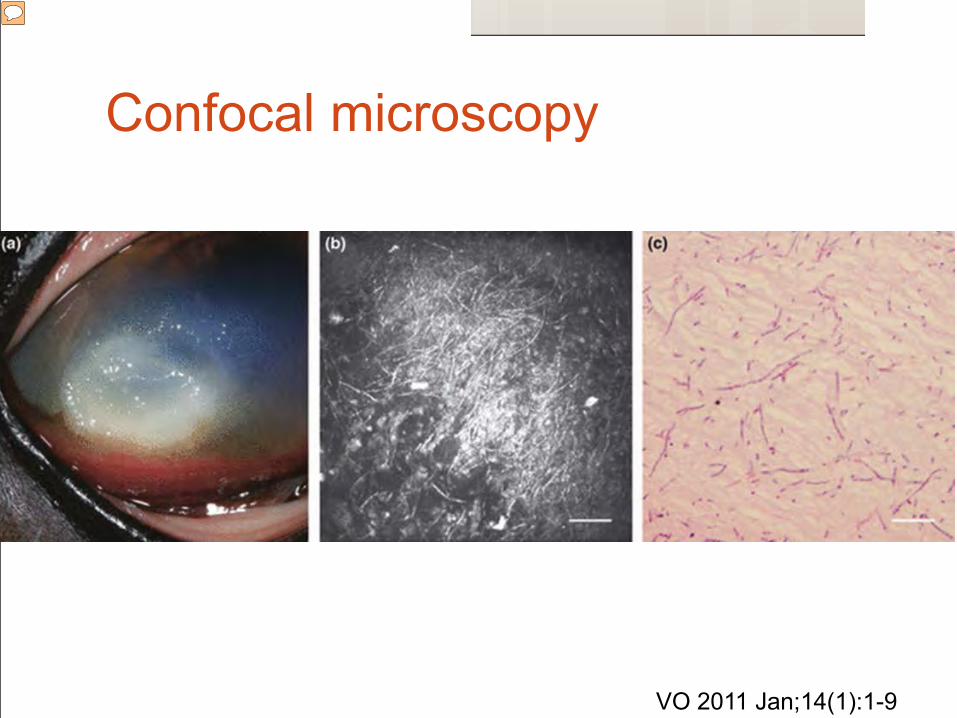
Unfortunately, fungal hyphae tend to migrate into Descemet’s membrane making diagnosis more difficult.
Unfortunately, fungal hyphae tend to migrate into Descemet’s membrane making diagnosis more difficult.
Treatment Sensitivities to standard drugs vary by region.
Voriconazole - 1% topical solution - broad spectum of activity and can penetrate into the anterior chamber.
Silver sulfadiazine can be applied every 4 to 5 hours.
No fungi were routinely sensitive to fluconazole.
Keratomycosis
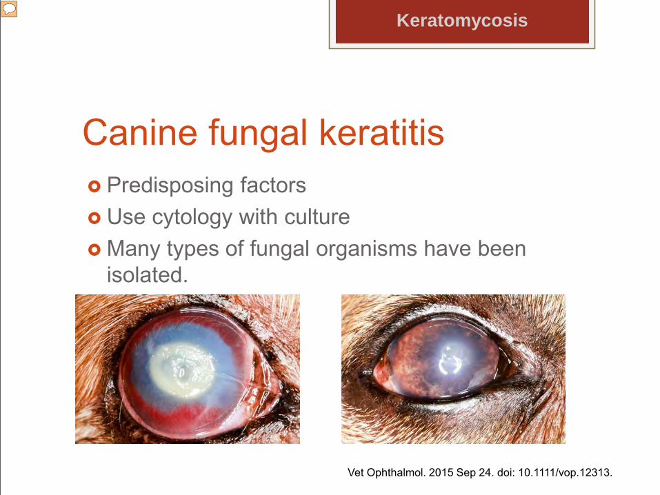
Canine fungal keratitis Predisposing factors Use cytology with culture Many types of fungal organisms have been
isolated.
Keratomycosis
Vet Ophthalmol. 2015 Sep 24. doi: 10.1111/vop.12313.
Presenter
Presentation Notes
such as underlying endocrinopathies, pre-existing corneal disease, intraocular surgery, and/or prolonged use of either topical antibiotics or corticosteroids. including Cladosporium spp., Chrysosporium spp., Curvularia spp., Aspergillus spp., Penicillium spp., and Phialemonium spp.(Scott 2014) Specific antifungal treatment can include 1% voriconazole solution, 1% itraconazole ointment or natamycin or others. Keratectomy and conjunctival grafting surgery may be necessary.
Systemic Mycoses
Blastomyces
Cryptococcus
Coccidioides
Histoplasma
Aspergillus
Most of these fungi are dimorphic.
All cause uveitis, chorioretinitis, endophthalmitis and optic neuritis.
The host inflammatory response is generally suppurative acutely and pyogranulomatouschronically.
Host tissues can be damaged directly by inflammatory processes or by degradativeenzymes produced by the fungi.
Systemic Mycoses
Presenter
Presentation Notes
Host tissues can be damaged directly by inflammatory processes or by degradative enzymes produced by the fungi.
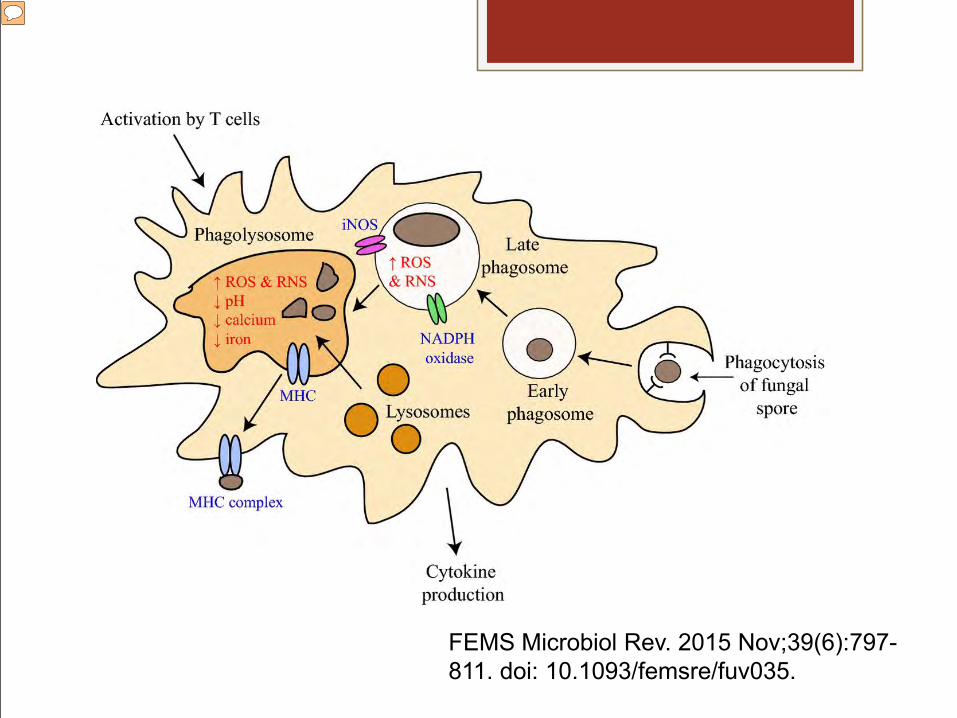
Phagocytes of the host immune system destroy fungal cells through the phagolysosomal pathway. In the lungs of an immunocompetent host, the fungal infectious propagules (usually asexual spores) are recognized by host innate immune cells such as macrophages and neutrophils. Specific fungal cell-wall components termed PAMPs (pattern-associated molecular patterns or PAMPs) are recognized via membrane-associated pattern recognition receptors on macrophage membranes. Once recognized, fungal cells are phagocytosed into an early phagosome. The early phagosome matures to a late phagosome by producing both Reactive oxygen species ROS and reactive nitrogen species RNS to damage the fungal cell from NADPH oxidase and iinducible nitric oxide synthase NOS. Lysosomes fuse with the late phagosome to produce the phagolysosome, a compartment with low pH and which contains hydrolytic enzymes to further damage the fungal cell. The MHC complex displays peptide fragments from the destroyed fungal cell for recognition by T cells. The activated macrophage releases cytokines to stimulate other cells of the immune system. Fungal cells may survive within the macrophage by the neutralization or adaptation to Reactive oxygen species ROS and reactive nitrogen species RNS production, by preventing the release of cytokines and by inducing genes allowing the acquisition of iron and calcium.
Pathogenesis Dimorphic switching
Residing in phagocytes
Remodeling cell walls
Adherence
Systemic Mycosis
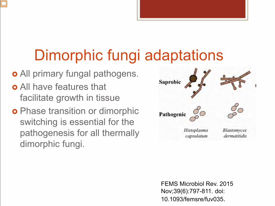
Dimorphic fungi adaptations All primary fungal pathogens. All have features that
facilitate growth in tissue Phase transition or dimorphic
switching is essential for the pathogenesis for all thermally dimorphic fungi.
Primary: Adaptations to circumvetn the effectiveness of the host defense responses Growth: production of small-sized conidia that can penetrate deep into the respiratory tree ability to grow at 37°C (i.e., thermotolerance) expression of yeast-phase–specific virulence factors evasion of host immune cells.
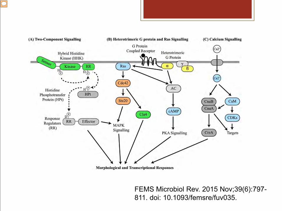
Signalling pathways for dimorphic switch
These signalling pathways also co-regulate processes important for adaptation to this environment, such as adaptation to oxidative stress.
Phase specific genes have been identified, but little is known about controlling expression.
The predominant stimulus for switch is temperature. Conversion to yeast is accelerated following
phagocytosis of B.dermatitidis spores by alveolar macrophages.
Presenter
Presentation Notes
TEMP: In the soil (22–25°C), B. dermatitidis grows as filamentous hyphae that produce conidia (asexualspores). Following soil disruption from human activities (e.g.,construction), conidia and hyphal fragments are aerosolized and when inhaled into the lungs of a human host (37°C) convert into budding yeast that cause pneumonia MACROPHAGES: In addition to intracellular spore germination, yeasts are able to survive and replicate with in macrophages during the early stages of infection. Thus, B. dermatitidis exhibits an intracellular lifestyle, which is similar to other dimorphic pathogens including H. capsulatum and Coccidioides spp. (Smith 2015).
Some fungi reside within phagocytic cells of the host as this shields organisms from the rest of the immune system.
Other dimorphic fungi use the yeast cell form to avoid phagocytosis and the cytotoxic environment of the phagolysosomal system; instead, they are adapted to tolerating the adaptive immune responses.
Presenter
Presentation Notes
Thus, dimorphic switching allows for the colonization of unique environmental niches within the host and the failure to switch almost always attenuates pathogenicity in these fungi. In other dimorphic fungi that exist predominately as a yeast vegetative growth form outside the host, such as Candida albicans, the dimorphic switch from a yeast to a filamentous growth form can facilitate tissue penetration during infection.
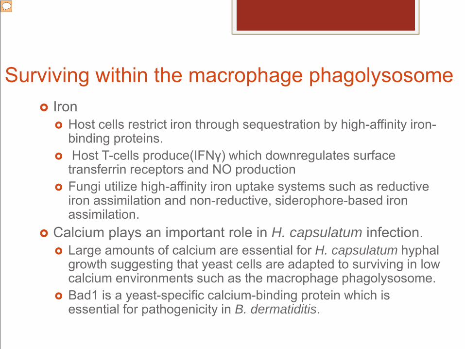
Surviving within the macrophage phagolysosome Iron
Host cells restrict iron through sequestration by high-affinity iron-binding proteins.
Host T-cells produce(IFNγ) which downregulates surface transferrin receptors and NO production
Fungi utilize high-affinity iron uptake systems such as reductive iron assimilation and non-reductive, siderophore-based iron assimilation.
Calcium plays an important role in H. capsulatum infection. Large amounts of calcium are essential for H. capsulatum hyphal
growth suggesting that yeast cells are adapted to surviving in low calcium environments such as the macrophage phagolysosome.
Bad1 is a yeast-specific calcium-binding protein which is essential for pathogenicity in B. dermatiditis.
Presenter
Presentation Notes
poses many metabolic challenges for intracellular dimorphic pathogens, including a lack of essential trace elements such as iron and zinc. Host cells restrict available iron through sequestration by high-affinity iron-binding proteins such as transferrin and ferritin to prevent intracellular microbial proliferation. Host T-cells produce the cytokine interferon gamma (IFNγ) which downregulates surface transferrin receptors and NO production by activated macrophages, which also restricts iron availability. Under iron-limiting conditions, fungi utilize high-affinity iron uptake systems such as reductive iron assimilation and non-reductive, siderophore-based iron assimilation. Calcium plays an important role in H. capsulatum infection. Large amounts of calcium are essential for H. capsulatum hyphal but not yeast growth suggesting that yeast cells are adapted to surviving in low calcium environments such as the macrophage phagolysosome (Batanghari 1997). Bad1 is a yeast-specific calcium-binding protein which is essential for pathogenicity in B. dermatiditis (Brandhorst et al. 1999). Bad1 contains 41 copies of a tandem repeat each with a calcium-binding EF-hand motif, facilitating calcium binding and growth in a poor calcium environment (Boyce 2015).
Other survival strategies Rapid remodeling of the cell wall to prevent
recognition by phagocytic cell PRRs. Proteins required for oxidative stress
resistance are components of the extracellular proteome of H. capsulatum yeast cells.
Heat shock proteins have developed to allow for a response to changes in environmental temperature.
BAD-1 protein allows binding to host lung tissue, the extracellular matrix, and cellular receptors via glycosaminoglycans.
Presenter
Presentation Notes
For example, B. dermatitidis decreases the ß(1,3)-glucan content and increases the α(1,3)-glucan content of the cell wall during infection and the hyphal-to-yeast dimorphic switch. This may prevent recognition of ß(1,3)-glucan, which is hidden by layer of α(1,3)-glucan, by the dectin-1 PPR. Additionally, proteins required for oxidative stress resistance were recently found to be components of the extracellular proteome of H. capsulatum yeast cells and heat shock proteins have developed to allow for a response to changes in environmental temperature (Caruso et al. 1987). , such as heparan sulfate. Pathogenic fungi and other microbes must adhere to host tissue to initiate infection. Surface adhesins promote this event and may be required for disease pathogenesis (Beaussart 2015).
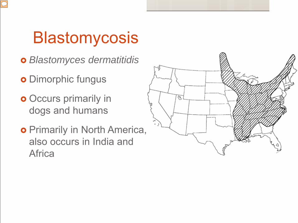
Blastomycosis Blastomyces dermatitidis
Dimorphic fungus
Occurs primarily in dogs and humans
Primarily in North America, also occurs in India and Africa
Presenter
Presentation Notes
and the Canadian provinces of Quebec, Manitoba, and Ontario. While the precise microecology of Blastomyces dermatitidis is unknown, it has been found in harsh or rapidly changing environmental conditions and associated with changing water levels, sand soils, relatively low elevation waterways, relative drought, with ground-dwelling mammals that utilize burrows containing latrine chambers, and other avian and mammalian waste products.(Baumgardner, Med Mycology 2009)
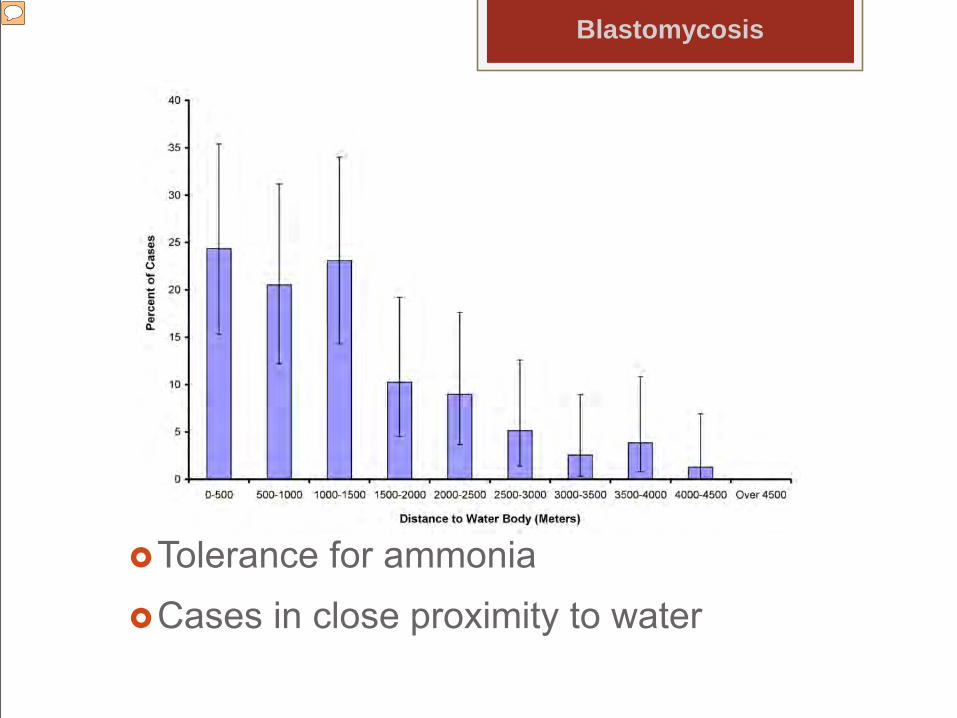
Tolerance for ammoniaCases in close proximity to water
Blastomycosis
Presenter
Presentation Notes
Growth and sporulation of the strains were observed at low glucose concentration and calculated ammonia concentrations of 4.2 mmol/l when plates were inoculated with either mold or yeast forms, and 4/5 strains tolerated ammonia concentrations of 42-62 mmol/l. Growth of virtually all soil fungi from the northern USA and Canada was inhibited at ammonia concentrations of 2.1-4.2 mmol/l. The ability of B. dermatitidis to survive and grow in organic carbon-poor, high ammonia microenvironments may be important to the competitive success of this fungus. (Baumgardner, Med Mycology 2009) One study that only looked at cases from Knox County, TN found that important risk factors associated with blastomycosis were sex, breed, age, and proximity to water. Soil type, pH, and organic matter content had no significant associations in this study area. The discrepancy with other studies may be due to little variation in pH and organic matter content of soils in this study area. It is also possible that these factors are not predictors of risk of blastomycosis in our study area thus strengthening our hypothesis that different variables may be more important predictors of the disease in certain geographical locales and not others. One other possibility is that exposure was not soil related. It has been noted that many isolates are collected above ground in manure or decomposed plant debris. (Chen, Med Myc 2008) The most significant route of transmission for B. dermatitidis is inhalation of airborne conidia which occurs from disruption of wet soil or organic matter containing micro-foci of B. dermatitidis mycelia. The mycelia release infectious conidia, which are subsequently inhaled by a susceptible host. Shared environmental exposures explain the occurrence of disease in humans and their canine companions. (Saccente, Clin Micro 2010 Review) Figure 2: Relationship between percentage of cases of canine blastomycosis (and their 95% confidence intervals) in Knox County, Tennessee and distance to the nearest water body No cultures from the nares of 110 dogs in an area of Wisconsin that is highly endemic for blastomycosis grew B. dermatitidis.(Varani, Med Mycol 2009)
Trends Increase in prevalence over 2 decades Increasing trend in diagnoses from February
to November No correlation between temperatures and
rainfall
Blastomycosis
Presenter
Presentation Notes
Saskatchewan. Signalment was similar to that previously reported. All cases originated in southern Saskatchewan and Manitoba. Case numbers showed a significant increase in the period 2001 to 2010 compared to 1990 to 2000.
Human cases in endemic states Increased percentage of population over age
65 Decreased maximum temperature Increased mercury and decreased copper soil
content.
Blastomycosis
Presenter
Presentation Notes
Saskatchewan. Signalment was similar to that previously reported. All cases originated in southern Saskatchewan and Manitoba. Case numbers showed a significant increase in the period 2001 to 2010 compared to 1990 to 2000.
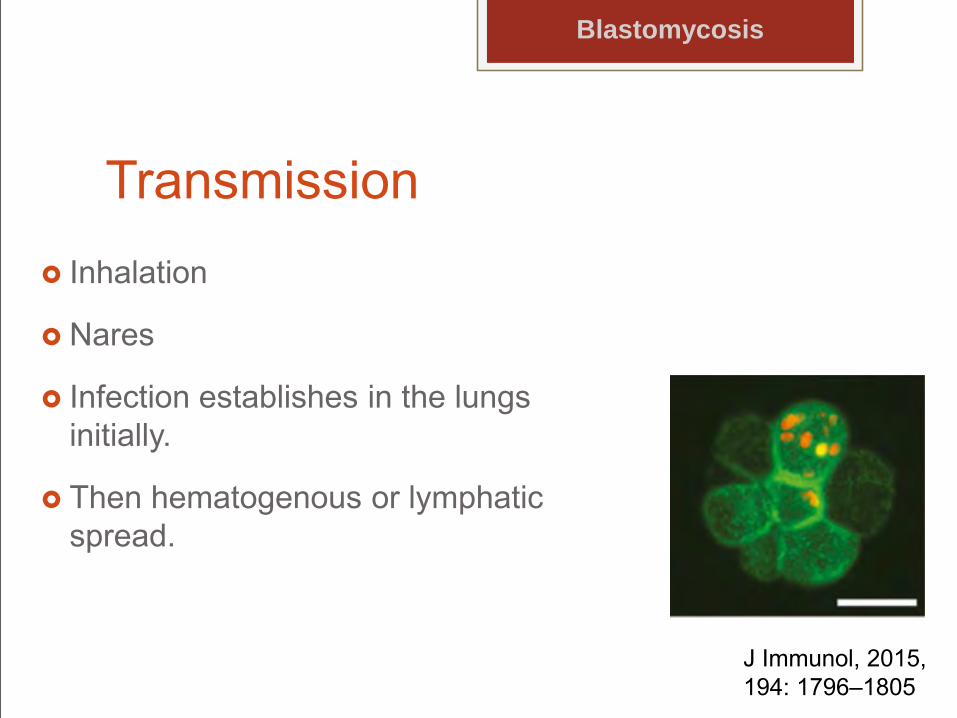
Transmission Inhalation
Nares
Infection establishes in the lungs initially.
Then hematogenous or lymphatic spread.
Blastomycosis
J Immunol, 2015,194: 1796–1805
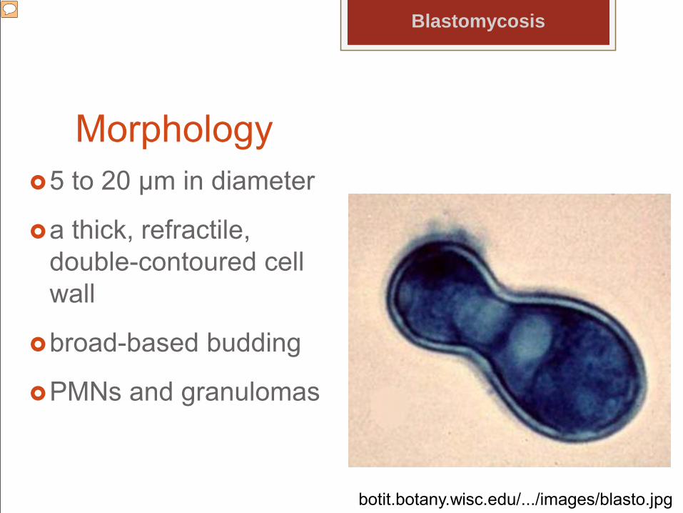
Morphology5 to 20 µm in diameter
a thick, refractile, double-contoured cell wall
broad-based budding
PMNs and granulomas
Blastomycosis
botit.botany.wisc.edu/.../images/blasto.jpg
Presenter
Presentation Notes
The characteristic host response to infection with B. dermatitidis are polymorphonuclear leukocytes and granulomas, with epitheloid histiocytes and giant cells. Early in the infection, polymorphonuclear leukocytes predominate and organisms generally are found easily. (Saccente, Clin Micro 2010 Review)
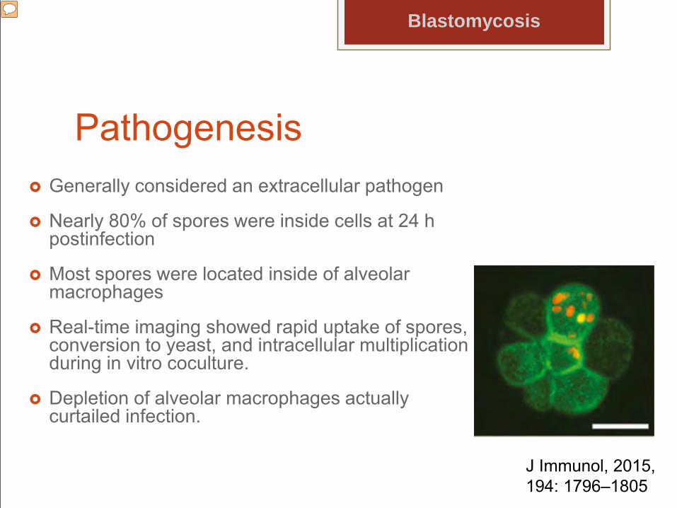
Pathogenesis Generally considered an extracellular pathogen
Nearly 80% of spores were inside cells at 24 h postinfection
Most spores were located inside of alveolar macrophages
Real-time imaging showed rapid uptake of spores, conversion to yeast, and intracellular multiplication during in vitro coculture.
Depletion of alveolar macrophages actually curtailed infection.
Blastomycosis
J Immunol, 2015,194: 1796–1805
Presenter
Presentation Notes
Intracellular residence of spores within alveolar macrophages from lung homogenates of mice infected with mCherry spores for 12 h. Alveolar macrophages were identified with anti–CD11c-FITC. Uvitex 2B stain of chitin was used to determine whether the spores were extracellular (only extracellular spores bind
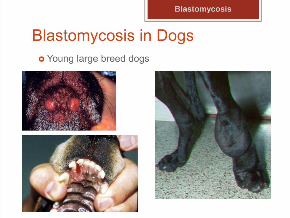
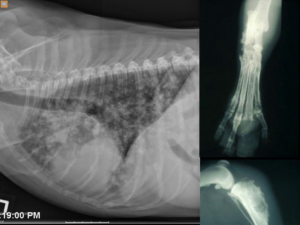
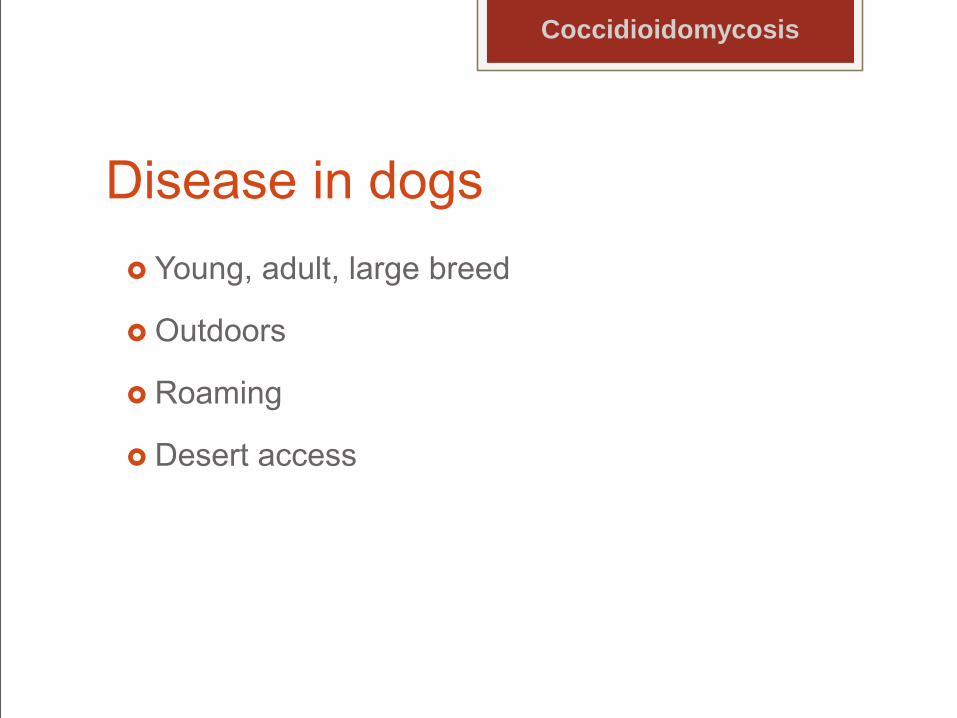
Blastomycosis in Dogs Young large breed dogs
Blastomycosis
Presenter
Presentation Notes
Bone involvement in ~1/3rd. Primarily long bones; vertebrate., Osteolysis, Periosteal proliferations, Usually below stifle, elbow. Soft tissue swelling
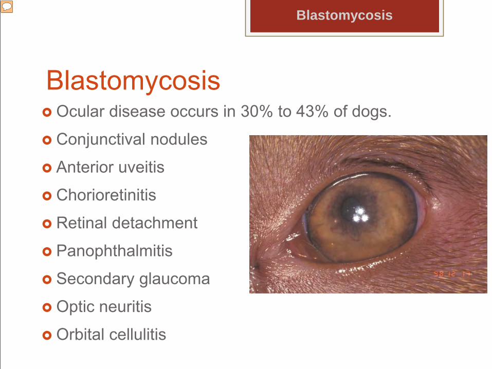
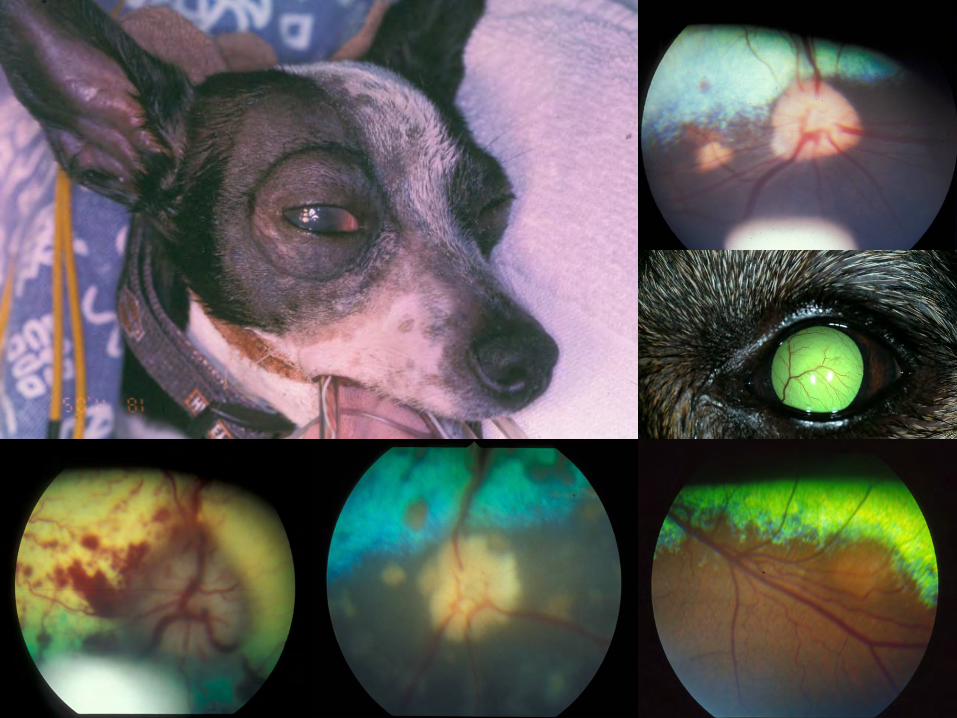
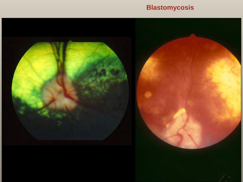
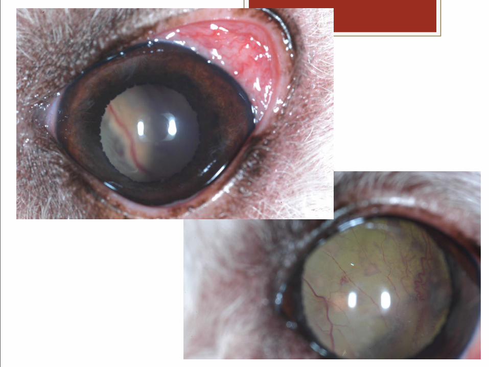
Blastomycosis Ocular disease occurs in 30% to 43% of dogs.
Conjunctival nodules
Anterior uveitis
Chorioretinitis
Retinal detachment
Panophthalmitis
Secondary glaucoma
Optic neuritis
Orbital cellulitis
Blastomycosis
Presenter
Presentation Notes
Basically panophthalmitis
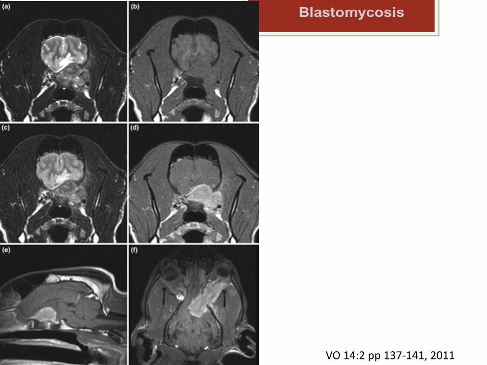
Blastomycosis
VO 14:2 pp 137-141, 2011
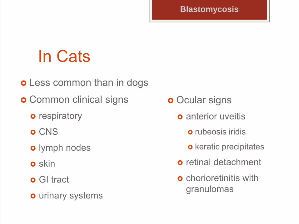
In Cats Less common than in dogs
Common clinical signs respiratory
CNS
lymph nodes
skin
GI tract
urinary systems
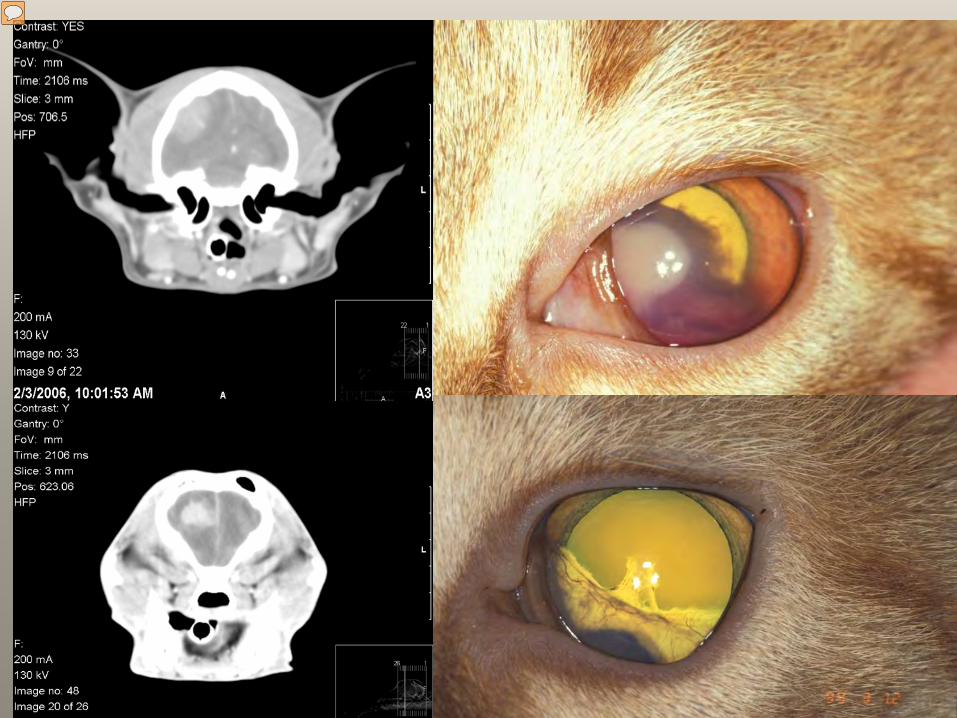
Ocular signs anterior uveitis
rubeosis iridis
keratic precipitates
retinal detachment
chorioretinitis with granulomas
Blastomycosis
Presenter
Presentation Notes
An 8-year-old domestic shorthair cat was evaluated because of signs of depression, circling, and visual deficits. Clinical Findings—The cat had no other signs. Computed tomography of the brain revealed a mass lesion involving the right parietal, temporal, and occipital lobes; the mass was in broad-based contact with the skull and smoothly marginated and had strong homogenous enhancement after contrast agent administration. During craniectomy, samples of the mass were collected for cytologic and histopathologic evaluations and microbial culture. A diagnosis of Blastomyces dermatitidis–associated meningoencephalitis with secondary pyogranulomatous inflammation was made. Treatment and Outcome—Amphotericin B then fluconazole, did well, no evidence of systemic disease
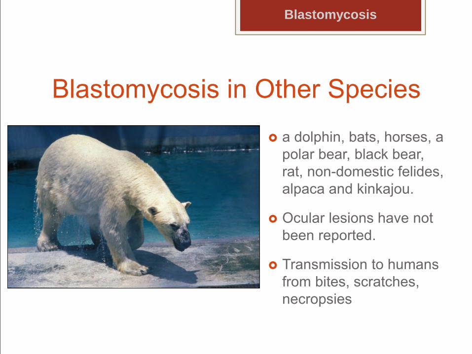
Blastomycosis in Other Species
a dolphin, bats, horses, a polar bear, black bear, rat, non-domestic felides, alpaca and kinkajou.
Ocular lesions have not been reported.
Transmission to humans from bites, scratches, necropsies
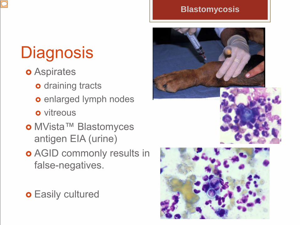
Ease of diagnosis is variable. Blastomyces antigen was detected in the urine in 93.5% of cases of blastomycosis in dogs using MVista™ Blastomyces antigen EIA. Blastomyces antigen detection in urine was more sensitive than antibody detection determined using AGID (17.4%). Blastomyces urinary antigen concentrations should be monitored, at minimum, at the time of diagnosis and when treatment discontinuation is being considered, as well as at any time during treatment when clinical efficacy is in doubt. The urine test should be repeated in any dogs with clinical suspicion of relapse, especially if Blastomyces organisms cannot yet be detected.(Foy 2014)
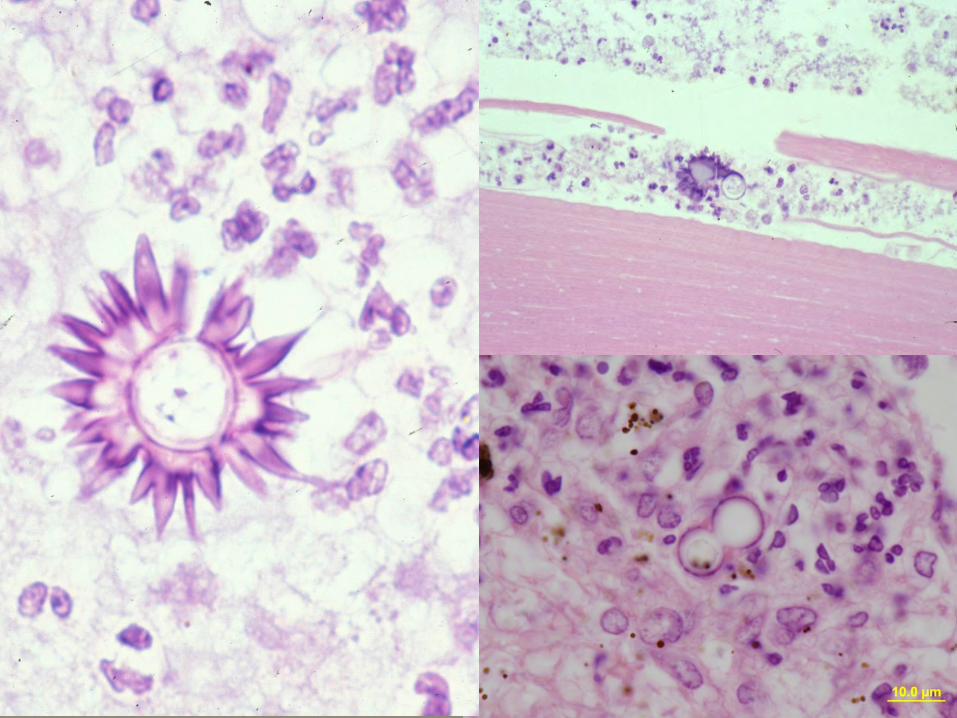
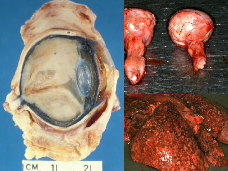
Histopathology Most commonly: choroiditis and retinal detachment Pyogranulomatous inflammation Lens rupture is seen in 50% of enucleated eyes. Choroidal inflammation is more severe in the
nontapetal choroid Organisms are observed primarily in the choroid
Blastomycosis
Presenter
Presentation Notes
No parts of the eye are spared involvement Additional histologic findings include: retinal degeneration, vitreal protein and or cells, protein and/or cells in anterior chamber, optic nerve inflammation, preiridial fibrovascular membranes and changes associated with secondary glaucoma.
Treatment Itraconazole
Commercial vs generic vs compounded Fluconazole Amphotericin B +/- Prednisone
Blastomycosis
Presenter
Presentation Notes
No parts of the eye are spared involvement Additional histologic findings include: retinal degeneration, vitreal protein and or cells, protein and/or cells in anterior chamber, optic nerve inflammation, preiridial fibrovascular membranes and changes associated with secondary glaucoma.
Prognosis Varies by location and extent of inflammation within
the eye and response to therapy.
Blastomycosis
Presenter
Presentation Notes
Itraconazole
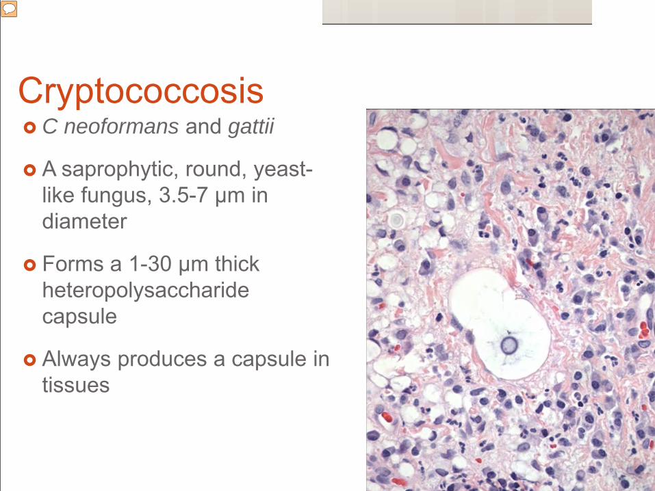
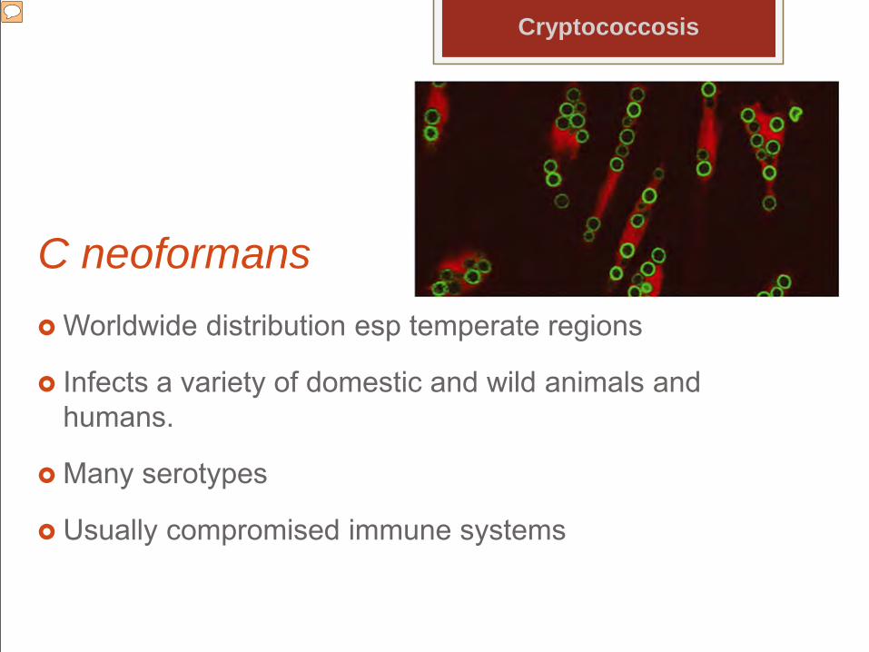
Cryptococcosis C neoformans and gattii
A saprophytic, round, yeast-like fungus, 3.5-7 µm in diameter
Forms a 1-30 µm thick heteropolysaccharide capsule
Always produces a capsule in tissues
Presenter
Presentation Notes
+/- capsule when growing on an artificial media or when growing naturally in the environment,
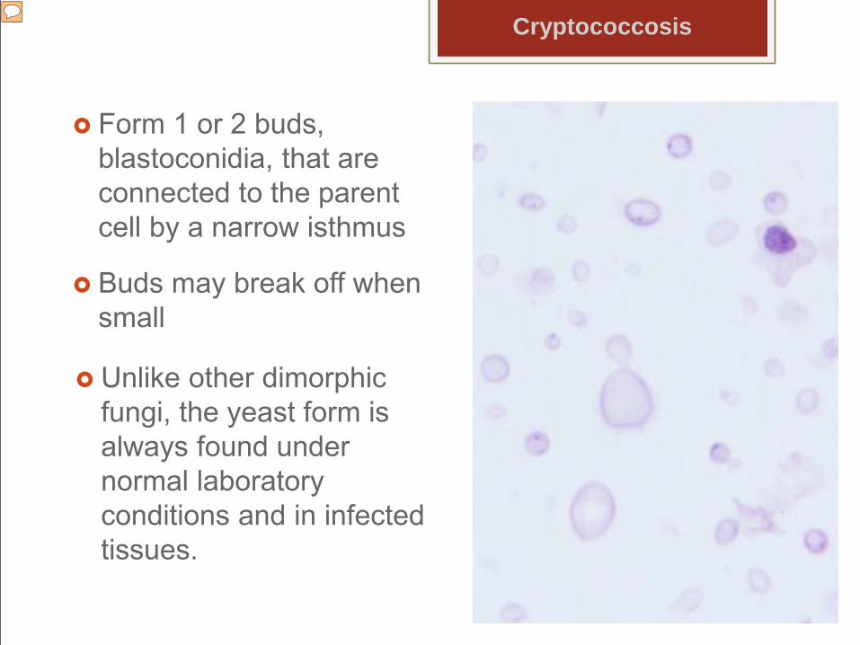
Form 1 or 2 buds, blastoconidia, that are connected to the parent cell by a narrow isthmus
Buds may break off when small
Unlike other dimorphic fungi, the yeast form is always found under normal laboratory conditions and in infected tissues.
Cryptococcosis
Presenter
Presentation Notes
the yeast population varies in size
C neoformans Worldwide distribution esp temperate regions
Infects a variety of domestic and wild animals and humans.
Many serotypes
Usually compromised immune systems
Cryptococcosis
Presenter
Presentation Notes
Much work has been done evaluating the serotypes of cryptococcosis due to its importance in humans with the AIDS virus. .
C gattii Isolated from the bark and leaf litter of eucalyptus
trees and trees in Pacific Northwest.
Incidence is increasing in Pacific Northwest and California
Usually non–immunocompromised people
Incubation has been > 8 years
Presenter
Presentation Notes
Also cases in British Columbia Neoformans is associated with immunocompromised individuals and gatti with non immunocompromised.
Pathogenesis The prevalence in cats is ≥ that of dogs Inhalation of airborne organisms Unencapsulated in the environment Arrival in upper respiratory tract causes nasal
granulomas. 7-14% of animals have asymptomatic colonization of
the nasal passages. Production of a thick capsule and abundant release
of glycoprotein into the circulation are hallmarks of virulence.
Cryptococcosis
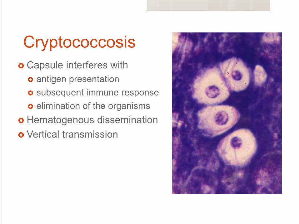
Cryptococcosis Capsule interferes with
antigen presentation subsequent immune response elimination of the organisms
Hematogenous dissemination Vertical transmission
Cryptococcosis neoformans Establishment and spread of infection in the host are
highly dependent on immunity.
Incidence in AIDS patients is decreasing dramatically.
Immunosuppression does not appear to play a role in veterinary patients.
Presenter
Presentation Notes
Incidence of crypto in nonHIV persons varied from 0.4 -5 cases per million population in mid 1990’s . Incidence in HIV infected patients is from 23-66 cases per 1,000 persons in 1992 to 2-7 cases per 1,000 in 2000 It is the leading cause of death in HIV infected individuals with an incidence of 30 % and a mortality of 30 – 60 %. The mortality rate in transplant patients is even higher (20 – 100%)While the presence of underlying disease has been evaluated in dogs and cats, very few dogs and cats are shown to have immunosuppressive disease that are confirmed to have cryptococcosis.
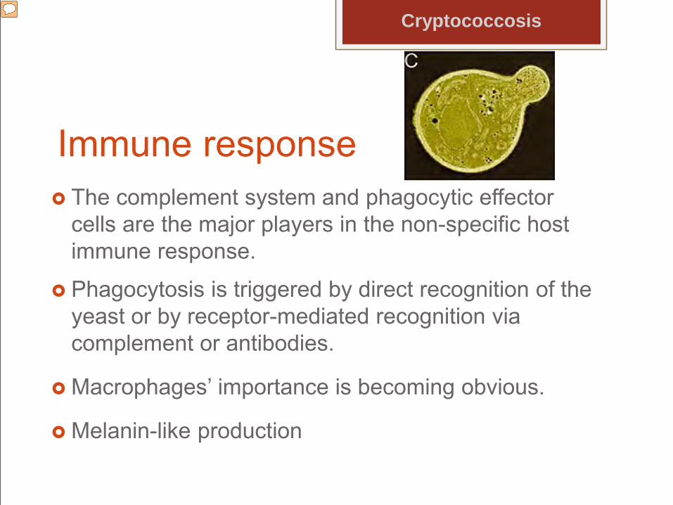
Immune response The complement system and phagocytic effector
cells are the major players in the non-specific host immune response.
Phagocytosis is triggered by direct recognition of the yeast or by receptor-mediated recognition via complement or antibodies.
Macrophages’ importance is becoming obvious.
Melanin-like production
Cryptococcosis
Presenter
Presentation Notes
The two major functions of the complement system during cryptococcosis are to stimulate the chemotaxis of phagocytic effector cells and enhance the uptake of cryptococcal cells by these phagocytes. The cryptococcal polysaccharide capsule is a well-known factor required for the pathogen’s virulence, e.g. by inhibiting phagocytosis. C.neoformans mutants with a capsule-deficient phenotype are avirulent in mice. Several studies with encapsulated and nonencapsulated C. neoformans strains also showed a difference in complement activation dependent on capsulation. Conserved structures such as the components of the cryptococcal capsule can be directly recognized by pattern recognition receptors Research has revealed an intriguing interaction between the phagocytic effectors and yeast showing that C. neoformans as an intracellular parasite. Cryptococcus has developed a unique method to manipulate host macrophages. After phagocytosis, C. neoformans can survive and proliferate within these infected host cells, eventually leading to host cell lysis. Additionally, the yeast can exit macrophages without killing the host cell, thus avoiding a local inflammatory response and the yeast can be laterally transferred from one macrophage to another.
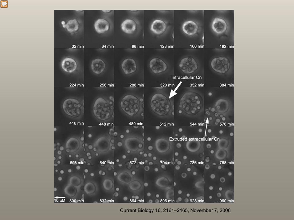
Current Biology 16, 2161–2165, November 7, 2006
Presenter
Presentation Notes
FIGURE 2: Cryptococcal expulsion from within a macrophage. Cryptococcus can exit macrophages in a novel non-lytic way that does not involve killing of the host cell or the yeast. Panels A-C show timelapse images of two intracellular yeast within a macrophage. Four hours into the experiment (panel D) the yeast is suddenly expelled. The macrophage and the yeast both remain alive after this process, as shown by continuing proliferation (panel E-F). Trogan horse analogy
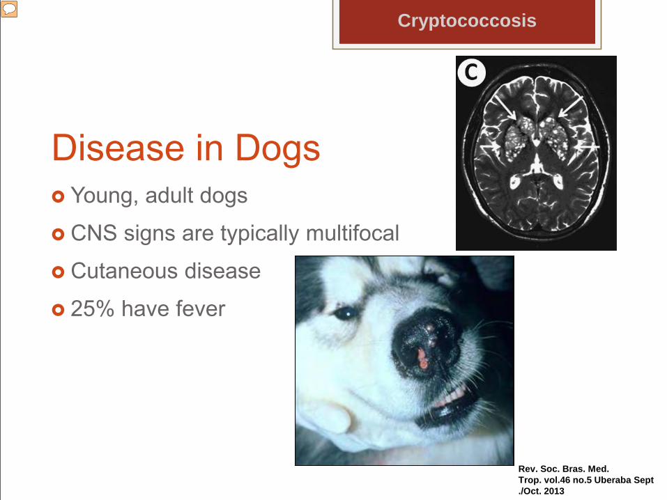
Doberman Pinschers, Great Danes and American Cocker Spaniels seem to be over-represented. CNS signs are attributable to meningitis or encephalomyelitis and may include: head tilt, nystagmus, facial paralysis, ataxia, mild paresis to complete paralysis, depressed reflexes, circling, and seizures. Cutaneous involvement has been found in dogs but is much less likely than in cats
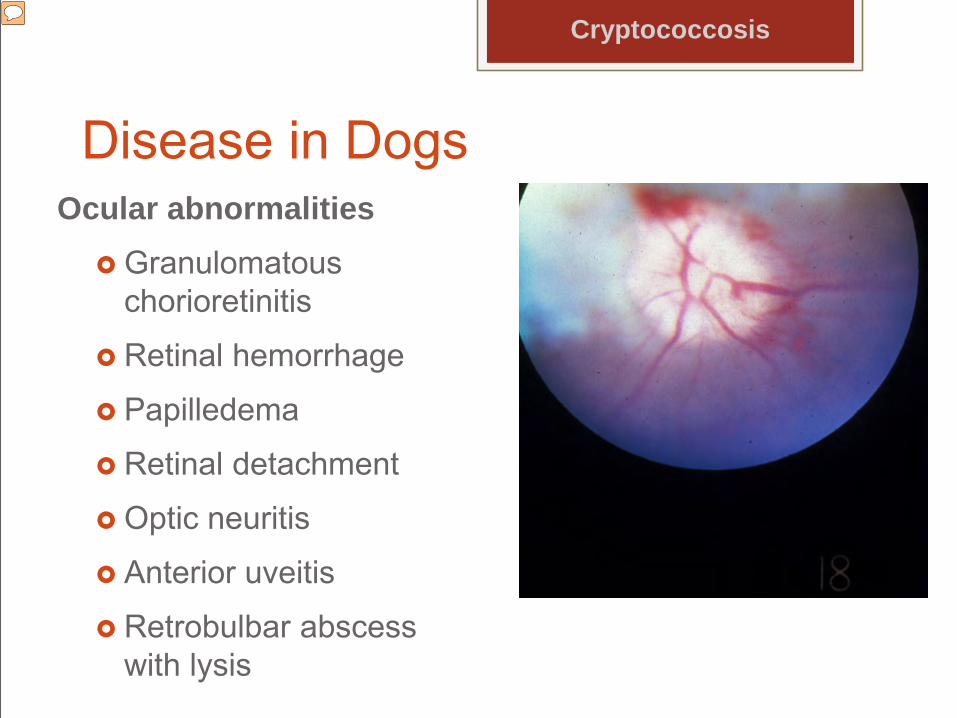
Disease in DogsOcular abnormalities
Granulomatous chorioretinitis
Retinal hemorrhage
Papilledema
Retinal detachment
Optic neuritis
Anterior uveitis
Retrobulbar abscess with lysis
Cryptococcosis
Presenter
Presentation Notes
The eye is involved secondary to hematogenous spread or extension from the brain along the optic nerve.
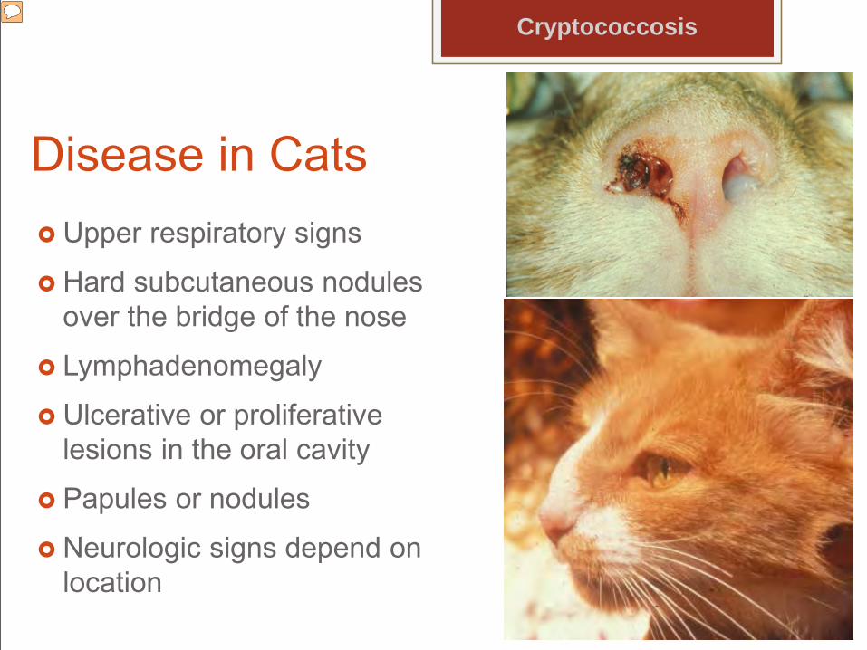
Disease in Cats Upper respiratory signs
Hard subcutaneous nodules over the bridge of the nose
Lymphadenomegaly
Ulcerative or proliferative lesions in the oral cavity
Papules or nodules
Neurologic signs depend on location
Cryptococcosis
Presenter
Presentation Notes
Thoracic radiographs are often normal, although small nodular lesions may be present. Cutaneous lesions, including papules or nodules varying from 1-10 mm in diameter are also common in cats and occur in 40-50% of them
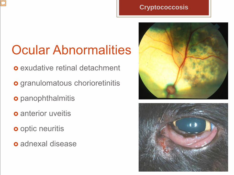
Ocular Abnormalities exudative retinal detachment
granulomatous chorioretinitis
panophthalmitis
anterior uveitis
optic neuritis
adnexal disease
Cryptococcosis
Presenter
Presentation Notes
Adnexal disease is not unusual and lesions have been seen both within third eyelid and the palpebral conjunctiva. Lesions may cause hyperemia and roughened proliferative areas on the conjunctiva with focal white incrustations and subepithelial deposits. Cytology of conjunctival scrapings is diagnostic.
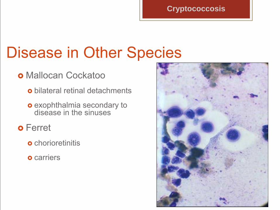
Disease in Other Species Mallocan Cockatoo
bilateral retinal detachments
exophthalmia secondary to disease in the sinuses
Ferret chorioretinitis
carriers
Cryptococcosis
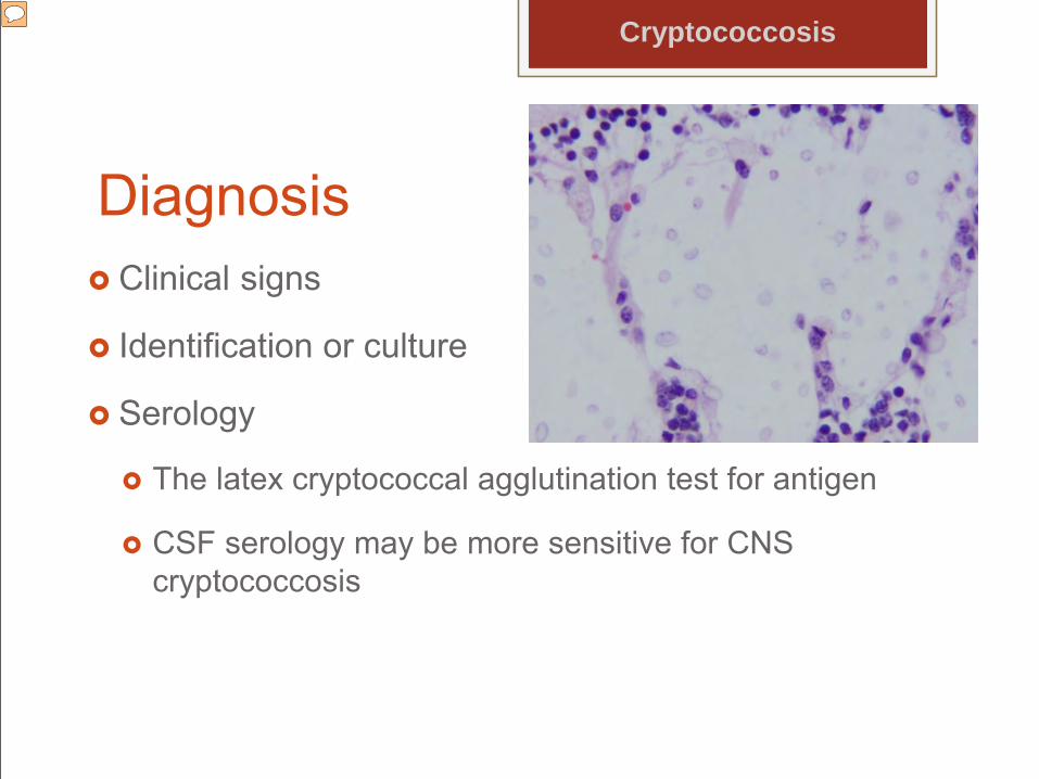
Diagnosis Clinical signs
Identification or culture
Serology
The latex cryptococcal agglutination test for antigen
CSF serology may be more sensitive for CNS cryptococcosis
Cryptococcosis
Presenter
Presentation Notes
Ocular or other tissue aspirates CSF HistopathologyLatex agglutination is considered to be the most reliable serologic test, because it can detect the presence of cryptococcal antigen in body fluids including serum, urine, CSF, aqueous humor, and vitreous The magnitude of antigen titers tends to correlate with severity of disease and response to therapy.
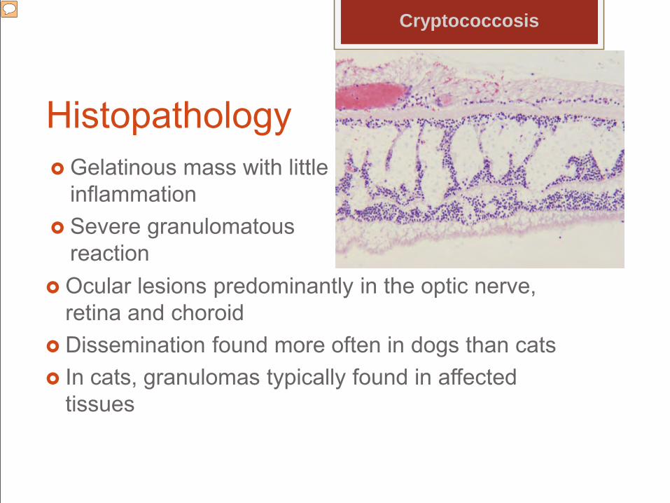
Histopathology Gelatinous mass with little
inflammation Severe granulomatous
reaction Ocular lesions predominantly in the optic nerve,
retina and choroid Dissemination found more often in dogs than cats In cats, granulomas typically found in affected
tissues
Cryptococcosis
Presenter
Presentation Notes
In those rare cases with severe inflammation, organisms are typically scarce. As with other mycoses, even when anterior uveitis is present, organisms are rarely demonstrated in the anterior uvea. In respiratory tract, the kidneys, lymph nodes, spleen, liver, thyroid gland, adrenal glands, pancreas, bone, GI tract, muscle, myocardium, prostate gland, heart valve and tonsils
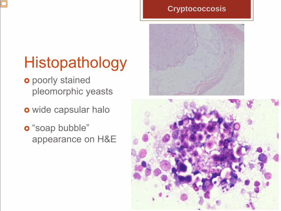
Histopathology poorly stained
pleomorphic yeasts
wide capsular halo
“soap bubble” appearance on H&E
Cryptococcosis
Presenter
Presentation Notes
The organisms are typically numerous and vary in size and shape.
Treatment Azole antifungals
Amphotericin B
Strain resistance
Judicious use of steroids
Cryptococcosis
Coccidioidomycosis Coccidioides immitis and Coccidioides posadasii
Only found in the mycelial phase in specific locals
Lower Sonoran Life Zone
sandy, alkaline soil, high environmental temperatures, low annual rain fall, and low elevation
Presenter
Presentation Notes
Geographically this region is within the southwestern United States, Mexico and Central and South America, including Guatemala, Honduras, Columbia, Venezuela, Paraguay and Argentina. Almost all cases are diagnosed within this region; however, occasionally stray cases have a history of residence or travel within this area.
Pathogenesis Inhalation
Incubation period is 1-3 weeks
Dissemination involves a reproductive cycle of spherules to endospores to new spherules.
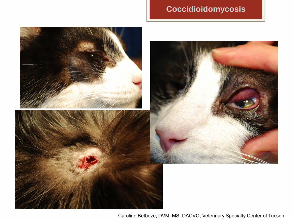
Caroline Betbeze, DVM, MS, DACVO, Veterinary Specialty Center of Tucson
Disease in Other SpeciesRing tailed lemurLlamaRhinocerosChimpanzeeKoala
Coccidioidomycosis
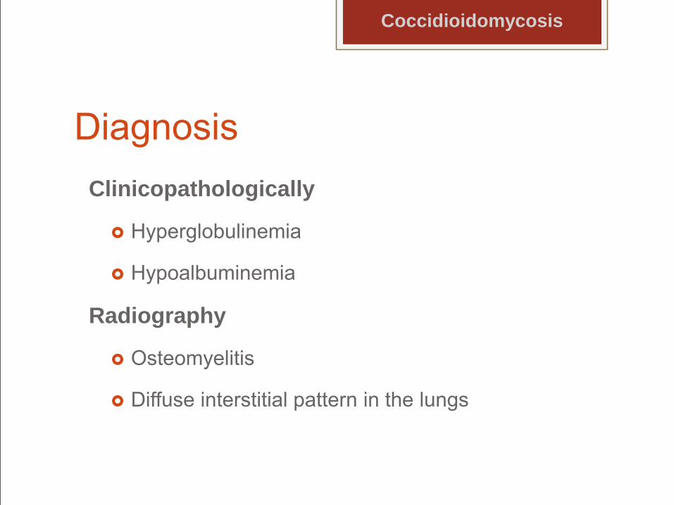
DiagnosisClinicopathologically
Hyperglobulinemia
Hypoalbuminemia
Radiography
Osteomyelitis
Diffuse interstitial pattern in the lungs
Coccidioidomycosis
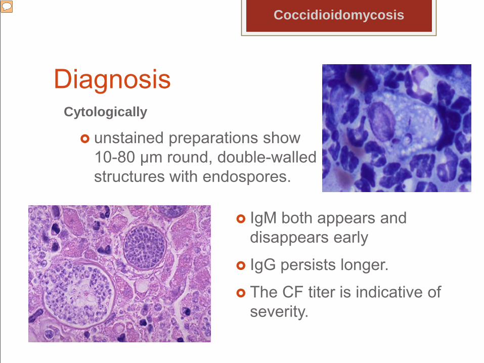
DiagnosisCytologically
unstained preparations show 10-80 µm round, double-walled structures with endospores.
Coccidioidomycosis
IgM both appears and disappears early
IgG persists longer.
The CF titer is indicative of severity.
Presenter
Presentation Notes
Wright stain, and Papanicolaou’s and periodic acid-Schiff stains are most helpful. Not all spherules will contain recognizable endospores and smaller spherules may have crumpled, transparent walls. Histopathology specimens, using H & E or PAS are also helpful for diagnosis, and the fungus may be isolated. 1 test can be positive and the other negative depending on the stage of infection; thus, the 2 tests should be performed in parallel. A titer of >1:32 or greater is indicative of disseminated disease. It is possible to have one test come out positive and the other negative depending on the stage of infection; thus, the two tests should be performed in parallel. Latex agglutination and ELISA are now employed by some laboratories. AGID is specific but relatively insensitive and should be done following ELISA or and latex particle agglutination which are good screening tests as they are very sensitive but have false positives.
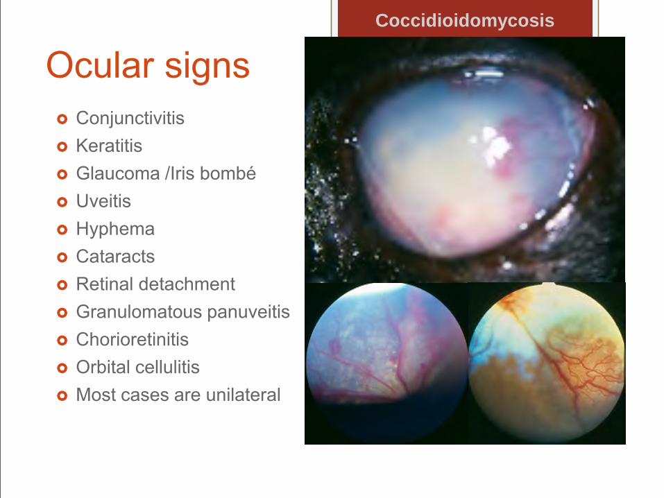
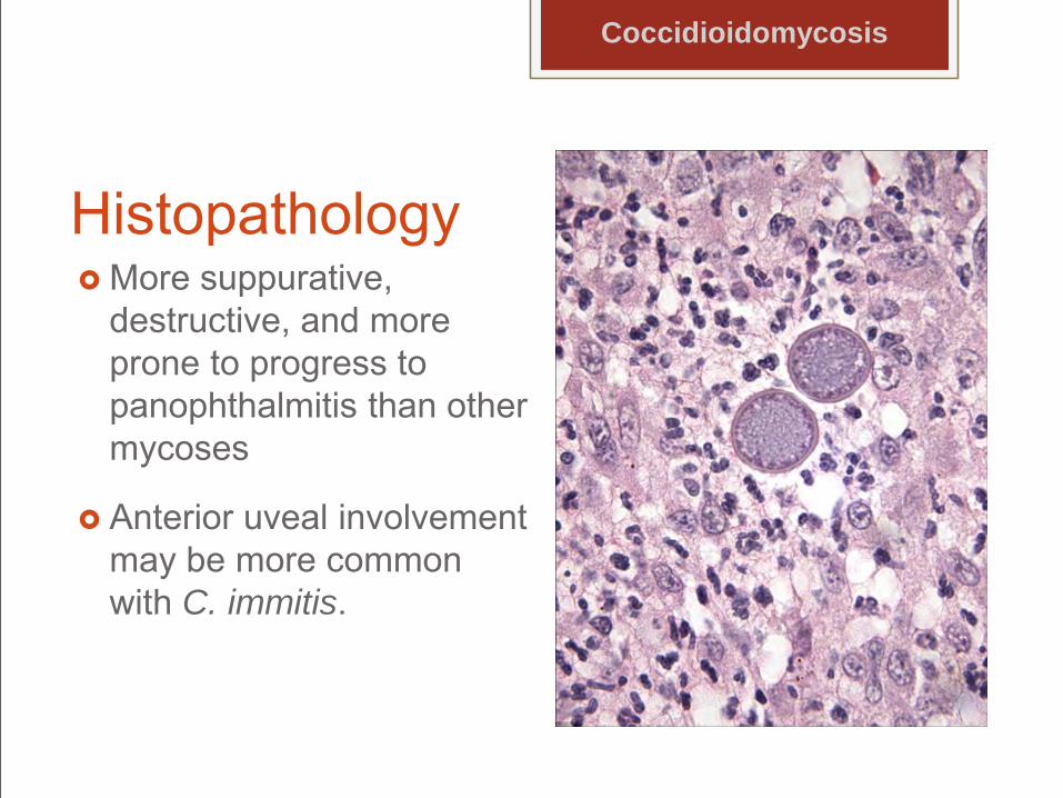
destructive, and more prone to progress to panophthalmitis than other mycoses
Anterior uveal involvement may be more common with C. immitis.
Coccidioidomycosis
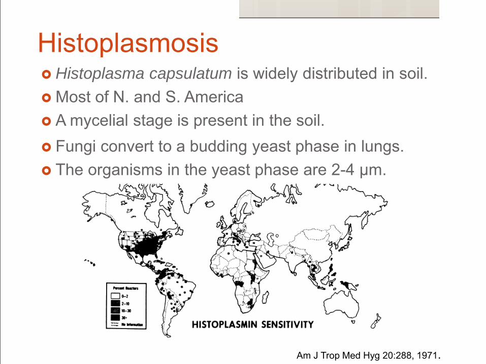
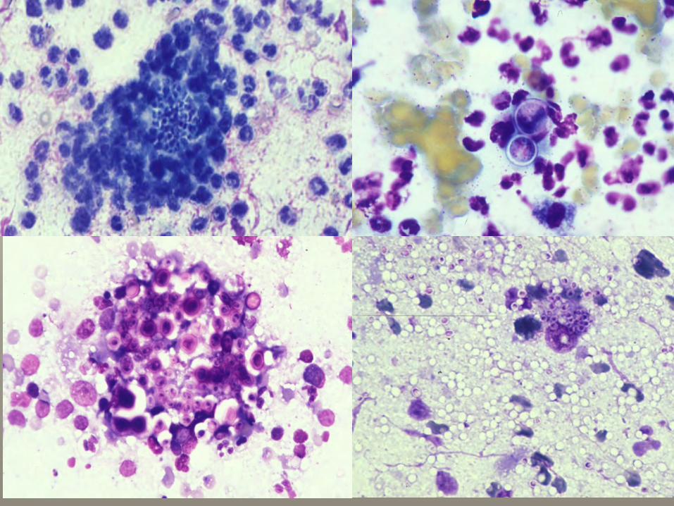
Histoplasmosis Histoplasma capsulatum is widely distributed in soil. Most of N. and S. America A mycelial stage is present in the soil. Fungi convert to a budding yeast phase in lungs. The organisms in the yeast phase are 2-4 µm.
Am J Trop Med Hyg 20:288, 1971.
Pathogenesis Grow as mold preferably on bird and bat guano at
ambient temps
Probably acquired by inhalation of microconidia
Incubation ~ 12-16 days
Reproduce by budding
Histoplasmosis
Presenter
Presentation Notes
that are small enough to reach the lower respiratory tract.
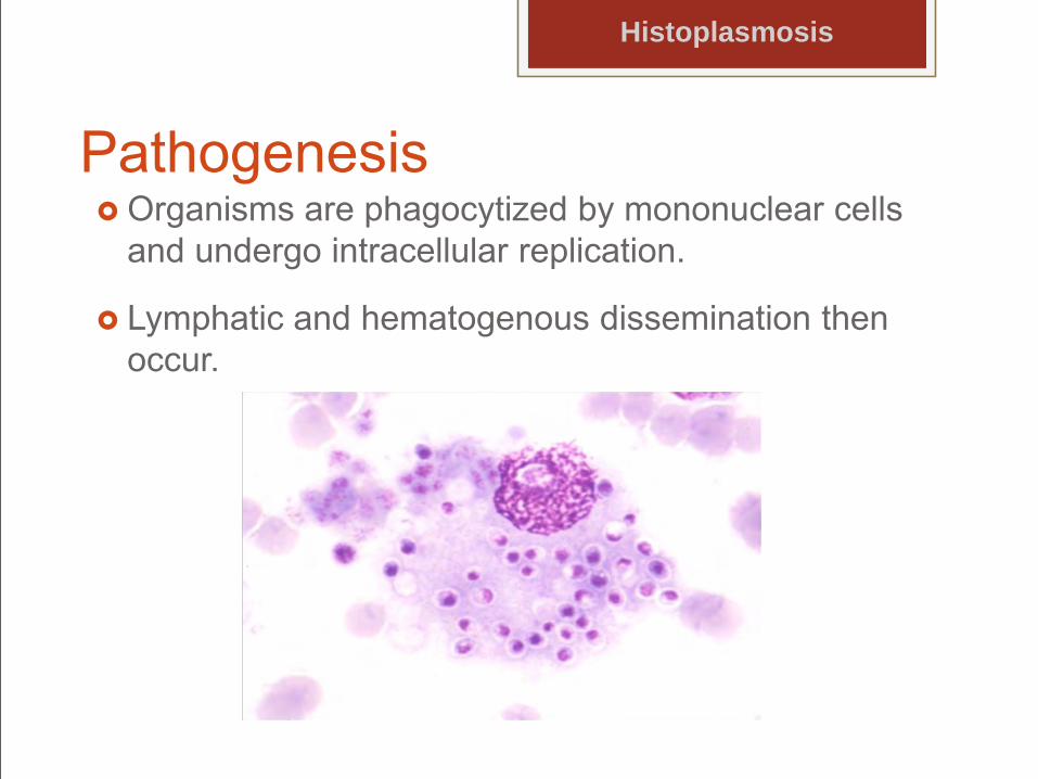
Pathogenesis Organisms are phagocytized by mononuclear cells
and undergo intracellular replication.
Lymphatic and hematogenous dissemination then occur.
Histoplasmosis
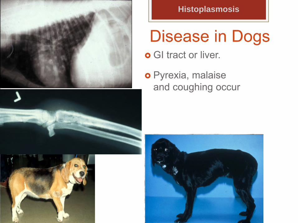
Disease in Dogs GI tract or liver.
Pyrexia, malaise and coughing occur
Histoplasmosis
Ocular signs in dogs Relatively rare
Blepharitis
Conjunctivitis
Pyogranulomatous chorioretinitis
Anterior uveitis
Optic neuritis
Experimentally, ocular lesions are seen in 66% of cases.
Histoplasmosis
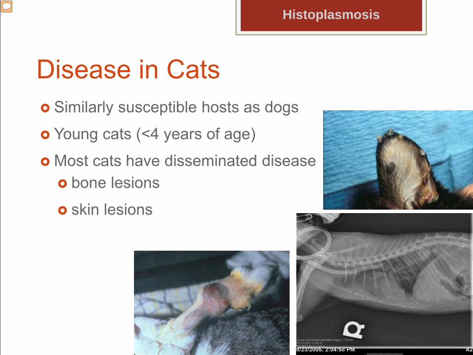
Disease in Cats Similarly susceptible hosts as dogs
Young cats (<4 years of age)
Most cats have disseminated disease
Histoplasmosis
bone lesions
skin lesions
Presenter
Presentation Notes
PE reveals dyspnea, tachypnea and abnormal lung sounds in more than half the cats. Visceral lymphadenomegaly, splenomegaly �and hepatomegaly are also seen.
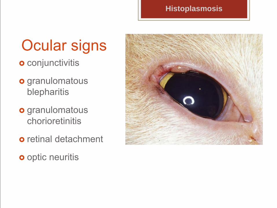
Ocular signs conjunctivitis
granulomatous blepharitis
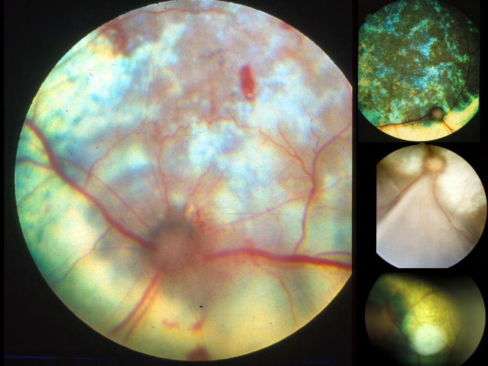
granulomatous chorioretinitis
retinal detachment
optic neuritis
Histoplasmosis
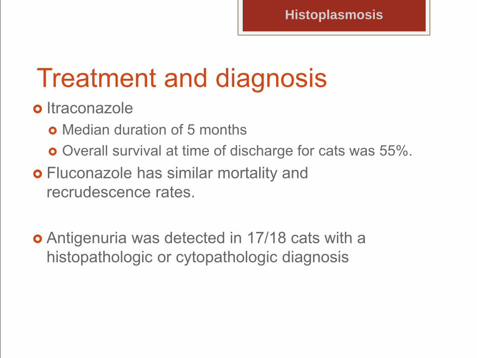
Treatment and diagnosis Itraconazole
Median duration of 5 months Overall survival at time of discharge for cats was 55%.
Fluconazole has similar mortality and recrudescence rates.
Antigenuria was detected in 17/18 cats with a histopathologic or cytopathologic diagnosis
Histoplasmosis
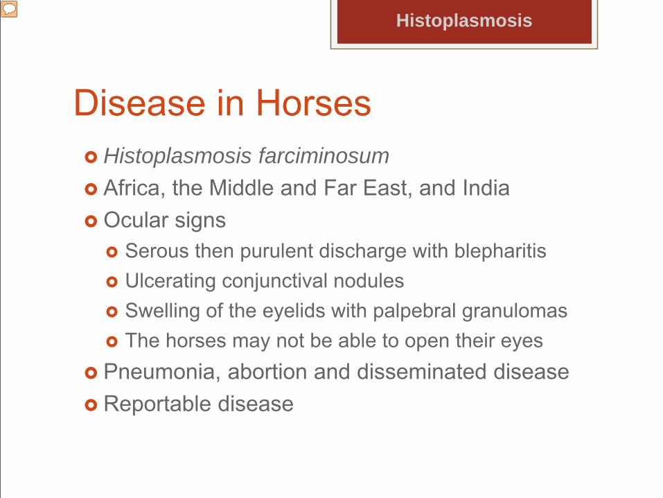
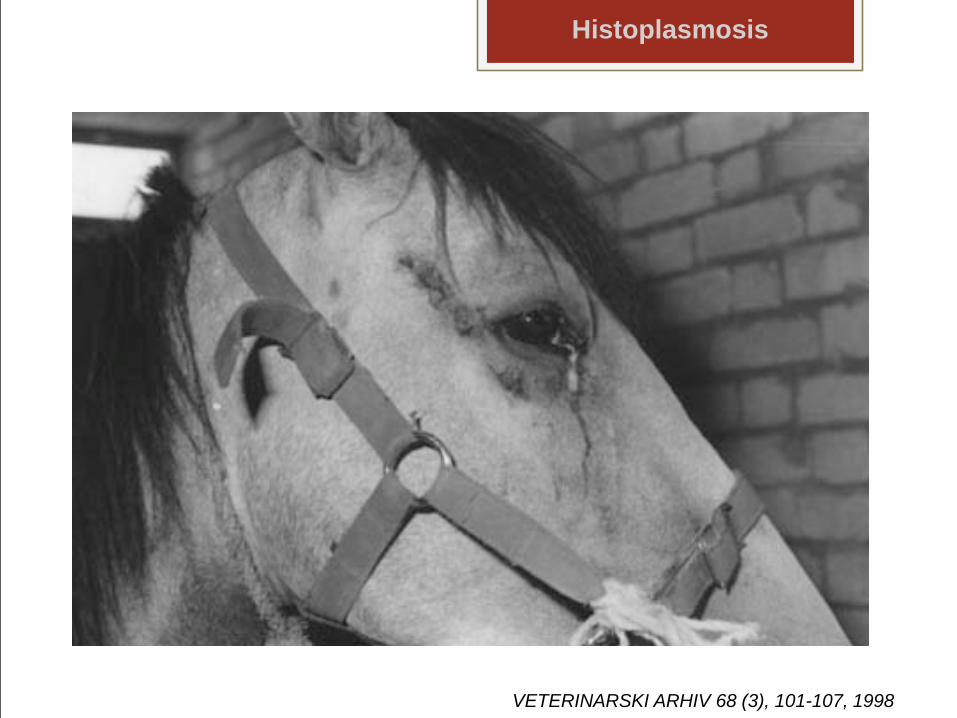
Disease in Horses Histoplasmosis farciminosum Africa, the Middle and Far East, and India Ocular signs
Serous then purulent discharge with blepharitis Ulcerating conjunctival nodules Swelling of the eyelids with palpebral granulomas The horses may not be able to open their eyes
Pneumonia, abortion and disseminated disease Reportable disease
Histoplasmosis
Presenter
Presentation Notes
H capsulatum was diagnosed in a horse with ulcerative keratitis that responded to topical fluconazole.
Histoplasmosis
VETERINARSKI ARHIV 68 (3), 101-107, 1998
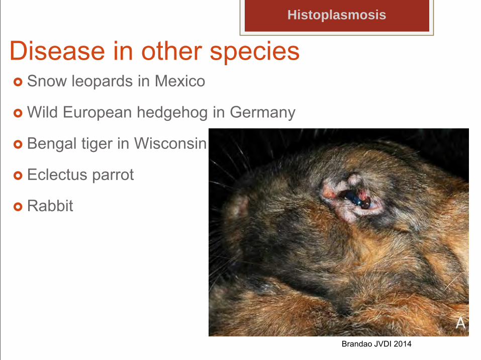
Disease in other species Snow leopards in Mexico
Wild European hedgehog in Germany
Bengal tiger in Wisconsin
Eclectus parrot
Rabbit
Histoplasmosis
Brandao JVDI 2014
Histoplasmosis
Presenter
Presentation Notes
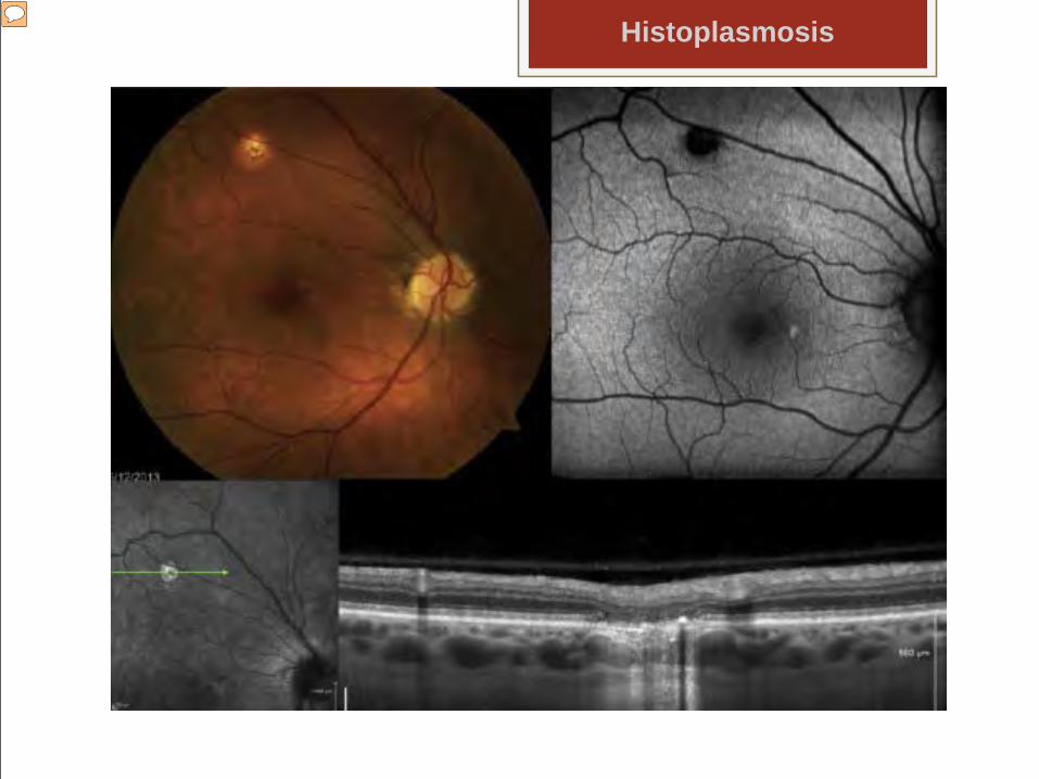
Ocular histoplasmosis syndrome (OHS) is a chorioretinal disorder with a distinct fundus appearance that is commonly found in regions endemic for Histoplasma capsulatum. Choroidal neovascularization (CNV) secondary to OHS is considered one of the principal causes of central vision loss among young adults in endemic areas. Although there is no consensus regarding its pathogenesis, evidence points to Histoplasma capsulatum as the most probable etiology. Once considered an intractable hemorrhagic maculopathy, CNVs are now treatable. Extrafoveal CNVs are successfully treated with laser photocoagulation. Subfoveal and juxtafoveal CNVs are managed with anti-vascular endothelial growth factor therapy, photodynamic therapy, or a combination of both. Imaging characteristics of chorioretinal scars secondary to ocular histoplasmosis syndrome. A color fundus photograph demonstrates a dispersedly pigmented, one-third disk diameter chorioretinal scar along the superior arcade, subtle peripapillary atrophy, and dispersed pigment nasal to fovea (upper left); fundus autofluorescence clearly depicts a superior, hypoautofluorescent chorioretinal scar, probably chronic, with an absence of RPE underneath and a small hyperautofluorescent lesion nasal to the fovea, probably an acute lesion with high metabolic activity (upper right); infrared image with green raster across the superior chorioretinal scar (bottom left); corresponding EDI SD-OCT image demonstrating the complete loss of the outer retinal architecture. There is an absence of the hyper-reflective ellipsoid and RPE band with subsequent increased transmission of light through the choroid. The image has a collapsing appearance, commonly referred to as a “punched out” chorioretinal lesion (bottom right). Often pigment extends around nerve OHS is a clinical diagnosis with distinctive posterior segment findings in the absence of vitritis or anterior segment inflammation. It is generally agreed that one or both eyes should manifest at least two of the classic triad components (Fig. 1):45, 128 and 162 1. Chorioretinal peripapillary atrophy (PPA) 2. Chorioretinal scars in the macula and mid-periphery (“histo spots” or “punched out” lesions) 3. Choroidal neovascularization (CNV) or corresponding sequelae, such as disciform scars
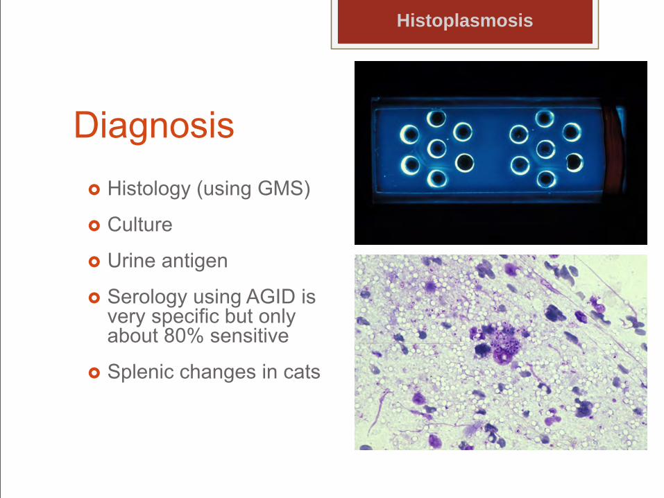
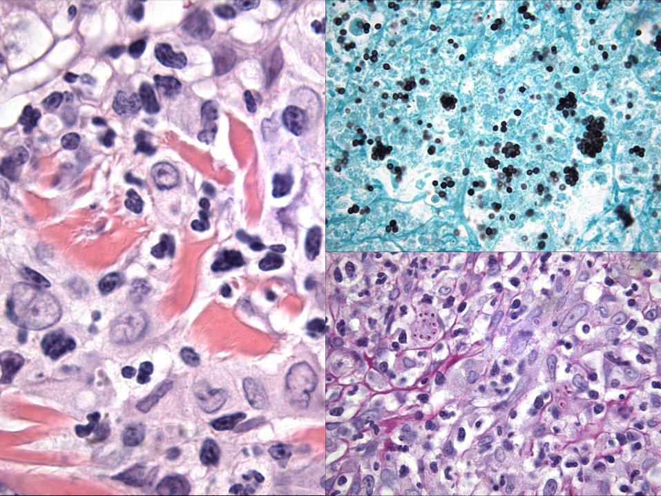
Diagnosis Histology (using GMS)
Culture
Urine antigen
Serology using AGID is very specific but only about 80% sensitive
Splenic changes in cats
Histoplasmosis
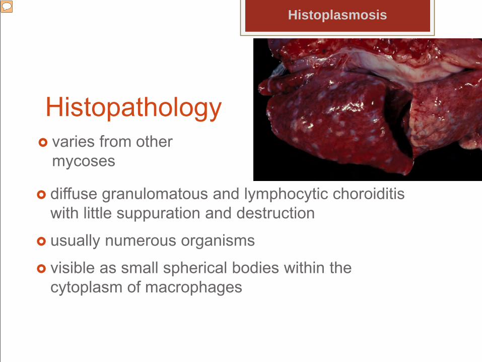
Histopathology varies from other
mycoses
Histoplasmosis
diffuse granulomatous and lymphocytic choroiditis with little suppuration and destruction
usually numerous organisms
visible as small spherical bodies within the cytoplasm of macrophages
Presenter
Presentation Notes
that characterizes blastomycosis and coccidioidomycosis is seen.
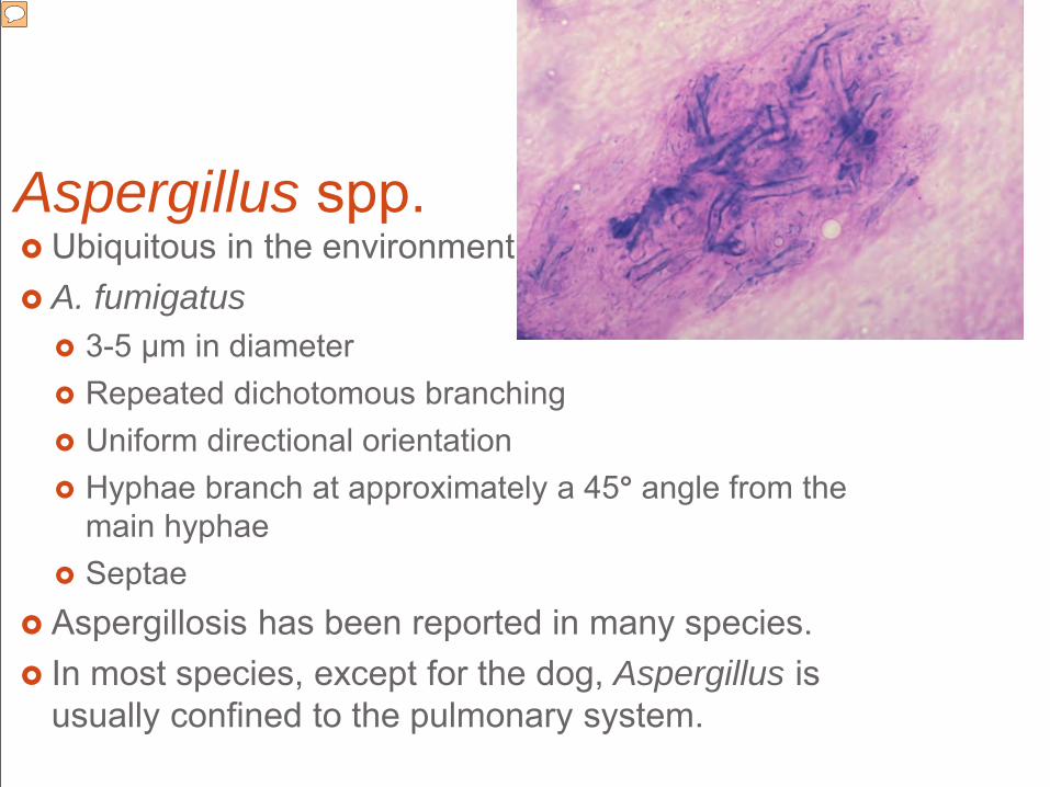
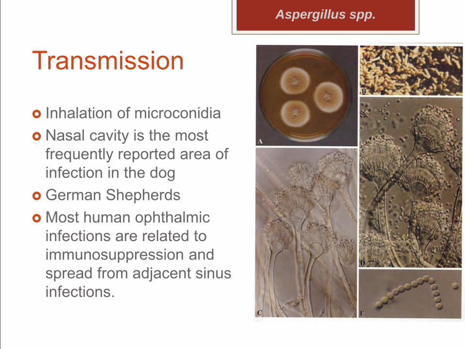
Aspergillus spp. Ubiquitous in the environment A. fumigatus
3-5 µm in diameter Repeated dichotomous branching Uniform directional orientation Hyphae branch at approximately a 45° angle from the
main hyphae Septae
Aspergillosis has been reported in many species. In most species, except for the dog, Aspergillus is
usually confined to the pulmonary system.
Presenter
Presentation Notes
°°°°°
Transmission
Inhalation of microconidia Nasal cavity is the most
frequently reported area of infection in the dog
German Shepherds Most human ophthalmic
infections are related to immunosuppression and spread from adjacent sinus infections.
Aspergillus spp.
A felis
Novel heterothallic species in Aspergillus section Fumigati Human with chronic invasive pulmonary aspergillosis Cats with invasive fungal rhinosinusitis Dog with disseminated invasive aspergillosis. Refractory to aggressive antifungal therapeutic regimens.
Aspergillus spp.
Presenter
Presentation Notes
unilateral exophthalmia Phenotypic analyses show that A. felis can be distinguished from the related species A. viridinutans by its ability to grow at 45°C and from A. fumigatus by its inability to grow at 50°C. Itraconazole and voriconazole cross-resistance was common in vitro.(Barrs 2013)
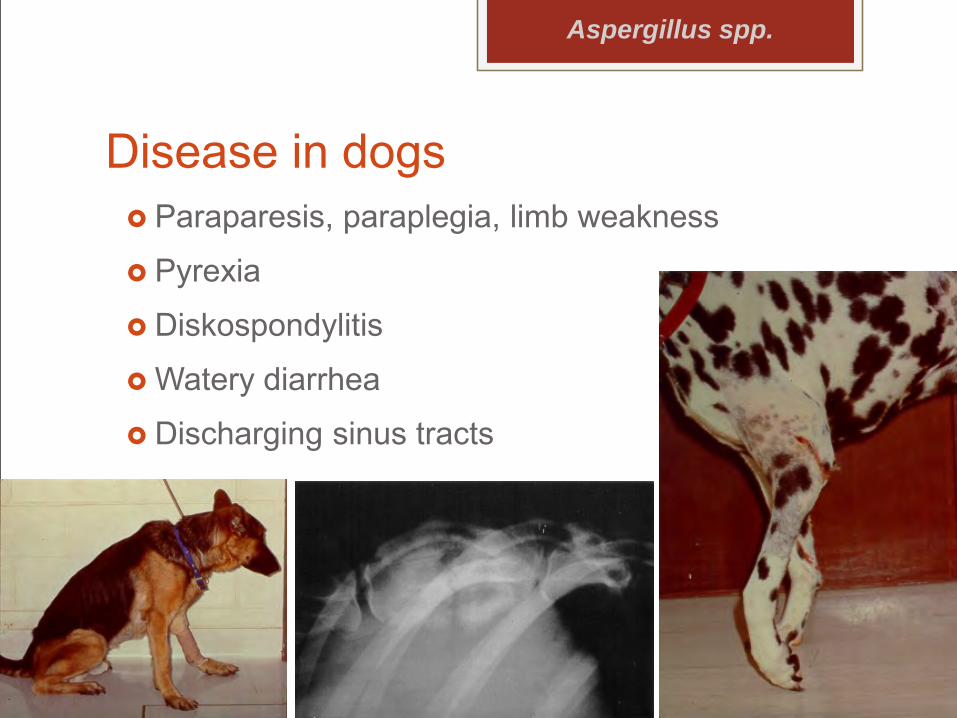
Disease in dogs Paraparesis, paraplegia, limb weakness
Pyrexia
Diskospondylitis
Watery diarrhea
Discharging sinus tracts
Aspergillus spp.
Ocular signs Swollen irides
Vitreal cells
Chorioretinitis
Retinal detachments
Orbital aspergillosis 2° to invasion from the sinus
Ocular signs often occur several mos before generalized illness
Aspergillus spp.
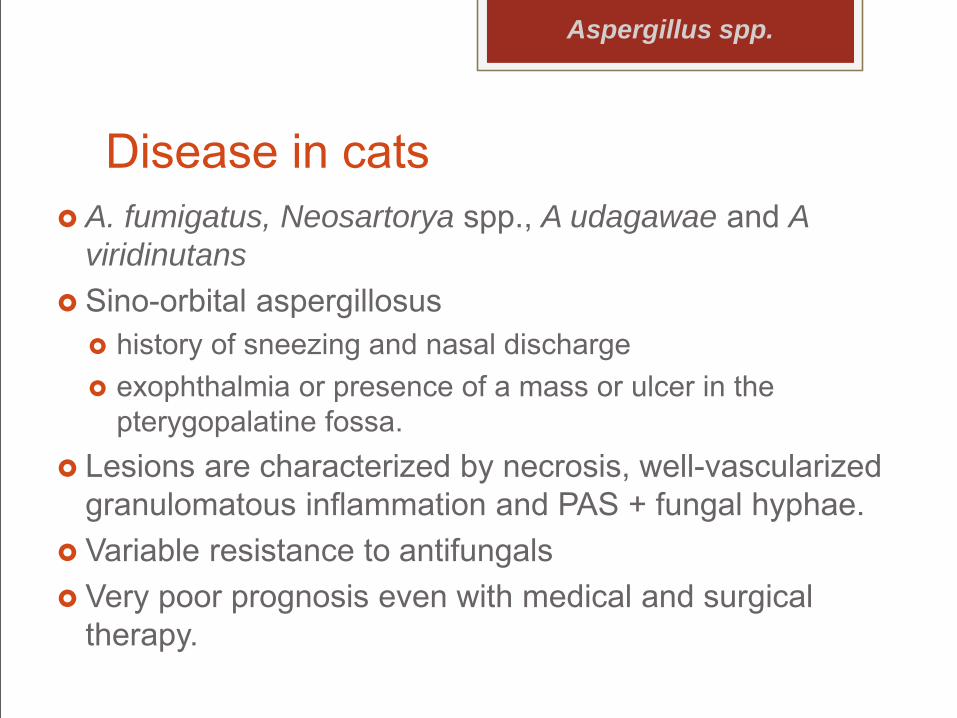
Disease in cats A. fumigatus, Neosartorya spp., A udagawae and A
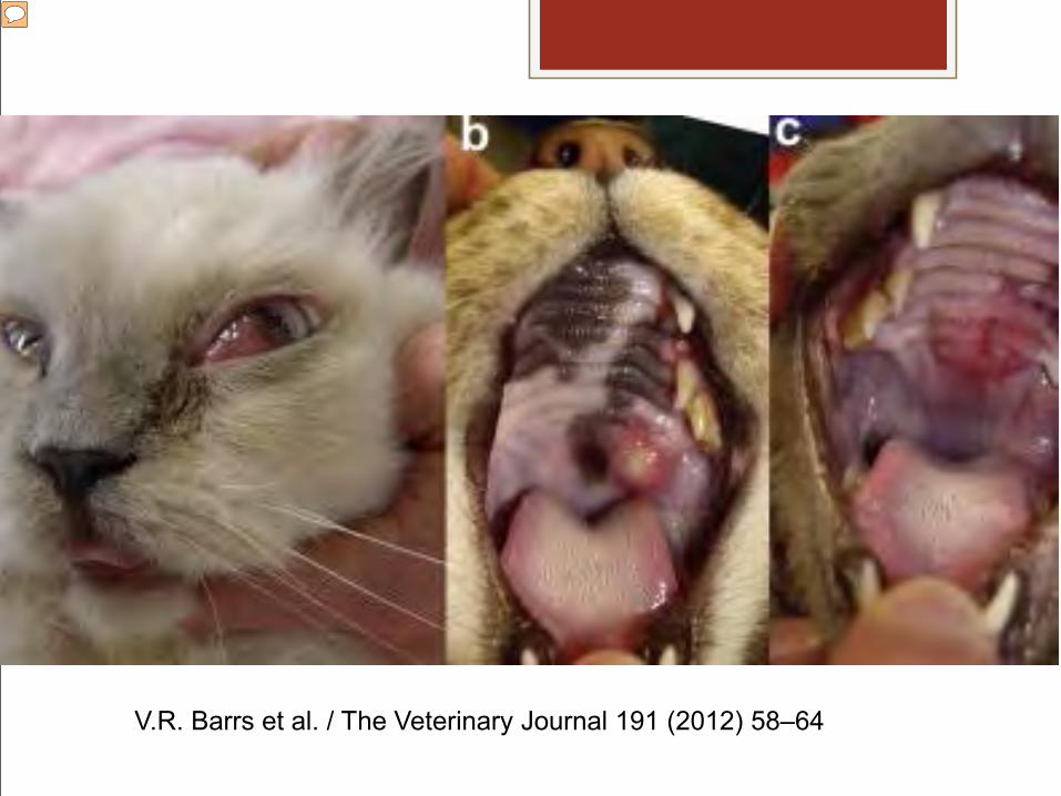
viridinutans Sino-orbital aspergillosus
history of sneezing and nasal discharge exophthalmia or presence of a mass or ulcer in the
pterygopalatine fossa. Lesions are characterized by necrosis, well-vascularized
granulomatous inflammation and PAS + fungal hyphae. Variable resistance to antifungals Very poor prognosis even with medical and surgical
therapy.
Aspergillus spp.
V.R. Barrs et al. / The Veterinary Journal 191 (2012) 58–64
Presenter
Presentation Notes
exophthalmia or presence of a mass or ulcer in the pterygopalatine fossa.
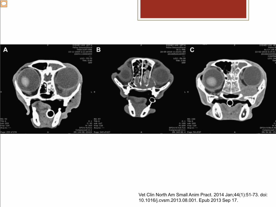
Vet Clin North Am Small Anim Pract. 2014 Jan;44(1):51-73. doi: 10.1016/j.cvsm.2013.08.001. Epub 2013 Sep 17.
Presenter
Presentation Notes
CT features of sino-orbital aspergillosis (A), including nasal cavity soft tissue attenuation, lysis of paranasal bones, and mass-effect (ventromedial orbital mass), overlap those of other mycoses (eg, cryptococcosis) (B), and neoplasia (eg, lymphoma) (C).
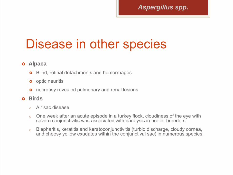
Disease in other species Alpaca
Blind, retinal detachments and hemorrhages
optic neuritis
necropsy revealed pulmonary and renal lesions
Birdso Air sac disease
o One week after an acute episode in a turkey flock, cloudiness of the eye with severe conjunctivitis was associated with paralysis in broiler breeders.
o Blepharitis, keratitis and keratoconjunctivitis (turbid discharge, cloudy cornea, and cheesy yellow exudates within the conjunctival sac) in numerous species.
Aspergillus spp.
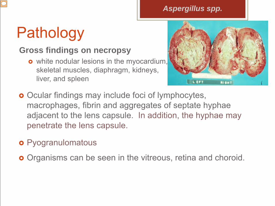
PathologyGross findings on necropsy
white nodular lesions in the myocardium, skeletal muscles, diaphragm, kidneys, liver, and spleen
Aspergillus spp.
Ocular findings may include foci of lymphocytes, macrophages, fibrin and aggregates of septate hyphae adjacent to the lens capsule. In addition, the hyphae may penetrate the lens capsule.
Pyogranulomatous
Organisms can be seen in the vitreous, retina and choroid.
Presenter
Presentation Notes
Since the posterior segment is the most often severely affected part of the eye, Masses of fungal hyphae in the renal pelvis (German Shepherd)
Pathology
Aspergillus spp.
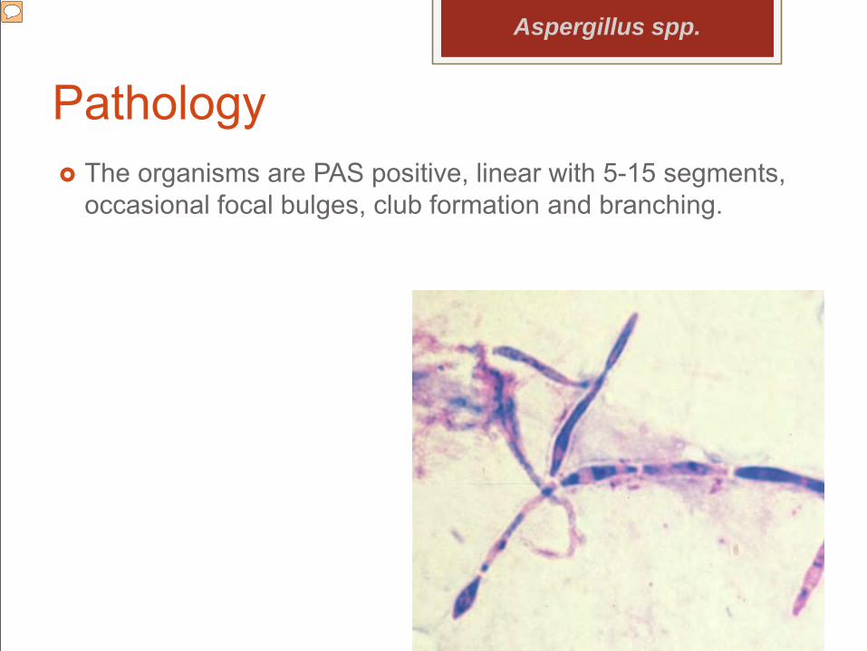
The organisms are PAS positive, linear with 5-15 segments, occasional focal bulges, club formation and branching.
Presenter
Presentation Notes
Since the posterior segment is the most often severely affected part of the eye, Masses of fungal hyphae in the renal pelvis (German Shepherd)
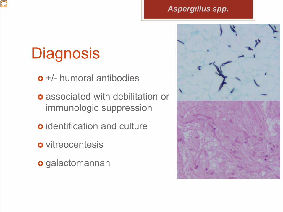
Diagnosis +/- humoral antibodies
associated with debilitation or immunologic suppression
identification and culture
vitreocentesis
galactomannan
Aspergillus spp.
Presenter
Presentation Notes
Because cellular immunity is important to the host response against Aspergillus, from improper housing, stress, primary disease or prolonged antibiotic or corticosteroid usage. Identification and culture from urine sediment, serum, synovial fluid, vitreous, lymph node, CSF or intervertebral disc centesis specimens.
Disseminated Rhizopus Rhizopus is an
opportunistic fungus.
Calf with disseminated Rhizopus infection
Bilateral ocular lesions, including endophthalmitis and intumescent immature cataracts
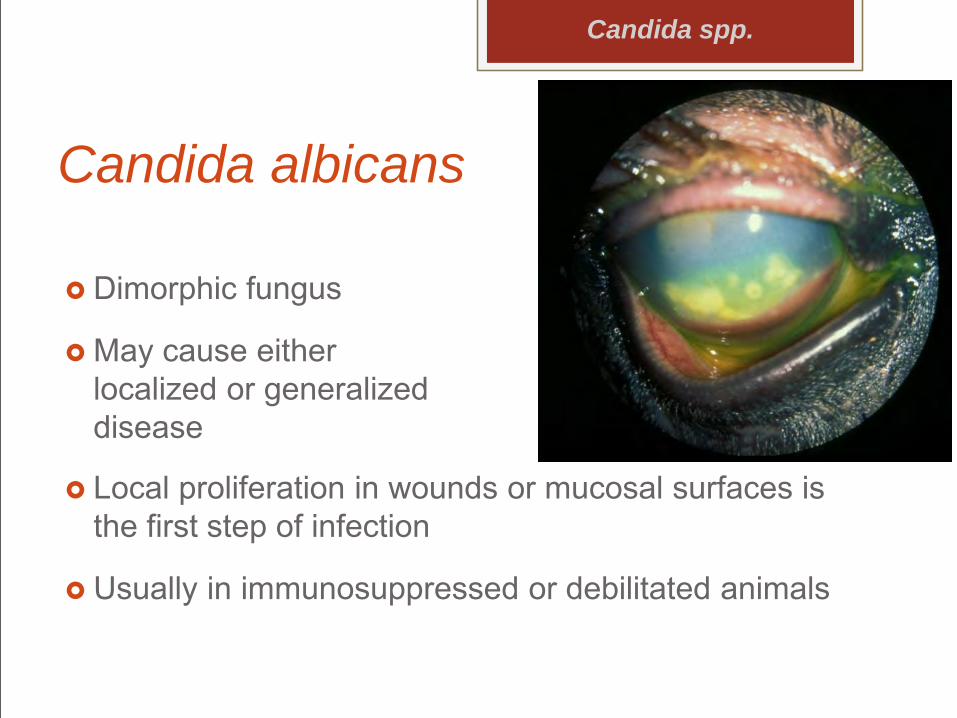
Local proliferation in wounds or mucosal surfaces is the first step of infection
Usually in immunosuppressed or debilitated animals
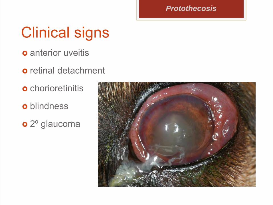
Protothecosis Achlorophyllous algae
P zopfii and P wickerhamii are pathogenic
P. zopfii is most commonly isolated from dogs
Presenter
Presentation Notes
The organism is found in water systems and soil. In North America the disease is primarily in the southeastern US.
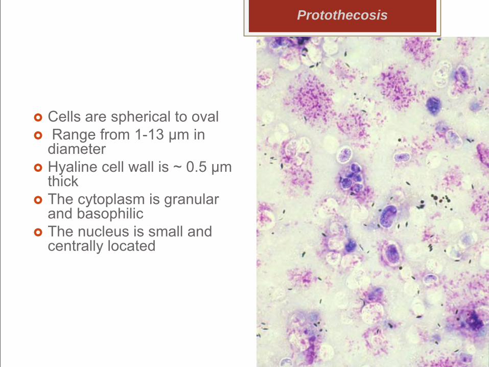
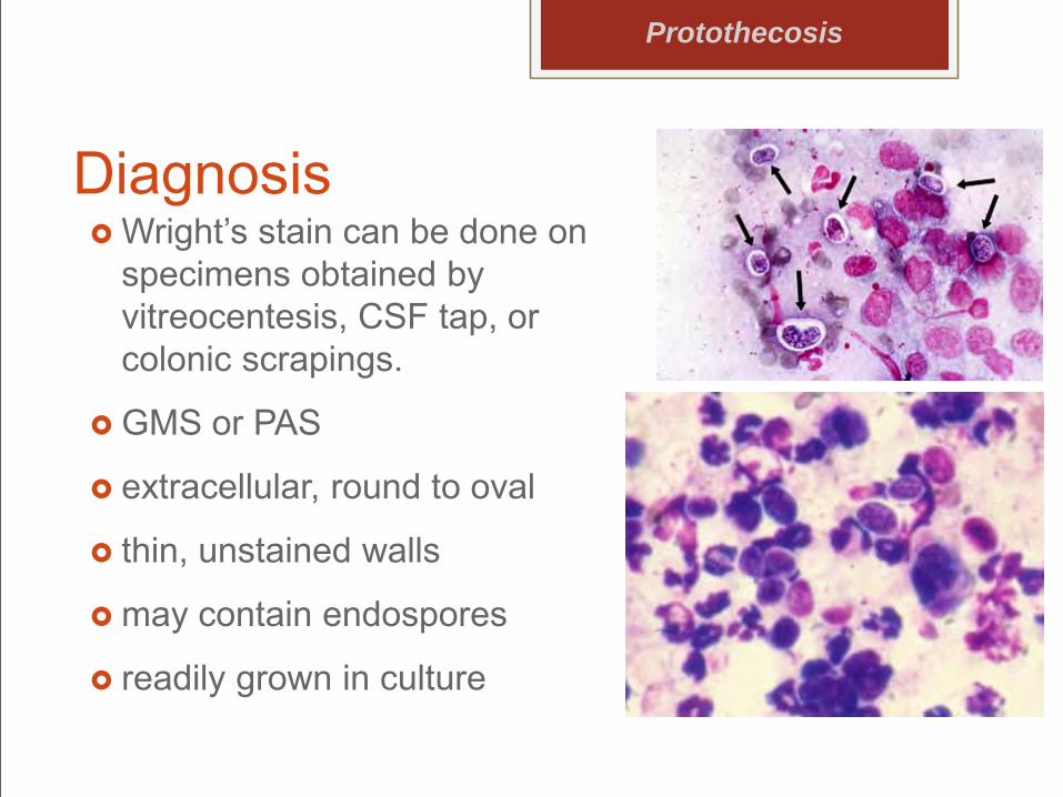
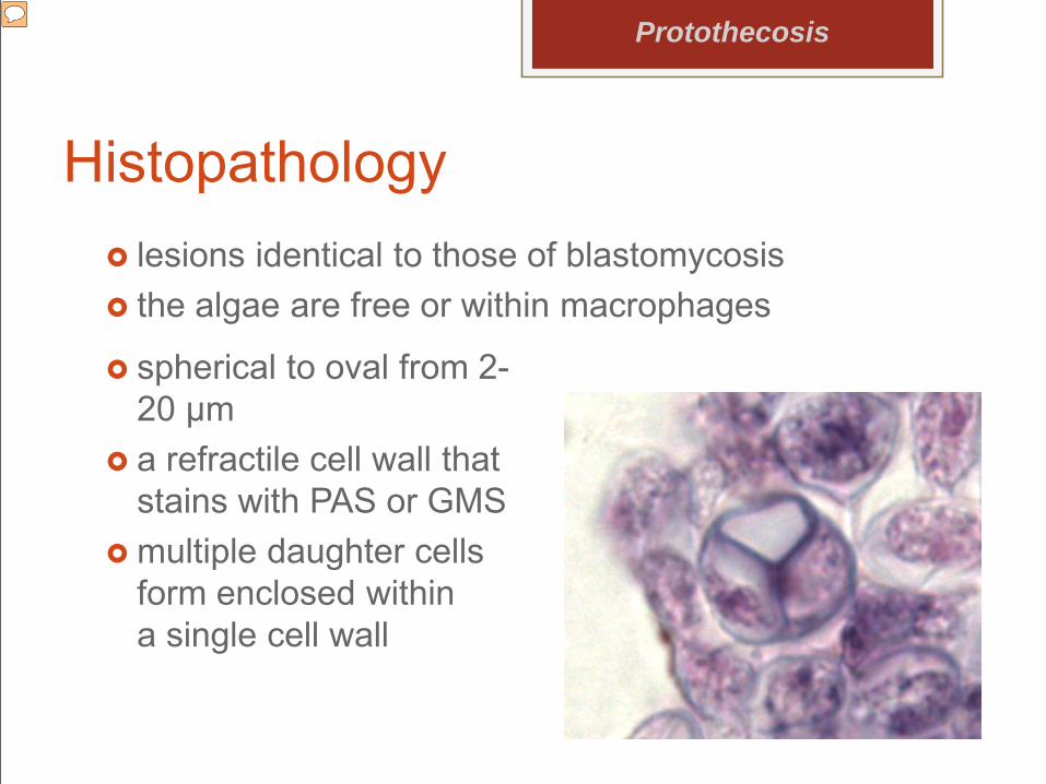
Cells are spherical to oval Range from 1-13 µm in
diameter Hyaline cell wall is ~ 0.5 µm
thick The cytoplasm is granular
and basophilic The nucleus is small and
centrally located
Protothecosis
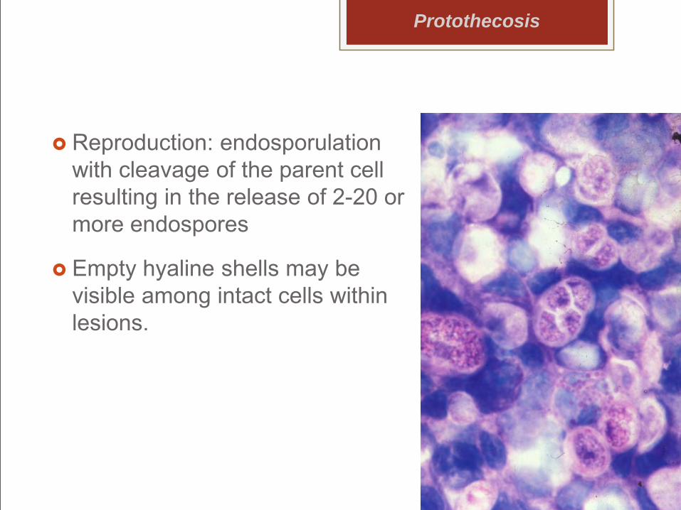
Reproduction: endosporulation with cleavage of the parent cell resulting in the release of 2-20 or more endospores
Empty hyaline shells may be visible among intact cells within lesions.
Protothecosis
Protothecosis
environmental contaminant
minimally pathogenic
does not spread between hosts
Europe, Asia, Oceania, and N. America
Pathogenesis Immunosuppression, especially if cell-mediated
immunity, favors establishment of infection. Another mechanism is the inablility of the host’s
PMNs to specifically destroy Prototheca after phagocytosis.
Biofilms may assist in pathogenesis In humans, infection is usually cutaneous,
subcutaneous, or bursal. The colon is the probable site of primary infection
and entry for disseminated disease in dogs.
Protothecosis
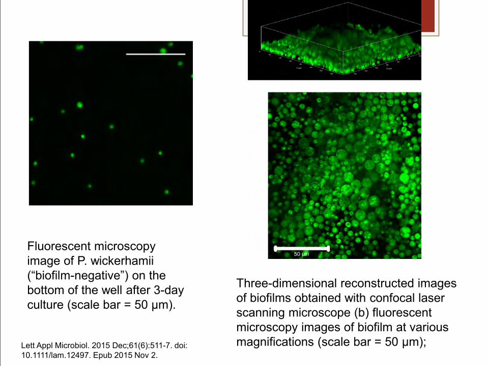
Fluorescent microscopy image of P. wickerhamii(“biofilm-negative”) on the bottom of the well after 3-day culture (scale bar = 50 µm).
Three-dimensional reconstructed images of biofilms obtained with confocal laser scanning microscope (b) fluorescent microscopy images of biofilm at various magnifications (scale bar = 50 μm); Lett Appl Microbiol. 2015 Dec;61(6):511-7. doi:
10.1111/lam.12497. Epub 2015 Nov 2.
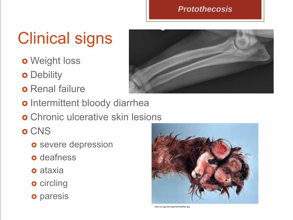
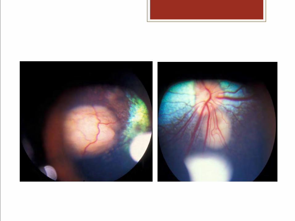
Disease in Dogs hemorrhagic diarrhea or colitis is primary sign
blindness may be initial complaint
predisposition for the Collie breed and females
lymphatic, nervous, renal and pulmonary systems are commonly involved
Model of the signalling pathways controlling dimorphic switching and yeast growth at 37°C. The colours indicate characterized roles in dimorphism: green, mutation or RNAi inhibition affects dimorphism; yellow, expression is induced during the dimorphic switch, blue, chemical inhibition or exogenous addition affects the dimorphic switch; and orange, mutation indicates no role in dimorphism. (A) Two-component signalling systems in fungi require four sequential phosphorylation events (H→D→H→D) and comprising one or multiple membrane-associated HHK, an intermediate histidine phosphotransfer protein (HPt) and two RRs. The HK perceives the environmental stimulus and is autophosphorylated at a conserved histidine in the kinase domain. The phosphate group is then transferred to a conserved aspartate in the HHK's RR domain and then to a conserved histidine in the HPt phosphotransfer protein (HPt). The HPt protein transfers the phosphate to an aspartate residue in the RR. The RR can either bind DNA directly or activate MAPK pathways to elucidate a morphological and transcriptional response. (B) Binding of the ligand to the G protein coupled receptor triggers GDP to GTP exchange in the Gα subunit of a heterotrimeric G protein which dissociates from the ß and γ subunits. The Gα subunit activates adenylate cyclase (AC) and subsequent cAMP/protein kinase A (PKA) signalling, in addition to, the Ras GTPase. Ras activates the Rho GTPase Cdc42, which in addition to controlling actin-mediated polarized growth, regulates the p21 activated kinases Ste20 and Cla4 to signal via MAPK pathways and regulate cellular division. (C) Entry of calcium (Ca2+) into the cell or release from internal stores acts as a transient intracellular signal. Binding of Ca2+ to calmodulin (CaM) enables it to activate the calmodulin-dependent kinases (CDKs) and the calcineurin phosphatase (CnaB/CnaA). Ca2+ binding to the regulatory subunit of calcineurin (CnaB) allows activation of the catalytic subunit (CnaA). CnaA dephosphorylates the CrzA transcription factor to allow entry into the nucleus to activate genes controlling processes such as cell-wall synthesis, germination of spores (conidia), ion homeostasis, pH adaptation, polarity and conidiophore development.


![IDENTIFICATION OF · [DNLM: 1. Fungi–isolation & purification. 2. Fungi–pathogenicity. 3. Mycoses–diagnosis. QW 180] 579.5–dc23 2012044506 A catalogue record for this book](https://img.pdfslide.net/doc/110x75/5fd928bdcc2800392818e37f/identification-of-dnlm-1-fungiaisolation-purification-2-fungiapathogenicity.jpg)